

Submitted:
30 July 2026
Posted:
31 July 2026
You are already at the latest version
Abstract
More than 180 chemically distinct RNA modifications – the epitranscriptome – have now been reported across all kingdoms of life and all forms of RNA. Given the emerging evidence for critical roles played by RNA modifications in many diseases, it is imperative to identify the function of all RNA modifications in the nearly 250,000 types of RNA in human cells – the human RNome. Here we take a step toward this goal by reviewing the literature to date and assembling a catalog of all established or putative human RNA modifications. While the identities and locations of many human RNA modifications are well established, emerging technologies are revealing new ones as well as old ones in unexpected locations, with low abundance and poorly characterized structures hampering their validation. To this end, we introduce an analytical validation framework that scores each reported human RNA modification for chemical identity and transcriptomic localization and then assigns it to one of three confidence tiers: Authenticated, Provisional, or Putative. Applying this framework, we critically re-examine the primary literature underlying 24 recently reported additions to the human epitranscriptome, evaluating the mass spectrometric, spectroscopic, and sequencing-based evidence supporting each as well as discussing their emerging biological roles and disease associations. We further extend this scoring system to modifications previously summarized in reviews and databases but not yet subjected to this level of scrutiny, compiling a comprehensive, tier-ranked inventory of the human epitranscriptome as of July 2026. Together, this catalog and framework provide a benchmark for evaluating future epitranscriptome discoveries, mapping the human RNome, and for derisking the translation of validated modifications as diagnostic and therapeutic targets.
Keywords:
modified ribonucleoside
; epitranscriptome
; protein translation
; RNA stability
; RNA structure
1. Introduction
The nucleic acid alphabet of life extends far beyond the canonical nucleotides of adenine, cytosine, guanine, and thymine in DNA and uracil in RNA [1,2]. While transcription copies the basic genetic code from DNA, the resulting primary RNA transcripts are typically unstable and structurally immature [3,4]. This necessitates significant post-transcriptional enzymatic modification of RNA, including splicing and A-to-I editing as well as the hundreds of modifications ranging from simple methylations to complex multi-step hypermodifications, which shape RNA conformation and function and enable dynamic and granular control of gene expression [5]. Since the discovery of pseudouridine (Ψ), often referred to as the ‘fifth RNA nucleotide’ [6], in calf liver RNA hydrolysate by Cohn and Volkin in 1951 [7], the world of RNA modifications has expanded exponentially. To date, over 180 modified ribonucleosides have been reported across all kingdoms of life [8], with new modifications being discovered regularly and this number expected to climb continuously [9].
The human epitranscriptome represents the complete landscape of post-transcriptional modifications found on all RNA molecules, such as transfer RNA (tRNA), ribosomal RNA (rRNA), messenger RNA (mRNA), and various non-coding RNA species [10]. These are much more than chemical decorations and govern every stage of the RNA lifecycle, including splicing, nuclear export, stability, translation efficiency, and environmental stress adaptation [8]. Rather than acting as static structural features, these modifications are dynamically orchestrated by a broad network of ‘writers’ that install the modifications, ‘readers’ that recognize and bind to them, and ‘erasers’ that remove them in response to cellular demands [11]. Consequently, this dynamic chemical layer serves as a master regulator of cellular fate, and its dysregulation is now associated with a broad spectrum of human diseases, from neurological disorder to cancer [12,13]. For example, in glioblastoma, overexpression of the epitranscriptome writer METTL1 installs m7G46 modifications across a subset of tRNAs including the isodecoder tRNA-Arg-TCT-4-1, which stabilizes and increases its copy numbers, ultimately boosting the translation of cancer-driving mRNAs enriched in corresponding cognate codons [13,14]. Given the critical roles these modifications play in cellular function, human health, and disease, and because the field is still in its relative infancy, further research is essential to fully decipher the spectrum of modifications across various RNA species, elucidate their underlying molecular mechanisms, and facilitate the development of novel diagnostic and therapeutic methods.
Not all the 180 reported modifications, however, rest on equally solid evidence. Beyond the most abundant, well-characterized epitranscriptome marks such as Ψ, m6A, and 2′-O-methylation (Nm) in human RNAs [15], recent years have witnessed the discovery of novel RNA modifications of low abundance from highly limited sample materials. Typical examples include 5-taurinomethyluridine (τm5U) and 5-taurinomethyl-2-thiouridine (τm5s2U) in human mitochondrial tRNAs (mt-tRNAs) [16,17], the identification of which was facilitated by breakthroughs in RNA purification, isolation, and enrichment in tandem with continuous improvement in the sensitivity and specificity of modern analytical instrumentation. As many of these newly reported modifications often occur at ultra-low abundance, lack orthogonal validation, and rely on unique, non-commercially available synthetic standards, the literature remains complicated by conflicting datasets, inadvertent technical artifacts, and false positives generated during sample handling or enzymatic hydrolysis. This growing gap between discovery and validation highlights an urgent need for rigorous validation frameworks and standardized criteria to cross-examine and validate novel epitranscriptomic discoveries.
To address these pressing challenges, this review provides a systematic and critical evaluation of recent additions to the human epitranscriptome, comprehensively cataloging all modified ribonucleosides reported across the entire spectrum of human RNA species. While existing literature typically addresses RNA modifications broadly across all kingdoms of life or focuses exclusively on the human tRNA epitranscriptome, a unified reference documenting all modified ribonucleosides across all human RNA classes has been conspicuously absent. To bridge this gap, we build on the 52 RNA modifications identified in the landmark review by Tsutomu Suzuki and coworkers on tRNA modifications [18], the publication by Taoka et al. on rRNA modifications [19], and the MODOMICS database to update the inventory of the human epitranscriptome [20].
We then expand this baseline through a systematic literature search to capture newly discovered and previously overlooked modifications. More importantly, we establish a standardized set of analytical validation criteria to score the detection confidence of these novel ribonucleosides and discuss their biological significance and associations with diseases. We further compile all human RNA modifications reported as of July 2026 into two ready-to-use tables for the field: one listing their detection confidence tier and RNA species of occurrence, and another detailing their instrumental parameters. Ultimately, we hope this review serves as a definitive resource and a rigorous benchmark for high-fidelity epitranscriptomic research, paving the way for future discoveries and the development of dependable diagnostic and therapeutic innovations.
2. Methods for Mapping RNA Modifications
The accurate mapping of the human epitranscriptome requires the strategic integration of targeted sample preparation and complementary analytical instrumentation (Figure 1). Historically, candidate modifications have emerged either from targeted hypotheses formulated by researchers or as unassigned peaks discovered during unrelated experiments. Regardless of their origin, authenticating a novel RNA modification requires resolving a complex, multi-tiered analytical puzzle. Because no single method can independently validate a new mark, researchers must first isolate or enrich the specific RNA species of interest, combine multiple orthogonal analytical technologies, synthesize chemical standards to cross-validate chemical identities and structures, and deploy sequencing platforms to capture exact transcriptomic maps. Ultimately, proving the presence of a novel mark requires satisfying two criteria: establishing its absolute chemical structure and identifying its precise location a specific transcript.
Liquid chromatography-coupled mass spectrometry (LC-MS) has routinely been used for identifying, structurally characterizing, and quantifying nucleic acid modifications given its ability to chromatographically resolve complex mixtures, uniquely select specific molecules as ions, and break them into predictable fragments, often with exact mass measurements [21]. Reassembly of the resulting data provides rigorous chemical identification and quantification. In epitranscriptome studies, researchers generally hydrolyze enriched RNA samples into individual monoribonucleosides using a combination of endonucleases, phosphodiesterases, and alkaline phosphatases to maximize analytical sensitivity and chromatographic resolution [22], then analyze the resulting hydrolysates by an MS1 full scan to determine accurate molecular mass. Candidate peaks are then subjected to tandem mass spectrometry (MS2 or MSn), in which collision-induced dissociation (CID) or higher-energy collisional dissociation (HCD) generates characteristic product ions for deep structural elucidation [23]. To corroborate these mass-centric findings, researchers can pre-purify target candidate peaks or employ complementary assays, such as ultraviolet spectrophotometry, infrared spectrophotometry, pulse labeling, and nuclear magnetic resonance (NMR) spectroscopy [23,24], to confirm the presence and exact stereochemical positions of specific functional groups within the core structure. Once a structure is proposed, researchers synthesize matching chemical standards to rigorously cross-validate the endogenous candidate mark [24]. High-confidence confirmation demands that the candidate peak in the sample and the synthetic standard exhibit identical chromatographic retention times, high-resolution exact masses, and CID/HCD tandem mass spectra.
After successfully validating the chemical identity of a novel modification, completing the epitranscriptomic puzzle still requires mapping its precise coordinates within the transcriptome. To achieve this, the field has deployed various RNA sequencing (RNA-seq) methodologies, including short-read sequencing (SRS), long-read sequencing (LRS), and LC-MS-based RNA-seq (Figure 1) [4]. It is important to note that none of these methods provide the unequivocal identification or quantification provided by LC-MS ribonucleoside analysis.
Historically anchored in SRS, this methodology is widely regarded as an indirect approach because RNA must first be reverse-transcribed into cDNA before sequencing: it is the cDNA, not the modified ribonucleotide itself, that is actually read [4,25]. What differs between methods is how a modification is rendered visible within that cDNA. Reverse transcriptase (RT) signature-based methods are applied to modifications that intrinsically disrupt RT through misincorporation, stalling, or abortive cDNA synthesis, thereby leaving specific and interpretable signatures at the modified sites in the cDNA [25,26]. These signatures arise because the modified base no longer forms normal Watson-Crick pairs with its canonical partner, or because bulky or highly hydrophobic substituents slow the rate of cDNA synthesis [26]. For RT-silent modifications, chemical- and biological-based methods can instead be used to react selectively with the modified base and introduce a distinctive RT signature or cleave modified and unmodified bases differently to convert chemical identity into a positional cut site [25,26]. In addition, antibody-based methods are used to enrich modification-bound RNA fragments prior to RT and sequencing [27], but this enrichment generally sacrifices single-nucleotide resolution as they typically achieve peak-level resolution on the order of ~100 – 200 nt [26]. However, most SRS methods are optimized to interrogate one specific modification at a time, and each modification requires substantial effort to develop a dedicated antibody, probe, or enzyme. This specialization creates a major bottleneck for using SRS in the discovery of novel RNA modifications.
In contrast to SRS, LRS can sequence native RNA molecules as they are threaded through a protein nanopore, without prior fragmentation, amplification, or chemical labeling [28]. As each short stretch of RNA resides within the pore, it produces a characteristic disruption in the ionic current passing through. RNA modifications perturb this expected current signature and can alter the time each stretch spends in the pore (the dwell time), providing a direct readout of modification presence at single-nucleotide resolution [29]. This signal can be interpreted in three main ways: (1) as “errors” when a canonical basecaller is forced to represent a modified base as one of the four standard nucleotides; (2) as a direct comparison of raw current intensity and dwell-time distributions against an unmodified control sample; or (3) through basecalling models trained on known modifications to call a specific modification [30]. Current LRS methods are therefore restricted to modifications for which suitable training data exist. A novel modification will produce, at best, an unassigned current or dwell-time anomaly relative to a control sample and will still require orthogonal methods to validate its presence and define its chemical identity.
Alternatively, researchers can leverage LC-MS-based RNA-seq methods, which represent a direct, modification-type-independent approach to simultaneously and accurately identify and locate multiple modifications in RNA samples at single-base resolution [31]. Instead of fully digesting the RNA down to monoribonucleosides, this approach utilizes site-specific endoribonucleases, such as RNase T1, to generate oligonucleotide fragments of varying lengths [32]. By coupling data-dependent/data-independent acquisition scans for these oligonucleotides with specialized data-processing software suites, such as Thermo BioPharma Finder and the OpenMS Nucleic Acid Search Engine (NASE) [33], researchers can simultaneously decode the nucleotide sequences and pinpoint the exact transcriptomic coordinate of a modification based on fragment ions. However, LC-MS-based RNA-seq is comparatively weak at distinguishing isobaric modifications and at pinpointing the exact functional group and position responsible for a given mass shift [33]. For example, a +14 Da shift from methylation could arise from a methyl group on the base (m6A), on the 2′-O position of the ribose (Am), or even at different positions on the same base (m1A). Achieving that level of structural resolution still requires cross-validation against monoribonucleoside-level LC–MS and the dedicated analytical chemistry methods.
Taken together, LC-MS ribonucleoside analysis is essential for identifying a modified ribonucleoside and establishing whether its structure actually exists in the human epitranscriptome. Once a modification has been structurally validated, it must then be localized within RNA sequences by SRS, LRS, or LC-MS-based RNA-seq. Because each analytical method and sequencing platform has its own strengths and weaknesses, orthogonal validation across multiple platforms is critical for increasing rigor and confidence. Replication by independent laboratories is also highly desirable to further strengthen detection claims. By integrating ribonucleoside-level structural validation, high-resolution spatial mapping data, and orthogonal validation, we can confidently authenticate a new component of the human epitranscriptome.
3. A Scoring System for Confidence in Identifying RNA Modifications
Many of the modifications described in the literature vary widely in the rigor with which their structures and RNA locations have been established. To assess the confidence for the recently reported additions to the human epitranscriptome, we introduce a standardized analytical validation scoring system that evaluates the integration of monoribonucleoside characterization, synthetic standard cross-validation, and transcriptomic mapping. Rather than treating all literature reports with equal weight, this framework classifies modifications into three progressive confidence tiers (Table 1), based on the depth and stringency of the analytical evidence provided in the original discovery papers.
Authenticated modifications represent the highest confidence tier: both chemical identity and transcriptomic location must be established beyond analytical ambiguity. Identification requires a full precursor and product ion scan, complementary structural methods such as NMR, cross-validation against a synthetic standard, and orthogonal confirmation by an independent laboratory. Transcriptomic mapping requires single-base or fragment-level mapping via LC-MS-based RNA-seq. Provisional modifications meet nearly the same identification bar, but complementary structural methods and independent orthogonal confirmation become optional. The larger distinction from Authenticated status lies in localization, where mapping is confined to bulk RNA fractions rather than a precise sequence coordinate, and no single sequencing platform is required to reach this tier. Putative modifications fall short on both fronts. Identification rests on only a low-resolution precursor ion scan, with the product ion scan, complementary structural methods, synthetic-standard cross-validation, and orthogonal confirmation all optional, and mapping data are absent entirely. This tier is not necessarily a dead end, as a Putative candidate can be promoted to Provisional or Authenticated status with further structural validation and precise mapping, while others may instead be reclassified as technical artifacts or false positives upon closer scrutiny.
4. The Expanding Repertoire of Human RNA Modifications and Their Biological Significance
The purpose of this review is to provide an accurate inventory of the human RNA modifications, building on previous reviews of tRNA modifications [18], analysis of rRNA modifications [19], and the MODOMICS database [20]. As depicted in Figure 2, we expand the repertoire by 24 RNA modifications, assessing the degree to which the structures have been validated. Supplementary Table 1 summarizes these 24 RNA modifications and the other 52 that together comprise the human epitranscriptome.
1,N6-Dimethyladenosine (m1,6A). m1,6A was first identified in 1991 in the urine of a patient with stage IV lung adenocarcinoma, characterized as its trimethylsilyl derivative by gas chromatography-MS and confirmed against a synthetic standard [34]. This work established its chemical identity but not biological origin, as the authors concluded the study by noting that m1,6A had not been identified as a component of tRNA, though they speculated that a methyltransferase might install a second methyl group to m1A or m6A [34]. This modification remained unassigned to an RNA species or enzymatic pathway for over thirty years, until 2022, when You et al. unambiguously identified m1,6A in human tRNAs [35].
You et al. synthesized an m1,6A standard and cross-validated its structure by high-resolution MS/MS and MS3 fragmentation as well as NMR [35]. Its presence in cellular RNA was confirmed both by spiking the synthetic standard into tRNA hydrolysate and by stable-isotope tracing with deuterated methionine, and m1,6A was undetectable in the other RNA species tested such as mRNA and 28S/18S/5.8S/5S rRNAs. m1,6A occurs at frequencies ranging from 0.0049% to 0.0084% in tRNAs from human cell lines, including HEK293T, HeLa, HepG2, K562, Jurkat-T, MCF-7, and HL-7702 [35]. Site-specific mapping was equally rigorous: using a targeted DNA hybridization protection/S1 nuclease digestion strategy on the purified tRNAGly(GCC) to isolate the exact oligonucleotide fragment containing the modification, the authors located m1,6A at position 58 of the tRNA, the same site occupied by the well-characterized m1A58 mark. While You et al. closed the gap the 1991 urinary finding had left open, no other publication has provided orthogonal evidence to show the presence of m1,6A in human RNA. On this basis, we assign m1,6A as a Provisional modification: definitive identification and location but no independent validation.
Mechanistically, the authors incubated purified tRNAs with TRMT6/61A and observed an increased level of m1,6A, confirming TRMT6/61A is responsible for m1,6A formation. Because no dedicated methyltransferase for m6A58 has been identified, they propose a two-step pathway in which a fraction of m1A58 first undergoes Dimroth rearrangement to m6A58, which TRMT6/61A then methylates at N1 position to form m1,6A58. The authors also demonstrated that m1,6A can be reversed by the demethylase ALKBH3. To assess its disease relevance, the authors compared m1,6A levels in tRNAs from human breast cancer tissues against matched adjacent normal tissues. They found that m1,6A, along with m1A and m6A, was significantly elevated in cancer tissue and that this increase correlated with reduced mRNA expression of the demethylases ALKBH1 and ALKBH3, whereas mRNA levels of TRMT6 and TRMT61A were only slightly increased. These observations point to the demethylase loss, rather than increased methyltransferase activity, as the more likely driver of elevated m1,6A, m1A, and m6A levels in cancer tissue. Finally, they noted that m1,6A may represent either an RNA damage product or a genuine regulatory mark, since its proposed biosynthetic route involves a non-enzymatic Dimroth rearrangement step.
3,2′-O-Dimethylcytidine (m3Cm) and N4,2′-O-dimethylcytidine (m4Cm). Cytidine dual methylations occurring at both the nucleobase and the ribose 2′-hydroxyl were already known in two forms before the discovery of m3Cm: m4Cm, found at position 1402 of the 16S rRNA of Escherichia coli, where it fine-tunes the local structure of the decoding site [36], and 5,2′-O-dimethylcytidine (m5Cm), found in archaeal and mammalian tRNA [37]. However, the third possible combination, m3Cm, had never been reported in any domain of life and m4Cm itself had never been identified outside bacteria. Cheng et al. addressed both gaps in a single study by synthesizing an m3Cm standard, characterizing it with high-resolution MS/MS and NMR, and developing an LC-MS/MS method capable of resolving the three structural isomers m3Cm, m4Cm, and m5Cm, which share identical molecular weights and multiple reaction monitoring (MRM) transitions [38].
The authors detected both m3Cm and m4Cm in the total RNA of HeLa cells, confirmed each identity by high-resolution MS/MS, spiking synthetic standards into RNA hydrolysate, and stable-isotope tracing with deuterated methionine. Following experiments showed that m3Cm and m4Cm are localized to small RNA (<200 nt) and 18S rRNA in HeLa cells. To rule out contamination from bacterial 16S rRNA as the source of the signal, the authors performed additional confirmation by agarose-gel purification of 18S rRNA to over 99.999% purity. Across four human cell lines (HeLa, HepG2, HL-7702, and MCF-7), m3Cm was quantified at 0.0003–0.0008% in total RNA and 0.002–0.007% in small RNA, while m4Cm was quantified at 0.0004–0.001% in total RNA and 0.0009–0.003% in 18S rRNA. In both cases, the mapping stopped at the RNA species level, and for m3Cm, the authors infer it most likely originates from tRNA, since tRNA accounts for roughly 90% of the small RNA pool. As this is the only study reporting their presence in human RNA and the modifications are only localized to a bulk RNA species, both m3Cm and m4Cm are Provisional modifications under our scoring framework.
To date, no enzyme has been experimentally linked to the formation or removal of both modifications in human cells. The authors propose METTL6 and FTSJ1 as candidate writers that could act sequentially to generate m3C and then m3Cm, since METTL6 is known to install m3C in serine tRNA and FTSJ1 carries out 2′-O-methylation in the tRNA anticodon region, respectively. No analogous candidate pathway is proposed for m4Cm formation. The authors also speculate that both modifications could enhance the structural stability of tRNA and 18S rRNA. Future studies will be necessary to better understand the biological roles and disease associations of both m3Cm and m4Cm.
5,2′-O-Dimethylcytidine (m5Cm). m5Cm was first structurally characterized in 1987 by Edmonds et al., who identified it in the tRNA of three extremely thermophilic archaebacteria, Sulfolobus solfataricus, Thermoproteus neutrophilus, and Pyrodictium occultum [39]. Three decades later, Huber et al. used a commercially obtained m5Cm standard and LC-MS to report the first detection of m5Cm in total RNA from HEK293T cells, as part of a study primarily focused on a related oxidative derivative, 2′-O-methyl-5-hydroxymethylcytidine (hm5Cm); no attempt to detect m5Cm in different RNA fractions was made [37]. Using a similar commercial standard and LC-MS, Feng et al. and Cheng et al. later verified that m5Cm is present in small RNA (<200 nt) in human cells [38,40].
Feng et al. developed a chemical-labeling method with 2-bromo-1-(3,4-dimethoxyphenyl)-ethanone (BDMOPE) that improved detection sensitivity of m5Cm by over 100-fold over unlabeled analysis [40]. They then confirmed the identity of endogenous m5Cm using its chemical standard and high-resolution MS/MS, quantified m5Cm in total RNA across six human cell lines (HEK293T, HL-7702, HepG2, Huh7.5, HeLa, and MCF-7; 0.0001%–0.0047%), and later localizing it predominantly to small RNA (<200 nt) in HEK293T and HeLa cells (0.0022%–0.044%). Cheng et al., by contrast, analyzed m5Cm in unlabeled form and confirmed its presence in both total RNA and small RNA (<200 nt) across four human cell lines (HeLa, HepG2, HL-7702, and MCF-7) by spiking the standard into RNA hydrolysate and by high-resolution MS/MS [38]. In total RNA, m5Cm was detected at 0.0001% to 0.0003%, and at 0.0006%–0.002% in small RNA, comparable with the findings of Feng et al.. However, mapping in both papers stopped at the RNA species level. The only site-specific mapping data in the literature comes from Kawarada et al., who used oligonucleotide MS and detected m5Cm at position 34 of cytoplasmic tRNALeu(CAA) in ALKBH1-KO HEK293T cells but not in wild-type cells [41]. Because this finding reflects a single tRNA species under an artificial KO condition, it cannot be used to confirm the specific source of the m5Cm detected in studies by Feng et al. and Cheng et al. On this basis, the characterization of m5Cm in human cells meets Provisional standard under our scoring framework.
Mechanistically, Kawarada et al. showed that in ALKBH1-KO cells, NSUN2 methylates C34 to form m5C34 in the primary transcript of cytoplasmic tRNALeu(CAA) in the nucleus, which is then exported to the cytoplasm and methylated by FTSJ1 to form m5Cm34 [41]. Under normal conditions, the same m5C34 intermediate is instead oxidized by ALKBH1 before 2′-O-methylation occurs, so the biosynthetic route to m5Cm specifically under physiological conditions still requires further validation. The clearest functional signal for m5Cm comes from Feng et al.’s comparison of 12 paired human lung carcinoma tissues against matched tumor-adjacent normal tissue, which found that m5Cm was significantly elevated in tumor tissue, while its downstream oxidation products hm5Cm and 5-formyl-2′-O-methylcytidine (f5Cm) were both significantly decreased [40]. The authors linked this observation to decreased expression of ALKBH1 in tumor tissue, proposing that reduced ALKBH1 activity could cause m5Cm to accumulate without being converted into hm5Cm and then f5Cm, as demonstrated in Kawarada et al. that ALKBH1 is responsible for this conversion [41]. This positions m5Cm as one node in a multi-step oxidative cascade (m5Cm → hm5Cm → f5Cm) whose balance shifts measurably in cancer tissue, suggesting that altered levels of these three modifications may contribute to lung carcinoma development and could serve as a candidate diagnostic indicator.
5-Carboxylcytidine (ca5C). Driven by TET family of enzymes, DNA demethylation occurs through sequential oxidation of m5C, generating three intermediates, hm5C, f5C, and ca5C, in DNA [42]. Given the structural similarity between DNA and RNA, researchers speculated that RNA might undergo the same demethylation pathway. This hypothesis gained support when Fu et al. showed that TET proteins also oxidize m5C to hm5C in RNA from human cells [43], and Zhang et al. later also confirmed the presence of f5C in human RNA [44], leaving ca5C unexplored. Huang et al. closed this gap by developing a chemical-labeling method using 2-bromo-1-(4-diethylaminophenyl)-ethanone, which improves detection sensitivity for ca5C for 313-fold in LC-MS/MS analysis [45].
Using this method, the authors identified endogenous ca5C in total RNA from HEK293T cells and mouse liver tissue, confirming its identity by comparing retention times and high-resolution MS/MS fragmentation against its commercial standard [45]. Of note, ca5C was detected at substantially lower frequency (~1 per 106 rG) than m5C, f5C, and hm5C (0.1 – 20 per 104 rG) in mouse liver total RNA. By examining different RNA species from mouse liver tissue, the authors found that ca5C, similar to f5C and hm5C, was concentrated almost exclusively in mRNA (~20 per 106 rG), with little to no signal in 28S rRNA, 18S rRNA, or small RNA (<200 nt). The trace level of ca5C was also reported by Arguello et al., who were unable to reliably detect ca5C in total RNA or small RNA from HEK293T cells by LC-MS/MS. The mapping stopped at RNA species level. On this basis, the discovery of ca5C in human RNA is Provisional under our scoring framework.
A plausible biosynthetic route for this endogenous ca5C was supported by Basanta-Sanchez et al. [46], who demonstrated that the catalytic domain of TET1 can oxidize f5C to ca5C in vitro across single-stranded, double-stranded, and hairpin RNA substrates, though conversion yields were substantially lower (<7% in the most permissive RNA context) than those observed during TET-mediated oxidation in DNA. The authors noted that the oxidative activities of other TET enzymes warrant further investigation, given their distinct RNA-binding domains. To date, ca5C in RNA has not shown a significant disease association. Although Huang et al. detected ca5C in total RNA from 20 paired human colorectal carcinoma tissues and 8 paired human hepatocellular carcinoma (HCC) tissues [45], they found no significant difference in ca5C levels between tumor and adjacent normal tissues; the same held true when the comparison was narrowed specifically to mRNA from paired HCC tissues. While the existence of ca5C in RNA is now established, further studies are required to define its functional and clinical significance.
5-Formyluridine (f5U) and 2′-O-methyl-5-formyluridine (f5Um). Following the discovery of DNA formylation, shown by the TET-mediated conversion of m5dC to hm5dC to f5dC [42], and the finding that f5C exists in human mt-tRNAMet [47], Jiang et al. questioned whether other types of endogenous DNA and RNA formylation exist in humans [48]. In this study, they specifically searched for f5C, f5U, f5Cm, and f5Um in RNA. Given the known extremely low abundance of f5dC and f5C in DNA and RNA [42,49,50], they developed a chemical-labeling strategy pairing Girard’s P reagent (GirP) with in-tube solid-phase microextraction and LC-MS/MS, improving method sensitivity for f5C and f5U by 350- and 880-fold, respectively, relative to unlabeled analysis [48].
The authors synthesized standards for f5U and its GirP-labeled derivative and confirmed their identities by high-resolution MS/MS [48]. Using the developed approach, they detected f5U for the first time in total RNA from three cultured human cell lines (HEK293T, MCF-7, and HeLa) and human thyroid tissue, with identity confirmed by matched retention time and high-resolution MS/MS product-ion spectra against the synthesized standard. Detected levels of f5U ranged from 0.8 to 1.6 per 106 nucleosides in cultured cells and from 0.4 to 3.9 per 106 nucleosides in thyroid tissues, comparable to the levels detected for f5C. f5Um, by contrast, was detected without any standards in either its native or GirP-labeled form. The authors monitored its predicted MRM transition (m/z 420.3→274.2) and observed a single chromatographic peak in total RNA hydrolysate from human thyroid tissue, further supported by a high-resolution MS/MS spectrum consistent with the predicted fragment masses. Considering all the data presented and the fact that this is the only study reporting their occurrence in human RNA, we assign f5U as a Provisional modification and f5Um as a Putative modification.
The authors did not investigate the endogenous biosynthetic pathway of f5U or f5Um, and to our knowledge no subsequent study has identified the enzyme responsible for either modification. The clearest functional signal comes from the comparison of paired human thyroid carcinoma and tumor-adjacent normal tissue: f5U and f5C in RNA was significantly elevated in tumor tissue [48], while f5Um was not included in this quantitative comparison. The authors interpreted this increase in RNA formylation as potentially contributing to tumor formation and development, though the underlying mechanism was not established. Independent replication, a synthetic f5Um standard, and RNA-species- or site-resolved mapping for both marks remain outstanding needs before their biological significance can be considered established.
2′-Thiouridine (s2U). 2-Thiolation at the wobble position (position 34) of the anticodon loop is among the most extensively characterized modifications in the human tRNA epitranscriptome, occurring in tRNAGln, tRNAGlu, and tRNALys [51,52]. At this position, s2U is rarely found in its standalone form and is almost universally further decorated at C5 to generate hypermodified 5-substituted-2-thiouridine derivatives (xm5s2U) [53], such as 5-methoxycarbonylmethyl-2-thiouridine (mcm5s2U) in cytoplasmic tRNAs and τm5s2U in mt-tRNAs [16,17,54]. Whether standalone s2U persists as a steady-state modification in human tRNAs has remained a subject of debate, and two independent studies have approached this question using different analytical approaches, providing preliminary evidence for its existence.
Dai et al. developed a chemical derivatization strategy using 2-bromo-1-(3,4-dimethoxyphenyl)-ethanone (BDMOPE) coupled with LC-MS/MS, achieving a 27-fold improvement in s2U detection sensitivity over underivatized analysis [55]. By comparing chromatographic retention time and high-resolution MS/MS fragmentation against a synthesized BDMOPE-s2U standard, they detected s2U in total RNA and small RNA from five human cell lines (HEK293T, HeLa, HepG2, HL-7702, and MCF-7) at levels ranging from 0.0057 to 0.0125% (s2U/U) in total RNA and 0.0380 to 0.0860% (s2U/U) in small RNA. BDMOPE-s2U was not detectable in isolated 18S rRNA, 28S rRNA, or mRNA fractions, consistent with s2U and its hypermodified derivatives being confined to tRNAs. The authors did not proceed to map the transcriptomic coordinates of the detected s2U signal. Using a different strategy, Suzuki et al. isolated mt-tRNAGln from human placenta, digested it with RNase T1, and analyzed the resulting oligonucleotide fragments by high-resolution MS/MS. At the wobble position, they resolved a mixture of modification states: 67% τm5s2U, 12% τm5U, 11% s2U, 1.3% cmnm5s2U, and 8% unmodified U [17]. This quantitative stoichiometric breakdown demonstrates that standalone s2U exists as a genuine, steady-state species at the wobble position of human mt-tRNAGln. On this basis, we assign standalone s2U as a Provisional modification, as nucleoside-level detection and site-specific oligonucleotide mapping do not support each other.
The biosynthetic pathways leading to mcm5s2U and τm5s2U are enzymatically independent: cytoplasmic 2-thiolation is carried out by the CTU1–CTU2 complex, which draws sulfur through an upstream relay involving NFS1, TUM1, UBA4, and the sulfur carrier protein URM1 [56], while mitochondrial 2-thiolation is catalyzed by TRMU and NFS1 [57]. Notably, neither pathway has been demonstrated to generate standalone s2U as an endpoint, and s2U is therefore best understood as a biosynthetic intermediate en route to the fully hypermodified xm5s2U species. No disease association has yet been attributed specifically to standalone s2U as distinct from the broader xm5s2U hypermodification landscape. Nevertheless, given that CTU1, CTU2, and TRMU are individually linked to cancer, neurological disorders, and mitochondrial disease, respectively [58], it remains possible that aberrant accumulation of unprocessed s2U intermediates contributes to these phenotypes.
5-Hydroxyuridine (ho5U). ho5U is a naturally occurring modified ribonucleoside enzymatically installed at the wobble position of bacterial tRNAs [59]. Depending on the type of bacteria, ho5U is further modified to cmo5U (Gram-negative bacteria) or mo5U (Gram-positive bacteria) to expand the decoding capacity of the modified tRNA to read codons from fourfold degenerate codon boxes [60]. In eukaryotes, by contrast, ho5U has historically been characterized as an oxidation product alongside 8-hydroxyguanosine and 8-hydroxyadenosine in yeast RNA, rather than as a naturally occurring modification [61]. More recently, in view of reports of two other hydroxylated ribonucleosides, N6-hydroxymethyladenosine (hm6A) and hm5C [43,62], both of which act as intermediates in RNA demethylation pathways, Dai et al. explored whether a parallel uridine hydroxylation modification, ho5U, might exist in human RNA, using chemical derivatization with BDMOPE coupled with LC-MS/MS [55]. This approach achieved a 32-fold improvement in ho5U detection sensitivity over the underivatized approach.
Using an authentic BDMOPE-ho5U standard as reference, Dai et al. detected a chromatographic peak with matching retention time and high-resolution MS/MS product-ion spectra in total RNA and small RNA isolated from five human cell lines (HEK293T, HeLa, HepG2, HL-7702, MCF-7), confirming the presence of ho5U in RNA of unstressed human cells. ho5U was quantified at 0.0011 – 0.0017% (ho5U/U) in total RNA and 0.0052 – 0.0062% (ho5U/U) in small RNA across the five cell lines, but was absent in 18S rRNA, 28S rRNA, and mRNA. Furthermore, Fleming et al. also detected ho5U in total RNA from both untreated and hydrogen peroxide-treated HEK293T cell using HPLC coupled with ultraviolet (UV) and electrochemical detection, which enables sensitive detection of ho5U based on UV absorbance and redox properties [63]. However, no orthogonal analysis, such as high-resolution MS/MS, was performed to verify the identities of the observed chromatographic peaks. Also, neither study performed additional experiments to define the transcriptomic coordinate of the detected ho5U. We therefore assign ho5U as a Provisional modification.
To date, no mammalian writer enzyme for ho5U has been identified, and no disease-association study specific to endogenous ho5U in human RNA has been reported.
N6-Formyladenosine (f6A) and N6-hydroxymethyladenosine (hm6A).N6-Methyladenosine (m6A) is a well-characterized and abundant internal modification on human mRNA, constituting 1–2% of all adenosines in mRNA and installed by the METTL3/METTL14 methyltransferase complex [62,64]. Studies have shown that m6A plays broad regulatory roles in RNA metabolism, including pre-mRNA processing, nuclear export, stability, translation efficiency, and non-canonical translation initiation [65]. Jia et al. had previously discovered that the human fat-mass-and-obesity-associated protein (FTO) possesses efficient oxidative demethylation activity toward this mark in human cells [66], providing the first example of reversible methylation in RNA and establishing m6A as a dynamic epitranscriptomic mark. Motivated by the fact that FTO is a Fe(II)- and α-ketoglutarate–dependent dioxygenase with versatile catalytic activity [67], Fu et al. sought to further dissect the mechanism of FTO-mediated m6A removal and discovered that FTO oxidizes m6A to hm6A and then to f6A in a stepwise manner [62].
Using high-resolution MALDI-TOF MS to monitor FTO-mediated oxidation of a synthetic m6A-RNA oligonucleotide, Fu et al. observed the expected demethylation product, unmodified A, along with two unexpected species: an m6A−2 Da species and an m6A+14 Da species [62]. They proposed that the m6A−2 Da species corresponded to N6-methyleneadenosine, formed by dehydration of hm6A and serving as indirect evidence for hm6A itself, while the m6A+14 Da species corresponded to f6A, arising from a second oxidation of hm6A, analogous to TET-mediated oxidation of m5C to hm5C to f5C during DNA demethylation. To confirm the identity of both species, the authors synthesized hm6A and f6A standards, characterizing hm6A by high-resolution MS/MS and f6A by both high-resolution MS/MS and NMR. On the ribonucleoside level, both FTO reaction products co-eluted with their respective synthetic standards and produced identical MS/MS fragmentation patterns, providing rigorous chemical confirmation. The authors then used an RNase T1/nuclease P1 digestion protocol at neutral pH to detect both hm6A and f6A in poly(A)-selected mRNA from cultured human HeLa cells and mouse liver tissue. Peak identity was confirmed by matched retention time and MRM transition (m/z 298.1→136.0 for hm6A; m/z 296.1→164.1 for f6A) against the synthetic standards. Both hm6A and f6A were estimated at a minimum of 0.5–1% of total m6A, since both may decompose to unmodified A during RNA isolation. No further mapping on poly(A)-selected RNA was performed. On this basis, hm6A and f6A are Provisional modifications.
Both modifications proved stable under simulated physiological conditions, with half-lives of 186 min (hm6A) and 188 min (f6A), comparable to the average half-life of mammalian mRNA. Mechanistically, both arise as sequential products in the two-step FTO-mediated oxidative demethylation of m6A, with oxidation of hm6A to f6A proceeding at a relatively slower rate than the oxidation of m6A to hm6A. The authors also pointed out that because the m6A writers (METTL3/METTL14 complex) and erasers (FTO and ALKBH5) partially colocalize in nuclear speckles, demethylated RNA could in principle be remethylated in a directly reversible manner. The transient hydroxymethyl and formyl intermediates at the N6 position of adenosine, however, may allow oxidized RNA to diffuse or be exported from nuclear speckles before remethylation can occur. The authors further showed that the m6A reader protein YTHDF2 does not bind either hm6A- or f6A-RNA, suggesting that they could serve as distinct marks recruiting different sets of RNA-binding proteins and regulating downstream RNA-related pathways. Current knowledge mostly links disease to m6A itself and its regulators in cancer, neurodegeneration, inflammatory disease, and metabolic disorders [65,68,69,70], but has not yet identified hm6A or f6A themselves as separate, functionally distinct modifications linked to human disease.
5-Methyl-4-pyrimidinone (h2U), its derivatives (xm5h2U), 5-carboxymethyluridine (cm5U), and 5-carboxymethyl-2-thiouridine (cm5s2U). As discussed earlier, s2U and its derivatives xm5s2U are wobble modifications found on both human cytoplasmic and mitochondrial tRNAGlu, tRNAGln, and tRNALys, where they are essential for efficient tRNA aminoacylation, accurate and efficient decoding of A/G-ending codons, and rapid translocation on the ribosome [16,17,53,54]. However, the thiocarbonyl group of the s2U moiety is chemically unstable and has been shown to be susceptible to oxidative desulfuration under in vitro oxidative stress conditions, generating the 4-pyrimidinone nucleoside (h2U) and unmodified U as end products [71]. While earlier studies remained largely confined to in vitro conditions, whether cellular oxidative stress could drive natural desulfuration in vivo went untested until two recent, independent studies demonstrated the presence of xm5h2U in RNA from human cells.
Sierant et al. applied LC-TOF-MS/MS with synthetic standards, including s2U, h2U, mcm5s2U, mcm5h2U, mcm5U, cm5s2U, cm5h2U, and cm5U, to test for the presence of xm5s2U and their desulfuration products in small RNA isolated from untreated human cells, including HEK293, HeLa, A375, MCF-7, MOLT-4, K562, U-87 MG, and A431 [72]. Their analysis revealed that mcm5s2U and cm5s2U were the most abundant of all the targets, at 29–53% and 33–57% of the modified nucleoside respectively, followed by mcm5h2U (2–11%), mcm5U (37% in HeLa and 4–8% in other cell lines), and cm5U (4–8% and detectable only in four cell lines); cm5h2U was detectable (3–5%) only when cells were treated with oxidants. While LC and MS parameters were provided in this paper, chromatograms and MS/MS spectra comparing sample analyses with synthetic standards were not shown. The presence of cm5U in human cells has also been independently reported by two studies. Fu et al. detected cm5U in small RNA from HEK293T cells using LC-MS/MS and confirmed the peak identity using high-resolution QTOF MS [73], while Qian et al. detected cm5U in tRNA from HCT116 cells by matching LC-MS/MS retention times to its synthetic standard [74].
More recently, Mo et al. prepared authentic xm5h2U by in vitro oxidation of mouse tRNA and pig mt-tRNALys, then co-injected nucleosides from untreated and oxidized tRNA for high-resolution MS/MS analysis [75]. Their analysis detected peaks at m/z 301 and 366, i.e., 32 Da less than mcm5s2U and τm5s2U, respectively, in both untreated and oxidized tRNA, with a marked increase in signal in oxidized tRNA. CID of the two peaks generated a BH2+ ion with loss of the unmodified ribose, indicating that the 32 Da loss originates from the base and confirming the presence of mcm5h2U and τm5h2U in mammalian tissue. Importantly, a spike-in experiment using E. coli tRNA as a tracer demonstrated that artificial formation of h2U derivatives during tRNA extraction and purification was negligible. The authors further mapped mcm5h2U, τm5h2U, and h2U to position 34 of individual mouse tRNA species at single-nucleoside resolution using RNase T1 oligonucleotide MS and fragment ion analysis. Quantitatively, mcm5h2U was present at 9.9% and 7.2% in mouse cytoplasmic tRNALys and tRNAGln, respectively, and τm5h2U was detected at 2.3% and 2.9% in mouse mitochondrial tRNALys and tRNAGlu, respectively; h2U itself was detected at 0.4% and 1.3% in the same two mitochondrial tRNAs. In human cells, mcm5h2U was detected at ~15% of tRNALys in HeLa cells, based on peak intensities of the RNase T1-digested fragments containing the anticodon, but at much lower levels in MOLM-13, RAW 264.7, MKN45, A549, T24, MCF-7, HuH-7, and HepG2. On this basis, we assign mcm5h2U as an authenticated modification, and cm5U and cm5s2U as Provisional modifications. τm5h2U and h2U are assigned as Putative modifications as they lack direct evidence in human RNA. cm5h2U is also assigned as a Putative modification as it was detected only under oxidative stress conditions.
Mechanistically, cm5U at wobble U34 is formed by the Elongator complex (ELP1-6) and serves as the first intermediate in the biosynthesis of mcm5U and mcm5s2U, whereas cm5s2U arises when this cm5-modified base is further subjected to 2-thiolation, probably by CTU1-CTU2 complex and associated factors, and it functions as an intermediate on the pathway toward mature xm5s2U [58,76]. To date, no human disease has been attributed specifically to cm5U or cm5s2U; instead, disease associations involve defects in the broader Elongator/ALKBH8- and CTU1–CTU2-dependent wobble-U modification pathways and their fully matured products such as mcm5s2U. For h2U derivatives, Mo et al. further demonstrated that mcm5h2U at the wobble position reduces aminoacylation efficiency for tRNALys, tRNAGlu, and tRNAGln, and impairs recognition of AAA/AAG codons at the ribosomal A-site, therefore negatively affecting codon-specific translation efficiency [75]. Whether xm5h2U accumulation is sufficient to trigger downstream consequences such as ribosome stalling or stress signaling cascades under oxidative stress conditions remains to be tested. Together, these observations support a model in which s2U-to-h2U conversion at the wobble position functions as an oxidative stress sensor and dynamically regulates codon recognition and protein synthesis during oxidative stress.
N4-Methylcytidine (m4C).N4-Cytosine DNA methylation is a widespread epigenetic modification in prokaryotes and serves important roles in bacterial evolution and gene regulation [77,78,79]. m4C has been found in the small subunit rRNAs of bacteria and in mitochondria of eukaryotic cells [79]. In E. coli, the SAM-dependent methyltransferase RsmH installs m4C at C1402 in the decoding center of 16S rRNA [36]. Although m4C had long been regarded as a bacteria-specific mark, an equivalent modification was reported in hamster mitochondrial rRNA in 1978 by Dubin et al. [80], but with the responsible enzyme unidentified. Given this conservation, and the presence of a human protein, METTL15, with clear sequence and structural homology to RsmH, Van Haute et al. set out to test whether METTL15 is responsible for introducing m4C at C839 in human 12S mt-rRNA and to characterize its functional role [81].
The authors generated METTL15 knockout (KO) HAP1 cells and quantified m4C in total RNA and mitochondria-enriched RNA from wild-type (WT) and KO cells by LC-MS/MS [81]. Using an LC method that resolves the positional isomers m3C, m4C, and m5C, they detected m4C readily in WT cells but not in METTL15 KO cells, while total RNA showed no m4C signal in either genotype. This indicates that mitochondrial RNA is the major cellular source of m4C and that METTL15 is required for its formation. Peak identity was confirmed by matched retention time against a synthetic m4C standard, and with no high-resolution MS/MS spectra were provided. The authors further used targeted RNA bisulfite sequencing, a method that is widely used to study m5C but also works for m4C, to confirm a modified base at C839 in 12S mt-rRNA from WT HAP1 and HeLa cells, consistent with the presence of m4C at this position. On this basis, we assign m4C as a Provisional modification.
By analogy to the N4-methylation of C1402 by RsmH in E. coli, the authors proposed that m4C839 also sits near the mRNA P-site codon and may help fine-tune P-site geometry, thereby improving decoding fidelity, particularly correct recognition of the initiation codon, though this remains to be tested [81]. They further showed that loss of functional METTL15 impairs mitoribosome biogenesis, mitochondrial translation, and mitochondrial oxidative phosphorylation performance. Because the loss of METTL15 also disrupts assembly of the small subunit of the mitochondrial ribosome, and given its position in the ribosome, the authors speculate that m4C at C839 may act together with the neighboring m5C841 to stabilize 12S rRNA folding, thereby facilitating the late stages of mitoribosome assembly.
N2,7-Dimethylguanosine (m2,7G) and N2,N2-7-trimethylguanosine (m2,2,7G). Human mRNAs are capped co-transcriptionally at their 5′ end with a 7-methylguanosine (m7G) linked to the first transcribed nucleotide via a reverse 5′–5′ triphosphate bridge [82], as soon as the nascent 5′ end emerges from RNA polymerase II (RNAP II) [83]. This m7G cap protects transcripts from 5′-exoribonuclease-mediated degradation, promotes proper splicing and polyadenylation, and signals for nuclear export. It is also the key determinant of eIF4E-dependent translation initiation, and thereby strongly influences mRNA stability and protein production [84]. Besides protein-coding mRNAs, a subset of RNAP II transcripts, including spliceosomal small nuclear RNAs (snRNAs) and small nucleolar RNAs (snoRNAs), undergo similar 5′ capping but are further hypermethylated to m2,7G cap and m2,2,7G cap [84].
Abdelhamid et al. combined orthogonal detection strategies on snRNAs and snoRNAs isolated from THP-1 cells [84]. They first released 5′ cap structures by treating size-fractionated RNA (<50 nt, 50–100 nt, and 100–200 nt) with tobacco acid pyrophosphatase, radiolabeled the products, and resolved them by two-dimensional thin-layer chromatography (TLC) against radiolabeled m2,7Gp and m2,2,7Gp standards. Spots consistent with both m2,7Gp and m2,2,7Gp were observed across all three length fractions, with m2,2,7Gp emerging as the most intense signal. To corroborate the TLC assignments, the authors digested fractionated RNA with RNase T2 and nuclease P1 to release intact 5′–5′ cap dinucleotides and analyzed the products by high-resolution MS. With mass error ≤ 3 ppm between observed masses and calculated masses, they confirmed the m2,2,7G cap structure in all three length fractions, while the m2,7G cap structure was found primarily in the fraction longer than 100 nt.
However, it should be noted that the 5′–5′ triphosphate bridge linking these caps may not be efficiently hydrolyzed by the endonucleases and alkaline phosphatases conventionally used for ribonucleoside analysis, raising some uncertainty as to whether m2,7G and m2,2,7G could be released as free ribonucleosides and detected during ribonucleoside analysis. Two earlier papers, by Ro-Choi et al. and Shibata et al. in 1975 [85,86], isolated mouse U2 snRNA and digested it into a 5′-oligonucleotide (m2,2,7GpppAmUmC) using pancreatic RNase followed by snake venom phosphodiesterase and alkaline phosphatase, and analyzed by LC-UV. They detected a peak matching the retention time of an m2,2,7G nucleoside standard, however, this remains the only report of m2,2,7G released in free ribonucleoside, and no m2,7G ribonucleoside has been reported in the literature. It is possible that the enzyme preparations used in 1970s were co-purified with contaminants capable of cleaving the 5′–5′ triphosphate bridge, allowing the alkaline phosphatase to further dephosphorylate m2,2,7Gp to its ribonucleoside, similar to the well-documented contamination of commercial alkaline phosphatases with adenosine deaminase, which causes artifactual deamination of adenosine to inosine in nucleoside analyses [22]. Further experiments will be needed to confirm the presence of m2,7G and m2,2,7G in RNA hydrolysates. As m2,2,7G cap has been reported in multiple instances, we assign m2,2,7G cap as an Authenticated modification, while m2,7G cap as a Provisional modification.
Benarroch et al. showed that the catalytic domain of human TGS1 is capable of two separable transmethylation steps: conversion of m7G to m2,7G, and conversion of m2,7G to m2,2,7G [87]. Using purified recombinant enzyme and a panel of synthetic cap-like nucleotide and dinucleotide substrates, they found that TGS1 requires prior guanine-N7 alkylation for both methylation steps and was completely unreactive toward unmethylated G or N2-monomethylated nucleotides. Functionally, m2,2,7G is well established as playing roles in splicing regulation and long RNA editing, and the cap methylation status influences RNA turnover, nuclear export, and translational competence [11]. By contrast, no biological function has been assigned specifically to m2,7G-capped transcripts.
5-Methyl-2-thiouridine (m5s2U) and 5-carbamoylmethyl-2′-O-methyluridine (ncm5Um). Both m5s2U and ncm5Um have been mentioned in the literature as human RNA modifications, but the supporting evidence for each is weak. The claim that m5s2U occurs in mammalian tRNA rests on a single study by Kimura-Harada et al. (1971), who reported detecting m5s2U in rat liver tRNAGlu and tRNALys using TLC with UV detection, identified by comparing its migration profile to a synthetic standard [88]. MODOMICS lists m5s2U as a naturally occurring modification in eukaryotic tRNA, installed by TrmU (Homo sapiens) [20]. However, we were unable to find any additional, independent evidence to corroborate its presence in human RNA. Similarly, ncm5Um is listed as a human cytoplasmic tRNA modification in a review by Chujo et al., formed by 2′-O-methylation of ncm5U at position 34 by FTSJ1-WDR6 [89]. Notably, we could not locate any primary reference either within this review or in the literature that demonstrates the presence of ncm5Um at the anticodon wobble position of human tRNA. Taken together, both modifications appear in secondary sources without verified primary sources establishing their occurrence in human RNA. Further investigation is needed to clarify whether these two modifications genuinely exist in human RNA.
Wybutosine (yW) and derivatives. yW is one of the most extensively characterized hypermodified nucleosides in eukaryotic tRNA, sitting at position 37 in tRNAPhe [90]. Its bulky, rigid tricyclic structure stabilizes the codon-anticodon interaction and helps maintain the reading frame during translation, preventing frameshifting on Phe codons [91]. While yW itself was first structurally defined in yeast, several publications over the years have suggested that yW is also present in human tRNA, likely by analogy to the yeast yW biosynthetic pathway, as direct evidence confirming its presence in human cells has not been reported [58,92]. Work from Noma et al. clarified this ambiguity by showing that human TYW5 recognizes the N4-methyl group on yW-72 and hydroxylates the β-carbon of the propyl side chain to form OHyW-72, which TYW4 then further methylates and methoxycarbonylates OHyW-72 to form OHyW [18,93]. Because hydroxylation occurs before methylation and methoxycarbonylation of the side chain, yW is unlikely to form as a discrete intermediate along the normal human pathway. Unmodified yW was only detected in TYW5 knockout cells in this study, where TYW4 acts directly on yW-72 in the absence of hydroxylation on the propyl side chain, producing yW rather than OHyW. Moreover, the side-chain hydroxyl group of OHyW was also reported to undergo further peroxidation by unidentified enzymes to form peroxywybutosine (o2yW) in human tRNA [18,94]. Besides OHyW-72, MODOMICS has listed another intermediate OHyWy in the biosynthesis of OHyW, i.e., OHyW-72 (named as OHyWx in MODOMICS) → OHyWy → OHyW [20], however, we are not able to locate the primary reference for this intermediate. On this basis, we assign yW and OHyWy as Putative modifications.
5. Overview of the Current Landscape of Human Transcriptome (as of July 2026)
Besides the 24 modifications critically evaluated here in their original publications, a substantial number of RNA modifications summarized in earlier review articles and modification databases (e.g., MODOMICS) have not been subjected to this level of systematic scrutiny. To extend our assessment to this broader landscape, we applied the same scoring framework to these RNA modifications and listed their assigned confidence tiers in Table 2. Taken together, 76 total modifications represent the current state human epitranscriptome as of July 2026.
Concluding Remarks and Perspectives
Advances in RNA enrichment, chemical derivatization, and MS sensitivity have enabled the detection of modifications present at ≤ 1 per 106 nt, while also lowering the threshold for reporting modifications whose chemical identity, biological origin, or reproducibility remain uncertain. The analytical validation framework introduced in this review is intended to provide the field with a practical guide for assessing how confidently we should assume that a reported modification exists in humans, and to clarify the evidence that should be presented when researchers submit a manuscript describing a novel modification. As a resource for the field, we have critically evaluated 24 modified ribonucleosides that expand the landscape of the human epitranscriptome to 76 modifications (Supplementary Table 1).
Several patterns emerged from applying the validation framework (Table 1) across the modifications we evaluated. First, most of the modifications discussed lack independent replication by at least one other research group, with the entirety of the evidence for their existence in human RNA traces back to a single publication. This does not necessarily imply a reliability concern, but it does reflect the inherent difficulty of detecting, characterizing, and mapping modifications present at ultra-trace abundance or in unstable or intermediate forms in cells. As a field, there is an urgent need to establish a public repository of synthetic standards and raw MS spectra for novel modifications, which would substantially accelerate the pace at which Provisional and Putative modifications can be orthogonally validated.
Second, most of the modifications discussed are mapped to bulk RNA fractions, with their transcriptomic coordinates at single-base or fragment resolution yet to be precisely determined. Chemical identity validation and site-specific mapping inherently require orthogonal approaches, instruments, and platforms. While it is understandable that not every laboratory is equipped with a full suite of instruments and methodologies to generate the full body of evidence needed to authenticate a novel modification, we encourage collaboration among research groups to strengthen the integration of ribonucleoside-level LC-MS analysis with LC-MS-based RNA-seq or LRS platforms within the same study. Through such collaborative endeavors, we expect the long tail of Provisional and Putative modifications to be reclassified into the Authenticated tier, with others identified as technical artifacts.
Finally, there is an evident gap between what is technically detectable and what is biologically present, as not every new addition to the epitranscriptome has clear genetic validation. Most of the modifications discussed here were confirmed by matching retention time, exact mass, and fragmentation pattern of a candidate peak to its synthetic standard, but comparatively few were also shown to disappear upon knockdown or knockout of a candidate writer enzyme. Without this genetic layer of evidence, a chemically well-defined peak in RNA cannot be fully distinguished from one arising through a non-enzymatic process, a co-purifying contaminant, or non-human origin RNA, most notably from microbial sources such as mycoplasma, a common and frequently overlooked contaminant of cultured human cell lines. We therefore encourage researchers to routinely inspect and report contamination controls alongside genetic perturbation of the candidate writer or eraser enzyme, as complementary lines of evidence to the identification and mapping of a modification.
Taken together, we hope the validation framework, the tiered list, and the priorities outlined above provide the field with both a practical checklist for reporting new modifications and a shared benchmark for revisiting existing ones, so that the human epitranscriptome can be built upon with confidence. Once these epitranscriptomic marks are validated, studies on their biological functions and disease associations will bloom, thus accelerating research and development toward their clinical and therapeutic utilities.
Supplementary Materials
The following supporting information can be downloaded at the website of this paper posted on Preprints.org.
Institutional Review Board Statement
Not applicable.
Informed Consent Statement
Not applicable.
Acknowledgments
We gratefully acknowledge support from the National Institutes of Health in grants AG090585, AG095773, AG096052, ES038107, ES026856; the MIT Abdul Latif Jameel Water and Food Systems Lab; and the Warren Alpert Foundation for supporting this study to advance the Human RNome Project (https://humanrnomeproject.org; PD, C-KC, and AR are members). C-KC is also supported by a Human Frontier Science Program Fellowship LT0013/2025-C.
References
- Liu, Q.; et al. Detection of DNA base modifications by deep recurrent neural network on Oxford Nanopore sequencing data. Nat. Commun. 2019, 10, 2449. [Google Scholar] [CrossRef] [PubMed]
- The International Human RNome Project Consortium. Unlocking the regulatory code of RNA: launching the Human RNome Project. Genome Biol. 2025, 26, 367. [Google Scholar] [CrossRef] [PubMed]
- Cramer, P. Organization and regulation of gene transcription. Nature 2019, 573, 45–54. [Google Scholar] [CrossRef] [PubMed]
- National Academies of Sciences, Engineering, and Medicine. Charting a Future for Sequencing RNA and Its Modifications: A New Era for Biology and Medicine; National Academies Press, 2024. [Google Scholar]
- Ohira, T.; Suzuki, T. Transfer RNA modifications and cellular thermotolerance. Mol. Cell 2024, 84, 94–106. [Google Scholar] [CrossRef] [PubMed]
- Li, X.; Ma, S.; Yi, C. Pseudouridine: the fifth RNA nucleotide with renewed interests. Curr. Opin. Chem. Biol. 2016, 33, 108–116. [Google Scholar] [CrossRef] [PubMed]
- Cohn, W. E.; Volkin, E. Nucleoside-5′-Phosphates from Ribonucleic Acid. Nature 1951, 167, 483–484. [Google Scholar] [CrossRef]
- Wei, J.; He, C. RNA modifications in gene regulation: Functions and pathways. Cell 2026, 189, 1591–1619. [Google Scholar] [CrossRef] [PubMed]
- Helm, M.; Motorin, Y. Detecting RNA modifications in the epitranscriptome: predict and validate. Nat. Rev. Genet 2017, 18, 275–291. [Google Scholar] [CrossRef] [PubMed]
- Miano, V.; Codino, A.; Pandolfini, L.; Barbieri, I. The non-coding epitranscriptome in cancer. Brief. Funct. Genom. 2021, 20, 94–105. [Google Scholar] [CrossRef] [PubMed]
- Artika, I. M.; Arianti, R.; Demeny, M. A.; Kristof, E. RNA modifications and their role in gene expression. Front Mol. Biosci. 2025, 12, 1537861. [Google Scholar] [CrossRef] [PubMed]
- Delaunay, S.; Frye, M. RNA modifications regulating cell fate in cancer. Nat. Cell Biol. 2019, 21, 552–559. [Google Scholar] [CrossRef] [PubMed]
- Dedon, P. C.; Begley, T. J. Dysfunctional tRNA reprogramming and codon-biased translation in cancer. Trends Mol. Med. 2022, 28, 964–978. [Google Scholar] [CrossRef] [PubMed]
- Orellana, E. A.; et al. METTL1-mediated m(7)G modification of Arg-TCT tRNA drives oncogenic transformation. Mol. Cell 2021, 81, 3323–3338 e3314. [Google Scholar] [CrossRef] [PubMed]
- Deng, X.; et al. Epitranscriptomic control of cancer hallmarks: Functions, mechanisms, and therapeutics of RNA modifications. Cancer Cell 2026, 44, 50–76. [Google Scholar] [CrossRef] [PubMed]
- Suzuki, T.; Suzuki, T.; Wada, T.; Saigo, K.; Watanabe, K. Taurine as a constituent of mitochondrial tRNAs: new insights into the functions of taurine and human mitochondrial diseases. EMBO J. 2002, 21, 6581–6589. [Google Scholar] [CrossRef] [PubMed]
- Suzuki, T.; et al. Complete chemical structures of human mitochondrial tRNAs. Nat. Commun. 2020, 11, 4269. [Google Scholar] [CrossRef] [PubMed]
- Suzuki, T. The expanding world of tRNA modifications and their disease relevance. Nat. Rev. Mol. Cell Biol. 2021, 22, 375–392. [Google Scholar] [CrossRef] [PubMed]
- Taoka, M.; et al. Landscape of the complete RNA chemical modifications in the human 80S ribosome. Nucleic Acids Res. 2018, 46, 9289–9298. [Google Scholar] [CrossRef] [PubMed]
- Sordyl, D.; et al. MODOMICS: a database of RNA modifications and related information. 2025 update and 20th anniversary. Nucleic Acids Res. 2026, 54, D219–D225. [Google Scholar] [PubMed]
- Pitt, J. J. Principles and applications of liquid chromatography-mass spectrometry in clinical biochemistry. Clin. Biochem Rev. 2009, 30, 19–34. [Google Scholar] [PubMed]
- Hengesbach, M.; et al. Toward standardized epitranscriptome analytics: an inter-laboratory comparison of mass spectrometric detection and quantification of modified ribonucleosides in human RNA. Nucleic Acids Res. 2025, 53. [Google Scholar] [CrossRef] [PubMed]
- Matuszewski, M.; et al. A hydantoin isoform of cyclic N6-threonylcarbamoyladenosine (ct6A) is present in tRNAs. Nucleic Acids Res. 2017, 45, 2137–2149. [Google Scholar] [PubMed]
- Kang, B. I.; et al. Identification of 2-methylthio cyclic N6-threonylcarbamoyladenosine (ms2ct6A) as a novel RNA modification at position 37 of tRNAs. Nucleic Acids Res. 2017, 45, 2124–2136. [Google Scholar] [CrossRef] [PubMed]
- Motorin, Y.; Helm, M. General Principles and Limitations for Detection of RNA Modifications by Sequencing. Acc. Chem. Res. 2024, 57, 275–288. [Google Scholar] [PubMed]
- Schwartz, S.; Motorin, Y. Next-generation sequencing technologies for detection of modified nucleotides in RNAs. RNA Biol. 2017, 14, 1124–1137. [Google Scholar] [PubMed]
- Tian, L.; Vallabhaneni, B.; Chang, Y. H. Advances in Quantitative Techniques for Mapping RNA Modifications. Life 2025, 15. [Google Scholar] [CrossRef] [PubMed]
- Meyer, D.; et al. Unlocking the full potential of nanopore sequencing: tips, tricks, and advanced data analysis techniques. Nucleic Acids Res. 2026, 54. [Google Scholar] [CrossRef] [PubMed]
- Furlan, M.; et al. Computational methods for RNA modification detection from nanopore direct RNA sequencing data. RNA Biol. 2021, 18, 31–40. [Google Scholar] [CrossRef] [PubMed]
- Cruciani, S.; et al. De novo basecalling of RNA modifications at single molecule and nucleotide resolution. Genome Biol. 2025, 26, 38. [Google Scholar] [CrossRef] [PubMed]
- Zhang, N.; et al. A general LC-MS-based RNA sequencing method for direct analysis of multiple-base modifications in RNA mixtures. Nucleic Acids Res. 2019, 47, e125. [Google Scholar] [CrossRef] [PubMed]
- Jiang, T.; et al. Oligonucleotide Sequence Mapping of Large Therapeutic mRNAs via Parallel Ribonuclease Digestions and LC-MS/MS. Anal. Chem. 2019, 91, 8500–8506. [Google Scholar] [CrossRef] [PubMed]
- Wein, S.; et al. A computational platform for high-throughput analysis of RNA sequences and modifications by mass spectrometry. Nat. Commun. 2020, 11, 926. [Google Scholar] [CrossRef] [PubMed]
- Hammargren, W. M.; Schram, K. H. Identification of a novel nucleoside, 1,N6-dimethyladenosine, in human cancer urine. Anal. Chim. Acta 1991, 247, 201–209. [Google Scholar] [CrossRef]
- You, X. J.; et al. Formation and removal of 1,N6-dimethyladenosine in mammalian transfer RNA. Nucleic Acids Res. 2022, 50, 9858–9872. [Google Scholar] [CrossRef] [PubMed]
- Kimura, S.; Suzuki, T. Fine-tuning of the ribosomal decoding center by conserved methyl-modifications in the Escherichia coli 16S rRNA. Nucleic Acids Res. 2010, 38, 1341–1352. [Google Scholar] [PubMed]
- Huber, S. M.; van Delft, P.; Tanpure, A.; Miska, E. A.; Balasubramanian, S. 2′-O-Methyl-5-hydroxymethylcytidine: A Second Oxidative Derivative of 5-Methylcytidine in RNA. J. Am. Chem. Soc. 2017, 139, 1766–1769. [Google Scholar] [CrossRef] [PubMed]
- Cheng, M. Y.; et al. Novel dual methylation of cytidines in the RNA of mammals. Chem. Sci. 2021, 12, 8149–8156. [Google Scholar] [CrossRef] [PubMed]
- Edmonds, C. G.; et al. Structural characterization of four ribose-methylated nucleosides from the transfer RNA of extremely thermophilic archaebacteria. J. Chem. Soc. Chem. Commun. 1987, 23, 909–910. [Google Scholar] [CrossRef]
- Feng, Y.; et al. Chemical labeling - Assisted mass spectrometry analysis for sensitive detection of cytidine dual modifications in RNA of mammals. Anal. Chim. Acta 2020, 1098, 56–65. [Google Scholar] [CrossRef] [PubMed]
- Kawarada, L.; et al. ALKBH1 is an RNA dioxygenase responsible for cytoplasmic and mitochondrial tRNA modifications. Nucleic Acids Res. 2017, 45, 7401–7415. [Google Scholar] [CrossRef] [PubMed]
- Ito, S.; et al. Tet proteins can convert 5-methylcytosine to 5-formylcytosine and 5-carboxylcytosine. Science 2011, 333, 1300–1303. [Google Scholar] [CrossRef] [PubMed]
- Fu, L.; et al. Tet-mediated formation of 5-hydroxymethylcytosine in RNA. J. Am. Chem. Soc. 2014, 136, 11582–11585. [Google Scholar] [CrossRef] [PubMed]
- Zhang, H. Y.; Xiong, J.; Qi, B. L.; Feng, Y. Q.; Yuan, B. F. The existence of 5-hydroxymethylcytosine and 5-formylcytosine in both DNA and RNA in mammals. Chem. Commun. (Camb) 2016, 52, 737–740. [Google Scholar] [CrossRef] [PubMed]
- Huang, W.; et al. Formation and determination of the oxidation products of 5-methylcytosine in RNA. Chem. Sci. 2016, 7, 5495–5502. [Google Scholar] [CrossRef] [PubMed]
- Basanta-Sanchez, M.; et al. TET1-Mediated Oxidation of 5-Formylcytosine (5fC) to 5-Carboxycytosine (5caC) in RNA. Chembiochem 2017, 18, 72–76. [Google Scholar] [PubMed]
- Nakano, S.; et al. NSUN3 methylase initiates 5-formylcytidine biogenesis in human mitochondrial tRNA(Met). Nat. Chem. Biol. 2016, 12, 546–551. [Google Scholar] [CrossRef] [PubMed]
- Jiang, H. P.; et al. Determination of formylated DNA and RNA by chemical labeling combined with mass spectrometry analysis. Anal. Chim. Acta 2017, 981, 1–10. [Google Scholar] [CrossRef] [PubMed]
- Dehnen, J. A.; Gopanenko, A. V.; Scholz, C.; Musheev, M. U.; Niehrs, C. 5-Formylcytosine is not a prevalent RNA modification in mammalian cells. Nat. Commun. 2025, 16, 9925. [Google Scholar] [CrossRef] [PubMed]
- Yuan, B. F.; Feng, Y. Q. Recent advances in the analysis of 5-methylcytosine and its oxidation products. Trac-Trend Anal. Chem. 2014, 54, 24–35. [Google Scholar] [CrossRef]
- Zhou, J. B.; Wang, E. D.; Zhou, X. L. Modifications of the human tRNA anticodon loop and their associations with genetic diseases. Cell Mol. Life Sci. 2021, 78, 7087–7105. [Google Scholar] [CrossRef] [PubMed]
- Schaffrath, R.; Leidel, S. A. Wobble uridine modifications-a reason to live, a reason to die?! RNA Biol. 2017, 14, 1209–1222. [Google Scholar] [CrossRef] [PubMed]
- Noma, A.; Sakaguchi, Y.; Suzuki, T. Mechanistic characterization of the sulfur-relay system for eukaryotic 2-thiouridine biogenesis at tRNA wobble positions. Nucleic Acids Res. 2009, 37, 1335–1352. [Google Scholar] [CrossRef] [PubMed]
- Katanski, C. D.; et al. Analysis of queuosine and 2-thio tRNA modifications by high throughput sequencing. Nucleic Acids Res. 2022, 50, e99. [Google Scholar] [CrossRef] [PubMed]
- Dai, Y.; et al. Sensitive and Simultaneous Determination of Uridine Thiolation and Hydroxylation Modifications in Eukaryotic RNA by Derivatization Coupled with Mass Spectrometry Analysis. Anal. Chem. 2021, 93, 6938–6946. [Google Scholar] [CrossRef] [PubMed]
- Pabis, M.; et al. Molecular basis for the bifunctional Uba4-Urm1 sulfur-relay system in tRNA thiolation and ubiquitin-like conjugation. EMBO J. 2020, 39, e105087. [Google Scholar] [CrossRef] [PubMed]
- Umeda, N.; et al. Mitochondria-specific RNA-modifying enzymes responsible for the biosynthesis of the wobble base in mitochondrial tRNAs. Implications for the molecular pathogenesis of human mitochondrial diseases. J. Biol. Chem. 2005, 280, 1613–1624. [Google Scholar] [PubMed]
- de Crecy-Lagard, V.; et al. Matching tRNA modifications in humans to their known and predicted enzymes. Nucleic Acids Res. 2019, 47, 2143–2159. [Google Scholar] [CrossRef] [PubMed]
- Frommeyer, Y. N.; et al. tRNA hydroxylation is an epitranscriptomic modulator of metabolic states affecting Pseudomonas aeruginosa pathogenicity. Nucleic Acids Res. 2025, 53, gkaf71. [Google Scholar] [CrossRef] [PubMed]
- Sugita, R.; et al. tRNA(Val) allows four-way decoding with unmodified uridine at the wobble position in Lactobacillus casei. RNA 2024, 30, 1608–1619. [Google Scholar] [CrossRef] [PubMed]
- Yanagawa, H.; Ogawa, Y.; Ueno, M. Redox ribonucleosides. Isolation and characterization of 5-hydroxyuridine, 8-hydroxyguanosine, and 8-hydroxyadenosine from Torula yeast RNA. J. Biol. Chem. 1992, 267, 13320–13326. [Google Scholar] [CrossRef]
- Fu, Y.; et al. FTO-mediated formation of N6-hydroxymethyladenosine and N6-formyladenosine in mammalian RNA. Nat. Commun. 2013, 4, 1798. [Google Scholar] [CrossRef] [PubMed]
- Fleming, A. M.; Dingman, J. C.; Burrows, C. J. CO(2) protects cells from iron-Fenton oxidative DNA damage in Escherichia coli and humans. Proc. Natl. Acad. Sci. U S A 2024, 121, e2419175121. [Google Scholar] [CrossRef] [PubMed]
- He, P. C.; He, C. m(6) A RNA methylation: from mechanisms to therapeutic potential. EMBO J. 2021, 40, e105977. [Google Scholar] [CrossRef] [PubMed]
- Jiang, X. L.; et al. The role of m6A modification in the biological functions and diseases. Signal Transduct. Target Ther. 2021, 6, 74. [Google Scholar] [CrossRef] [PubMed]
- Jia, G.; et al. N6-methyladenosine in nuclear RNA is a major substrate of the obesity-associated FTO. Nat. Chem. Biol. 2011, 7, 885–887. [Google Scholar] [CrossRef] [PubMed]
- Loenarz, C.; Schofield, C. J. Physiological and biochemical aspects of hydroxylations and demethylations catalyzed by human 2-oxoglutarate oxygenases. Trends Biochem Sci. 2011, 36, 7–18. [Google Scholar] [CrossRef] [PubMed]
- Deng, X.; Qing, Y.; Horne, D.; Huang, H.; Chen, J. The roles and implications of RNA m(6)A modification in cancer. Nat. Rev. Clin. Oncol. 2023, 20, 507–526. [Google Scholar] [CrossRef] [PubMed]
- Fan, Y.; Lv, X.; Chen, Z.; Peng, Y.; Zhang, M. m6A methylation: Critical roles in aging and neurological diseases. Front Mol. Neurosci. 2023, 16, 1102147. [Google Scholar] [CrossRef] [PubMed]
- Li, Z.; et al. N6-methyladenosine in inflammatory diseases: Important actors and regulatory targets. Gene 2025, 936, 149125. [Google Scholar] [CrossRef] [PubMed]
- Sochacka, E.; et al. The 2-thiouridine unit in the RNA strand is desulfured predominantly to 4-pyrimidinone nucleoside under in vitro oxidative stress conditions. Chem. Commun. (Camb) 2011, 47, 4914–4916. [Google Scholar] [CrossRef] [PubMed]
- Sierant, M.; et al. Studies on the Oxidative Damage of the Wobble 5-Methylcarboxymethyl-2-Thiouridine in the tRNA of Eukaryotic Cells with Disturbed Homeostasis of the Antioxidant System. Int. J. Mol. Sci. 2024, 25. [Google Scholar] [CrossRef] [PubMed]
- Fu, D.; et al. Human AlkB homolog ABH8 Is a tRNA methyltransferase required for wobble uridine modification and DNA damage survival. Mol. Cell Biol. 2010, 30, 2449–2459. [Google Scholar] [CrossRef] [PubMed]
- Qian, Y.; et al. ALKBH8-mediated codon-specific translation promotes colorectal tumorigenesis. Nat. Commun. 2025, 16, 9075. [Google Scholar] [CrossRef] [PubMed]
- Mo, Y.; et al. Translational regulation by oxidative desulfuration of tRNA modifications. Nat. Commun. 2026, 17. [Google Scholar] [CrossRef] [PubMed]
- Abbassi, N. E.; et al. Cryo-EM structures of the human Elongator complex at work. Nat. Commun. 2024, 15, 4094. [Google Scholar] [CrossRef] [PubMed]
- Manzer, H. S.; Doran, K. S. Complete m6A and m4C methylomes for group B streptococcal clinical isolates CJB111, A909, COH1, and NEM316. Microbiol. Resour. Announc 2024, 13, e0073323. [Google Scholar] [CrossRef] [PubMed]
- Kumar, S.; et al. N4-cytosine DNA methylation regulates transcription and pathogenesis in Helicobacter pylori. Nucleic Acids Res. 2018, 46, 3429–3445. [Google Scholar] [CrossRef] [PubMed]
- Mao, S.; et al. Base pairing, structural and functional insights into N4-methylcytidine (m4C) and N4,N4-dimethylcytidine (m42C) modified RNA. Nucleic Acids Res. 2020, 48, 10087–10100. [Google Scholar] [CrossRef] [PubMed]
- Dubin, D. T.; Taylor, R. H.; Davenport, L. W. Methylation status of 13S ribosomal RNA from hamster mitochondria: the presence of a novel riboside, N4-methylcytidine. Nucleic Acids Res. 1978, 5, 4385–4397. [Google Scholar] [CrossRef] [PubMed]
- Van Haute, L.; et al. METTL15 introduces N4-methylcytidine into human mitochondrial 12S rRNA and is required for mitoribosome biogenesis. Nucleic Acids Res. 2019, 47, 10267–10281. [Google Scholar] [CrossRef] [PubMed]
- Decroly, E.; Ferron, F.; Lescar, J.; Canard, B. Conventional and unconventional mechanisms for capping viral mRNA. Nat. Rev. Microbiol. 2011, 10, 51–65. [Google Scholar] [CrossRef] [PubMed]
- Garg, G.; et al. Structural insights into human co-transcriptional capping. Mol. Cell 2023, 83, 2464–2477 e2465. [Google Scholar] [CrossRef] [PubMed]
- Abdelhamid, R. F.; et al. Multiplicity of 5′ cap structures present on short RNAs. PLoS ONE 2014, 9, e102895. [Google Scholar] [CrossRef] [PubMed]
- Shibata, H.; et al. The primary nucleotide sequence of nuclear U-2 ribonucleic acid. The 5′-terminal portion of the molecule. J. Biol. Chem. 1975, 250, 3909–3920. [Google Scholar] [CrossRef]
- Ro-Choi, T. S.; Choi, Y. C.; Henning, D.; McCloskey, J.; Busch, H. Nucleotide sequence of U-2 ribonucleic acid. The sequence of the 5′-terminal oligonucleotide. J. Biol. Chem. 1975, 250, 3921–3928. [Google Scholar] [CrossRef]
- Benarroch, D.; Jankowska-Anyszka, M.; Stepinski, J.; Darzynkiewicz, E.; Shuman, S. Cap analog substrates reveal three clades of cap guanine-N2 methyltransferases with distinct methyl acceptor specificities. RNA 2010, 16, 211–220. [Google Scholar] [CrossRef] [PubMed]
- Kimura-Harada, F.; Saneyoshi, M.; Nishimura, S. 5-methyl-2-thiouridine: A new sulfur-containing minor constituent from rat liver glutamic acid and lysine tRNAs. FEBS Lett. 1971, 13, 335–338. [Google Scholar] [CrossRef] [PubMed]
- Chujo, T.; Tomizawa, K. Neurological Diseases Caused by Loss of Transfer RNA Modifications: Commonalities in Their Molecular Pathogenesis. J. Mol. Biol. 2025, 437, 169047. [Google Scholar] [CrossRef] [PubMed]
- Perche-Letuvee, P.; Molle, T.; Forouhar, F.; Mulliez, E.; Atta, M. Wybutosine biosynthesis: structural and mechanistic overview. RNA Biol. 2014, 11, 1508–1518. [Google Scholar] [CrossRef] [PubMed]
- Noma, A.; Kirino, Y.; Ikeuchi, Y.; Suzuki, T. Biosynthesis of wybutosine, a hyper-modified nucleoside in eukaryotic phenylalanine tRNA. EMBO J. 2006, 25, 2142–2154. [Google Scholar] [CrossRef] [PubMed]
- Heiss, M.; Hagelskamp, F.; Marchand, V.; Motorin, Y.; Kellner, S. Cell culture NAIL-MS allows insight into human tRNA and rRNA modification dynamics in vivo. Nat. Commun. 2021, 12, 389. [Google Scholar] [CrossRef] [PubMed]
- Noma, A.; et al. Expanding role of the jumonji C domain as an RNA hydroxylase. J. Biol. Chem. 2010, 285, 34503–34507. [Google Scholar] [CrossRef] [PubMed]
- Li, J.; et al. Intellectual disability-associated gene ftsj1 is responsible for 2′-O-methylation of specific tRNAs. EMBO Rep. 2020, 21, e50095. [Google Scholar] [CrossRef] [PubMed]
Figure 1.
A combination of high-fidelity RNA purification, discovery and structural validation, and high-resolution sequencing technology are essential for accurate mapping of the human epitranscriptome.
Figure 1.
A combination of high-fidelity RNA purification, discovery and structural validation, and high-resolution sequencing technology are essential for accurate mapping of the human epitranscriptome.
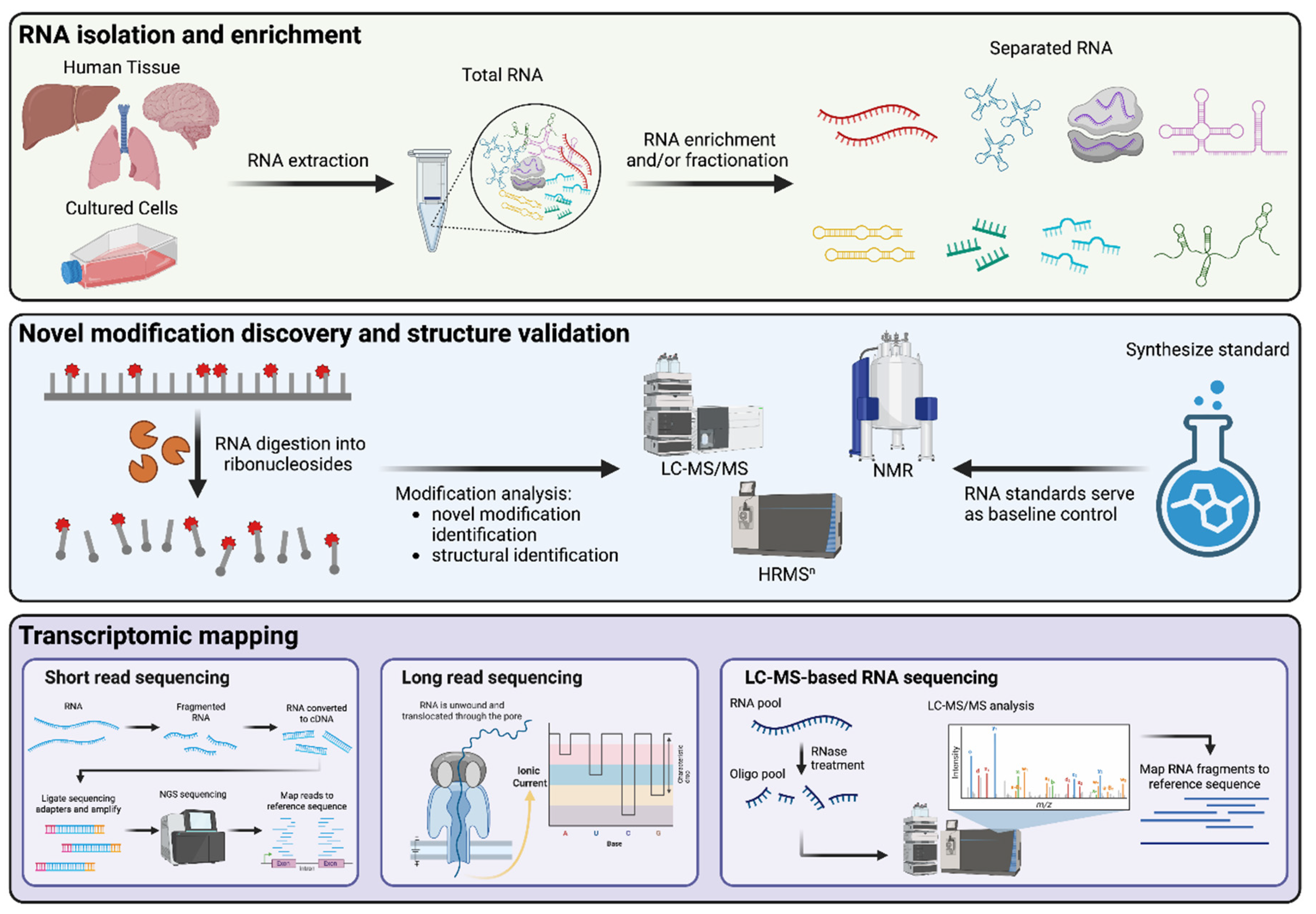
Figure 2.
Structures of 24 RNA modifications that expand the repertoire of human RNA modifications to 76. Using the analytical criteria outlined in Table 1, the modified ribonucleosides are categorized as authenticated, provisional, and putative. Analytical parameters and RNA locations are detailed in Supplementary Table 1.
Figure 2.
Structures of 24 RNA modifications that expand the repertoire of human RNA modifications to 76. Using the analytical criteria outlined in Table 1, the modified ribonucleosides are categorized as authenticated, provisional, and putative. Analytical parameters and RNA locations are detailed in Supplementary Table 1.
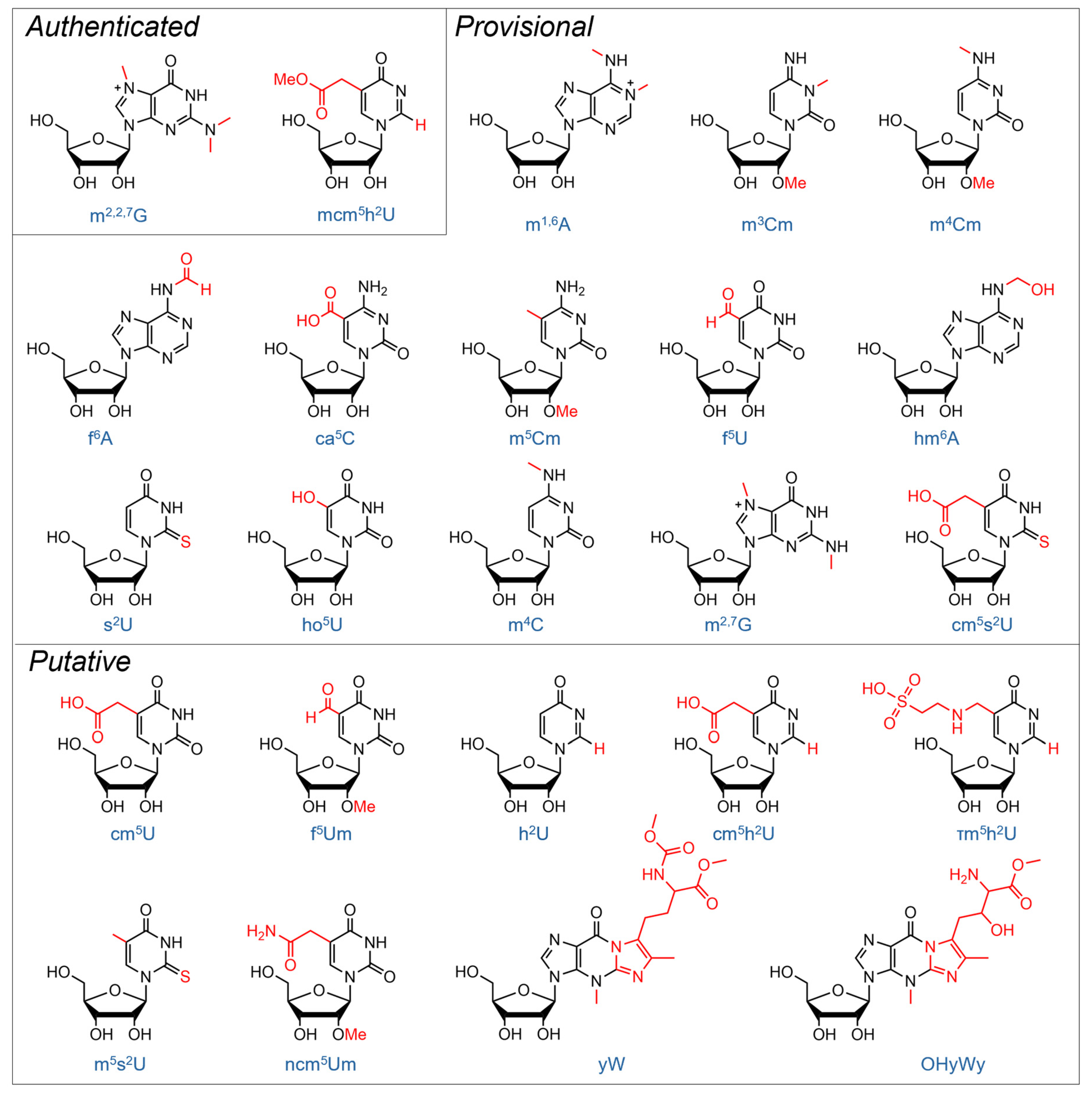

Table 1.
Analytical criteria for different confidence tiers in validating human RNA modifications.
| Confidence Tiers | |||
|---|---|---|---|
| Authenticated | Provisional | Putative | |
| Identity Validation | |||
| Precursor ion scan (MS1 full scan) | Mandatory | Mandatory | Low-resolution |
| Product ion scan (MS2 or MSn by CID/HCD) | Mandatory | Mandatory | Absent |
| Complementary methods for structural elucidation (e.g., NMR) | Mandatory | Optional | Absent |
| Cross-validate with synthetic standard (identical RT, exact mass, and fragments) | Mandatory | Mandatory | Absent |
| Orthogonal validation by multiple independent laboratories | Mandatory | Optional | Absent |
| Mapping Validation | |||
| Mapping resolution | Single-base or fragment resolution |
Bulk RNA fractions, no precise location |
Absent |
| LC-MS-based RNA-seq | Mandatory | Optional | Absent |
| Long-read sequencing | Optional | Optional | Absent |
| Short-read sequencing | Mandatory for isobaric methyl modifications | Optional | Absent |
| Orthogonal validation by multiple independent laboratories | Optional | Optional | Absent |
Disclaimer/Publisher’s Note: The statements, opinions and data contained in all publications are solely those of the individual author(s) and contributor(s) and not of MDPI and/or the editor(s). MDPI and/or the editor(s) disclaim responsibility for any injury to people or property resulting from any ideas, methods, instructions or products referred to in the content. |
© 2026 by the authors. Licensee MDPI, Basel, Switzerland. This article is an open access article distributed under the terms and conditions of the Creative Commons Attribution (CC BY) license.
Copyright: This open access article is published under a Creative Commons CC BY 4.0 license, which permit the free download, distribution, and reuse, provided that the author and preprint are cited in any reuse.


