

Submitted:
27 July 2026
Posted:
30 July 2026
You are already at the latest version
Abstract
Background/Objectives: Non-Hodgkin Lymphoma (NHL) remains a challenging malignancy where treatment success is often limited by drug resistance and severe side effects, even with the standard R-CHOP regimen. In this study, rituximab (RTX)-conjugated nanoparticles (NPs) enhance mitochondrial accumulation of their cargo, which are the standard regimen chemotherapeutics cyclophosphamide, doxorubicin, and vincristine. Methods: These nanoparticles, based on guanidine-amphiphilic cyclodextrin (ACD) or guanidine-cyclodextrin polymer (PCD), underwent comprehensive in vitro characterization, including particle size, surface charge, drug-loading efficiency, and drug release profiles as reported in our previous article. Two- and three-dimensional cell culture studies, involving both RTX-resistant and sensitive models, were conducted. Additionally, in vivo studies on BALB/c mice included plasma pharmacokinetic profiling for nanoparticle-bound RTX, doxorubicin, cyclophosphamide, and vincristine in comparison to free drugs, safety evaluations after single and multiple dose administrations, and antitumoral efficacy tests in NHL tumor-induced animals. Results: RTX-conjugated drug-loaded NPs significantly reduced the viability of lymphoma cell lines compared to multidrugs in solution form (p < 0.05). These experiments highlighted the superior therapeutic performance of the RTX-conjugated Gua-CD nanoparticle formulations, particularly in improving therapeutic outcome by overcoming drug resistance, when compared to conventional therapies. Conclusions: These novel rituximab-conjugated, multi-drug-loaded nanoparticles present a promising strategy for improving the current NHL treatment and set a precedent for nanoparticle-bound delivery of combination therapies in malignancies.
Keywords:
Non-Hodgkin’s Lymphoma
; drug resistance
; active targeting
; subcellular targeting
; rituximab
; cyclodextrin
1. Introduction
The lymphatic system plays a critical role in immune defense and consists of organs that transport lymph fluid through a network of vessels. Lymphoma, a type of cancer originating from lymphocytes, can affect various regions of the lymphatic system. Lymphomas are classified into two main categories that are Hodgkin Lymphoma (HL) and Non-Hodgkin Lymphoma (NHL). NHL accounts for 85-90% of all lymphomas and is the most common group among hematologic cancers. Despite the standard R-CHOP (Rituximab, Doxorubicin, Cyclophosphamide, Vincristine) regimen, the efficacy of NHL treatments is limited by drug resistance, leading to high relapse rates [1,2].
Drug resistance is a phenomenon where cancer cells acquire the ability to evade the effects of chemotherapy or other therapeutic agents. Resistance can be either intrinsic or acquired. Intrinsic resistance refers to the inherent ability of cancer cells to resist drug effects due to their natural characteristics, such as altered expression of drug transporters or mutations in the drug’s target [3]. Acquired resistance occurs as cancer cells undergo genetic mutations or adapt to their environment during treatment, leading to the loss of drug efficacy [4]. Drug resistance is a phenomenon where cancer cells acquire the ability to evade the effects of chemotherapy or other therapeutic agents. Resistance can be either intrinsic or acquired. Intrinsic resistance refers to the inherent ability of cancer cells to resist drug effects due to their natural characteristics, such as altered expression of drug transporters or mutations in the drug’s target [3]. Acquired resistance occurs as cancer cells undergo genetic mutations or adapt to their environment during treatment, leading to the loss of drug efficacy [4].
The development of resistance is influenced by various factors, including the tumor microenvironment. The microenvironment can affect resistance mechanisms by altering cellular behaviors through interactions with surrounding stromal cells, immune cells, and extracellular matrix components. Moreover, the tumor cells may adapt by activating survival pathways, such as autophagy, that help them resist treatment [5,6].
In the context of Non-Hodgkin lymphoma (NHL), resistance mechanisms play a significant role in treatment failure. NHL, particularly B-cell lymphomas, are often characterized by immune evasion and changes in the tumor microenvironment, which contribute to resistance. Genetic mutations, such as those affecting the B-cell receptor signaling pathway, can lead to resistance to chemotherapy [4].
Overcoming drug resistance in cancer treatment, especially in NHL, requires innovative therapeutic strategies [6]. One promising approach is the use of nanoparticle-based drug delivery systems. Nanoparticles can bypass cellular efflux pumps, enhance drug penetration into tumors, and target specific cells, improving the therapeutic efficacy. Targeting resistance mechanisms, such as P-glycoprotein (P-gp) inhibitors or enhancing apoptosis, is a promising strategy for overcoming resistance and improving treatment outcomes.
This study aims to address the critical challenge of chemotherapy resistance in non-Hodgkin lymphoma (NHL) by developing an advanced nanotechnology-based drug delivery system. Conventional chemotherapeutic regimens often fail due to multidrug resistance mechanisms, which significantly reduce treatment efficacy and worsen patient outcomes. To overcome this barrier, we focused on the design of actively targeted, multi-drug-loaded nanoparticles capable of achieving both cellular and subcellular (mitochondrial) precision in drug delivery.
For this purpose, amphiphilic cyclodextrin and polymeric cyclodextrin derivatives containing guanidino groups were employed as nanocarriers. The guanidino moieties confer an intrinsic mitochondrial targeting potential by exploiting the elevated mitochondrial membrane potential characteristic of malignant cells, thereby enabling subcellular-level active targeting beyond conventional receptor-mediated mechanisms. The nanoparticles were loaded with doxorubicin, cyclophosphamide, and vincristine and then they mixed physically and further surface functionalized with rituximab to achieve active cellular targeting of CD20 expressing malignant B cells. To rigorously evaluate the therapeutic potential and targeting specificity of these dual-targeted formulations, a multi-tiered experimental framework was established.
Following physicochemical characterization, drug loading efficiency, and in vitro release profiling, cellular uptake studies were conducted to confirm mitochondrial localization of the nanoparticle-bound cargo. Targeting efficacy and cytotoxic activity were evaluated in both conventional 2D monolayer cultures and 3D multicellular tumor sphere models. Subsequently, in vivo studies were conducted to validate the nanomedicine’s antitumor efficacy, pharmacokinetic properties, and systemic safety in experimental models related to NHL in comparison to drugs in solution form. Based on the comprehensive findings obtained across all experimental stages, the developed rituximab functionalized, guanidino-cyclodextrin based nanoparticles demonstrate considerable promise as a next-generation drug delivery platform for NHL treatment. The convergence of receptor-mediated cellular targeting via rituximab and mitochondria-directed subcellular localization via guanidino moieties, combined with the synergistic co-delivery of doxorubicin, cyclophosphamide, and vincristine, resulted in markedly enhanced cytotoxic efficacy compared to free drug combinations. The therapeutic superiority observed in 2D cultures was further corroborated in 3D tumor spheroid models, where the formulation demonstrated effective penetration and sustained antitumor activity under conditions more reflective of the in vivo tumor microenvironment. In vivo evaluation additionally confirmed anitumoral efficacy and an acceptable systemic safety profile and prolonged half life and slower clearance for most of the nanoparticle-bound chemotherapeutics. Collectively, these results position the proposed nanoparticulate system as a highly promising candidate for overcoming multidrug resistance in NHL, warranting further translational investigation.
2. Materials and Methods
2.1. Materials
The following chemicals and reagents were used in this study: Anti-mCD20 (Invivogen), acetonitrile (HPLC grade, ≥99.8%, Sigma & Aldrich, Germany), dimethyl sulfoxide (DMSO, cell culture grade, ≥99.5%, Sigma & Aldrich, Germany), dialysis membrane (MwCO: 14,000 Da, Sigma & Aldrich, Germany), doxorubicin (Sigma & Aldrich, Germany), Dulbecco's modification of Eagle’s medium (DMEM, Sigma & Aldrich, Germany), ethanol (HPLC grade, ≥99.8%, Sigma & Aldrich, Germany), guanidino-β-cyclodextrin polymer-epichlorohydrin HCl (CycloLab, Cyclodextrin R&D Ltd., Hungary), heptakis (6-deoxy-6-guanidino) β-cyclodextrin heptahydrochloride (CycloLab, Cyclodextrin R&D Ltd., Hungary), cholesterol assay kit (MAK043, Sigma & Aldrich, Germany), Matrigel® basement membrane (Corning, USA), penicillin/streptomycin (Sigma, Israel), poly(2-hydroxyethyl methacrylate) (Poly-HEMA, Sigma & Aldrich, USA), rituximab (Biorbyt, USA), cyclophosphamide (Sigma & Aldrich, Germany), survivin assay kit (ab119607, Abcam, UK), tetrahydrofuran (HPLC grade, ≥99.8%, Sigma & Aldrich, Germany), trypan blue (Sigma & Aldrich, USA), trypsin-EDTA (Sigma & Aldrich, Germany), Triton-X 100 (Neofroxx, Germany), TUNEL assay kit (ApopTag®, S7101, Millipore, USA), WST-1 (water-soluble tetrazolium salt, Roche, Germany), and vincristine (Sigma & Aldrich, Germany).
For biological materials, fetal bovine serum (Sigma, Germany) was used as a supplement in cell culture studies. Human Non-Hodgkin’s lymphoma cell lines, Daudi (CCL-213) and BJAB (CRL-2230), were obtained from the American Type Culture Collection (ATCC, USA). Additionally, the mouse fibroblast cell line L929 was acquired from ATCC (USA) for cytotoxicity assays.
2.2. Preparation and Characterization of Active Targeted and Multi-Drug Loaded Nanoparticles
As reported in our previous article all nanoparticle formulations were prepared by nanoprecipitation method. The nanoparticle formulations were characterized by in vitro studies including mean particle size distribution, zeta potential, drug loading efficacy and drug release profile [7].
2.3. Cell Culture Studies
Cell culture studies were designed in order to assess the potential of the Gua-CD nanoparticles conjugated with RTX in overcoming drug resistance for NHL. BJAB cells are a human-derived B-lymphocyte cell line, originally established in 1973 from a patient with Epstein-Barr virus (EBV)-negative Burkitt's lymphoma. As a human-derived model, BJAB cells retain key characteristics of B cells, making them valuable for studying lymphoma biology and therapeutic responses in a human context.
Their EBV-negative status further enhances their utility in investigating lymphomagenesis and drug resistance mechanisms without the confounding effects of viral infection. These properties make BJAB cells an essential tool for evaluating the efficacy of anti-cancer drugs and exploring molecular pathways relevant to human lymphomas.
2.3.1. Drug Resistance Development Studies
BJAB human lymphoma cells were cultured in RPMI 1640 medium supplemented with 10% fetal bovine serum (FBS) and maintained at 37 °C in a humidified atmosphere containing 5% CO₂. RTX-resistant BJAB cells were generated using a modified protocol adapted from previously reported methods [8]. Briefly, cells were exposed to rituximab (RTX) at concentrations ranging from 0.125 to 8 µg/mL for 24 h, followed by incubation in fresh medium for 72 h. The RTX concentration was doubled in each subsequent treatment cycle, and cells were treated with RTX twice weekly. After the seventh passage, RTX-resistant BJAB cells were successfully established [8,9].
To confirm RTX resistance, flow cytometry was performed. RTX-sensitive and -resistant BJAB cells were passaged, and 5 × 10⁵ cells were distributed into separate tubes. RTX-resistant BJAB cells were maintained in RTX-free medium for 48 hours before staining. The cells were first washed once with PBS, followed by incubation with anti-human CD20 antibody (BD Pharmingen, Cat no: 555622) at room temperature for 20 minutes. After incubation, cells were washed with PBS and centrifuged at 1800 rpm for 5 minutes. Finally, samples were analyzed using a BD FACSCanto II flow cytometer, and the data were processed with FlowJo™ 10 software.
2.3.2. Determination of Antiproliferative Activity of Nanoparticles
The anticancer activity was evaluated in the following groups:
- blank guanidino-modified cyclodextrin nanoparticles (Blank Gua-CD NPs),
- doxorubicin-loaded nanoparticles (DOX-NPs),
- cyclophosphamide-loaded nanoparticles (CYC-NPs),
- vincristine-loaded nanoparticles (VRC-NPs),
- the combination of free doxorubicin, cyclophosphamide, and vincristine (Free DOX + CYC + VRC),
- the combination of DOX, CYC, and VRC loaded , physically mixed nanoparticles (DOX + CYC + VRC NPs)
- rituximab-conjugated DOX + CYC + VRC loaded, physically mixed nanoparticles (RTX-DOX + CYC + VRC NPs),
were evaluated using human lymphoma cell lines, Daudi and BJAB (both RTX-resistant and sensitive). For this purpose, cells were first seeded into U-bottom 96-well cell culture plates containing RPMI-1640 medium supplemented with 10% fetal bovine serum (FBS). After incubation periods of 24 and 48 hours, cell viability was assessed using the WST-1 assay. Cells incubated only with the culture medium served as the control group, and the viability of test groups was calculated relative to the control [10].
2.3.3. Determination of Cell Death Mechanism
The mechanism of cell death induced by the prepared NP formulations was determined by flow cytometry. For this purpose, BJAB (RTX-sensitive and -resistant) human lymphoma cells were incubated with ACD and PCD NP formulations, as well as free drug solutions. After incubation, the cells were treated with 5 μL Annexin V-FITC and 5 μL PI for 15 minutes at 37°C. The proportion of apoptotic and necrotic cells was then determined by flow cytometry (11)
2.3.4. Determination of Antibody-Dependent Cellular Cytotoxicity
To evaluate the biological activity of adamantane-conjugated RTX present in NP formulations, an antibody-dependent cellular cytotoxicity (ADCC) assay was performed. BJAB (both RTX-resistant and sensitive) lymphoma cells were used as target cells, while PBMCs served as effector cells.
Each well of a 96-well U-bottom plate was seeded with 2.5 × 10⁵ PBMC (effector cells) and 1x10⁴ Daudi or BJAB cells (target cells) in 100 µL of culture medium, maintaining an effector-to-target cell ratio of 25:1. The cells were subsequently incubated at 37°C for 4 hours in the presence of RTX solution or RTX-loaded CD NP formulations to assess cytotoxic activity [12]. Following incubation, the protocol provided in the commercially available Lactate Dehydrogenase (LDH) kit (Biovision K-311) was applied to measure cytotoxicity. For this purpose, the cells were centrifuged at 5000 rpm for 20 minutes using a microplate centrifuge. Subsequently, 100 µL of supernatant from each sample was transferred to a new 96-well plate in eight replicates per sample. Then, 100 µL of LDH detection reagent was added to each well, and the plate was incubated at room temperature for 30 minutes. Absorbance was measured at 495 nm using a microplate reader to determine LDH release [13]. During the study, cells incubated with 1% Triton X-100 were used as the positive control group. The absorbance obtained from this group was considered as representing 100% LDH release. Based on this value, the percentage of LDH release in the experimental groups was calculated accordingly.
2.3.5. Cholesterol Extraction Study
In addition to their use as drug delivery systems, cyclodextrins (CDs) are also utilized to alter cholesterol composition in various cell types [14,15]. This is accepted as one of the mechanisms with which CDs help overcome drug resistance by extracting the cholesterol from drug resistant cancer cell membrane and thus increasing cell membrane permeability. Varan et al. demonstratred that amphiphilic CD nanoparticles of positive or negative surface charge can induce apoptosis in a variety of cancer cell types through a mitochondrial pathway (REF). Thus, cholesterol extraction capability of the new CD nanoparticles was also evaluated in this study.
The cholesterol removal efficacy from the membrane varies depending on the type of cyclodextrin (CD) derivative, its concentration, incubation time with the cells, temperature, and the cell type. Specifically, it has been demonstrated that Methylated-β-CD is the most effective CD derivative for cholesterol removal [16].
The cholesterol affinity of the synthesized Gua-CD derivatives in NHL cancer cells was determined. For this purpose, the cholesterol content in BJAB (both RTX-resistant and sensitive) cells, incubated with various Gua-CD derivatives, was measured using a commercially available cholesterol assay kit [17]. For comparison, Methyl-β-CD, known for its high cholesterol removal efficacy, was used as the reference standard.
2.3.6. Survivin Assay
Survivin is an anti-apoptotic gene that is highly expressed in cancer cells. The survivin protein produced because of its expression inhibits the activation of Caspase-9, preventing cells from undergoing apoptosis. Furthermore, in non-Hodgkin lymphoma (NHL), the increased expression of survivin has been shown to prevent cell apoptosis, leading to enhanced proliferation and an increase in drug resistance [18,19].
BJAB (both RTX-resistant and sensitive) cells were incubated with drug-loaded and actively targeted NPs prepared using CD derivatives. After incubation, the effect of NPs on the expression of the survivin gene in NHL cells was determined using a commercially available Human Survivin ELISA Kit, following the protocol recommended by the kit. Cells incubated only with the medium were used as the control group. For this experiment, 100 µL of cell suspension, containing 5x10³ cells per well, was added to each well of a 96-well cell culture plate. After 24 hours, the cell culture medium was replaced with DMEM containing NPs. After an additional 48 hours of incubation, the protocol of the kit was followed. Subsequently, the absorbance of the samples was measured at 450 nm, and the concentration of survivin protein in the samples was determined using the equation derived from standard solutions [20].
2.3.7. Assessment of Mitochondrial Directed Delivery Capability
The Gua-CD derivatives used in this study have the potential to target the cell mitochondria due to the guanidine groups present in their structures. Based on this property, to determine the accumulation of Gua-CD bound drug in the mitochondria, 20 × 10⁶ Daudi and BJAB (both RTX-resistant and sensitive) cells were incubated for 24 hours at 37°C in a 5% CO₂ atmosphere with DOX-loaded Gua-CD derivatives or an equal amount of DOX solution [21]. After incubation, the mitochondria of the cells were isolated according to the protocol provided in the commercial Mitochondria Isolation Kit (Thermo 89874). The incubated cells were centrifuged at 5000 rpm for 20 minutes, and the supernatant was removed. Then, 800 µL of Reagent A from the kit was added to the cells, and they were kept on ice for 2 minutes. Subsequently, 10 µL of Reagent B was added, and the cells were kept on ice for an additional 5 minutes, mixing gently 5 times for 10 seconds each minute using vortex. After this, 800 µL of Reagent C was added, and the cells were centrifuged at 700 g for 10 minutes at +4°C. The supernatant was discarded, and the pellet containing the mitochondria was resuspended and centrifuged again at 4000 g for 15 minutes. The supernatant was discarded again, and the pellet containing the mitochondria was resuspended in 500 µL of Reagent C and centrifuged at 4sensitive000 g for 5 minutes. Finally, the pellet containing the mitochondria was taken, and the amount of DOX was measured using a fluorescence microplate reader at excitation and emission wavelengths of 470 nm and 560 nm, respectively.
2.3.8. Determination of Pgp Inhibition
The P-glycoprotein (P-gp) inhibition caused by the CD derivatives through their ATPase activity was determined colorimetrically using a commercially available cell-based ATPase kit. For this purpose, both resistance-developed and sensitive cells were incubated with the prepared nanoparticles (NPs), and after incubation, the kit protocol was applied. The Calcein AM extrusion assay was performed on both resistant and sensitive BJAB cells according to the manufacturer's instructions and analyzed.
Resistant and sensitive BJAB cells were seeded at a density of 1 × 10⁵ cells/well in 24-well plates and incubated for 24 hours. The cells were then incubated with PBS, DOX formulations, or Verapamil (2.5 μM) at 37 °C for 30 minutes. Afterward, the cells were incubated with 0.5 μM Calcein AM at 37 °C for an additional 30 minutes. PBS-treated cells served as the negative control. Following incubation, the cells were washed three times with cold PBS and read using a microplate reader (ex: 495 nm, em: 515 nm). Verapamil, a known characteristic P-gp substrate, was used as a positive control (n=6) during the experiment.
2.3.9. 3D Tumor Model Studies
To develop a 3D tumor model using BJAB (RTX-resistant and sensitive) human lymphoma cell lines, round-bottomed 96-well plates were coated with a Poli-HEMA solution. Poli-HEMA was dissolved by mixing 1.2 g of it in 40 mL of 95% ethanol, and 50 μL of this solution was added to each well. After incubation overnight, 200 μL of cell seeding medium containing 5000 cells per well and 3% (v/v) Matrigel® was added to the plates. The plates were then centrifuged at 10,000 rpm for 10 minutes. Every two days, the medium was changed. On the third day, the formation of the spheroid structure was observed, and to allow further development, the evaluation of the formulations' efficacy began on the fifth day. The cells were incubated with drug-loaded NP formulations and drug solutions for 48 and 72 hours. After incubation, cell viability was determined by WST-1 analysis [10]. Cells incubated with only the medium served as the control group, with their viability considered to be 100%.
2.4. Animal Studies
Experiments were performed using male BALB/c mice (7–8 weeks old, 22–25 g). The animals were kept in an environment with a constant temperature of 23 ± 2 °C, 50% humidity, and filtered air. All procedures were carried out following ethical guidelines for animal protection, with approval from the Hacettepe University Animal Experiments Ethical Committee (Approval number 2022/57 and decision number 2020/10-05).
2.4.1. Plasma Profiling for Free and Nanoparticle-Bound Drugs
in vitro studies using ACD and PCD-based NP systems revealed successful formulations, with the optimum formulation determined using ACD. Following this, in vivo bioavailability assessments were performed using 7–8-week-old male BALB/c mice, with drug-loaded ACD NPs in solution form serving as the reference.
Healthy BALB/c mice received intravenous administration via the tail vein of the anticancer drugs at the following doses: doxorubicin (DOX, 1 mg/kg), vincristine (VRC, 2 mg/kg), cyclophosphamide (CYC, 30 mg/kg), and rituximab (RTX, 2 mg/kg). Blood samples (approximately 200 µL) were collected by cardiac puncture under ether anesthesia at predetermined time points based on the biological half-lives of the drugs in mice (DOX: 0.25, 1, 4, 8, 12, and 24 h; VRC: 0.08, 0.25, 0.5, 1, and 2 h; CYC: 0.25, 0.5, 1, 2, and 4 h). Three mice were used for each sampling time point (n = 3).
Blood samples were transferred into heparinized tubes and centrifuged at 4,000 rpm for 10 min to obtain plasma. Plasma concentrations of DOX, VRC, and CYC were determined using an LC–MS/MS system (Shimadzu LCMS-8030, Shimadzu Corporation, Kyoto, Japan). The chromatographic and mass spectrometric conditions were adapted from the validated LC–ESI–MS/MS method reported by Nussbaumer et al. [22]. Pharmacokinetic parameters, including the elimination rate constant (kd), elimination half-life (t½), volume of distribution (Vd), clearance (Cl), area under the plasma concentration–time curve (AUC), maximum plasma concentration (Cmax), and time to reach maximum plasma concentration (Tmax), were calculated from the plasma concentration–time profiles.
Reference stock solutions of DOX, CYC, and VRC were prepared by accurately weighing 10 mg of each compound and dissolving them individually in methanol. Working standard solutions were prepared by serial dilution of the stock solutions with methanol:water (1:1, v/v). Calibration standards were prepared by spiking 50 µL of blank mouse plasma with 50 µL of the appropriate working standard solution in an Eppendorf tube, followed by vortex mixing for 15 min. The final calibration concentrations in plasma were 4, 10, 20, 100, 400, 1000, and 2000 ng/mL for DOX; 5, 12.5, 25, 125, 500, 1250, and 2500 ng/mL for CYC; and 6, 15, 30, 150, 600, 1500, and 3000 ng/mL for VRC.
For plasma sample preparation, 50 µL of plasma was mixed with 100 µL of acetonitrile for protein precipitation. The mixture was vortex-mixed and centrifuged at 10,000 rpm for 10 min. The resulting clear supernatant was transferred into autosampler vials equipped with glass inserts and analyzed using the Shimadzu LCMS-8030 LC–MS/MS system.
2.4.2. Safety Study
The potential toxicity of the synthesized Gua-CD derivatives in a rodent model was determined through in vivo studies using empty Gua-CD NP. For this purpose, empty Gua-CD NP was administered intravenously to healthy BALB/c mice in a manner that reflects the clinical R-CHOP regimen, with a single dose and repeated doses (one dose every two days for a total of five doses). Following the final dose, blood samples were collected from the animals 24 hours later, and biomarkers such as alkaline phosphatase and alanine aminotransferase were measured to assess potential toxicity. Four animals were used in each group for this experiment.
2.4.3. Antitumoral Efficacy Study
To determine the antitumor efficacy of anticancer drug-loaded and actively targeted ACD NPs, 11-12-week-old male BALB/c mice were first injected subcutaneously in the right flank with 100 µL of PBS containing 2.5x10⁶ A20 murine lymphoma cells. Tumor development was confirmed when the tumor reached a size of 0.5 cm, approximately 4 weeks post-injection. The mice were then divided into appropriate groups based on the NP formulations to be administered, with 6 animals per group. Control groups received only physiological serum. The other groups received treatment formulations intravenously via the tail vein at a volume of 100 µL per injection, twice a week, for 4 weeks. To achieve therapeutic response with RTX, anti-CD20 was used for the mice, and characterization processes were performed. The anticancer drug doses were as follows: DOX = 1 mg/kg, VRC = 100 mg/kg, and CYC = 2 mg/kg. The NP formulations were encapsulated at 2 mg/kg of anti-CD20 in physiological serum [23,24]. The NP formulations were prepared aseptically immediately before administration and sterilized by passing through a 0.22 µm filter. Throughout the treatment period, the animals were monitored for changes in body weight and tumor size. After the treatments, the animals were euthanized using a high-dose anesthetic. The formulation groups for the mice were optimized based on in vitro and cell/tumor culture studies using the best Gua-CD derivative, and the treatment was carried out as follows. Six groups of mice were used for intravenous treatments with 6 animals per group. The groups were as follows:
- Free drug solution group: RTX, DOX, VRC, CYC solution (intravenous in physiological serum)
- Nanoparticle group: RTX-DOX-ACD + RTX-VRC-ACD + RTX-CYC-ACD NP (intravenous in physiological serum)
- Rituximab solution and nanoparticle group: RTX solution and DOX + VRC + CYC-loaded ACD NP (intravenous in physiological serum)
- Blank nanoparticle group (intravenous in physiological serum)
- RTX-conjugated blank nanoparticle group (intravenous in physiological serum)
- Control group: Physiological serum only.
2.5. Statistical Analysis
The differences between groups were considered statistically significant when p < 0.05. During the 2D and 3D cell culture experiments, each group was repeated at least 6 times. The obtained data were analyzed using Student's t-test. In the cell culture studies, cells incubated with only the medium were used as the control group, and the formulations were evaluated against the control group and/or each other. Differences between groups were considered statistically significant when p < 0.05. In vivo activity results were analyzed using a suitable statistical program with a two-way ANOVA. The formulations were evaluated against the control group and/or each other, and differences between groups were considered statistically significant when p < 0.05 (* p < 0.05, ** p < 0.001, *** p < 0.0001).
3. Results
3.1. Preparation and Characterization of Active Targeted and Multi-Drug Loaded Nanoparticles
The detailed characterization results of the nanoparticles have been reported previously [25] In brief, all drug-loaded ACD NP formulations exhibited particle sizes below 200 nm (typically in the range of 120–180 nm) with mostly positive zeta potentials, while the incorporation of RTX led to a slight reduction in particle size. The loading efficiency, calculated as the ratio of the drug amount quantified by HPLC to the initially added amount, was found to be high >70% for most formulations). RTX content in the formulations was determined using a BCA assay.
3.2. Cell Culture Studies
3.2.1. Drug Resistance Development Studies
The expression of CD20 in BJAB cells with different rituximab-related resistance characteristics was analyzed in terms of mean fluorescence intensity (MFI). It was determined that CD20 expression decreased in association with rituximab resistance. Specifically, CD20 expression in rituximab-resistant BJAB cells was found to be 31.3% with an MFI of 136, while in rituximab-sensitive BJAB cells, CD20 expression was observed at 73% with an MFI of 488 (Figure 1).
3.2.2. Determination of Cell Death Mechanism
The mechanisms of cell death in resistant and sensitive BJAB cell lines treated with different formulations are shown in Figure 2. Flow cytometry analysis using Annexin V-FITC and PI staining classified cells as viable (Annexin V-/PI-), early apoptotic (Annexin V+/PI-), or late apoptotic/necrotic (Annexin V+/PI+).
In sensitive cells, RTX-Drug-ACD nanoparticles induced a ~3-fold increase in both early and late apoptotic populations compared to the control. Interestingly, early apoptosis decreased in the RTX-Drug-ACD group, while late apoptosis increased nearly 10-fold (p < 0.05). The proportion of viable cells in the RTX-Drug-ACD group was 4-fold lower than in the solution-treated group (p < 0.05).
In resistant BJAB cells, RTX-Drug-ACD nanoparticles significantly enhanced necrosis and late apoptosis compared to other treatments (p < 0.05). Early apoptosis was reduced, while necrosis and late apoptosis increased up to 3-fold (p < 0.05). The viable cell population was significantly lower in the RTX-Drug-ACD group compared to the solution-treated group (p < 0.05).
Overall, ACD nanoparticle formulations showed the most pronounced and statistically significant effects on cell death in both resistant and sensitive cells (p < 0.05).
3.2.3. Antiproliferative Activity of Nanoparticles
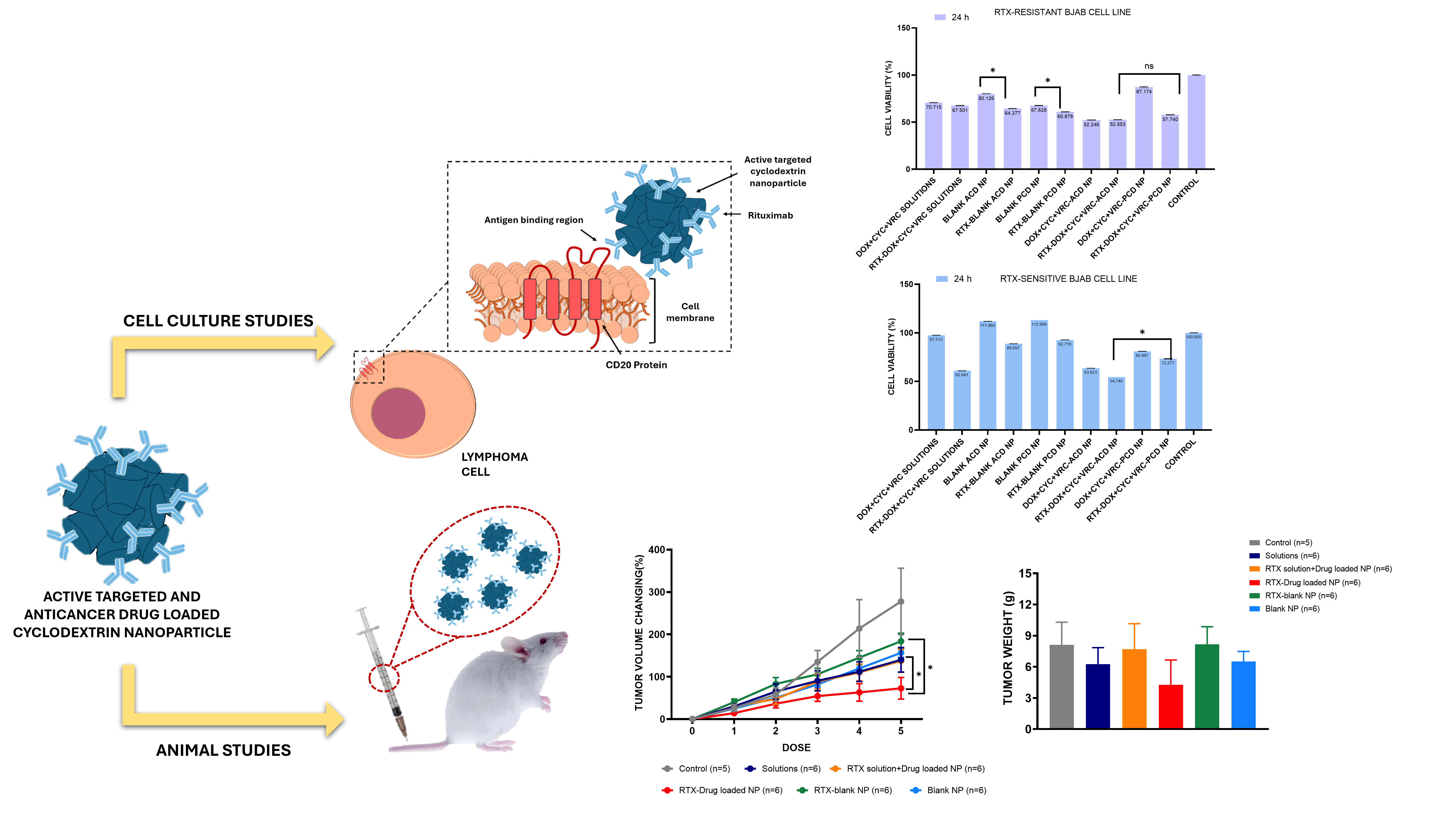
The antiproliferative activity of different formulations were evaluated on BJAB human lymphoma cell lines both resistant and sensitive. The results of the 24-hour and 48-hour cell viability assays conducted on human lymphoma cells are presented in Figure 3.
3.2.4. Determination of Antibody-Dependent Cellular Cytotoxicity
According to the cell lysis results shown in Figure 4, it was determined that RTX-containing GuaCD nanoparticles stimulated effector cells at least as effectively as free RTX in both cases with ACD nanoparticles showing a higher ADCC (p<0.05).
3.2.5. Cholesterol Extraction Study
The findings obtained from the cholesterol extraction studies are shown in Figure 5 for all formulations in sensitive or resistant BJAB cells.
3.2.6. Survivin Assay
The effect of drugs in free form and nound to ACD or PCD nanoparticles on the survivin levels in BJAB cells are shown in Table 1.
3.2.7. Assessment of Accumulation in Mitochondria
According to the mitochondrial isolation analysis results shown in Figure 6, ACD nanoparticles delivered a higher amount of DOX to the mitochondria in both sensitive and resistant BJAB cells compared to PCD nanoparticles. PCD nanoparticles enhanced mitochondrial DOX delivery only in the sensitive cells compared to the DOX solution, while in resistant cells, their effect was similar to the solution (p>0.05). Overall, ACD nanoparticles were the most effective formulation for promoting mitochondrial drug accumulation across both cell types.
3.2.8. Determination of Pgp Inhibition
The comparative Pgp results of ACD, PCD, and the positive control group (Verapamil) are shown in Figure 7.
3.2.9. 3D Tumor Model Studies
According to the findings shown in Figure 8, after 48 hours of incubation, more than 70% cell viability was observed in both drug-loaded nanoparticles and drug solutions. Based on the results of the WST-1 assay after 48 hours of incubation in BJAB 3D lymphoma spheroids, a significant difference was observed between the drug-loaded, RTX-targeted ACD NP formulations and the free drug solutions (p<0.05).
3.3. Animal Studies
3.3.1. Safety Study
The safety study results shown in Figure 9 were obtained based on serum samples collected from healthy Balb/c mice, with the analysis conducted.
3.3.2. Determination of Plasma Profile Free and Nanoparticle-Bound Drug
The calibration curves for DOX, CYC, and VRC were linear over the tested concentration ranges, with correlation coefficients (R²) of 0.9921, 0.9863, and 0.9972, respectively.
Figure 10.
Comparative plasma profiles of DOX, CYC and VRC in free form and bound to CD nanoparticles (n=3).
Figure 10.
Comparative plasma profiles of DOX, CYC and VRC in free form and bound to CD nanoparticles (n=3).
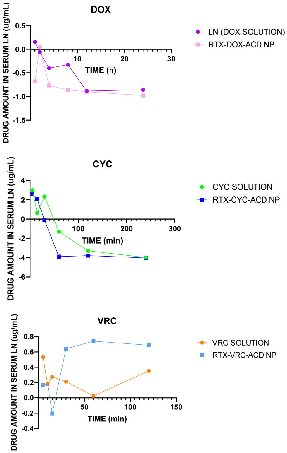
The following results were obtained based on the evaluation of pharmacokinetic parameters derived from the graphs (Table 2).
3.3.3. Antitumoral Efficacy Study
4. Discussion
4.1. Preparation and Characterization of Active Targeted and Multi-Drug Loaded Nanoparticles
This study utilized RTX-targeted cyclodextrin nanoparticles (ACD and PCD) previously characterized for their critical quality attributes and suitability for NHL treatment [25]. In brief, ACD nanoparticles exhibited more homogeneous size distribution, lower PDI, and positive zeta potential compared to PCD nanoparticles, which likely contributes to enhanced cellular uptake. Both systems demonstrated high drug-loading efficiency and stability, as reported in our earlier study.
4.2. Conventional Cell Culture Studies
4.2.1. Drug Resistance Development Studies and Determination of Cell Death Mechanism
CD20 is a surface protein that is highly expressed in B cells while being minimally present in other tissues, making it an ideal target for immunotherapy against B cell-related malignancies. CD20 expression begins at the pre-B cell stage and continues until B cells differentiate into plasma cells. CD20 is neither secreted, altered, nor internalized. Although the exact function of CD20 is not fully understood, it is believed to be involved in the release of calcium ions into the cell and to be responsible for maintaining calcium ion concentration. In this way, it is thought to play a role in the activation of B lymphocytes [26]. As shown in Figure 1, following the application of resistance development protocols in BJAB cells, the expression of CD20 was reduced by more than half. This has demonstrated the development of resistance by flow cytometry. In this study, both resistant and sensitive BJAB cells were used, as shown in Figure 1.
In sensitive BJAB cells, RTX-Drug-ACD nanoparticles induced a modest decrease in early apoptotic cells (Annexin V+/PI-) while causing a nearly 10-fold increase in late apoptotic/necrotic cells (Annexin V+/PI+). This suggests that the formulation accelerates the progression from early to late apoptosis, effectively pushing cells more rapidly toward irreversible cell death. Such a shift indicates enhanced cytotoxic efficiency compared to non-targeted solutions or single-drug treatments. The marked increase in late apoptotic cells could also reflect time-dependent effects, as cells exposed to the nanoparticles progress through apoptotic stages faster than in control treatments.
Moreover, the observation that the viable cell population remained four times higher than in the solution-treated group may indicate improved selectivity, potentially sparing non-malignant or less susceptible cells. The balance between apoptosis and necrosis is also critical: while early apoptosis represents reversible cell stress, late apoptosis and necrosis represent irreversible cell damage, suggesting that RTX-mediated targeting enhances the delivery of cytotoxic drugs specifically to lymphoma cells, maximizing therapeutic effect while potentially limiting off-target toxicity.
In resistant BJAB cells, RTX-Drug-ACD nanoparticles significantly increased late apoptotic and necrotic populations while early apoptosis decreased. This indicates that the formulation overcomes resistance mechanisms, such as reduced drug uptake or altered apoptotic signaling, by efficiently triggering irreversible cell death. In contrast, PCD nanoparticles or non-targeted formulations induced smaller increases in late apoptosis and necrosis, highlighting the critical role of ACD nanoparticle properties (smaller size, positive zeta potential, homogeneous distribution) and active RTX targeting in enhancing cytotoxicity. The differences between sensitive and resistant cells demonstrate that CD20 expression directly affects nanoparticle uptake and apoptotic induction, with lower expression in resistant cells requiring targeted delivery to achieve comparable cytotoxic effects.
Developing resistance in BJAB cells, a human lymphoma cell line, is of critical importance for simulating drug resistance, one of the most significant challenges in clinical practice. In clinical settings, most patients develop resistance to treatments involving RTX over time, complicating disease progression and limiting available treatment options [27,28]. Therefore, Jazirehi et al. investigated the biological alterations in rituximab-resistant cells. Several key mechanisms were identified in the study, highlighting the ineffectiveness of rituximab treatment in resistant cells. Firstly, the study found that the hyperactivation of ERK1/2 and NF-κB signaling pathways in resistant cells contributed to their evasion of apoptosis. Additionally, rituximab's effect was inhibited in resistant cells via complement-dependent cytotoxicity (CDC), rendering the treatment ineffective. The study also revealed that the overexpression of anti-apoptotic proteins, such as Bcl-2 and Bcl-xL, in resistant cells played a critical role in protecting these cells from treatment-induced apoptosis. These findings suggest that rituximab resistance is associated with mechanisms such as deregulated signaling pathways, CDC resistance, and enhanced anti-apoptotic protein expression [8].
In this study, developing RTX resistance in NHL cells provided a more physiologically relevant model to evaluate the efficacy of the nanoparticle formulations, as it mimics the natural progression of treatment resistance observed in patients. In this context, RTX-Drug-ACD nanoparticles demonstrated superior cytotoxicity compared to free drugs or non-targeted nanoparticles, in both sensitive and resistant BJAB cells. In sensitive cells, the formulation accelerated progression from early to late apoptosis, resulting in nearly tenfold increases in late apoptotic/necrotic populations, while preserving a higher number of viable cells relative to solution-treated controls, suggesting selective targeting and efficient intracellular drug delivery. In resistant cells, RTX-Drug-ACD NPs overcame reduced CD20 expression and other resistance mechanisms by significantly increasing late apoptosis and necrosis (~threefold), while early apoptosis decreased and viable cells declined. These differences indicate that the observed effects are formulation-dependent with ACD nanoparticles providing optimized size, surface charge, and homogeneity and cell type-dependent, as resistant cells required targeted delivery to achieve comparable cytotoxicity. Overall, these findings suggest that active targeting via RTX, combined with the physicochemical properties of ACD nanoparticles, allows the formulations to effectively induce irreversible cell death in both sensitive and resistant NHL cells, fulfilling the primary objective of overcoming acquired drug resistance.
4.2.2. Determination of Antiproliferative Activity of Nanoparticles
The results of cell viability analyses performed with BJAB (resistant and sensitive) human lymphoma cell lines after 24 is presented in Figure 2.
Upon reviewing all the results, it is evident that the final formulation obtained by physically mixing the three drug-loaded NP formulations led to a statistically significant decrease in cell viability compared to drug solutions. Among the formulations, the groups treated with actively targeted ACD NPs exhibited the lowest cell viability.
In the BJAB cell line experiments, both resistant and sensitive cells maintained over 80% viability when treated with RTX solution alone at 24 h, indicating that RTX by itself did not exert direct cytotoxic effects. However, incorporation of RTX into the nanoparticle formulations significantly enhanced the cytotoxicity of both PCD and ACD nanoparticles compared to nanoparticles without RTX (p<0.05). This improvement is likely due to multiple complementary mechanisms. First, RTX binds specifically to CD20 on B cells, promoting receptor-mediated endocytosis of the nanoparticles. This effect is particularly pronounced in ACD nanoparticles, which exhibit smaller particle sizes, higher positive surface charge, and a more compact molecular packing as reflected by their lower polydispersity index, characteristics that favor stronger electrostatic interactions with the negatively charged cell membranes and more efficient internalization compared to the more flexible, polymeric PCD nanoparticles. Second, in an environment containing only tumor cells, RTX may crosslink CD20 molecules on the cell surface, initiating downstream apoptotic signaling pathways and further enhancing cell death. Together, these factors explain why RTX-ACD nanoparticles demonstrated the greatest cytotoxic effect, highlighting the importance of active targeting, favorable physicochemical properties, and potential receptor-mediated signaling in achieving enhanced efficacy against both sensitive and resistant BJAB cells [29].
The observed difference in cytotoxicity between ACD and PCD nanoparticles cannot be attributed solely to the more complex and larger polymeric architecture of PCD NPs. While structural complexity may reduce cellular uptake due to steric hindrance or aggregation tendencies, other physicochemical properties also play important roles. ACD nanoparticles exhibited smaller particle sizes, more homogeneous distributions, and higher positive surface charge, which facilitate stronger interactions with negatively charged cell membranes and more efficient endocytosis. Additionally, the drug-loading efficiency and total payload of anticancer drugs were generally higher in ACD NPs, and their release profiles favored a more sustained and controlled intracellular drug delivery. In contrast, PCD nanoparticles, with their larger and branched structure, demonstrated slightly lower drug-loading efficiency and a tendency for aggregation, which likely reduced effective cellular internalization and led to a less pronounced reduction in cell viability. Therefore, the superior cytotoxicity of ACD nanoparticles arises from a combination of smaller size, optimized surface charge, higher drug loading, and favorable release kinetics, rather than structural complexity alone.
4.2.3. Determination of Antibody-Dependent Cellular Cytotoxicity
Antibody-dependent cell-mediated cytotoxicity (ADCC) is a type of immune reaction in which target cells are coated with antibodies and then killed by macrophage cells. This mechanism is commonly used to assess the biological activity of antibodies and in the comparison of biosimilars (30). One of the common methods to assess cytotoxicity is by measuring the activity of cytoplasmic enzymes released from damaged cells. Lactate dehydrogenase (LDH) is a stable cytoplasmic enzyme present in all cells, and it is rapidly released into the culture medium when the cell membrane is damaged. This property makes it one of the primary indicators of cellular damage, such as apoptosis, necrosis, or other types of cellular injury. [31].
Fasano et al. developed an in vitro LDH cytotoxicity assay using NK cells isolated from PBMCs of healthy donors to evaluate the ADCC induction potential of IgG1 isotype monoclonal antibody treatment. The H460 and H1299 cell lines, which exhibit high levels of PD-L1 and EGFR and low MHC-I expression, were identified as the most sensitive to NK-mediated ADCC. The combination of Avelumab and Cetuximab significantly increased cytotoxicity in lung cancer cells [32].
Based on the literature, LDH activity was used as a marker in the study. RTX specifically binds to Daudi and BJAB (both resistant and sensitive) cells, stimulating effector cells. The amount of LDH released from the lysed lymphoma cells by the activated effector cells was measured to determine the percentage of lysed cells. According to the results shown in Figure 3, it was found that ACD and PCD NPs containing RTX stimulated effector cells at least as effectively as free RTX in both cases. This result demonstrates that RTX retained its biological activity during the nanoparticle preparation process. Furthermore, circular dichroism (CD) analysis performed within the scope of characterization studies further supported these findings. The CD spectra of free RTX solution and vincristine-loaded RTX-conjugated ACD nanoparticles exhibited highly similar profiles in the far-UV region, with no significant spectral shifts or appearance of new characteristic peaks (Figure 13.). The CD curves fluctuated close to the baseline, indicating that the conjugation and nanoparticle preparation processes did not induce major conformational alterations in the secondary structure of RTX. In addition, the HT profiles showed comparable trends among the samples and remained within acceptable limits, confirming the reliability of the obtained spectra. These results suggest that RTX preserved its structural integrity after nanoparticle functionalization and it confirms that active targeting was successfully achieved, and RTX, which reached the cells in its intact form, not only functioned as a targeting agent but also contributed to the therapy through ADCC.
4.2.4. Cholesterol Extraction Study
Cholesterol plays a critical role in regulating the fluidity of cell membranes. High cholesterol levels can make the membrane stiffer, increasing drug resistance. This rigidity can hinder the entry of drugs into cancer cells, reducing the effectiveness of treatment. Lower cholesterol levels, on the other hand, increase membrane fluidity, which facilitates the entry of drugs into cells, but may also increase cell mobility, potentially supporting metastasis. In addition, cholesterol affects the activity of transporter proteins related to the ATP binding region. High membrane cholesterol can enhance the activity of these transporters, leading to the expulsion of chemotherapeutic agents from the cells, which further contributes to drug resistance. Furthermore, cancer cells often undergo metabolic adaptations by altering their lipid profiles, affecting cholesterol metabolism. These adaptations can help cells survive under treatment conditions, leading to increased drug resistance [33,34].
As mentioned in the literature, the ability of cyclodextrins (CDs) to form complexes with cell membrane lipids is well known, and this is supported by the work of Varan et al. The results clearly show that anionic and polycationic CD nanoparticles (NPs) disrupt the structure of the cell membrane through their cholesterol affinity. It has been noted that surface charge and lipophilicity are key factors in the apoptotic effect and the rate of membrane disruption in cancer cells. It was concluded that apoptosis occurs due to the cholesterol affinity of CDs, and the rate of this interaction is determined by molecular weight and the ability to form complexes (17). Akkın et al. investigated the cholesterol-reducing efficacy of erlotinib-loaded amphiphilic CD NPs in A549 and HepG2 cell lines. Both cell lines showed lower or equivalent cholesterol content compared to the positive control group. The groups treated with the formulation showed statistically significant reductions in cholesterol levels. Additionally, it was noted that the zeta potential did not affect the cholesterol affinity [35].
The findings from the cholesterol extraction studies conducted as part of the study are shown in Figure 4. During the studies, methyl-β-CD solution was used as a positive control group. Upon evaluating the obtained data, it was observed that the cholesterol content in cells incubated with all three anticancer drug-loaded and RTX-containing ACD NP formulations was the lowest (%31.9 ± 0.2) and significantly lower than that of cells incubated with drug-loaded ACD NP without RTX and the drug solution group (p < 0.05). It was determined that the cholesterol removal efficacy of drug-loaded but RTX-free NPs was similar to that of the drug solution-incubated group.
Furthermore, it was observed that the cholesterol content in cells incubated with blank ACD NPs was higher than in cells incubated with drug-loaded ACD NPs. As mentioned earlier, cholesterol plays a crucial role in the development of drug resistance. High membrane cholesterol increases rigidity, hinders drug entry, and can enhance the activity of transporter proteins that expel chemotherapeutic agents, collectively contributing to resistance. In the context of our study, the developed ACD nanoparticle formulations were able to reduce cholesterol levels in target cells, thereby mitigating these resistance pathways. Cells treated with blank ACD nanoparticles exhibited higher cholesterol content compared to drug-loaded ACD NPs, as blank nanoparticles interact with the membrane through their cholesterol affinity but do not induce cytotoxic stress, allowing cholesterol levels to remain relatively high or redistribute without depletion. In contrast, drug-loaded, RTX-containing ACD nanoparticles deliver anticancer agents that induce cellular stress, disrupt membrane integrity, and trigger apoptotic pathways, which promotes enhanced cholesterol extraction or redistribution from the membrane. Active targeting via RTX further facilitates selective binding to CD20 on B cells and receptor-mediated uptake, increasing interactions that perturb lipid organization and reduce cholesterol content. Moreover, the smaller size, more homogeneous distribution, and higher positive surface charge of ACD nanoparticles compared to PCD nanoparticles enhance electrostatic interactions with the negatively charged membrane, improving internalization efficiency. Altogether, the combination of drug-induced cytotoxicity, active targeting, and cholesterol-binding properties of ACD nanoparticles synergistically decreases membrane cholesterol, disrupts membrane structure, and increases permeability, contributing to overcoming drug resistance in B-cell lymphoma.
4.2.5. Survivin Assay
Resistance to cancer therapies is considered one of the main reasons for treatment failures and low patient survival rates. This resistance arises from the influence of multiple factors, such as genetic, epigenetic, and tumor microenvironmental factors. Major mechanisms contributing to the development of drug resistance in cancer cells include drug inactivation, reduced drug uptake or increased efflux, alterations in drug targets, activation of compensatory survival pathways, DNA repair and regulation of cell death, tumor cell plasticity, and the regulatory role of the tumor microenvironment (TME). Various strategies have been proposed to overcome these resistance mechanisms, including the identification of biomarkers that predict drug response and resistance, discovery of new targets, development of novel targeted drugs, combination therapies targeting multiple signaling pathways, and modulation of the tumor microenvironment [36].
The survivin protein, known as an anti-apoptotic protein, is overexpressed in many tumor cells but is rarely found in normal cells. Kesharwani et al. designed a dendrimer formulation loaded with DOX and lycopene for triple-negative breast cancer treatment. In studies performed on B16F10 melanoma cells, it was shown that including anti-survivin siRNA in the dendrimer formulation inhibited tumor growth without causing cardiac toxicity (37). Reducing the levels of survivin, which contributes to resistance by enabling cancer cells to survive chemotherapy-induced cell death, helps prevent the development of resistance.
In our study, the development of resistance in BJAB cells allowed for a more realistic evaluation of the effectiveness of the ACD and PCD nanoparticle formulations. The evaluation of survivin protein levels in both resistant and sensitive BJAB cells revealed a clear impact of the nanoparticle formulations and RTX targeting on anti-apoptotic signaling. In resistant cells, treatment with free DOX+CYC+VRC solutions reduced survivin to 61.3%, while the addition of RTX further decreased it to 16.0%, indicating that RTX enhances drug efficacy, likely through CD20-mediated targeting and increased intracellular accumulation. Blank ACD and PCD nanoparticles had minimal effects on survivin levels, reflecting their lack of cytotoxic drug content, with survivin levels remaining high (74.8% and 90.3%, respectively) (Figure 5.).
Drug-loaded ACD NPs decreased survivin to 34.4% in resistant cells, demonstrating the cytotoxic effect of the encapsulated drugs. RTX conjugation further reduced survivin to 27.8%, confirming that active targeting enhances the anti-survivin effect. In sensitive cells, the effect was even more pronounced, with RTX-DOX+CYC+VRC-ACD NPs lowering survivin to 5.8%, suggesting that sensitive cells are more responsive to drug-induced apoptosis. PCD NPs showed similar trends but with slightly higher survivin levels in some cases, likely due to their larger size, more complex structure, and less efficient cellular uptake compared to ACD NPs.
These results indicate that the combination of drug loading and RTX targeting in ACD nanoparticles effectively suppresses survivin, an anti-apoptotic protein that contributes to chemoresistance. By reducing survivin, the nanoparticles facilitate apoptotic pathways and help overcome both intrinsic and acquired drug resistance, consistent with observations in the literature where targeted delivery systems or survivin inhibition enhanced chemosensitivity in resistant tumor models [37]. Overall, the data demonstrate that ACD nanoparticles, particularly when conjugated with RTX, provide a mechanistically effective strategy to downregulate survivin and potentiate apoptosis in NHL cells.
4.2.6. Assessment of Accumulation in Mitochondria
Mitochondria produce energy for cells and can alter their morphology through processes known as mitochondrial dynamics. Recent studies have shown that mitochondrial dynamics can be involved in the immune cell response and activation. Cancer cells undergo metabolic reprogramming to maintain growth and survival against chemotherapy drugs, highlighting the vital importance of mitochondrial dynamics and energy [38]. Metabolic reprogramming is considered one of the most important distinguishing features of cancer [39].
In recent years, organelle-specific bioactive molecule delivery has attracted attention in cancer therapy in order to achieve high selectivity, maximum therapeutic effect, minimal side effects, and reduced resistance. Therapeutic strategies targeting mitochondria can directly affect the mitochondrial membrane or matrix, mitochondrial metabolism, and mitochondrial apoptosis or regulatory signaling pathways [40]. Necrosis and apoptosis are two of the death mechanisms that cells will experience when mitochondrial membrane permeability (MMP) rises. Mitochondrial dysfunction, including increased oxidative stress and dysregulated apoptosis and/or impaired oxidative phosphorylation, plays a vital role in pathophysiological mechanisms. It also controls the pathogenesis of cancer and other acquired pathologies, as well as congenital anomalies [41]. Healthy and cancerous mitochondria differ structurally and functionally, including their energy production pathways, respiration rate, membrane potential, and gene mutations. In healthy normal cells, mitochondria regulate various functions necessary for maintaining the cell's growth and death cycle [39].
In cancer cells, mitochondrial metabolic irregularities occur due to the higher metabolic demand of rapidly proliferating cells. Cancer cells undergo extensive metabolic reprogramming and are much more sensitive to mitochondrial dysfunctions than some healthy cells. As noted in the literature, small molecules such as guanidine, berberine, and rhodamine can also be used for mitochondrial targeting, and mitochondrial-targeted nano-carrier systems loaded with DOX, chlorambucil, and camptothecin have been studied [38].
NP-based mitochondrial targeting mechanisms include mitochondria-specific lipophilic cations (TPP), mitochondria-specific peptides (MPP), mitochondrial permeability transition pores (mPTP), and reactive oxygen species (ROS)-sensitive release mechanisms [42]. The literature suggests that mitochondrial oxidative damage leads to many degenerative diseases, and mitochondrial-specific antioxidants can be used to prevent this damage. Antioxidants (e.g., ubiquinol or α-tocopherol) conjugated with positively charged triphenylphosphonium (TPP) cations accumulate several hundred times more in mitochondria, preventing oxidative damage. These compounds provide protection in tissues where oxidative damage is intense, such as muscle cells and the blood-brain barrier. As a result, these compounds can form the basis of protective mitochondrial therapies [43].
Guanidine is known as a potent organic base and has gained attention as an important component of the cell-penetrating peptide (TAT peptide), which has high cell entry capability [44]. Wang et al. demonstrated that guanidine-functionalized HPMA copolymers significantly enhanced intracellular delivery. The charge-reversible multifunctional HPMA copolymer nanocomplex (MPC) remained stable under physiological pH but exposed the positive charge of the guanidine group in response to the mildly acidic tumor microenvironment (pH 6.5), thereby facilitating endocytosis, endosomal/lysosomal escape, and mitochondrial targeting simultaneously. Compared with unmodified HPMA copolymers, guanidine-modified copolymers exhibited approximately 4.3-fold higher cellular uptake and 23.8-fold greater mitochondrial-targeting ability under tumor-mimicking conditions. In vivo studies in B16F10 tumor-bearing C57 mice further showed the highest tumor accumulation and a peak tumor inhibition rate of 82.9% [45].
The mitochondrial isolation analysis conducted as part of study shows, in Figure 6, that both GuaCD derivatives deliver significantly more DOX to the mitochondria compared to the solution form. In resistant BJAB cells, the ACD formulation delivered approximately three times more DOX to the mitochondria compared to the solution form, whereas no significant difference was observed between the PCD formulation and the solution. In sensitive Bjab cells, however, the ACD formulation increased DOX delivery by 4.5 times, while the PCD formulation increased it by approximately two times. This demonstrates that the ACD NP formulation can enhance drug delivery to resistant cells, suggesting that the dual targeting strategy (with guanidine and RTX) could also be effective in resistant cells.
The increased DOX delivery to mitochondria through NP formulations is linked to the previously mentioned increase in mitochondrial membrane permeability in cancer cells treated with these formulations, as well as the cationic charge of the nanoparticles derived from the guanidine group. It is believed that, similar to the cell membrane, cyclodextrins could alter the lipid ratio of the mitochondrial membrane. Moreover, the increase in the amount of drug delivered to the tumor cell also increases the amount of drug accumulated around the mitochondria. Although an increase was observed in both cell groups compared to the solution form, the increase was less pronounced in the resistant cells, and no significant difference was found between PCD and the solution. Resistant cells have a more compact structure, both in the mitochondria and cell membrane, compared to sensitive cells (Table 1). Therefore, it is expected that sensitive cells will take up more drug. The difference between the PCD and ACD formulations can be explained by their size. PCD NPs are larger than ACD NPs, which makes their passage through membranes more difficult. Furthermore, ACD NPs are more cationic than PCD NPs, which increases their interaction with the negatively charged mitochondrial membrane. The literature includes studies on mitochondrial targeting using guanidine groups with various NPs [21], but no studies have been found that use cyclodextrins for this purpose. Therefore, this study is the first to explore this approach.
4.2.7. Determination of Pgp Inhibition
P-glycoprotein (P-gp) is widely distributed in the body as an efflux membrane pump, responsible for limiting the intake of foreign or toxic substances into cells. Structurally, hundreds of therapeutically distinct agents are substrates for this transporter, which works by expelling these agents out of the cell, thereby preventing their absorption, permeability, and retention within the cell. P-gp is overexpressed in cancer cells, and while it prevents the entry of chemotherapeutic agents, it is also responsible for the development of drug resistance in cancer cells. This situation poses a threat to the absorption and targeting of drugs, leading to the development of strategies aimed at overcoming P-gp-mediated drug efflux [46].
The aim of these strategies is to enhance drug bioavailability, cellular uptake in the targeted organ, and more effective cancer treatment through selective blockade of P-gp's effect. Inhibitors are structurally diverse, much like substrates. Many inhibitors (such as verapamil, cyclosporine A, trans-flupentixol, etc.) are themselves substrates of P-gp [47]. By incorporating both chemotherapeutic agents and inhibitor agents into the carrier system, P-gp-related challenges can be overcome.
Another strategy involves the use of anti-P-gp monoclonal antibodies to prevent P-gp from expelling drugs. In this process, the antibody binds to the drug-loaded carrier system, sufficiently inhibiting drug efflux. Matsuo et al. found that lipid nanoparticles loaded with VRC and bound to anti-P-gp monoclonal antibodies (MRK-16) exhibited greater cytotoxicity in resistant human myeloid leukemia cell lines compared to non-targeted nanoparticles. This helped partially overcome P-gp-mediated resistance mechanisms [48]. Goren et al. used folate-targeted DOX-loaded liposomes to target tumor cells overexpressing folate receptors. The folate receptor-mediated targeted liposomal DOX uptake in the M109-HiFR cells’ multi-drug-resistant subtype (M109R-HiFR) was unaffected by P-gp-mediated drug efflux, showing an effect completely opposite to that of free DOX uptake (49).
Additionally, the siRNA-mediated silencing of the P-gp gene is another strategy used. In a study by Malmo et al., they worked with a rat endothelial cell line and incubated the cells with chitosan nanoparticles loaded with anti-P-gp-siRNA. This resulted in the transfection of the anti-P-gp siRNA into the cells, leading to a reduction in P-gp levels. Subsequently, the effectiveness of DOX in the cells was determined using the Alamar Blue assay, which measures the metabolic activity of the cells. A significant decrease in metabolic activity was observed in cells with low P-gp levels [50].
In the study, statistical differences were observed between the two formulation groups, RTX-targeted DOX-loaded ACD and PCD, compared to the negative (PBS) and positive (Verapamil) control groups (Figure 7). The observed increase in drug uptake in both resistant and sensitive BJAB cells following treatment with RTX-targeted DOX-loaded ACD and PCD nanoparticles can be mechanistically explained by several factors. Cyclodextrin nanoparticles are known to interact with the plasma membrane and modulate its lipid composition, including cholesterol content, which can indirectly affect P-gp activity by altering membrane fluidity and transporter function. In resistant cells, where P-gp is overexpressed, ACD NPs likely facilitated greater drug internalization by combining active RTX targeting with favorable physicochemical properties, such as smaller particle size, higher positive surface charge, and more homogeneous distribution. These properties enhance membrane interaction and receptor-mediated endocytosis, effectively bypassing P-gp efflux mechanisms. The similarity of ACD NP effects to verapamil in resistant cells supports the notion that these nanoparticles can functionally inhibit P-gp activity or circumvent its drug-export function. In contrast, in sensitive cells where P-gp expression is lower, the effect of nanoparticle-mediated P-gp modulation is less pronounced, which explains the comparatively smaller increase in drug uptake with ACD NPs. PCD NPs, due to their larger and more complex structure, may interact less efficiently with the cell membrane, resulting in slightly reduced enhancement of drug internalization compared to ACD NPs. These findings align with previous literature showing that nanoparticle-mediated delivery systems, particularly those with active targeting ligands, can overcome P-gp-mediated drug efflux by promoting receptor-mediated uptake and modulating membrane properties, thereby increasing intracellular drug accumulationSimilarly, in a study by Pawar et al., they developed a drug delivery system (β-CDP/QD NCs) loaded with quercetin (QCT) and DOX to overcome P-gp-mediated multidrug resistance (MDR). They exploited the P-gp inhibitory effect of QCT. Additionally, they stated that CD-based delivery systems increase cellular drug uptake. Using Rhodamine-123 staining, they examined changes in mitochondrial membrane potential (MMP) in P-gp overexpressing MDR KB-ChR 8–5 cancer cells. The results demonstrated that β-CDP/QD NCs induced apoptosis in cancer cells by inhibiting P-gp activity and affecting MMP [51].
Based on the literature and our study, since DOX is a P-gp substrate, its cellular uptake is reduced in its solution form. However, with the ACD NP formulation, it can be concluded that P-gp-mediated efflux is decreased, and DOX cellular uptake is enhanced through drug delivery systems. It is believed that with the increased drug uptake, mitochondrial targeting becomes more effective. The enhanced DOX uptake observed with ACD NPs is consistent with our other in vitro findings. The increased intracellular accumulation correlates with the higher cytotoxicity and more pronounced induction of late apoptosis and necrosis in both resistant and sensitive BJAB cells treated with RTX-Drug-ACD NPs. Moreover, the significant reduction in survivin levels in these groups supports the idea that effective drug delivery via ACD NPs overcomes anti-apoptotic resistance mechanisms. Cholesterol extraction studies further reinforce this mechanism, as drug-loaded, RTX-containing ACD NPs decreased membrane cholesterol content, thereby increasing membrane permeability and likely facilitating DOX internalization. Additionally, RTX-mediated targeting enhanced receptor-specific uptake, promoting ADCC activity, which also contributes to tumor cell death. Collectively, these results indicate that the P-gp bypassing effect and increased drug uptake provided by ACD NPs are mechanistically supported by multiple complementary cellular outcomes, demonstrating the consistency and robustness of our nanoparticle-based drug delivery strategy.
4.3. 3D Tumor Model Studies
Three-dimensional (3D) cell cultures have emerged as valuable tools in cancer research, offering significant advantages over traditional two-dimensional (2D) cell culture systems. One of the main benefits of 3D cell culture in cancer research is its ability to summarize the complex interactions between cancer cells and the surrounding stroma. Tumors are composed not only of cancer cells but also of various other cell types, including stromal cells, immune cells, and blood vessels. These models bridge the gap between traditional 2D cell cultures and animal models, providing a cost-effective, scalable, and ethical alternative for preclinical research [52].
Foxall et al. developed a new diffuse large B-cell lymphoma (DLBCL) model in a 3D culture system, replicating the key components of the tumor microenvironment (TME), which facilitates the in vitro testing of both existing and new therapies. They compared the potential of DLBCL for phagocytosis in traditional 2D and 3D culture systems. Anti-CD-targeted RTX was applied as a control. In the DLBCL 2D culture, 98% of the cells died in the in vitro environment because they did not experience the complex interactions found in natural environments. In contrast, in the 3D environment, the cells survived for a longer period, as this structure better mimicked the natural microenvironment. In the 3D collagen-based spheroid model, they observed lower levels of phagocytosis in RTX-treated DLBCL cells compared to 2D cultures. Additionally, other cells, such as macrophages and fibroblasts, were involved in the 3D system, allowing for more realistic observations of RTX effects [53].
When analyzing the 48-hour data for both resistant and sensitive cells, as expected, higher cell death was observed in sensitive cells, with the most effective formulation being RTX-DOX+CYC+VRC-ACD NP (Figure 8). In the resistant group, cell viability decreased to approximately 60%, and once again, the most effective formulation was RTX-DOX+CYC+VRC-ACD NP. Compared to the solution form, NP formulations were statistically significantly more effective, and the inclusion of RTX in the formulation further enhanced efficacy. This suggests that targeting is working as intended. When the 3D cell culture results were evaluated alongside conventional cell culture studies, it was determined that the RTX-DOX+CYC+VRC-ACD NP formulation was the most effective in both groups. The fact that we confirmed the results obtained in 2D cells in 3D cultures further clarified the effects of the formulations. The 3D cell culture results provided more realistic outcomes due to interactions with the tumor microenvironment (TME). This is crucial for determining the optimal formulation before moving to animal studies, and it has enabled us to better predict the outcomes of these studies.
4.4. Animal Studies
4.4.1. Safety Study
Based on the results of in vitro characterization and cell culture studies of the two different CD NPs developed, the formulation showing the best results, RTX-targeted drug-loaded ACD NPs, was selected for in vivo efficacy, safety, and pharmacokinetic studies. Additionally, tumor tissues obtained at the end of the experimental study were histopathologically examined.
ALT (Alanine aminotransferase) and AST (Aspartate aminotransferase) are two important biochemical enzymes commonly used to assess liver function. These enzymes are involved in amino acid metabolism and are found in high concentrations in liver cells. When liver cells are damaged, these enzymes are released into the bloodstream, and their levels serve as important indicators of liver injury. AST levels rise not only in liver damage but also in conditions related to other organs, such as muscle damage or heart disorders (54). Nanoparticles (NP) can cause liver damage through various mechanisms, including oxidative stress, inflammation, cell membrane damage, mitochondrial dysfunction, and apoptosis. This leads to the release of enzymes like ALT and AST into the bloodstream, signaling liver dysfunction. The accumulation of NPs in the liver and damage to intracellular structures can raise the levels of these enzymes, indicating toxicity. Therefore, monitoring these enzymes is crucial when evaluating the safety of NP-based treatments [55].
In a study by Abdelhalim et al., rats were exposed to gold nanoparticles (GNP) of 10 and 50 nm sizes for 3 days, and the toxicity was compared by assessing enzymes such as AST, gamma-glutamyl transferase (GGT), ALT, alkaline phosphatase (ALP), urea and creatinine (CREA). AST levels increased in the groups treated with 10 and 50 nm GNPs compared to the control group. However, ALT levels decreased with GNP treatment compared to the control. Notably, the group treated with 10 nm GNPs showed significantly higher ALT and AST levels compared to the 50 nm GNP-treated group, emphasizing the importance of optimizing the physical properties of particles to minimize toxicity [56]. In another example, Sakai et al. evaluated the safety of an Adamantan-HSA conjugate and FA-MβCD complex in Balb/c nu/nu mice. Their study showed that repeated application of FA-MβCD alone increased serum AST and ALT levels, suggesting that long-term repeated applications might cause liver damage. However, the Ad-HSA/FA-MβCD complex did not increase serum AST or ALT levels upon repeated application. These results indicated that the Ad-HSA/FA-MβCD complex improved the safety profile of FA-MβCD by prolonging its circulation time in the bloodstream [57].
The levels of ALT and AST were measured in Balb/c mice to evaluate the effects of the CD derivatives used. No statistically significant differences were found between the ACD and PCD derivatives and the control group, indicating that these derivatives did not elevate ALT/AST levels. This study was conducted at the beginning of the animal experiments to assess whether the CD derivatives had any toxic effects on the animals (Figure 9). Compared to the control group, the CD derivatives at the specified dose did not show any toxic effects on the animals, allowing the animal experiments to proceed under these conditions.
4.4.2. Determination of Plasma Profile of Free and Nanoparticle-Bound Drugs
Hydrophilic drug molecules administered intravenously typically undergo rapid renal clearance due to insufficient reabsorption after glomerular filtration. In contrast, lipophilic drugs undergo biotransformation in the liver into hydrophilic metabolites before being excreted via bile or kidneys. Encapsulation of drugs in nanoparticles (NPs) reduces renal clearance due to the increased particle size (the renal clearance threshold is <15 nm). Additionally, NP formulations can protect lipophilic drugs from metabolizing enzymes in the liver [58]
In the light of these findings, drug carrier systems are frequently employed to alter the plasma profile of hydrophilic drugs.
Gilkey et al. used a physiologically based pharmacokinetic (PBPK) model to study the distribution of fluorescently labeled NPs in mice during controlled dexamethasone release for the treatment of acute lymphoblastic leukemia (ALL). The model simulated the concentrations of NPs in the plasma and organs (liver, spleen, kidneys) of the mice, showing that NPs rapidly accumulated in the liver, spleen, and kidneys, while plasma concentrations quickly decreased. These results were consistent with previously published in vivo data [59]
When analyzing the results obtained by LC/MSMS, as expected, at the first time point, the serum drug concentration in the group receiving the drug solution is higher compared to the NP-released drug (Figure 10, Table 2).
In the case of DOX solution and RTX-DOX-ACD NP, the plasma profile shows a decreasing trend over time, whereas the DOX-loaded ACD NP formulation shows an increase in concentration at the 2-hour mark, followed by a decrease. Although there was no significant difference in AUC values (p > 0.05), the half-life was found to be two times longer with the NP formulation. This suggests that the drug will be eliminated from the body at half the rate, staying at therapeutic levels for a longer period, thus prolonging its effect. Additionally, this provides the advantage of potentially reducing the frequency of dosing compared to the solution. The volume of distribution (Vd) is lower for the solution, indicating that the drug is more likely to remain in the plasma and has more limited tissue penetration compared to the NP formulations. The observed differences in the plasma profiles between DOX solution, RTX-DOX-ACD NP, and DOX-loaded ACD NP can be explained by the physicochemical and structural properties of the nanoparticles. The encapsulation of DOX within ACD NPs provides a protective matrix that reduces rapid clearance from circulation, thereby extending the half-life approximately twofold. The initial increase in plasma concentration at the 2-hour mark may be due to sustained release of DOX from the nanoparticles into the bloodstream, whereas free DOX solution is immediately subject to distribution and metabolism. The lower volume of distribution (Vd) for the solution form indicates limited tissue penetration, likely because free DOX rapidly binds plasma proteins and distributes less efficiently to peripheral tissues. In contrast, the NP formulations, due to their nanoscale size, positive surface charge, and steric stabilization, can circulate longer, evade rapid renal clearance, and achieve better tissue distribution. Furthermore, RTX conjugation may enhance selective accumulation in CD20-expressing cells, contributing to prolonged systemic exposure and therapeutic effect. Overall, the differences in half-life and Vd are primarily driven by the nanoparticle-mediated protection of DOX, controlled release, and enhanced interaction with target cells, which collectively improve pharmacokinetic behavior compared to free drug.
For CYC solution and RTX-CYC-ACD NP, both formulations show a decreasing profile. The AUC value is almost halved, and the half-life is shortened by about 3 minutes. Moreover, with the NP formulation, the clearance and Vd values increased (p < 0.05), suggesting that the NP formulation might be eliminated more quickly compared to the solution form. The observed pharmacokinetic behavior of CYC-loaded ACD NPs, showing a decreased half-life and increased clearance and Vd compared to the solution, can be attributed to several factors. First, CYC is a small, highly water-soluble molecule, and when loaded into nanoparticles, it may be released rapidly into circulation, leading to a faster distribution into tissues. The increased Vd suggests that the NP-associated CYC distributes more widely across body compartments than the solution, possibly due to the enhanced tissue penetration conferred by the nanoparticles’ small size. Additionally, the faster clearance could result from opsonization or uptake by the mononuclear phagocyte system, which can remove nanoparticles from circulation more quickly, especially for formulations lacking specific targeting or with lower plasma stability. In contrast to DOX, the chemical stability, solubility, and interaction with plasma proteins of CYC may make it more prone to rapid release and distribution, explaining the shorter half-life observed.
In the case of VRC solution and RTX-VRC-ACD NP, the VRC concentrations remain similar up to the last time point in the solution form, while the NP formulation shows an increasing profile. This indicates that no "burst effect" is observed and that controlled release is achieved. With the NP formulation, clearance decreases, and the half-life more than doubles. This suggests that elimination will be much slower than the solution form, leading to increased drug exposure. The observed pharmacokinetic profile of VRC-loaded ACD NPs, showing increased half-life and decreased clearance compared to the solution, can be explained by the controlled release behavior of the nanoparticles. The small size, positive surface charge, and structural properties of ACD NPs likely protect VRC from rapid metabolism or renal clearance, allowing it to remain in circulation longer. The absence of a “burst effect” indicates that the drug is gradually released from the nanoparticles, maintaining plasma concentrations over time. Statistically, the extended half-life and reduced clearance for the NP formulation are significant when compared to the solution group (p < 0.05), confirming that the nanoparticles enhance drug exposure and prolong systemic availability, which could improve therapeutic efficacy while potentially reducing dosing frequency.
Tmax and Cmax values showed prolonged and increased peak concentrations for DOX and VRC bound to NP formulations, indicating that these formulations provide controlled release, allowing the drug to stay at a more stable concentration in the body and potentially reduce side effects. For CYC, there was no significant difference between the solution and NP formulations, suggesting that the NP formulation rapidly releases the drug (likely due to a "burst effect"), leading to faster elimination.
Although no significant effect was observed on the plasma pofile for VRC, cell culture data indicate that encapsulating VRC in nanoparticles prove to be more effective in cellular uptake and apoptotic activity. Therefore, all drugs were administered in nanoparticle-bound form for the tumor induced animal model studies.
4.4.3. Antitumoral Efficacy Study
In this study, a syngeneic mouse model was established using A20 murine lymphoma cells, and it was utilized to evaluate antitumor efficacy [60]. Although this animal model is present in the literature, there is no study formulating the R-CHOP treatment regimen with a drug delivery system, making our animal studies a first in this regard. Each mouse in the group received its corresponding formulation five times, and 48 hours after the fifth dose, all the animals were sacrificed, and their tumors and organs were excised. During this process, some mice showed signs of bleeding and scabbing due to angiogenesis, visible on the skin surface. Additionally, tissue damage, which was apparent on the surface, suggested that in certain groups, the tumors had undergone necrosis. This phenomenon was particularly evident in the control group, where one animal ate the entire tumor (therefore excluded from the study), while another expelled necrotic fluid from the tumor.
Clynes et al.'s studies provide strong evidence for the importance of ADCC. They found that the antibody was ineffective in mice lacking the common FcRγ chain, but effective in normal mice. Furthermore, in mice deficient in the inhibitory receptor FcγRIIb, the antitumor effect of monoclonal antibody therapy was observed to be enhanced. These findings emphasize the central role of interactions between antibodies and Fc receptors in determining therapeutic efficacy [61]. In another study by Voltan et al., nutlin-3-loaded RTX-coated PLGA NPs demonstrated significantly higher therapeutic activity and survival rates in JVM-2 (human leukemic cell line) xenograft mice compared to solution, non-targeted, or blank NP groups. As shown in in vitro cell culture studies, RTX-coated NPs were able to activate the complement cascade and initiate cell-mediated cytotoxicity. Thus, the success in in vivo experiments was attributed to both the cytotoxic effect of nutlin-3 and the ability of RTX to initiate cell-mediated cytotoxicity [62].
In the study, when the tumors were excised from the sacrificed animals, a fatty tissue layer was observed around the tumors. This fatty tissue was more solid in the blank ACD NP and control groups, whereas in the RTX-containing groups, it became more fluid, indicating a more free-flowing state. It was also hypothesized that this fatty tissue grew over time along with the tumor, potentially affecting drug resistance. Tumors in animals with a geometric mean over 1 continued to grow rapidly despite treatment, while in the RTX-DOX+CYC+VRC ACD NP treatment group with a geometric mean around 0.8-0.9, tumor size stabilized after the third dose. In all other groups, tumor growth continued, indicating that RTX-DOX+CYC+VRC ACD NP was the most effective treatment. When compared to the solution form used in the clinic, it was observed to slow tumor growth, suggesting more effective therapy (Figure 11).
When evaluating tumor weights, the lowest weight was found in the RTX-DOX+CYC+VRC ACD NP-treated group. None of the groups showed metastasis when examining the liver, lungs, and spleen of the sacrificed mice. However, fatty tissue was seen growing from the back toward the abdominal cavity, surrounding the organs. In conclusion, when evaluating tumors based on size, growth rate, and weight, the most effective group was RTX-DOX+CYC+VRC ACD NP. Furthermore, the greater efficacy of RTX-DOX+CYC+VRC ACD NP compared to RTX solution+DOX+CYC+VRC ACD NP demonstrated the successful interaction of CD-adamantane, and the fact that RTX was not released until reaching the targeting area (Figure 12).
Thus, it is concluded that both active and passive targeting were successfully achieved, and the antitumor drugs in the NP system reached the tumor area. Due to the cytotoxic effects of these drugs, and as noted in the cell culture results, RTX-induced ADCC, CDC, or direct apoptosis signaling via induction of cell death are believed to have contributed to the therapeutic success.
5. Conclusions
This study provides strong evidence that actively targeted, multi-drug loaded cyclodextrin-based nanoparticles represent a promising therapeutic strategy for overcoming acquired resistance in non-Hodgkin lymphoma, particularly in resistant BJAB models. By combining rituximab-mediated active targeting with a rationally designed multidrug approach, the developed nanoformulations significantly enhanced cellular uptake, improved intracellular drug accumulation, and restored therapeutic responsiveness in cells that had previously exhibited resistance to conventional treatment. These findings suggest that resistance, one of the major limitations in lymphoma therapy, may be effectively circumvented through advanced nanocarrier engineering strategies.
In vitro investigations demonstrated that RTX-functionalized nanoparticles exhibited markedly greater cytotoxic activity than both non-targeted nanoparticles and free drug combinations in resistant and sensitive lymphoma cells. The enhanced therapeutic efficacy may be attributed not only to improved cellular internalization and prolonged intracellular retention, but also to the ability of the nanosystem to modulate resistance-associated pathways at the subcellular level. In particular, guanidine-mediated effects likely contributed to improved endosomal escape, enhanced intracellular trafficking, and disruption of resistance-related cellular mechanisms, thereby facilitating more efficient drug delivery to intracellular targets. Furthermore, LDH-based ADCC analyses demonstrated that RTX preserved its biological activity following nanoparticle conjugation, indicating that the antibody retained both its targeting capability and immunotherapeutic function. Circular dichroism studies additionally confirmed the structural integrity of RTX after nanoparticle functionalization, supporting the preservation of its conformational stability throughout the formulation process.
From a physicochemical perspective, the nanoparticles displayed favorable characteristics, including nanoscale particle size, narrow size distribution, appropriate surface charge, high encapsulation efficiency, and controlled drug release behavior. Collectively, these properties contribute to formulation stability and support the translational potential of the developed system.
The in vivo findings further reinforced the therapeutic potential of the targeted nanoparticles. In BALB/c xenograft models, RTX-conjugated multidrug-loaded nanoparticles produced significantly greater tumor growth inhibition compared with free drug solutions and non-targeted formulations. Histopathological examinations revealed extensive tumor necrosis and substantial reductions in tumor burden, demonstrating enhanced antitumor efficacy at the tissue level. Importantly, these therapeutic improvements were achieved without evidence of significant systemic toxicity, as indicated by stable body weight profiles, preserved organ histology, and the absence of detectable pathological abnormalities in major organs. The simultaneous achievement of high therapeutic efficacy and favorable biocompatibility highlights the clinical promise of this nanosystem.
Overall, the present study demonstrates that actively targeted cyclodextrin-based multidrug nanoparticles constitute a highly promising platform for lymphoma treatment by simultaneously enhancing therapeutic efficacy, preserving safety, and overcoming acquired drug resistance. The ability of the developed system to re-sensitize resistant lymphoma cells, maintain rituximab functionality, and achieve robust in vivo antitumor activity underscores its potential clinical relevance. Future studies should focus on optimizing elucidating detailed intracellular and molecular resistance-modulating mechanisms, evaluating long-term pharmacokinetics and biodistribution, and validating efficacy across additional resistant lymphoma models to facilitate clinical translation of this platform.
Author Contributions
Conceptualization, N.D. and E.B.; methodology, N.D., G.V., S.G., E.N.; software, N.D., G.V., S.G., E.N.; validation, N.D., G.V., S.G.; formal analysis, N.D. and E.N.; investigation, N.D., G.V. and S.G.; resources, G.E. and E.B ; data curation, N.D.; writing—original draft preparation, N.D. G.V..; writing—review and editing, G.V., S.G., E.N., G.E. and E.B.; visualization, N.D., G.V. and S.G.; supervision, G.E. and E.B.; project administration, E.B.; funding acquisition, E.B. All authors have read and agreed to the published version of the manuscript.
Funding
This research was funded by the Scientific and Technological Research Council of Türkiye (TÜBİTAK) ARDEB, grant number 220S051. This project has received funding from the European Union's Horizon Europe research and innovation programme under the Marie Skłodowska-Curie grant agreement BICYCLOS No. 101130235. Views and opinions expressed are however those of the authors only and do not necessarily reflect those of the European Union. Neither the European Union nor the granting authority can be held responsible for them. The APC was funded by the European Union's Horizon Europe research and innovation programme under the Marie Skłodowska-Curie grant agreement BICYCLOS No. 101130235.
Institutional Review Board Statement
The animal study protocol was approved by the Hacettepe University Animal Experiments Ethical Committee (approval number 2022/57 and decision number 2020/10-05).
Informed Consent Statement
Not applicable.
Data Availability Statement
The data presented in this study are available within the article.
Conflicts of Interest
The authors declare no conflicts of interest. The funders had no role in the design of the study; in the collection, analyses, or interpretation of data; in the writing of the manuscript; or in the decision to publish the results.
Abbreviations
The following abbreviations are used in this manuscript:
| 2D | Two-Dimensional |
| 3D | Three-Dimensional |
| ACD | Amphiphilic Cyclodextrin |
| ALT | Alanine Transferase |
| AST | Aspartate Transaminase |
| CD | Cyclodextrin |
| CV | Coefficient of Variation |
| CYC | Cyclophosphamide |
| DLS | Dynamic Light Scattering |
| DMEM | Dulbecco's Modified Eagle's Medium |
| DOX | Doxorubicin |
| EGF(R) | Epidermal Growth Factor (Receptor) |
| ELISA | Enzyme-Linked Immunosorbent Assay |
| EMA | European Medicines Agency |
| EP | European Pharmacopoeia |
| FBS | Fetal Bovine Serum |
| FDA | Food and Drug Administration |
| HPLC | High-Performance Liquid Chromatography |
| LDH | Lactate Dehydrogenase |
| LOD | Limit of Detection |
| LOQ | Limit of Quantification |
| NK | Natural Killer Cells |
| NMR | Nuclear Magnetic Resonance |
| PB | Particle Size |
| PBS | Phosphate-Buffered Saline |
| PCD | Polymeric Cyclodextrin |
| PDI | Polydispersity Index |
| RTX | Rituximab |
| SDS-PAGE | Sodium Dodecyl Sulfate–Polyacrylamide Gel Electrophoresis |
| SEM | Scanning Electron Microscopy |
| TUNEL | Terminal Deoxynucleotidyl Transferase |
| TÜBİTAK | Scientific and Technological Research Council of Turkey |
| USP | United States Pharmacopeia |
| VEGF(R) | Vascular Endothelial Growth Factor (Receptor) |
| VRC | Vincristine |
| WHO | World Health Organization |
| WST-1 | Water-Soluble Tetrazolium Salt-1 |
| ZP | Zeta Potential |
References
- Foundation, L.R. Understanding Non-Hodgkin Lymphoma, A guide for Patients, Survivors and Loved Ones. 2024. Available online: https://lymphoma.org/wp-content/uploads/2019/01/NHL-Booklet_July-2018.pdf.
- Crockett, D.G.; Armitage, J.O.; Vose, J.M. Diagnosis and treatment of non-hodgkin’s lymphoma of adults. Neoplast. Dis. Blood 2018, 951–973. [Google Scholar]
- Housman, G.; Byler, S.; Heerboth, S.; Lapinska, K.; Longacre, M.; Snyder, N.; Sarkar, S. Drug resistance in cancer: an overview. Cancers 2014, 6(3), 1769–1792. [Google Scholar] [CrossRef] [PubMed]
- Mansoori, B.; Mohammadi, A.; Davudian, S.; Shirjang, S.; Baradaran, B. The Different Mechanisms of Cancer Drug Resistance: A Brief Review. Adv. Pharm. Bull. 2017, 7(3), 339–348. [Google Scholar] [CrossRef] [PubMed]
- Vasan, N.; Baselga, J.; Hyman, D.M. A view on drug resistance in cancer. Nature 2019, 575(7782), 299–309. [Google Scholar] [CrossRef] [PubMed]
- Anderson, N.M.; Simon, M.C. The tumor microenvironment. Curr. Biol. 2020, 30(16), R921–R925. [Google Scholar] [PubMed]
- Demirtürk, N.; Varan, G.; Kağa, S.; Malanga, M.; Bilensoy, E. Optimization and characterization of Rituximab targeted multidrug loaded cyclodextrin nanoparticles against Non-Hodgkin Lymphoma. Int. J. Pharm. 2024, 662, 124488. [Google Scholar] [CrossRef] [PubMed]
- Jazirehi, A.R.; Vega, M.I.; Bonavida, B. Development of rituximab-resistant lymphoma clones with altered cell signaling and cross-resistance to chemotherapy. Cancer Res. 2007, 67(3), 1270–1281. [Google Scholar] [CrossRef] [PubMed]
- Li, H.; Zhang, G.; Jiang, C.; Zhang, F.; Ke, C.; Zhao, H.; Sun, Y.; Zhao, M.; Chen, D.; Zhu, X.; Zhang, L.; Li, B.; Dai, J.; Li, W. Suppression of Rituximab-resistant B-cell lymphoma with a novel multi-component anti-CD20 mAb nanocluster. Oncotarget 2015, 6(27), 24192–24204. [Google Scholar] [CrossRef] [PubMed]
- Gamze Varan, V.P.; Borchard, Gerrit; Bilensoy, Erem. Cellular Interaction and Tumoral Penetration Properties of Cyclodextrin Nanoparticles on 3D Breast Tumor Model. Nanomaterials 2018, 8(2). [Google Scholar] [CrossRef] [PubMed]
- Song, L. Z.W.; Chen, H.; Zhang, X.; Wu, H.; Ma, M.; Wang, Z.; Gu, N.; Zhang, Y. Apoptosis-promoting effect of rituximab-conjugated magnetic nanoprobes on malignant lymphoma cells with CD20 overexpression. Int. J. Nanomed. 2019, 14, 921–936. [Google Scholar] [CrossRef] [PubMed]
- Li, H.; Zhang, G.; Jiang, C.; Zhang, F.; Ke, C.; Zhao, H.; Sun, Y.; Zhao, M.; Chen, D.; Zhu, X.; Zhang, L.; Li, B.; Dai, J.; Li, W. Suppression of Rituximab-resistant B-cell lymphoma with a novel multi-component anti-CD20 mAb nanocluster. Oncotarget 2015, 6(27). [Google Scholar] [CrossRef] [PubMed]
- Nupur, N.; Chhabra, N.; Dash, R.; Rathore, A.S. Assessment of structural and functional similarity of biosimilar products: Rituximab as a case study. mAbs 2018, 10(1), 143–158. [Google Scholar] [CrossRef] [PubMed]
- Szente, L. S.A.; Domokos, A.; Song, B. Cyclodextrins: Assessing the Impact of Cavity Size, Occupancy, and Substitutions on Cytotoxicity and Cholesterol Homeostasis. Molecules 2018, 23(5). [Google Scholar] [CrossRef] [PubMed]
- Tian, B.; Hua, S.; Liu, J. Cyclodextrin-based delivery systems for chemotherapeutic anticancer drugs: A review. Carbohydr. Polym. 2020, 232, 115805. [Google Scholar] [CrossRef] [PubMed]
- López, C.; Vries, A.; Marrink, S. Molecular Mechanism of Cyclodextrin Mediated Cholesterol Extraction. PLoS Comput. Biol. 2011, 7, e1002020. [Google Scholar] [CrossRef] [PubMed]
- Varan, G.; Öncül, S.; Ercan, A.; Benito, J.M.; Ortiz Mellet, C.; Bilensoy, E. Cholesterol-Targeted Anticancer and Apoptotic Effects of Anionic and Polycationic Amphiphilic Cyclodextrin Nanoparticles. J. Pharm. Sci. 2016, 105(10), 3172–3182. [Google Scholar] [CrossRef] [PubMed]
- Ansell, S.M.; Arendt, B.K.; Grote, D.M.; Jelinek, D.F.; Novak, A.J.; Wellik, L.E.; Remstein, E.D.; Bennett, C.F.; Fielding, A. Inhibition of survivin expression suppresses the growth of aggressive non-Hodgkin's lymphoma. Leukemia 2004, 18(3), 616–623. [Google Scholar] [CrossRef] [PubMed]
- Li, J.; Wu, H. Expression of survivin in human non-Hodgkin lymphoma and its correlation with proliferation and angiogenesis. J. Huazhong Univ. Sci. Technol. 2006, 26(5), 504–507. [Google Scholar] [CrossRef] [PubMed]
- Ercan, A.; Çelebier, M.; Oncul, S.; Varan, G.; Kocak, E.; Benito, J.M.; Bilensoy, E. Polycationic cyclodextrin nanoparticles induce apoptosis and affect antitumoral activity in HepG2 cell line: An evaluation at the molecular level. Int. J. Pharm. 2021, 598, 120379. [Google Scholar] [CrossRef] [PubMed]
- Ahn, J.; Lee, B.; Choi, Y.; Jin, H.; Lim, N.; Park, J.; Kim, J.; Bae, J.; Jung, J. Non-peptidic guanidinium-functionalized silica nanoparticles as selective mitochondria-targeting drug nanocarriers. J. Mater. Chem. B 2018, 6. [Google Scholar] [CrossRef] [PubMed]
- Nussbaumer, S.; Fleury-Souverain, S.; Antinori, P.; Sadeghipour, F.; Hochstrasser, D.F.; Bonnabry, P.; Veuthey, J.-L.; Geiser, L. Simultaneous quantification of ten cytotoxic drugs by a validated LC–ESI–MS/MS method. Anal. Bioanal. Chem. 2010, 398(7), 3033–3042. [Google Scholar] [CrossRef] [PubMed]
- Serafini, P.; Mgebroff, S.; Noonan, K.; Borrello, I. Myeloid-Derived Suppressor Cells Promote Cross-Tolerance in B-Cell Lymphoma by Expanding Regulatory T Cells. Cancer Res. 2008, 68(13), 5439–5449. [Google Scholar] [CrossRef] [PubMed]
- Leigh, N.D.; Bian, G.; Ding, X.; Liu, H.; Aygun-Sunar, S.; Burdelya, L.G.; Gudkov, A.V.; Cao, X. A flagellin-derived toll-like receptor 5 agonist stimulates cytotoxic lymphocyte-mediated tumor immunity. PLoS ONE 2014, 9(1), e85587. [Google Scholar] [CrossRef] [PubMed]
- Demirtürk, N.; Varan, G.; Kağa, S.; Malanga, M.; Bilensoy, E. Optimization and characterization of Rituximab targeted multidrug loaded cyclodextrin nanoparticles against Non-Hodgkin Lymphoma. Int. J. Pharm. 2024, 662, 124488. [Google Scholar] [CrossRef] [PubMed]
- Gabriela, P.; Marek, M. The regulation and function of CD20: an “enigma” of B-cell biology and targeted therapy. Haematologica 2020, 105(6), 1494–1506. [Google Scholar] [CrossRef] [PubMed]
- Riaz, W.; Hernandez-Ilizaliturri, F.J.; Czuczman, M.S. Strategies to enhance rituximab anti-tumor activity in the treatment of CD20-positive B-cell neoplasms. Immunol. Res. 2010, 46(1), 192–205. [Google Scholar] [PubMed]
- Hernandez-Ilizaliturri, F.J.; Czuczman, M.S. Understanding the Mechanisms of Resistance to Rituximab: Paving the Road for the Development of Therapeutic Strategies to Overcome Rituximab-Resistance. In Resistance to Immunotherapeutic Antibodies in Cancer: Strategies to Overcome Resistance; Bonavida, B., Ed.; Springer New York: New York, NY, 2013; pp. 73–92. [Google Scholar]
- Weiner, G.J. Rituximab: Mechanism of Action. Semin. Hematol. 2010, 47(2), 115–123. [Google Scholar] [CrossRef] [PubMed]
- Singh, S.K.; Pokalwar, S.; Bose, S.; Gupta, S.; Almal, S.; Ranbhor, R.S. Structural and functional comparability study of anti-CD20 monoclonal antibody with reference product. Biol. Targets Ther. 2018, 12(null), 159–170. [Google Scholar] [CrossRef] [PubMed]
- Kumar, P.; Nagarajan, A.; Uchil, P.D. Analysis of Cell Viability by the Lactate Dehydrogenase Assay. Cold Spring Harb. Protoc. 2018, 2018(6). [Google Scholar] [CrossRef] [PubMed]
- Fasano, M.; Della Corte, C.M.; Di Liello, R.; Barra, G.; Sparano, F.; Viscardi, G.; Iacovino, M.L.; Paragliola, F.; Famiglietti, V.; Ciaramella, V.; Cimmino, F.; Capasso, M.; Iolascon, A.; Sforza, V.; Morabito, A.; Maiello, E.; Ciardiello, F.; Morgillo, F. Induction of natural killer antibody-dependent cell cytotoxicity and of clinical activity of cetuximab plus avelumab in non-small cell lung cancer. ESMO Open 2020, 5(5). [Google Scholar] [CrossRef] [PubMed]
- Szlasa, W.; Zendran, I.; Zalesińska, A.; Tarek, M.; Kulbacka, J. Lipid composition of the cancer cell membrane. J. Bioenerg. Biomembr. 2020, 52(5), 321–342. [Google Scholar] [CrossRef] [PubMed]
- Wang, Za; Li, Y.; Xue, Z.; Hu, W.; Kong, S.X. Lipid metabolism as a target for cancer drug resistance: progress and prospects. In Frontiers in Pharmacology; 2023. [Google Scholar]
- Varan, G.; Akkın, S.; Demirtürk, N.; Benito, J.M.; Bilensoy, E. Erlotinib entrapped in cholesterol-depleting cyclodextrin nanoparticles shows improved antitumoral efficacy in 3D spheroid tumors of the lung and the liver. J. Drug Target. 2021, 29(4), 439–453. [Google Scholar] [CrossRef] [PubMed]
- Lei, Z.-N.; Tian, Q.; Teng, Q.-X.; Wurpel, J.N.D.; Zeng, L.; Pan, Y.; Chen, Z.-S. Understanding and targeting resistance mechanisms in cancer. MedComm 2023, 4(3), e265. [Google Scholar] [CrossRef] [PubMed]
- Kesharwani, P.; Sheikh, A.; Abourehab, M.A.S.; Salve, R.; Gajbhiye, V. A combinatorial delivery of survivin targeted siRNA using cancer selective nanoparticles for triple negative breast cancer therapy. J. Drug Deliv. Sci. Technol. 2023, 80, 104164. [Google Scholar] [CrossRef]
- Sharma, A.; Virmani, T.; Kumar, G.; Sharma, A.; Virmani, R.; Gugulothu, D.; Singh, K.; Misra, S.K.; Pathak, K.; Chitranshi, N.; Coutinho, H.D.M.; Jain, D. Mitochondrial signaling pathways and their role in cancer drug resistance. Cell. Signal. 2024, 122, 111329. [Google Scholar] [CrossRef] [PubMed]
- Jeena, M.T.; Kim, S.; Jin, S.; Ryu, J.H. Recent Progress in Mitochondria-Targeted Drug and Drug-Free Agents for Cancer Therapy. Cancers 2019, 12(1). [Google Scholar] [CrossRef] [PubMed]
- Kroemer, G. Mitochondria in cancer. Oncogene 2006, 25(34), 4630–4632. [Google Scholar] [CrossRef] [PubMed]
- Wisnovsky, S.; Lei Eric, K.; Jean Sae, R.; Kelley Shana, O. Mitochondrial Chemical Biology: New Probes Elucidate the Secrets of the Powerhouse of the Cell. Cell Chem. Biol. 2016, 23(8), 917–927. [Google Scholar] [CrossRef] [PubMed]
- Yin, Y.; Shen, H. Common methods in mitochondrial research (Review). Int. J. Mol. Med. 2022, 50(4), 126. [Google Scholar] [CrossRef] [PubMed]
- Murphy, M.P.; Smith, R.A.J. Targeting Antioxidants to Mitochondria by Conjugation to Lipophilic Cations. Annu. Rev. Pharmacol. Toxicol. 2007, 47((Volume 47), 629–656. [Google Scholar] [CrossRef] [PubMed]
- Li, Q.; Huang, Y. Mitochondrial targeted strategies and theirapplication for cancer and other diseases treatment. J. Pharm. Investig. 2020, 50(3), 271–293. [Google Scholar] [CrossRef]
- Wang, F.; Sun, W.; Li, L.; Li, L.; Liu, Y.; Zhang, Z-r; Huang, Y. Charge-Reversible Multifunctional HPMA Copolymers for Mitochondrial Targeting. ACS Appl. Mater. Interfaces 2017, 9(33), 27563–27574. [Google Scholar] [CrossRef] [PubMed]
- Amin, M.L. P-glycoprotein Inhibition for Optimal Drug Delivery. Drug Target Insights 2013, 7(1). [Google Scholar] [CrossRef] [PubMed]
- Sharom, F.J. The P-glycoprotein multidrug transporter. Essays Biochem. 2011, 50, 161–178. [Google Scholar] [CrossRef] [PubMed]
- Matsuo, H.; Wakasugi, M.; Takanaga, H.; Ohtani, H.; Naito, M.; Tsuruo, T.; Sawada, Y. Possibility of the reversal of multidrug resistance and the avoidance of side effects by liposomes modified with MRK-16, a monoclonal antibody to P-glycoprotein. J. Control. Release 2001, 77(1), 77–86. [Google Scholar] [CrossRef] [PubMed]
- Goren, D.; Horowitz, A.T.; Tzemach, D.; Tarshish, M.; Zalipsky, S.; Gabizon, A.A. Nuclear delivery of doxorubicin via folate-targeted liposomes with bypass of multidrug-resistance efflux pump. Clin. Cancer Res. An. Off. J. Am. Assoc. Cancer Res. 2000, 6 5, 1949–1957. [Google Scholar]
- Malmo, J.; Sandvig, A.; Vårum, K.M.; Strand, S.P. Nanoparticle mediated P-glycoprotein silencing for improved drug delivery across the blood-brain barrier: a siRNA-chitosan approach. PLoS ONE 2013, 8(1), e54182. [Google Scholar] [CrossRef] [PubMed]
- Pawar, C.S.; Rajendra Prasad, N.; Yadav, P.; Muthu Vijayan Enoch, I.V.; Manikantan, V.; Dey, B.; Baruah, P. Enhanced delivery of quercetin and doxorubicin using β-cyclodextrin polymer to overcome P-glycoprotein mediated multidrug resistance. Int. J. Pharm. 2023, 635, 122763. [Google Scholar] [CrossRef] [PubMed]
- Abuwatfa, W.H.; Pitt, W.G.; Husseini, G.A. Scaffold-based 3D cell culture models in cancer research. J. Biomed. Sci. 2024, 31(1), 7. [Google Scholar] [CrossRef] [PubMed]
- Foxall, R.; Narang, P.; Glaysher, B.; Hub, E.; Teal, E.; Coles, M.; Ashton-Key, M.; Beers, S.; Cragg, M. Developing a 3D B Cell Lymphoma Culture System to Model Antibody Therapy. Front. Immunol. 2021, 11. [Google Scholar] [CrossRef] [PubMed]
- van Beek, J.H.D.A.; de Moor, M.H.M.; de Geus, E.J.C.; Lubke, G.H.; Vink, J.M.; Willemsen, G.; Boomsma, D.I. The Genetic Architecture of Liver Enzyme Levels: GGT, ALT and AST. Behav. Genet. 2013, 43(4), 329–339. [Google Scholar] [CrossRef] [PubMed]
- Das, S.K.; Sen, K.; Ghosh, B.; Ghosh, N.; Sinha, K.; Sil, P.C. Molecular mechanism of nanomaterials induced liver injury: A review. World J. Hepatol. 2024, 16(4), 566. [Google Scholar] [CrossRef] [PubMed]
- Abdelhalim, M.A.K.; Abdelmottaleb Moussa, S.A. The gold nanoparticle size and exposure duration effect on the liver and kidney function of rats: In vivo. Saudi J. Biol. Sci. 2013, 20(2), 177–181. [Google Scholar] [CrossRef] [PubMed]
- Sakai, A.; Yamashita, Y.; Misumi, S.; Kishimoto, N.; Onodera, R.; Higashi, T.; Arima, H.; Motoyama, K. Nanoparticles of folic acid-methyl-β-cyclodextrin (FA-MβCD)/adamantane-albumin exhibit enhanced antitumor activity compared with FA-MβCD alone. FEBS Open Bio 2023, 13(2), 233–245. [Google Scholar] [CrossRef] [PubMed]
- Kadam, R.S.; Bourne, D.W.A.; Kompella, U.B. Nano-Advantage in Enhanced Drug Delivery with Biodegradable Nanoparticles: Contribution of Reduced Clearance. Drug Metab. Dispos. 2012, 40(7), 1380–1388. [Google Scholar] [CrossRef] [PubMed]
- Gilkey, M.; Krishnan, V.; Scheetz, L.; Jia, X.; Rajasekaran, A.; Dhurjati, P. Physiologically Based Pharmacokinetic Modeling of Fluorescently Labeled Block Copolymer Nanoparticles for Controlled Drug Delivery in Leukemia Therapy. CPT Pharmacomet. Syst. Pharmacol. 2015, 4(3), 167–174. [Google Scholar] [CrossRef] [PubMed]
- Bascuas, T.; Moreno, M.; Mónaco, A.; Reyes, L.; Paolino, A.; Oliver, P.; Kramer, M.G.; Engler, H.; Pacheco, J.P.; Grille, S.; Chabalgoity, J.A. A novel non-Hodgkin lymphoma murine model closer to the standard clinical scenario. J. Transl. Med. 2016, 14(1), 323. [Google Scholar] [CrossRef] [PubMed]
- Clynes, R.A.; Towers, T.L.; Presta, L.G.; Ravetch, J.V. Inhibitory Fc receptors modulate in vivo cytoxicity against tumor targets. Nat. Med. 2000, 6(4), 443–446. [Google Scholar] [CrossRef] [PubMed]
- Voltan, R.; Secchiero, P.; Ruozi, B.; Forni, F.; Agostinis, C.; Caruso, L.; Vandelli, M.A.; Zauli, G. Nanoparticles Engineered with Rituximab and Loaded with Nutlin-3 Show Promising Therapeutic Activity in B-Leukemic Xenografts. Clin. Cancer Res. 2013, 19(14), 3871–3880. [Google Scholar] [CrossRef] [PubMed]
Figure 1.
The CD20 expression levels of rituximab sensitive and resistant BJAB cells. BJAB RTX-resistant cells were generated by gradual exposure to increasing concentrations of RTX over multiple passages using a modified protocol adapted from previously reported methods (Jazirehi et al., 2007). Resistance was confirmed by flow cytometric analysis of CD20 expression.
Figure 1.
The CD20 expression levels of rituximab sensitive and resistant BJAB cells. BJAB RTX-resistant cells were generated by gradual exposure to increasing concentrations of RTX over multiple passages using a modified protocol adapted from previously reported methods (Jazirehi et al., 2007). Resistance was confirmed by flow cytometric analysis of CD20 expression.
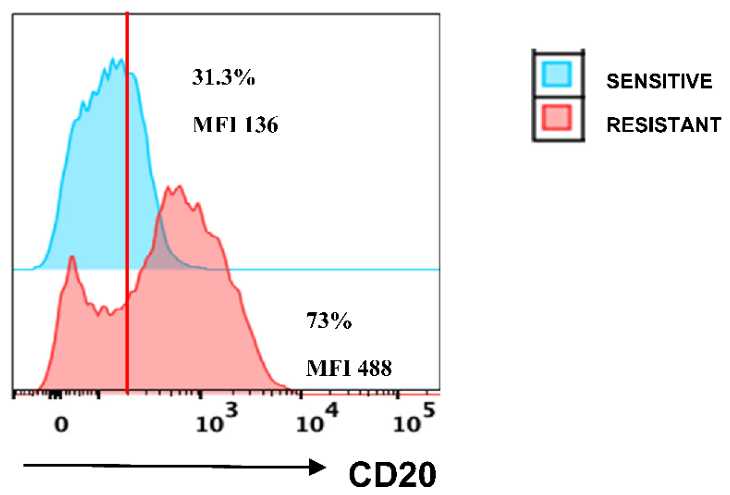
Figure 2.
Th. stages (a) and mechanisms (b) of cell death in resistant and sensitive BJAB cell lines treated with different formulations. (n= 3).
Figure 2.
Th. stages (a) and mechanisms (b) of cell death in resistant and sensitive BJAB cell lines treated with different formulations. (n= 3).
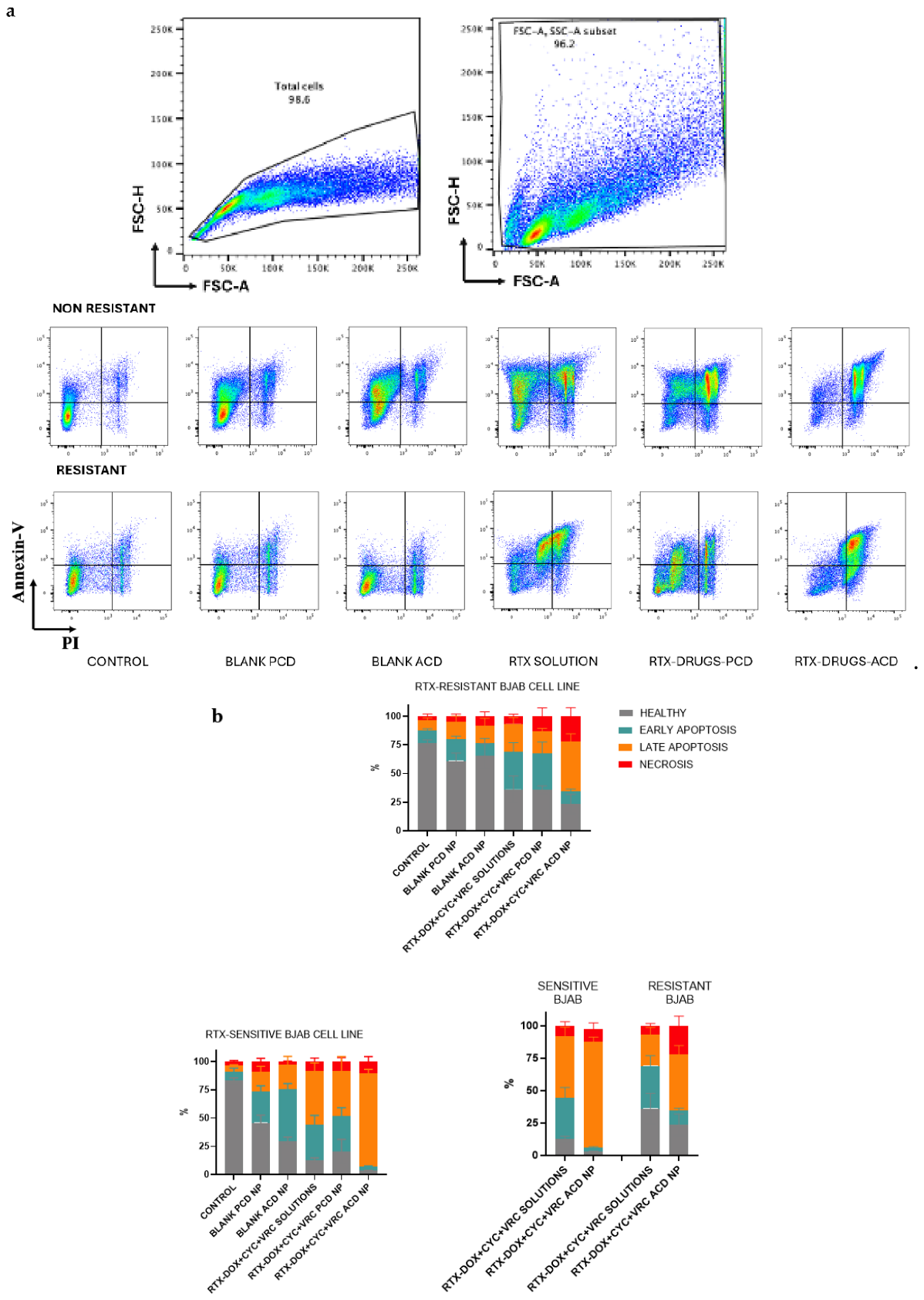
Figure 3.
Cell viability (%) (n=3, ±SD) was assessed using BJAB human lymphoma cell lines (resistant and sensitive) incubated with various NP formulations (DOX, VRC, CYC-loaded three different anticancer drugs), blank CD NPs, and three different drug solutions for 24 and 48 hours. Cell viability was evaluated using the WST-1 assay. p < 0.05.
Figure 3.
Cell viability (%) (n=3, ±SD) was assessed using BJAB human lymphoma cell lines (resistant and sensitive) incubated with various NP formulations (DOX, VRC, CYC-loaded three different anticancer drugs), blank CD NPs, and three different drug solutions for 24 and 48 hours. Cell viability was evaluated using the WST-1 assay. p < 0.05.
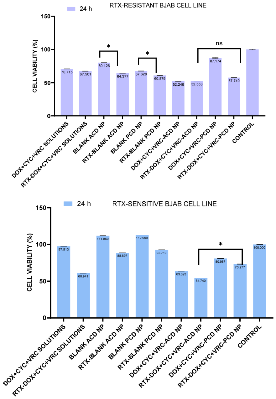
Figure 4.
The percentage of lysis ratio in BJAB (resistant and sensitive) lymphoma cells incubated with different nanoparticle formulations and drug solutions (n=8 ± SD) BJAB cells (RTX-sensitive and RTX-resistant) were co-cultured with macrophages at a 25:1 effector-to-target ratio and treated with RTX solution or RTX-containing CD NP formulations for 4 hours at 37°C. LDH release was measured using a commercial LDH assay kit (Biovision K-311) to quantify cell lysis.
Figure 4.
The percentage of lysis ratio in BJAB (resistant and sensitive) lymphoma cells incubated with different nanoparticle formulations and drug solutions (n=8 ± SD) BJAB cells (RTX-sensitive and RTX-resistant) were co-cultured with macrophages at a 25:1 effector-to-target ratio and treated with RTX solution or RTX-containing CD NP formulations for 4 hours at 37°C. LDH release was measured using a commercial LDH assay kit (Biovision K-311) to quantify cell lysis.
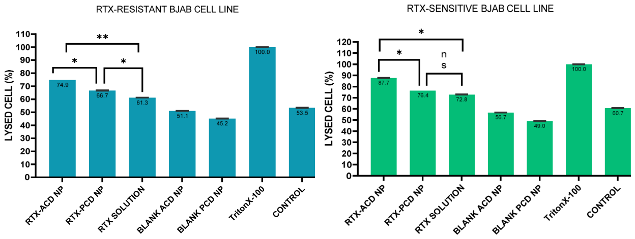
Figure 5.
Cholesterol content in BJAB human lymphoma cells (RTX-sensitive and RTX-resistant) following treatment with various nanoparticle formulations. Cells were incubated with blank CD nanoparticles, anticancer drug-loaded nanoparticles (DOX, VRC, CYC), free drug solutions, and Gua-CD derivatives. Cholesterol levels were quantified using a commercial cholesterol assay kit. Methyl-β-CD was used as a reference standard for cholesterol depletion efficiency. (n=3, ±SD) (p<0.05).
Figure 5.
Cholesterol content in BJAB human lymphoma cells (RTX-sensitive and RTX-resistant) following treatment with various nanoparticle formulations. Cells were incubated with blank CD nanoparticles, anticancer drug-loaded nanoparticles (DOX, VRC, CYC), free drug solutions, and Gua-CD derivatives. Cholesterol levels were quantified using a commercial cholesterol assay kit. Methyl-β-CD was used as a reference standard for cholesterol depletion efficiency. (n=3, ±SD) (p<0.05).
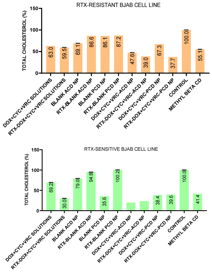
Figure 6.
Mitochondrial accumulation of DOX-loaded Gua-CD derivatives in BJAB cells (RTX-sensitive and RTX-resistant). Cells (20 × 10⁶) were incubated with DOX-loaded Gua-CD derivatives or free DOX for 24 hours at 37°C under 5% CO₂. Mitochondria were isolated using a commercial mitochondrial isolation kit (Thermo 89874), and DOX fluorescence was measured at excitation/emission wavelengths of 470/560 nm using a microplate reader (n=8, ±SD) (p<0.05).
Figure 6.
Mitochondrial accumulation of DOX-loaded Gua-CD derivatives in BJAB cells (RTX-sensitive and RTX-resistant). Cells (20 × 10⁶) were incubated with DOX-loaded Gua-CD derivatives or free DOX for 24 hours at 37°C under 5% CO₂. Mitochondria were isolated using a commercial mitochondrial isolation kit (Thermo 89874), and DOX fluorescence was measured at excitation/emission wavelengths of 470/560 nm using a microplate reader (n=8, ±SD) (p<0.05).
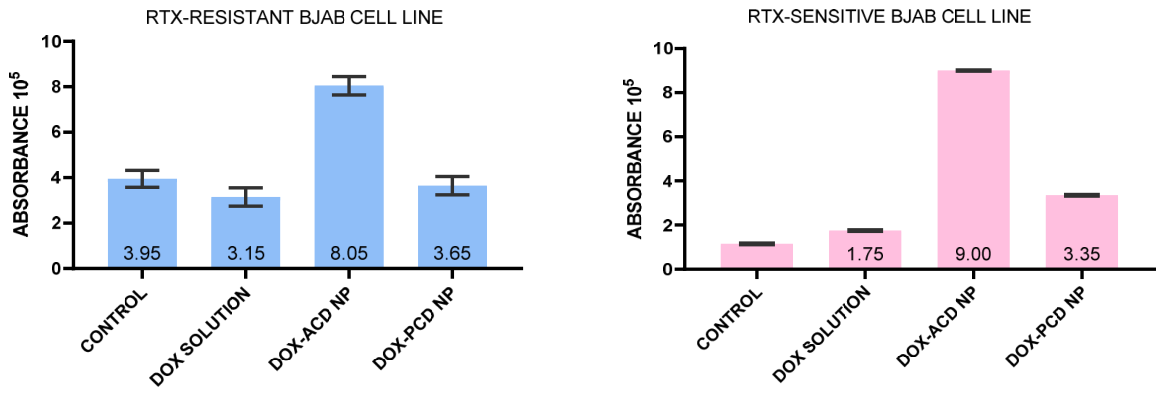
Figure 7.
P-gp inhibition and Calcein AM extrusion assay in BJAB (resistant and sensitive) human lymphoma cells incubated with RTX-DOX-ACD NP formulations and Verapamil (n=6 ± SD) P-gp ATPase activity was measured using a commercial cell-based ATPase assay kit, and Calcein AM fluorescence was analyzed by microplate reader (ex: 495 nm, em: 515 nm).
Figure 7.
P-gp inhibition and Calcein AM extrusion assay in BJAB (resistant and sensitive) human lymphoma cells incubated with RTX-DOX-ACD NP formulations and Verapamil (n=6 ± SD) P-gp ATPase activity was measured using a commercial cell-based ATPase assay kit, and Calcein AM fluorescence was analyzed by microplate reader (ex: 495 nm, em: 515 nm).
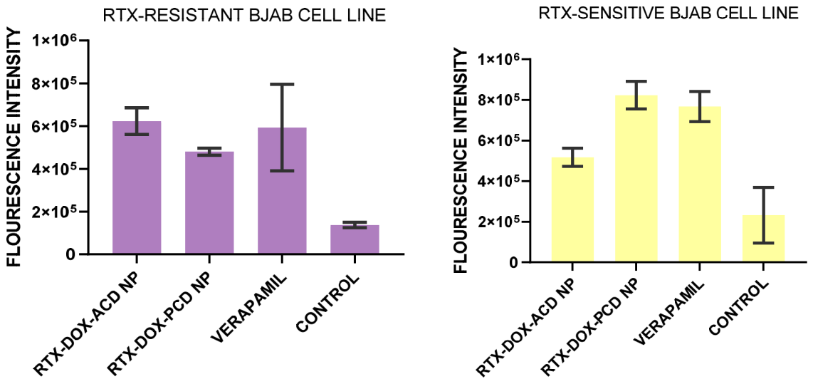
Figure 8.
Cell viability after 48 hours of incubation in BJAB (drug-resistant and sensitive) 3D lymphoma spheroids with different formulations (n=6 ± SD) Drug-loaded NP formulations or free drug solutions were added, and cell viability was assessed using the WST-1 assay after 48 hours. Control groups were incubated with medium alone, and viability was normalized to 100%.
Figure 8.
Cell viability after 48 hours of incubation in BJAB (drug-resistant and sensitive) 3D lymphoma spheroids with different formulations (n=6 ± SD) Drug-loaded NP formulations or free drug solutions were added, and cell viability was assessed using the WST-1 assay after 48 hours. Control groups were incubated with medium alone, and viability was normalized to 100%.
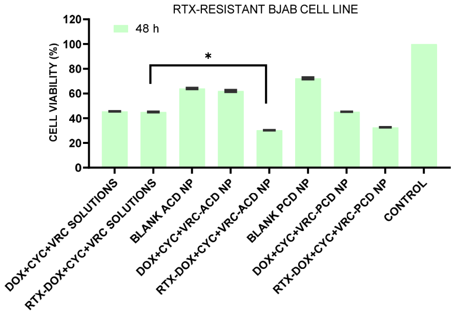
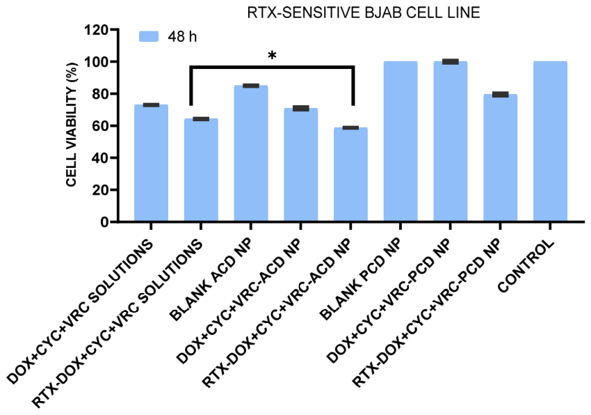
Figure 9.
The ALT and AST levels and ratios obtained from serum collected from healthy Balb/c mice administered with single/multiple doses of blank nanoparticles (n=4) NP were administered intravenously as a single dose or repeated doses (5 doses, every 2 days) to mimic the R-CHOP clinical regimen. Blood samples were collected 24 hours after the last administration, and toxicity was assessed by measuring Aspartate aminotransferase (AST) and alanine aminotransferase (ALT) levels.
Figure 9.
The ALT and AST levels and ratios obtained from serum collected from healthy Balb/c mice administered with single/multiple doses of blank nanoparticles (n=4) NP were administered intravenously as a single dose or repeated doses (5 doses, every 2 days) to mimic the R-CHOP clinical regimen. Blood samples were collected 24 hours after the last administration, and toxicity was assessed by measuring Aspartate aminotransferase (AST) and alanine aminotransferase (ALT) levels.
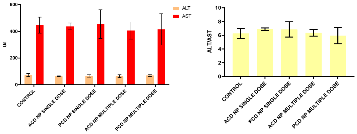
Figure 11.
Tumor growth in Balb/c mice, measured before each injection, showed a highly significant difference between the control group and the drug-loaded, RTX-targeted nanoparticle (NP) group (p<0.05).
Figure 11.
Tumor growth in Balb/c mice, measured before each injection, showed a highly significant difference between the control group and the drug-loaded, RTX-targeted nanoparticle (NP) group (p<0.05).
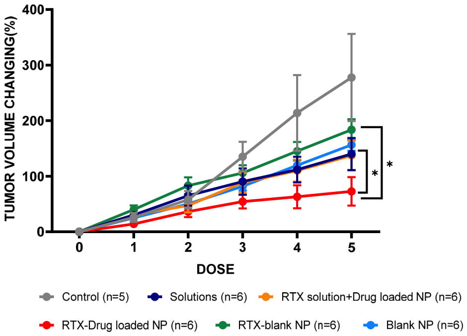
Figure 12.
Tumor tissues excised from sacrificed mice and their average weights.
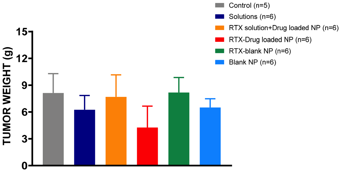
Figure 13.
Circular dichroism spectra of free RTX and vincristine-loaded RTX-conjugated ACD nanoparticles.
Figure 13.
Circular dichroism spectra of free RTX and vincristine-loaded RTX-conjugated ACD nanoparticles.
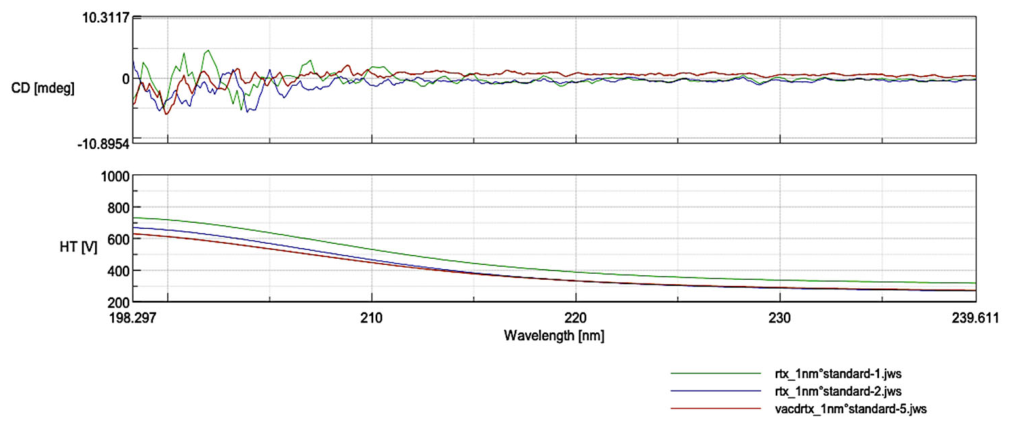

Table 1.
Survivin levels in resistant and sensitive cell line by different formulations. Survivin levels were measured using a Human Survivin ELISA Kit after 48 hours of incubation. Absorbance was read at 450 nm, and concentrations were calculated using a standard curve (n=6). Control cells were incubated with medium alone. ) (p<0.05).
Table 1.
Survivin levels in resistant and sensitive cell line by different formulations. Survivin levels were measured using a Human Survivin ELISA Kit after 48 hours of incubation. Absorbance was read at 450 nm, and concentrations were calculated using a standard curve (n=6). Control cells were incubated with medium alone. ) (p<0.05).
| SURVIVIN PROTEIN AMOUNT % | ||
| RESISTANT | SENSITIVE | |
| DOX+CYC+VRC SOLUTIONS | 61.29 | 53.87 |
| RTX-DOX+CYC+VRC SOLUTIONS | 15.96 | 10.73 |
| BLANK ACD NP | 74.78 | 54.83 |
| RTX-ACD NP | 97.72 | 101.80 |
| DOX+CYC+VRC-ACD NP | 34.41 | 72.56 |
| RTX-DOX+CYC+VRC-ACD NP | 27.79 | 5.80 |
| BLANK PCD NP | 90.33 | 83.27 |
| RTX-PCD NP | 102.51 | 101.56 |
| DOX+CYC+VRC-PCD NP | 21.40 | 21.25 |
| RTX-DOX+CYC+VRC-PCD NP | 27.79 | 44.61 |
| CONTROL | 100.00 | 100.00 |
Table 2.
Pharmacokinetic parameters of solution and nanoparticle formulations.
| PHARMACOKINETIC PARAMETERS |
DOX SOLUTION |
RTX-DOX- ACD-NP |
CYC SOLUTION |
RTX-CYC- ACD-NP |
VRC SOLUTION |
RTX-VRC- ACD-NP |
|
| AUC (µg·dk/mL) | 12,7 | 10,4 | 6,2 | 3,2 | 2,3 | 3,6 | |
| Cmax (µg/mL) | 1,16 | 1,04 | 19,92 | 13,6 | 1,75 | 1,89 | |
| tmax (sa) | 1 | 2* | 0,083 | 0,083 | 0,083 | 0,5* | |
| Cl (mL/dk) | 0,78 | 0,96 | 32,15 | 62,26* | 17,14 | 11,22 | |
| Vd (mL) | 8,55 | 19,75* | 10,03 | 14,69* | 23,46 | 33,88* | |
| t1/2 (sa) | 7,56 | 14,25* | 0,21 | 0,16 | 0,94 | 2,09* | |
Disclaimer/Publisher’s Note: The statements, opinions and data contained in all publications are solely those of the individual author(s) and contributor(s) and not of MDPI and/or the editor(s). MDPI and/or the editor(s) disclaim responsibility for any injury to people or property resulting from any ideas, methods, instructions or products referred to in the content. |
© 2026 by the authors. Licensee MDPI, Basel, Switzerland. This article is an open access article distributed under the terms and conditions of the Creative Commons Attribution (CC BY) license (http://creativecommons.org/licenses/by/4.0/).
Copyright: This open access article is published under a Creative Commons CC BY 4.0 license, which permit the free download, distribution, and reuse, provided that the author and preprint are cited in any reuse.