Preprint
Article

This version is not peer-reviewed.

GPCRs Share an Allosteric Regulatory Site Located Within the Extracellular Agonist-Binding Pocket: Cross-Binding of GPCR Ligands Can Knockout or Revitalize Receptors and Affect Tolerance and Addiction

Submitted:

29 July 2026

Posted:

30 July 2026

You are already at the latest version

Abstract

Angiotensin II (AngII) binds to the AngII type 1 receptor (AT1R) in either A-mode (on-switch; G-protein-mediated contraction) or D-mode (off-switch; arrestin-mediated desensitization/tachyphylaxis). Angiotensin receptor blockers (ARB) can desensitize AT1Rs by binding to an allosteric regulatory B-site, which overlaps with the AngII A/D binding site in the extracellular binding pocket of the receptor. ARBs shift the receptor conformation from A/D-mode to agonist-independent D*-mode, resulting in receptor internalization. Binding of ligands to the inverse agonist binding site (B-site) can promote (lock in) the D*-mode conformation of the receptor (desensitization) or disengage (lock out) the D-mode conformation (upsensitization, reduction of tolerance). Computer-aided docking (CAD) studies show that ARBs, particularly bisartan ACC519TT, bind with high affinity to several G protein-coupled receptors (GPCRs), including AT1R, adrenergic (alpha 1 and 2), opioid (mu and delta), and muscarinic 3 receptors, suggesting that the electrostatic architecture of the B-site has been preserved across a broad spectrum of GPCRs. Consistent with CAD findings, ACC519TT significantly reduced dilation responses of mouse colon and small intestine to the opioid receptor agonist, oxycodone. Functional studies in rabbit iliac artery rings demonstrated the ability of lisinopril to significantly reduce AngII-induced contraction, comparable to candesartan. However, CAD studies show that lisinopril has a markedly lower affinity than candesartan for AT1R, illustrating that the similar effect to AngII dose-response may be due to the time-dependent receptor internalization. For agonists, D-site versus A-site activity may provide insight regarding dependency potential (addiction ratio, D/A). Agonists cross-talk among different GPCRs creates the potential for a sophisticated interplay, possibly involving endogenous ligands not yet identified, with the notable exception of angiotensin antipeptide.

Keywords: 
;  ;  ;  ;  

1. Introduction

The octapeptide, angiotensin II (AngII) acts on Ang II type 1 (AT1R) and type 2 (AT2R) receptors in a variety of tissues to elicit contraction (via AT1R) or dilation (via AT2R), inflammation (activation of AT1R on regulatory T cells), and various other physiological and behavioral effects, including memory, seizures, and pain [1,2]. AngII is the major component of the renin-angiotensin system (RAS) that is pivotal for blood pressure homeostasis and fluid balance, controlling hemodynamic flow, electrolyte absorption, excretion, and vascular resistance [1,3]. The physiological actions of the RAS are mediated by highly sophisticated biomolecular cascades involving enzymatic cleavage and the modification of pro-hormones, intermediary proteins, and other bioactive molecules, which exert their effects primarily by activating G protein–coupled receptors (GPCRs) [4,5]. The intricacy of RAS is further evident by the existence of two opposing axes, which are integral to maintaining cardiovascular and renal homeostasis [6,7]. The classical axis serves as the main effector pathway of the RAS and relies on the coordinated actions of angiotensin-converting enzyme (ACE), AngII, and AT1R [8,9]. Conversely, the alternative pathway provides counter-regulatory balance, primarily through ACE2, alamandine, angiotensin (1-7), and Mas1 proto-oncogene receptor [10,11].
Under normal physiological states, AngII exerts its regulatory effects via AT1R activation, which promotes the release of antidiuretic hormone and aldosterone secretion, enhances sodium reabsorption and water conservation, stimulation of sympathetic pathways, and induces vascular smooth muscle contraction [12]. However, dysregulated production and release of AngII have been implicated in diverse pathological processes, such as endothelial and vascular impairment, heightened inflammation and oxidative stress, fibrosis, cellular hypertrophy, and the progression of diseases affecting multiple organ systems. Renin inhibitors, ACE inhibitors, and angiotensin receptor blockers (ARBs) [13,14] are commonly prescribed medications to reduce the detrimental cardiovascular and renal effects of dysregulated classical RAS activity. Increased levels of AngII and AT1R overstimulation have been associated with pathological processes affecting the cardiovascular [8,11,12,15], renal [16,17], central nervous [2,18,19,20], and endocrine [21,22,23] systems.
The synthesis of analogues of AngII showed that substitution of the Asp1 residue with Sar1 increased activity, and that replacement of the Phe8 residue of AngII with an aliphatic amino acid, as in [Sar1Ile8]AngII (sarilesin), produced a noncompetitive antagonist which desensitized AT1Rs [24]. However, methylation of the Tyr4 hydroxyl group led to the competitive antagonist [Sar1Tyr(Me)4]AngII (sarmesin) [24]. Methylation of the Tyr4 hydroxyl of sarilesin also expunged bioactivity, illustrating that the Tyr hydroxyl proton [and/or tyrosinate anion] is important for both agonist and desensitizing actions. Crystal structures of AT1R bound to angiotensin ligands have confirmed interactions of Tyr hydroxyl with receptor-based groups [25]. Conformational investigations also established the presence of a charge relay system (CRS) in AngII, in which the Tyr4 hydroxyl can interact with the C-terminal Phe8 carboxylate, either directly or via a relay mechanism provided by a His4 imidazole bridge [26]. These two conformers are also recreated when AngII binds to the AT1R and could reflect desensitizing (D-mode) and agonist (A-mode) binding, respectively [27].
The discovery of ARBs launched a new era in RAS research, aided by crystal structures for AT1R bound to nonpeptide sartans [28,29,30] and to angiotensin peptides [25]. Principal interactions involve the receptor residue K199 for binding AngII peptides [25], whereas an adjacent residue, R167, appears to anchor ARBs [29]. This suggests that ARB sartans occupy an overlapping binding site (B-site), potentially compromising AngII peptide binding while promoting an inverse agonist response mechanism. This site, which is proximal to that occupied by AngII and peptide desensitizers like sarilesin [25], binds sartans with high affinity and appears to shift the receptor conformation from A-mode to D*-mode (similar but not identical to D-mode produced by peptides). Taken together, these findings illustrate the complexity of highly flexible peptides versus rigid nonpeptides for receptor interactions and may also explain the apparently permissive nature of mechanisms for inverse agonism by nonpeptide ligands at GPCR [31]. Nonpeptide sartans, which are almost exclusively inverse agonists rather than agonists at AT1R, may also lack the sophistication afforded by the length and flexibility of peptides, which is required to promote positive cooperatively between AT1R dimers with G-protein that activates the contractile response [32]. Our finding that angiotensin receptor blockers (ARBs) (i.e., sartans and bisartans) can block the contractile response to phenylephrine, an alpha-1 adrenergic receptor (α1AR) agonist, in iliac arteries [33] led to the investigation of computer-aided docking (CAD) studies of ARB binding to αARs. This study reported that the observed biological effects likely originated from receptor-based effects rather than direct influences on receptor signaling pathways [31].
An extension of these studies to include opioid receptors (OR) demonstrated high-affinity cross-binding of ligands normally associated with each G-protein coupled receptor (i.e., AT1R, αAR, and OR) [31]. Moreover, cross-over binding of ligands at each of the alternative GPCRs often coincides with bioactivity, suggesting that these modulatory binding sites (B-sites) may have a biological function and perhaps a common evolutionary origin. Parallel findings occur at ORs, although peptides (e.g., enkephalins and endorphins) promote agonist activity with nanomolar affinities which are several orders of magnitude higher than the micromolar affinities observed for nonpeptide ligands (morphine, fentanyl, etc.). Notably, oxycodone (4 µM), morphine (2 µM), codeine (1 µM), and fentanyl (0.4 µM) have similar CAD receptor affinities [31] yet display markedly different potencies, varying by orders of magnitude. This suggests that nonpeptide opioids possess substantially different intrinsic agonist efficacies, which are not necessarily matched by differences in dependency potential. Thus, the importance of fully understanding these receptor mechanisms cannot be overstated and is essential to elucidate and mitigate drug addiction and dependence.

2. Materials and Methods

2.1. CAD Studies

Docking of ligands to AT1R, α1AR, α2AR, and mu OR (µOR) using crystal structures downloaded from the Protein Data Bank (PDB) was assessed by methods as previously described [31].

2.2. Ex Vivo Experiments

2.2.1. Materials

Candesartan (Cat#9003239) and lisinopril (Cat#000-19862) were purchased from Sapphire Bioscience (Sydney, NSW, Australia); naloxone (Cat#HY-17417A) was purchased from Focus Bioscience Pty Ltd (St Lucia, QLD, Australia); human AngII sequence DRVYIHPF was purchased from Mimotopes (Mulgrave, VIC, Australia); and CaCl2 (Cat#C1016), glucose (Cat#G7021), KCl (Cat#P9541), KH2PO4 (Cat#P0662), MgSO4·7H2O(Cat#230391), NaHCO3 (Cat#S5761), NaCl (Cat#S9888) and oxycodone (Cat#PHR8802) were purchased from Millipore Sigma (North Ryde, NSW, Australia).

2.2.2. Animal Models, Husbandry, and Ethics Approval

Male New Zealand White rabbits (n = 4) at 8 weeks of age were purchased from Flinders City University (Adelaide, SA, Australia. Animals were housed at Victoria University, Werribee Campus Animal Facilities (VIC, Australia). Rabbits were housed in pairs until 10 weeks of age before reaching sexual maturity. Rabbits were fed a normal chow diet (Specialty Feeds, Glen Forrest, WA, Australia), and food and water were supplied ad libitum. Animals were aged to 16 weeks and were kept on a 12-h day/night cycle and maintained at a constant temperature of 21°C and relative humidity level between 40 and 70%. Small intestines and colons were harvested from 16-week-old male wildtype (C57BL/6) surplus mice (n = 8) that were not required for breeding, supporting the principles of the 3Rs by minimizing the use of additional animals. All experimental procedures were conducted in accordance with the National Health and Medical Research Council ‘Australia Code of Practice for the Care and Use of Animals for Scientific Purposes’ (8th edition) (https://www.nhmrc.gov.au/about-us/publications/australian-code-care-and-use-animals-scientific-purposes; accessed on 21 March 2026) and were approved by the Victoria University Animal Ethics Committee (VUAEC#17/013 and 22/009B).

2.2.3. Anesthesia and Humane Dispatch

Rabbits: To minimize stress, rabbits were sedated via subcutaneous injection of medetomidine (0.25 mg/kg) at the base of the neck. Animals were then placed in a plastic chamber and anesthetized with isoflurane (4% in 1.5 L/min O2) [33]. Once the righting reflex was lost, anesthesia was maintained via a face mask (4% isoflurane in 0.8 L/min O2) [33]. Following the loss of corneal and palpebral pain reflexes, a lower abdominal incision was made, and the subcutaneous tissue and muscle layers were carefully dissected to expose the inferior vena cava [33]. Rabbits were humanely dispatched by exsanguination by dissecting the inferior vena cava, with death confirmed by dissection of the diaphragm [33]. A T-tube was inserted distal to the aortic arch to allow thorough flushing of the aorta, aortic bifurcation, and iliac arteries with cold (4°C), oxygenated Krebs–Henseleit solution (Krebs; 118 mM NaCl, 4.7 mM KCl, 1.2 mM MgSO4·7H2O, 1.2 mM KH2PO4, 25 mM NaHCO3, 11.7 mM glucose, 1.25 mM CaCl2; pH 7.4) [33]. The left and right iliac arteries were then excised, cleaned of connective and adipose tissue under a light microscope, and cut into 2 mm rings for functional isometric tension myography studies [33].
Mice: Animals were first placed into a plastic chamber and anesthetized with isoflurane (4% in 1.2 L/min O2). Once loss of righting reflex was observed, animals were then placed onto a mask for continuous delivery of anesthesia (4% in 0.8 L/min O2). When loss of palpebral pain reflex was demonstrated, mice were humanely dispatched by cardiac puncture. The colon and small intestine were excised, cleaned of connective tissue, fat, and fecal matter, and cut into 3 mm rings for functional isometric tension myography studies.

2.2.4. Drug Incubation and Functional Isometric Tension Studies

Iliac artery, small intestine, and colon rings were immediately transferred to adjacent organ baths (OB16, Zultek Engineering, Melbourne, VIC, Australia) containing 5 mL of Krebs solution and allowed to equilibrate for 30 minutes [33]. Baths were maintained at 37 °C and continuously aerated with carbogen (95% O2 / 5% CO2) to approximate physiological conditions. Rings were then mounted on metal hooks connected to force-displacement transducers, stretched (iliac artery: 0.4-0.5 g, small intestine: 1 g; and colon: 0.5 g) and equilibrated for an additional 30 minutes [33]. Subsequently, the baths were refreshed, rings re-stretched, and equilibrated for a further 30 min before drug incubation [33].
To determine the effect that lisinopril had on modulating AngII-induced contraction, iliac artery rings were incubated with (i) no drug to serve as controls, (ii) the angiotensin-converting enzyme inhibitor, lisinopril [10−12 M], or (ii) the commercially available ARB, candesartan [10−12 M], for 10 minutes. Following drug incubations, organ baths were refreshed, and an AngII [10−11 M –10−5 M] dose-response was performed [33]. After dose–response studies, rings were refreshed, allowed to return to baseline tension, and contracted with high potassium physiological solution (125 mM/L KCl; 1.2 mM/L MgSO4·7H2O; 1.2 mM/L KH2PO4; 25 mM/L NaHCO3; and 11.7 mM/L glucose; and 1.25 mM CaCl2) (pH: 7.4) to determine maximal standard contraction responses [33].
To determine the ability of the bisartan, ACC519TT, to reduce relaxation responses to oxycodone, small intestine and colon ring were incubated with (i) no drug to serve as controls, (ii) ACC519TT [10−6 M or 10−9 M], (iii) candesartan [10−6 M], or (iv) the OR antagonist, naloxone [10−6 M], for 30 minutes. Krebs solution was then replaced with 40 mM KPSS to induce contraction and, once a plateau was reached, an oxycodone dose response [10−10 M–10−5 M].

2.2.5. Statistical Analysis

GraphPad Prism version 11 was used to analyze data, and the significant p-value was set at p < 0.05. A two-way analysis of variance followed by Sidak’s multiple comparisons post hoc test was performed to determine significance. All data are represented as mean ± standard error of the mean (SEM).

3. Results

3.1. CAD Studies

CAD of AngII to AT1R (Figure 1A) closely matches docking observed in the crystal structure of AT1R bound to AngII [25]. Binding affinities (Kd) of selected compounds, shown in Table 1, were calculated directly from binding energies [31]. The affinity constants in Table 1 do not discriminate between ligand binding to the agonist (A or D) site or to the overlapping (B) site, nor whether ligand interaction produces agonism or competitive antagonism (A-site), or inverse agonism or upsensitization (B-site).
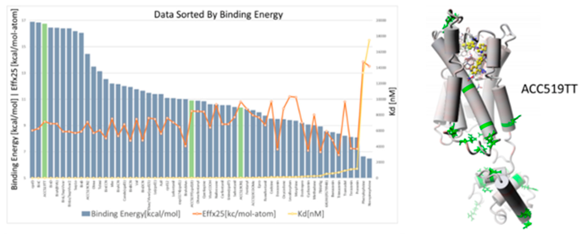
When binding energy is adjusted for the number of heavy atoms or surface contact area as shown in Figure 1B and C, the binding efficiency metric favors smaller sartans over bisartans. This is not necessarily reflective of relative bioassay potencies, and the binding efficiency metric may be useful for comparing similar size molecules, i.e. sartans with sartans, or bisartans with bisartans, Apparently the binding pocket can readily accommodate the larger and often more potent bisartan molecule which, due to rotation at the carbon atoms attached to imidazole N, allows for conformations anywhere between that in which the biphenyl sidechains run in the same direction (cis), to that in which biphenyls run in the opposite direction (trans), with the latter approaching the length of the octapeptide AngII. According to the binding efficiency metric, the best sartan appears to be elsartan (also called vivartan) followed by ACC519TT, and the best bisartan is Bis A followed by ACC519TT (Figure 1C). There is an interesting footnote to the data in Figure 1C, namely that the best binder, elsartan (which has relatively weak in vivo antagonist activity), has a much higher CAD affinity for AT1R than its highly active metabolite elsartan-COOH [resulting from conversion of -CH2OH to -COOH in vivo]. This implies that the prodrug elsartan could inhibit its active metabolite, at least until sufficient conversion to elsartan-COOH has occurred. More importantly it demonstrates that it should be possible to create a competitive inhibitor of inverse agonism, which would be particularly useful at µOR where addiction profiles could potentially be altered (see subsection 3.3.3 below).
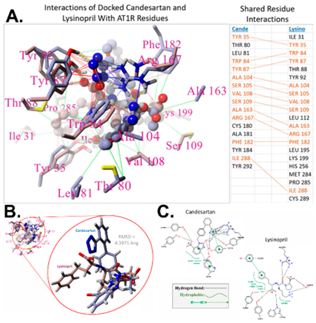
Interactions for candesartan and lisinopril binding to AT1R are shown in Figure 3 (and are discussed in subsection 3.3.4 below).
Figure 2. Interactions of candesartan and lisinopril binding at AT1R.
Preprints 225586 g002
Binding energies for docking of ligands to M3R are shown in Figure 4 and the resulting affinities, which are noticeably higher than for the other receptors studied, are shown in Table 1. Bisartans feature as the most potent ligands based on relative affinities (0.1-10 pM), with ACC5119TT (0.3 pM) again demonstrating notably high affinity (Table 1). Sartans are generally less potent (1-10 nM) than bisartans. Carfentanyl (5 nM) and doxazosin (50 nM) also have significant affinities for M3R and can be anticipated to modulate the activities of other GPCRs.
Figure 3. CAD energies for ligands binding to M3R.
Preprints 225586 g003
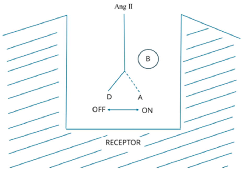
Figure 4. Switching mechanism in the extracellular ligand binding pocket of GPCRs. AngII binds to AT1R in two different binding modes shown here as A-mode (agonism, on-switch) and D-mode (desensitization/tachyphylaxis, off-switch). The motion of a single side chain in AngII (see subsection 4.6) switches the conformation of AngII [and thereby the receptor conformation also] between binding in A-mode (coupling to G protein) or in D-mode (coupling to β-arrestin). Agonist-induced cooperativity of receptor dimers with G protein increases the affinity of the A-site for agonist substantially (10-100X) so that at low concentrations of AngII binding is exclusively in A-mode. As the concentration of AngII increases, it begins binding in D-mode, invoking receptor desensitization and internalization and creating a tunnel-shaped dose-response curve (DRC) [27].
Preprints 225586 g004
Biased agonism: crystal studies of AT1R show that whereas peptide desensitizers like sarilesin bind exclusively in D-mode [25] and evoke receptor internalization, small molecule inverse agonists like ARB sartans bind to an overlapping allosteric B-site [29] which also promotes receptor desensitization/internalization by converting the receptor into a D*-mode conformation. Tachyphylactic D-mode and agonist-independent D*-mode are not identical because the latter does not affect KCl-induced contraction. Reciprocal inhibition of adrenoceptors by sartans [31] and of AT1R by adrenergic blockers [34] appears to operate by shifting the receptor conformation from A-mode to D*-mode. It is also possible that binding of select ligands to B-site [or to an allosteric site on the extracellular loops] could “lock out” the D-mode conformation of the receptor and prevent desensitization: certain angiotensin (ARB), and adrenergic and muscarinic ligands appear to upsensitize opioid receptors and reduce tolerance [35,36]. Whereas the A/D site is super-selective, the B-site is relatively unselective and demonstrates cross-over binding of ligands for other GPCRs [31], leading to cross-talk among GPCRs [36].

3.2. Ex Vivo Studies

3.2.1. Lisinopril Reduces AngII-Meditated Contraction in Rabbit Iliac Arteries

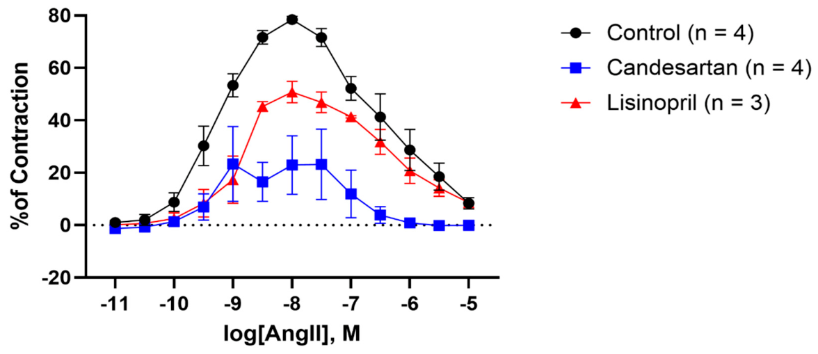
In untreated iliac artery rings, an AngII dose-response invoked a biphasic vasoactive response (Figure 5, Table 2). Rings treated with the commercially available ARB, candesartan, had significantly diminished contraction response to AngII (Figure 5, Table 2). Unexpectedly, rings treated with the ACE inhibitor, lisinopril, showed the capacity to reduce contraction to AngII (Figure 5, Table 2).

3.2.2. ACC519TT Reduces Oxycodone-Induced Relaxation in Mouse Small Intestine and Colon

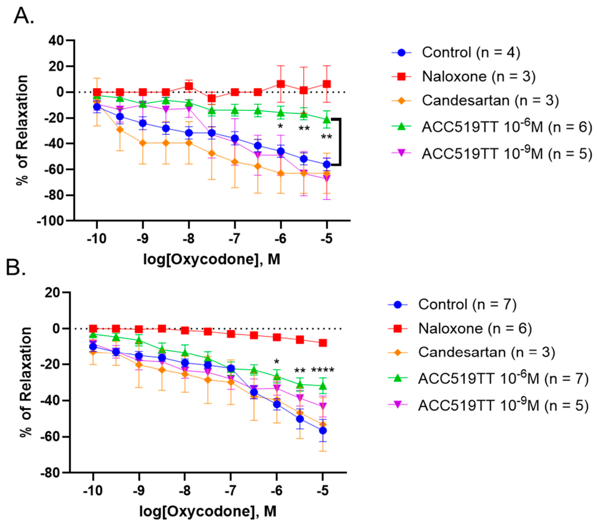
Oxycodone relaxes tissues via ORs located on the smooth muscles, which may be counteracted by ARBs binding to the B-site. Expectedly, the OR antagonist, naloxone, abolished relaxation responses to oxycodone in both the small intestine and colon rings (Figure 6; Table 3 and Table 4). Pre-incubation with candesartan or the lower dose of ACC519TT had no effect on relaxation responses to oxycodone in either the small intestine or colon (Figure 6; Table 3 and Table 4). Interestingly, the higher dose of ACC519TT significantly reduced relaxation responses in small intestinal and colonic rings at higher doses of oxycodone (Figure 6; Table 3 and Table 4).

3.3. Results Continued

3.3.1. Cross-Binding of Ligands at AT1R

Nonpeptide ligands other than ARB sartans, including various αAR and OR ligands, which appear to be structurally unrelated to sartans, were investigated (Table 1). The best ligand was the benzimidazole-bisartan, ACC519TT (Kd ~0.2 nM), with bisartans (1-10 nM) generally having higher affinities than sartans (10-100 nM). Surprisingly, the best alpha adrenergic inverse agonist, doxazosin, had high affinity (~50 nM), and the best opioid ligand carfentanyl, also retained significant affinity (~500 nM). The affinity of the ACE inhibitor (ACEI), lisinopril, was also significant (1 µM), suggesting a possible antihypertensive role involving blocking of AT1R in addition to ACE inhibition. The affinities of alpha ligands and even opioid ligands for the AT1R are unexpectedly high and significant enough to warrant serious consideration for possible biological relevance. In this regard, reciprocal inhibition of angiotensin receptors and adrenoceptors is already established because ARB sartans inhibit adrenoceptors [33] and beta-adrenergic blockers inhibit AngII contractions [34].

3.3.2. Cross-Binding of Ligands at α1AR and α2AR

Nonpeptide ligands other than αAR ligands, including various ARB sartans and bisartans, which are structurally unrelated to αAR ligands, were evaluated for docking/binding to α1AR and α2AR subtypes (Table 1). The best ligand at α1AR was ACC519TT (~0.5 nM), and bisartans and sartans generally had similar affinities (1-50 nM). The best α1AR ligand was the inverse agonist doxazosin (~50 nM), which had unexpectedly lower affinity than the ARBs. In addition, the ACE inhibitor lisinopril retained significant affinity for α1AR (~10 µM). Unfortunately, opioid ligands were not tested.
For α2ARs, the best ligand was ACC519TT (5 nM), and bisartans (10-50 nM) generally had higher affinities than sartans (10-100 nM). The best alpha ligand was again doxazosin (10 nM). As with α1ARs, bisartans appear to have significantly higher affinities than alpha ligands like doxazosin for α2ARs, suggesting the possible existence of a regulatory site on αAR that is able to bind angiotensin-related ligands. Indeed, ARBs are known to block alpha adrenergic receptor responses to phenylephrine [33].

3.3.3. Cross-Binding of Ligands at µOR

Nonpeptide ligands other than OR ligands, including various ARB sartans and αAR ligands, which are structurally unrelated to opiates, were investigated for docking/binding to mu opioid receptors (Table 1). The best ligands were telmisartan and ACC519TT (10 nM), and bisartans and sartans had generally similar affinities (0.1-1 µM). The best alpha ligand was doxazosin (0.2 µM), and the best opioid ligand was carfentanyl (0.4 µM). In addition, lisinopril (2 µM) retained measurable affinity. These unexpected findings may be partly explained by the behavior of nonpeptide opiates, which bind with much lower affinities (0.1-10 µM) than physiologically relevant peptide ligands like enkephalin (~1 nM). In other words, plant-based and synthetic opiates have very poor binding characteristics despite exerting significant nociceptive effects. Indeed, this anomaly could be at the root of the severe addiction issues associated with synthetic opiates. Pharmacologically, synthetic opiates can be categorized as partial agonists that evoke a dose-dependent agonist response followed by a large desensitization/tachyphylaxis effect [32,37]. In order to produce a significant agonist response, large doses must be given, which in turn evoke a very large tachyphylaxis effect associated with receptor desensitization, tolerance, dependence, withdrawal, and ultimately severe addiction. Perhaps the best intervention to prevent factors that lead to addiction via receptor desensitization would be the identification of an allosteric (B-site) ligand that renders the D-mode of the receptor inactive/inoperable without affecting the agonist (A-mode). ARBs, candesartan and valsartan, are known to reduce tolerance to opiates [35,38] by mechanisms unknown, but potentially by interfering with D-site viability (shifting the receptor conformation away from D-mode (Figure 4). In Figure 6, candesartan showed no activity at intestinal opioid receptors, whereas ACC519TT blocked opioid receptors in a dose-dependent manner, in agreement with CAD studies where ACC519TT binds to µOR more strongly than candesartan (Table 1). Naloxone was the best blocker in Figure 6, in line with its use for alleviating symptoms associated with opioid intoxication.

3.3.4. Cross-Binding of Ligands at M3R

ARB sartans and particularly bisartans demonstrate high affinity for M3R (Figure 3). As with the other three receptors investigated, ACC519TT features strongly (Table 1). In addition, opioids retain moderate affinities, whereas a lower affinity was observed for the alpha-adrenergic inverse agonist doxazosin (Table 1). Surprisingly, the muscarinic inverse agonist, atropine, and other muscarinic ligands had affinities similar to the alpha ligand doxazosin. In this regard, it should be noted that ligand affinity is not the issue here, because the potency of an inverse agonist is largely dependent on the association rate constant (ka) [rather than equilibrium dissociation constant (KD)] which determines the extent of receptor knockout (see sections 3.2.5 and 4.3 below).

3.3.5. Lisinopril Is an ARB

Detailed investigation of the ACEI lisinopril by CAD (Table 1) has shown that it binds to AT1R (1 µM) and binds to αAR (1-10 µM). Accordingly, lisinopril could act as an AT1R blocker. Indeed, bioassays of contraction of isolated smooth muscle have confirmed that lisinopril, despite displaying much lower affinity than ARB sartans by CAD (Table 2), has blocking activity similar to ARB candesartan (Figure 5). Whereas CAD binding affinity appears to correlate somewhat with biological potency for sartans/bisartans, it does not for lisinopril. The explanation for lisinopril’s low affinity (Table 2) but high potency (Figure 5) resides in the irreversibility of inverse agonist behavior wherein potency is dependent only on the time of contact with the tissue and the ‘on’ rate (mass action law does not apply when measuring response, see Discussion section).
It seems likely that the blood pressure lowering effects of lisinopril are to some degree due to ARB actions on vascular smooth muscle. ACEIs were originally developed to treat patients with renal hypertension who have high circulating levels of AngII, but it turned out that ACEI also worked on the 80% of patients with essential hypertension and normal levels of AngII. The present findings provide an explanation for this unexpected bonus. However, this dual interaction of ACEIs may not provide an advantage over ARBs for treating hypertension and other cardiovascular ailments, because ACEIs will still retain the side effect of dry cough associated with reduced ACE activity which increases plasma bradykinin levels.
Actions of lisinopril on smooth muscle could originate from similarities in structure between lisinopril and sartans, both of which contain two acid groups with similar spatial characteristics (~1nm, or ~10 Angstroms apart). Carboxylate and/or tetrazole groups in ARB sartans/ bisartans can be superimposed with the two carboxylates of lisinopril (Figure 2). For example, when the tetrazole group of candesartan is overlapped with one carboxylate of lisinopril, the other carboxylate of lisinopril can overlay the carboxylate of candesartan. Similarly, the two tetrazole groups of bisartans can be positioned to overlay the two carboxylates of lisinopril. The two acid groups of captopril (sulphydryl and carboxylate) are closer (7 Angstroms) but may be able to mimic the role of lisinopril’s two carboxylates, suggesting captopril may also act as an ARB, and it is possible that many other ACEIs (e.g. ramipril) will turn out to be ARBs as well.
Individual ion pairing interactions can contribute more binding energy than other electrostatic interactions of peptides/proteins, although the summing up of multiple dipole, quadrupole, and hydrogen bonds can have a significant influence on binding outcomes. Close inspection of the docking of lisinopril and candesartan to AT1R (Figure 2) shows that the binding of ACEI and ARB involves similar ion-pairing contact residues, confirming the importance of the spatial equivalence of acid groups outlined above. Carboxylate groups in both molecules interact with the hydroxyl protons of Y87 and Y92 of the receptor, whereas R167 anchors the tetrazole group of candesartan and, correspondingly, the second carboxylate of lisinopril. The latter ion pairing interactions probably account for much of the binding energy for lisinopril and candesartan because the 3D conformations of the two ligands are quite different; that is to say that, aside from the twin acid moieties, the docked poses of these ligands do not superimpose well. The difference in affinity between candesartan and lisinopril (x100) is equivalent to a binding energy difference of ~2kcal/mol. The similarity in potency in bioassays show that whilst both ligands likely have similar “on” rates, candesartan binds tighter and has a ~100X slower “off rate”, accounting for its much higher affinity. Part of this increased affinity may derive from the stronger interaction of tetrazole in candesartan, compared to carboxylate in lisinopril, with R167 of the receptor (in Figure 1 there is less favorable geometry of interaction for lisinopril).
In this regard, we conducted a comparison of interaction energies for methyltetrazolate and acetic anion with methylguanidinium using semiempirical RM1 calculations in vacuo, allowing for geometric optimization and followed by molecular mechanics (MM+ and AMBER99) to yield a low energy conformer pair after >1000 iterations. The interaction energies with methylguanidinium were similar for acetate (-272 kcal/mol) and methyltetrazolate (-223 kcal/mol), and in both cases there was deprotonation of methylguanidinium. In Figure 2, the interaction of tetrazolate with R167 is accompanied by multiple H-bonds, which are absent for lisinopril despite the proximity of an ionic interaction between carboxylate and R167 guanidino. Apparently, the alignment of R167 guanidinium with tetrazolate in candesartan is more complete than for carboxylate in lisinopril, and there is less favourable geometry optimization for the latter interaction, which could account in part for the binding energy deficit associated with lisinopril.
There is a profound difference in the energy of interaction of ion pairs when calculated in isolation as outlined above (>200 kcal/mol) compared to that involved in receptor interactions, where total binding energy per molecule is less than 20 kcal/mol. Contributions to total binding energy by ion pairing are clearly much muted for receptor interactions with the whole molecule, presumably because of the increased complexity of the situation, which balances many inputs across the entire molecular surface (see section 4.3 on binding efficiency). The architecture of the binding pocket likely limits the accessibility of ion pairs and precludes a full-on electrostatic interaction, and ion pairing may have more to do with correctly orienting and aligning the incoming molecule as it enters the binding pocket and settles in. Factors related to entropy are also poorly understood and largely imponderable but may have a significant influence on receptor binding outcomes. Remarkably, doxazosin, which is devoid of acid groups implicit to ARB or ACEI activity, appears to have high affinity for AT1R (Table 1), suggesting that doxazosin could block AngII-induced contractions. As discussed below, receptor interactions and molecular mechanisms can be extremely subtle and involve small changes in binding energy, which ripple through the tentacles of receptor-based peptide chains.

4. Discussion

4.1. Receptor Knockout by Inverse Agonists (Receptor Desensitizers)

The shape of the dose-response curve for an agonist (A) is determined by two competing influences (Figure 5). Functional results from our isolated iliac artery isometric tension experiments exhibit a biphasic, parabolic shape, a pattern that has been observed in previous studies demonstrating similar vascular behavior in human [39,40,41,42], rabbit [43], rat [44,45,46] and mouse [47,48,49] arteries in response to cumulative doses of Ang II. At low doses, agonist-induced positive cooperativity of receptor dimers with G protein is present (binding in A-mode), and the curve turns upwards. However, at higher doses the competing influence of negative cooperativity (in D-mode, tachyphylaxis, receptor saturation and internalization) becomes apparent, and the curve begins to turn downwards [26,33].
The inflection point can be diagnosed by plotting dose/response against dose, which also permits estimations of Hill coefficients for the positive and negative cooperativity phases of the DRC [50,51,52]. If another ligand is introduced, which binds in a competitive manner to the A-site, the DRC will be parallel shifted to the right, but if this ligand binds competitively to the D-site, the upper part of the DRC will be shifted to the left and the maximum response will be elevated. In both of these scenarios, the affinity of the competing ligand will determine potency, and the ligand will have to be present during the establishment of the DRC. However, if the secondary ligand is washed out of the tissue prior to establishing the DRC, then any subsequent effects on the DRC are now due to noncompetitive behavior, meaning that the receptor has been semi-permanently transformed by ligand action at the receptor (e.g., desensitization or upsensitization), and the law of mass action no longer applies. The isolated tissue experiments used in our studies, which involve preincubating the drug for 15 minutes, followed by washing out of tissues prior to establishing a DRC to the agonist (Figure 5), capture the knockout of receptors and measure desensitization [or upsensitization] of receptors. This bioassay technique will not detect competitive antagonist activity, which requires the drug to remain in contact with the tissue for the duration.
Measuring inverse agonism at AT1R is not as straightforward as quantifying agonist activity. Contraction of isolated smooth muscle by the agonist AngII yields a log dose versus response curve which is quantifiable according to parameters like ED50 and maximum response, and the slope of the double reciprocal Hill plot of the data (Hill coefficient) provides a measure of efficacy [32,37,53]. At high doses, AngII desensitizes AT1R by binding in D-mode (tachyphylaxis; Figure 5), and likewise partial agonists, which by definition cannot produce the maximum response, have been shown to act as agonists at low concentrations (A-mode binding), but act as desensitizers at higher doses (D-mode binding) [53].
Peptides like sarilesin, which desensitize tissues and produce no contractile response in rat tissues, have been shown by crystallography to bind in D-mode [25]. Conversely, small molecule inverse agonists, such as ARB sartans, bind to a secondary B-site (Figure 5), overlapping the A/D site [29,30]. ARB binding at the B-site appears to shift the receptor conformation towards D*-mode (equivalent to D-mode for tachyphylaxis by peptides). Desensitization of receptors is prolonged, and the potency of an inverse agonist can be estimated according to the concentration required to block AngII. However, the concept of pA2 values is not applicable because mass action law does not apply due to the irreversible nature of inverse agonism within the time frame of a typical bioassay experiment. Nevertheless, the duration of desensitization has been used to compare relative potencies of sarilesin peptide analogs [54].
Inverse agonism at the B-site is agonist-independent and, as with agonist-dependent tachyphylaxis via D-site, desensitization is the result of coupling of AT1R to β-arrestin, which renders the receptor unresponsive/inaccessible to the agonist AngII [55,56,57] and has long-lasting effects that manifest as irreversible behavior within the timeframe of a typical bioassay. This is the result of receptor internalization (endocytosis), although the ligand does not remain attached to the receptor during this process [58], as evidenced by CAD and by radioligand binding studies. Essentially, receptors are knocked out for extended periods (hours), and it is difficult to assess ligand potency by standard bioassay methods. For example, the data suggest that ARBs, such as candesartan, have nanomolar affinity (Kd from CAD, Table 1) and apparently nanomolar potency (ID50 from bioassay, Table 1); lisinopril has micromolar affinity (CAD) but apparently nanomolar potency [which would imply higher efficacy for lisinopril over candesartan if the mass action law was applicable]. However, the irreversible nature of inverse agonism means that efficacy becomes difficult to define. The effect of the inverse agonist on the receptor accumulates over time, making the magnitude of the observed response depend mainly on the duration of contact time between the ligand and tissue. Despite having significantly different Kd values, as determined by CAD (Table 1), lisinopril and candesartan exhibit similar apparent potencies (ID50, Figure 5), most likely because they share a comparable “on rate” or association rate constant. Inverse agonists desensitize tissues on an hourly basis [54], whereas bioassay measurements are usually completed within minutes.
Inverse agonists desensitize receptors in a manner that is pharmacologically indistinguishable from the tachyphylaxis produced by supramaximal doses of agonist [31,54]. Importantly, both processes are mediated by β-arrestin and culminate in internalization. Subtle differences may reside in actions involving KCl-contracted tissues (see subsection 4.4). It is important to distinguish between partial agonists, which have intrinsic agonist activity and act on A/D site, from inverse agonists, which act at the B site. Accordingly, opioid ligands, such as morphine and fentanyl, retain significant agonist activity at ORs and are partial agonists (A/D-site). In contrast, naloxone appears to act like an inverse agonist (B-site), simultaneously obfuscating A-mode binding. The ability of carfentanyl to bind to receptors other than ORs (Table 1) probably involves B-site binding, underscoring that the physical overlap of A/D and B sites could result in diverse ligand binding and be associated with receptor couplings.

4.2. Cross-Binding of Inverse Agonists and Structural Overlap

In the CAD affinity ranking (Table 1), the most unexpected finding is that bisartan, ACC519TT, consistently displayed the highest affinity across all four GPCRs, including αAR, muOR and M3R, for which it is not designed. ACC519TT acts as a highly potent inverse agonist at the AT1R and presumably has high affinity to the active site responsible for inverse agonist effects (featuring Arg167), which is proximal to the agonist binding site (featuring Lys199). Interestingly, bisartans and sartans also bind to αAR (Table 1), and this unexpected finding has been confirmed by bioassays wherein candesartan blocks contractile responses to phenylephrine in vascular smooth muscle [33] by a mechanism possibly originating from “B-site” binding on αAR. Moreover, bisartans and sartans were also found to bind to ORs with affinities similar to or greater than nonpeptide opiate ligands (Table 1), and bioassays confirmed that ACC519TT blocks OR (Figure 6). Notably, candesartan did not block ORs (Figure 6); however, less potent ARBs, such as candesartan and valsartan, are known to interfere with the development of tolerance to morphine [35,59], potentially by preventing desensitization of receptors by opiates through interactions at an allosteric site on the receptor. This site may be part of the B-site or potentially another region located on the extracellular loops of the receptor. Even so, the ability of ACC519TT to block all four GPCRs suggests that GPCRs have retained B-sites with similar electrostatic binding characteristics, despite containing different amino acid compositions. This convergence may indicate that GCPRs have evolved from a common precursor.
Equally surprising is the finding that the αAR inverse agonist, doxazosin, has a high affinity for AT1R that is similar to its affinity for αARs (Table 1). αAR inverse agonists, such as doxazosin and prazosin, would be expected to block AT1R responses mediated by AngII in bioassays, as βAR blockers have been reported to inhibit AT1R [34]. However, doxazosin also displayed unexpectedly high affinity for the µOR, showing greater affinity than carfentanyl and all other opiates tested [31]. Opioid and adrenergic ligands have been reported to bind to conserved extracellular regions of each other’s receptors, and crosstalk between these receptors is a well-established phenomenon in anesthesia and analgesia [36]. Cross-binding of adrenergic inverse agonists may interfere with the development of tolerance to synthetic opiates, paralleling the manner observed with ARB sartans [35,59].
Structural considerations: if the B-sites of GPCRs share similar structural features that allow them to bind a range of ligands, then the ligands themselves must also share similar features in order to fit a common receptor site. Nitazenes are a class of highly potent benzylbenzimidazole opioid ligands, and it may be no coincidence that the strongest binders to µOR identified in our studies, namely telmisartan, ACC519TT and olmesartan [31] contain benzimidazole. Similarly, doxazosin contains a structurally similar benzpyrimidine, and these common structural features may represent the foundation of the observed binding affinities in Table 1. It is also well-established that opioids (e.g., meperidine) and muscarinic antagonists (e.g., atropine) share common structural and therapeutic properties, as do opioids and adrenergic ligands [36]. This raises the possibility that inverse agonists with vaguely similar structural properties are common in all four GPCRs investigated herein, accounting for their respective CAD affinities at different GPCRs (Table 1). This finding suggests that there is a permissive set of binding criteria for these shared B-sites in the binding pockets of GPCRs, which seem to recognize simplistic structural elements, namely a positively charged amino group (secondary or tertiary, often bridged) together with an aromatic moiety and various hydroxyl/carboxyl groups. The increased affinity of bisartans relative to other sartans (Table 1) may originate from the positively charged imidazole present on bisartans, as opposed to the extra biphenyltetrazole group, which in our CAD studies often appears not to interact with receptor-based groups.
In summary, GPCRs may have evolved a B-site that retains sufficiently similar electrostatic architectural characteristics within each binding pocket to allow GPCRs to accommodate the same sets of synthetic ligands. The GPCR binding pockets that these inverse agonist compounds inhabit do not exhibit sequence homology, although it is possible to construct similar basic electrostatic architectures using alternative amino acid sequences.
Opiates, such as carfentanyl, were found to bind to AT1R with affinities similar to that for OR (Table 1). However, it should be noted that all the ligands investigated, including nonpeptide opiates, generally bind with low affinity to OR, especially when compared to natural peptide ligands like endorphin. Crosstalk between opioid receptors and adrenoceptors is well-established [36], and the present findings suggest that this phenomenon may extend to angiotensin receptors. Crosstalk between opioid and muscarinic receptors is similarly exemplified by well-known interactions between atropine and morphine in analgesia. The potentiation of morphine analgesia by muscarinic or adrenergic ligands could originate from binding of these ligands in a manner that interferes with desensitization of the opiate receptor, preventing morphine from desensitizing itself, and raising the bar for the onset of inhibition by morphine. This could also be the mechanism by which sartans inhibit tolerance to opioids, by blocking opioid binding to a desensitizing site, thereby preventing knockout of receptors. These findings offer the hope that it is possible to find compounds which will lower tolerance and dependence on opiates without diminishing nociceptive activity.
It is only a short step from these observations to infer that all GPCRs are involved in crosstalk of this kind. Almost all the ligands investigated are synthetic inverse agonists (with the notable exception of the opioid ligands, which tend to be partial agonists) designed to bind to their own individual receptors but were discovered to cross-bind to alternative receptors (Table 1). In addition, sartans and bisartans are known to block contractile responses to alpha agonists at αARs [31,33], oxycodone at µOR (Figure 6), and are also known to interfere with tolerance to opiates [35,59]. Taken together, these findings suggest that inverse agonists are capable of binding to other alternate GPCRs and modulating their activity. The strength of these effects raises the possibility that cross-regulation among GPCRs may confer an evolutionary advantage. It is possible that there are naturally occurring endogenous ligand equivalents to these synthetic inverse agonists. In the case of angiotensin, human mRNA complementary to that for AngII, encodes an inverse agonist peptide analogue, or “angiotensin antipeptide” [60]. Thus, demonstrating that endogenous inverse agonist peptides can exist. However, endogenous inverse agonists for αARs, ORs and muscarinic receptors are presently unknown. It has been proposed that OR and adrenergic receptors evolved from a common ancestor [36]. The present findings suggest that this common evolutionary ancestor can be extended to include angiotensin and muscarinic receptors, and therefore probably all GPCRs.
Hybrid receptors: there is yet another mechanism that enables crosstalk between receptors, known as transinhibition. During this process, GPCRs that normally function as homodimers can form heterodimers with other GPCR subunits. In these configurations, one receptor may interfere with the signaling of another receptor (e.g., ARB inhibition of adrenoceptors). This mechanism is not relevant to CAD data and cannot explain the present findings, although it may complicate the interpretation of certain pharmacological data.

4.3. On-Switch Versus Off-Switch and Binding Energy Versus Efficiency

Figure 4 illustrates GPCR function in its simplest form, in which an agonist can bind in two different overlapping modes within the extracellular ligand binding pocket, namely A-mode (on-switch), which is responsible for activating the receptor, and D-mode (off-switch), which desensitizes the receptor. Peptide blockers, such as like sarilesin, bind exclusively to the D-site [25] causing desensitization, whereas small molecule ARBs (inverse agonists) bind [29] to an adjacent B-site (Figure 4), preventing binding to the A/D site but may “lock in” the D*-mode conformation of the receptor causing desensitization. The on-switch evokes positive cooperativity (positive efficacy), while the off-switch invokes negative cooperativity (negative efficacy) when measured in contractile responses. Positive cooperativity, achieved through interaction between receptor dimers and G protein, is a clever mechanism in which the agonist increases its own affinity for the receptor, allowing the agonist to amplify the response over a tighter concentration range. In practical terms, the receptor transitions from a low affinity resting state to a high affinity excited state in the presence of agonist, which favors A-mode binding over D-mode binding at low concentrations of agonist. However, as agonist concentration increases it begins to bind to the lower affinity D-site. Negative cooperativity (D-mode) begins to set in at high concentrations of agonist, which implies decoupling from the positive cooperativity invoked by the interaction of receptor dimers with G protein, as well as recoupling to a new signaling molecule (β-arrestin). More importantly, coupling to β-arrestin leads to events (internalization) which are not easily reversible, even after the ligand has dissociated from the receptor. For sarilesin acting on the isolated rat uterus, it takes about one hour for receptor responsiveness to return to baseline [54].
Although AngII (at high concentrations), sarilesin, and sartans can induce receptor knockout through β-arrestin-mediated signaling and internalization, crystal studies show that small molecule ARBs occupy a different receptor binding site in the extracellular cleft of the receptor [29,35] that is distinct from peptide ligands [25]. Peptide ligands are known to interact with A/D sites [25], whereas inverse agonists act independently of agonist occupancy by interacting with an allosteric site (B-site in Figure 4) within the extracellular ligand binding pocket, causing knockout of receptors. However, binding to the B-site may also prevent agonist binding in A-mode. Binding affinity for an inverse agonist is accessible by CAD or from radioligand binding studies. Binding energies/affinities shown in Figure 2 do not permit site location but may represent binding of ligands to the B-site for most compounds listed, except known agonists (or competitive antagonists). For muscarinic agonists, such as acetylcholine and oxotremorine (Figure 4), binding presumably occurs at the A-site of MRs, but reflects the low-affinity resting state (equivalent in binding studies when GTP is used to decouple receptors) because CAD does not take into account cooperativity between receptor dimers leading to the high-affinity excited state of the receptor.
Binding efficiency, an alternative metric that provides information on binding affinity in relation to molecular size and shape, tends to favor smaller molecules over larger ones (Figure 2 and Figure 3). On the one hand, it might be expected that bigger molecules that exhibit more potential contacts with receptor-based groups than smaller molecules may have higher affinity and therefore be poorly represented by the efficiency metric, provided that there is adequate space in the binding cleft, and indeed this is observed for bisartans over sartans at angiotensin receptors (Figure 2). However, muscarinic ligands appear to be better represented by the binding efficiency metric, as antimuscarinic drugs, such as atropine and scopolamine, shift from low binding energy to high binding efficiency (Figure 3). It is possible that binding efficiency relates to entropy and efficacy because efficacy (negative or positive) arises from ligand-induced movements of receptor peptide chains, a process which is conceivably dependent on organizational efficiency (entropy). Interestingly, for the M3R the CAD data indicate that, unlike the pattern observed for angiotensin receptors (Figure 3), size does not matter for ACC519TT, which registers near the top of the ranking table in both bar graphs (Figure 3). Opioids also score higher for efficiency than for energy at M3R, perhaps reflecting symbiosis between muscarinic ligands, such as atropine, and opioids, like morphine. Unexpectedly, the αAR ligand, phenylephrine, demonstrated low affinity yet high binding efficiency (Figure 3). A full appreciation of the binding “efficiency versus energy” paradigm shown in Figure 2 and Figure 3 will ultimately require corroborating bioassay data for all these ligands.
The bisartan, ACC519TT, which has unexpectedly high affinity for all receptors investigated by CAD [31], also displays significant bioassay potencies at receptors (ARs and ORs) other than AT1R (Figure 6). CAD findings represent the initial step in ligand action at receptors (knock on door) and do not reveal the subsequent conformational events that follow (door opening). Molecular dynamics simulations [31] have shed some light on the events that follow the initial ligand-receptor engagement; however, the ~200 ns timescale achievable with current computational resources is likely only sufficient to cover the time it takes for the ligand to settle into the membrane, and not for capturing the subsequent, slower conformational machinations with the receptor. MD simulations expanding to 5000 ns, require the power of a quantum computer to expose many of the motions which unfold following ligand-receptor contact, and perhaps explain why the very high CAD affinity of ACC519TT is qualitatively, but not quantitatively, matched by biological potency. It is possible that the extra biphenyltetrazole “leg” in bisartans, whilst increasing binding affinity compared to sartans, becomes a steric hindrance when the receptor undergoes transitions in conformation which enable coupling and signaling.

4.4. Tachyphylaxis and Inverse Agonism

GPCR tachyphylaxis may result from: (i) the agonist binds to GPCR triggering a signalling response, (ii) continued activation leads to phosphorylation of the receptor’s tail by G-protein coupled kinase (GPK), (iii) β-arrestin binds to phosphorylated receptor and blocks further G-protein coupling, and (iv) clathrin-mediated endocytosis (internalization) removes the receptor from the cell surface. The actions of inverse agonists, which suppress the basal agonist-independent activity of receptors by a process involving β-arrestin [25] indicate a possible downstream mechanistic overlap between desensitization invoked by agonist tachyphylaxis with that evoked by inverse agonist. Although inverse agonism is pharmacologically indistinguishable from tachyphylaxis, in routine isolated tissue assays [54] agonist-induced desensitization can sometimes affect KCL-mediated contraction. In other words, D-mode tachyphylaxis by an agonist can influence voltage-gated calcium channels, whereas D*- mode induced by an inverse agonist apparently does not.
Tachyphylaxis likely arises because, at high concentrations, the agonist and also partial agonists [53] begin binding to a secondary binding site (D-mode) within the binding pocket. Engagement of this site induces negative cooperativity, and thereby negative efficacy, a phenomenon demonstrated in dose-response relationships for ligands acting on angiotensin [32] and muscarinic [53] GPCRs. These findings [32,53] indicate that partial agonists are defined by their tendency to engage with the secondary inhibitory D-site (tachyphylaxis) before they achieve a maximal response by interacting with the A-site. In this receptor model (Figure 4), peptide inverse agonists like sarilesin bind exclusively in D-mode, whereas small molecule inverse agonists bind to an adjacent B-site that overlaps with both the A-mode (on-switch) and D-mode (off-switch). Binding at the B-site induces the receptor into a distinct D*-mode conformation that also couples with β-arrestin. Accordingly, the peptide inverse agonist sarilesin, which induces a tachyphylaxis-like state similar to that produced by ARBs [54] binds predominantly to a different residue Lys199 (D-site) [25], in contrast to non-peptide inverse agonists that interact primarily with Arg167 (B-site [29,30]) within the binding pocket. However, as shown in Figure 1B, Arg167 and Lys199 are in close proximity, suggesting sufficient spatial overlap between these binding sites. Thus, inferring that binding of a small molecule inverse agonist to the B-site will interfere with agonist engagement.
Positive homotropic cooperativity of receptor dimers has been linked to heterotropic cooperativity with the G protein [37], and appears to rely on the inherent length and flexibility that only peptides can provide. For example, the active “charge relay” region of AngII resides in the C-terminal half of the molecule, while the N-terminal residues and in particular Arg2 have a critical role for receptor binding and function. Similarly, the movement of a single Arg2 guanidino sidechain at the receptor appears to determine if a peptide analogue displays agonist [A-mode] or desensitization/tachyphylaxis [D-mode] activity [27]. Such dynamic variability is not available to smaller, more rigid synthetic inverse agonists, which appear to bind to an overlapping B-site and operate in a more unified or “one size fits all” basis, inducing D*-mode desensitization.

4.5. Factors Affecting Addiction

Although ORs, like AT1R, are peptidergic, small molecule opiates, such as morphine, desensitize ORs located in the ileum that have been precontracted with KPSS. In contrast, ARBs do not inhibit KPSS-induced contraction at AT1R, suggesting that they interact with the D-site rather than the B-site (Figure 6). Thus, morphine congeners, which retain significant agonist activity, can be viewed as partial agonists acting at A or D sites, whereas the opioid blocker, naloxone, is an inverse agonist primarily acting on the B-site. According to the theory that tolerance begets addiction, the least addictive opiates will be molecules that have low potency at the D-site but high potency at the A-site, such that the ensuing potency ratio (D/A) provides a possible measure of addictive potential. Weak partial agonists, such as morphine and oxycodone, score low on both measures, and fentanyls and nitazenes, which are stronger partial agonists, score higher on both measures. However, all these opioid molecules have similar D/A scores. In contrast, endorphin, which scores high on A-site potency but low on D-site potency, provides a low addiction ratio (D/A), probably only achievable because of the structural length and flexibility afforded by a peptide. This versatility, providing for almost infinite conformational sampling, is a unique property of peptides which contrast sharply with the chronic lack of specificity outlined herein for nonpeptide entities
In previous decades, peptides were widely considered as impractical drug candidates due to their metabolic instability, but advances in formulation, chemical modification, and delivery technologies have now largely overcome these limitations. In earlier decades, peptides were widely regarded as impractical drug candidates due to their metabolic instability, but advances in formulation, chemical modification, and delivery technologies have now largely overcome these limitations. Nature has evolved peptides to perform highly specific physiological functions, and, unlike many small synthetic molecules, they are not generally associated with addictive potential. Addicted individuals could potentially be treated with long-acting beta-endorphin, which would maximize desirable central effects while minimizing tachyphylaxis, tolerance, dependence, and the escalating opioid doses typically required to satisfy drug-driven cravings.
In contrast, methamphetamine addiction presents a challenge because the endogenous adrenoreceptor ligand is not a peptide. Tachyphylaxis is less prominent with small and inflexible catecholamines. Interestingly, the ARB, candesartan, acts like an inverse agonist at adrenoceptors [31], presumably by binding to the B-site and promoting a D*-mode conformation (Figure 4). However, at ultralow doses, candesartan upsensitizes adrenoceptors [33], possibly by preventing desensitization through a biphasic mechanism. In regard to treating addiction, a ligand which binds to B-site and “locks out’ the D-mode conformation may be effective. Alternatively, an allosteric ligand targeting a distinct region on the receptor, such as an extracellular loop, may represent an opportunity to interfere with A/D-site and B-site binding.
Neuronal pathways connect ORs and ARs to cortico-mesolimbic dopamine neurons associated with “reward” reinforcement behavior. Whereas dopamine agonists evoke euphoria during enjoyable activities (e.g., gambling, sex, alcohol intake and drug use), dopamine antagonists (i.e., inverse agonists) cause inertia, depression and are antipsychotics. The presence of a shared B-site on GPCR means that sartans, and particularly the extraordinarily potent zwitterionic supersartans, could influence the effects of amphetamines, opioids, and alcohol. By binding directly to the dopamine receptor in addition to ORs. ARBs could also affect serotonergic, ARs, and non-gating GABA receptors, which mediate positivity and excitation alongside dopamine effects. However, since candesartan blocks ARs (GPCR cross-over binding) after being washed out of tissues [31,33], ARBs may act like inverse agonist desensitizers and reduce dopamine and serotonin responses. However, our recent findings show that candesartan actions are biphasic, and pre-incubations at ultralow concentrations shift the DRC for phenylephrine to the left [33], and may prevent desensitization (reduce tolerance) of ARs. It may be no coincidence that ultralow doses of the potent opioid inverse agonist naloxone (Figure 6) also reduce the tolerance to morphine [61]. Apparently, at doses well below threshold in pharmacological experiments, there is another world of activity in which ligand actions can be reversed.
The challenge will be to “knockout” or eliminate the desensitization effect (tolerance/addiction switch) without disrupting the patient’s normal physiological response to endogenous agonists. In other words, an attractive therapeutic response to addiction would be to screen for ligands that bind to the B-site or another allosteric site and prevent the desensitization, resulting in reduced receptor function. Future bioassays of ligands, which have been shown to have high affinity for GPCRs [31], will reveal if these ligands are desensitizers, upsensitizers, or simply ineffectual at various GPCRs. Findings from opioid bioassays confirm the CAD prediction findings (Table 1), which indicate that the bisartans, ACC519TT, should effectively block ORs (Figure 6). Conversely, candesartan, whose predicted affinity for ORs is lower (Table 2), shows reduced activity in the bioassays (Figure 6). Candesartan is known to reduce tolerance to opioids [59], presumably at doses below those that block ORs, and it is possible that concentrations of ACC519TT below that which blocks oxycodone (Figure 6) may reduce tolerance. This deduction is based on findings which show that alleviation of tolerance is observed at ultralow doses (subthreshold in pharmacological assays) for naloxone at ORs [61] and candesartan at ARs [26]. There is accumulating evidence that GPCR ligands are biphasic and that ultralow doses, well below what is required to desensitize receptors, can upsensitize (prevent desensitization) of receptors [33]. If the cross-over binding of GPCR ligands shown herein (Table 1) also occurs at ultralow doses, then fine tuning of receptors at this level may confer subtle metabolic influences, potentially regulating the sensitivity of other GPCRs in ways that have not been previously recognized. To date, there is no anecdotal evidence in patients taking sartans that they are more sensitive or exhibit less tolerance to morphine.
Alternatively, drugs that inhibit GPK and thus arrestin binding provide an alternative approach to preventing receptor desensitization. For example, paroxetine, a GPK-2 inhibitor, can help with alcohol recovery, and it is known that ORs are linked to GPK-2,3 or 5, providing targets, which might yield a treatment for addiction. The presence of a universal off-switch (B-site) on 370 brain GPCRs, and the lack of diversity of GPKs (7 subspecies in brain), probably explains why it has been so difficult to obtain receptor-selective psychoactive drugs.

4.6. Receptor Interactions and Molecular Mechanisms

Two different receptor-based molecular mechanisms have been proposed to explain agonist versus inverse agonist behavior at AT1R. The first mechanism, referred to as the “ring-rotation model”, claims that the aromatic ring of the C-terminal Phe8 residue of AngII, normally orientated to interact with Lys199 in the receptor crystal structure, stabilizes the GPCR active state [25,62]. Conversely, inverse agonism is believed to arise when the Phe8 ring undergoes a 90° rotation (repelling away from Lys199). Thus, disrupting the GPCR cascade and favors β-arrestin signaling [63]. The Tyr4 hydroxyl exists in an H-bond with the Phe8 carboxylate, which is in turn anchored to K199 of the receptor. The second mechanism, charge relay, Tyr swing or proton hopping model, originates from the observation that in solution, Ang II adopts a confirmation featuring a tripartite charge relay system involving Tyr4OH- -His6(imN)- -Phe8CO2, which is also recreated at the receptor [in the format Tyr4OH- -His6(imN)- -Asp281], may produce agonist coupling to the G-protein, and therefore the crystal structure [25] may represent the inverse agonist pose [27]. This mechanism requires the Tyr4 sidechain to swing 5-6 Angstroms away from Lys199 towards the His6 imidazole ring of receptor-bound AngII (which is activated by Asp281 of the receptor) in order to evoke an agonist response [27]. Thus, the TyrOH proton “hops” from carboxylate to imidazole (as it does in the free peptide). The positioning of the Tyr4 hydroxyl, for agonist versus inverse agonist activity, is triggered by upstream peptide-receptor interactions, which determine if Arg2 interacts preferentially with: (i) Asp263, allowing for unencumbered activation of His6 by D281, thereby facilitating CRS formation and agonist activity, or (ii) Asp281, which effectively neutralizes the CRS interaction and thereby prevents formation of the agonist pose, as is seen in saralasin pose [27].
The two proposed receptor mechanisms (ring rotation versus proton hopping) differ substantially in the magnitude of the energy gradient between agonist and inverse agonist conformations. The “ring rotation” model involves a switch from a relatively weak ion-quadrupole ring interaction to non-interaction, and represents a relatively minor perturbation (“hair trigger”) which could be consistent with the observed super sensitivity (quantum) effects associated with ARBs [33]. In contrast, the “proton hopping” model, resulting from swinging of the Tyr sidechain between two localities, incorporates more robust energy interactions involving a switch from a Tyr4OH hydrogen bond with Phe8 carboxylate, to a charge relay interaction in which Tyr4OH could theoretically be deprotonated by the interacting pair D281 – His6. It remains possible that these mechanisms may operate in concert with each other. However, the two models differ regarding the identity of the single molecular species observed in the crystal structure of receptor-bound Ang II. The “ring rotation” model supports an agonist pose (A-mode), based on targeted structure-activity data [63], whereas the “proton hopping” model favours an inverse agonist pose [27] (D-mode), possibly because the agonist conformation is not visualized in the absence of G-protein, which was not co-crystallized in the X-ray structure.
The “charge relay” or “proton hopping” mechanism derives not only from decades of studying the solution conformation of AngII and its analogues, but also from the structural differences observed between receptor-bound Ang II and the inverse agonist saralasin, wherein the Arg2 guanidinium group moves from interaction with Asp263 in the former to interaction with Asp281 in saralasin [25]. Since the interaction of Arg2 with Asp281 (observed for the inverse agonist saralasin) would obfuscate the likelihood of charge relay formation, this observation points to the charge relay conformer as a plausible agonist pose. In other words, the Phe8 ring of AngII may play a secondary regulatory role, in which its interaction with Lys199 serves as an arbiter for Arg2 positioning, which in turn triggers the movement of Tyr4OH towards a stronger interaction with the Asp281-His6 pair. As previously discussed, receptor interactions and their associated molecular mechanisms can be extremely subtle and involve small changes in binding energy, initiating knock-on effects that ripple through the length of the peptide chain of AngII and the receptor. Interpreting these complex mechanistic models is further complicated by the inherent flexibility of peptides. This is exemplified by the apparently inexplicable role of Sar1, which increases the potency of AngII analogues across all functional classes (i.e., agonist, antagonist and inverse agonist) despite the crystal structure not providing any obvious rational explanation. Such considerations emphasize the need for a holistic approach to interpret the peptide-receptor interactions, involving the entire molecule rather than selected side chains.
The proposed agonist confirmation involving Tyr4OH bonding to paired Asp281—His6 is not visible by crystallography [59]. However, this is not unexpected, as when studying enzymes, capturing an active-state confirmation requires a transition state or suicide inhibitor to stabilize the action and capture an X-ray visualization. Likewise, the positive cooperativity associated with GPCR agonist activity involves transient, short-lived transition states, which may present a similar challenge for visually capturing these conformations. Suicide inhibitors of AngII receptors, such as those used in photoaffinity labelling, result in dose-response characteristics similar to those of inverse agonists [64], illustrating the knockout longevity of the inverse agonist effect.

4.7. Reciprocal Modulation of Receptors and Physiological Relevance

There is a clear therapeutic advantage associated with transinhibitory effects at AT1R and αAR by inverse agonists cross-inhibiting each other’s receptors, particularly when lowering of blood pressure is the desired outcome. In addition, a “fight or flight” hormone, such as AngII, elevating the pain threshold through modulation of OR activity could be beneficial during an acute stress response. This is notable given that the competitive AngII antagonist, sarmesin, has been reported to lower the pain threshold in male Wistar rats subjected to mechanical hind-paw pressure nociception testing [2]. Moreover, sarmesin dose-dependently impaired memory retention and consolidation in rats during passive avoidance testing [2], suggesting that AngII improves memory (useful as a reminder not to repeat a crisis event) [65]. On the other hand, the significance of the observed low-affinity opiate binding to AT1R remains unclear. One possibility is that it moderates AngII responsiveness (i.e., antihypertensive and anti-inflammatory effects) to produce relaxation and moderation of stress. For example, long-distance runners may rely on the advantages conferred by elevated AngII for the initial stages of a race, whereas the subsequent release of endorphins helps to alleviate the pain of exertion and may reset AngII signaling for sustained performance.
It is noteworthy that all ligands investigated in this study displayed low affinity for ORs. Nonpeptide opiates bind with much lower affinities (~1 µM) than physiologically relevant peptide ligands, such as enkephalins and endorphins (~1 nM) [66]. This may suggest that plant-based and synthetic opiates may be partial agonists that have very poor binding characteristics despite exerting significant analgesic effects. This discrepancy may underlie the profound high abuse, dependence, and addiction with synthetic opiates. To elicit an agonist response, comparatively large doses are required, which in turn provoke a pronounced inverse-agonist–like cascade characterized by receptor desensitization, tolerance, dependence, withdrawal, and ultimately a high risk of severe addiction. In order to induce an inverse agonist response, comparatively large doses are required, which subsequently evoke a pronounced inverse agonist effect characterized by receptor desensitization, tolerance, dependence, withdrawal, and ultimately a high risk of severe addiction. It is possible that peptide opiates would incur lower dependency per unit pain relief than nonpeptide opiates, since the agonist/inverse agonist affinity ratio is higher for peptides due to higher affinity at the A-site, and there is a case for using enkephalins or endorphins to treat pain via intramuscular injection to extend duration. In our experience, small peptides, unlike mRNA, do not benefit from encapsulation in liposomes because they undergo first-pass degradation effects. However, an approach similar to that used for GLP-1 agonists in the treatment of diabetes and obesity [67], in which endorphin is modified by the attachment of long-chain fatty acids to Lys residues that are not essential for bioactivity, could substantially prolong duration of action. The C-terminal region of the endorphin molecule contains multiple Lys residues that are spatially distant from the N-terminal enkephalin pharmacophore, providing suitable sites for attachment of fatty acids.

4.8. Sartans and Bisartans

Bisartans exhibit physiochemical properties that are markedly different from traditional sartans because disubstitution of both imidazole ring nitrogens, with two bulky biphenyl tetrazole or biphenylnitrile groups, creates a positive charge on the imidazole ring, resulting in a large zwitterionic molecule enriched with aromatic rings. Moreover, the ability of the two biphenyls to swivel relative to each other creates significant conformational diversity, expanding and enhancing binding opportunities, including the ability to form an exceptionally strong double interaction with the guanidinium side chain of Arg residues in target enzymes and receptors [68]. However, the key feature that gives bisartans their high affinity for the B-site (off-switch) of GPCRs is their zwitterion property. This characteristic likely enables these “supersartans” to moderate the activities of a broad repertoire of GPCRs, with consequences yet to be fully understood. The extraordinary properties of supersartans are not limited to their GPCR-linked cardiovascular effects [33,69] but extend to antiviral properties that are largely driven by their inhibition of neuraminidase/sialidase enzymes used by viruses to enter host cells [53]. This target duplicity suggests that the binding sites of GPCRs and neuraminidases have similar electrostatic architectures, and perhaps a common evolutionary ancestor. The broad range of viruses inhibited by bisartans (e.g., influenza, respiratory syncytial virus, and SARS-CoV-2) positions these compounds as candidate pan-antivirals [70].
The combined ability to inhibit neuraminidase and AT1R and provide anti-inflammatory and immune axis protection can be regarded as an added therapeutic bonus. However, ARBs bind GPCRs with higher affinity (~1 nM) than neuraminidases (~1µM). Consequently, while blood pressure lowering effects occur at low-milligram doses, achieving pan-antiviral effects is anticipated to require doses in substantially higher milligram doses. Fortunately, because the blood pressure lowering effects of ARBs seem to have a natural lower limit in most humans, larger doses required to elicit broad-spectrum antiviral actions might not present a serious problem in normotensive individuals. Indeed, the actions of bisartans at AT1R on regulatory T cells could have beneficial immunosuppressive and anti-inflammatory co-benefits.

4.9. From Angiotensin to Sartans and Bisartans: Mechanism of AngII and Bisartans Action as Pan-Antiviral Drugs

Pioneer research on AngII, including structure-activity studies, nuclear magnetic resonance and crystallography, has revealed a CRS mechanism, analogous to serine proteases, involving the three aromatic residues (Tyr, His, and Phe) and C-terminal carboxylate, resulting in a tyrosinated anion, which, together with the Phe residue, activates AT1R, triggering increased blood pressure, vascular dysfunction and congestive heart failure [71,72,73]. Earlier research on the design and development of losartan analogues has led to the discovery of a new class of ARBs where the imidazole substituents, butyl and hydroxy methylene groups at positions 2 and 4, respectively, are in reversed positions compared to losartan [74,75]. These analogs were the basis for further developing bisalkylated derivatives, which symmetrically bear two biphenyl tetrazole groups on the imidazole ring, called bisartans, with notable properties relevant to hypertension and coronavirus therapies [69,76,77].
Bisartans differ from sartans because desubstitution of both imidazole ring nitrogens with two bulky biphenyl tetrazole or biphenylnitrile groups in bisartans creates a positive charge on the imidazole ring, which results in a large zwitterionic molecule with multiple aromatic rings and consequently very different properties from sartans. The ability of the two biphenyls to swivel relative to each other also creates conformational diversity and increased binding opportunities, including the ability to form a very strong double interaction with the guanidinium side chain of Arg residues of target enzymes and receptors. These interactions could be electronegative due to the opposite charges between biphenyl tetrazole/Arg guanidinium or to pi-pi electrons between Phe/Arg [70,78]. The extraordinary properties of bisartans are not only observed from their cardiovascular effects but extend to antiviral properties, which largely result from inhibition of neuraminidase/sialylase enzymes used by viruses to enter host cells [79]. The broad range of different viruses inhibited by bisartans positions these compounds as candidate pan-antivirals [79] and possibly also to treat cancer through interference with cell adhesion mechanisms [80,81,82,83,84]. The concomitant ability to inhibit AT1R and provide anti-inflammatory and immune axis protection can be viewed as a bonus side effect. However, ARBs have a higher affinity for GPCR (~1 nM) than for neuraminidases (~1 μM), so that whereas blood pressure-lowering effects require doses in the low mg range, pan-antiviral effects are anticipated to require doses in the high mg range. Fortunately, because the blood pressure-lowering effects of ARBs seem to have a natural lower limit in most humans, larger doses required to elicit broad-spectrum antiviral actions might not present a serious problem in normotensive individuals. Indeed, actions of bisartans at AT1R on regulatory T cells could have beneficial immunosuppressive and anti-inflammatory effects.

5. Conclusions

GPCR ligands, like AngII, can bind in A-mode (on-switch) for agonist activity and positive cooperativity, and in D-mode (off-switch) for receptor desensitization and negative cooperativity. Inverse agonists cause receptor desensitization by binding to an overlapping B-site, which appears to ‘lock in” the D-mode conformation of the receptor. Since receptor desensitization is a causal factor for addiction, the discovery of B-site ligands which “lock out” the D-mode conformation of GPCR, and evoke resensitization, could provide a treatment for addiction. Reciprocity of binding by inverse agonist ligands for AT1R, αARs, ORs and MR, could imply a common evolutionary origin for the B-sites of GPCRs. The ACE inhibitor, lisinopril, may act as an ARB with activity similar to candesartan. New generation bisartans are pan-antivirals and potential anticancer agents

Author Contributions

Conceptualization, G.J.M; methodology, L.K.G., H.R., and G.J.M.; writing—original draft, G.J.M; writing—review and editing, G.J.M., H.R., L.K.G., V.A., A.Z., and J.M.M. All authors have read and agreed to the published version of the manuscript.

Institutional Review Board Statement

The study was conducted in accordance with the Declaration of Helsinki and approved by Victoria University Animal Ethics Committee (VUAEC#17/013 approved 14 August 2018).

Acknowledgments

This publication is part of the ELPanvir Consortium. J.M.M. would like to thank Patras Science Park, Greece, and the Region of Western Greece (Research and Technology) for supporting his research in multiple sclerosis and COVID-19. V.A. would like to thank RMIT University for supporting the research and via the Distinguished Professors Scheme, as well as Victoria University for their support via the Vice-Chancellors Distinguished Professorial Fellows Scheme. H.R. and V.A. would like to thank the Institute for Sustainable Industries and Liveable Cities, and the Institute for Health and Sport, for supporting the computational research studies and providing the required infrastructure. V.A. and H.R. were supported in part by a Planetary Health Grant PH098 from Victoria University. V.A. would like to thank the Greek Orthodox Archdiocese of Australia Funds, whose generous support made the research of this paper possible. The authors would also like to acknowledge the outstanding support of Steven Holloway, Tricia Murphy and Anne Luxford for their assistance with animal care (Victoria University Animal Services, Werribee Campus, VIC Australia).

Conflicts of Interest

Harry Ridgway is the CEO and founder of THERAmolecular, LLC (Rodeo, New Mexico, USA). Graham J. Moore is employed at Pepmetics Inc. (Victoria, Canada). John M. Matsoukas are employed at NewDrug PC (Patras, Greece). A.Z. co-owns Zultek Engineering (Melbourne, VIC, Australia), the provider of the OB8 and OB16 products used for the isometric tension studies. These authors, while employed by companies, declare no conflicts of interest, and these companies had no role in the study design, data collection, analysis, interpretation, manuscript preparation, or the decision to publish the results. The remaining authorssss declare that they have no commercial or financial relationships that could be construed as a potential conflict of interest.

Abbreviations

The following abbreviations are used in this manuscript:
ACE Angiotensin-converting enzyme
ACEI Angiotensin-converting enzyme inhibitor
AngII Angiotensin II
AR Adrenergic receptor
ARB Angiotensin receptor blocker
AT1R Angiotensin type 1 receptor
CAD Computer-aided docking
DRC Dose-response curve
GPCR G-protein coupled receptor
M3R Muscarinic 3 receptor
OR Opiod receptor

References

  1. Ruan, Y.; Yu, Y.; Wu, M.; Jiang, Y.; Qiu, Y.; Ruan, S. The renin-angiotensin-aldosterone system: An old tree sprouts new shoots. Cell. Signal. 2024, 124, 111426. [Google Scholar] [CrossRef] [PubMed]
  2. Tchekalarova, J.; Pechlivanova, D.; Kambourova, T.; Matsoukas, J.; Georgiev, V. The effects of sarmesin, an Angiotensin II analogue on seizure susceptibility, memory retention and nociception. Regul. Pept. 2003, 111, 191–197. [Google Scholar] [CrossRef] [PubMed]
  3. Lino, C.A.; Barreto-Chaves, M.L. Beta-arrestins in the context of cardiovascular diseases: Focusing on angiotensin II type 1 receptor (AT1R). Cell. Signal. 2022, 92, 110253. [Google Scholar] [CrossRef] [PubMed]
  4. Tóth, A.D.; Turu, G.; Hunyady, L.; Balla, A. Novel mechanisms of G-protein-coupled receptors functions: AT1 angiotensin receptor acts as a signaling hub and focal point of receptor cross-talk. Best Pract. Res. Clin. Endocrinol. Metab. 2018, 32, 69–82. [Google Scholar] [CrossRef] [PubMed]
  5. Ferrario, C.M.; Groban, L.; Wang, H.; Sun, X.; VonCannon, J.L.; Wright, K.N.; Ahmad, S. The renin–angiotensin system biomolecular cascade: a 2022 update of newer insights and concepts. Kidney Int. Suppl. 2022, 12, 36–47. [Google Scholar] [CrossRef] [PubMed]
  6. Paz Ocaranza, M.; Riquelme, J.A.; García, L.; Jalil, J.E.; Chiong, M.; Santos, R.A.; Lavandero, S. Counter-regulatory renin–angiotensin system in cardiovascular disease. Nat. Rev. Cardiol. 2020, 17, 116–129. [Google Scholar] [PubMed]
  7. McFall, A.; Nicklin, S.A.; Work, L.M. The counter regulatory axis of the renin angiotensin system in the brain and ischaemic stroke: Insight from preclinical stroke studies and therapeutic potential. Cell. Signal. 2020, 76, 109809. [Google Scholar] [CrossRef] [PubMed]
  8. Wu, C.-H.; Mohammadmoradi, S.; Chen, J.Z.; Sawada, H.; Daugherty, A.; Lu, H.S. Renin-angiotensin system and cardiovascular functions. Arterioscler. Thromb. Vasc. Biol. 2018, 38, e108–e116. [Google Scholar] [CrossRef] [PubMed]
  9. Eguchi, S.; Kawai, T.; Scalia, R.; Rizzo, V. Understanding angiotensin II type 1 receptor signaling in vascular pathophysiology. Hypertension 2018, 71, 804–810. [Google Scholar] [CrossRef] [PubMed]
  10. Dominici, F.P.; Gironacci, M.M.; Narvaez Pardo, J.A. Therapeutic opportunities in targeting the protective arm of the renin-angiotensin system to improve insulin sensitivity: a mechanistic review. Hypertens. Res. 2024, 47, 3397–3408. [Google Scholar] [CrossRef] [PubMed]
  11. Li, X.C.; Zhang, J.; Zhuo, J.L. The vasoprotective axes of the renin-angiotensin system: physiological relevance and therapeutic implications in cardiovascular, hypertensive and kidney diseases. Pharmacol. Res. 2017, 125, 21–38. [Google Scholar] [CrossRef] [PubMed]
  12. Balakumar, P.; Jagadeesh, G. A century old renin–angiotensin system still grows with endless possibilities: AT1 receptor signaling cascades in cardiovascular physiopathology. Cell. Signal. 2014, 26, 2147–2160. [Google Scholar] [CrossRef] [PubMed]
  13. Ramya, K.; Suresh, R.; Kumar, H.Y.; Kumar, B.P.; Murthy, N.S. Decades-old renin inhibitors are still struggling to find a niche in antihypertensive therapy. A fleeting look at the old and the promising new molecules. Bioorganic Med. Chem. 2020, 28, 115466. [Google Scholar] [CrossRef] [PubMed]
  14. Chen, R.; Suchard, M.A.; Krumholz, H.M.; Schuemie, M.J.; Shea, S.; Duke, J.; Pratt, N.; Reich, C.G.; Madigan, D.; You, S.C. Comparative first-line effectiveness and safety of ACE (angiotensin-converting enzyme) inhibitors and angiotensin receptor blockers: a multinational cohort study. Hypertension 2021, 78, 591–603. [Google Scholar] [CrossRef] [PubMed]
  15. Kahlon, T.; Carlisle, S.; Otero Mostacero, D.; Williams, N.; Trainor, P.; DeFilippis, A.P. Angiotensinogen: more than its downstream products: evidence from population studies and novel therapeutics. Heart Fail. 2022, 10, 699–713. [Google Scholar]
  16. Sequeira-Lopez, M.L.S.; Gomez, R.A. Renin cells, the kidney, and hypertension. Circ. Res. 2021, 128, 887–907. [Google Scholar] [CrossRef] [PubMed]
  17. Bhandari, S.; Mehta, S.; Khwaja, A.; Cleland, J.G.; Ives, N.; Brettell, E.; Chadburn, M.; Cockwell, P. Renin–angiotensin system inhibition in advanced chronic kidney disease. N. Engl. J. Med. 2022, 387, 2021–2032. [Google Scholar] [CrossRef] [PubMed]
  18. Lv, J.; Zhu, D.; Song, S.; Zhou, J.; Zhang, X.; Zhou, W.; Liu, Y.; Si, Z. AT1R in Central Nervous System Disorders: Unveiling Novel Mechanisms and Therapeutic Avenues for Addiction. Addctn. Biol. 2026, 31, e70152. [Google Scholar] [CrossRef] [PubMed]
  19. Singh, G.; Aran, K.R. The Brain Renin–Angiotensin System in Parkinson’s Disease: Friend or Foe? Mechanistic Insights and Therapeutic Implications. Ageing Res. Rev. 2026, 103230. [Google Scholar] [CrossRef] [PubMed]
  20. Young, C.N.; Davisson, R.L. Angiotensin-II, the brain, and hypertension: an update. Hypertension 2015, 66, 920–926. [Google Scholar] [CrossRef] [PubMed]
  21. Goossens, G.; Blaak, E.; Arner, P.; Saris, W.; Van Baak, M. Angiotensin II: a hormone that affects lipid metabolism in adipose tissue. Int. J. Obes. 2007, 31, 382–384. [Google Scholar]
  22. Ramalingam, L.; Sopontammarak, B.; Menikdiwela, K.R.; Moustaid-Moussa, N. Endoplasmic reticulum (ER) stress in part mediates effects of angiotensin II in pancreatic beta cells. Diabetes Metab. Syndr. Obes. 2020, 2843–2853. [Google Scholar] [CrossRef] [PubMed]
  23. Graus-Nunes, F.; Souza-Mello, V. The renin-angiotensin system as a target to solve the riddle of endocrine pancreas homeostasis. Biomed. Pharmacother. 2019, 109, 639–645. [Google Scholar] [CrossRef] [PubMed]
  24. Scanlon, M.N.; Matsoukas, J.M.; Franklin, K.J.; Moore, G.J. A new approach to angiotensin antagonists: methylation of the tyrosine hydroxyl in angiotensin II. Life Sci. 1984, 34, 317–321. [Google Scholar] [CrossRef] [PubMed]
  25. Wingler, L.M.; Skiba, M.A.; McMahon, C.; Staus, D.P.; Kleinhenz, A.L.; Suomivuori, C.-M.; Latorraca, N.R.; Dror, R.O.; Lefkowitz, R.J.; Kruse, A.C. Angiotensin and biased analogs induce structurally distinct active conformations within a GPCR. Science 2020, 367, 888–892. [Google Scholar] [CrossRef] [PubMed]
  26. MOORE, G.J. Kinetics of acetylation-deacetylation of angiotensin II: Intramolecular interactions of the tyrosine and histidine side-chains. Int. J. Pept. Protein Res. 1985, 26, 469–481. [Google Scholar] [CrossRef]
  27. Moore, G.J.; Ridgway, H.; Gadanec, L.K.; Apostolopoulos, V.; Zulli, A.; Matsoukas, J.M. Gating Mechanism for Biased Agonism at Angiotensin II Type 1 Receptors. Molecules 2025, 30, 2399. [Google Scholar] [CrossRef] [PubMed]
  28. Evans, A.; Bernatchez, P. A Retrospective Analysis of Human Angiotensin II Receptor Blockers (ARB) Pharmacokinetics, Dosages and C max Values for the Experimental Modelling of Pleiotropic and Supratherapeutic Activities. Basic Clin. Pharmacol. Toxicol. 2026, 138, e70203. [Google Scholar] [CrossRef] [PubMed]
  29. Zhang, H.; Unal, H.; Desnoyer, R.; Han, G.W.; Patel, N.; Katritch, V.; Karnik, S.S.; Cherezov, V.; Stevens, R.C. Structural basis for ligand recognition and functional selectivity at angiotensin receptor*♦. J. Biol. Chem. 2015, 290, 29127–29139. [Google Scholar] [CrossRef] [PubMed]
  30. Takezako, T.; Unal, H.; Karnik, S.S.; Node, K. Current topics in angiotensin II type 1 receptor research: Focus on inverse agonism, receptor dimerization and biased agonism. Pharmacol. Res. 2017, 123, 40–50. [Google Scholar] [CrossRef] [PubMed]
  31. Ridgway, H.; Moore, G.J.; Gadanec, L.K.; Matsoukas, J.M. Docking Simulations of G-Protein Coupled Receptors Uncover Crossover Binding Patterns of Diverse Ligands to Angiotensin, Alpha-Adrenergic and Opioid Receptors: Implications for Cardiovascular Disease and Addiction. Biomolecules 2025, 15, 855. [Google Scholar] [CrossRef] [PubMed]
  32. Moore, G.J.; Scanlon, M.N. Methods for analyzing and interpreting cooperativity in dose-response curves--I. Antagonist effects on angiotensin receptors in smooth muscle. General. Pharmacol. 1989, 20, 193–198. [Google Scholar] [CrossRef] [PubMed]
  33. Gadanec, L.K.; Swiderski, J.; Apostolopoulos, V.; Kelaidonis, K.; Vidali, V.P.; Canko, A.; Moore, G.J.; Matsoukas, J.M.; Zulli, A. Existence of quantum pharmacology in Sartans: Evidence in isolated rabbit iliac arteries. Int. J. Mol. Sci. 2023, 24, 17559. [Google Scholar] [CrossRef] [PubMed]
  34. Barki-Harrington, L.; Luttrell, L.M.; Rockman, H.A. Dual inhibition of β-adrenergic and angiotensin II receptors by a single antagonist: a functional role for receptor–receptor interaction in vivo. Circulation 2003, 108, 1611–1618. [Google Scholar] [CrossRef] [PubMed]
  35. Kaeidi, A.; Amirteimoury, M.; Zare, M.-S.; Nazari, A.; Hakimizadeh, E.; Hassanshahi, J.; Fatemi, I. Effects of valsartan on morphine tolerance and dependence in rats. Res. Pharm. Sci. 2021, 16, 286–293. [Google Scholar] [CrossRef] [PubMed]
  36. Root-Bernstein, R.; Churchill, B. Co-evolution of opioid and adrenergic ligands and receptors: shared, complementary modules explain evolution of functional interactions and suggest novel engineering possibilities. Life 2021, 11, 1217. [Google Scholar] [CrossRef] [PubMed]
  37. Scanlon, M.; Koziarz, P.; Moore, G. The relationship between homotropic and heterotropic cooperativity for angiotensin receptors in smooth muscle. General. Pharmacol. 1990, 21, 59–65. [Google Scholar] [CrossRef] [PubMed]
  38. Zhao, W.; Shen, F.; Yao, J.; Su, S.; Zhao, Z. Angiotensin II receptor type 1 blocker candesartan improves morphine tolerance by reducing morphine-induced inflammatory response and cellular activation of BV2 cells via the PPARγ/AMPK signaling pathway. Mol. Med. Rep. 2022, 26, 1–10. [Google Scholar] [CrossRef] [PubMed]
  39. van Geel, P.P.; Pinto, Y.M.; Voors, A.A.; Buikema, H.; Oosterga, M.; Crijns, H.J.; van Gilst, W.H. Angiotensin II type 1 receptor A1166C gene polymorphism is associated with an increased response to angiotensin II in human arteries. Hypertension 2000, 35, 717–721. [Google Scholar] [CrossRef] [PubMed]
  40. Voors, A.A.; van Geel, P.P.; Buikema, H.; Oosterga, M.; van Veldhuisen, D.J.; van Gilst, W.H. High angiotensin II responsiveness is associated with decreased endothelium-dependent relaxation in human arteries. J. Renin-Angiotensin-Aldosterone Syst. 2005, 6, 145–150. [Google Scholar] [CrossRef] [PubMed]
  41. van der Harst, P.; Asselbergs, F.W.; Buikema, H.; Voors, A.A.; van Veldhuisen, D.J.; van Gilst, W.H. Effects of C-reactive protein and cholesterol on responsiveness in vitro of the internal thoracic artery to angiotensin II in patients having coronary artery bypass grafting. Am. J. Cardiol. 2006, 98, 751–753. [Google Scholar] [CrossRef] [PubMed]
  42. Hussain, M.B.; Püntmann, V.O.; Mayr, M.; Khong, T.; Singer, D.R. The role of oxidant stress in angiotensin II-mediated contraction of human resistance arteries in the state of health and the presence of cardiovascular disease. Vasc. Pharmacol. 2006, 45, 395–399. [Google Scholar] [CrossRef] [PubMed]
  43. Habiyakare, B.; Alsaadon, H.; Mathai, M.L.; Hayes, A.; Zulli, A. Reduction of angiotensin A and alamandine vasoactivity in the rabbit model of atherogenesis: differential effects of alamandine and A ng (1-7). Int. J. Exp. Pathol. 2014, 95, 290–295. [Google Scholar] [CrossRef] [PubMed]
  44. Loot, A.E.; Roks, A.J.; Henning, R.H.; Tio, R.A.; Suurmeijer, A.J.; Boomsma, F.; van Gilst, W.H. Angiotensin-(1–7) attenuates the development of heart failure after myocardial infarction in rats. Circulation 2002, 105, 1548–1550. [Google Scholar] [CrossRef] [PubMed]
  45. Moreira, J.D.; Pernomian, L.; Gomes, M.S.; Pernomian, L.; Moreira, R.P.; do Prado, A.F.; da Silva, C.H.; de Oliveira, A.M. Acute restraint stress increases carotid reactivity in type-I diabetic rats by enhancing Nox4/NADPH oxidase functionality. Eur. J. Pharmacol. 2015, 765, 503–516. [Google Scholar] [CrossRef] [PubMed]
  46. Pernomian, L.; Gomes, M.S.; Restini, C.B.A.; de Oliveira, A.M. Mas-Mediated Antioxidant Effects Restore the Functionality of Angiotensin Converting Enzyme 2-Angiotensin-(1–7)-Mas Axis in Diabetic Rat Carotid. BioMed Res. Int. 2014, 2014, 640329. [Google Scholar] [CrossRef] [PubMed]
  47. Zhou, Y.; Dirksen, W.P.; Babu, G.J.; Periasamy, M. Differential vasoconstrictions induced by angiotensin II: role of AT1 and AT2 receptors in isolated C57BL/6J mouse blood vessels. Am. J. Physiol.-Heart Circ. Physiol. 2003, 285, H2797–H2803. [Google Scholar] [CrossRef] [PubMed]
  48. Zhou, Y.; Chen, Y.; Dirksen, W.P.; Morris, M.; Periasamy, M. AT1b receptor predominantly mediates contractions in major mouse blood vessels. Circ. Res. 2003, 93, 1089–1094. [Google Scholar] [CrossRef] [PubMed]
  49. Xie-Zukauskas, H.; Das, J.; Short, B.L.; Gutkind, J.S.; Ray, P.E. Heparin inhibits angiotensin II-induced vasoconstriction on isolated mouse mesenteric resistance arteries through Rho-A-and PKA-dependent pathways. Vasc. Pharmacol. 2013, 58, 313–318. [Google Scholar] [CrossRef] [PubMed]
  50. Guo, D.F.; Sun, Y.L.; Hamet, P.; Inagami, T. The angiotensin II type 1 receptor and receptor-associated proteins. Cell Res. 2001, 11, 165–180. [Google Scholar] [CrossRef] [PubMed]
  51. Bian, J.; Zhang, S.; Yi, M.; Yue, M.; Liu, H. The mechanisms behind decreased internalization of angiotensin II type 1 receptor. Vasc. Pharmacol. 2018, 103, 1–7. [Google Scholar] [CrossRef] [PubMed]
  52. Duarte, D.A.; Parreiras-e-Silva, L.T.; Oliveira, E.B.; Bouvier, M.; Costa-Neto, C.M. Angiotensin II type 1 receptor tachyphylaxis is defined by agonist residence time. Hypertension 2022, 79, 115–125. [Google Scholar] [CrossRef] [PubMed]
  53. Moore, G.J. Methods for analyzing and interpreting cooperativity in dose-response curves--II. Partial agonists acting on muscarinic receptors in smooth muscle. General Pharmacol. 1989, 20, 199–203. [Google Scholar] [CrossRef] [PubMed]
  54. Moore, G.J.; Franklin, K.J.; Nystrom, D.M.; Goghari, M.H. Structure–desensitization relationships of angiotensin analogues in the rat isolated uterus. Can. J. Physiol. Pharmacol. 1985, 63, 966–971. [Google Scholar] [CrossRef] [PubMed]
  55. Cao, Y.; van der Velden, W.J.; Namkung, Y.; Nivedha, A.K.; Cho, A.; Sedki, D.; Holleran, B.; Lee, N.; Leduc, R.; Muk, S. Unraveling allostery within the angiotensin II type 1 receptor for Gαq and β-arrestin coupling. Sci. Signal. 2023, 16, eadf2173. [Google Scholar] [CrossRef] [PubMed]
  56. Singh, K.D.; Karnik, S.S. Implications of β-Arrestin biased signaling by angiotensin II type 1 receptor for cardiovascular drug discovery and therapeutics. Cell. Signal. 2024, 124, 111410. [Google Scholar] [CrossRef] [PubMed]
  57. Liu, J.; Xue, L.; Ravier, M.A.; Eshak, F.; Acher, F.C.; Goupil-Lamy, A.; Cimadevila, M.; Drube, J.; Hoffmann, C.; Inoue, A. Multi-faceted roles of β-arrestins in G protein-coupled receptor endocytosis. Nat. Commun. 2025. [Google Scholar] [CrossRef] [PubMed]
  58. Sung, Y.; Choi, Y.; Kim, E.S.; Ryu, J.H.; Kwon, I.C. Receptor-ligand interactions for optimized endocytosis in targeted therapies. J. Control. Release 2025, 380, 524–538. [Google Scholar] [CrossRef] [PubMed]
  59. Zhao, W.; Shen, F.; Yao, J.; Su, S.; Zhao, Z. Angiotensin II receptor type 1 blocker candesartan improves morphine tolerance by reducing morphine-induced inflammatory response and cellular activation of BV2 cells via the PPARγ/AMPK signaling pathway. Mol. Med. Rep. 2022, 26, 318. [Google Scholar] [CrossRef] [PubMed]
  60. Moore, G.J.; Ganter, R.C.; Franklin, K.J. Angiotensin ‘antipeptides’:(−) messenger RNA complementary to human angiotensin II (+) messenger RNA encodes an angiotensin receptor antagonist. Biochem. Biophys. Res. Commun. 1989, 160, 1387–1391. [Google Scholar] [CrossRef] [PubMed]
  61. Wang, H.-Y.; Friedman, E.; Olmstead, M.; Burns, L. Ultra-low-dose naloxone suppresses opioid tolerance, dependence and associated changes in mu opioid receptor–G protein coupling and Gβγ signaling. Neuroscience 2005, 135, 247–261. [Google Scholar] [CrossRef] [PubMed]
  62. Wingler, L.M.; McMahon, C.; Staus, D.P.; Lefkowitz, R.J.; Kruse, A.C. Distinctive activation mechanism for angiotensin receptor revealed by a synthetic nanobody. Cell 2019, 176, 479–490. e412. [Google Scholar] [CrossRef] [PubMed]
  63. Suomivuori, C.-M.; Latorraca, N.R.; Wingler, L.M.; Eismann, S.; King, M.C.; Kleinhenz, A.L.; Skiba, M.A.; Staus, D.P.; Kruse, A.C.; Lefkowitz, R.J. Molecular mechanism of biased signaling in a prototypical G-protein-coupled receptor. Biophys. J. 2020, 118, 162a. [Google Scholar] [CrossRef]
  64. Kwok, Y.; Moore, G.J. Photoaffinity labeling of the rat isolated portal vein: determination of affinity constants and" spare" receptors for angiotensins II and III. J. Pharmacol. Exp. Ther. 1984, 231, 137–140. [Google Scholar] [CrossRef]
  65. Frenkel, L.; Maldonado, H.; Delorenzi, A. Retrieval improvement is induced by water shortage through angiotensin II. Neurobiol. Learn. Mem. 2005, 83, 173–177. [Google Scholar] [CrossRef] [PubMed]
  66. Kaczor, A.; Matosiuk, D. Non-peptide opioid receptor ligands-recent advances. Part I-Agonists. Curr. Med. Chem. 2002, 9, 1567–1589. [Google Scholar] [CrossRef] [PubMed]
  67. Peri, R.V.; Anchan, H.; Jonnalagadda, K.; Varghese, R.; Gupta, P. Designing GLP-1 delivery: structural perspectives and formulation approaches for optimized therapy. Nutr. Diabetes 2025. [Google Scholar] [CrossRef] [PubMed]
  68. Matsoukas, J.M.; Panagiotopoulos, D.; Keramida, M.; Mavromoustakos, T.; Yamdagni, R.; Wu, Q.; Moore, G.J.; Saifeddine, M.; Hollenberg, M.D. Synthesis and contractile activities of cyclic thrombin receptor-derived peptide analogues with a Phe-Leu-Leu-Arg motif: Importance of the Phe/Arg relative conformation and the primary amino group for activity. J. Med. Chem. 1996, 39, 3585–3591. [Google Scholar] [CrossRef] [PubMed]
  69. Ridgway, H.; Moore, G.J.; Mavromoustakos, T.; Tsiodras, S.; Ligielli, I.; Kelaidonis, K.; Chasapis, C.T.; Gadanec, L.K.; Zulli, A.; Apostolopoulos, V. Discovery of a new generation of angiotensin receptor blocking drugs: Receptor mechanisms and in silico binding to enzymes relevant to SARS-CoV-2. Comput. Struct. Biotechnol. J. 2022, 20, 2091–2111. [Google Scholar] [CrossRef] [PubMed]
  70. Ridgway, H.; Apostolopoulos, V.; Moore, G.J.; Gadanec, L.K.; Zulli, A.; Swiderski, J.; Tsiodras, S.; Kelaidonis, K.; Chasapis, C.T.; Matsoukas, J.M. Computational evidence for bisartan arginine blockers as next-generation pan-antiviral therapeutics targeting SARS-CoV-2, influenza, and respiratory syncytial viruses. Viruses 2024, 16, 1776. [Google Scholar] [CrossRef] [PubMed]
  71. Moore, G.J.; Matsoukas, J.M. Angiotensin as a model for hormone–receptor interactions. Biosci. Rep. 1985, 5, 407–416. [Google Scholar] [CrossRef] [PubMed]
  72. Matsoukas, J.M.; Hondrelis, J.; Keramida, M.; Mavromoustakos, T.; Makriyannis, A.; Yamdagni, R.; Wu, Q.; Moore, G.J. Role of the NH2-terminal domain of angiotensin II (ANG II) and [Sar1] angiotensin II on conformation and activity. NMR evidence for aromatic ring clustering and peptide backbone folding compared with [des-1, 2, 3] angiotensin II. J. Biol. Chem. 1994, 269, 5303–5312. [Google Scholar] [CrossRef]
  73. Agelis, G.; Roumelioti, P.; Resvani, A.; Durdagi, S.; Androutsou, M.-E.; Kelaidonis, K.; Vlahakos, D.; Mavromoustakos, T.; Matsoukas, J. An efficient synthesis of a rationally designed 1, 5 disubstituted imidazole AT1 angiotensin II receptor antagonist: Reorientation of imidazole pharmacophore groups in losartan reserves high receptor affinity and confirms docking studies. J. Comput.-Aided Mol. Des. 2010, 24, 749–758. [Google Scholar] [CrossRef] [PubMed]
  74. Agelis, G.; Resvani, A.; Durdagi, S.; Spyridaki, K.; Tůmová, T.; Slaninová, J.; Giannopoulos, P.; Vlahakos, D.; Liapakis, G.; Mavromoustakos, T. The discovery of new potent non-peptide Angiotensin II AT1 receptor blockers: A concise synthesis, molecular docking studies and biological evaluation of N-substituted 5-butylimidazole derivatives. Eur. J. Med. Chem. 2012, 55, 358–374. [Google Scholar] [CrossRef] [PubMed]
  75. Agelis, G.; Resvani, A.; Koukoulitsa, C.; Tůmová, T.; Slaninová, J.; Kalavrizioti, D.; Spyridaki, K.; Afantitis, A.; Melagraki, G.; Siafaka, A. Rational design, efficient syntheses and biological evaluation of N, N′-symmetrically bis-substituted butylimidazole analogs as a new class of potent Angiotensin II receptor blockers. Eur. J. Med. Chem. 2013, 62, 352–370. [Google Scholar] [CrossRef] [PubMed]
  76. Ridgway, H.; Chasapis, C.T.; Kelaidonis, K.; Ligielli, I.; Moore, G.J.; Gadanec, L.K.; Zulli, A.; Apostolopoulos, V.; Mavromoustakos, T.; Matsoukas, J.M. Understanding the driving forces that trigger mutations in SARS-CoV-2: mutational energetics and the role of arginine blockers in COVID-19 therapy. Viruses 2022, 14, 1029. [Google Scholar] [CrossRef] [PubMed]
  77. Maragoudakis, M.E.; Kraniti, N.; Giannopoulou, E.; Alexopoulos, K.; Matsoukas, J. Modulation of angiogenesis and progelatinase a by thrombin receptor mimetics and antagonists. Endothelium 2001, 8, 195–206. [Google Scholar] [CrossRef] [PubMed]
  78. Konain, K.; Faheem, M.; Ullah, K.; Ayub, S.; Ahmed, J.; Huma, Z.; Javed, A.; Khan, T.; Hussain, D.; Khan, I. Biomarker-guided drug repurposing and molecular validation of angiotensin-2 receptor type-1 in brain tumor. Precis. Med. Commun. 2023, 3, 27–42. [Google Scholar] [CrossRef]
  79. O’Rawe, M.; Wickremesekera, A.C.; Pandey, R.; Young, D.; Sim, D.; FitzJohn, T.; Burgess, C.; Kaye, A.H.; Tan, S.T. Treatment of glioblastoma with re-purposed renin-angiotensin system modulators: Results of a phase I clinical trial. J. Clin. Neurosci. 2022, 95, 48–54. [Google Scholar] [CrossRef] [PubMed]
  80. Hajji, N.; Garcia-Revilla, J.; Soto, M.S.; Perryman, R.; Symington, J.; Quarles, C.C.; Healey, D.R.; Guo, Y.; Orta-Vázquez, M.L.; Mateos-Cordero, S. Arginine deprivation alters microglial polarity and synergizes with radiation to eradicate non-arginine-auxotrophic glioblastoma tumors. J. Clin. Investig. 2022, 132. [Google Scholar] [CrossRef] [PubMed]
  81. Perryman, R.; Renziehausen, A.; Shaye, H.; Kostagianni, A.D.; Tsiailanis, A.D.; Thorne, T.; Chatziathanasiadou, M.V.; Sivolapenko, G.B.; El Mubarak, M.A.; Han, G.W. Inhibition of the angiotensin II type 2 receptor AT2R is a novel therapeutic strategy for glioblastoma. Proc. Natl. Acad. Sci. USA 2022, 119, e2116289119. [Google Scholar] [CrossRef] [PubMed]
  82. Juillerat-Jeanneret, L. The other angiotensin II receptor: AT2R as a therapeutic target. J. Med. Chem. 2020, 63, 1978–1995. [Google Scholar] [CrossRef] [PubMed]
  83. Arrieta, O.; Pineda-Olvera, B.; Guevara-Salazar, P.; Hernández-Pedro, N.; Morales-Espinosa, D.; Cerón-Lizarraga, T.; González-De la Rosa, C.; Rembao, D.; Segura-Pacheco, B.; Sotelo, J. Expression of AT1 and AT2 angiotensin receptors in astrocytomas is associated with poor prognosis. Br. J. Cancer 2008, 99, 160–166. [Google Scholar] [CrossRef] [PubMed]
  84. Li, X.; Wang, X.; Xie, J.; Liang, B.; Wu, J. Suppression of angiotensin-(1–7) on the disruption of blood-brain barrier in rat of brain glioma. Pathol. Oncol. Res. POR 2019, 25, 429–435. [Google Scholar] [CrossRef] [PubMed]
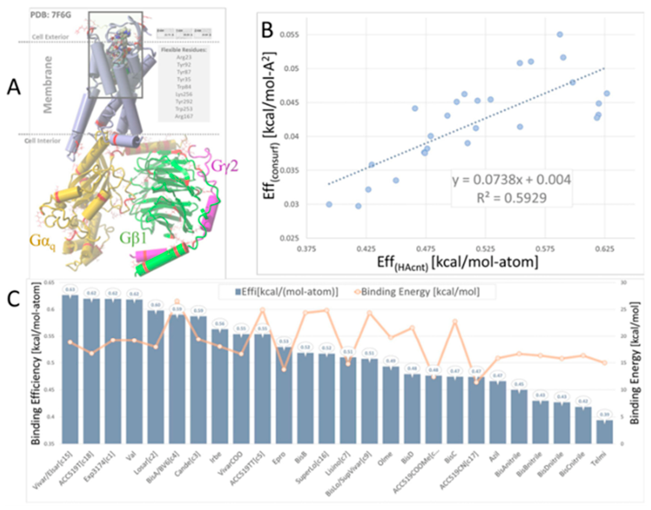
Figure 1. Flexible Receptor Docking (FRD) of 26 selected FDA-approved and experimental ARBs to AT1R (PDB 7F6G). (A) FRD setup included removal of co-crystalized water from 7F6G, optimization of the hydrogen-bonding network, bond order corrections, and so forth. FRD was run using AutoDockLGA in the Yasara Structure (www.Yasara.org) modelling suite driven by a custom script that specified 750 runs per ligand and AMBER14 parameters for partial atomic charge and dihedral bond assignments. Nine flexible receptor residues inside the cell surface binding domain were designated, including Arg23, Tyr92, Tyr87, Tyr35, Trp84, Lys256, Tyr293, Trp253, and Arg167. Docking was performed using GPU (NVIDIA 4090) acceleration. (B) The best-scoring ligand pose from the 750 runs was reported. Scoring functions included (1) binding energy expressed as kcal/mol, (2) a binding efficiency metric defined as the binding energy divided by the ligand heavy atom count expressed as kcal/mol-atom (Eff[HAcnt]), and (3) a second binding efficiency metric defined as the binding energy divided by the contacting surface area of the ligand with the interacting pocket residues (Eff[consurf]) expressed as kcal/mol-A2. (C) Ligand FRD results as a function of Eff[HAcnt] (blue bars) and binding energy (orange line with markers). Abbreviations: azil, azilsartan; bis, bisartans; cande, candesartan; elsar, elsartan; epro, eprosartan; ibre, ibresaratn; lisino, lisinopril; los, losartan; olme, olmesartan; telmi, telmisartan; vivar, vivartan.
Preprints 225586 g001
Figure 5. Contraction responses to AngII dose-response in rabbit iliac arteries. Rings pre-treated with candesartan demonstrated reduced contraction responses to AngII (mean ± SEM, significance shown in Table 1). Remarkably, rings pre-treated with lisinopril also displayed markedly reduced contraction responses to AngII (mean ± SEM, significance shown in Table 1).
Preprints 225586 g005
Figure 6. Relaxation responses in the small intestine and colon. (A) The higher dose of ACC519TT markedly reduced relaxation responses in small intestine rings to oxycodone (mean ± SEM is shown, * p<0.05, ** p<0.01) (significance presented in Table 3). (B) Similarly, the higher dose of ACC519TT significantly reduced relaxation responses in colon rings to oxycodone (mean ± SEM is shown, * p<0.05, ** p<0.01, **** p<0.0001) (significance presented in Table 4).
Preprints 225586 g006
Table 1. CAD-derived Kd values for selected compounds at AT1R, α1AR, α2AR, muOR, muscarinic receptors (M3R).
Compound Receptor
AT1R α1AR α2AR µOR M3R
ACC519TT 0.2 nM 0.5 nM 5 nM 10 nM 0.5 pM
AngII 10 nM - - - -
Bisartans 1-10 nM 1-50 nM 10-50 nM 0.1-1µM 0.1-10
Carfentanyl 0.5 µM - - 0.4 µM 5 nM
Doxazosin 50 nM 50 nM 10 nM 0.2 µM 50 nM
Lisinopril 1 µM 10 µM 1 µM 2 µM -
Sartans 10-100 nM 1-50 nM 10-100 nM 0.1-1µM 1-10 nM
Table 2. Significant differences in contraction responses to AngII dose-response after pretreatment with candesartan or lisinopril.
log[AngII], M Control vs. candesartan Control vs. lisinopril Candesartan vs. lisinopril
-11.0 No significance No significance No significance
-10.5 No significance No significance No significance
-10.0 No significance No significance No significance
-9.5 p=0.0115 p=0.0325 No significance
-9.0 p=0.0008 p=0.0002 No significance
-8.5 p<0.0001 p=0.0078 p=0.0033
-8.0 p<0.0001 p=0.0051 p=0.0047
-7.5 p<0.0001 p=0.0138 p=0.0191
-7.0 p<0.0001 No significance p=0.0026
-6.5 p<0.0001 No significance p=0.0046
-6.0 p=0.0021 No significance No significance
-5.5 No significance No significance No significance
-5.0 No significance No significance No significance
Table 3. Significant differences in small intestine relaxation responses to oxycodone after pre-treatment with naloxone, candesartan, or ACC519TT.
log[oxycodone], M Control vs. naloxone Control vs. candesartan Control vs. ACC519TT [10−6 M] Control vs. ACC519TT [10−9 M]
-10.0 No significance No significance No significance No significance
-9.5 No significance No significance No significance No significance
-9.0 No significance No significance No significance No significance
-8.5 No significance No significance No significance No significance
-8.0 No significance No significance No significance No significance
-7.5 No significance No significance No significance No significance
-7.0 No significance No significance No significance No significance
-6.5 p=0.0248 No significance No significance No significance
-6.0 p=0.0019 No significance p=0.0370 No significance
-5.5 p=0.0014 No significance p=0.0090 No significance
-5.0 p<0.0001 No significance p=0.0089 No significance
Table 4. Significant differences in colon relaxation responses to oxycodone after pre-treatment with naloxone, candesartan, or ACC519TT.
log[oxycodone], M Control vs. naloxone Control vs. candesartan Control vs. ACC519TT [10−6 M] Control vs. ACC519TT [10−9 M]
-10.0 No significance No significance No significance No significance
-9.5 No significance No significance No significance No significance
-9.0 p=0.0468 No significance No significance No significance
-8.5 p=0.0205 No significance No significance No significance
-8.0 p=0.0065 No significance No significance No significance
-7.5 p=0.0043 No significance No significance No significance
-7.0 p=0.0032 No significance No significance No significance
-6.5 p<0.0001 No significance No significance No significance
-6.0 p<0.0001 No significance p=0.0231 No significance
-5.5 p<0.0001 No significance p=0.0024 No significance
-5.0 p<0.0001 No significance p<0.0001 No significance
Disclaimer/Publisher’s Note: The statements, opinions and data contained in all publications are solely those of the individual author(s) and contributor(s) and not of MDPI and/or the editor(s). MDPI and/or the editor(s) disclaim responsibility for any injury to people or property resulting from any ideas, methods, instructions or products referred to in the content.
Copyright: This open access article is published under a Creative Commons CC BY 4.0 license, which permit the free download, distribution, and reuse, provided that the author and preprint are cited in any reuse.
Prerpints.org logo

Preprints.org is a free preprint server supported by MDPI in Basel, Switzerland.

Subscribe

© 2026 MDPI (Basel, Switzerland) unless otherwise stated

Accessibility

Disclaimer

Terms of Use

Privacy Policy

Privacy Settings