

Submitted:
29 July 2026
Posted:
30 July 2026
You are already at the latest version
Abstract
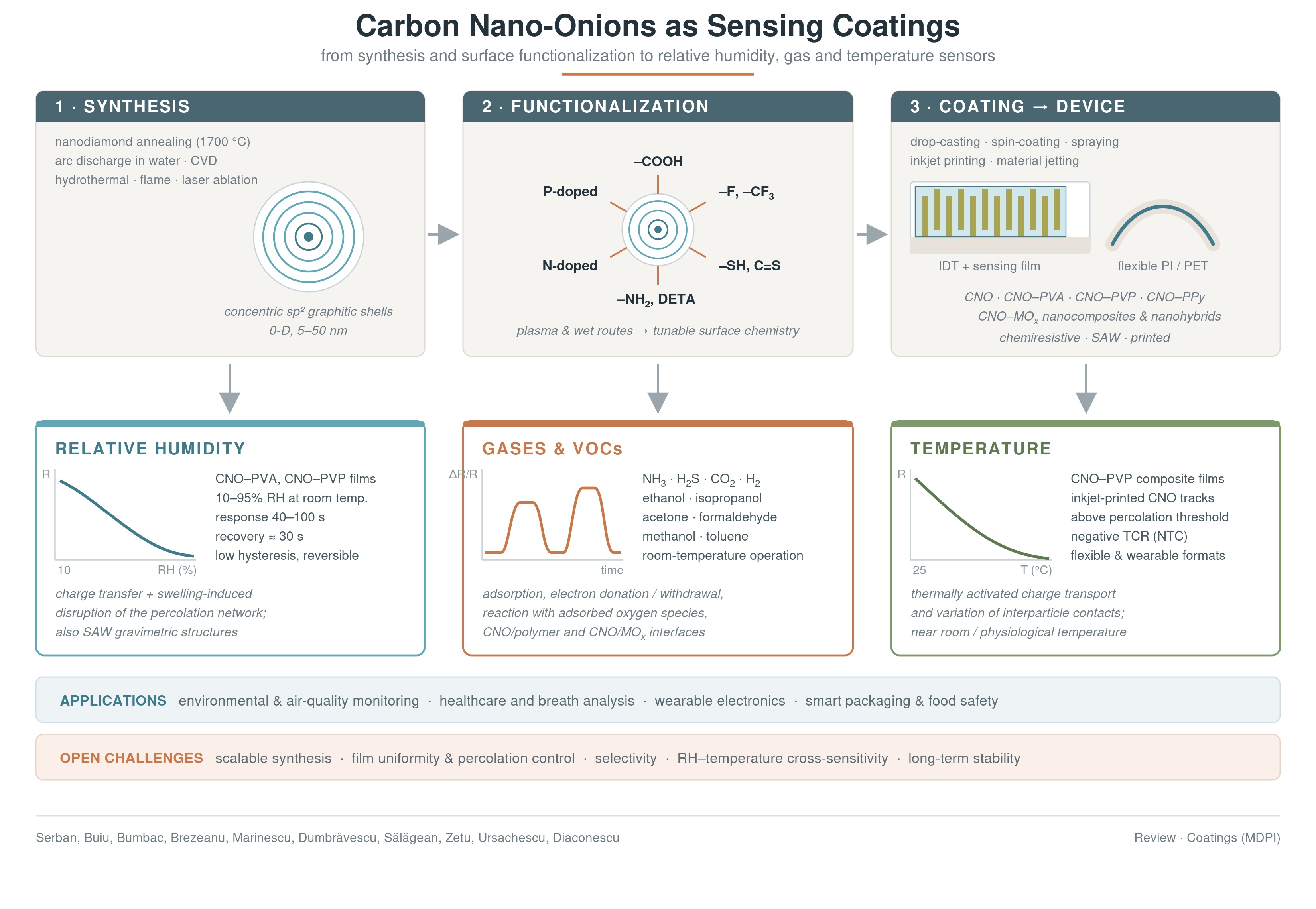
In recent years, carbon nano-onions (CNOs), together with their functionalized derivatives, nanocomposites, and nanohybrids, have attracted increasing attention as sensing materials for monitoring relative humidity (RH), gases, and temperature. Their concentric graphitic structure provides good electrical conductivity, chemical and thermal stability, accessible surface sites, tunable surface chemistry, and compatibility with polymer matrices and flexible substrates. This review highlights recent advances in the synthesis and functionalization of CNOs and examines their integration into chemiresistive, surface acoustic wave, flexible, and printed sensing platforms. Particular attention is devoted to pristine and oxidized CNOs, heteroatom-doped materials, and composites incorporating hydrophilic or conducting polymers, metal oxides, and other functional fillers. CNOs-based sensing layers demonstrate room-temperature (RT) detection of RH, hydrogen, ammonia, acetone, ethanol, isopropanol, carbon dioxide, hydrogen sulfide, and other volatile organic compounds. In addition, CNOs and CNOs–polymer films exhibit significant temperature-dependent variations in resistance, supporting their potential use in flexible and wearable temperature sensors. Although several CNOs-based devices show superior performance in sensitivity, response, recovery characteristics, mechanical flexibility, and low-power operation, the studies on CNOs available in the literature remain limited compared with those on carbon nanotubes, graphene derivatives, and other carbonaceous materials. Further progress on CNOs-based structures requires reproducible, large-scale synthesis; improved film uniformity and selectivity; standardized testing; compensation for temperature–humidity cross-sensitivity; and long-term stability studies. This review concludes by highlighting research directions to bridge the gap between laboratory prototypes and commercially viable CNOs-based sensing devices.
Keywords:
carbon nano-onions
; relative humidity
; gas sensors
; temperature
; volatile organic compounds
; chemiresistive
; nanocomposites
; functionalization
1. Introduction
Gas sensing is the technology used to detect specific gases and measure their concentrations in the surrounding environment. In recent decades, gas sensing has become a vital research area, driven by the need for accurate and reliable detection of low concentrations of hazardous, toxic, and flammable gases, as well as environmental pollutants [1,2].
Gas sensors are essential tools in safety, industrial, environmental, and health-monitoring systems, as follows:
- Life and Safety Protection: Gas sensors enable real-time detection of hazardous gases, preventing fatal accidents, explosions, and acute poisoning by identifying leaks (e.g., carbon monoxide (CO), chlorine, hydrogen sulfide (H2S), ammonia (NH3)) and monitoring oxygen depletion in confined spaces such as storage tanks or mines [3,4].
- Health, safety, disease diagnosis, and hospital infrastructure monitoring: Gas sensors are fundamental pillars for patient monitoring and diagnosis (metabolic analysis, capnography, breath biomarkers, precise anaesthesia delivery) [7,8], gas pipeline and hyperbaric chamber monitoring, and sterilisation safety [9,10].
- Regulatory compliance: Approved gas sensors guarantee compliance with rigorous industrial safety and health regulations (workplace safety, explosion prevention, environmental tracking) [13].
Beyond the hazardous gases, relative humidity (RH) monitoring represents a particularly important area of gas sensing. Monitoring RH has a significant impact on several industrial, medical, commercial, and residential applications [21,22,23,24,25]. A more detailed description of the gas and RH sensor applications is presented in the chart above (Figure 1). Beyond the sensing principle, electronics, architecture, miniaturisation, and power consumption, the nature of the sensing layer is crucial. It dictates the chemical reactivity, operating temperature, and overall performance of the sensors [26]. Consequently, a wide range of materials have been tested as sensing layers within the design of gas and RH sensors, including metal oxides (MOXs), metal-organic frameworks (MOFs), covalent-organic frameworks (COFs), conducting polymers, dielectric polymers, perovskites, nanoclays, black phosphorus/phosphorene/ two-dimensional materials, and a plethora of hybrid or composite systems [27,28,29,30,31,32,33,34]. Last but not least, due to their outstanding physicochemical properties, carbon-based materials and their nanocomposites/nanohybrids have been successfully employed as sensing layers for gas and RH monitoring across various sensor types. Thus, beyond of the well-known graphene, graphene oxide (GO), reduced graphene oxide (rGO), carbon nanotubes (multi and single–walled), fullerenes, carbon quantum dots, carbon nanohorns, nanodiamond [35,36,37,38], numerous less conventional carbon-based materials such as graphdiyne nanoribbons, carbide-derived carbon, bamboo-derived porous carbon, MOF-derived carbon, and hydrogenated amorphous carbon (a-C: H) film were used as key sensing film in the design of the RH and gas sensors [39,40,41,42,43].
At the same time, temperature sensors are important devices for measuring and monitoring temperature in various environments. They are widely used in home appliances (refrigerators to storage the food at the appropriate temperature, ovens, air conditioners), medical field (thermometers, incubators, patient monitoring system), automotive industry (for monitoring engine temperature, coolant temperature, and the temperature inside the car), chemical industry (to monitor machines, chemical processes, and production lines), agriculture, electronics, weather stations and so forth [44,45,46,47]. Flexible temperature sensors have become increasingly interesting in recent years due to their outstanding properties, such as high flexibility, lightweight design, excellent conformability to curved or irregular surfaces, high sensitivity, and potential integration into wearable and portable electronic devices. These characteristics make them suitable for applications in healthcare monitoring, human–machine interfaces, electronic skin, smart textiles, soft robotics, and advanced industrial systems (Figure 2) [48,49].
Nanocarbonic materials are excellent candidates for sensing in the manufacturing of flexible temperature sensors because they have demonstrated unique advantages, including high sensitivity, mechanical flexibility, lightweight structure, and good thermal stability. Materials such as graphene, carbon nanotubes (CNTs), carbon black, and carbon nanofibers can form conductive networks that respond sensitively to temperature changes through variations in electrical resistance [50,51,52,53,54,55].
In recent years, carbon nano-onions (CNOs), together with their nanocomposites, have attracted increasing attention as promising materials for gas, RH, and temperature sensing applications. Although research on CNOs-based sensing materials remains relatively limited, existing studies indicate growing interest in their use as key sensing elements in the design of advanced sensors [56,57,58].
This review article provides an overview of recent advances and emerging perspectives on CNOs, as well as their nanocomposites and nanohybrids, for gas, RH, and temperature sensors. The review is structured into seven main sections. The first section discusses the principal strategies employed for CNOs synthesis, with particular emphasis on the functionalization of these nanocarbonic structures to enhance their sensing performance towards water, VOCs, and other gases. The second section highlights the key physicochemical and electronic properties of CNOs that make them promising sensing materials. The third section briefly addresses the design of RH, gas, and temperature sensors based on CNOs-derived materials used as sensing films.
The fourth section discusses the synthesis and performance of various CNOs-based sensors for RH, VOCs (formaldehyde, ethanol, isopropanol), and other gases, including NH3, H2S, and CO2. In this context, pristine and functionalized CNOs and nanocomposites/nanohybrids incorporating several dielectric, hydrophilic polymers, semiconducting MOX, and conducting polymers are evaluated and compared in terms of their sensitivity, response time, and recovery time. The fifth section presents the main sensing mechanisms involved in the RH and gas detection structures discussed. It analyses the properties of each component, together with their mutual interactions and influence on the sensing behaviour. The sixth section is devoted to CNOs-based temperature sensors, highlighting their sensing performance and potential relevance for medical applications. Finally, the seventh section provides a critical discussion of the main challenges, current limitations, and future research directions associated with the development of CNOs-based sensing platforms. Particular attention is given to assessing their potential for the transition from laboratory-scale prototypes to commercial sensors.
2. Structure and Synthesis of Pristine CNOs and Their Derivatives Used in RH, Gas, and Temperature Monitoring
2.1. Structure of CNOs
CNOs are zero-dimensional carbon nanostructures consisting of multilayered, concentric graphitic shells arranged around a central core (Figure 3).
These shells are usually composed of sp2-hybridized carbon atoms, similar to those found in CNTs and fullerenes [59,60]. CNOs generally have nanoscale dimensions, with particle diameters strongly dependent on the synthesis procedure, starting material, and post-annealing conditions. In many synthesis protocols, CNOs obtained from annealed nanodiamond precursors show relatively small diameters, typically around 5–10 nm, whereas other synthesis methods can produce larger particles, commonly in the range of 30–100 nm [61,62]. Structural defects, such as vacancies, sp3-hybridized carbon atoms, dangling bonds, and pentagonal and/or heptagonal carbon rings, may be present in the graphitic layers. These defects strongly influence the electronic properties and functionalization pathways of CNOs [63]. The high curvature of the graphitic shells gives CNOs unique surface reactivity compared with flat graphite [64].
2.2. Synthesis of Pristine CNOs
Carbon nano-onions were first observed by S. Iijima in 1980 using transmission electron microscopy (TEM) [65]. Later, D. Ugarte’s 1992 work significantly increased worldwide scientific interest in these carbonaceous nanostructures [66].
The synthesis of CNOs can be achieved through several physical and chemical methods, and the selected pathway strongly influences their size, crystallinity, degree of graphitisation, and defect density. One of the most widely used methods is the thermal annealing of nanodiamond particles under vacuum or an inert atmosphere at high temperatures (1700 °C) [67].
During this process, the diamond-like sp3 carbon structure gradually transforms into sp2 graphitic layers with a substantial degree of carbon ordering. Arc discharge in water represents another straightforward and efficient route for producing appreciable amounts of CNOs [68]. In this process, a high-temperature plasma arc is established between two graphite electrodes, typically under an inert atmosphere such as argon or helium. The extreme temperatures generated during the discharge lead to the vaporisation of graphite, followed by the condensation and reorganisation of carbon species into CNO structures. This synthetic protocol allows the formation of high-purity CNOs with minimal contamination [69,70].
Chemical vapour deposition (CVD) is another widely employed technique for obtaining CNOs, relying on the thermal decomposition of hydrocarbon precursors such as methane or acetylene. At elevated temperatures, these gaseous carbon sources decompose on the surfaces of metallic catalysts, promoting the nucleation and growth of concentric graphitic carbon layers. Catalyst composition and reaction temperature play pivotal roles in controlling both the structure and the production yield of the resulting CNOs [71,72]. Hydrothermal synthesis has emerged as a promising approach to producing CNOs, owing to its simplicity, low cost, and environmental friendliness [73]. This method is based on the hydrothermal carbonisation of organic precursors, such as citric acid, which gradually transforms into carbon-based nanostructures. Moreover, the incorporation of organic solvents such as toluene, hexane, or butyl acetate can influence particle growth, improve production scalability, and modify the final properties of the synthesised CNOs [74,75].
Other reported approaches include annealing of heavy oil precursors [76], flame synthesis [77], laser ablation [78], detonation [79], ion implantation [80], and high-energy ball milling [81]. The synthesis method must be carefully selected for the intended application, as the electronic properties, chemical reactivity, and functionalization of CNOs are directly related to their structure.
2.3. Synthesis of Functionalized CNOs for RH and Gas Sensing
The targeted functionalization of CNOs with specific chemical groups enables the tailoring of their surface chemistry for RH and gas-sensing applications. By introducing appropriate functional groups with a high affinity for water molecules or gaseous analytes, the sensitivity, selectivity, response time, and hysteresis of the designed sensors can be significantly improved. In addition, incorporating pristine and functionalized CNOs into polymer matrices to form nanocomposites may enhance their processability, mechanical stability, and sensing performance.
Due to their largely nonpolar carbon framework, pristine CNOs are inherently hydrophobic. Consequently, surface functionalization is required to introduce hydrophilic groups and enhance their interaction with water molecules.
Carboxyl-functionalized CNOs have been proposed as hydrophilic nanomaterials for RH sensing applications. The presence of surface carboxyl groups enhances water adsorption and improves the interaction of the sensing layer with moisture, thereby enabling a measurable electrical response to changes in RH [82].
Synthesis of hydrophilic CNOs can be performed in several ways. The pristine CNOs are oxidised either by refluxing them in 3 M nitric acid for 48 h, or by exposure to Ar/O2 plasma. These oxidation/hydrophilization treatments introduce polar, oxygen-containing surface functionalities, increase the affinity of the CNOs for water molecules, and facilitate their dispersion and incorporation into nanocomposite sensing films for RH monitoring [83,84].
Fluorinated CNOs represent another promising class of nanomaterials for resistive RH sensing, owing to their tunable surface chemistry, high electrical conductivity, and enhanced interactions with water molecules. Synthesis of fluorinated CNOs is performed in N2/F2 plasma [85]. The high electronegativity of fluorine atoms increases the surface polarity of the nanocarbon material, generating temporary dipoles that promote interactions with water molecules.
Serban et al. proposed an RT chemiresistive formaldehyde (CH2O) sensor based on a nitrogen-doped CNOs/polyvinylpyrrolidone (PVP) nanocomposite, in which the electrical resistance increases upon exposure to CH2O due to charge-transfer interactions and disruption of the conductive percolation network. N-doped CNOs are synthesised according to a procedure that starts with fluorinated CNOs that are obtained by treatment in an F2/Ar plasma, using a volumetric gas ratio of 1:6, at a pressure of 0.4 bar. The process is carried out in a nickel reactor at RT. The gas injection time is 5 minutes, while the exposure time varies from 2 to 6 minutes. Heating the fluorinated CNOs at 500 °C under an NH3 atmosphere leads to their defluorination, generating vacancies within the carbon nanostructure. Nitrogen atoms subsequently occupy these vacancies, resulting in the formation of N-CNOs [86,87].
A nanohybrid based on CNOs functionalized with trifluoromethyl groups (CNOs–CF3) and NiO was proposed as a sensing layer for resistive monitoring of ethanol vapour [88,89]. The synthesis of CNOs functionalized with CNO–CF3 is carried out by treatment in CF4 plasma at a pressure of 1 bar, in a nickel reactor, at RT. The injection time is 3 minutes, while the exposure time ranges from 2 to 20 minutes.
CNOs functionalized with mercapto (C-SH) groups and carbonothioyl (C=S) groups were proposed as the key sensing element within the design of the H2S resistive sensor. S-based CNOs are prepared using H2S/He plasma (60/40 v/v) [90,91].
Oxy-fluorinated CNOs have been proposed as sensing layers for the resistive monitoring of ethanol [92]. Oxy-fluorinated CNOs can be synthesised through two distinct functionalization routes, which differ with respect to the order of the fluorination and oxidation steps.
Route 1: Fluorination of CNOs Followed by Oxidation
In the first route, pristine CNOs are initially fluorinated to obtain CNOs-F. Fluorination is performed in an F2/N2 plasma using an equimolar gas mixture at 0.5 bar in a nickel reactor at RT. The gas injection time is 5 min, while the plasma exposure time ranges from 2 to 4 min. The resulting CNOs-F are subsequently oxidised in an Ar/O2 plasma using a 2:1 volumetric gas mixture. The oxidation treatment is carried out in a quartz tube at 3 Torr and at RT. The gas injection time is 5 min, followed by a plasma exposure time of 2–4 min.
Route 2: Oxidation of CNOs Followed by Fluorination
In the second route, pristine CNOs are first oxidised to obtain ox-CNOs. The oxidation treatment is performed in an Ar/O2 plasma using a 2:1 volumetric mixture in a quartz tube at 3 Torr and at RT. The gas injection time is 5 min, while the exposure time ranges from 2 to 4 min. The resulting ox-CNOs are then fluorinated in an F2/N2 plasma using an equimolar gas mixture at 0.5 bar in a nickel reactor at RT. In this case, the gas injection time is 4 min, while the plasma exposure time ranges from 2 to 4 min.
Diethylenetriamine-functionalized CNOs (CNO–DETA) [93] and amino-CNOs [94] were used as a sensing layer for CO2 detection.
Raghu et al. developed phosphorus-doped onion-like carbon nanostructures using triphenylphosphine, P(C6H5)3, as the P precursor. During thermal treatment, P atoms are incorporated into the carbon lattice, forming P–C bonds. Subsequent exposure to air or oxygen causes partial oxidation of the P species at the surface. As a result, functional groups such as P–O, P=O, and P–OH are formed. These highly surface-active nanostructures can be successfully applied in ultrasensitive, fully reversible, and portable NH3 gas sensors [95].
All the synthesis routes described above are depicted in Figure 4.
3. Properties of CNOs
The distinctive concentric graphitic architecture of CNOs gives rise to a combination of structural, surface, chemical, thermal, and electrical properties. The most relevant features of CNOs are summarised below:
- High external surface accessibility – Because of their small diameter and spherical morphology, CNOs expose a large external surface that ions, molecules or functional groups can easily access. This property is important for electrochemical storage, sensing, adsorption, and catalysis [96].
- High specific surface area – CNOs can exhibit high specific surface areas, especially when produced under conditions that limit particle sintering or when they are chemically activated. Nanodiamond-derived CNOs usually exhibit specific surface areas around 300–600 m2/g depending on the annealing temperature. Chemical activation can be used to create a porous structure within the outer carbon shells, leading to a remarkable increase in specific surface area, reaching values above 3,800 m2/g. As a result, these materials show strong potential for use in adsorption, catalysis, and gas-sensing technologies [97,98].
- Electrical conductivity - CNOs possess good to moderate electrical conductivity, but they are not as highly conductive as continuous carbon structures like single-walled CNTs or graphene. In composite films, the CNOs form conductive pathways once they reach a “percolation threshold” (often around 20 wt% in polymer matrices) [99,100].
- Porous interparticle network – This characteristic is highly advantageous because it prevents nanoparticles from aggregating, which would otherwise limit their performance. This architecture addresses key structural issues while dramatically improving the way CNOs operate in high-demand applications [101].
- Tunable surface chemistry – The surface of CNOs can be modified through covalent functionalization (oxidation, amidation/esterification, electrografting), noncovalent functionalization (π-π stacking and encapsulation), and heteroatom doping. These surface treatments enable the tailoring of the material’s surface chemistry by introducing several functional groups, thereby enhancing its compatibility with solvents, polymers, biomolecules, and metal nanoparticles [102].
- Good thermal stability – CNOs exhibit high thermal stability in air, with negligible mass loss up to approximately 500–550 °C. The main degradation or oxidation of the carbon framework typically occurs at higher temperatures, around 630–700 °C, indicating that CNOs can withstand elevated thermal conditions before significant structural decomposition [103].
- Chemical stability and corrosion resistance – Due to their highly ordered, closed-cage sp2 graphitic structure, pristine CNOs exhibit a remarkably high resistance to carbon corrosion and oxidation, surpassing other well-known carbon supports (like amorphous carbon or carbon black) in harsh operating environments [104].
- Low toxicity and biocompatibility potential – CNOs are widely recognised for their low toxicity, high biocompatibility, minimal cellular & immune impact, and in vivo safety [105].
- Catalytic and catalyst-support behaviour – CNOs can act as active carbon materials or as supports for metal nanoparticles and redox-active compounds [106].
- High absorption capacity - CNOs exhibit exceptional absorption capacities as a consequence of their unique sp2 graphitic shells, large available outer surface area, and curved edges. They are highly effective at absorbing environmental pollutants, including toxic heavy metals, oxoanions, and dyes [107,108].
- Potential use in polymer nanocomposites – Due to their conductivity, versatile covalent and noncovalent functionalization, small size, and chemical stability, CNOs can be incorporated into polymer matrices to obtain electrically conductive, mechanically reinforced, or thermally improved nanocomposites [109].
Taken together, these properties indicate that CNOs are promising nanocarbon materials either as a sensing layer alone (pristine or functionalized) or as nanocomposites for RH, gas, and temperature sensing.
4. Structure of CNOs-Based Resistive RH, Gas, and Temperature Sensors
CNOs-based resistive RH and gas sensors typically include a substrate, a sensing layer, and two metal electrodes. An example of the RH-sensing structure is shown in Figure 5. The sensor was fabricated on a 470 µm-thick Si substrate and covered by an approximately 1 µm-thick electrically insulating SiO2 layer. An interdigitated transducer (IDT) consisting of two interlocking gold electrode combs was patterned on top of the SiO2 surface. The RH-sensing film was a CNOs/polyvinyl alcohol (PVOH) nanocomposite prepared using CNOs mass ratios of 1:1 and 2:1.
Another example of an RH-sensing structure is a flexible chemiresistive sensor based on a CNOs/PVP nanocomposite deposited onto gold interdigitated electrodes patterned on a polyimide substrate. Gold interdigitated electrodes were patterned on the polyimide, with an active transducer area of approximately 5 × 7 mm2. The sensing layer was a CNOs/PVP nanocomposite with a 1:1 mass ratio, containing CNOs approximately 5–8 nm in diameter and PVP with a molecular weight of 10,000 Da [111].
In the study by Dhonge et al. [112], the CNOs film was used as the sensing film to monitor RH and organic vapours, such as ethanol, acetone, and methanol. The device was based on a monocrystalline Si (100) substrate, with the carbon material formed in situ on its surface via laser photolysis of toluene rather than being transferred as a separately prepared powder or embedded in a polymer matrix.
CNOs-based layers were used as key sensing elements in the design of surface acoustic wave (SAW)-based sensors for CO2 detection [93]. The SAW structure employed was a dual delay-line device fabricated on a piezoelectric quartz substrate (Figure 6). It featured a double delay-line configuration designed to compensate for thermal drift. One delay line was coated with functionalized carbon nanohorns (CNHs), CNHs–R–NH–R–NH2 or CNOs–R–NH–R–NH2, while the second delay line consisted of the piezoelectric substrate without a sensing layer. To obtain a signal arising exclusively from the chemical interaction between CNHs–R–NH–R–NH2 or CNOs–R–NH–R–NH2 and CO2, the signal associated with the uncoated delay line was subtracted from that of the delay line coated with CNHs–R–NH–R–NH2 or CNOs–R–NH–R–NH2, using the differential configuration.
In the work of Pinto et al. [113,114], the sensor was fabricated on a flexible polyethylene terephthalate (PET) substrate and consisted of an inkjet-printed CNOs track approximately 2 cm in length. The printed line had a width of about 220–245 µm, while its thickness depended on the number of printing passes and reached approximately 0.72 µm for 50 deposited layers. Electrical contact was provided by nickel conductive paste pads applied at both ends of the CNOs track, forming a simple two-terminal, two-wire resistive configuration. During manufacturing and testing, the flexible substrate was temporarily supported on a glass slide to ensure mechanical stability.
In the study by Lawaniya et al. [115], N-doped CNOs and polypyrrole (PPy) were used to fabricate the sensing layer of a flexible chemiresistive ammonia sensor. The sensor had a flexible structure fabricated on a polyvinylidene fluoride (PVDF) substrate. Interdigitated aluminum electrodes were deposited on the substrate by electron-beam evaporation, with a reported spacing of 200 µm between adjacent fingers and a thickness of 400 µm. The sensing layer consisted of a nanocomposite of PPy and N-CNOs, containing 5 wt.% N-CNOs. The nanocomposite powder was mixed with α-terpineol at a mass ratio of 20:1 to obtain a viscous paste. The paste was uniformly deposited over the active area of the interdigitated electrodes and dried at RT. The final sensor architecture can be described as PVDF/(aluminum interdigitated electrodes) Al-IDE/N-CNOs–PPy, with the nanocomposite film electrically bridging the two interdigitated electrode networks.
Olariu et al. [116] employed CNOs nanoparticles as the sensing layer of an RT chemiresistive hydrogen sensor. The device was constructed on a 22.8 × 7.6 × 0.7 mm3 glass substrate carrying photolithographically patterned gold interdigitated electrodes. The electrode structure comprised two interdigitated arrays with 5 µm-wide fingers, 5 µm interelectrode gaps, and a finger length of 6760 µm. The active material consisted of spherical CNOs particles approximately 5–7 nm in diameter, each containing 6 to 8 concentric graphitic shells. Dielectrophoresis was used to trap and distribute the CNOs agglomerates within the interelectrode regions.
Raghu et al. [95] fabricated a portable chemiresistive ammonia sensor by coating P-doped CNOs nanostructures onto gold interdigitated-array electrodes formed on an alumina substrate. In this planar configuration, the P-doped CNO film electrically bridged adjacent Au fingers and acted as the sole gas-sensitive resistor. The active material contained substitutional P atoms together with surface metaphosphate and phosphonate species, which generated polar adsorption sites and facilitated charge transfer during interaction with ammonia.
Mongwe et al. [117] developed a chemiresistive acetone sensor based on a ternary nanocomposite deposited onto an interdigitated-electrode platform. The sensing layer consisted of PVP, CNOs or N-doped CNOs, and MnO2 nanorods, mixed at a 3:1:1 mass ratio. The components were dispersed in dimethylformamide, and a 10 µL aliquot of the resulting suspension was drop-cast directly onto the interdigitated electrodes. The interdigitated structure comprised 40 electrode strips, with a reported strip dimension of approximately 72 µm and an inter-strip spacing of approximately 125 µm. Exposure to acetone altered the sensing film’s electrical properties, and the sensor response was determined by monitoring the resulting changes in resistance or impedance between the electrodes.
Serban et al. [118] developed a resistive isopropanol sensor using a sensing layer composed of CNOs and PVP in 1:1 (w/w) ratio. The sensing device, comprising a Si/SiO2 substrate and gold electrodes, was fabricated by drop-casting an aqueous CNOs–PVP suspension onto the sensing platform.
Serban et al. [119] developed a resistive ethanol sensor using a sensing layer composed of CNOs and PVP in a 2:1 (w/w) ratio. The sensing device, composed of a polyimide substrate and gold electrodes, was fabricated by drop-casting an CNOs-PVP suspension prepared in a dimethylformamide-ethanol mixture onto the sensing platform.
Compared with graphene, GO, and CNTs, CNOs remain considerably less explored as RH and gas-sensing materials, with relatively few dedicated studies and sensor configurations reported in the literature [120,121]. Most reported CNOs-based gas sensors employ a resistive architecture, in which a CNOs or CNOs-based nanocomposite layer is deposited over or between interdigitated electrodes, and gas detection is achieved by monitoring changes in the sensing layer’s electrical resistance. Although other sensing principles have occasionally been explored, resistive configurations currently dominate CNOs-based gas-sensing studies.
Only a limited number of temperature sensors based on CNOs have been reported, and the available devices are mainly resistive structures in which either a pristine CNOs film or a CNOs–based nanocomposite acts as the active sensing layer.
Pinto et al. [114] introduced a flexible two-terminal temperature-sensor architecture based on a directly printed CNOs thin layer. The manufactured device comprised a 50 µm-thick PET substrate onto which a linear CNOs track was deposited by drop-on-demand piezoelectric inkjet printing. Electrical contacts were formed at the two ends of the printed strip, creating a planar resistive element connected to an external resistance-measurement circuit.
Pinto et al. [113] developed a more detailed version of the same printed architecture using a 75 µm-thick PET substrate. A 2 cm-long CNOs track was deposited directly onto the PET surface by piezoelectric inkjet printing, while the track thickness was adjusted through the number of printing passes. For a ten-pass structure, the printed layer was approximately 256 µm wide and 176 nm thick. Nickel conductive paste pads were manually applied to both ends of the CNOs strip to provide an electrical connection.
Serban et al. [100] investigated a planar resistive structure based on a CNOs–PVP nanocomposite film. The device consisted of a supporting polyimide substrate carrying measurement electrodes, over which the CNOs–PVP composite was deposited, forming a film that bridged the electrode gap and defined the active sensing region. Within the composite layer, the CNOs particles constituted the electrically conductive phase, while PVP formed the surrounding polymer matrix.
The limited number of CNOs-based temperature sensors reported to date likely reflects the difficulty of producing uniform, reproducible CNOs films with stable electrical contacts and controlled sensitivity to interfering factors such as humidity. Moreover, most studies on CNOs have focused on synthesis and electrical characterization rather than on the manufacturing, calibration, and validation of temperature-sensing devices.
5. RH and Gas Sensors Based on CNOs and Their Nanocomposites
5.1. CNOs and Their Derivatives/Nanocomposites as Sensing Layers in RH Sensors
The idea of using CNOs and their derivatives in sensing layers employed for resistive RH detection was recently explored. Historically, Dhonge et al. [112] reported the first use of a pristine CNOs layer for the resistive detection of RH. The CNOs-based nanostructures were synthesized by laser photolysis of toluene. The morphology of the deposited CNOs depended strongly on the laser power density. At approximately 2.54 W cm−2, the CNOs formed hierarchical coin-tower-like islands, whereas power densities of approximately 6.62 and 9.17 W cm−2 produced flower-like aggregates composed of CNO nanoparticles. For low RH levels, conductivity decreased as RH increased to 18.7%; beyond this threshold, conductivity was shown to increase with further increases in RH.
Pinto et al. [113] subsequently reported a more detailed evaluation of the RH dependence of a standalone CNOs film. The study focused on the formulation and inkjet printing of CNOs inks for flexible printed electronics. The printed CNOs structures exhibited an electrical resistivity of approximately 593 ± 79 Ω·m. Their resistance was measured during increasing and decreasing RH sweeps performed at a constant temperature of 40 °C. RH was varied from 20% to 80% in 5-percentage-point steps, with an RH-setting tolerance of approximately ±1% RH. When RH increased from 20% to 80%, the resistance of the printed CNOs line decreased by as much as 91% relative to its initial value at 20% RH. The resistance–RH characteristic was sigmoidal. The average RH sensitivity of CNOs over the entire RH range was estimated as βCNO = −1.29 × 10⁻² ± 1.03 × 10⁻³ RH%−1. However, the variation in resistance did not strictly follow a linear relationship. Sensitivity decreased at RH values below 40%, while signs of saturation become evident above 70% RH.
Serban et al. [110,122] reported the seminal use of a CNOs–polymer nanocomposite as the sensing layer in a resistive RH sensor. The study demonstrated its potential for sensitive, rapid, and low-hysteresis RH monitoring, evaluating the RH response of a resistive sensor employing a novel sensing film composed of pristine CNOs and polyvinyl alcohol (PVA) in weight ratios of 1:1 and 2:1. The CNO/PVA sensing layers were prepared by dispersing pristine CNOs in an aqueous PVA solution under ultrasonication. The resulting suspensions were drop-cast onto metallic interdigitated electrodes fabricated on a Si/SiO2 substrate, while the contact pads were masked. After deposition, the sensing films were thermally treated under vacuum and subsequently dried. The same preparation procedure was used for both compositions, differing only in the CNO/PVA mass ratio.
The surface topography of the CNOs/PVA-based sensing layer was examined using scanning electron microscopy (SEM) (Figure 7).
The SEM micrographs of the CNOs/PVA composite with a 1:1 mass ratio (Figure 7) recorded at 3,000× and 7,000× magnification revealed rough, heterogeneous, and predominantly granular surface morphology. At lower magnification, the sample appeared relatively compact, with fine particles distributed throughout the polymer matrix, together with scattered, rounded agglomerates and small pores. The higher magnification image confirmed the presence of densely packed irregular clusters and approximately spherical particles, indicating that the CNOs phase was incorporated into the PVA matrix but not completely uniformly dispersed. Local agglomeration and micron-scale voids were visible, while no major cracks or pronounced phase separation could be observed in the analyzed regions.
Raman analysis (Figure 8) showed the characteristic D and G bands of CNOs at 1,331 and 1,559 cm−1, respectively. The increase in the ratio from 0.558 for reference CNOs to 1.126 for CNOs/PVA indicated greater structural disorder after PVA incorporation. Additional low-wavenumber bands confirmed the presence of PVA in the nanocomposite.
Atomic force microscopy (AFM) analysis yielded key indications about the morphology of the sensing layer (Figure 9). The surface roughness of the CNOs/PVA 1/1 (w/w) sensing layer was characterized by an SDQ of 0.759 nm, an SA of 353.83 nm, and an SQ of 435.40 nm. The relatively high SA and SQ values confirmed the presence of a well-developed, heterogeneous surface topography. In contrast, the moderate SDQ value indicated that the height variations occur through comparatively gradual slopes, suggesting a rough but relatively evenly distributed morphology rather than a sharply irregular or highly jagged surface.
The RT RH sensing performance of the manufactured sensor employing CNOs/PVA sensing films with weight ratios of 1:1 and 2:1 was investigated by applying a constant electric current between the interdigitated electrodes and recording the corresponding voltage as the RH was varied from 5% to 95%. As shown in Figure 10, changing the CNOs-to-PVA ratio markedly influences the resistance response to RH variations within the test chamber. In the sensing layer with a higher proportion of CNOs, the resistance exhibited persistent, pronounced fluctuations over a broad range, even when the RH remains stable.
The operational stability of the proposed RH-sensing materials was systematically assessed by monitoring thin-film resistance across repeated measurement cycles. Three sensors fabricated under quasi-identical conditions were exposed to successive humidity variations spanning the 5–95% RH range. Figure 11 illustrates the dependence of the normalized resistance variation ((Ri - Rf)/Ri) on RH. The experimental results revealed that the CNOs–PVA 1:1 (w/w) sensing layer exhibited markedly superior stability compared with the CNOs–PVA 2:1 (w/w) formulation, indicating a substantially improved resistance to performance degradation under repeated humidity cycling.
The response time and sensitivity analyses were conducted only for the sensor with the CNOs/PVA sensing layer at a 1:1 (w/w) ratio and compared to the performance of a commercially available RH sensor, used as reference sensor, as shown in Figure 12.
The analysis of the ΔR/ΔRH variation across different relative humidity steps revealed distinct behavior in the CNOs/PVA layer at a 1:1 weight ratio at RH values above 80%. The response times of the CNOs/PVA-based sensor ranged from 40 to 100 s, with the longest values recorded when the RH decreased to 70%. The response time ratio shown in Figure 12c indicates that the microsensor based on the 1:1 CNOs/PVA layer generally exhibited performance comparable to or better than that of the reference sensor. The only exception was the 30–40% RH interval, in which the CNOs/PVA sensor responded more slowly than the reference sensor across all three testing cycles.
When the RH decreased from 95% to 10%, the recovery time of the CNOs/PVA sensor was approximately 30 s, nearly half compared to the response time of the reference sensor, estimated 65 s. A key characteristic of the sensor employing CNOs/PVA at 1:1 (w/w) was the presence of an inflection point in the evolution of the (Rf - Ri) / Ri factor in the 80 – 85% RH domain, as shown in Figure 13. This behavior was observed during both the water molecule sorption and desorption stages.
Dumbravescu et al. [123] investigated two resistive RH sensors based on CNOs/PVA and CNOs–PVP nanocomposites. The study focused on improving their electrical sensing characteristics by modifying the preparation procedure, particularly by doubling the sonication time of the nanocomposite suspensions. This processing alteration produced sensors with high sensitivity, fast response, and practically overlapping resistance curves during the humidification and dehumidification stages (zero hysteresis). The authors identified the longer sonication treatment as the key factor responsible for the enhanced reversibility of the electrical response. Extended sonication most likely promoted a more homogeneous distribution of the CNOs within the polymer matrices and reduced the presence of large carbon agglomerates. As a result, the conductive network and the water sorption–desorption processes became more uniform and reproducible, leading to zero hysteresis. The improved electrical characteristics of both sensor types were analyzed and discussed in detail, demonstrating the importance of suspension preparation for the performance of CNOs–polymer sensing films.
Serban et al. [124] functionalized CNOs using argon plasma treatment at 1 bar in a nickel reactor at RT. Ar was injected for 3 minutes, while the nanomaterial was exposed to the plasma for 5 or 10 minutes to modify its surface properties before incorporation into the PVA matrix.
The electrical resistance of the sensing layer increased progressively as the RH increased throughout the range investigated. At low and intermediate RH values, the resistance increased relatively gradually, whereas at higher RH values the increase was much steeper. The experimental data were analyzed in relation to both the type of plasma employed and the duration of plasma exposure. The combined electrical and wettability results demonstrate that treatment with pure Ar plasma substantially modified the surface characteristics of the nanocarbonic layers. In particular, exposure to pure Ar plasma led to a clear increase in surface electrical resistance, reaching approximately 6.6% after 5 minutes of treatment and 13.2% after 10 minutes.
Serban et al. [111] reported a chemiresistive platform developed for RH sensing, in which the active coating was a newly formulated nanocomposite of pristine CNOs and PVP, mixed in equal mass proportions. The material was drop-cast onto a polyimide support bearing gold electrodes. Electron and atomic force microscopy were employed to examine the film morphology and surface features, while FTIR and Raman measurements provided information on its chemical and structural characteristics.
The SEM images of the CNOs/PVP composite prepared at a 1:1 mass ratio (Figure 14) revealed a highly agglomerated morphology composed of rounded to quasi-spherical nanoparticles. The primary particles appeared to be strongly interconnected, forming irregular clusters with visible necking and partial coalescence between adjacent particles. A relatively broad particle-size distribution and interparticle voids could also be observed.
The RH sensing performance of the novel sensor was evaluated by applying a current between the two electrodes and recording the resulting voltage difference as the RH was varied from 0% to 100%. Measurements performed over three operating cycles (Figure 15, Figure 16 and Figure 17) revealed that the novel sensor’s resistance increased across the entire RH range. Below 65% RH, the resistance exhibited an approximately linear dependence on RH, with a relatively constant slope, whereas above this threshold the sensor response became significantly more stronger.
5.2. CNOs and Their Derivatives/Nanocomposites as Sensing Layers in Gas Sensors
CNOs dispersed ultrasonically in ethanol and dielectrophoretically assembled across gold interdigitated electrodes were used as sensing layer for a RT chemiresistive hydrogen sensor [116]. At 10 ppm H2 in nitrogen, all five fabricated devices exhibited response times of less than 10 s. Recovery generally required approximately 40–60 s, depending on the device, with the best sensor recovering in less than about 40 s.
Raghu et al. [95] introduced P-doped CNOs as the sensitive layer of a portable, RT chemiresistive NH3 sensor. Phosphorus was incorporated both substitutionally into the carbon framework and through polar phosphorus-containing surface groups, including metaphosphate and phosphonate-like sites, thereby increasing the number and strength of adsorption centers for ammonia. The P-CNOs sensor exhibited a response time of 2.8 s and a recovery time of 4.5 s, making it the fastest CNOs-based ammonia sensor reported in the literature. The work therefore demonstrated that heteroatom doping can greatly accelerate adsorption/desorption kinetics.
Lawaniya et al. [115] developed a flexible NH3 sensor based on a nanocomposite consisting of N-doped CNOs and PPy, deposited on a flexible membrane. The CNOs were obtained via flame pyrolysis of waste cooking oil; nitrogen doping was carried out hydrothermally using urea; PPy was grown by in situ oxidative polymerization. The optimal composition was approximately 5 wt% N-CNOs in PPy, with the material exhibiting a specific surface area of about 237 m2/g and a mesoporous structure. The sensor was shown to operate at RT over an NH3 concentration range of 1–200 ppm. At 100 ppm, a response of 17.32% was reported, with a response time of 26 s and a lower detection limit of 1 ppm. The device showed selectivity toward NH3 compared with H2, CO2, CO, ethanol, and NO2, remained stable for five weeks, and continued to operate after 500 bending cycles.
Mongwe et al. [117] investigated RT acetone sensing using four ternary nanocomposites: PVP/pristine CNOs/MnO2 nanorods (pS), PVP/N-CNOs-100/MnO2 nanorods (a1S), PVP/N-CNOs-150/MnO2 nanorods (a1.5S), and PVP/N-CNOs-200/MnO2 nanorods (a2S). The study compared how different nitrogen-doping conditions (ammonia was used as the doping agent) affected the chemiresistive performance of the sensing films. Among the tested materials, the composite containing pristine CNOs exhibited the highest acetone sensitivity.
The labels indicate the nitrogen-doping conditions used for the carbon nano-onions:
- a1S: sensor containing N-doped CNOs prepared with an ammonia flow of 100 sccm;
- a1.5S: sensor containing N-doped CNOs prepared with an ammonia flow of 150 sccm;
- a2S: sensor containing N-doped CNOs prepared with an ammonia flow of 200 sccm;
The letter S refers to the final sensor, while pS denotes the sensor based on pristine, undoped CNOs. The experimental acetone concentrations ranged from 136 to 678 ppm. Increasing the acetone concentration decreased resistance, indicating apparent n-type behavior. The most sensitive device was shown to be the one containing undoped CNOs, with a sensitivity of 2.0 × 10⁻⁴ ppm−1 and a detection limit of 2.9 ppm; one nitrogen-doped material provided the lowest detection limit, namely 1.2 ppm.
Panda et al. [125] synthesized CNOs via a solvent-free hydrothermal process and investigated them as a dual-mode sensing platform using fluorescence and electrochemical measurements. The material was tested for the detection of several volatile organic compounds. Exposure to ethylenediamine and diisopropylamine caused fluorescence quenching, whereas exposure to dioxane increased the fluorescence intensity. For diisopropylamine, the authors reported a detection limit of approximately 16.5 nM. Electrochemical measurements supported the trends observed in the fluorescence experiments.
In [119], Serban et al. presented preliminary findings on the ethanol vapor-sensing behavior of a chemiresistive device incorporating a thin sensing film composed of CNOs and PVP in a 2:1 weight ratio. The sensor consisted of a polyimide substrate with gold interdigitated electrodes and was manufactured by drop-casting a CNOs–PVP suspension prepared in a dimethylformamide–ethanol mixture onto the sensing platform. The composition and morphology of the deposited layer were characterized by SEM, AFM, and Raman spectroscopy. The SEM images of the CNOs–PVP layer at a 2:1 ratio revealed a highly agglomerated and heterogeneous morphology (Figure 18).
At 50,000× magnification, the surface consisted of irregular, cauliflower-like clusters separated by numerous voids, indicating a relatively porous and interconnected structure. The higher-magnification image at 100,000× showed that these aggregates consisted of smaller, rounded particles that were closely packed and partially fused. The smoother connections between neighboring particles may be associated with the presence of the PVP matrix, which acted as a binder between the CNOs agglomerates. The sensing performance was evaluated by applying a constant current across the electrodes and recording the resulting voltage as the ethanol vapor concentration was varied from 5 to 100 ppm. The CNOs–PVP film exhibited a positive chemiresistive response throughout the investigated range, with a clear dependence on ethanol concentration. At low-to-moderate concentrations (approximately 10–60 mg/L), the response was nearly linear, whereas noticeable deviations from linearity occurred at higher concentrations (Figure 19). The transition from a quasi-linear to a nonlinear response at elevated ethanol concentrations was attributed to the limited number of available sorption sites and the finite swelling capacity of the nanocomposite.
Maubane et al. [126] investigated a chemiresistive sensing material composed of nitrogen-doped onion-like carbon nanoparticles and manganese oxide for detecting toluene at room temperature (RT). Nitrogen doping was used to modify the defect density and electronic properties of the carbon material, whereas manganese oxide provided additional adsorption and charge-transfer sites. The reported toluene sensitivity was approximately 8 × 10⁻⁴ ppm−1, and the estimated limit of detection was 0.4 ppm.
Serban et al. [118,127] evaluate the isopropyl alcohol (IPA) sensing properties of a chemiresistive sensor incorporating a nanohybrid sensing layer composed of CNOs and PVP in a 1:1 weight ratio. The stability of the sensor response was evaluated by performing 7 repeated measurements during IPA exposure and 4 during IPA removal. At concentrations below 30 mg/L, no consistent resistive response could be detected. Between 30 and 70 mg/L, the sensor response increased approximately linearly with IPA concentration. It showed good reproducibility, particularly during the second operating cycle, when enhanced signal stability was observed (Figure 20). At concentrations above 70 mg/L, the response became unstable and irregular, most likely because of saturation of the sensing layer.
Mathe et al. [128] developed a flexible, RT ammonia sensor based on a ternary nanocomposite comprising CNOs, polyaniline (PANI), and indium oxide (In2O3). Two microwave-assisted methods were used to prepare the In2O3 component, and the synthesized materials were incorporated into a CNOs/polyaniline conductive network. The addition of In2O3 enhanced the ammonia response of the CNOs/PANI composite by introducing semiconductor–polymer interfaces and additional gas-adsorption sites.
6. Sensing Mechanisms for RH and Gas Monitoring Using CNOs and Their Nanocomposites/Nanohybrids
In this chapter, several mechanisms that may explain the RH and gas sensing behavior of CNOs-based sensing layers are presented and discussed.
One mechanism proposed to explain the RH-sensing process was proposed by Dhonge et al. [112], who observed an increase in electrical resistance with increasing RH: since CNOs behave as p-type semiconductors, adsorbed water molecules donate electrons, thereby reducing the concentration of holes, which act as the majority charge carriers. However, the increase of resistance with RH was observed only up to an RH value of 18.7%. The same authors suggested that, at RH values above 27.9%, CNOs may undergo a transition from p-type to n-type semiconducting behavior.
The sensing mechanism proposed by Dhonge et al. is based on the interaction between water molecules and electrically active sites within the CNOs structure. In the first step, a water molecule from the gas phase is adsorbed onto the CNOs surface. It undergoes ionization, producing an adsorbed positively charged water species and releasing an electron:
H2O(g) → H2O+(ad) + e− (1)
Water molecules may also interact with oxygen species associated with structural defects or vacancies in the CNOs. These reactions lead to the formation of adsorbed hydroxyl groups and the simultaneous release of electrons:
H2O(g) + O02− + V− → 2OH0−(ad) + e− (2)
H2O(g) + O02− + V2− → 2OH0−(ad) + e− (3)
In equations [2] and [3], V− and V2− represent vacancy sites capable of trapping one and two electrons, respectively. The vacancies, therefore, act as electrically active defects that participate in the interaction between the CNOs surface and the adsorbed water molecules. As the RH increases, more water molecules become adsorbed, resulting in the release of more electrons.
Because CNOs exhibit p-type semiconducting behaviour for low RH levels, holes are the majority charge carriers. The electrons generated through the adsorption reactions recombine with or compensate for these holes, reducing the concentration of available positive charge carriers. At relatively low RH values, this decrease in hole concentration is expected to reduce the electrical conductivity and, consequently, increase the sensing material’s resistance.
Dhonge et al. further proposed a second RH-sensing mechanism according to which, at sufficiently high RH values, the concentration of water-generated electrons may become high enough to exceed the hole concentration. Under these conditions, electrons would become the dominant charge carriers, causing a transition from p-type to n-type semiconducting behaviour. If such a transition occurred, a further increase in RH would be expected to enhance electron conduction and reduce sensor resistance.
A third possible RH-sensing mechanism involves the self-ionisation of adsorbed water molecules on the surface of the nanocarbonic material, particularly at high RH levels, yielding protons and hydroxyl ions. The ionic species generated through water dissociation, together with proton hopping between adjacent water molecules, may enhance the overall electrical conductivity of the thin sensing film. Although this mechanism is plausible under high humidity conditions, protonic conduction is not expected to play a significant role in the resistive RH response of the investigated sensing materials.
Various studies showed that, for RH below 82% (sensing layer based on CNOs/PVA at 1/1 w/w ratio), below 50.5% (sensing layer based on CNOs/PVA at 2/1 w/w ratio), or below 65% (sensing layer based on CNOs/PVP at 1/1 w/w ratio), the resistance increases linearly with RH, with a moderate slope [110]. In contrast, the slope turns much steeper for RH higher than the above-mentioned threshold value. An explanation for this behaviour might be that, at higher RH levels, the adsorption of water molecules may induce volumetric expansion (swelling) of the polymer matrix by disrupting a significant fraction of the hydrogen bonds formed between its hydroxyl groups (Figure 21). This expansion increases the separation between neighbouring CNOs, thereby reducing the number of interparticle contact points and disrupting the conductive percolation pathways. As a result, the electrical resistance of the CNOs/PVA or CNOs/PVP sensing layers increases. Therefore, polymer swelling may be considered the dominant RH-sensing mechanism above the RH threshold value discussed.
Each of the presented CNOs-based nanocomposites exhibited a characteristic RH threshold, defined as the RH level above which the slope of the electrical resistance versus RH sharply increases. This threshold can be adjusted by modifying several parameters, including the degree of polymerisation and cross-linking, the interactions between the polymer and the nanocarbon material, and water-vapour permeability [129].
The p-type to n-type transition process proposed by Dhonge et al. cannot be excluded from occurring in the CNOs/PVA and CNOs/PVP nanocomposites solely based on some of the measured resistance responses. The absence of a resistance decrease with increasing RH does not necessarily indicate that the transition does not occur, but rather that its contribution is not dominant in the overall electrical behaviour of the sensing films.
According to Mongwe et al. [117], acetone sensing in PVP/CNOs/MnO2 nanocomposites can be explained by a surface adsorption and oxidation mechanism. Initially, oxygen molecules are adsorbed onto the sensing surface and capture electrons from the composite, forming negatively charged superoxide species:
O2(g) + e− ⇌ O2−(ads) (4)
The adsorbed superoxide ions may subsequently capture additional electrons and dissociate into atomic oxygen species:
O2−(ads) + e− ⇌ O22−(ads) ⇌ 2O−(ads) (5)
Upon exposure to acetone, the gas molecules react with the negatively charged oxygen species adsorbed on the CNOs/MnO2 surface. The overall oxidation reaction can be expressed as follows:
CH3COCH3(g) + 8O−(ads) → 3CO2(g) + 3H2O(g) + 8e− (6)
During this reaction, acetone is oxidised to carbon dioxide and water, while the released electrons are returned to the electron-depletion layer of the sensing material. Consequently, the electron concentration increases and the electrical resistance of the nanocomposite decreases. This process accounts for the experimentally observed n-type chemiresistive response of the PVP/CNOs/MnO2 sensors toward acetone at RT.
To explain ammonia sensing, Lawaniya et al. 115] proposed a theory according to which NH3 molecules are adsorbed at the defect-rich surface of the N-CNOs–PPy nanocomposite, where they interact with the protonated PPy chains and the electronic states of the N-CNOs. Because ammonia acts as a Lewis base, it promotes the deprotonation of PPy, which can be represented as
where A− denotes the polymer counterion. Simultaneously, NH3 acts as an electron donor and transfers electron density to the sensing layer. The nitrogen-doped CNOs introduce defects and active adsorption sites and provide the composite with an n-type-like chemiresistive response. Consequently, ammonia adsorption increases the electron concentration and electrical conductivity of the N-CNOs–PPy network, resulting in a decrease in resistance.
PPy–H+A− + NH3 ⇌ PPy + NH4+A− (7)
In [118], two sensing mechanisms were considered for explaining IPA detection using a sensing layer composed of CNOs and PVP in a 1:1 (w/w) ratio. Firstly, charge transfer between IPA, acting as an electron donor, and the CNOs network, which reduces the hole concentration; and secondly, swelling of the PVP matrix. The sensitivity results indicated that PVP absorbed IPA vapours and underwent hydrogen-bond-induced swelling, although this effect was less pronounced than that observed in the presence of water.
7. CNOs-Based Temperature Sensors
CNOs are emerging as promising functional materials for the development of robust resistive temperature sensors. Their concentric fullerene-like shells combine good electrical conductivity, high thermal and chemical stability, and compatibility with solution-based deposition techniques. Nevertheless, compared with other nanocarbonic materials such as CNTs and graphene, the use of CNOs as the primary temperature-sensitive material remains underexplored, and the available literature remains relatively limited [130].
Kunetsov et al. [131] provided one of the earliest experimental foundations for understanding the thermally activated electrical behaviour of onion-like carbon. The authors investigated CNOs produced by annealing ultra-dispersed diamond at different temperatures. The study demonstrated that the degree of graphitisation, defect density, and size of the conductive domains can modify both the initial resistance and the slope of R(T). From a sensor design perspective, the results showed that the CNOs synthesis temperature and thermal treatment must be tightly controlled to obtain reproducible devices.
Serban et al. [100] investigated the temperature-dependent electrical properties of nanocomposite films based on CNOs dispersed in a PVP matrix. CNOs–PVP composite suspensions containing between 2 and 85 wt% CNOs were prepared using water and isopropyl alcohol as dispersion media. The addition of isopropyl alcohol improved the stability and homogeneity of the suspensions and facilitated their deposition on the substrate. The resulting mixtures were drop-cast onto test structures containing interdigitated Au/Cr electrodes separated by 50 µm gaps. Following deposition, the films were thermally treated at 100 °C to promote consolidation. Their electrical resistance was subsequently measured as a function of both CNOs concentration and temperature.
Electrical measurements performed at RT showed that the percolation threshold of CNOs in the PVP matrix occurred at approximately 20 wt%. At this concentration, the composite resistance decreased by several orders of magnitude, indicating the formation of a continuous conductive network through the polymer film. The comparatively high percolation threshold was attributed to the nearly spherical, zero-dimensional geometry and low aspect ratio of CNOs particles. Unlike one- or two-dimensional carbon fillers, such as CNTs or graphene sheets, a larger concentration of CNOs was required to establish interconnected electrical pathways.
For CNOs concentrations above the percolation threshold, the films exhibited a negative temperature coefficient of resistance. During heating from 25 to 150 °C, the resistance decreased rapidly in the interval between approximately 25 and 50–75 °C, followed by a weaker variation or near-saturation at higher temperatures (Figure 22 and Figure 23). The most pronounced response was therefore observed close to RT, which is particularly relevant for practical temperature-sensing applications. In the 25–50 °C range, the temperature coefficient of resistance was approximately −1.05 × 10⁻² K−1.
The negative resistance–temperature dependence resembles the electrical behaviour of graphite. It may result from the competition between the temperature-induced increase in charge-carrier concentration and the simultaneous reduction in carrier mobility. In the composite films, additional contributions may arise from the CNOs/PVP interfaces and from thermally induced dimensional changes in the polymer. The decrease in PVP film thickness during heating may modify the distance and contact resistance between neighbouring CNOs agglomerates.
A degree of hysteresis was observed between the heating and cooling cycles, with the resistance generally remaining lower during cooling than during heating. This behaviour was attributed to irreversible or slowly reversible thermo-mechanical stress relaxation, changes in particle contacts, and variations in the distribution of CNOs agglomerates.
The pronounced variation in resistance observed in the 25–50 °C temperature range suggests that the CNOs/PVP composite film could be considered for low-temperature sensing applications. After further optimisation and calibration in the physiological temperature range, the composite may be integrated into flexible or wearable devices for skin-temperature monitoring, fever screening, or local temperature assessment in smart wound dressings. However, its use as a medical temperature sensor requires improved reversibility, reduced heating–cooling hysteresis, evaluation of the response and recovery times, mechanical decoupling, and validation of the biocompatibility of the encapsulated sensing layer.
Pinto et al. [113] used CNOs to manufacture a flexible temperature sensor. The authors reported synthesis of particles with an average diameter of approximately 33 nm, a specific surface area of about 160 m2 g−1, droplet volumes of roughly 52 pL, and a lateral printing resolution of about 220 µm. After 10 printing passes, the deposited layer had a thickness of approximately 180 nm and a resistivity of about 600 Ω·m. The reported temperature coefficient was αCNO = −4.350 × 10⁻² ± 2.54 × 10⁻³ K−1, which is comparatively large in magnitude compared with many other printed carbon inks, and the resistance decreased during heating due to thermally activated charge transport. The film also responded to humidity, with a coefficient of approximately −1.29 × 10⁻² per %RH, meaning that a practical sensor would require encapsulation, simultaneous humidity measurement, or a compensation algorithm.
8. Why Are CNOs Used Less Frequently Than CNTs and Graphene Derivatives for Resistive RH Sensing? Possible Opportunities and Future Research Directions
The limited use of CNOs does not indicate that these nanocarbonic materials are intrinsically inappropriate as sensing materials. Rather, CNOs-based sensors are at a much earlier stage of technological development than CNTs- and graphene-based sensors. CNTs-based gas sensing was demonstrated as early as 2000 through the strong conductance response of individual single-walled CNTs to NO2 and NH3, while results on graphene achieved molecular-scale gas detection were published in 2007. By comparison, representative CNOs studies - including phosphorus-doped CNOs for NH3 detection, nitrogen-doped CNOs composites for acetone and NH3 sensing and CNOs–polymer films for RH sensing - have been published mainly during the past several years. CNTs and graphene derivatives have been tested against a plethora of gases and RH conditions using chemiresistive, field-effect, capacitive, electrochemical, gravimetric and optical transducers. By comparison, CNO-oriented studies examined fewer analytes, and only a few functionalized CNO layers and their nanocomposites/nanohybrids were tested as sensing films.
Compared with CNTs and graphene, carbon nano-onions generally have a lower accessible active surface and less efficient long-range charge transport because they form networks of discrete particles. In addition, CNOs synthesis and surface functionalization are less standardized, and their sensing mechanisms and long-term performance have been studied less extensively.
Moreover, CNOs-based temperature sensors are also less extensively studied and lack standardised fabrication procedures, making their temperature coefficient of resistance, linearity, hysteresis and long-term stability more difficult to control.
However, compared with other carbon-based materials, the potential advantage of CNOs lies in their concentric structure, composed of multiple graphitic shells. This architecture allows the outer layer to be functionalized for gas or water adsorption, while the inner layers remain conductive and structurally stable. Their highly curved surface can generate defects and reactive regions that favour adsorption, while their nearly spherical shape can promote a more uniform distribution in films and composites. In addition, the contacts between CNOs particles can be highly sensitive to changes caused by gas. Moreover, their numerous interparticle tunnelling junctions may produce significant temperature-dependent resistance variations. At the same time, their high curvature, defect-tunable outer shells, chemical stability, and compatibility with polymers could be advantageous for low-cost, flexible, and printable temperature sensors.
CNOs are promising materials, but they still need improvement in large-scale synthesis, reproducibility, film uniformity, and selectivity. Future research on CNOs for RH, gas, and temperature sensing should focus on developing advanced nanocomposites that combine the structural stability and conductive properties of CNOs with the tunable functionality of other materials. In particular, CNOs–polymer composites may provide improved flexibility, processability, water affinity and thermal sensitivity, while CNOs–MOF and CNOs–COF hybrids may introduce controlled porosity, high adsorption capacity and selective interactions with water molecules. Incorporating ionic liquids into CNOs-based sensing layers can further enhance ionic conductivity, low-humidity response and temperature-dependent charge transport. By carefully adjusting the CNOs loading, interfacial chemistry, pore structure and electrical percolation network, these hybrid materials could offer superior performance in terms of sensitivity, linearity, selectivity, response–recovery behaviour and long-term stability. Such approaches are especially promising for the fabrication of flexible, printable and low-power sensors suitable for environmental monitoring, wearable electronics and smart-packaging applications.
CNOs-based sensors’ performance should be evaluated using:
- several independently synthesised material batches and multiple sensors fabricated from each batch, to confirm reproducibility;
- realistic ambient-air conditions rather than only dry nitrogen or dry synthetic air;
- measurements of temperature and relative humidity cross-sensitivity;
- mixed-gas experiments to evaluate selectivity under realistic conditions;
- hysteresis and baseline-drift measurements during repeated sensing cycles;
- poisoning and recovery tests after exposure to strongly adsorbed contaminants;
- accelerated ageing and long-term stability tests over several months;
- validation against calibrated commercial temperature and relative-humidity instruments
- response and recovery times over a wide temperature and relative-humidity range;
- sensor-to-sensor variability and statistical analysis of measurement uncertainty;
- mechanical stability under bending, stretching or repeated deformation for flexible devices;
- power consumption and compatibility with low-power portable electronics;
- performance after prolonged exposure to dust, UV radiation, temperature cycling, and fluctuating humidity.
9. Conclusions
This paper reviewed and analyzed recent advances in RH, gas, and temperature sensors based on CNOs, their functionalized derivatives, nanocomposites, and nanohybrids. The first section discussed the structure of CNOs, the main synthesis routes, and the functionalization procedures used to tailor their surface chemistry. Thermal annealing of nanodiamonds, arc discharge, chemical vapour deposition, hydrothermal synthesis, flame synthesis, and laser ablation are among the most relevant methods for their preparation. Oxidation, fluorination, oxyfluorination, amination, sulfur-containing functionalization, and heteroatom doping introduce active sites for water molecules and gaseous analytes, thereby improving the compatibility of CNOs with polymeric and inorganic matrices.
The next section summarized the physicochemical and electronic properties that make CNOs attractive sensing materials, including their accessible external surface, tunable surface chemistry, thermal and chemical stability, adsorption capacity, and electrical conductivity. It was demonstrated that their concentric graphitic architecture is advantageous because the outer shells can be functionalized, while the inner shells preserve structural and electrical stability. However, their nearly spherical geometry leads to relatively high electrical percolation thresholds in insulating polymers, making CNOs loading, dispersion, and interparticle contact resistance essential parameters.
The review then presented the main architecture of CNOs-based RH, gas, and temperature sensors. Most devices were shown to use resistive or chemiresistive configurations in which pristine or functionalized CNOs, or CNOs-based composites, were deposited between interdigitated electrodes. Rigid Si/SiO2, glass, quartz, and alumina substrates, as well as flexible polyimide, PET, and PVDF supports, have been employed. Surface acoustic wave devices, dielectrophoretically assembled layers, and inkjet-printed tracks further demonstrated the compatibility of CNOs with microfabrication, solution processing, and flexible electronics. For RH sensing, pristine CNOs films exhibited marked but generally nonlinear resistance variations.
In contrast, CNOs/PVA and CNOs/PVP nanocomposites offered improved processability and more reproducible responses. The CNOs/PVA 1:1 w/w composition exhibited better stability than the 2:1 formulation during repeated humidity cycles, with response times of approximately 40–100 s and a recovery time of about 30 s for a decrease from 95% to 10% RH. Longer sonication improved CNOs dispersion and reduced hysteresis, confirming that polymer type, CNOs/polymer ratio, suspension preparation, and percolation-network homogeneity strongly influence sensitivity and stability.
The gas-sensing section showed that CNOs-based materials can detect hydrogen, ammonia, acetone, ethanol, isopropanol, toluene, and other volatile compounds, frequently at room temperature. Phosphorus-doped CNOs exhibited very fast NH3 response and recovery, while flexible nitrogen-doped CNO–polypyrrole sensors combined selectivity, five-week stability, and resistance to repeated bending. CNO/MnO2- and CNO/PVP-based materials also responded to acetone and alcohol vapours, although saturation and deviations from linearity appeared at higher concentrations. These results confirm that heteroatom doping and the incorporation of polymers or metal oxides can modify adsorption, charge transfer, selectivity, and response–recovery kinetics.
The sensing-mechanism section indicated that RH detection involves charge transfer between adsorbed water and CNOs and, in polymer nanocomposites, swelling-induced disruption of conductive pathways. Gas detection is governed by analyte adsorption, electron donation or withdrawal, reactions with adsorbed oxygen species, and changes at CNOs/polymer or CNOs/metal-oxide interfaces.
Temperature sensing was shown to be mainly associated with thermally activated charge transport and variations in interparticle contacts. CNOs/PVP films and inkjet-printed CNOs tracks showed a negative temperature coefficient of resistance, with their strongest response near room and physiological temperatures, but heating–cooling hysteresis and humidity cross-sensitivity remain important limitations.
Finally, CNOs sensing technology was demonstrated to be less mature than CNTs- and graphene-based sensing. Further progress requires reproducible, scalable synthesis; improved film uniformity; better control of functionalization and percolation; mixed-gas and cross-sensitivity studies; long-term ageing tests; and sensor-to-sensor evaluation. Advanced CNOs/polymer, CNOs/metal oxide, CNOs/MOF, CNOs/COF, and ionic-liquid-containing systems may improve porosity, selectivity, flexibility, and stability. Addressing these challenges has the potential to enable the manufacturing of low-cost, printable, flexible, and low-power CNOs-based sensors for environmental monitoring, healthcare, wearable electronics, and smart packaging.
Author Contributions
Conceptualization, B.C.S., O.B. and M.B. (Marius Bumbac); methodology, B.C.S., N.D., and R.M.; validation, O.B., and M.B. (Mihai Brezeanu); formal analysis, B.C.S., O.B. and M.B. (Marius Bumbac); investigation, O.B., R.M., N.D.; resources, B.C.S., and O.B.; data curation, B.C.S., and M.B. (Marius Bumbac); writing—original draft preparation, all authors; writing—review and editing, all authors.; visualization, M.R.S., C.M.Z., M.U., and V.D.; supervision, B.C.S., and O.B.; project administration, B.C.S, and O.B.; funding acquisition, B.C.S., and O.B. All authors have read and agreed to the published version of the manuscript.
Funding
Authors from IMT acknowledge the funding of the project “National Platform for Semiconductor Technologies”, contract no. G 2024-85828/390008/27.11.2024, SMIS code 351364, funded by the European Regional Development Fund under the Operational Program for Smart Growth, Digitization, and Financial Instruments (POCIDIF), Priority 4 – Development of Strategic Technologies for Europe – STEP.
Institutional Review Board Statement
Not applicable.
Informed Consent Statement
Not applicable.
Conflicts of Interest
The authors declare no conflicts of interest.
Abbreviations
The following abbreviations are used in this manuscript:
| Al-IDE | aluminum interdigitated electrodes |
| COF | covalent-organic framework |
| CNHs | carbon nanohorns |
| CNOs | carbon nano-onions |
| CNOs-CF3 | carbon nano-onions functionalized with trifluoromethyl groups |
| CNOs-DETA | diethylenetriamine-functionalized carbon nano-onions |
| CNTs | carbon nanotubes |
| CVD | chemical vapor deposition |
| Da | Dalton |
| GO | graphene oxide |
| IPA | isopropanol alcohol |
| MOF | metal-organic framework |
| MOX | metal oxide |
| N-CNO | nitrogen-doped carbon nano-onions |
| PANI | polyaniline |
| P-CNO | phosphorus-doped carbon nano-onions |
| PET | polyethylene terephthalate |
| PPy | polypyrrole |
| PVA | polyvinyl alcohol |
| PVDF | polyvinylidene fluoride |
| PVOH | polyvinyl alcohol |
| PVP | polyvinylpyrrolidone |
| rGO | reduced graphene oxide |
| RH | relative humidity |
| RT | room temperature |
| SAW | surface acoustic wave |
| SEM | scanning electron microscopy |
| SUT | sensor under testing |
| TEM | transmission electron microscopy |
| VOC | volatile organic compound |
References
- Liu, X.; Cheng, S.; Liu, H.; Hu, S.; Zhang, D.; Ning, H. A survey on gas sensing technology. Sensors 2012, 12, 9635–9665. [Google Scholar] [CrossRef] [PubMed]
- Nazemi, H.; Joseph, A.; Park, J.; Emadi, A. Advanced micro-and nano-gas sensor technology: A review. Sensors 2019, 19, 1285. [Google Scholar] [CrossRef] [PubMed]
- Ambeth, K.V.D. Human security from death-defying gases using an intelligent sensor system. Sens. Bio-Sens. Res. 2016, 7, 107–114. [Google Scholar] [CrossRef]
- Devadoss, A.K. Human life protection in trenches using a gas detection system. Biomed. Res. 2016, 27, 475–484. [Google Scholar]
- Lee, D.D.; Lee, D.S. Environmental gas sensors. IEEE Sens. J. 2001, 1, 214–224. [Google Scholar] [CrossRef]
- Tsujita, W.; Yoshino, A.; Ishida, H.; Moriizumi, T. Gas sensor network for air-pollution monitoring. Sens. Actuators B Chem. 2005, 110, 304–311. [Google Scholar] [CrossRef]
- Lourenço, C.; Turner, C. Breath analysis in disease diagnosis: methodological considerations and applications. Metabolites 2014, 4, 465–498. [Google Scholar] [CrossRef] [PubMed]
- Lagopati, N.; Valamvanos, T.F.; Proutsou, V.; Karachalios, K.; Pippa, N.; Gatou, M.A.; et al. The role of nano-sensors in breath analysis for early and non-invasive disease diagnosis. Chemosensors 2023, 11, 317. [Google Scholar] [CrossRef]
- Jildeh, Z.B.; Kirchner, P.; Oberlaender, J.; Vahidpour, F.; Wagner, P.H.; Schöning, M.J. Development of a package-sterilization process for aseptic filling machines: A numerical approach and validation for surface treatment with hydrogen peroxide. Sens. Actuators A Phys. 2020, 303, 111691. [Google Scholar] [CrossRef]
- Kot, J. Medical equipment for multiplace hyperbaric chambers. Part I: Devices for monitoring and cardiac support. Eur. J. Underw. Hyperb. Med. 2005, 6, 115–120. [Google Scholar]
- Abarna, P.S.; Priya, S.P.; Pavalarajan, S.; Sivatharani, S.; Manshi, M. IoT-based leakage detection and emergency shutdown. In Proceedings of the 6th International Conference on Mobile Computing and Sustainable Informatics (ICMCSI 2025), 2025; pp. 316–323. [Google Scholar]
- Lee, J.H.; Lim, S.Y.; Lee, J.J.; Shin, H.; Kim, Y.; Kim, I. Reducing risks in petrochemical plants through the integration of existing and emerging gas leak detection technologies. Sensors 2025, 25, 7197. [Google Scholar] [CrossRef] [PubMed]
- Liu, H.; Wang, Z.; Zhou, G. Wearable sensors for monitoring workplace chemical exposures in occupational health management. Anal. Methods 2025, 17, 7863–7889. [Google Scholar] [CrossRef] [PubMed]
- Kumar, A.; Singh, A.; Kumar, A.; Singh, M.K.; Mahanta, P.; Mukhopadhyay, S.C. Sensing technologies for monitoring intelligent buildings: A review. IEEE Sens. J. 2018, 18, 4847–4860. [Google Scholar] [CrossRef]
- Schieweck, A.; Uhde, E.; Salthammer, T.; Salthammer, L.C.; Morawska, L.; Mazaheri, M.; Kumar, P. Smart homes and the control of indoor air quality. Renew. Sustain. Energy Rev. 2018, 94, 705–718. [Google Scholar] [CrossRef]
- Wales, D.J.; Grand, J.; Ting, V.P.; Burke, R.D.; Edler, K.J.; Bowen, C.R.; et al. Gas sensing using porous materials for automotive applications. Chem. Soc. Rev. 2015, 44, 4290–4321. [Google Scholar] [CrossRef] [PubMed]
- Bhattacharya, S.; Agarwal, A.K.; Prakash, O.; Singh, S.; Pandey, M.; Kant, R. Introduction to sensors for aerospace and automotive applications. In Sensors for Automotive and Aerospace Applications; Springer: Singapore, 2018; pp. 1–6. [Google Scholar]
- Neethirajan, S.; Jayas, D.S.; Sadistap, S. Carbon dioxide (CO2) sensors for the agri-food industry—a review. Food Bioprocess Technol. 2009, 2, 115–121. [Google Scholar] [CrossRef]
- Shaalan, N.M.; Ahmed, F.; Saber, O.; Kumar, S. Gases in food production and monitoring: Recent advances in target chemiresistive gas sensors. Chemosensors 2022, 10, 338. [Google Scholar] [CrossRef]
- Ma, M.; Yang, X.; Ying, X.; Shi, C.; Jia, Z.; Jia, B. Applications of gas sensing in food quality detection: A review. Foods 2023, 12, 3966. [Google Scholar] [CrossRef] [PubMed]
- Wang, Q.; Duan, Z.; Liu, D.; Yuan, Z.; Jiang, Y.; Tai, H. Recent advances in flexible paper-based humidity sensors: Fabrication, mechanisms, performances, and applications. Nanoscale Horiz. 2026, 11, 1801–1822. [Google Scholar] [CrossRef] [PubMed]
- Wen, Y.; Liu, Y.; Han, S.-T.; Roy, V.A.L.; Zhou, Y. Humidity sensing systems: From manufacturing to intelligent perception applications. Int. J. Extrem. Manuf. 2026, 8, 033001. [Google Scholar] [CrossRef]
- Dong, X.; Li, D.; Chen, A.; Zheng, D. Humidity Sensing in Extreme Environments: Mechanisms, Materials, Challenges, and Future Directions. Chemosensors 2026, 14, 20. [Google Scholar] [CrossRef]
- Lao, S.; Duan, Z.; Zhao, Q.; Yuan, Z.; Jiang, Y.; Tai, H. Recent advances in mass-sensing humidity sensors: mechanisms, materials, and applications. Nanoscale 2025, 17, 23763–23787. [Google Scholar] [CrossRef] [PubMed]
- Jiang, J. Research progress on temperature and humidity composite sensor technology. In Proceedings of the 6th International Conference on Sensors and Information Technology (ICSI 2026), 2026; pp. 77–81. [Google Scholar]
- Machín, A.; Márquez, F. Next-generation chemical sensors: The convergence of nanomaterials, advanced characterization, and real-world applications. Chemosensors 2025, 13.9, 345. [Google Scholar]
- Korotcenkov, G. Handbook of Gas Sensor Materials: Properties, Advantages and Shortcomings for Applications. In Conventional Approaches; Springer: New York, NY, USA, 2013; Volume 1. [Google Scholar]
- Zhou, T.; Zhang, T. Recent progress of nanostructured sensing materials from 0D to 3D: overview of structure–property-application relationship for gas sensors. Small Methods 2021, 5, 2100515. [Google Scholar] [CrossRef] [PubMed]
- Yang, S.; Jiang, C.; Wei, S.-H. Gas sensing in 2D materials. Appl. Phys. Rev. 2017, 4, 021304. [Google Scholar] [CrossRef]
- Jiménez-Cadena, G.; Riu, J.; Rius, F.X. Gas sensors based on nanostructured materials. Analyst 2007, 132, 1083–1099. [Google Scholar] [CrossRef] [PubMed]
- Wang, H.; Ma, J.; Zhang, J.; Feng, Y.; Vijjapu, M.T.; Yuvaraja, S.; et al. Gas sensing materials roadmap. J. Phys. Condens. Matter 2021, 33, 303001. [Google Scholar] [CrossRef] [PubMed]
- Zhang, J.; Liu, X.; Neri, G.; Pinna, N. Nanostructured materials for room-temperature gas sensors. Adv. Mater. 2016, 28, 795–831. [Google Scholar] [PubMed]
- Dong, R.; Yang, M.; Zuo, Y.; Liang, L.; Xing, H.; Duan, X.; Chen, S. Conducting polymers-based gas sensors: Principles, materials, and applications. Sensors 2025, 25(9), 2724. [Google Scholar] [CrossRef] [PubMed]
- Wang, J.; Gong, H.; Tian, F.; Li, Z.; Li, J.; Jiang, J.; Li, Y.; Chen, X. Chemiresistive gas sensors: Materials, mechanisms, and applications on the road to intelligence and multifunctionality. Small 2026, 22, e12547. [Google Scholar] [CrossRef] [PubMed]
- Llobet, E. Gas sensors using carbon nanomaterials: A review. Sens. Actuators B Chem. 2013, 179, 32–45. [Google Scholar] [CrossRef]
- Mao, S.; Lu, G.; Chen, J. Nanocarbon-based gas sensors: progress and challenges. J. Mater. Chem. A 2014, 2, 5573–5579. [Google Scholar] [CrossRef]
- Kyokunzire, P.; Zaraket, J.; Fierro, V.; Celzard, A. Recent developments in the use of activated carbon-based materials for gas sensing applications. J. Environ. Chem. Eng. 2024, 12, 113702. [Google Scholar] [CrossRef]
- Dariyal, P.; Sharma, S.; Chauhan, G.S.; Singh, B.P.; Dhakate, S.R. Recent trends in gas sensing via carbon nanomaterials: outlook and challenges. Nanoscale Adv. 2021, 3, 6514–6544. [Google Scholar] [CrossRef] [PubMed]
- Wu, Y.; Chen, X.; Weng, K.; Arramel; Jiang, J.; Ong, W.J.; et al. Highly sensitive and selective gas sensor using heteroatom doping graphdiyne: a DFT study. Adv. Electron. Mater. 2021, 7, 2001244. [Google Scholar] [CrossRef]
- Presser, V.; Heon, M.; Gogotsi, Y. Carbide-derived carbons–from porous networks to nanotubes and graphene. Adv. Funct. Mater. 2011, 21, 810–833. [Google Scholar] [CrossRef]
- Qin, Z.; Wu, Z.; Sun, Q.; Sun, J.; Zhang, M.; Shaymurat, T.; et al. Biomimetic gas sensor derived from disposable bamboo chopsticks for highly sensitive and selective detection of NH3. Chem. Eng. J. 2023, 462, 142203. [Google Scholar] [CrossRef]
- Xu, X.; Sun, G.; Wang, M.; Jiang, H.; Ma, W.; Liu, W.; et al. Room Temperature Flexible Gas Sensor Based on MOF-Derived Porous Carbon Skeletons Loaded with ZnO Nanoparticles and DMF Detection. ACS Appl. Mater. Interfaces 2025, 17, 52510–52521. [Google Scholar] [CrossRef] [PubMed]
- Epeloa, J.; Repetto, C.E.; Gómez, B.J.; Nachez, L.; Dobry, A. Resistivity humidity sensors based on hydrogenated amorphous carbon films. Mater. Res. Express 2019, 6, 025604. [Google Scholar]
- Bentley, J.P. Temperature sensor characteristics and measurement system design. J. Phys. E Sci. Instrum. 1984, 17, 430–439. [Google Scholar] [CrossRef]
- Pinet, É.; Ellyson, S.; Borne, F. Temperature fiber-optic point sensors: Commercial technologies and industrial applications. Inf. MIDEM 2010, 40, 273–284. [Google Scholar]
- Boano, C.A.; Tsiftes, N.; Voigt, T.; Brown, J.; Roedig, U. The impact of temperature on outdoor industrial sensornet applications. IEEE Trans. Ind. Inform. 2009, 6, 451–459. [Google Scholar]
- Fahmy, H.M.; Helmy, H.I.; Ali, F.E.; Motei, N.E.; Fathy, M.S. Industrial applications of sensors. In Handbook of Nanosensors: Materials and Technological Applications; Springer Nature Switzerland: Cham, Switzerland, 2023; pp. 1–34. [Google Scholar]
- Liu, R.; He, L.; Cao, M.; Sun, Z.; Zhu, R.; Li, Y. Flexible temperature sensors. Front. Chem. 2021, 9, 539678. [Google Scholar] [CrossRef] [PubMed]
- Li, Q.; Zhang, L.N.; Tao, X.M.; Ding, X. Review of flexible temperature sensing networks for wearable physiological monitoring. Adv. Healthc. Mater. 2017, 6, 1601371. [Google Scholar] [CrossRef] [PubMed]
- Davaji, B.; Cho, H.D.; Malakoutian, M.; Lee, J.K.; Panin, G.; Kang, T.W.; Lee, C.H. A patterned single layer graphene resistance temperature sensor. Sci. Rep. 2017, 7, 8811. [Google Scholar] [CrossRef] [PubMed]
- Tang, C.; Wang, Y.; Li, Y.; Zeng, S.; Kong, L.; Li, L.; et al. A review of graphene-based temperature sensors. Microelectron. Eng. 2023, 278, 112015. [Google Scholar] [CrossRef]
- Štulík, J.; Musil, O.; Josefík, F.; Kadlec, P. Graphene-based temperature sensors–comparison of the temperature and humidity dependences. Nanomaterials 2022, 12, 1594. [Google Scholar] [CrossRef] [PubMed]
- Karimov, K.S.; Chani, M.T.S.; Khalid, F.A. Carbon nanotubes film-based temperature sensors. Phys. E Low.-Dimens. Syst. Nanostruct. 2011, 43, 1701–1703. [Google Scholar] [CrossRef]
- Monea, B.F.; Ionete, E.I.; Spiridon, S.I.; Ion-Ebrasu, D.; Petre, E. Carbon nanotubes and carbon nanotube structures used for temperature measurement. Sensors 2019, 19, 2464. [Google Scholar] [CrossRef] [PubMed]
- Di Bartolomeo, A.; Sarno, M.; Giubileo, F.; Altavilla, C.; Iemmo, L.; Piano, S.; et al. Multiwalled carbon nanotube films as small-sized temperature sensors. J. Appl. Phys. 2009, 105, 064518. [Google Scholar] [CrossRef]
- Plonska-Brzezinska, M.E.; Lapinski, A.; Wilczewska, A.Z.; Dubis, A.T.; Villalta-Cerdas, A.; Winkler, K.; Echegoyen, L. The synthesis and characterization of carbon nano-onions produced by solution ozonolysis. Carbon 2011, 49, 5079–5089. [Google Scholar] [CrossRef]
- Borgohain, R.; Yang, J.; Selegue, J.P.; Kim, D.Y. Controlled synthesis, efficient purification, and electrochemical characterization of arc-discharge carbon nano-onions. Carbon 2014, 66, 272–284. [Google Scholar] [CrossRef]
- Bian, Y.; Liu, L.; Liu, D.; Zhu, Z.; Shao, Y.; Li, M. Electrochemical synthesis of carbon nano-onions. Inorg. Chem. Front. 2020, 7, 4404–4411. [Google Scholar] [CrossRef]
- Bartelmess, J.; Giordani, S. Carbon nano-onions (multi-layer fullerenes): Chemistry and applications. Beilstein J. Nanotechnol. 2014, 5, 1980–1998. [Google Scholar] [CrossRef] [PubMed]
- Zeiger, M.; Jäckel, N.; Mochalin, V.N.; Presser, V. Carbon onions for electrochemical energy storage. J. Mater. Chem. A 2016, 4, 3172–3196. [Google Scholar] [CrossRef]
- McDonough, J.K.; Gogotsi, Y. Carbon onions: Synthesis and electrochemical applications. Electrochem. Soc. Interface 2013, 22, 61–66. [Google Scholar] [CrossRef]
- Palkar, A.; Melin, F.; Cardona, C.M.; Elliott, B.; Naskar, A.K.; Edie, D.D.; Kumbhar, A.; Echegoyen, L. Reactivity differences between carbon nano-onions (CNOs) prepared by different methods. Chem. Asian J. 2007, 2, 625–633. [Google Scholar] [CrossRef] [PubMed]
- Montesino Castillo, S.M.; Méndez Hernández, R.R.; Codorniú Pujals, D.; Márquez Mijares, M. Computational analysis of electronic properties of carbon nano-onions and fullerenes with point defects. J. Nanopart. Res. 2026, 28, 120. [Google Scholar] [CrossRef]
- Thamizhchelvan, A.M.; Lien, N. Unique nanostructures of carbon nano-onions. In Handbook of Functionalized Carbon Nanostructures: From Synthesis Methods to Applications; Springer International Publishing: Cham, Switzerland, 2023; pp. 1–49. [Google Scholar]
- Iijima, S. Direct observation of the tetrahedral bonding in graphitized carbon black by high-resolution electron microscopy. J. Cryst. Growth 1980, 50, 675–683. [Google Scholar] [CrossRef]
- Ugarte, D. Curling and closure of graphitic networks under electron-beam irradiation. Nature 1992, 359, 707–709. [Google Scholar] [CrossRef] [PubMed]
- Kuznetsov, V.L.; Chuvilin, A.L.; Butenko, Y.V.; Mal’kov, I.Y.; Titov, V.M. Onion-like carbon from ultra-disperse diamond. Chem. Phys. Lett. 1994, 222, 343–348. [Google Scholar] [CrossRef]
- Sano, N.; Wang, H.; Chhowalla, M.; Alexandrou, I.; Amaratunga, G.A. Synthesis of carbon’onions’ in water. Nature 2001, 414, 506–507. [Google Scholar] [CrossRef] [PubMed]
- Lange, H.; Sioda, M.; Huczko, A.; Zhu, Y.Q.; Kroto, H.W.; Walton, D.R.M. Nanocarbon production by arc discharge in water. Carbon 2003, 41, 1617–1623. [Google Scholar] [CrossRef]
- Alessandro, F.; Scarcello, A.; Basantes Valverde, M.D.; Coello Fiallos, D.C.; Osman, S.M.; Cupolillo, A.; et al. Selective synthesis of turbostratic polyhedral carbon nano-onions by arc discharge in water. Nanotechnology 2018, 29, 325601. [Google Scholar] [CrossRef] [PubMed]
- Merlano, A.S.; Hoyos Palacio, L.M.; Cacua, K.; Rudas, J.S.; Meneses Munera, S.; Vázquez-Fletes, R.C.; Cornelio, J.A.C. Facile chemical vapor deposition (CVD) method for synthesis of high-purity carbon nano-onions. Fuller. Nanotub. Carbon Nanostruct. 2025, 33, 171–177. [Google Scholar] [CrossRef]
- Manawi, Y.M.; Ihsanullah; Samara, A.; Al-Ansari, T.; Atieh, M.A. A review of carbon nanomaterials’ synthesis via the chemical vapor deposition (CVD) method. Materials 2018, 11, 822. [Google Scholar] [CrossRef] [PubMed]
- Sang, S.; Yang, S.; Guo, A.; Gao, X.; Wang, Y.; Zhang, C.; et al. Hydrothermal Synthesis of Carbon Nano-Onions from Citric Acid. Chem. Asian J. 2020, 15, 3428–3431. [Google Scholar] [CrossRef] [PubMed]
- Najafi, A.S.G.; Alizadeh, T. One-step hydrothermal synthesis of carbon nano-onions anchored on graphene sheets for potential use in electrochemical energy storage. J. Mater. Sci. Mater. Electron. 2022, 33, 7444–7462. [Google Scholar] [CrossRef]
- Xin, Y. Carbon nano-onion as next-generation functional nanomaterial: Synthesis methods and practical applications. Funct. Mater. Lett. 2023, 16, 2330001. [Google Scholar] [CrossRef]
- Adam, M.; Hart, A.; Stevens, L.A.; Wood, J.; Robinson, J.P.; Rigby, S.P. Microwave synthesis of carbon nano-onions in fractal aggregates using heavy oil as a precursor. Carbon 2018, 138, 427–435. [Google Scholar] [CrossRef]
- Dhand, V.; Prasad, J.S.; Rao, M.V.; Bharadwaj, S.; Anjaneyulu, Y.; Jain, P.K. Flame synthesis of carbon nano-onions using liquefied petroleum gas without catalyst. Mater. Sci. Eng. C 2013, 33, 758–762. [Google Scholar] [CrossRef] [PubMed]
- Dorobantu, D.; Bota, P.M.; Boerasu, I.; Bojin, D.; Enachescu, M. Pulse laser ablation system for carbon nano-onions fabrication. Surf. Eng. Appl. Electrochem. 2014, 50, 390–394. [Google Scholar] [CrossRef]
- Bagge-Hansen, M.; Bastea, S.; Hammons, J.A.; Nielsen, M.H.; Lauderbach, L.M.; Hodgin, R.L.; et al. Detonation synthesis of carbon nano-onions via liquid carbon condensation. Nat. Commun. 2019, 10, 3819. [Google Scholar] [CrossRef] [PubMed]
- Thune, E.; Cabioc’h, T.; Guérin, P.; Denanot, M.F.; Jaouen, M. Nucleation and growth of carbon onions synthesized by ion implantation: A transmission electron microscopy study. Mater. Lett. 2002, 54, 222–228. [Google Scholar] [CrossRef]
- Patiño-Carachure, C.; Flores-Chan, J.E.; Gil, A.F.; Rosas, G. Synthesis of onion-like carbon-reinforced AlCuFe quasicrystals by high-energy ball milling. J. Alloys Compd. 2017, 694, 46–50. [Google Scholar] [CrossRef]
- Șerban, B.-C.; Buiu, O.; Cobianu, C.; Marinescu, M.R. New sensitive layer for relative humidity sensor and its manufacturing method. Romanian Patent Application RO 134519 A2, Application No. a 2019 00156, 30 October 2020. [Google Scholar]
- Șerban, B.-C.; Buiu, O.; Cobianu, C.; Avramescu, V.M.; Marinescu, M.R. Humidity sensor. Romanian Patent Application RO 134520 A2, Application No. a 2019 00161, 30 October 2020. [Google Scholar]
- Șerban, B.-C.; Buiu, O.; Cobianu, C.; Marinescu, M.R. New chemiresistive sensor for humidity detection. Romanian Patent Application RO 134521 A2, Application No. a 2019 00164, 30 October 2020. [Google Scholar]
- Șerban, B.-C.; Buiu, O.; Marinescu, M.R. Resistive humidity sensor based on fluorinated nanocarbon materials. Romanian Patent Application RO 137256 A2, Application No. a 2021 00393, 30 January 2023. [Google Scholar]
- Șerban, B.-C.; Buiu, O.; Bumbac, M.; Nicolescu, C.M. Formaldehyde chemiresistive sensor. 2025. [Google Scholar] [CrossRef] [PubMed]
- Șerban, B.-C.; Buiu, O.; Bumbac, M.; Nicolescu, C.M.; Brezeanu, M. Binary nanohybrid of nitrogen-doped carbon nanohorns with copper oxide as formaldehyde resistive sensor. In Book of Abstracts, International Symposium “The Environment and the Industry” (SIMI 2024); 2024; pp. 21–22. [Google Scholar] [CrossRef]
- Șerban, B.-C.; Buiu, O.; Bumbac, M.; Nicolescu, C.M. Chemiresistive ethanol sensor. Romanian Patent Application RO 139523 A1, 30 April 2026. [Google Scholar]
- Șerban, B.-C.; Buiu, O.; Bumbac, M.; Nicolescu, C.M.; Sălăgean, M.R.; Diaconescu, V.; Ursăchescu, M.-G. Resistive ethanol sensors based on binary nanohybrids of onion-type nanocarbon materials functionalized with trifluoromethyl groups and nickel oxide. In Book of Abstracts, International Symposium “The Environment and the Industry” (SIMI 2025); 2025; pp. 57–58. [Google Scholar] [CrossRef]
- Șerban, B.-C.; Buiu, O.; Cobianu, C.; Marinescu, M.R. Resistive hydrogen sulphide sensor. Romanian Patent Application a 2020 00472, Publication No. RO 135484 A2, 28 January 2022. [Google Scholar]
- Șerban, B.-C.; Buiu, O.; Bumbac, M. Thiolated carbon nano-onions as sensing layer for resistive hydrogen sulphide sensor. In Proceedings of the III International Architectural Sciences and Applications Symposium, Naples, Italy, 14–15 September 2023; p. 195. [Google Scholar]
- Șerban, B.-C.; Buiu, O.; Brezeanu, G.; Marinescu, M.R. Chemiresistive ethanol sensor and manufacturing process thereof. 2025. [Google Scholar] [PubMed]
- Șerban, B.-C.; Buiu, O.; Cobianu, C.; Marinescu, M.R. Carbon dioxide sensor. Romanian Patent Application RO 135485 A2, 28 January 2022. [Google Scholar]
- Șerban, B.-C.; Buiu, O.; Cobianu, C.; Marinescu, M.R. Sensing layer for gravimetric carbon dioxide sensor. Romanian Patent Application RO 135490 A2, 28 January 2022. [Google Scholar]
- Raghu, A.V.; Karuppanan, K.K.; Pullithadathil, B. Highly surface-active phosphorus-doped onion-like carbon nanostructures: Ultrasensitive, fully reversible, and portable NH3 gas sensors. ACS Appl. Electron. Mater. 2019, 1, 2208–2219. [Google Scholar] [CrossRef]
- Jung, S.; Myung, Y.; Das, G.S.; Bhatnagar, A.; Park, J.W.; Tripathi, K.M.; Kim, T. Carbon nano-onions from waste oil for application in energy storage devices. New J. Chem. 2020, 44, 7369–7375. [Google Scholar] [CrossRef]
- Jin, H.; Wu, S.; Li, T.; Bai, Y.; Wang, X.; Zhang, H.; et al. Synthesis of porous carbon nano-onions derived from rice husk for high-performance supercapacitors. Appl. Surf. Sci. 2019, 488, 593–599. [Google Scholar] [CrossRef]
- Moussa, G.; Ghimbeu, C.M.; Taberna, P.L.; Simon, P.; Vix-Guterl, C. Relationship between the carbon nano-onions (CNOs) surface chemistry/defects and their capacitance in aqueous and organic electrolytes. Carbon 2016, 105, 628–637. [Google Scholar] [CrossRef]
- Portet, C.; Yushin, G.; Gogotsi, Y. Electrochemical performance of carbon nano-onions, nanodiamonds, carbon black and multiwalled nanotubes in electrical double layer capacitors. Carbon 2007, 45, 2511–2518. [Google Scholar] [CrossRef]
- Serban, B.; Dumbravescu, N.; Buiu, O.; Dumbravescu, C.; Bumbac, M.; Cobianu, C.; et al. Temperature behavior of the electrical conductivity of CNO–PVP nanocomposite films beyond the percolation threshold. In Proceedings of the 2025 International Semiconductor Conference (CAS), 2025; pp. 69–72. [Google Scholar]
- Han, B.; Gabriel, J.C.P. Thin-film nanocomposite (TFN) membrane technologies for the removal of emerging contaminants from wastewater. J. Clean. Prod. 2024, 480, 144043. [Google Scholar] [CrossRef]
- Bartkowski, M.; Zhou, Y.; Penna, I.; Russo, D.; Mohan, H.; Serodre, T.; Anglaret, E.; Bandiera, T.; Giordani, S. Surface engineering of carbon nano-onions for folic-acid-mediated targeted chemotherapeutic delivery. ACS Appl. Nano Mater. 2026, 9, 1234–1251. [Google Scholar] [CrossRef]
- Parvez, M.M.H.; Rahman, M.M.; Moniruzzaman, M.; Uddin, M.N. Recent advances and challenges of carbon nano-onions in polymer nanocomposites. In Carbon Nanomaterials—Recent Trends on Synthesis, Properties and Applications; IntechOpen: London, UK, 2025. [Google Scholar] [CrossRef]
- Kausar, A. Carbon nano-onions and related multifunctional hybrids—Radiation shielding, tribological and anticorrosion potentials. Adv. Mater. Sci. 2026, 26, 21–45. [Google Scholar] [CrossRef]
- Ahlawat, J.; Asil, S.M.; Barroso, G.G.; Nurunnabi, M.; Narayan, M. Application of carbon nano-onions in the biomedical field: Recent advances and challenges. Biomater. Sci. 2021, 9, 626–644. [Google Scholar] [CrossRef] [PubMed]
- Han, T.H.; Mohapatra, D.; Mahato, N.; Parida, S.; Shim, J.H.; Nguyen, A.T.N.; et al. Effect of nitrogen doping on the catalytic activity of carbon nano-onions for the oxygen reduction reaction in microbial fuel cells. J. Ind. Eng. Chem. 2020, 81, 269–277. [Google Scholar] [CrossRef]
- Kumari, P.; Chholak, A.; Tripathi, K.M.; Awasthi, K.; Gupta, R. Synthesis and applications of carbon nano-onions and their composites for the remediation of organic pollutants from wastewater. RSC Sustain. 2025, 3, 5070–5088. [Google Scholar] [CrossRef]
- Sakulthaew, C.; Chokejaroenrat, C.; Poapolathep, A.; Satapanajaru, T.; Poapolathep, S. Hexavalent chromium adsorption from aqueous solution using carbon nano-onions (CNOs). Chemosphere 2017, 184, 1168–1174. [Google Scholar] [CrossRef] [PubMed]
- Kausar, A.; Ghavanloo, E. Carbon nano-onions reinforced nanocomposites: fabrication, computational modeling techniques, and mechanical properties. Crit. Rev. Solid State Mater. Sci. 2024, 49, 1179–1201. [Google Scholar] [CrossRef]
- Serban, B.C.; Dumbravescu, N.; Buiu, O.; Bumbac, M.; Dumbravescu, C.; Brezeanu, M.; et al. Carbon nano-onions–polyvinyl alcohol nanocomposite for resistive monitoring of relative humidity. Sensors 2025, 25, 3047. [Google Scholar] [CrossRef] [PubMed]
- Serban, B.C.; Dumbravescu, N.; Buiu, O.; Bumbac, M.; Brezeanu, M.; Pachiu, C.; et al. Carbon nano-onions-Based Matrix Nanocomposite as Sensing Film for Resistive Humidity Sensor. Rom. J. Inf. Sci. Technol. 2025, 28, 77–88. [Google Scholar] [CrossRef]
- Dhonge, B.P.; Motaung, D.E.; Liu, C.P.; Li, Y.C.; Mwakikunga, B.W. Nanoscale carbon onions produced by laser photolysis of toluene for detection of optical, humidity, acetone, methanol, and ethanol stimuli. Sens. Actuators B Chem. 2015, 215, 30–38. [Google Scholar] [CrossRef]
- Pinto, R.M.; Nemala, S.S.; Faraji, M.; Fernandes, J.; Ponte, C.; De Bellis, G.; et al. Material jetting of carbon nano-onions for printed electronics. Nanotechnology 2023, 34, 365710. [Google Scholar] [CrossRef] [PubMed]
- Pinto, R.M.; Nemala, S.S.; Faraji, M.; Capasso, A.; Vinayakumar, K.B. Inkjet-printing of carbon nano-onions for sensor applications in flexible printed electronics. In Proceedings of the 2022 IEEE International Conference on Flexible and Printable Sensors and Systems (FLEPS), 2022; pp. 1–4. [Google Scholar]
- Lawaniya, S.D.; Kumar, S.; Yu, Y.; Awasthi, K. Nitrogen-doped carbon nano-onions/polypyrrole nanocomposite-based low-cost flexible sensor for room-temperature ammonia detection. Sci. Rep. 2024, 14, 7904. [Google Scholar] [CrossRef] [PubMed]
- Olariu, M.; Arcire, A. Electrostimulated desorption hydrogen sensor based on onion-like carbons as a sensing element. J. Electron. Mater. 2018, 47, 6476–6483. [Google Scholar] [CrossRef]
- Mongwe, T.H.; Ntuli, T.D.; Sikeyi, L.L.; Coville, N.J.; Mamo, M.A.; Serbena, J.P.M.; Maubane-Nkadimeng, M.S. The use of ex situ nitrogen-doped olive-oil-derived carbon nano-onions for application in chemiresistive gas sensors to detect acetone at room temperature. S. Afr. J. Chem. 2022, 76, 38–48. [Google Scholar] [CrossRef]
- Șerban, B.-C.; Dumbravescu, N.; Buiu, O.; Marinescu, R.; Bumbac, M.; Brincoveanu, O.; Romanițan, C.; Ursăchescu, M.-G.; Cristina, P. Carbon nano-onions-based matrix nanocomposite for chemiresistive detection of isopropyl alcohol. Poster presented at the E-MRS 2026 Spring Meeting, Symposium F: Engineered Nanomaterials for Energy and Environment: From Synthesis to Applications, Strasbourg, France, 2026. [Google Scholar]
- Șerban, B.-C.; Dumbravescu, N.; Buiu, O.; Marinescu, R.; Bumbac, M.; Brezeanu, M.; Pachiu, C.; Brincoveanu, O.; Ursăchescu, M.-G.; Diaconescu, V.; Dumitrașcu, D. Onion-like carbon–PVP composite as a sensing layer for resistive ethanol detection. Presentation at ACS Fall 2026, Chicago, IL, USA and online; American Chemical Society: Washington, DC, USA, 23–27 August 2026. [Google Scholar]
- Zhang, T.; Mubeen, S.; Myung, N.V.; Deshusses, M.A. Recent progress in carbon nanotube-based gas sensors. Nanotechnology 2008, 19, 332001. [Google Scholar] [CrossRef] [PubMed]
- Tian, W.; Liu, X.; Yu, W. Research progress of gas sensors based on graphene and its derivatives: A review. Appl. Sci. 2018, 8, 1118. [Google Scholar] [CrossRef]
- Serban, B.C.; Dumbravescu, N.; Buiu, O.; Bumbac, M.; Brezeanu, M.; Pachiu, C.; et al. Resistive humidity sensor based on onion-like carbon–PVA composite sensing film. In Proceedings of the 2024 International Semiconductor Conference (CAS), 2024; pp. 65–68. [Google Scholar]
- Dumbravescu, N.; Dumbravescu, C.; Serban, B.; Buiu, O. Zero hysteresis, high sensitivity, and fast response of two RH sensors based on CNO–polymer nanocomposites. In Proceedings of the 2024 International Semiconductor Conference (CAS), 2024; pp. 309–312. [Google Scholar]
- Șerban, B.-C.; Buiu, O.; Dumbravescu, N.; Simionescu, O.G.; Bumbac, M.; Nicolescu, C.M.; Sălăgean, M.R.; Diaconescu, V.; Ursăchescu, M.-G. Argon-plasma-treated carbon nano-onions–PVA nanocomposite as sensing film for resistive humidity sensor. Book of Abstracts, International Symposium “The Environment and the Industry” (SIMI 2025), 2025; pp. 19–20. [Google Scholar] [CrossRef]
- Panda, A.; Arumugasamy, S.K.; Lee, J.; Son, Y.; Yun, K.; Venkateswarlu, S.; Yoon, M. Chemical-free sustainable carbon nano-onion as a dual-mode sensor platform for noxious volatile organic compounds. Appl. Surf. Sci. 2021, 537, 147872. [Google Scholar] [CrossRef]
- Maubane-Nkadimeng, M.S.; Mongwe, T.H.; Ntuli, T.D.; Sikeyi, L.L.; Coville, N.J.; Serbena, J.P.; Mamo, M.A. Facile use of in situ doped onion-like carbon nanoparticles for detecting toluene at room temperature. In Proceedings of the 2022 IEEE Sensors, 2022; pp. 1–4. [Google Scholar]
- Șerban, B.-C.; Dumbravescu, N.; Buiu, O.; Bumbac, M.; Cobianu, C.; Brezeanu, M.; Marinescu, R.; Zetu, C.M.; Pachiu, C.; Brincoveanu, O. Onion-like carbon–PVP nanocomposite films for chemiresistive detection of isopropanol vapor. Manuscript submitted to the International Semiconductor Conference (CAS 2026), Sinaia, Romania, 2026. [Google Scholar]
- Mathe, B.N.; Masemola, C.M.; Moma, J.; Tetana, Z.N.; Linganiso, E.C. Room-temperature ammonia gas sensing using polyaniline/indium oxide/onion-like carbon composite. In Proceedings of the 2023 IEEE Sensors, 2023; pp. 1–4. [Google Scholar]
- Serban, B.C.; Dumbravescu, N.; Buiu, O.; Bumbac, M.; Pachiu, C.; Brezeanu, M.; et al. Resistive humidity sensor based on a carbonaceous ternary nanocomposite as sensing layer. In Proceedings of the 2025 International Semiconductor Conference (CAS), 2025; pp. 57–60. [Google Scholar]
- Chen, Z.; Zhao, D.; Ma, R.; Zhang, X.; Rao, J.; Yin, Y.; et al. Flexible temperature sensors based on carbon nanomaterials. J. Mater. Chem. B 2021, 9, 1941–1964. [Google Scholar] [CrossRef] [PubMed]
- Kuznetsov, V.L.; Butenko, Y.V.; Chuvilin, A.L.; Romanenko, A.I.; Okotrub, A.V. Electrical resistivity of graphitized ultradisperse diamond and onion-like carbon. Chem. Phys. Lett. 2001, 336, 397–404. [Google Scholar] [CrossRef]
Figure 1.
Gas and RH sensor applications.

Figure 2.
Applications of flexible temperature sensors.
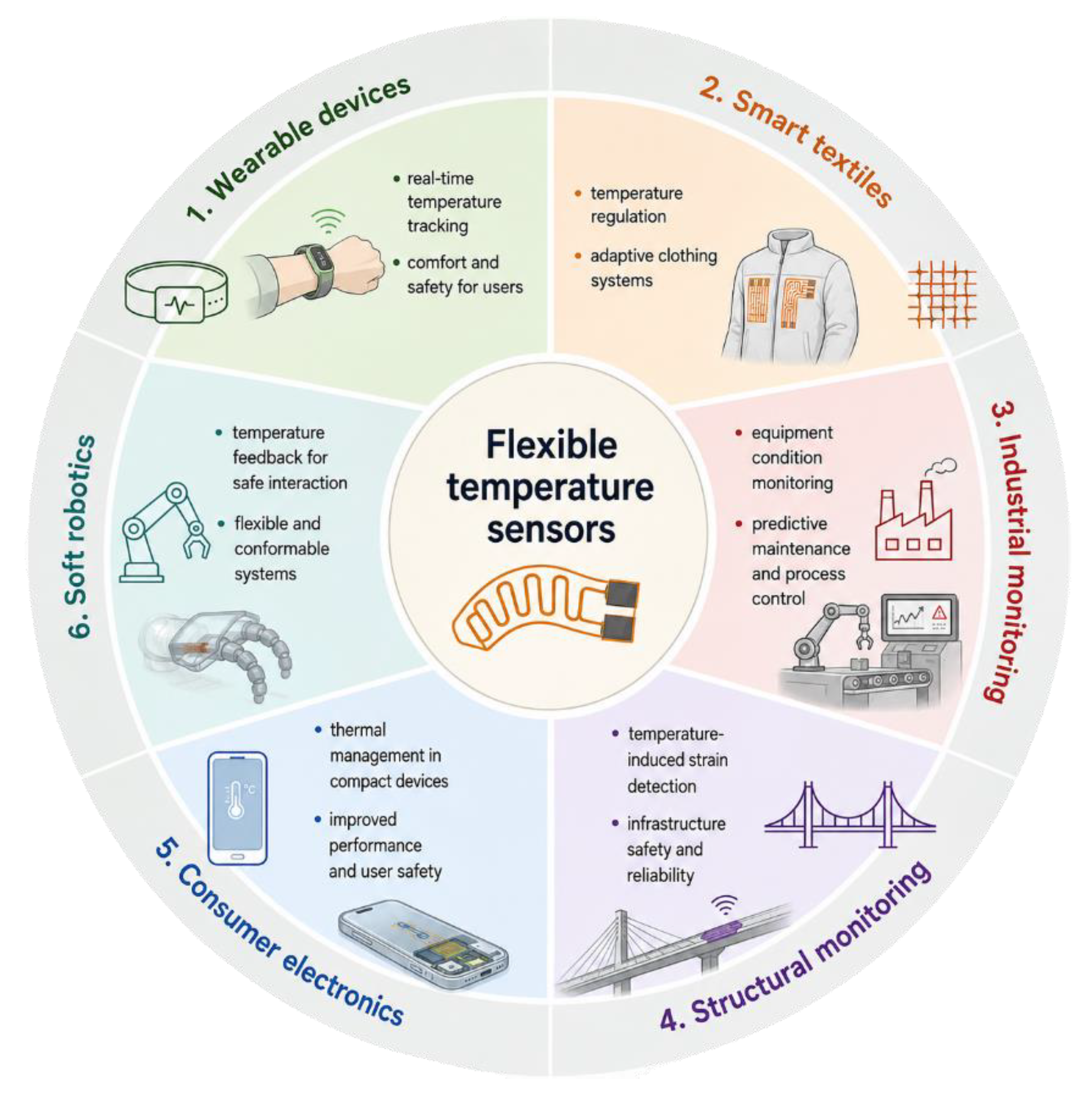
Figure 3.
The structure of pristine CNOs.

Figure 4.
Several functionalization routes of pristine CNOs for the synthesis of gas-sensing materials.
Figure 4.
Several functionalization routes of pristine CNOs for the synthesis of gas-sensing materials.
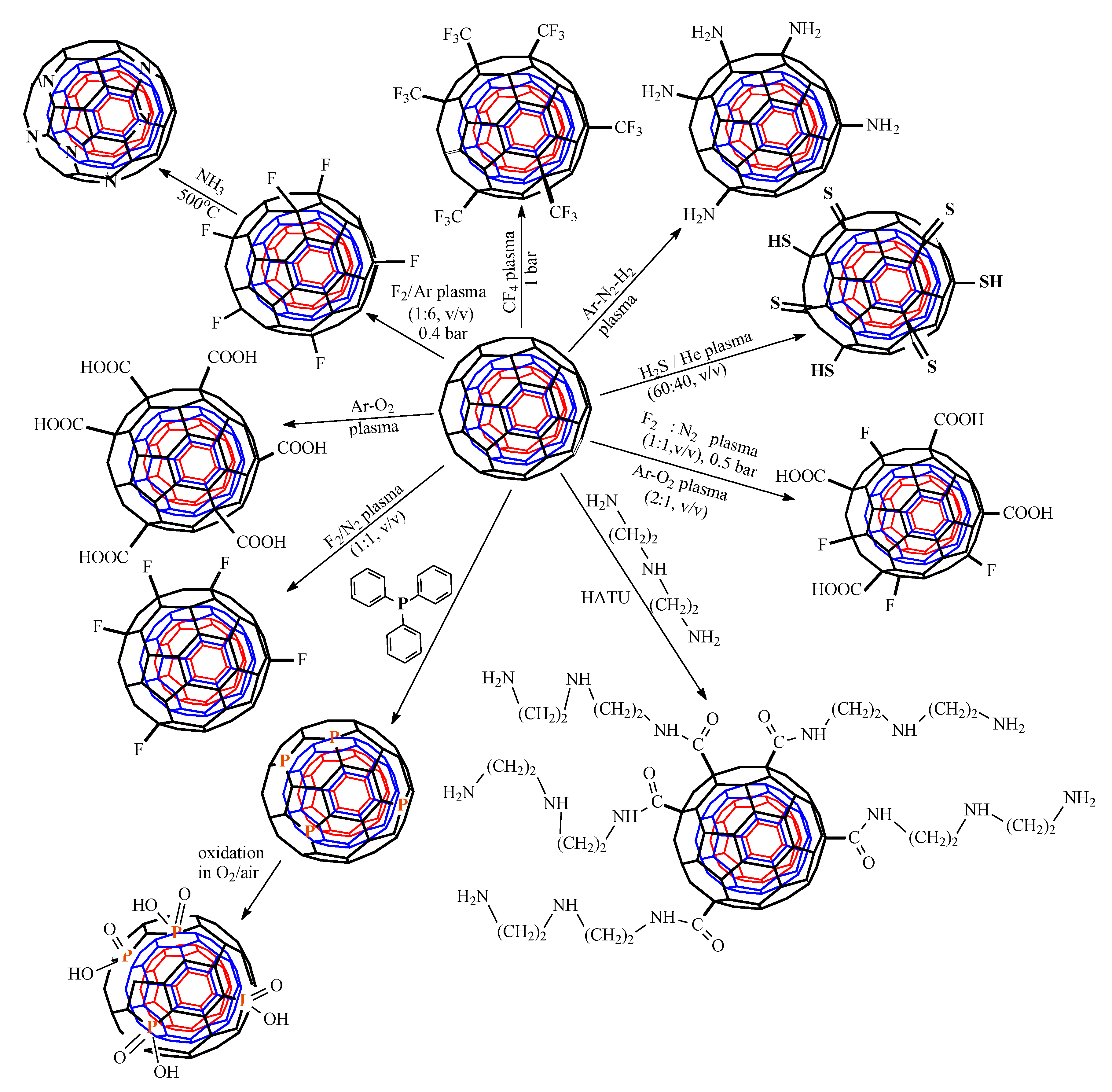
Figure 5.
The metal stripes of the IDT in an RH-resistive sensing structure employing a CNOs– PVOH nanocomposite as a sensing layer; reprinted from Ref. [110], licensed under CC BY4.0.
Figure 5.
The metal stripes of the IDT in an RH-resistive sensing structure employing a CNOs– PVOH nanocomposite as a sensing layer; reprinted from Ref. [110], licensed under CC BY4.0.
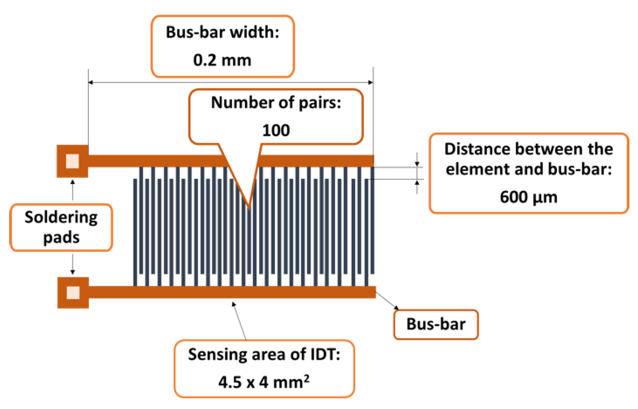
Figure 6.
The structure of the SAW delay-line sensor.
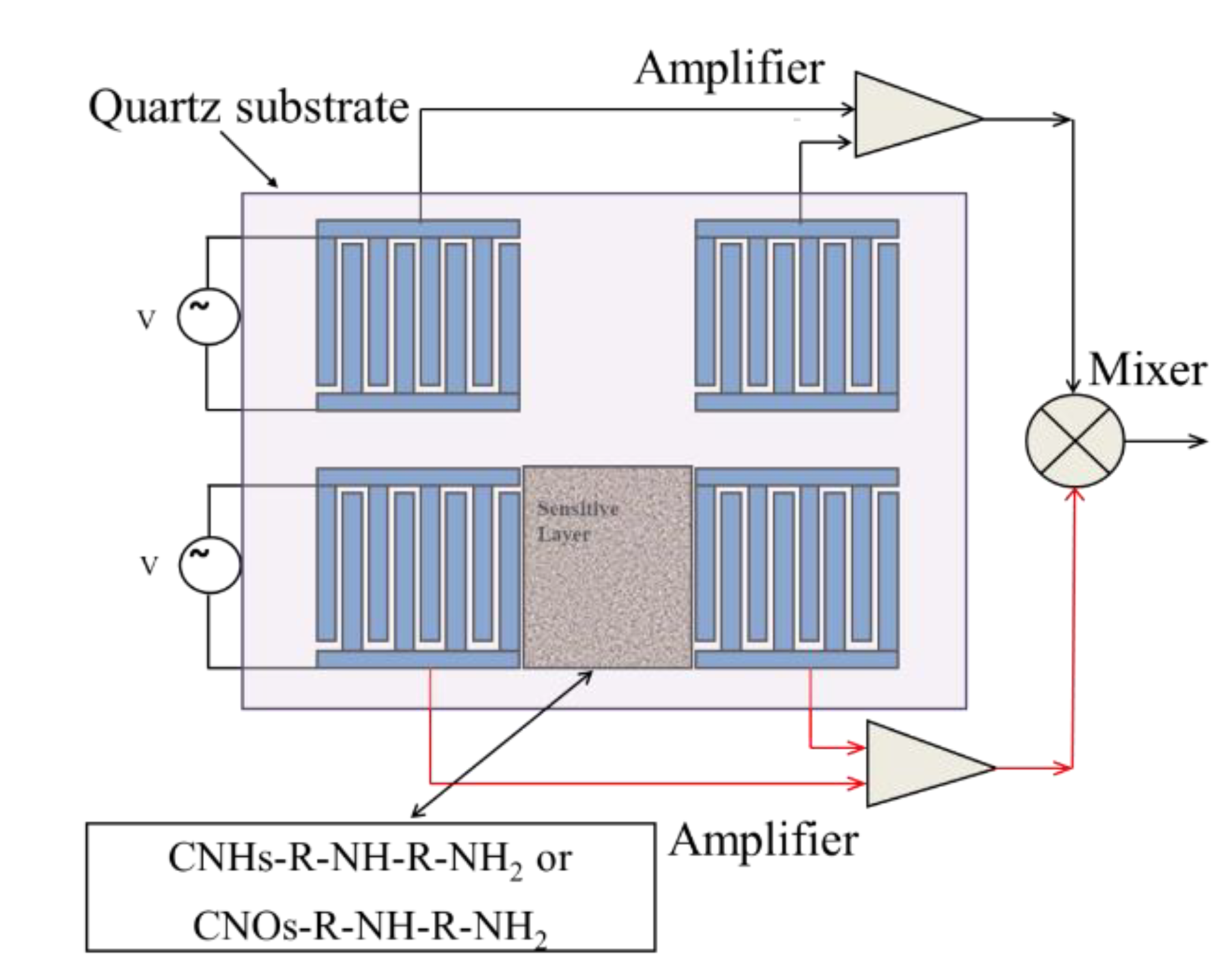
Figure 7.
SEM of CNOs/PVA layer, 1/1 w/w mass ratio, at: (a) x 7,000, (b) x 3,000,.
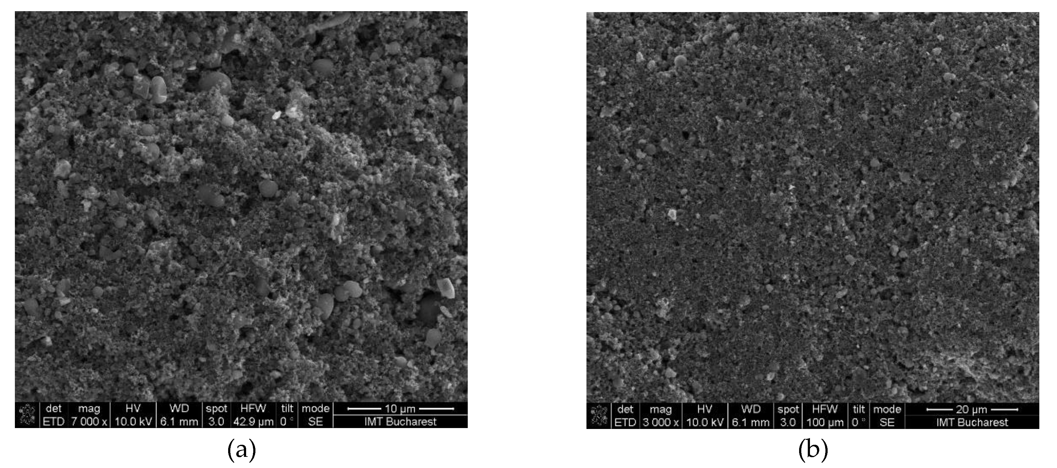
Figure 8.
Raman spectra of a solid-state film of CNOs/PVA (1/1 w/w mass ratio) deposited on the Si substrate. Reprinted from Ref. [110], licensed under CC BY4.0.
Figure 8.
Raman spectra of a solid-state film of CNOs/PVA (1/1 w/w mass ratio) deposited on the Si substrate. Reprinted from Ref. [110], licensed under CC BY4.0.
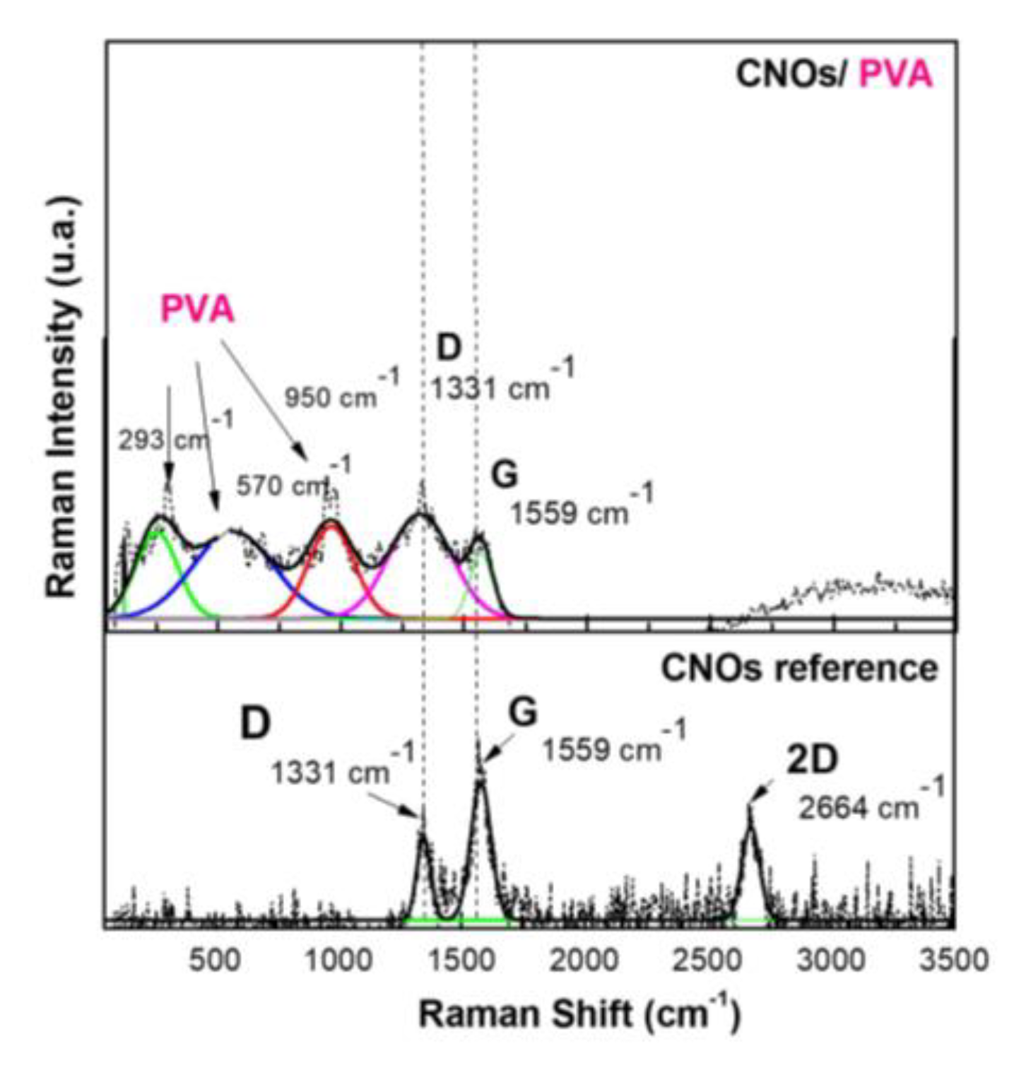
Figure 9.
AFM images of the CNOs/PVA RH sensing layer (1/1 w/w ratio) substrate. Reprinted from Ref. [110], licensed under CC BY4.0.
Figure 9.
AFM images of the CNOs/PVA RH sensing layer (1/1 w/w ratio) substrate. Reprinted from Ref. [110], licensed under CC BY4.0.
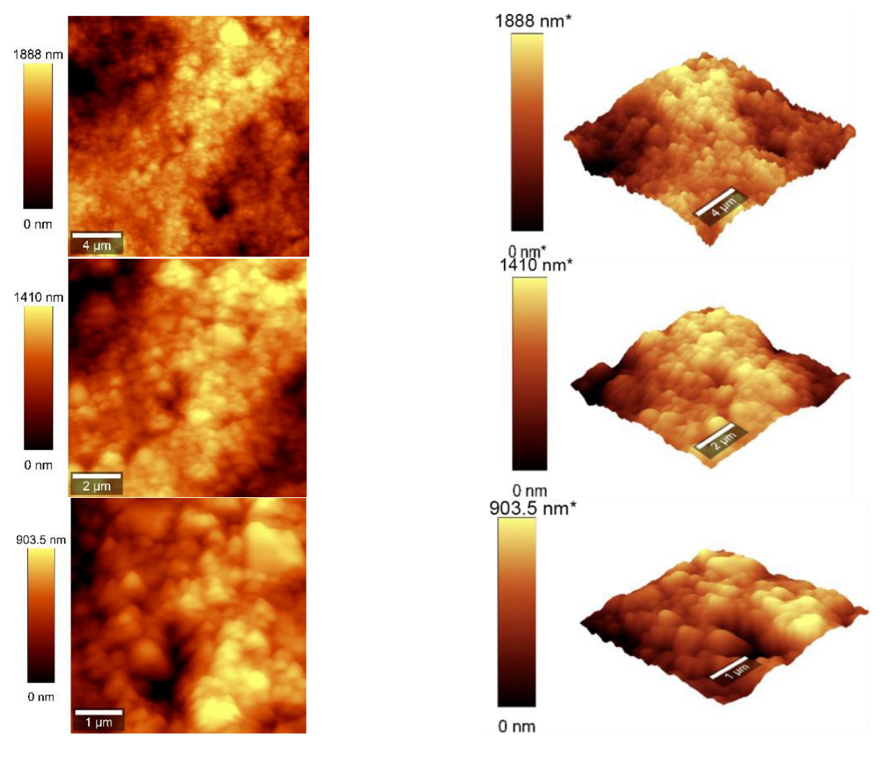
Figure 10.
RH and resistance variation vs. time for two types of CNOs/PVA sensing layers: (a) 1:1 (w/w), and (b) 2:1 (w/w). Reprinted from Ref. [110], licensed under CC BY4.0.
Figure 10.
RH and resistance variation vs. time for two types of CNOs/PVA sensing layers: (a) 1:1 (w/w), and (b) 2:1 (w/w). Reprinted from Ref. [110], licensed under CC BY4.0.
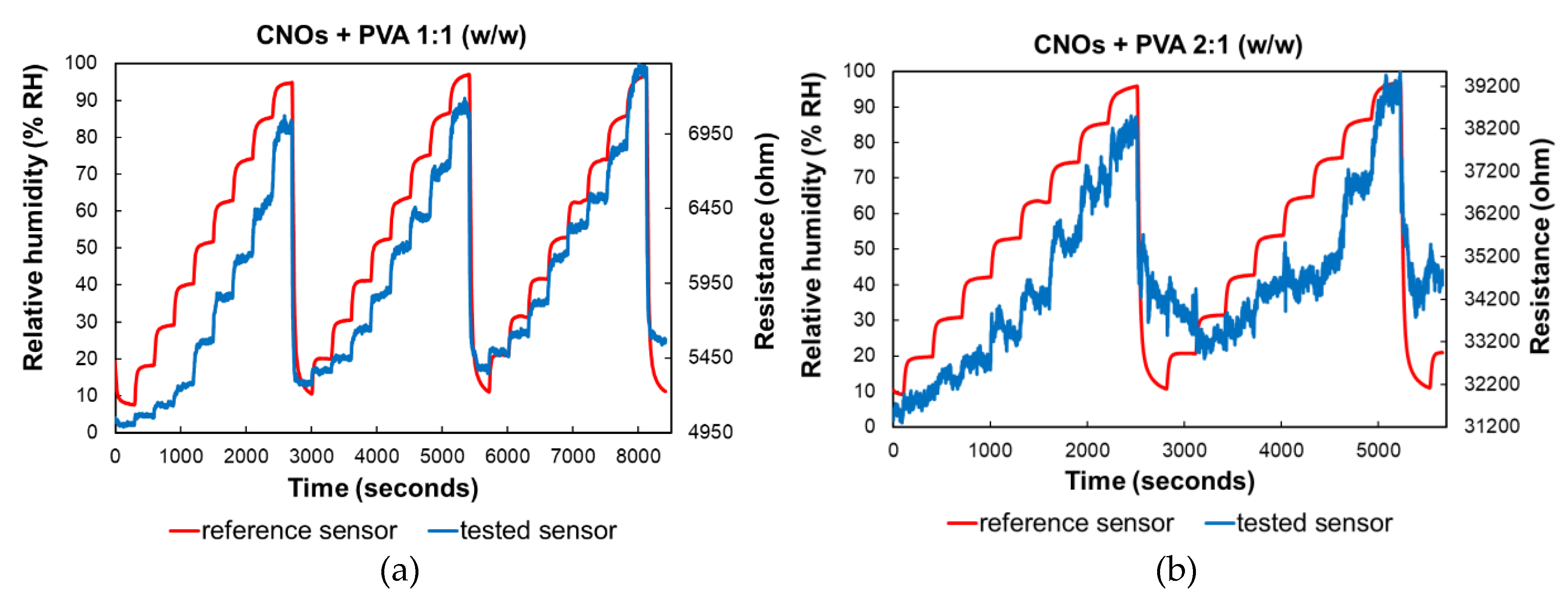
Figure 11.
– RH variation in repeated sorption-desorption cycles of three identically manufactured sensors employing as sensing layer: a) CNOs/PVA 1:1 (w/w), and b) CNOs/PVA 2:1 (w/w). Reprinted from Ref. [110], licensed under CC BY4.0.
Figure 11.
– RH variation in repeated sorption-desorption cycles of three identically manufactured sensors employing as sensing layer: a) CNOs/PVA 1:1 (w/w), and b) CNOs/PVA 2:1 (w/w). Reprinted from Ref. [110], licensed under CC BY4.0.
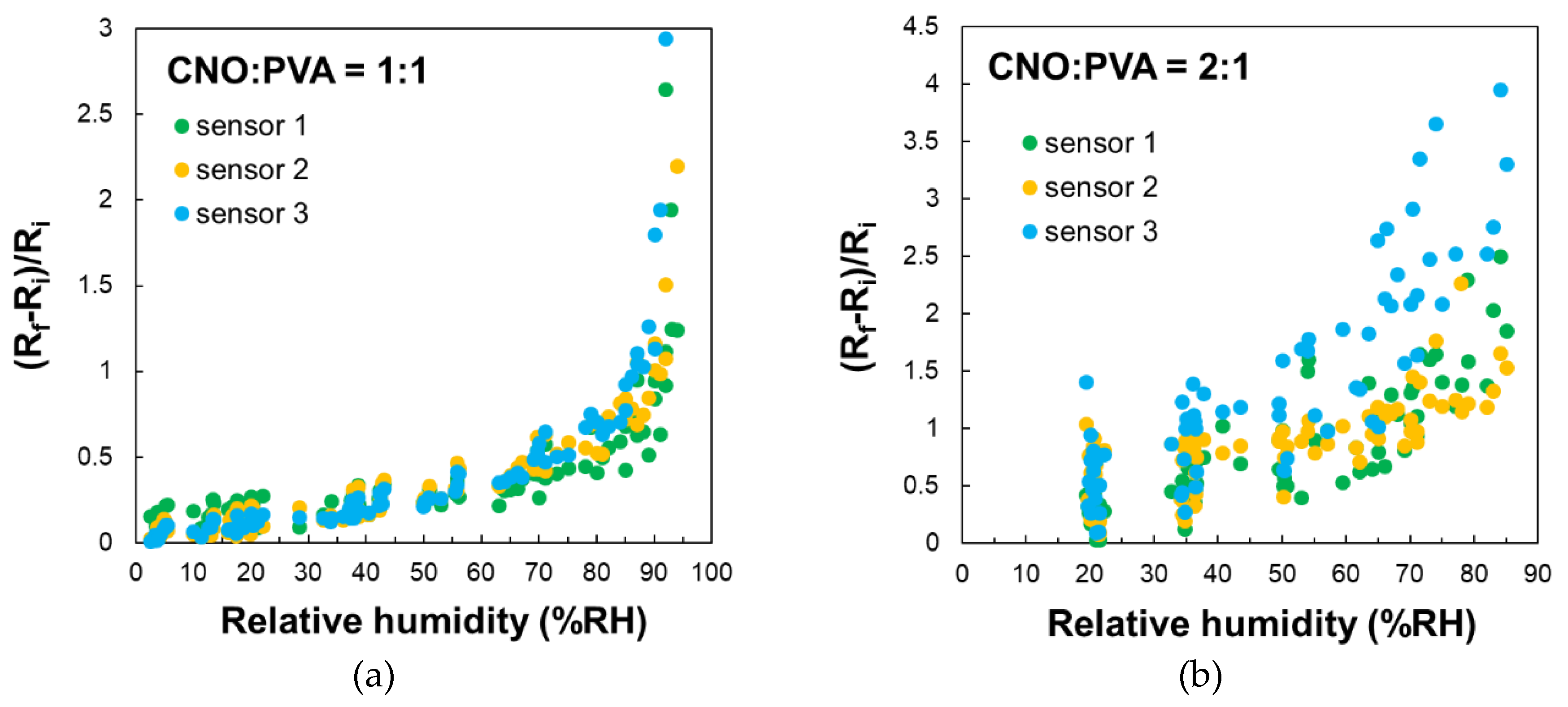
Figure 12.
Response time and relative resistance for each RH variation step: a) calculated parameters for a single RH variation step, b) ΔR/ΔRH values (red bars), and c) response time (green bars), at 1:1 (w/w) ratio. Reprinted from Ref. [110], licensed under CC BY4.0.
Figure 12.
Response time and relative resistance for each RH variation step: a) calculated parameters for a single RH variation step, b) ΔR/ΔRH values (red bars), and c) response time (green bars), at 1:1 (w/w) ratio. Reprinted from Ref. [110], licensed under CC BY4.0.
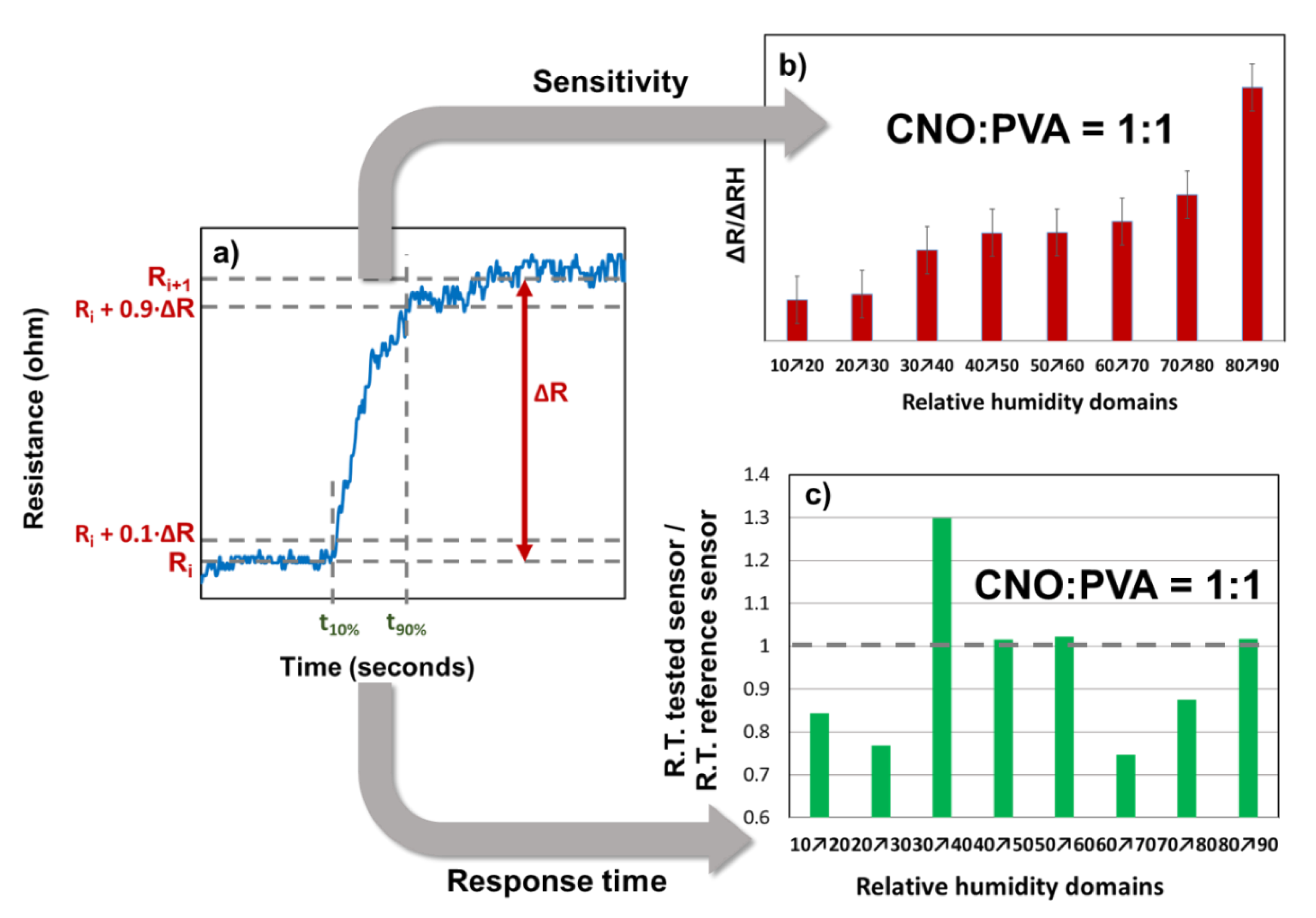
Figure 13.
Relative variation of the resistance (Rf-Ri)/Ri as a function of RH for MS employing as sensing layer: (a) CNOs/PVA at 1:1 (w/w), and (b) CNOs/PVA at 1:1 (w/w) (sorption – red dots, desorption – blue dots). (w/w Reprinted from Ref. [110], licensed under CC BY4.0.
Figure 13.
Relative variation of the resistance (Rf-Ri)/Ri as a function of RH for MS employing as sensing layer: (a) CNOs/PVA at 1:1 (w/w), and (b) CNOs/PVA at 1:1 (w/w) (sorption – red dots, desorption – blue dots). (w/w Reprinted from Ref. [110], licensed under CC BY4.0.
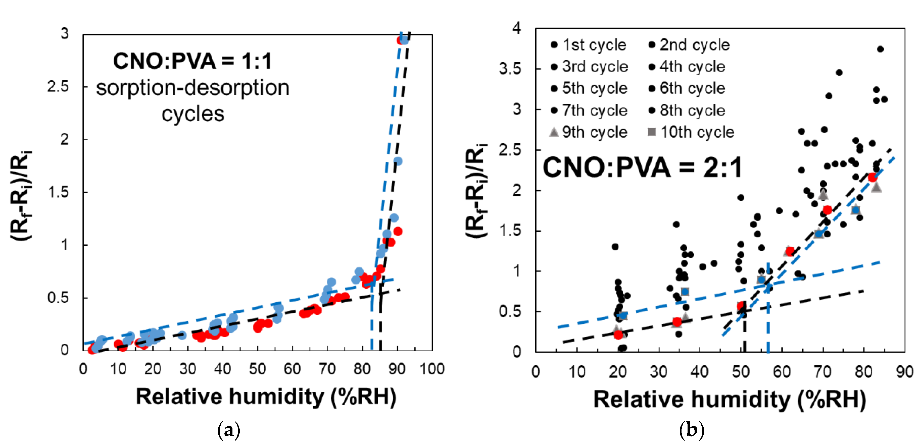
Figure 14.
SEM of CNOs/PVP layer, 1/1 (w/w) mass ratio, at: (a) x 200,000, (b) x 100,000.
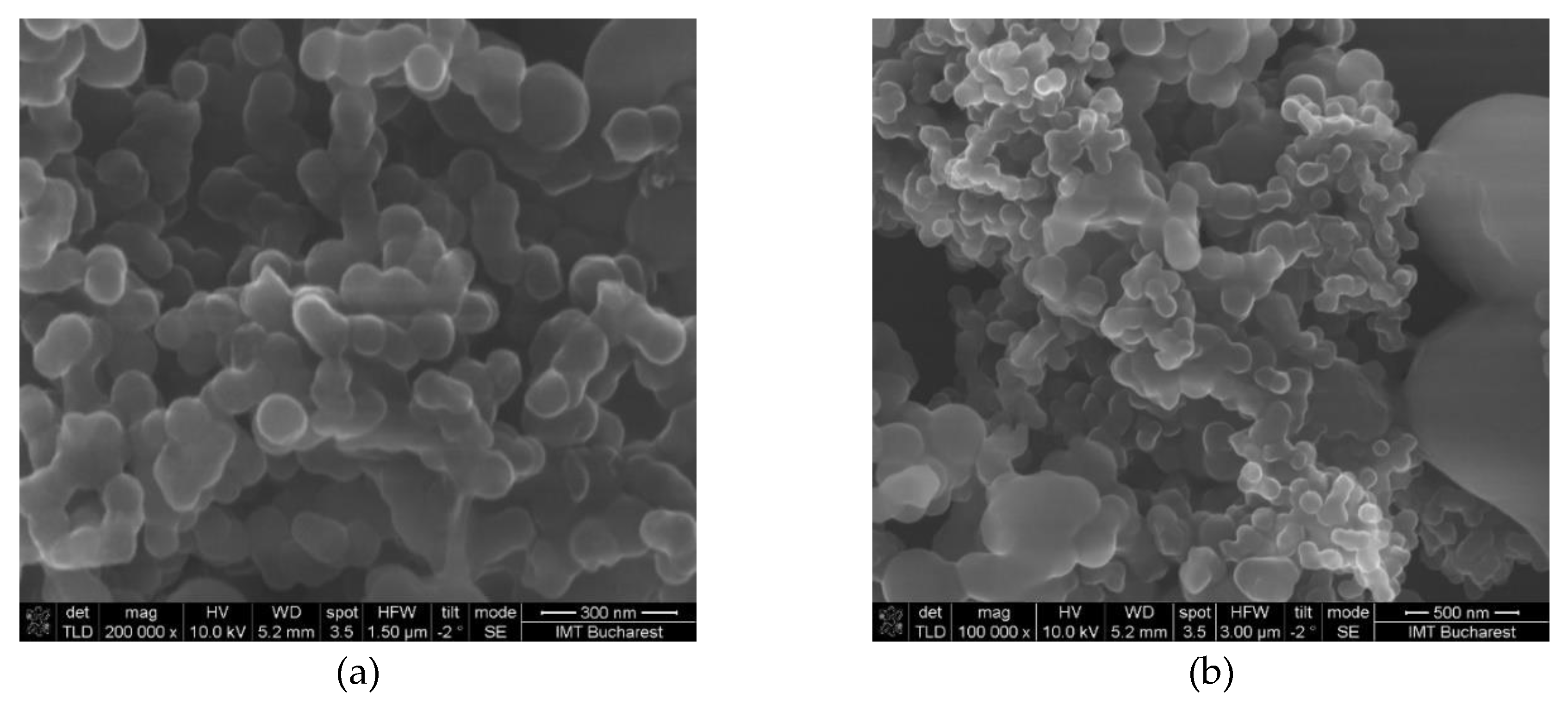
Figure 15.
Electrical resistance versus RH for the sensor under testing (SUT), for the 1st operating cycle (adsorption in red, desorption in grey). Reproduced with permission from ROMJIST, Ref. [111].
Figure 15.
Electrical resistance versus RH for the sensor under testing (SUT), for the 1st operating cycle (adsorption in red, desorption in grey). Reproduced with permission from ROMJIST, Ref. [111].
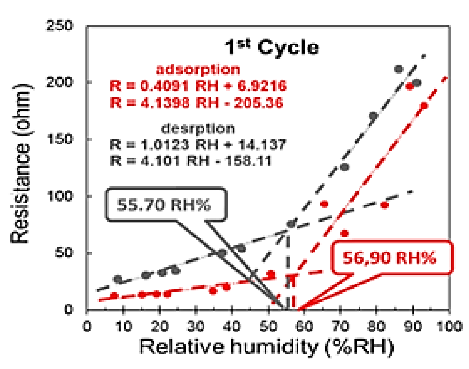
Figure 16.
Electrical resistance versus RH for SUT, for the 2nd operating cycle (adsorption in red, desorption in grey. Reproduced with permission from ROMJIST, Ref. [111].
Figure 16.
Electrical resistance versus RH for SUT, for the 2nd operating cycle (adsorption in red, desorption in grey. Reproduced with permission from ROMJIST, Ref. [111].
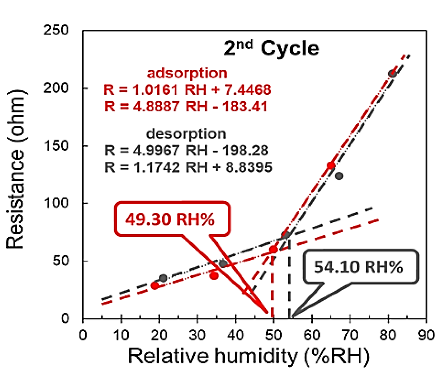
Figure 17.
Electrical resistance versus RH for SUT , for the 3rd operating cycle (adsorption in red, desorption in grey). Reproduced with permission from ROMJIST, Ref. [111].
Figure 17.
Electrical resistance versus RH for SUT , for the 3rd operating cycle (adsorption in red, desorption in grey). Reproduced with permission from ROMJIST, Ref. [111].
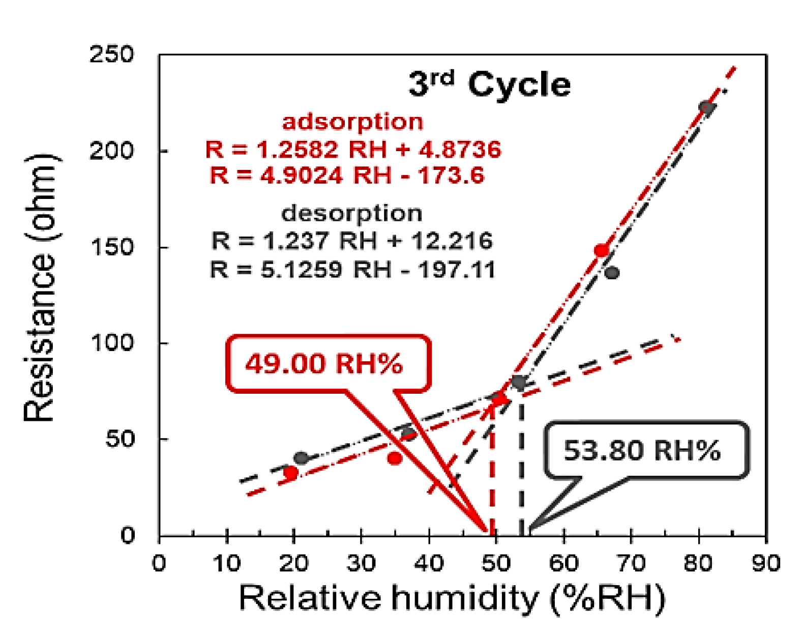
Figure 18.
SEM of CNOs/PVP layer, 2/1 (w/w) ratio, at: (a) x 50,000, (b) x 100,000,.
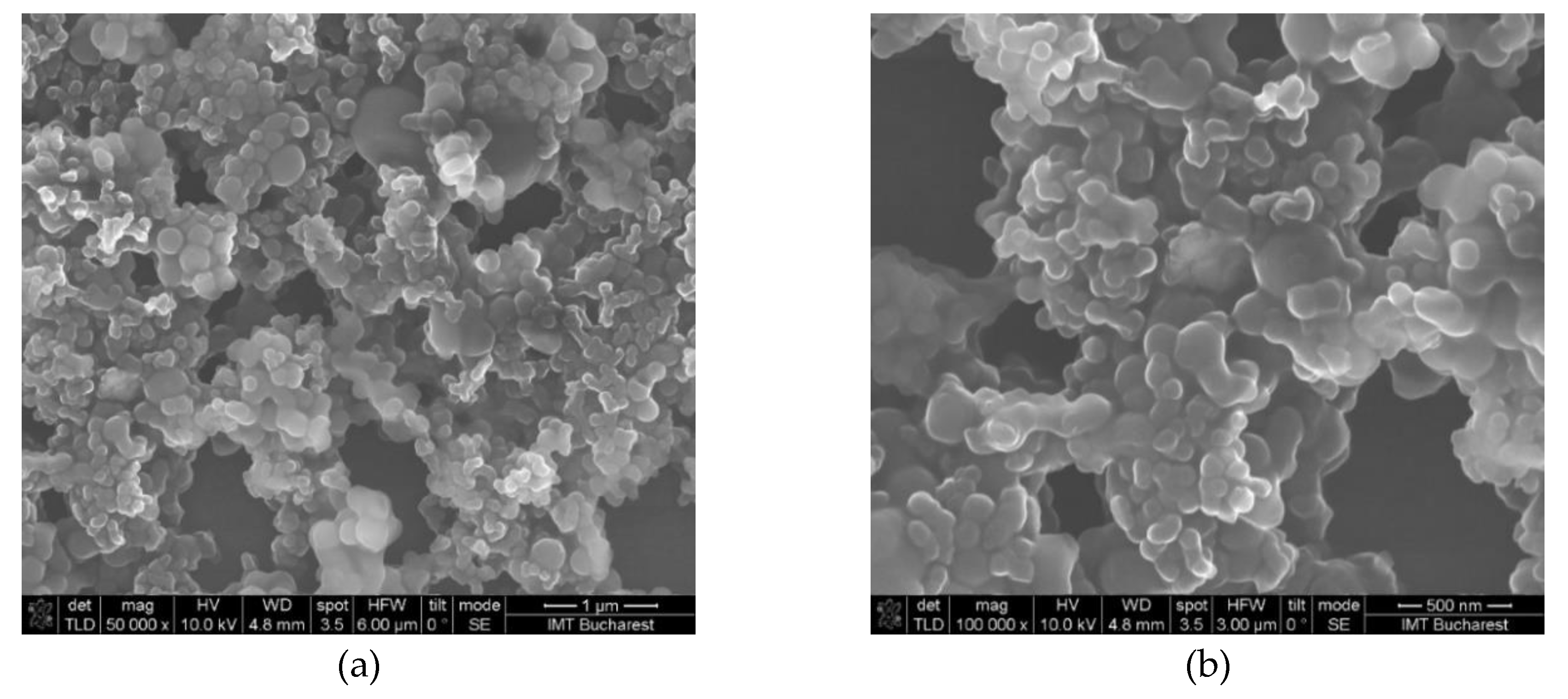
Figure 19.
Two-cycle resistive response of the CNO/PVP (2:1) sensor to ethanol vapor.
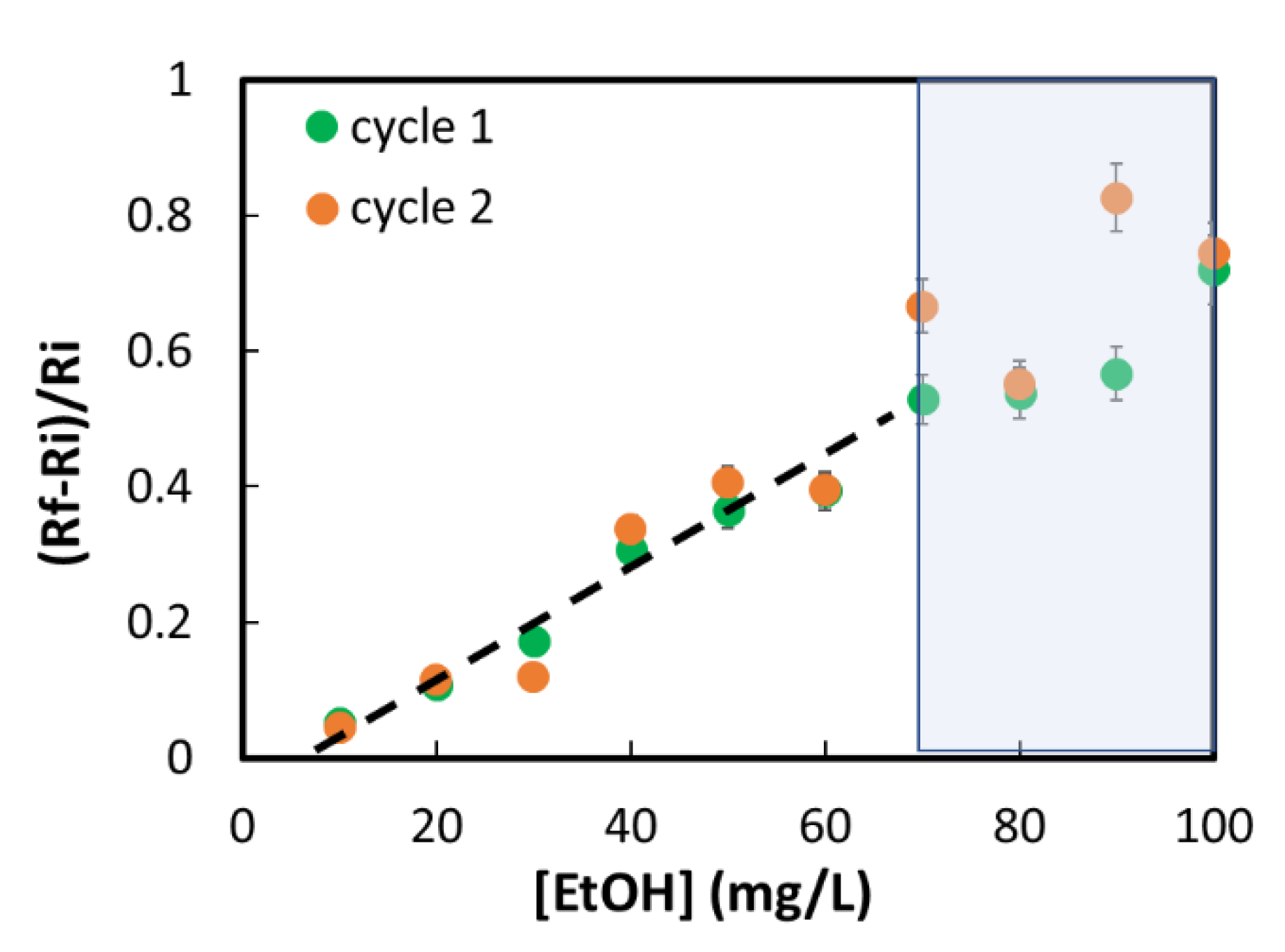
Figure 20.
Two-cycle resistive response of the CNO/PVP (2:1) sensor to isopropanol vapor.
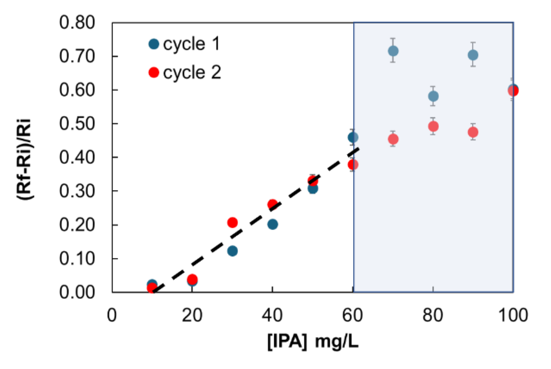
Figure 21.
The swelling effect occurring in the PVA in contact with water molecules leading to the disruption of hydrogen bonds between hydroxyl groups of PVA and the percolating pathways of the CNOs.
Figure 21.
The swelling effect occurring in the PVA in contact with water molecules leading to the disruption of hydrogen bonds between hydroxyl groups of PVA and the percolating pathways of the CNOs.
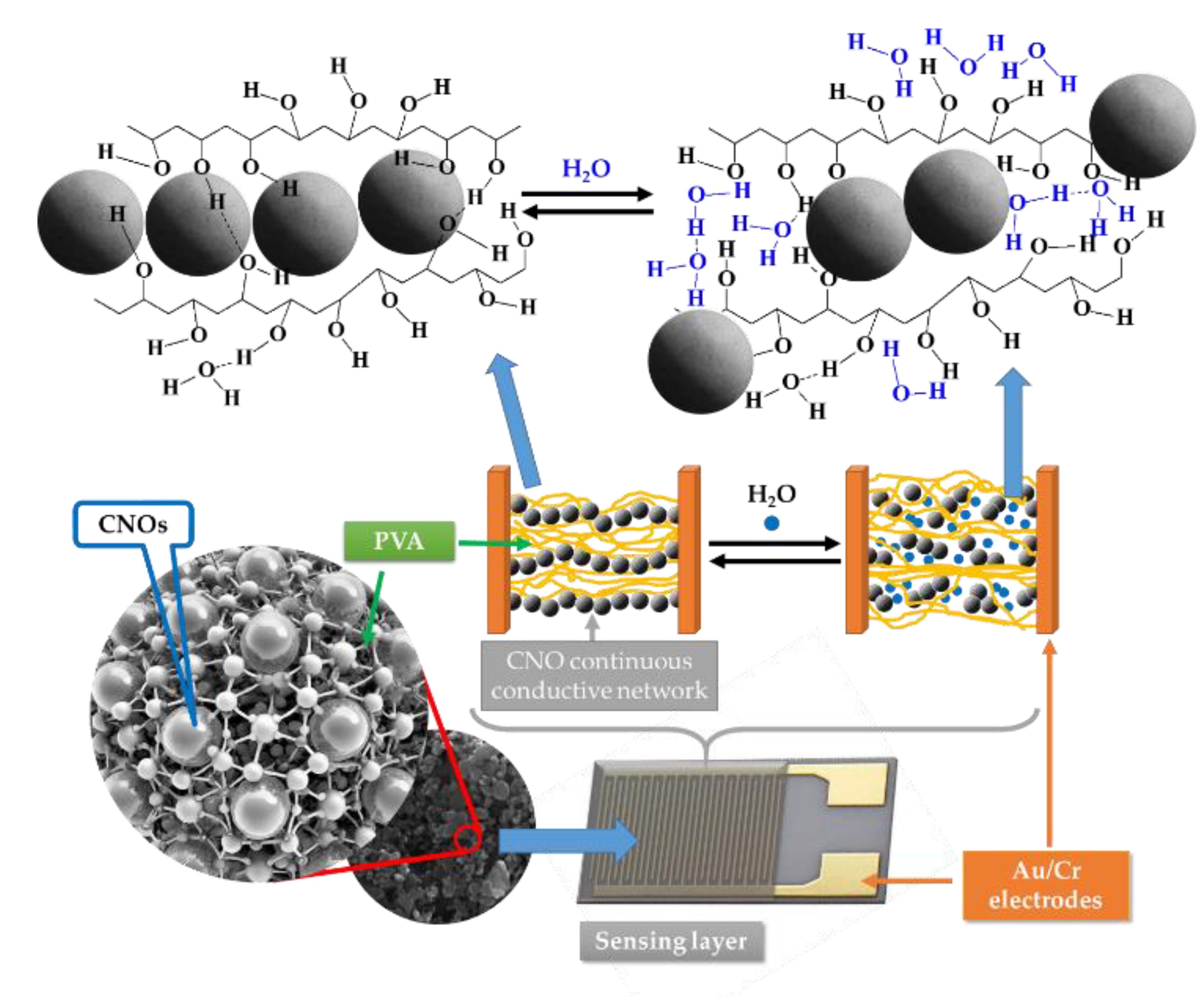
Figure 22.
Electrical resistance of CNOs/PVP composite films with different CNOs concentrations during: a) heating, and b) cooling.
Figure 22.
Electrical resistance of CNOs/PVP composite films with different CNOs concentrations during: a) heating, and b) cooling.
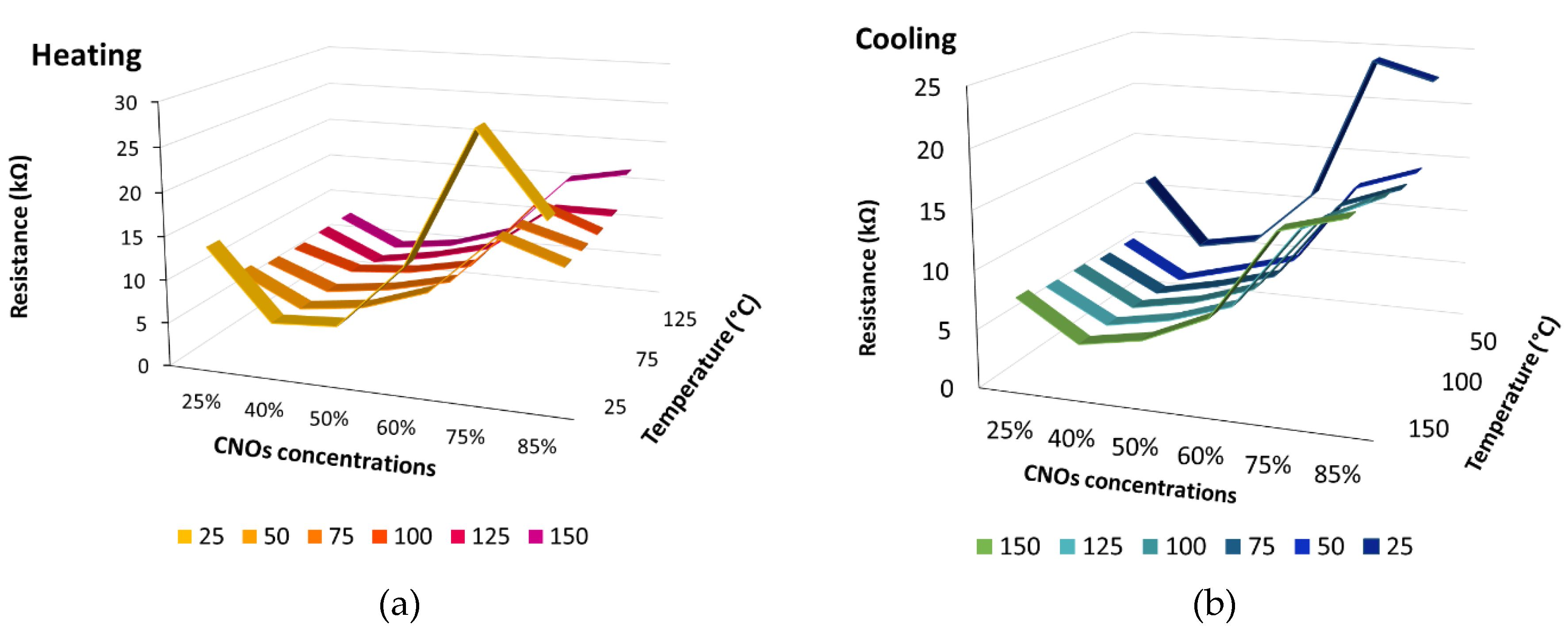
Figure 23.
Electrical resistance of CNOs/PVP composite films with different CNOs concentrations measured at selected temperatures during (a) heating from 25 to 150 °C and (b) cooling from 150 to 25 °C. Each data series corresponds to a fixed measurement temperature.
Figure 23.
Electrical resistance of CNOs/PVP composite films with different CNOs concentrations measured at selected temperatures during (a) heating from 25 to 150 °C and (b) cooling from 150 to 25 °C. Each data series corresponds to a fixed measurement temperature.
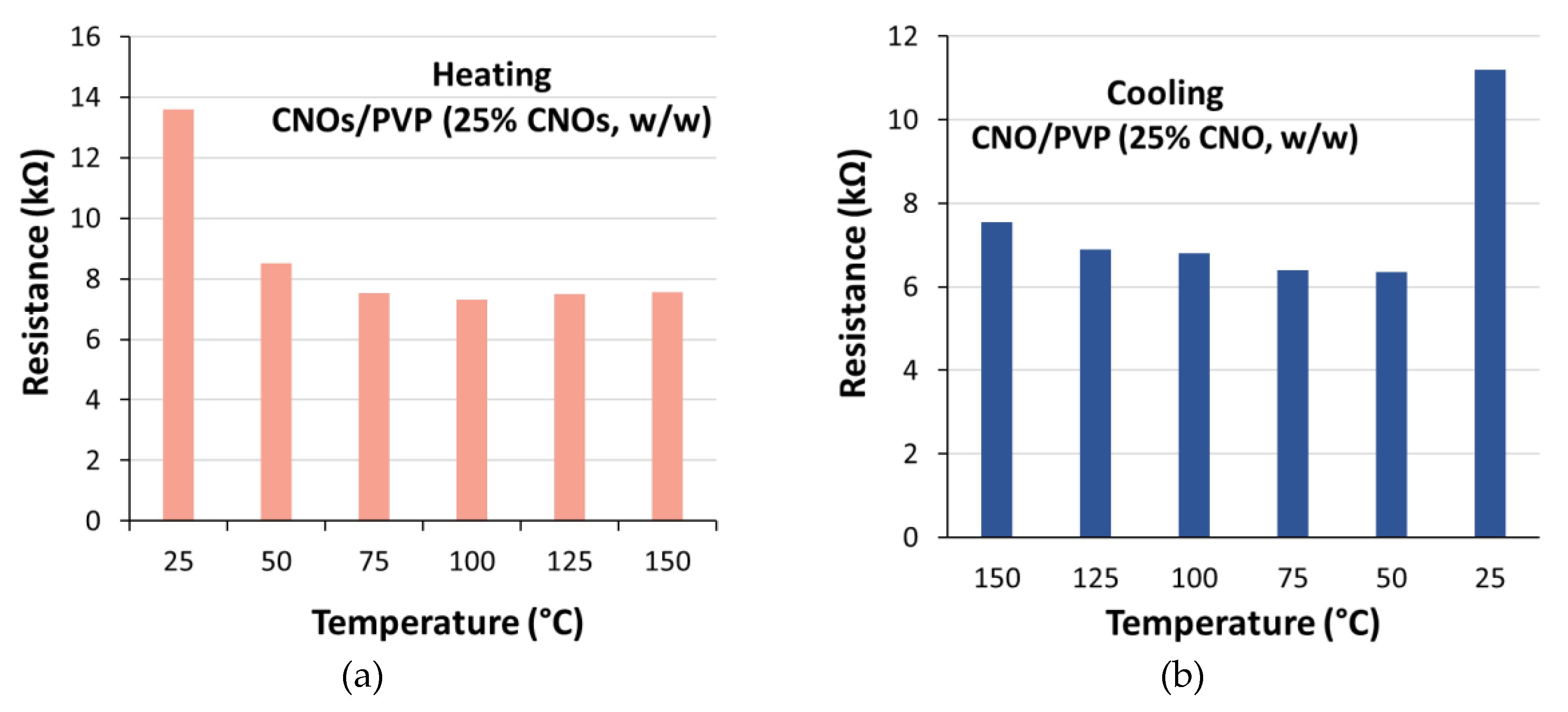
Disclaimer/Publisher’s Note: The statements, opinions and data contained in all publications are solely those of the individual author(s) and contributor(s) and not of MDPI and/or the editor(s). MDPI and/or the editor(s) disclaim responsibility for any injury to people or property resulting from any ideas, methods, instructions or products referred to in the content. |
© 2026 by the authors. Licensee MDPI, Basel, Switzerland. This article is an open access article distributed under the terms and conditions of the Creative Commons Attribution (CC BY) license.
Copyright: This open access article is published under a Creative Commons CC BY 4.0 license, which permit the free download, distribution, and reuse, provided that the author and preprint are cited in any reuse.