

Submitted:
29 July 2026
Posted:
30 July 2026
You are already at the latest version
Abstract
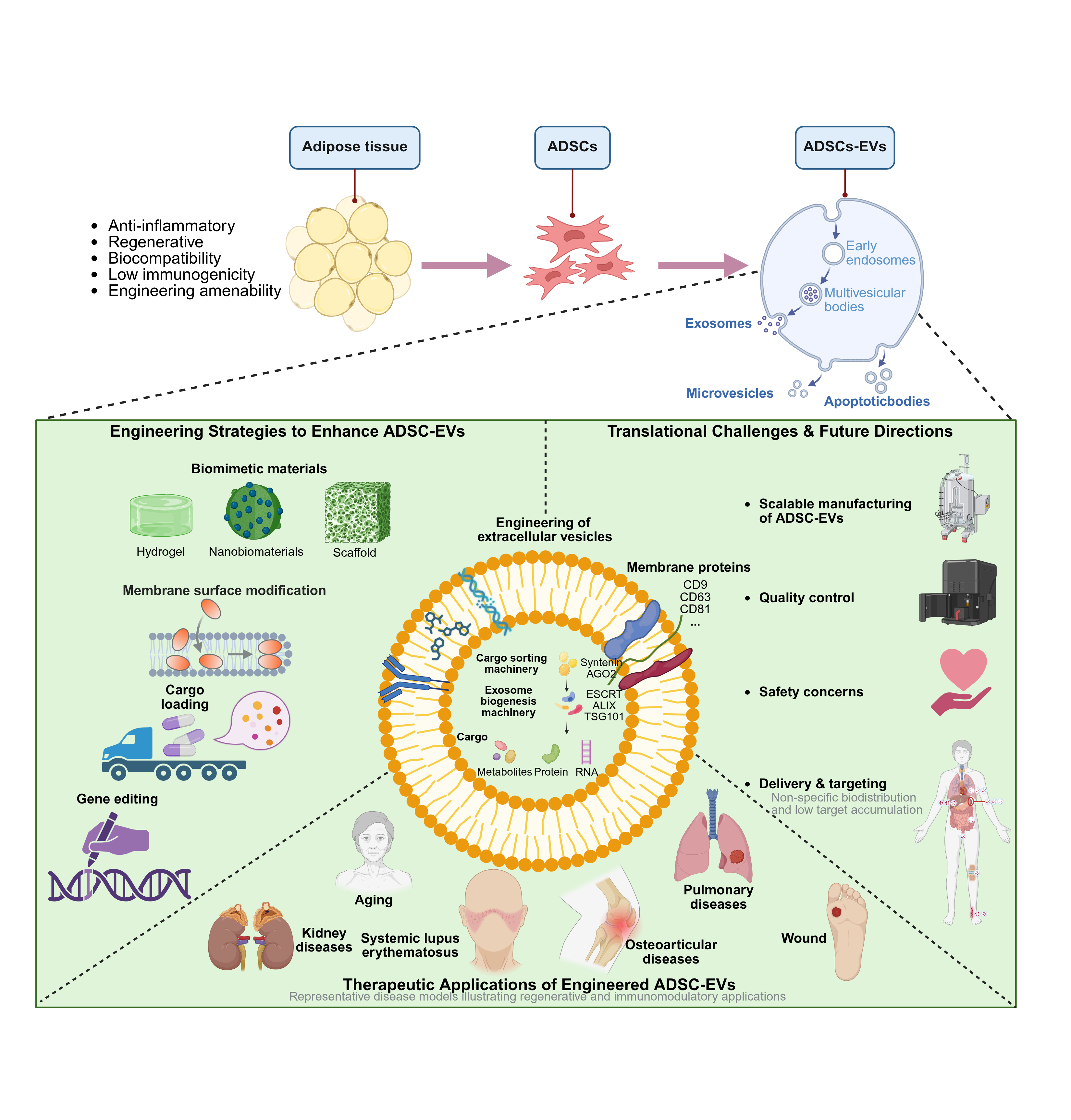
Adipose-derived stem cells (ADSCs) have garnered substantial attention within regenerative medicine due to their ready availability, ease of isolation, and potent immunomodulatory and tissue-repair capabilities. Growing evidence indicates that ADSC therapeutic efficacy stems predominantly from their secreted extracellular vesicles (EVs), rather than from direct cellular engraftment. However, clinical application of native ADSC-derived EVs (ADSC-EVs) is impeded by insufficient yield, inadequate targeting precision, and rapid systemic clearance. Recent advances in engineering technologies—including genetic modification, membrane functionalization, cargo encapsulation, and biomimetic integration—have provided innovative solutions to improve the production, stability, homing efficiency, and therapeutic potency of ADSC-EVs. In this review, we provide a systematic overview of the various engineered approach platforms for ADSC-EVs with a focus on parental cell reprogramming, cargo engineering, membrane protein engineering and hybrid or EV-mimetic platforms. We also report recent advances in engineered ADSC-EVs for multiple biomedical applications, including tissue regeneration, immunomodulation, anti-fibrotic therapies, cancer treatment and radiation-induced injury repair. We review key translational foibles surrounding the lack of standardization, reproducible production, quality assurance and safety. Lastly, we will provide perspectives on future directions to accelerate ADSC-EV clinical translation. Artificial smart cargo optimization, next-generation biomimetic targeting platforms and standardized GMP-compliant production systems offer the most promising prospects to overcome current translational bottlenecks.
Keywords:
adipose-derived stem cells
; engineered extracellular vesicles
; bioengineering
; drug delivery
; clinical translation
1. Introduction
Adipose-derived stem cells (ADSCs), a type of mesodermal origin mesenchymal stem cells (MSCs), are abundant, harvested in a minimally invasive manner with high proliferation capacity and low immunogenicity. They are currently recognized as having significant potential in regenerative medicine and immunomodulatory applications. ADSCs provide greater clinical availability and scalability than other tissue-derived sources of MSCs such as bone marrow, making them one of the most translationally exploitable stem cell sources [1].
Increasing evidence supports that the regenerative properties of ADSCs are due to paracrine rather than lineage differentiation mechanisms. ADSC-secreted extracellular vesicles contain a heterogeneous mixture of bioactive cargo: proteins, lipids and nucleic acids. EVs mediate intercellular communication through the transfer of functional molecules to suppress inflammation, promote tissue repair, and remodel injured microenvironments [2]. EVs as a cell-free therapeutic modality have unique advantages over the conventional cell-based approach with regard to safety, storage stability, immunocompatibility and quality controllability [3]. However, native EVs face many challenges during clinical translation, such as having limited drug-loading capacity [4], poor targeting efficiency [5], insufficient in vivo stability and significant batch-to-batch variability in bioactivity. In addition, the industrialization of these technologies is hampered by the lack of effective frameworks for large-scale production, quality control and standardization [6]. Thus, functional engineering of EVs is an important pathway for facilitating the efficacy of effective drug delivery to clinically relevant tissues via physical, chemical and bioengineering approaches.Engineered ADSC-EVs have advanced from fundamental mechanistic investigations to validation across diverse disease models, with preliminary clinical exploration emerging in select therapeutic areas. This review provides an overview of engineering strategies, functional optimization, and translational challenges, with an emphasis on EV construction, underlying mechanisms, biomedical applications, and critical bottlenecks in clinical translation.
Distinguished from prior reviews that broadly survey MSC-derived EVs or focus on individual engineering techniques, this review delivers a source-specific and systematically integrated analysis of ADSC-EV engineering strategies. We (i) provide a comprehensive mapping of the engineering toolbox, spanning from parental cell reprogramming to post-isolation modification and scalable bioreactor production; (ii) critically evaluate the strengths, limitations, and translational viability of each approach, thereby bridging the divide between experimental validation and large-scale industrial production; (iii) present an updated clinical landscape of ADSC-EV trials with critical assessment of translational bottlenecks; and (iv) identify key CMC and regulatory hurdles that must be resolved to propel engineered ADSC-EVs from bench to bedside.
2. Biological Properties of ADSC-EVs
2.1. EVs Classification and Functional Roles
Extracellular vesicles (EVs) are nanometer-sized, membrane-bound structures encased in a phospholipid bilayer, released by virtually all cell types and abundantly present in various body fluids. They are composed of proteins, lipids, nucleic acids and metabolites encapsulated in a membrane with high structural stability. EVs transfer bioactive cargos to recipient cells via membrane fusion or receptor-mediated endocytosis, thus actively integrating into a wide variety of physiological and pathological processes.
Extracellular vesicles are conventionally classified into three subtypes based on size, biogenetic origin, and molecular profiles, i.e., exosomes (Exo 30–150 nm) generated through the endocytosis–multivesicular body (MVB) pathway where cargo loading is regulated by ESCRT proteins such as TSG101 and Alix; Rab GTPases; and Ca²⁺ signaling with enrichment in tetraspanin markers (CD9, CD63/CD81); microvesicles (MVs 100–1000 nm), arising via direct plasma membrane budding; and apoptotic bodies (ABs 500–2000 nm), which detach from apoptotic cells [7,8]. Yet, because of the marked similarity in traits among these subtypes, the International Society for Extracellular Vesicles recommends using the umbrella term “EVs.”
EVs play unique roles in intercellular signaling during tissue regeneration and the inflammatory process. This is done by promoting angiogenesis, inhibiting apoptotic pathways, and overall improving organ performance. MSC-derived EVs are the most reported population, while ADSC-EVs have gained considerable attention as they can be easily isolated in large quantities [9,10]. ADSC-EVs are rich in proteins and nucleic acids that promote angiogenesis, modulate immune responses, and facilitate extracellular matrix remodeling [11,12].
Figure 1.
Biogenesis and secretion pathways of ADSC-EVs.
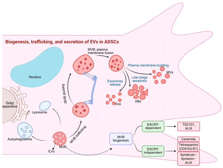
ADSC-EVs are produced through different pathways. ILVs, on the other hand, are generated by inward budding of the endosomal membrane and have been shown to be regulated by various players including the ESCRT machinery (e.g., TSG101), the Syndecan–Syntenin–ALIX axis, ceramide-dependent pathways, and tetraspanins (CD9, CD63, CD81). These MVBs are then trafficked to the plasma membrane and undergo docking and fusion, releasing ILVs as Exosome (Exo), with a subset also directed towards autophagy–lysosome-mediated degradation. Moreover, MVs are released as a result of plasma membrane budding and ABs are secreted late in the apoptotic process.
2.2. Biological Properties of ADSC-EVs
ADSC-EVs, which possess favorable biological characteristics yet have therapeutic potential. The proteomic analyses revealed high concentrations of growth factors in ADSC-EVs such as VEGF, HGF, and IGF-1 along with anti-inflammatory cytokines and regulatory miRNAs that promote angiogenesis, inhibit local inflammatory responses and assist in remodeling the extracellular matrix [13,14]. Interestingly, due to the favorable secretory profile of their parental cells, compared with EVs derived from other progenitor sources such as MSCs, ADSC-EVs display superior proliferation signals and immunoregulatory potential [15]. In addition, ADSC-EVs possess excellent antioxidant abilities by enhancing the Nrf2 pathway and decreasing oxidative stress in recipient cells [16]. With these properties along with their scalability and stable bioactivity from batch-to-batch, ADSC-EVs are an ideal candidate for a cell-free therapeutic platform [17].
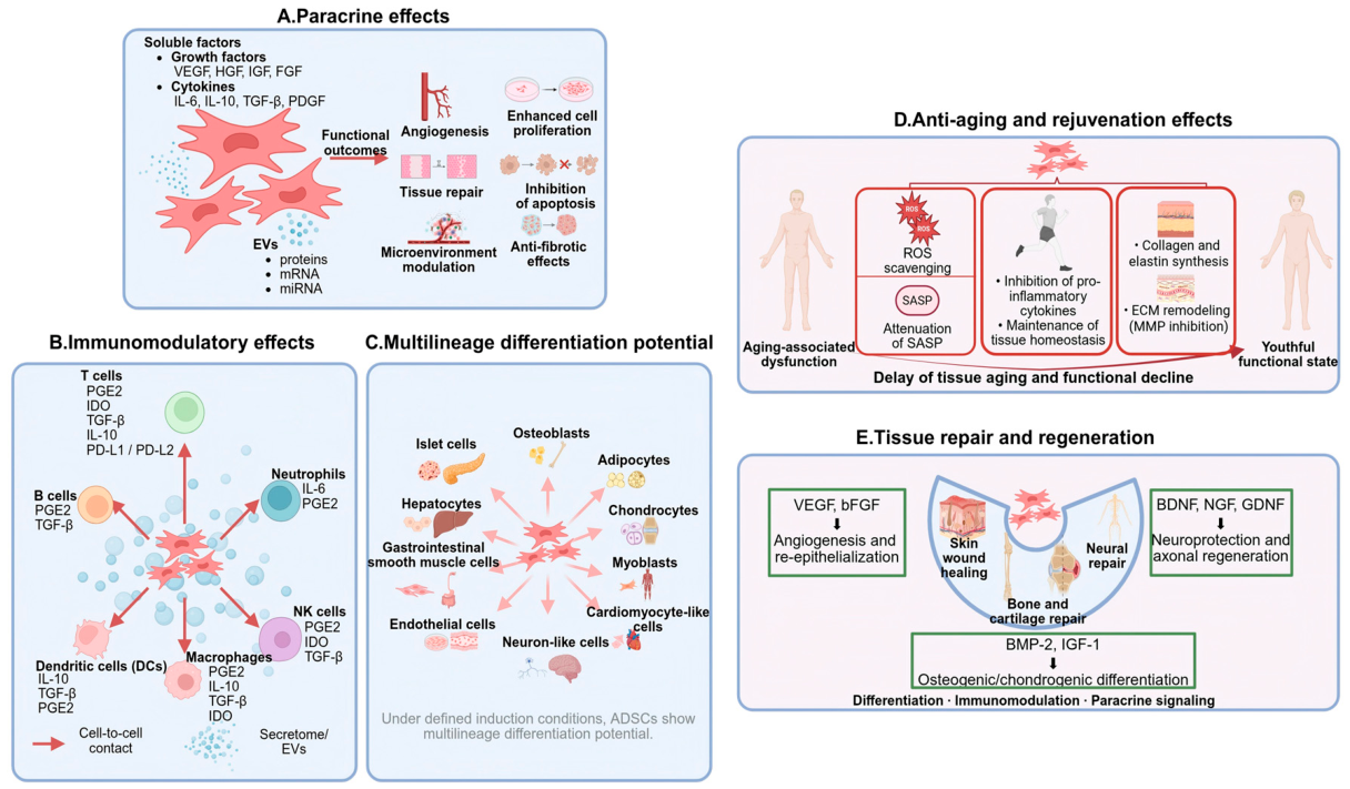
Figure 2.
Therapeutic biological properties of ADSCs. A. Paracrine Activity: The Therapeutic Effect of ADSCs is due to the secretome containing growth factors, cytokines, and EVs (containing proteins/RNA) B. Immunomodulation: ADSCs modulate immunity through mediators (PGE2, IDO, TGF-β and IL-10) affecting immune cells of various origins. C. Differentiation into multilineages: ADSCs differentiate into multiple cells during the induction phase. d. Anti-aging: ROS, inflammation, SASP and ECM remodeling by ADSCs E. Tissue Repair/Regeneration, which is the ability of ADSCs to promote angiogenesis neuroprotection; bone/cartilage formation.
Figure 2.
Therapeutic biological properties of ADSCs. A. Paracrine Activity: The Therapeutic Effect of ADSCs is due to the secretome containing growth factors, cytokines, and EVs (containing proteins/RNA) B. Immunomodulation: ADSCs modulate immunity through mediators (PGE2, IDO, TGF-β and IL-10) affecting immune cells of various origins. C. Differentiation into multilineages: ADSCs differentiate into multiple cells during the induction phase. d. Anti-aging: ROS, inflammation, SASP and ECM remodeling by ADSCs E. Tissue Repair/Regeneration, which is the ability of ADSCs to promote angiogenesis neuroprotection; bone/cartilage formation.
Although they are used to use a wide range of therapeutics, native ADSC-EVs confront with various translational barriers, such as low drug-loading efficiency and targeting specificity in addition to mainly rapid clearance inside the body which automatically blocking its therapeutic index [18]. Moreover, the heterogeneity in EV preparation results into batches with varied bioactivity [19]. This requires engineering strategies to improve ADSC–EV function.
2.3. Translational Barriers of Native ADSC-EVs
In order to overcome these limitations various engineering strategies have been employed for the properties of the ADSC-EVs to be optimized. Broadly speaking, these methods fall within two paradigms: cell engineering of parental source cells to modify the cargo and surface composition of secreted EVs; or post-isolation modifications which involve direct physical, chemical, or biological manipulation of purified EVs. [20] Parental cell engineering utilizes natural biogenesis pathways that enable efficient cargo loading and stable surface display of EVs but offers limited control over EV composition. In comparison, post-isolation strategies allow more specific functionalization options, but often at the cost of vesicle integrity and scalability [21]. Emerging bioreactor-based co-culture systems combine aspects of both paradigms, allowing the in vitro production of engineered EVs using scalable and controllable cultivation [22]. Different engineering strategies open new avenues for the targetable modification of stem cells for different technologies and applications, but the implementation of each comes with specific pros and cons depending on what type of therapeutic application is desired; therefore, choice should be made carefully considering the circumstances (Table 1) [23].
2.4. Engineering Strategies and Functional Advantages of EVs
Parental cell engineering modifies the parental cells—to examine how genetic or biochemical changes in source cells can affect both the composition and function of secreted EVs. This class of gene editing mean, which has been employed in ADSCs to achieve either stable overexpression or knockdown of target genes through the use of lentiviral vectors and CRISPR-Cas9, can augment derived EVs with desired therapeutic cargo (e.g., miRNAs [24], growth factors or immunomodulatory proteins). As an example, lentiviral transduction of ADSCs with miR-126 leads to the generation of more potent pro-angiogenic EVs and CRISPR-mediated knockout (KO) of genes in pro-inflammatory cytokines pathway generates potent anti-inflammatory EVs [25]. In addition to the genetic approach, a few preconditioning strategies (hypoxic culture, inflammatory stimulation or treatment with chemical agents) can induce certain pathways in parental cells and improve regenerative and immunomodulatory cargo of secreted EVs [26,27]. Nonetheless, a major drawback is that you cannot precisely control cargo composition to an EV because any synthetic changes made at the cellular level will propagate in an unpredictable way through endogenous biogenesis pathways [28]. In addition, ADSC donor source and passage number along with other parameters will need to be fully validated since the donor-dependent variability has been well documented to have a large impact on outcomes [29,47].
3. Engineering Approaches for ADSC-EVs
Moreover, the strategies for engineering parental ADSCs to produce functional eVs include three complementary approaches: pre-isolation engineering (genomic editing), post-extraction modification and bioreactor production. However, due to its limited ability to control EV content, parental cell engineering exploits endogenous biogenesis pathways for high cargo loading and stable surface display of exogenous proteins (hence the name). On the other hand, strategies that functionalize targets after isolation offer higher flexibility with potential loss of vesicle integrity and scalability [21]. Finally, novel bioreactor-based co-culture systems incorporate features from both paradigms to facilitate the large-scale production of engineered EVs under tightly controlled conditions [22]. Particular strategies have particular advantages, limitations, and translational considerations that must be appropriately weighed in the context of anticipated applications (Figure 3, Table 1) [23].
3.1. Parental Cell Engineering
Post-isolation modification involves direct manipulation of purified EVs to introduce exogenous cargo or functional moieties. Physical methods, including electroporation, sonication, and extrusion, transiently permeabilize the EV membrane to facilitate loading of nucleic acids, proteins, or small-molecule drugs [30]. Among these, electroporation is the most widely employed technique for siRNA and miRNA loading, though it may induce membrane damage and cargo aggregation [31]. Chemical conjugation approaches, such as click chemistry and PEGylation, enable stable attachment of targeting ligands or stealth polymers to the EV surface, enhancing tissue-specific homing and prolonging circulation time [32].
Biological modification strategies, including membrane fusion with liposomes and enzyme-mediated surface engineering, offer additional functionalization versatility. Membrane fusion generates hybrid vesicles combining EV biocompatibility with tunable properties of synthetic carriers, while enzymatic approaches enable site-specific protein conjugation [33]. However, post-isolation methods generally suffer from lower loading efficiency and potential structural disruption.

Table 1.
Genetic Engineering Approaches for Parental Cells and Their Functional Advantages.
| Engineering Strategy | Priming Approach | Representative Examples | Key Advantages |
|---|---|---|---|
| Non-genetic Priming Strategies | Hypoxic preconditioning | Hypoxia-preconditioned ADSC-Exo enhances angiogenesis and improves fat graft survival by activating VEGF/VEGF-R signaling pathway [36]. | Enhancing therapeutic efficacy |
| Inflammatory stimulation | Inflammatory cytokine preconditioning (TNF-α/IFN-γ) enhances the immunomodulatory properties of ADSC-EVs [37]. | Improving immunomodulatory and anti-inflammatory capacities | |
| Metabolic induction | Metabolic syndrome alters the mRNA cargo of ADSC-EVs and modulates their immunomodulatory function [38]. | Enhancing immunomodulatory capacity | |
| Genetic Engineering Strategies | CRISPR/Cas9-mediated genome editing | CRISPR/Cas9-mediated activation of TSG-6 in MSCs enhances the anti-inflammatory properties of ADSC-EVs [39]. | Modulating immune responses |
| CRISPRi/CRISPRa-mediated gene regulation | Activation of PDGFR-β via CRISPRa in ADSCs potentiates their migration and paracrine signaling, leading to enhanced angiogenesis and improved healing of diabetic wounds [40]. | Improving overall therapeutic performance | |
| miRNA overexpression | Ectopic overexpression of miR-126, miR-21, or miR-146a-5p drives the endogenous packaging of therapeutic RNAs into EVs, enhancing their potential for targeted delivery [41,42]. | Enhancing therapeutic efficacy by promoting anti-inflammatory and tissue repair effects | |
| Gene Delivery Strategies | Viral vector–mediated transduction | Lentiviral vector transduction enables stable gene expression in ADSCs and can be enhanced by LentiBOOST and protamine sulfate [43]. | Improving therapeutic performance and safety profile |
| Chemical transfection (plasmid-based) | Plasmid gene transfection mediated by HPAESA efficiently delivers the NGF gene into ADSCs and enhances their biological activity [44]. | Enhancing safety and therapeutic efficacy | |
| Physical transfection (electroporation-based) | Electroporation-mediated plasmid transfection enables stable gene integration in MSCs while preserving their differentiation capacity [45]. Engineered dendritic cell-derived exosomes expressing Lamp2b-RVG enable neuron-specific delivery of siRNA to the brain via electroporation-mediated cargo loading [46]. | Improving therapeutic performance by enhancing cargo-loading capacity |
3.2. Post-Isolation Modification Strategies of ADSC-EVs
Bioreactor-based co-culture systems represent an emerging strategy that integrates controlled microenvironmental conditions with scalable production. Hollow fiber bioreactors and microcarrier-based stirred-tank systems enable continuous EV harvest while maintaining cells in a physiologically relevant 3D architecture, thereby enhancing both yield and cargo quality [34]. Notably, these systems can be combined with preconditioning stimuli to produce engineered EVs with customized functional profiles in a single integrated platform, offering significant advantages for translational applications [35].
Table 2.
Physicochemical Modification Methods and Their Effects.
| Modification Method | Representative Example | Performance Enhancement | |
|---|---|---|---|
| Ligand/antibody-mediated targeting | Plasmid-engineered ADSC-Exo (M2pep-ADSC-Exo) achieve targeted delivery to M2 microglia, inhibiting ferroptosis and enhancing neurological recovery after stroke [48]. Conjugation of cardiac stem cell-derived exosomes with a cardiac homing peptide (CHP) via a DOPE-NHS linker enables targeted delivery to the infarcted myocardium [49]. | Enhanced targeting and cellular uptake; improved therapeutic efficacy. | |
| PEGylation | PEGylated and β-cell-aptamer-modified MSC exosomes (Apt-EXO) prolong circulation and enable targeted delivery, enhancing islet protection and therapeutic efficacy in type 2 diabetes [50]. | Prolonged circulation half-life; improved stability; enhanced directional delivery and therapeutic outcomes. | |
| Physical loading | Electroporation | This study compared MSC- and milk-derived EVs for drug loading, showing electroporation achieves maximal loading without damaging EV surface proteins [51]. | Significantly increased cargo loading efficiency while preserving carrier integrity and structure. |
| Freeze-thaw | Review of engineered EVs for skin regeneration, including physical loading (e.g., freeze-thaw) and other strategies to enhance therapeutic efficacy [52]. | ||
| Sonication | Sonication-mediated loading of doxorubicin into ADSC-Exo for delivery to breast cancer cells and cancer-associated fibroblasts [53]. | ||
| Membrane fusion and hybridization | Membrane fusion/hybrid engineering strategies for ADSC-Exos to enhance therapeutic potential [54]. | Improved transfection efficiency. | |
| Surface charge and hydrophobicity modulation | PPD-modified ADSC-EVs enhance membrane fusion and renal repair in AKI [55]. | Enhanced cellular uptake; increased bioavailability; reduced dosing frequency; improved therapeutic outcomes. | |
3.3. Bioreactor-Based Co-Culture Systems
The way to address this limitation is by the use of recent advances in microfluidic technologies that allow for precise, rapid and high-throughput EV engineering. Microfluidic platforms enable fast mixing and controlled reaction conditions to accomplish efficient cargo loading and surface modification with a very less single use of the samples [36]. In addition, the recent development of hybrid acoustofluidic and electrophoretic approaches enabling label-free EV isolation and functionalization in one step showed high potential for improving the engineering workflow [37]. These technologies are particularly promising for point-of-care applications, although challenges related to throughput and reproducibility at manufacturing scale remain to be addressed [38].
Table 3.
Classification and Characteristics of 3D Culture Systems for ADSC-EVs.
| Culture System | Applications | Representative Examples | Key Advantages |
|---|---|---|---|
| Scaffold-based and organoid-like 3D culture systems | Tissue engineering modeling; organ-on-a-chip platforms; enhanced ADSC-EV functionality | 3D scaffold/organoid-like bioreactors enhance ADSC-EV production and anti-inflammatory neuroprotective effects [58]. | Enhanced bioactivity of ADSC-EVs |
| Microfluidic-based 3D culture systems | Simulation of disease microenvironments; high-throughput ADSC-EV screening and quality control | Microfluidic 3D adipose tissue-on-chip enables automated formation, long-term culture, and functional analysis of adipose microtissues [59]. | Precise control of culture conditions enables standardized production |
| Bioreactor-based 3D culture systems | Large-scale expansion; industrial-scale production of ADSC-EVs | Hollow fiber bioreactor enables large-scale production of MSC-EVs with preserved potency for H-ARS therapy [60]. MSC-EV production was enhanced in a 3D bioreactor, improving yield and purity compared to 2D culture [56]. | GMP-compliant production with increased ADSC-EV yield, facilitating scalable manufacturing and clinical translation |
3.4. Comparative Analysis and Limitations of Engineering Strategies
A critical consideration in evaluating engineering strategies is the trade-off between functional enhancement and preservation of native EV properties. Over-engineering may compromise the inherent biocompatibility and low immunogenicity that make ADSC-EVs attractive therapeutics, while insufficient modification may fail to achieve meaningful therapeutic gains [39].
A comparison of cellular engineering methods indicates that parental cell engineering gives the best cargo loading but the worst control over composition and functionalization (although still better than using pure liposomes), while post-isolation methods provide precise analyses on subsequent functionalization from a compositional standpoint, at low yields [40]. Bioreactor systems offer a solution to scalability challenges but bring complication in terms of process validation and quality assurance [41]. Table 2 summarizes the trade-offs between engineering strategies with respect to key translational parameters.
To explore the imports and challenges of an engineered extracellular vesicle [4] (EV) may be produced in the 2D versus 3D culture systems. Although 3D architectures recapitulate the in vivo cellular microenvironment more faithfully, which can enhance EV yield and functional cargo profiles, they also introduce significant process complexity [42]. Critical process parameters, such as oxygen transfer, shear stress, nutrient gradients, and waste removal during the scale-up from laboratory to manufacturing scale must be optimized carefully since these factors can have a significant impact on both EV quantity and quality [43]. Additionally, the heterogeneous nature of 3D culture-derived EV populations underscores the need for analytical strategies that can characterize their profiles in-depth since bulk assays would dilute important signals and/or miss population-level differences in cargo content as well as surface marker expression [44].
Novel process analytical technology (PAT) and quality-by-design (QbD) frameworks are making alluring inroads into these challenges. Real-time monitoring of critical quality attributes (CQAs) using inline PAT allows for dynamic control of the process through reduced batch-to-batch variability [56,58,60]. Nonetheless, while 3D systems often yield EVs with altered surface marker profiles and cargo composition that may influence downstream purification after a 2D to 3D culture shift, there exist regulatory comparability concerns regarding compatibility with existing preclinical data.
A key limitation shared by these engineering approaches is the availability of standardized potency assays that can translate in vitro data to in vivo efficacy. In contrast to small-molecule drugs for which structure-activity relationships are very well defined, engineered EVs are complex biological products with activities that arise from synergisms between multiple cargo components, membrane properties and targeting ligands. This complexity makes it challenging to provide meaningful structure-function correlations that are necessary for rational design optimizations and regulatory approval [95,96]. Also, engineered modifications, particularly exogenous surface proteins and synthetic targeting ligands, could be immunogenic themselves and should thus be assessed for potential immune recognition that would lead to accelerated clearance of the ligand-modified Andromeda cells leading to a paradoxical decrease in therapeutic efficacy [106].
From an industrial perspective, engineered COG for ADSC-EVs is still prohibitively expensive compared to conventional pharmaceuticals due to the complexity of upstream cell culture, low EV yield and strict downstream purification.bounds. Achieving economically feasible production will necessitate integrated process intensification, including continuous manufacturing platforms, advanced analytical technologies, and platform processes adaptable across multiple product candidates [97,98].
4. Biomedical Applications of Engineered ADSC-EVs
Driven by rapid progress in engineering technologies, multifaceted modifications at the levels of parental cells, EV membranes, and intravesicular cargo have significantly improved the targeting delivery capability, stability, and therapeutic bioactivity of ADSC-EVs. Engineered ADSC-EVs display substantial promise across an extensive range of disease models, with markedly enhanced delivery efficiency and therapeutic performance, underscoring their promising prospects in translational medicine (Figure 4).
4.1. Regenerative Medicine and Tissue Engineering
ADSCs-EVs exhibit multi-target regulatory capabilities in tissue repair. In myocardial infarction (MI), melatonin-loaded ADSC-derived nanovesicles (Mel@NVs) have been shown to improve post-MI cardiac repair by mitigating oxidative stress and stimulating angiogenesis [61]. Additionally, lentiviral-mediated delivery of miR-210 into ADSCs produces ADSC-EVs enriched with anti-apoptotic molecules, resulting in a 37% reduction in infarct size and an increase in left ventricular ejection fraction (LVEF) to 29%, indicating effective attenuation of cardiomyocyte apoptosis [62].
In bone defect models, enhanced osteoblast differentiation and bone matrix deposition promote tissue regeneration [63]. ADSC-EVs enriched with osteoprotegerin (OPG) and regulatory miRNAs (miR-21-5p, let-7b-5p) suppress osteoclast differentiation and reduce bone loss [64]. Genetic modification of parental ADSCs and engineering of membrane proteins to enhance homing have been studied [65]. Some other methods to achieve local retention of ADSC-EVs are based on biomaterial scaffolds or hydrogels [66], allowing long-term release as well as better results. Importantly, miR-378-enriched ADSC-EVs that promote bone vascularization and osteogenic differentiation ameliorate glucocorticoid-induced osteonecrosis of the femoral head [67]. ConclusionA synergistic combination of lab-designed ADSC-EVs with scaffold-based biomaterials emerges to be an important lead direction for the clinical translation of regenerative medicine.
4.2. Precision Cancer Therapy
ADSC-EVs with advantageous biocompatibility, high tumor microenvironment (TME) homing ability and sequence specific programmable drug-loading functions has been identified to be an effective delivery carrier for precise cancer therapy. ADSC-EVs have the flexibility to encapsulate various kinds of chemotherapeutic agents, especially paclitaxel (PTX) and temozolomide (TMZ). ADSC-EVs loaded with PTX exert notable anti-proliferative effects in breast cancer models by cyclin downregulation, cell cycle arrest and apoptosis induction [68,69]. In glioblastoma (GBM), ADSCs have been designed to a bifunctional platform that allows the generation of an ADSC-Exos platform loaded with survivin siRNA and TMZ, allowing concurrent gene silencing and chemotherapy. It not only significantly prevents the proliferation, migration and invasion but also sensitizes TMZ, suggesting that EV-based multimodal combination strategies are promising [70]. Lou et al. engineered ADSC-Exos loaded with miR-199a (AMSC-Exo-199a) for the efficient delivery of miRNAs into hepatocellular carcinoma cells and found a significantly improved tumor sensitivity to doxorubicin, as evidenced by increased apoptosis and enhanced drug responsiveness in both in vitro and in vivo studies. [71]. Overall, drug/nucleic acid co-delivery systems utilizing ADSC-EVs offer enhanced local delivery of drugs from the engineered agonist IN extremis and reduced systemic toxicity, making it an emerging strategy for the precision targeting of refractory tumors. Nevertheless, ADSCs and their secretome may show pro-tumorigenic effects in certain microenvironments[101], detailed mechanistic studies paired with thorough safety assessments are crucial to enable clinical translation[102,105].
4.3. Neurodegenerative Diseases and Neural Repair
For treating neurological disorders, the unique advantages of converting AD stem cells into ADSC-EVs is their ability to incorporate and deliver a powerful combination payload: those proteins that promote neuroprotection while clearing pathogenic molecules. Engineered ADSC-EVs from multicomponent by sonication followed by intranasal administration may have the potential for AD therapy and have proven to reduce Aβ deposition, improve cognitive function, promote neuroregeneration, and suppress neuroinflammation [72]. Angiopep-2-modified ADSC-EVs, a BBB-targeting delivery system confers increased blood–brain barrier penetrability when administrated into SCI models and delivers more efficaciously than native ADSC-EVs both in terms of efficiency to the lesion site and also its therapeutic efficacy [73]. These results provides a proof-of-concept experimental platform for the future development of ADSC-EV-based neurological therapies with translational relevance.
4.4. Metabolic Disorders
Moreover, ADSC-EVs have been more widely used as more precision targeted delivery vehicles for antioxidant enzymes and metabolic regulatory molecules. Translating from basic to clinical contextA recent study considered the use of Ag nanoparticles that are conjugated to miRNAs for delivering miR-26a-5p in a similar path [73]. Using miR-223-3p-enriched engineered ADSC-EVs can specifically deliver to and be internalized by hepatocytes; in vitro/in vivo NAFLD models confirmed the vesicle’s profound inhibitory effect on lipid overaccumulation and hepatic fibrosis, its effect is significantly better than native ADSC-EVs [75]. Overall, engineered ADSC-EVs demonstrate significantly improved bioavailability and therapeutic efficacy in delivering functional molecules in a stabilized manner, thereby greatly enhancing their translational potential for the treatment of metabolic diseases.
4.5. Skin Regeneration and Hair Follicle Restoration
Small extracellular vesicles from adipose tissue (ADSC-EVs) have potential therapeutic benefits in skin injury and radiation-induced dermatitis that may be further enhanced with biomaterial-assisted delivery strategies. Wound healing-promoting exosomes: In a non-healing wound model, miR-21-5p mimics carried in hiPSCs-derived exosomes (hASC-exos) by electroporation markedly promoted keratinocyte proliferation and migration, thus accelerating re-epithelialization and angiogenesis in diabetic wounds [76]. ADSC-Exo will obtain controlled release in MMP-responsive PEG hydrogels for diabetic wound healing by Akt signaling and relieve oxidative stress [77]. Hypoxic preconditioning that enhances the antioxidant regulatory capability of ADSC-EVs accelerated wound closure in diabetic foot ulcers [78,79]. Tumor functionally ameliorated ADSC-EVs miR-122-5p on follicular microenvironment, exerting more effective against the inhibition of dihydrotestosterone (DHT) on hair follicles with higher regeneration efficiency as compared to controlling group [57,80].
4.6. Bone and Joint Diseases
Though, as new delivery vehicles for bioactive molecules that have pronounced anti-inflammatory and immunomodulatory properties, ADSC-EVs position the effective cells of their source (research findings) as therapeutic agents targeting osteoarticular disorders [81]. ADSC–EVs exerted greater chondrogenic stimulation than MSC–EVs from other sources, indicating that the source of these EVs should be taken into account to optimize their functions [82]. We identified the Nrf2/HO-1 pathway in ADSC-EVs following H₂O₂ preconditioning, which promotes osteogenic differentiation and bone regeneration [83]. Cytocompatibility and osteoinductive capacity are improved through physical adsorption to titanium implant surfaces of ADSC-EVs engineered onto the surface, mimic paracrine pro-regenerative effects, and promote osteoblast adhesion, spreading and differentiation [84]. Macrophage M1-to-M2 polarization and inhibition of ERK/HIF-1α/GLUT1 glycolytic pathway by Icariin (ICA)-loaded ADSC-Exo alleviate inflammation and cartilage damage in rheumatoid arthritis [85]. ADSC-EVs as a new class of EVs can directly modulate pathways for bone and cartilage regeneration, while also offering tangible modifications through engineering approaches such as microenvironmental modulation, material interface functionalization and drug loading; thus holding great potential for precision therapy of osteoarticular diseases.
The next section focuses on the current state of clinical application of ADSC-EVs in addition to persistent challenges in translation, building upon the previous discussions related to engineering approaches and biomedical. applications.
5. Clinical Landscape of ADSC-EVs
As translational research advances, several clinical teams have launched exploratory and early-phase studies to assess the safety and feasibility of ADSC-EVs across multiple conditions, including neurodegenerative disorders (e.g., AD)[86], wound repair [87], periodontal tissue regeneration, metabolic dysfunction, and gonadal dysfunction [88,89]. A systematic compilation of registered and ongoing clinical studies involving ADSC-EVs is presented in Table 4, organized by indication, donor source, clinical phase, and registration information.
While current evidence is primarily derived from small-scale preliminary studies, investigations have consistently revealed favorable tolerability and safety profiles, suggesting potential therapeutic benefits. However, clinical efficacy and safety for long-term use need to be confirmed by well-designed large-scale RCTs. Significantly, clinical interventions so far mostly rely on naturally secreted ADSC-EVs like autologous or allogeneic types, while engineered ADSC-EVs, as a functionally augmented and programmable therapeutic modality, are poorly developed clinically with inadequate systematic evidence. A large scale examination shows numerous common issues. Natural secreted, instead of engineered ADSC-EVs are being on trial for most registered trials which highlights a gap between Preclinical engineering advances and clinical implementation with regulatory burden, complexity manufacturing and now in safety concerns [94,104]. Second, several studies have small sample sizes, heterogeneous dosing schedules and varying potency metrics that make dose-response conclusions challenging. Third, the lack of harmonized outcome measures limits comparability and consequently meta-analytic synthesis, hindering evidence-based optimization [95,104]. Fourth, there are few long-term safety data particularly with respect to immunogenicity of repeated dosing, biodistribution and off-target effects—an especially important consideration in the light of evidence that ADSCs may have context-dependent pro-tumorigenic effects [35]. Altogether, these studies emphasize the urgent requirement for thoroughly-designed, randomized multi-centre clinical trials with controlled production methods and defined potency tests followed by durable follow up to produce strong scientific data to allow regulatory approval.
The clinical translation of engineered ADSC-EVs requires not only therapeutic innovation but also stringent manufacturing, quality control and regulatory compliance.
6. CMC and Regulatory Challenges in Translating Engineered ADSC-EVs
Translational obstacle There exists a complex matrix of CMC requirements combined with intense regulatory scrutiny that must be navigated to bridge preclinical proof-of-concept and clinical deployment for engineering ADSC-EVs. In contrast to conventional therapeutics, engineered EVs form a novel product class that poses challenges to existing regulatory pathways and will require guidelines for assessing quality, safety and efficacy [94,106].
6.1. Standardization and Characterization
The lack of standardised protocols for isolation, phenotypic analysis and quality control is a significant barrier. The current guidelines recommend a thorough documentation of EV purification approaches, analytical methods and functional assays [95], which has recently been reinforced in the MISEV update. Compliance is still suboptimal with almost 40% of the studies not even reaching minimal characterization thresholds defined by ISEV [23]. With engineered ADSC-EVs, however, as particular modification strategies are employed (e.g., to deliver bioactive molecules including miRNAs or proteins) the characterization requirements become even more stringent with regard to cargo encapsulation efficiency, surface functionalization uniformity, and structural integrity after manipulation and processing. Nevertheless, orthogonally analytical platforms to resolve the inherent heterogeneity of EVs at single-particle level – e.g., high-resolution flow cytometry NTA and cryo-electron microscopy–are crucial for routine quality control applications but currently limited by technical complexity and costs [96,106].
6.2. Scalable Manufacturing and GMP Compliance
Moving from production to GMP compliance is an essential bottleneck. Conventional ultracentrifugation, while academically preferred and golden standard today, does not translate well to GMP conditions as it has limitations regarding scalability, batch variability and co-isolation of contaminants. TFF and size-exclusion chromatography (SEC) provide higher scalability options, at the expense of yield, purity, and operational complexity [97]. Considering these engineering modifications also necessitating compliance with GMP protocols for viral vector, additional complexity comes into play since construct design already has to align with gene therapy compliance and then chemical conjugation of mAb through processes such as bioconjugation have additional steps that require confirmation of reagent clearance or residual toxicity. While additive manufacturing and even microfluidics may provide this type of modularity, there remains a large void in platform processes which would facilitate the development of diverse engineered EV products with minimal re-validation [97,98].
6.3. Quality Control and Potency Testing
Defining and quantifying Critical Quality Attributes (CQAs) for engineered ADSC-EVs remains debated. While established methodologies adequately evaluate physical parameters—particle dimensions, concentration, and surface markers—there is a conspicuous lack of functional potency assays predicting in vivo outcomes. This deficiency is particularly acute for engineered EVs, where efficacy depends on synergistic interactions between cargo elements and surface modifications. Prioritizing mechanism-driven potency assays validated against clinical endpoints is imperative. Additionally, comprehensive stability profiling across diverse storage and transport conditions is essential, as engineering interventions may compromise physicochemical stability. Recent advances in biomimetic cryoprotectant formulations present promising strategies for preserving EV bioactivity [103].
6.4. Regulatory Pathway and Product Classification
The regulatory status of engineered ADSC-EVs remains poorly defined, as these therapeutics do not fit neatly within existing classifications for biological drugs, medical devices, or gene therapies. The FDA classifies EV-based products through various pathways depending on intended application, whereas the EMA has placed specific EV products under the Advanced Therapy Medicinal Product (ATMP) framework [94,106]. Such regulatory ambiguity creates obstacles for clinical trial design, as requirements for preclinical safety evaluations, manufacturing documentation, and post-market surveillance vary considerably. The absence of harmonized international guidelines impedes multi-center trials and global development. Establishing a risk-proportionate regulatory framework addressing the unique characteristics of engineered EVs is imperative to balance innovation with patient safety [94,104,106].
7. Conclusions
Engineered EVs from ADSCs merge the benefits of natural nanocarriers and desirable characteristics, showing significant potential in drug delivery and tissue repair. However, there are still many challenges to address towards mass production of such CAR-Exosomes due to lack of GMP-compliant procedures, unsatisfactory drug encapsulation efficiency and targeting accuracy, risk of xenogeneic immune response and in addition batch-to-batch quality variation. Further long-term safety evaluations are still needed [58,92,93]. Upcoming priorities involve the establishment of scalable industrial platforms for “cell-to-formulation”, construction of intelligent delivery systems, and standardized mass-production along with rigorously designed clinical studies and optimization through artificial intelligence co-developing personalized therapy strategies.
The merger of artificial intelligence (AI) and machine learning (ML) technologies is expected to transform engineered ADSC-EV design and optimization procedures. AI-driven methods can expedite the process of choosing suitable cargos, forecasting tissue-targeting ligands and obtaining effective loading paradigms by using computational modeling to limit the need for empirical trial-and-errors [101,109]. Recent developments in this field follow-up with descriptor-based computational approaches, which find the most viable strategies for drug-loading by analyzing physicochemical properties and rational decision making [100]. AI powered single-particle analysis platforms also provide the highest resolution available to characterize the heterogeneity of EVs and support rigorous structure-function correlations needed for quality assurance [101,107].
Beyond AI, next-generation targeted delivery systems represent another groundbreaking frontier. Hybrid nanovesicle platforms, merging biological advantages of EVs with engineering precision of synthetic carriers (e.g., liposomes, polymeric nanoparticles), offer the potential to unify tunable physicochemical attributes with inherent biocompatibility [99,102,108]. Furthermore, stimulus-responsive EV systems capable of triggered cargo release in response to disease-specific microenvironmental signals (e.g., pH, enzymatic activity, redox conditions) represent an emerging paradigm for spatiotemporally controlled therapeutics. The synergy of these innovations with advancing regulatory standards and unified manufacturing frameworks will be essential to translate the therapeutic potential of engineered ADSC-EVs into clinically viable precision medicines.
Author Contributions
[X.Z.]: Conception and design, collection and/or assembly of data, data analysis and interpretation, manuscript writing. [W.Y.]: Conception and design, provision of study material, manuscript writing. [X.G.Z.]: Data analysis and interpretation, manuscript writing. [Y.L.]: Collection and/or assembly of data. [Y.H.]: Data analysis and interpretation.[J.W.]: Conception and design, financial support, administrative support, final approval of manuscript. [Q.W.]: Financial support, administrative support, final approval of manuscript.
Funding
This research was supported by the Sichuan Science and Technology Program (2024YFFK0398); the Joint Project between Luzhou Municipal People’s Government and Southwest Medical University (2024LZXNYDJ056); the National Natural Science Foundation of China (82374073); the Sichuan Science and Technology Program (2025ZYD0025); and the Luzhou Science and Technology Program (2025JBH004, 2024LZXNYDJ008).
Institutional Review Board Statement
Not applicable.
Informed Consent Statement
Not applicable.
Data Availability Statement
No new data were created or analyzed in this study.
Conflicts of Interest
The authors declare no conflicts of interest.
References
- Zheng, G.; Huang, L.; Tong, H.; et al. Treatment of acute respiratory distress syndrome with allogeneic adipose-derived mesenchymal stem cells: A randomized, placebo-controlled pilot study. Respir. Res. 2014, 15(1), 39. [Google Scholar] [CrossRef] [PubMed]
- Navarro-Perez, J.; Carobbio, S. Adipose tissue-derived stem cells, in vivo and in vitro models for metabolic diseases. Biochem Pharmacol. 2024, 222, 116108. [Google Scholar] [CrossRef] [PubMed]
- Xing, N.; Huo, R.; Wang, H.T.; et al. Research advances of adipose stem cell matrix gel in promoting wound healing. Zhonghua Shao Shang Yu Chuang Mian Xiu Fu Za Zhi 2023, 39(1), 81–84. [Google Scholar] [CrossRef] [PubMed]
- Huang, H.; Xu, W.; Hao, X.; et al. A comprehensive review on the storage stability of extracellular vesicles for clinical translation: Current status, challenges, and prospects. J. Control. Release Off. J. Control. Release Soc. 2026, 397, 115170. [Google Scholar] [CrossRef] [PubMed]
- Dieu, L.L.; Kazsoki, A. Drug-loaded extracellular vesicle-based drug delivery: Advances, loading strategies, therapeutic applications, and clinical challenges. Pharmaceutics 2025, 18(1). [Google Scholar] [CrossRef] [PubMed]
- Thakur, A.; Rai, D. Global requirements for manufacturing and validation of clinical grade extracellular vesicles. J. Liq. Biopsy 2024, 6, 100277. [Google Scholar] [CrossRef] [PubMed]
- Niel, G.; Angelo, G.; Raposo, G. Shedding light on the cell biology of extracellular vesicles. Nat. Rev. Mol. Cell Biol. 2018, 19(4), 213–228. [Google Scholar] [CrossRef] [PubMed]
- Brien, K.; Breyne, K.; Ughetto, S.; et al. RNA delivery by extracellular vesicles in mammalian cells and its applications. Nat. Rev. Mol. Cell Biol. 2020, 21(10), 585–606. [Google Scholar] [CrossRef] [PubMed]
- Mattei, V.; Santilli, F.; Pulcini, F.; et al. Validated methods for isolation and qualification of mesenchymal stromal/stem cells from different sources. J. Transl. Med. 2025, 23(1), 975. [Google Scholar] [CrossRef] [PubMed]
- Stefano, A.; Toia, F.; Urrata, V.; et al. Spheroids of adipose derived stem cells show their potential in differentiating towards the angiogenic lineage. Gene 2023. [Google Scholar] [CrossRef] [PubMed]
- Guo, S.; Wang, T.; Zhang, S.; et al. Adipose-derived stem cell-conditioned medium protects fibroblasts at different senescent degrees from UVB irradiation damages. Mol. Cell. Biochem. 2020, 476(2), 1269–1282. [Google Scholar] [CrossRef] [PubMed]
- Wang, B.; Li, L.; Yu, R. Exosomes from adipose-derived stem cells suppress the progression of chronic endometritis. Cell Transplant. 2023, 32, 9636897231219830. [Google Scholar] [CrossRef] [PubMed]
- James, A.W. Review of signaling pathways governing MSC osteogenic and adipogenic differentiation. Scientifica 2013, 2013, 684736. [Google Scholar] [CrossRef] [PubMed]
- Ma, X.; Fan, C.; Wang, Y.; et al. MiR-137 knockdown promotes the osteogenic differentiation of human adipose-derived stem cells via the LSD1/BMP2/SMAD4 signaling network. J. Cell Physiol. 2020, 235(2), 909–919. [Google Scholar] [CrossRef] [PubMed]
- Byun, S.; Sim, C.; Chung, Y.; et al. Skeletal muscle regeneration by the exosomes of adipose tissue-derived mesenchymal stem cells. Curr. Issues Mol. Biol. 2021, 43(3), 1473–1488. [Google Scholar] [CrossRef] [PubMed]
- Xu, P.; Xin, Y.; Zhang, Z.; et al. Extracellular vesicles from adipose-derived stem cells ameliorate ultraviolet B-induced skin photoaging by attenuating reactive oxygen species production and inflammation. Stem Cell Res. Ther. 2020, 11(1), 264. [Google Scholar] [CrossRef] [PubMed]
- Chen, S.; He, Z.; Xu, J. Application of adipose-derived stem cells in photoaging: Basic science and literature review. Stem Cell Res. Ther. 2020, 11(1), 491. [Google Scholar] [CrossRef] [PubMed]
- Chen, J.; Wang, L.; Liu, M.; et al. Implantation of adipose-derived mesenchymal stem cell sheets promotes axonal regeneration and restores bladder function after spinal cord injury. Stem Cell Res. Ther. 2022, 13(1), 503. [Google Scholar] [CrossRef] [PubMed]
- Qin, Y.; Ge, G.; Yang, P.; et al. An update on adipose-derived stem cells for regenerative medicine: Where challenge meets opportunity. Adv. Sci. 2023, 10(20), e2207334. [Google Scholar] [CrossRef] [PubMed]
- Jun, Y.; Kang, A.R.; Lee, J.S.; et al. Microchip-based engineering of super-pancreatic islets supported by adipose-derived stem cells. Biomaterials 2014, 35(17), 4815–4826. [Google Scholar] [CrossRef] [PubMed]
- Mir, B.; Goettsch, C. Extracellular vesicles as delivery vehicles of specific cellular cargo. Cells 2020, 9(7). [Google Scholar] [CrossRef] [PubMed]
- Zhao, R.; Zhao, T.; He, Z.; et al. Composition, isolation, identification and function of adipose tissue-derived exosomes. Adipocyte 2021, 10(1), 587–604. [Google Scholar] [CrossRef] [PubMed]
- Fusco, C.; Rosa, G.; Spatocco, I.; et al. Extracellular vesicles as human therapeutics: A scoping review of the literature. J. Extracell. Vesicles 2024, 13(5), e12433. [Google Scholar] [CrossRef] [PubMed]
- Mao, J.; Chen, S.; Li, J.; et al. Exosomes from adipose-derived stem cells: A potential therapeutic strategy for diabetic foot ulcers. World J. Diabetes 2025, 16(10), 109763. [Google Scholar] [CrossRef] [PubMed]
- Choi, H.; Choi, Y.; Yim, H.Y.; et al. Biodistribution of exosomes and engineering strategies for targeted delivery of therapeutic exosomes. Tissue Eng. Regen. Med. 2021, 18(4), 499–511. [Google Scholar] [CrossRef] [PubMed]
- Chen, H.; Wang, L.; Zeng, X.; et al. Exosomes, a new star for targeted delivery. Front Cell Dev. Biol. 2021, 9, 803800. [Google Scholar] [CrossRef] [PubMed]
- Wang, Y.; Zhu, J.; Ma, Q.; et al. Trends in mesenchymal stem cell-derived extracellular vesicles clinical trials 2014-2024: Is efficacy optimal in a narrow dose range? Front Med. 2025, 12, 1625787. [Google Scholar] [CrossRef] [PubMed]
- Zhu, X.; Gao, M.; Yang, Y.; et al. The CRISPR/Cas9 system delivered by extracellular vesicles. Pharmaceutics 2023, 16(1). [Google Scholar] [CrossRef] [PubMed]
- Cao, B.; Liu, G.; Gao, K.; et al. The role of extracellular vesicles derived from MicroRNA-146a-modified mesenchymal stem cells in modulating inflammation in experimental glenohumeral osteoarthritis. Iran. J. Allergy Asthma Immunol. 2024, 23(5), 578–587. [Google Scholar] [CrossRef] [PubMed]
- Liu, T.; Li, T.; Zheng, Y.; et al. Evaluating adipose-derived stem cell exosomes as miRNA drug delivery systems for the treatment of bladder cancer. Cancer Med. 2022, 11(19), 3687–3699. [Google Scholar] [CrossRef] [PubMed]
- Weng, Z.; Zhang, B.; Wu, C.; et al. Therapeutic roles of mesenchymal stem cell-derived extracellular vesicles in cancer. J. Hematol. Oncol. 2021, 14(1), 136. [Google Scholar] [CrossRef] [PubMed]
- Liu, F.; Xia, C.; Yu, H.; et al. Engineering exosomes for cancer therapy - modification technologies and subcellular targeting strategies: A review. Int. J. Pharm. X 2026, 12, 100606. [Google Scholar] [CrossRef] [PubMed]
- Zhao, S.; Di, Y.; Fan, H.; et al. Targeted delivery of extracellular vesicles: The mechanisms, techniques and therapeutic applications. Mol. Biomed. 2024, 5(1), 60. [Google Scholar] [CrossRef] [PubMed]
- Lin, A.; Yu, J.; Yuan, S.; et al. Extracellular vesicles from adipose-derived stem cells in bone regeneration: Mechanisms and therapeutic advances. World J. Stem Cells 2025, 17(10), 110248. [Google Scholar] [CrossRef] [PubMed]
- Seo, J.; Kim, K.S.; Park, J.; et al. Metastasis-on-a-chip reveals adipocyte-derived lipids trigger cancer cell migration via HIF-1α activation in cancer cells. Biomaterials 2021, 281, 121339. [Google Scholar] [CrossRef] [PubMed]
- Han, Y.; Ren, J.; Bai, Y.; et al. Exosomes from hypoxia-treated human adipose-derived mesenchymal stem cells enhance angiogenesis through VEGF/VEGF-R. Int. J. Biochem. Cell Biol. 2019, 109, 59–68. [Google Scholar] [CrossRef] [PubMed]
- Soltero-Rivera, M.; Arzi, B.; Bourebaba, L.; et al. Impact of pro-inflammatory cytokine preconditioning on metabolism and extracellular vesicles in feline mesenchymal stromal cells: A preliminary study. Stem Cells 2025. [Google Scholar] [CrossRef] [PubMed]
- Meng, Y.; Eirin, A.; Zhu, X.; et al. The metabolic syndrome modifies the mRNA expression profile of extracellular vesicles derived from porcine mesenchymal stem cells. Diabetol. Metab. Syndr. 2018, 10, 58. [Google Scholar] [CrossRef] [PubMed]
- Martinez-Zalbidea, I.; Wagner, G.; Bergendahl, N.; et al. CRISPR-dCas9 activation of TSG-6 in MSCs modulates the cargo of MSC-derived extracellular vesicles and attenuates inflammatory responses in human intervertebral disc cells In vitro. Cell. Mol. Bioeng. 2025, 18(1), 83–98. [Google Scholar] [CrossRef] [PubMed]
- Li, Y.; Li, D.; You, L.; et al. dCas9-based PDGFR-β activation ADSCs accelerate wound healing in diabetic mice through angiogenesis and ECM remodeling. Int. J. Mol. Sci. 2023, 25(1). [Google Scholar] [CrossRef] [PubMed]
- An, Y.; Zhao, J.; Nie, F.; et al. Exosomes from adipose-derived stem cells (ADSCs) overexpressing miR-21 promote vascularization of endothelial cells. Sci. Rep. 2019, 9(1), 12861. [Google Scholar] [CrossRef] [PubMed]
- Che, D.; Xiang, X.; Xie, J.; et al. Exosomes derived from adipose stem cells enhance angiogenesis in diabetic wound via miR-146a-5p/JAZF1 axis. Stem Cell Rev. Rep. 2024, 20(4), 1026–1039. [Google Scholar] [CrossRef] [PubMed]
- Collon, K.; Gallo, M.C.; Bell, J.A.; et al. Improving lentiviral transduction of human adipose-derived mesenchymal stem cells. Hum. Gene Ther. 2022, 33(23-24), 1260–1268. [Google Scholar] [CrossRef] [PubMed]
- Li, Q.; Sun, L.; Huang, X.; et al. Genetic engineering of adipose-derived stem cells using biodegradable and lipid-like highly branched poly(β-amino ester)s. ACS Macro Lett. 2022, 11(5), 636–642. [Google Scholar] [CrossRef] [PubMed]
- Peister, A.; Mellad, J.A.; Wang, M.; et al. Stable transfection of MSCs by electroporation. Gene Ther. 2004, 11(2), 224–228. [Google Scholar] [CrossRef] [PubMed]
- Alvarez-Erviti, L.; Seow, Y.; Yin, H.; et al. Delivery of siRNA to the mouse brain by systemic injection of targeted exosomes. Nat. Biotechnol. 2011, 29(4), 341–345. [Google Scholar] [CrossRef] [PubMed]
- Gabaran, S.G.; Ghasemzadeh, N.; Rahnama, M.; et al. Functionalized exosomes for targeted therapy in cancer and regenerative medicine: Genetic, chemical, and physical modifications. Cell Commun. Signal. CCS 2025, 23(1), 265. [Google Scholar] [CrossRef] [PubMed]
- Wang, Y.; Liu, Z.; Li, L.; et al. Anti-ferroptosis exosomes engineered for targeting M2 microglia to improve neurological function in ischemic stroke. J. Nanobiotechnology 2024, 22(1), 291. [Google Scholar] [CrossRef] [PubMed]
- Vandergriff, A.; Huang, K.; Shen, D.; et al. Targeting regenerative exosomes to myocardial infarction using cardiac homing peptide. Theranostics 2018, 8(7), 1869–1878. [Google Scholar] [CrossRef]
- Xia, L.; Yang, M.; Zang, N.; et al. PEGylated β-cell-targeting exosomes from mesenchymal stem cells improve β cell function and quantity by suppressing NRF2-mediated ferroptosis. Int. J. Nanomed. 2024, 19, 9575–9596. [Google Scholar] [CrossRef] [PubMed]
- Mukhopadhya, A.; Tsiapalis, D.; Namee, N.; et al. Doxorubicin loading into milk and mesenchymal stem cells’ extracellular vesicles as drug delivery vehicles. Pharmaceutics 2023, 15(3). [Google Scholar] [CrossRef] [PubMed]
- Cui, L.; Song, Y.; Hou, Z.; et al. From bench to bedside: The research status and application opportunity of extracellular vesicles and their engineering strategies in the treatment of skin defects. J. Nanobiotechnology 2025, 23(1), 375. [Google Scholar] [CrossRef] [PubMed]
- Attar, F.A.; Irani, S.; Oloomi, M.; et al. Doxorubicin loaded exosomes inhibit cancer-associated fibroblasts growth: In vitro and in vivo study. Cancer Cell Int. 2025, 25(1), 72. [Google Scholar] [CrossRef] [PubMed]
- Yang, S.; Sun, Y.; Yan, C. Recent advances in the use of extracellular vesicles from adipose-derived stem cells for regenerative medical therapeutics. J. Nanobiotechnology 2024, 22(1), 316. [Google Scholar] [CrossRef] [PubMed]
- Ping, W.; Xu, X.; Jiang, Y.; et al. Charge-reversed small extracellular vesicles from human adipose-derived mesenchymal stromal cells attenuate renal fibrosis postacute kidney injury by inhibiting epithelial-mesenchymal transition progression in SD rat model. Cytotherapy 2025, 27(6), 753–765. [Google Scholar] [CrossRef] [PubMed]
- Almeria, C.; Weiss, R.; Keck, M.; et al. Dynamic cultivation of human mesenchymal stem/stromal cells for the production of extracellular vesicles in a 3D bioreactor system. Biotechnol. Lett. 2024, 46(2), 279–293. [Google Scholar] [CrossRef] [PubMed]
- Feng, H.; Gong, S.; Liu, J.; et al. Adipose-derived stem cell exosomes: Mechanisms and therapeutic potentials in wound healing. Biomark. Res. 2025, 13(1), 88. [Google Scholar] [CrossRef] [PubMed]
- Ma, S.; Ene, J.; Garraugh, C.; et al. Extracellular vesicle secretion from 3D culture of human adipose-derived mesenchymal stem cells in scalable bioreactors. Bioengineering 2025, 13(1). [Google Scholar] [CrossRef] [PubMed]
- Compera, N.; Atwell, S.; Wirth, J.; et al. Adipose microtissue-on-chip: A 3D cell culture platform for differentiation, stimulation, and proteomic analysis of human adipocytes. Lab A Chip 2022, 22(17), 3172–3186. [Google Scholar] [CrossRef] [PubMed]
- Kink, J.A.; Bellio, M.A.; Forsberg, M.H.; et al. Large-scale bioreactor production of extracellular vesicles from mesenchymal stromal cells for treatment of acute radiation syndrome. Stem Cell Res. Ther. 2024, 15(1), 72. [Google Scholar] [CrossRef] [PubMed]
- Zhang, Y.; Yang, N.; Huang, X.; et al. Melatonin engineered adipose-derived biomimetic nanovesicles regulate mitochondrial functions and promote myocardial repair in myocardial infarction. Front. Cardiovasc. Med. 2022, 9, 1036928. [Google Scholar] [CrossRef] [PubMed]
- Cheng, H.; Chang, S.; Xu, R.; et al. Hypoxia-challenged MSC-derived exosomes deliver miR-210 to attenuate post-infarction cardiac apoptosis. Stem Cell Res. Ther. 2020, 11(1), 224. [Google Scholar] [CrossRef] [PubMed]
- Li, G.; Zhang, Y.; Wu, J.; et al. Adipose stem cells-derived exosomes modified gelatin sponge promotes bone regeneration. Front Bioeng. Biotechnol. 2023, 11, 1329959. [Google Scholar] [CrossRef] [PubMed]
- Lee, K.S.; Lee, J.; Kim, H.K.; et al. Extracellular vesicles from adipose tissue-derived stem cells alleviate osteoporosis through osteoprotegerin and miR-21-5p. J. Extracell. Vesicles 2021, 10(12), e12152. [Google Scholar] [CrossRef] [PubMed]
- Wu, Q.; Fu, X.; Li, X.; et al. Modification of adipose mesenchymal stem cells-derived small extracellular vesicles with fibrin-targeting peptide CREKA for enhanced bone repair. Bioact. Mater. 2023, 20, 208–220. [Google Scholar] [CrossRef] [PubMed]
- Yang, Z.; Li, X.; Gan, X.; et al. Hydrogel armed with Bmp2 mRNA-enriched exosomes enhances bone regeneration. J. Nanobiotechnology 2023, 21(1), 119. [Google Scholar] [CrossRef] [PubMed]
- Nan, K.; Zhang, Y.; Zhang, X.; et al. Exosomes from miRNA-378-modified adipose-derived stem cells prevent glucocorticoid-induced osteonecrosis of the femoral head by enhancing angiogenesis and osteogenesis via targeting miR-378 negatively regulated suppressor of fused (sufu). Stem Cell Res. Ther. 2021, 12(1), 331. [Google Scholar] [CrossRef] [PubMed]
- Pascucci, L.; Bonomi, A.; Ami, D.; et al. Paclitaxel is incorporated by mesenchymal stromal cells and released in exosomes that inhibit in vitro tumor growth: A new approach for drug delivery. J. Control. Release Off. J. Control. Release Soc. 2014, 192, 262–270. [Google Scholar] [CrossRef] [PubMed]
- Xie, Y.; Liu, Y.; Shen, W.; et al. Survivin-specific small interfering RNAs enhance sensitivity of glioma U-87MG cells to paclitaxel by promoting apoptosis. Neural Regen. Res. 2012, 7(13), 1013–1019. [Google Scholar] [CrossRef] [PubMed]
- Han, X.; Li, X.; Yan, X.; et al. 3D-ASC-exos as a novel drug carrier for glioma treatment. Front Cell Dev. Biol. 2025, 13, 1753180. [Google Scholar] [CrossRef] [PubMed]
- Lou, G.; Chen, L.; Xia, C.; et al. MiR-199a-modified exosomes from adipose tissue-derived mesenchymal stem cells improve hepatocellular carcinoma chemosensitivity through mTOR pathway. J. Exp. Clin. Cancer Res. CR 2020, 39(1), 4. [Google Scholar] [CrossRef] [PubMed]
- Sun, C.; Sha, S.; Shan, Y.; et al. Intranasal delivery of BACE1 siRNA and berberine via engineered stem cell exosomes for the treatment of alzheimer’s disease. Int. J. Nanomed. 2025, 20, 5873–5891. [Google Scholar] [CrossRef] [PubMed]
- Kong, G.; Liu, J.; Wang, J.; et al. Engineered extracellular vesicles modified by angiopep-2 peptide promote targeted repair of spinal cord injury and brain inflammation. ACS Nano 2025, 19(4), 4582–4600. [Google Scholar] [CrossRef] [PubMed]
- Duan, Y.; Luo, Q.; Wang, Y.; et al. Adipose mesenchymal stem cell-derived extracellular vesicles containing microRNA-26a-5p target TLR4 and protect against diabetic nephropathy. J. Biol. Chem. 2020, 295(37), 12868–12884. [Google Scholar] [CrossRef] [PubMed]
- Niu, Q.; Wang, T.; Wang, Z.; et al. Adipose-derived mesenchymal stem cell-secreted extracellular vesicles alleviate non-alcoholic fatty liver disease via delivering miR-223-3p. Adipocyte 2022, 11(1), 572–587. [Google Scholar] [CrossRef] [PubMed]
- Lv, Q.; Deng, J.; Chen, Y.; et al. Engineered Human Adipose Stem-Cell-Derived Exosomes Loaded with miR-21-5p to Promote Diabetic Cutaneous Wound Healing. Mol. Pharm. 2020, 17(5), 1723–1733. [Google Scholar] [CrossRef] [PubMed]
- Jiang, T.; Liu, S.; Wu, Z.; et al. ADSC-exo@MMP-PEG smart hydrogel promotes diabetic wound healing by optimizing cellular functions and relieving oxidative stress. Mater. Today Bio 2022, 18, 100538. [Google Scholar] [CrossRef] [PubMed]
- Wang, J.; Wu, H.; Peng, Y.; et al. Hypoxia adipose stem cell-derived exosomes promote high-quality healing of diabetic wound involves activation of PI3K/akt pathways. J. Nanobiotechnology 2021, 19(1), 202. [Google Scholar] [CrossRef] [PubMed]
- Li, X.; Xie, X.; Lian, W.; et al. Exosomes from adipose-derived stem cells overexpressing Nrf2 accelerate cutaneous wound healing by promoting vascularization in a diabetic foot ulcer rat model. Exp. Mol. Med. 2018, 50(4), 1–14. [Google Scholar] [CrossRef] [PubMed]
- Liang, Y.; Tang, X.; Zhang, X.; et al. Adipose mesenchymal stromal cell-derived exosomes carrying MiR-122-5p antagonize the inhibitory effect of dihydrotestosterone on hair follicles by targeting the TGF-β1/SMAD3 signaling pathway. Int. J. Mol. Sci. 2023, 25(1). [Google Scholar] [CrossRef] [PubMed]
- Zhou, B.; Chen, Q.; Zhang, Q.; et al. Therapeutic potential of adipose-derived stem cell extracellular vesicles: From inflammation regulation to tissue repair. Stem Cell Res. Ther. 2024, 15(1), 249. [Google Scholar] [CrossRef] [PubMed]
- Li, Q.; Yu, H.; Sun, M.; et al. The tissue origin effect of extracellular vesicles on cartilage and bone regeneration. Acta Biomater. 2021, 125, 253–266. [Google Scholar] [CrossRef] [PubMed]
- Li, Y.; Yue, G.; Yu, S.; et al. Extracellular vesicles derived from H(2)O(2)-stimulated adipose-derived stem cells alleviate senescence in diabetic bone marrow mesenchymal stem cells and restore their osteogenic capacity. Drug Des. Dev. Ther. 2024, 18, 2103–2124. [Google Scholar] [CrossRef] [PubMed]
- Chen, L.; Mou, S.; Hou, J.; et al. Simple application of adipose-derived stem cell-derived extracellular vesicles coating enhances cytocompatibility and osteoinductivity of titanium implant. Regen. Biomater. 2021, 8(1), 038. [Google Scholar] [CrossRef] [PubMed]
- Yan, Q.; Liu, H.; Sun, S.; et al. Adipose-derived stem cell exosomes loaded with icariin alleviates rheumatoid arthritis by modulating macrophage polarization in rats. J. Nanobiotechnology 2024, 22(1), 423. [Google Scholar] [CrossRef] [PubMed]
- Xie, X.; Song, Q.; Dai, C.; et al. Clinical safety and efficacy of allogenic human adipose mesenchymal stromal cells-derived exosomes in patients with mild to moderate alzheimer’s disease: A phase I/II clinical trial. General. Psychiatry 2023, 36(5), e101143. [Google Scholar] [CrossRef] [PubMed]
- Wu, S.; Hsueh, K.; Liao, Y.; et al. Wound healing effects of exosomes from hypoxia- and normoxia-expanded adipose-derived stem cells of infant and adult origin. Stem Cell Res. Ther. 2025, 16(1), 506. [Google Scholar] [CrossRef] [PubMed]
- Papadopoulos, K.S.; Piperi, C.; Korkolopoulou, P. Clinical applications of adipose-derived stem cell (ADSC) exosomes in tissue regeneration. Int. J. Mol. Sci. 2024, 25(14). [Google Scholar] [CrossRef] [PubMed]
- Wang, T.; Li, T.; Niu, X.; et al. ADSC-derived exosomes attenuate myocardial infarction injury by promoting miR-205-mediated cardiac angiogenesis. Biol. Direct 2023, 18(1), 6. [Google Scholar] [CrossRef] [PubMed]
- Zhu, Y.; Shi, M.; Monsel, A.; et al. Nebulized exosomes derived from allogenic adipose tissue mesenchymal stromal cells in patients with severe COVID-19: A pilot study. Stem Cell Res. Ther. 2022, 13(1), 220. [Google Scholar] [CrossRef] [PubMed]
- Shi, M.; Yang, Q.; Monsel, A.; et al. Preclinical efficacy and clinical safety of clinical-grade nebulized allogenic adipose mesenchymal stromal cells-derived extracellular vesicles. J. Extracell. Vesicles 2021, 10(10), e12134. [Google Scholar] [CrossRef] [PubMed]
- Garcia, S.G.; Clos-Sansalvador, M.; Tortajada, M.; et al. Hollow fiber bioreactor allows sustained production of immortalized mesenchymal stromal cell-derived extracellular vesicles. Extracell. Vesicles Circ. Nucleic Acids 2024, 5(2), 201–220. [Google Scholar] [CrossRef] [PubMed]
- Deuse, T.; Hu, X.; Gravina, A.; et al. Hypoimmunogenic derivatives of induced pluripotent stem cells evade immune rejection in fully immunocompetent allogeneic recipients. Nat. Biotechnol. 2019, 37(3), 252–258. [Google Scholar] [CrossRef] [PubMed]
- Limongi, T.; Milla, P.; Stella, B.; et al. Regulatory challenges and opportunities for cell-derived extracellular vesicles in pharmaceutical development: A European and global perspective. J. Extracell. Vesicles 2026, 15(7), e70332. [Google Scholar] [CrossRef] [PubMed]
- Akbar, N.; Burger, D.; Bussolati, B.; et al. Updating MISEV: When, what, how, and why? J. Extracell. Vesicles 2026, 15(2), e70230. [Google Scholar] [CrossRef] [PubMed]
- Salehi, M.; Torabi, S.; Seydi, H.; et al. Extracellular vesicles and the Gartner hype cycle. J. Extracell. Biol. 2025, 4(12), e70104. [Google Scholar] [CrossRef] [PubMed]
- Garduque, R.; Vijayavenkataraman, S. Additive manufacturing for extracellular vesicle therapeutics: Engineering strategies for production, isolation, and delivery. Adv. Healthc. Mater. 2026, e71439. [Google Scholar] [CrossRef] [PubMed]
- Xu, H.; Liu, R.; Zhou, H.; et al. Engineered small extracellular vesicles as bioactive materials: Integrating engineering strategies for cargo loading and targeted delivery systems. Bioact. Mater. 2026, 59, 96–134. [Google Scholar] [CrossRef] [PubMed]
- Brossa, A.; Arena, M.; Ceccotti, E.; et al. Extracellular vesicles, liposomes, and hybrid nanovesicles: Comparative strategies for targeted cancer therapy. Int. J. Mol. Sci. 2026, 27(13). [Google Scholar] [CrossRef] [PubMed]
- Zelko, R.; Kazsoki, A. Descriptor-guided selection of extracellular vesicle loading strategies for small-molecule drug delivery: A mechanistically interpretable decision-support framework. Pharmaceutics 2026, 18(4). [Google Scholar] [CrossRef] [PubMed]
- Hong, I.; Opadele, A.; Zhu, G.; et al. AI-assisted label-free single-particle analysis of milk-derived extracellular vesicles enabled by nanotweezers. npj Biosensing 2026, 3(1), 37. [Google Scholar] [CrossRef] [PubMed]
- Yoon, H.; Lee, G.; Jo, J.; et al. Precision engineering of extracellular vesicles as programmable carriers for mRNA therapeutics. Int. J. Nanomed. 2026. [Google Scholar] [CrossRef] [PubMed]
- Zhang, P.; Nie, X.; Mao, H.; et al. A biomimetic cryoprotectant preserves extracellular vesicles bioactivity for intravenous administration and potentiates wound healing. J. Nanobiotechnology 2026. [Google Scholar] [CrossRef] [PubMed]
- Han, Y.; Chen, N.; Song, P.; et al. Stem cell-derived extracellular vesicles for rare diseases: A clinical-bottleneck-to-mechanism framework for translation. Biosci. Trends 2026. [Google Scholar] [CrossRef] [PubMed]
- Hsu, C.; Murtazaev, S.; Zubair, S.; et al. Engineered small extracellular vesicles in hematologic malignancies: Mechanisms, therapeutic strategies, and translational challenges. Clin. Exp. Med. 2026, 26(1). [Google Scholar] [CrossRef] [PubMed]
- Rowland, A.; Kirkpatrick, C.; Rodrigues, A.; et al. Extracellular vesicles as enabling biomarkers for new approach methodologies to support the US Food and Drug Administration Modernization Act 3.0. J. Extracell. Vesicles 2026, 15(6), e70313. [Google Scholar] [CrossRef] [PubMed]
- Kumari, S.; Agnihotri, A.; Sharma, A.; et al. The potential of bioengineered exosomes in regenerative medicine: A next generation therapy. Hum. Cell 2026, 39(6). [Google Scholar] [CrossRef] [PubMed]
- Yamano, T.; Hanayama, R. Engineered extracellular vesicles as programmable immune interfaces: Surface and cargo engineering for cancer immunotherapy and tolerance. Cells 2026, 15(13). [Google Scholar] [CrossRef] [PubMed]
- Preetam, S.; Rath, P.; Al-Enazi, N.; et al. Engineering extracellular vesicle biogenesis for therapeutic gene delivery: Emerging genetic programming strategies and translational prospects. Mol. Biol. Rep. 2026, 53(1). [Google Scholar] [CrossRef] [PubMed]
Figure 3.
Engineering strategies for ADSC-EVs. This schematic summarizes key engineering strategies to enhance the efficacy and specificity of ADSC-EVs, including: (i) parental cell engineering (non-genetic priming, genetic modification, and gene delivery); (ii) 3D culture and bioreactor systems to improve EV yield and bioactivity; and (iii) physicochemical modifications, such as targeting ligands, PEGylation, and cargo loading (e.g., sonication, freeze–thaw, electroporation), membrane fusion, and surface modulation. Collectively, these approaches enhance cargo loading, targeting capability, and functional performance of EVs for therapeutic applications.
Figure 3.
Engineering strategies for ADSC-EVs. This schematic summarizes key engineering strategies to enhance the efficacy and specificity of ADSC-EVs, including: (i) parental cell engineering (non-genetic priming, genetic modification, and gene delivery); (ii) 3D culture and bioreactor systems to improve EV yield and bioactivity; and (iii) physicochemical modifications, such as targeting ligands, PEGylation, and cargo loading (e.g., sonication, freeze–thaw, electroporation), membrane fusion, and surface modulation. Collectively, these approaches enhance cargo loading, targeting capability, and functional performance of EVs for therapeutic applications.
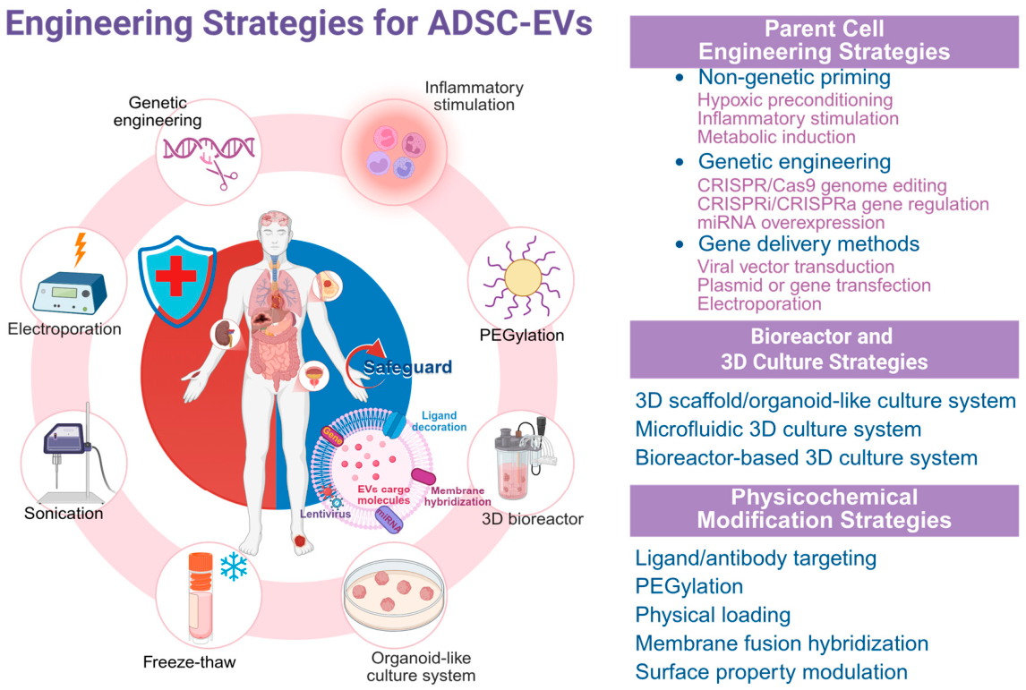
Figure 4.
An overview of engineered ADSCs and ADSC-EVs applications in a variety of disease models and regenerative medicine. Schematic representation of engineering techniques and therapeutic potentials of ADSC-EVs. Top Panel: Engineering strategies executed into parental ADSCs. The core features targeted ligands, PEGylation of cargo loading and membrane fusion (the space highlighted in orange as to the left). The outer ring shows clinical and regenerative uses: bone & joint diseases, skin hair regeneration, metabolic troubles, neurodegeneration and neural repair, particular oncology and more widely regenerative medicine + tissue engineering.
Figure 4.
An overview of engineered ADSCs and ADSC-EVs applications in a variety of disease models and regenerative medicine. Schematic representation of engineering techniques and therapeutic potentials of ADSC-EVs. Top Panel: Engineering strategies executed into parental ADSCs. The core features targeted ligands, PEGylation of cargo loading and membrane fusion (the space highlighted in orange as to the left). The outer ring shows clinical and regenerative uses: bone & joint diseases, skin hair regeneration, metabolic troubles, neurodegeneration and neural repair, particular oncology and more widely regenerative medicine + tissue engineering.
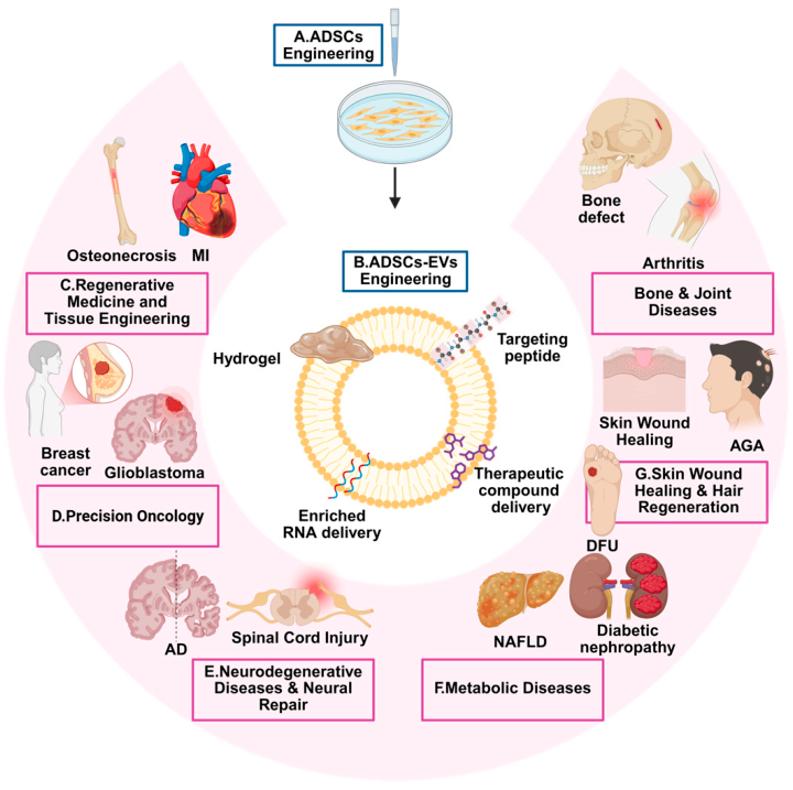
Table 4.
Overview of clinical studies involving ADSCs and their derived EVs.
| Disease / Indication | Study Overview | Cell Source | Study Timeline | Study Phase / Status | Data Source | Trial Identifier |
|---|---|---|---|---|---|---|
| AD | Phase I/II trial evaluating the safety and efficacy of allogeneic ADSC-Exo in mild to moderate AD. | Allogeneic ADSC-EVs | 2020-09 | Phase I/II—Recruiting | Ruijin Hospital | NCT04388982 |
| Atrial fibrillation/arrhythmia | Investigate epicardial fat-derived exosomes in atrial fibrillation as functional mediators and potential biomarkers. | Epicardial ADSC-EVs | 2021-04-08 | Unknown | Sheba Medical Center | NCT03478410 |
| Postpartum eclampsia (vascular endothelial injury) | Clinical evaluation of exosome therapy for vascular dysfunction after preeclampsia. | Fetal ADSC-EVs | 2025-09-19 | Recruiting | Universitas Padjadjaran | NCT07183384 |
| Coronavirus disease 2019 (COVID-19) | Aerosolized ADSC-Exo for severe COVID-19. | Allogeneic ADSC-EVs | 2020-09-07 | Completed | Ruijin Hospital | NCT04276987; [90] |
| Pulmonary diseases | Evaluating the safety and tolerability of aerosolized ADSC-Exo in healthy volunteers. | ADSC-EVs | 2021-08-04 | Completed | Ruijin Hospital | NCT04313647; [91] |
| Drug-resistant pulmonary infections | Evaluate ADSC-Exo via nebulization for efficacy and safety in carbapenem-resistant Gram-negative pulmonary infections. | ADSC-EVs | 2024-07-22 | Suspended | Ruijin Hospital | NCT04544215 |
| Diabetes-related skin ulcers/wound healing | Evaluate personalized nutritional intervention combined with MSC-Exo for wound healing and regeneration in diabetic ulcers. | ADSC-EVs | 2023-10-25 | Unknown | Maimónides Biomedical Research Institute of Córdoba | NCT05243368 |
| Obesity and metabolic syndrome | Clinical study exploring ADSC-EV profiles in obesity and insulin resistance. | ADSC-EVs | 2025-09-15 | Recruiting | Hasselt University | NCT06444646 |
| Postprandial dynamics of ADSC-EVs | Exosomal features of adipose tissue from normal-weight vs. obese individuals after meal stimulation. | Autologous ADSC-EVs | 2026-02-11 | Not yet recruiting | Mayo Clinic | NCT07031297 |
| Insulin resistance / metabolically healthy obesity | Role of ADSC-EVs in obesity-related insulin resistance. | ADSC-EVs and plasma | 2025-07-17 | Recruiting | Washington University School of Medicine | NCT05933707 |
| Erectile dysfunction (including type 2 diabetes-associated ED) | Evaluation of autologous ADSC injections for erectile dysfunction. | Autologous ADSC-EVs | 2025-08-26 | Recruiting | Jumeirah American Clinic | NCT06605508 |
| Gonadal dysfunction/testosterone deficiency | Evaluate ADSCs and exosomes for safety and efficacy in male and female gonadal failure. | Autologous ADSC-EVs | 2025-04-10 | Recruiting | Jumeirah American Clinic | NCT06841328 |
| Prostate cancer/obesity | Investigate ADSC-Exo mediating communication with prostate cancer cells in lean vs. obese patients. | Prostate tissue and ADSC-EVs | 2021-09-22 | Recruiting | Imperial College London | NCT04167722 |
| Bone grafting/osteogenesis promotion | Evaluate autologous ADSCs and conditioned medium for the quality and quantity of maxillary bone graft formation. | Conditioned medium from ADSCs (containing EVs) | 2025-06-05 | Recruiting | Pontifícia Universidade Católica do Rio Grande do Sul | NCT04998058 |
| Osteoarthritis | Evaluate the safety and efficacy of intra-articular injection of autologous ADSC-Exo for osteoarthritis. | Autologous ADSC-EVs | 2024-03-21 | Completed | Jagiellonian University | NCT05081921 |
| Burn wound repair | Evaluate the safety and efficacy of allogeneic ADSC-Exo-engineered construct for burn wound healing. | Allogeneic ADSC-EVs | 2017-04-26 | Unknown | Kyiv City Clinical Hospital No. 2 | NCT03113747 |
| Wound healing | Pilot study evaluating human ADSC-Exo dressings for wound healing. | Autologous ADSC-EVs | 2023-10-19 | Completed | Shanghai Ninth People’s Hospital, Shanghai Jiao Tong University | NCT05475418 |
| Chronic non-healing wounds/wound repair | Evaluate ADSC-EVs for the healing of chronic wounds. | Allogeneic ADSC-EVs | 2024-02-12 | Recruiting | Shanghai Ninth People’s Hospital, Shanghai Jiao Tong University | NCT06253975 |
| Tendon injury | Study of mechanisms and clinical translation of tendon injury treatment using exosomes from ADSCs with TGIF1 gene silencing. | Genetically engineered ADSC-EVs | 2020-10-21 | Approved | Shanghai Tenth People’s Hospital | ChiCTR2000039159 |
| Periodontitis | Clinical trial assessing autologous adipose stem cell exosomes for periodontal regeneration. | Autologous ADSC-EVs | 2020-02-17 | Early Phase I—Recruiting | Beni- Suef University | NCT04270006 |
| Facial rejuvenation / medical aesthetics | Evaluating the efficacy of ADSC therapy for facial rejuvenation. | Autologous ADSCs | 2016-10-04 | Completed | Xuzhou Medical University | NCT02923219 |
| Pancreatic ductal adenocarcinoma | Evaluate the safety and dosing of MSC-Exo carrying KrasG12D siRNA in metastatic pancreatic cancer. | Allogeneic ADSC-EVs | 2025-11-26 | Recruiting | MD Anderson Cancer Center | NCT03608631 |
Disclaimer/Publisher’s Note: The statements, opinions and data contained in all publications are solely those of the individual author(s) and contributor(s) and not of MDPI and/or the editor(s). MDPI and/or the editor(s) disclaim responsibility for any injury to people or property resulting from any ideas, methods, instructions or products referred to in the content. |
© 2026 by the authors. Licensee MDPI, Basel, Switzerland. This article is an open access article distributed under the terms and conditions of the Creative Commons Attribution (CC BY) license (http://creativecommons.org/licenses/by/4.0/).
Copyright: This open access article is published under a Creative Commons CC BY 4.0 license, which permit the free download, distribution, and reuse, provided that the author and preprint are cited in any reuse.


