

Submitted:
29 July 2026
Posted:
30 July 2026
You are already at the latest version
Abstract
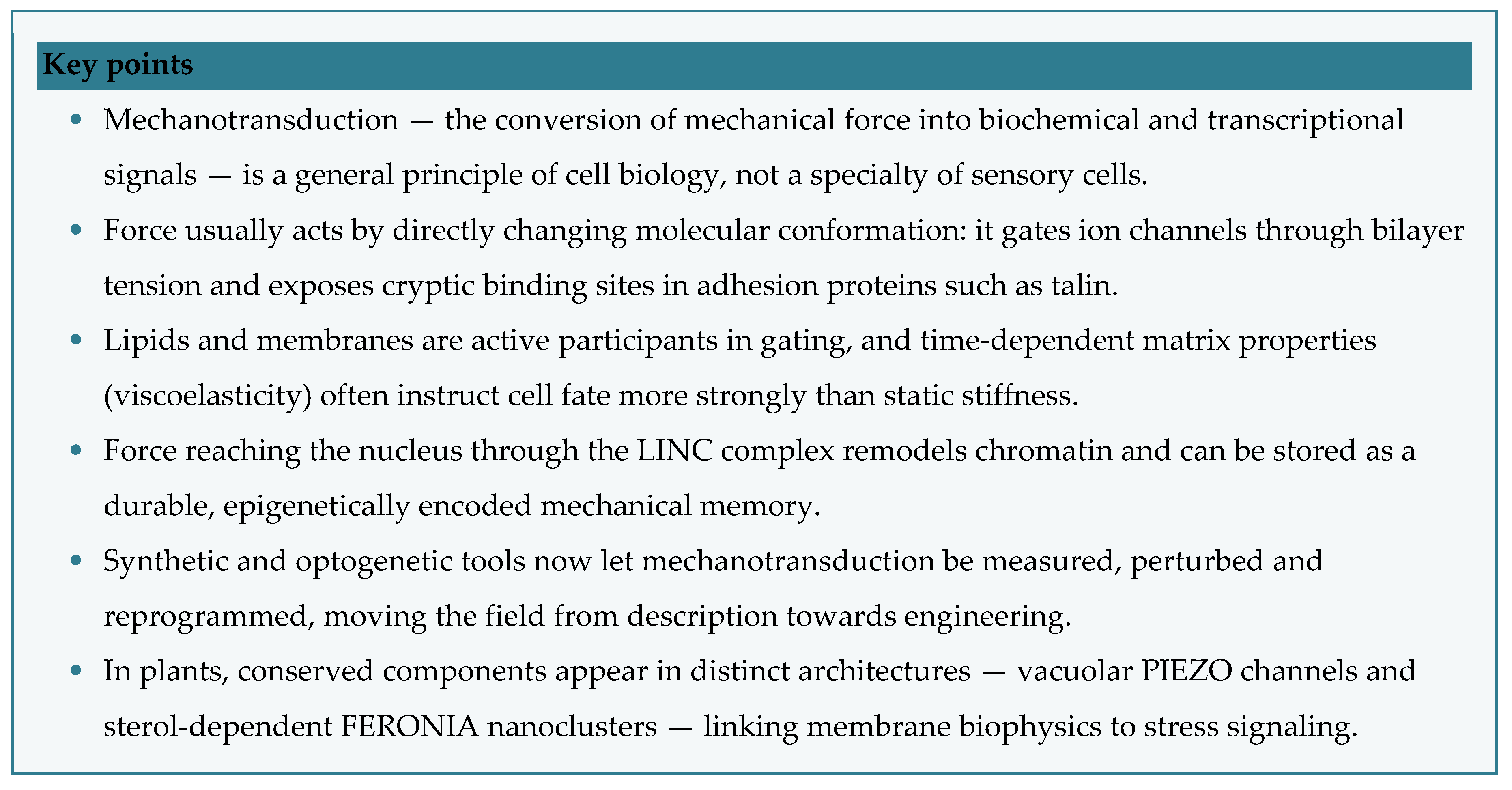
Mechanotransduction is how cells convert mechanical stimuli — tension, shear, substrate stiffness, viscoelasticity and confinement — into biochemical and transcriptional signals. Once viewed as a specialty of sensory cells, it is now a general principle of cell biology, spanning scales from single-bond lifetimes to tissue remodeling and timescales from millisecond channel gating to durable epigenetic memory. This review organizes the field into five threads: adhesion and cytoskeletal force sensors; mechanosensitive ion channels; the extracellular matrix as an instructor of cell fate; nuclear mechanotransduction and chromatin remodeling; and the engineering of mechanotransduction. A final section highlights plant mechanobiology, where PIEZO channels and the receptor kinase FERONIA reveal distinct architectures relevant to membrane biophysics and biomolecular condensate biology. Throughout, force acts mainly by directly altering molecular conformation; lipids and membranes are active participants; time-dependent matrix properties often outrank static stiffness; and the genome behaves as a mechanically responsive organelle that records a cell’s mechanical history.
Keywords:
mechanotransduction
; force
; molecules
; nuclei
; plants
; piezo
; feronia
; biomolecular condensates
Introduction
Mechanical force is as fundamental a cellular input as any soluble ligand. Mechanotransduction — the conversion of a mechanical stimulus into an electrical or biochemical signal — underlies touch, hearing, proprioception, osmoregulation and the mechanical homeostasis of nearly every tissue [1]. The central question today is how transient forces are integrated into lasting outcomes such as development, regeneration and disease [2]. Two ideas recur. First, mechanotransduction is multiscale: membrane sensing, cytoplasmic transmission and nuclear response are coupled across timescales, with proteins such as talin acting as mechanical filters [3]. Second, the cell behaves as a network of mechanically coupled compartments — proteins, membranes, condensates and organelles — that respond collectively to force [4]. Two receptor systems dominate the entry point for force — integrin adhesions that couple to the cytoskeleton, and mechanosensitive channels that convert tension into current — and both feed a shared route to the genome. We begin with that architecture, then dissect each module.
1. The Architecture of Force Transmission
Mechanotransduction can be viewed as a vertically integrated relay (Figure 1). Mechanical information in the extracellular matrix (ECM) — stiffness, viscoelasticity and ligand presentation — is read at the membrane by integrins, which couple to the cytoskeleton, and by mechanosensitive channels, which convert tension into ionic current. Force then passes through the actomyosin cytoskeleton and adhesion clutches to the nuclear envelope via the LINC (linker of nucleoskeleton and cytoskeleton) complex, reshaping chromatin and transcription. The relay is bidirectional: transcriptional output feeds back on tension and matrix remodeling, creating a mechanical memory that can outlast the stimulus (Figure 1).
2. Cytoskeletal and Adhesion-Based Force Sensors
2.1. The Integrin–Cytoskeleton Axis
Magnetic twisting first showed that force applied to integrin β1 — but not to non-adhesion receptors — triggers focal-adhesion assembly and cytoskeleton-dependent stiffening [5], establishing the integrin–cytoskeleton axis and the tensegrity model, in which interdependent struts and cables distribute load globally (Table 1). Adhesion mechanosensing converges on cytoskeleton–matrix–integrin connections [6] at load-bearing structures that are themselves dynamic [7]. Because force is shared across the actomyosin cortex, not adhesions alone, the cortex is both conduit and sensor [8].
2.2. Single-Molecule Force Switches
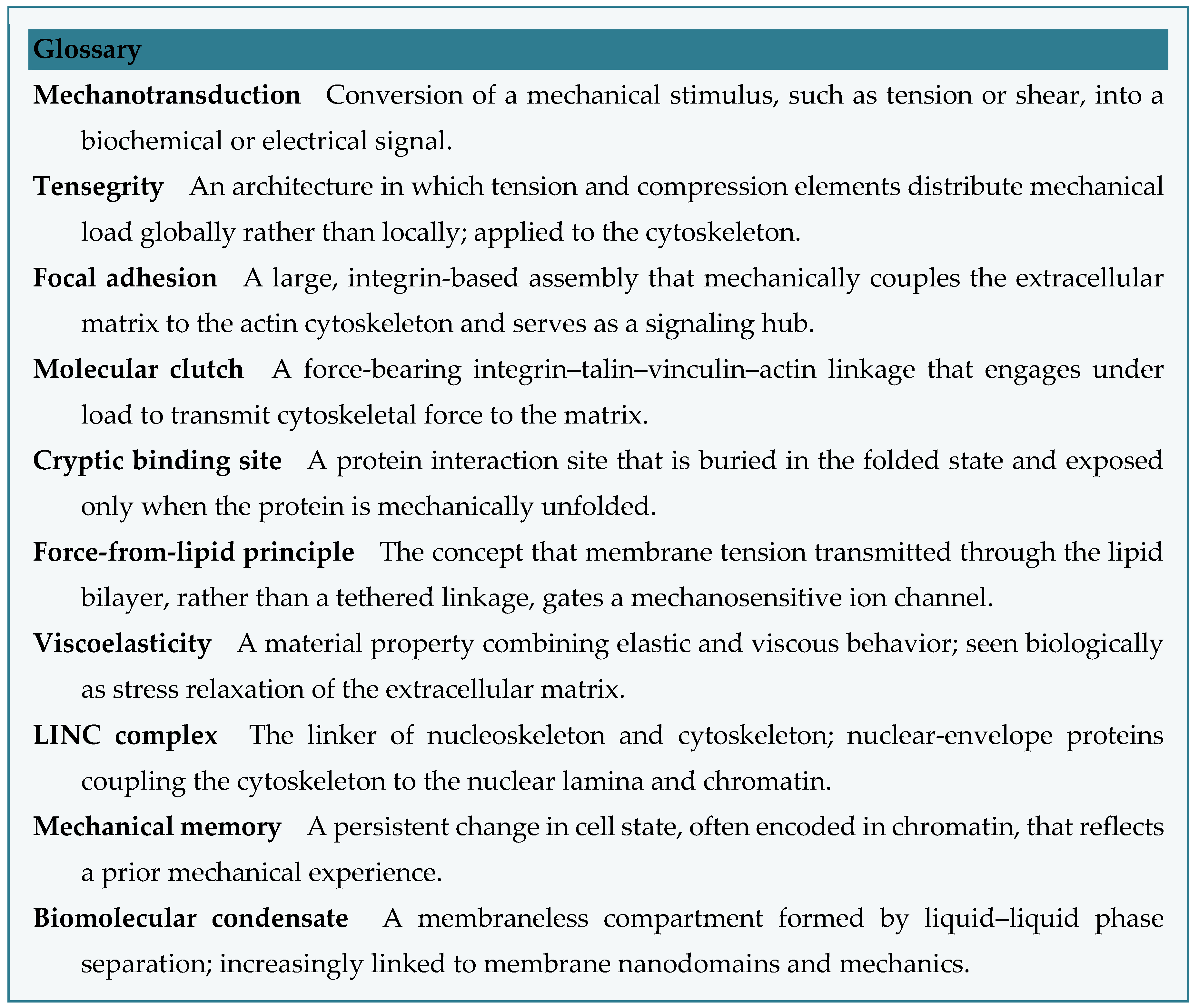
At the single-molecule level, a unifying theme is the force-induced exposure of cryptic binding sites (Figure 2). Stretching the talin rod unfolds its helices to reveal vinculin-binding sites, turning force directly into a new protein interaction that reinforces the adhesion [9]. Talin isoform linkages set the stiffness range cells probe [10], and a ~2-pN talin-dependent slip bond couples fibronectin to the cytoskeleton [11]. Similar switches operate elsewhere: strain regulates filamin A binding to FilGAP and integrins [34], and integrin-applied force releases active TGF-β from its latent complex [35]. Adhesion mechanotransduction is, in essence, a set of force-gated conformational switches.
3. Mechanosensitive Ion Channels
Ion channels convert force to current within milliseconds — fast enough to require direct gating by membrane tension rather than chemical relays [12]. The force-from-lipid principle holds that tension in the surrounding bilayer, not a tethered linkage, opens the pore (Figure 3).
3.1. PIEZO Channels
PIEZO1 and PIEZO2 are large, curved, three-bladed cation channels whose curvature-based gating gives high mechanosensitivity, governing vascular development and red-cell volume (PIEZO1) and touch, proprioception and interoception (PIEZO2) [16]. Force-controlled nanopipettes dissect how membrane tension tunes PIEZO1 [17], and microscale cell geometry modulates PIEZO1 through cytoskeletal redistribution [18].
3.2. Sensory Transduction Complexes
3.3. Bacterial Channels as Paradigms
The bacterial channels MscL and MscS remain the cleanest gating paradigms. Spectroscopy and structure captured open and intermediate states [36,37], and cryo-EM visualized MscS under defined tension [38]. All point to a lipids-first mechanism: the amphipathic MscL N-terminus senses tension [39], lipids in hydrophobic nano-pockets set channel state (40), and lipid reorganization — not protein deformation alone — gates the pore [41](Table 2).
4. The Extracellular Matrix as a Mechanical Instructor
Cells build, degrade and pre-stress the ECM, whose mechanics then act back on them — an active homeostatic loop central to tissue function [20]. Two properties dominate (Figure 4).
4.1. Stiffness and Viscoelasticity
Native ECM is viscoelastic, but classical hydrogels are purely elastic. Tuning stress-relaxation rate independently of stiffness showed that faster relaxation promotes spreading, proliferation and osteogenesis in mesenchymal stem cells (MSCs) (21), making viscoelasticity an instructive cue in its own right (22).
4.2. Cell-Remodelable Matrices
Fate also depends on what cells can do to the matrix. Degradation-mediated traction directs MSC lineage independent of stiffness or shape [42]; a nascent pericellular protein layer, not the bulk gel, guides mechanosensing [43]; and fast-dissociating dynamic crosslinks enhance force-induced remodeling and sensing [44,45]. In three dimensions, mechanotransduction is a negotiation between cell and matrix (Table 3).
5. Nuclear Mechanotransduction and Chromatin Remodeling
Force reaching the nucleus is converted into changes in genome organization and transcription (24), through a self-reinforcing loop (Figure 5).
5.1. Chromatin Condensation and Mechanical Memory
Dynamic loading condenses chromatin via actomyosin contractility and the methyltransferase EZH2, stiffening the nucleus and instilling a mechanical memory that persists after the stimulus [25]. Conversely, softening the nucleus — by HDAC inhibition or lamin A/C knockdown — speeds migration through dense tissue, so nuclear stiffness is both a barrier and a tunable variable [26].
5.2. Pathological Persistence
Sustained force is pathological: stiff substrates drive fibroblast-to-myofibroblast conversion through nuclear-envelope tension and HDAC activity [46]; nuclear deformation guides chromatin reorganization in cardiac development and disease [47]; and chemomechanical cues set nanoscale chromatin organization in diseased connective tissue [48].
5.3. Molecular Couplers
6. Engineering and Synthetic Mechanotransduction
6.1. Tools for Probing and Perturbing Force
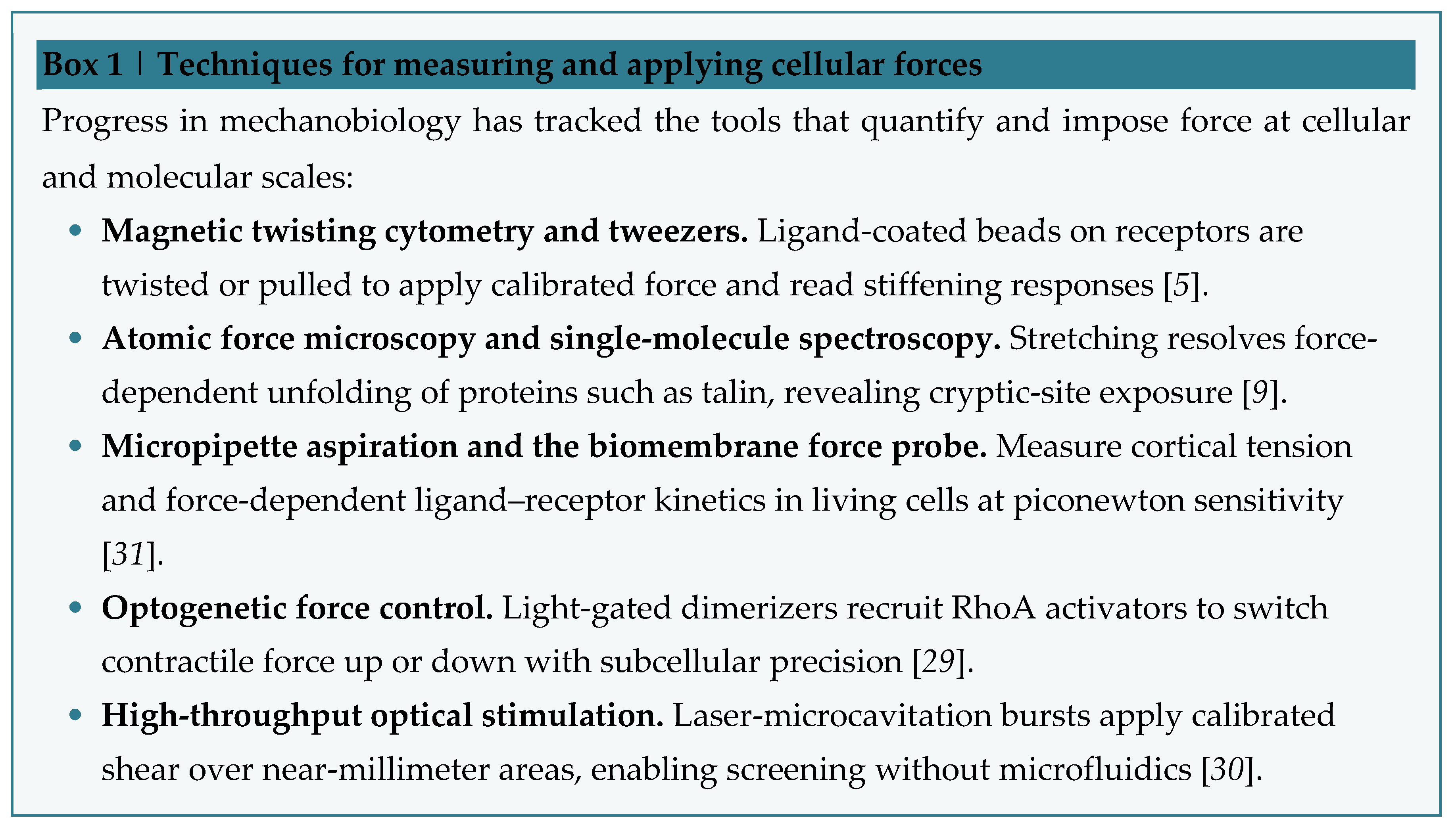
With the parts list filling in, the field now controls force. Optogenetic RhoA switches contractility up or down in subcellular regions [29]; laser-microcavitation applies calibrated shear for high-throughput screening [30]; and micropipette and biomembrane-force-probe assays measure force-dependent kinetics and conformational change in living cells [31].
6.2. Mechanogenetics and Synthetic Circuits
Synthetic mechanotransduction builds artificial force-to-gene circuits. Deconstructing TRPV4 signaling yielded mechanogenetic circuits that drive on-demand biologic production in engineered tissue [32], defining the field of mechanogenetics [33]; a recent framework generalizes such synthetic modules toward reverting malignant phenotypes and tuning immunotherapies [28].
6.3. Translational Mechanobiology
7. An Emerging Frontier: Plant Mechanotransduction
Most mechanisms above come from animals and bacteria, leaving plant mechanotransduction under-explored despite constant gravity, turgor, wind and touch. Conserved components appear in plant-specific architecture (Figure 6).
7.1. Vacuolar PIEZO
Plant PIEZO homologs in Physcomitrium patens and Arabidopsis thaliana localize to the vacuolar, not the plasma, membrane, where they support growth and cytoplasmic Ca²⁺ oscillations and promote vacuole tubulation — a kingdom-specific redeployment of a conserved channel [51] (Table 5).
The FERONIA case is notable for condensate research: dynamic nanodomain assembly — conceptually adjacent to liquid–liquid phase separation — operates in a mechanosensory, stress-signaling context, offering a natural entry point for phase-separation work.
7.2. FERONIA
The receptor kinase FERONIA (FER) links membrane mechanics to development and stress: it enables root penetration of hard substrates via the transcription factor PIF3 [52] and, activated by the RALF34 peptide and moderate heat, nucleates sterol-dependent membrane nanoclusters that drive heat acclimation [53] (Table 5).
8. Conclusions and Future Perspectives
Four principles recur. Force acts mainly by directly changing molecular conformation — gating channels [16,38,41] and exposing cryptic binding sites [9,35]. Lipids and membranes are active gating elements [39,40,41] and, in plants, partition signaling into nanodomains [53]. Viscoelasticity and cell-driven remodeling often outrank static stiffness [21,23,42,43,44]. And the genome stores mechanical history in chromatin [23,25,46,48].
Open questions remain: how are signals integrated across the millisecond-to-month range [3]; how do mechanically distinct compartments, including biomolecular condensates, compute a response [4]; and how far do animal paradigms transfer to plants, where vacuolar PIEZOs [51] and FER nanoclusters [53] suggest distinct logic? As measurement and synthetic tools mature [28,29,32], the field is poised to move from describing mechanotransduction to programming it.
Author Contributions
The authors contributed equally to all aspects of the article.
Conflicts of Interest
The authors declare no conflict of interest.
Acknowledgments
We acknowledge Huang Lab members for the fruitful discussion on this topic. Due to space limitations, we apologize to authors whose works were not cited. During the preparation of this work, the authors used Google Gemini to improve language and readability of figures. After using this tool, the authors reviewed and edited the content as needed and take full responsibility for the content of the publication. This research received no external funding.
References
- Gillespie, P. G.; Walker, R. G. Molecular basis of mechanosensory transduction. Nature 2001, 413, 194–202. [Google Scholar] [CrossRef] [PubMed]
- Paluch, E. K.; Nelson, C. M.; Biais, N.; Fabry, B.; Moeller, J.; Pruitt, B. L.; Wollnik, C.; Kudryasheva, G.; Rehfeldt, F.; Federle, W. Mechanotransduction: Use the force(s). BMC Biol. 2015, 13, 47. [Google Scholar] [CrossRef] [PubMed]
- Cheng, B.; Li, M.; Lin, M.; Guo, H.; Xu, F. Mechanobiology across timescales. Nat. Rev. Phys. 2025, 7, 621–644. [Google Scholar] [CrossRef]
- Kasuba, K. C.; Fläschner, G.; Jain, A.; Krieg, M.; Sharma, U.; Gensbittel, V.; Nava, M. M.; Camacho Rufino, M.; Treutlein, B.; Liberali, P.; Trepat, X.; Jülicher, F.; Piel, M.; Iber, D.; Lutolf, M. P.; Roska, B.; Vassalli, M.; Müller, D. J. Advancing mechanobiology from single molecules to complex cellular systems. Nat. Nanotechnol. 2026, 1–11. [Google Scholar]
- Wang, N.; Butler, J. P.; Ingber, D. E. Mechanotransduction across the cell surface and through the cytoskeleton. Science 1993, 260, 1124–1127. [Google Scholar] [CrossRef] [PubMed]
- Alenghat, F. J.; Ingber, D. E. Mechanotransduction: All signals point to cytoskeleton, matrix, and integrins. Sci. STKE 2002, 2002, pe6–pe6. [Google Scholar] [CrossRef] [PubMed]
- Hoffman, B. D.; Grashoff, C.; Schwartz, M. A. Dynamic molecular processes mediate cellular mechanotransduction. Nature 2011, 475, 316–323. [Google Scholar] [CrossRef] [PubMed]
- Luo, T.; Mohan, K.; Iglesias, P. A.; Robinson, D. N. Molecular mechanisms of cellular mechanosensing. Nat. Mater. 2013, 12, 1064–1071. [Google Scholar] [CrossRef] [PubMed]
- del Rio; Perez-Jimenez, R.; Liu, R.; Roca-Cusachs, P.; Fernandez, J. M.; Sheetz, M. P. Stretching single talin rod molecules activates vinculin binding. Science 2009, 323, 638–641. [Google Scholar] [CrossRef] [PubMed]
- Austen, K.; Ringer, P.; Mehlich, A.; Chrostek-Grashoff, A.; Kluger, C.; Klingner, C.; Sabass, B.; Zent, R.; Rief, M.; Grashoff, C. Extracellular rigidity sensing by talin isoform-specific mechanical linkages. Nat. Cell Biol. 2015, 17, 1597–1606. [Google Scholar] [CrossRef] [PubMed]
- Jiang, G.; Giannone, G.; Critchley, D. R.; Fukumoto, E.; Sheetz, M. P. Two-piconewton slip bond between fibronectin and the cytoskeleton depends on talin. Nature 2003, 424, 334–337. [Google Scholar] [CrossRef] [PubMed]
- Chalfie, M. Neurosensory mechanotransduction. Nat. Rev. Mol. Cell Biol. 2009, 10, 44–52. [Google Scholar] [CrossRef] [PubMed]
- Delmas, P.; Hao, J.; Rodat-Despoix, L. Molecular mechanisms of mechanotransduction in mammalian sensory neurons. Nat. Rev. Neurosci. 2011, 12, 139–153. [Google Scholar] [CrossRef] [PubMed]
- Tsunozaki, M.; Bautista, D. M. Mammalian somatosensory mechanotransduction. Curr. Opin. Neurobiol. 2009, 19, 362–369. [Google Scholar] [CrossRef] [PubMed]
- García-Añoveros, J.; Corey, D. P. The molecules of mechanosensation. Annu. Rev. Neurosci. 1997, 20, 567–594. [Google Scholar] [CrossRef] [PubMed]
- Xiao, B. Mechanisms of mechanotransduction and physiological roles of PIEZO channels. Nat. Rev. Mol. Cell Biol. 2024, 25, 886–903. [Google Scholar] [CrossRef] [PubMed]
- Lüchtefeld; Pivkin, I. V.; Gardini, L.; Zare-Eelanjegh, E.; Gäbelein, C.; Ihle, S. J.; Reichmuth, A. M.; Capitanio, M.; Martinac, B.; Zambelli, T.; Vassalli, M. Dissecting cell membrane tension dynamics and its effect on Piezo1-mediated cellular mechanosensitivity using force-controlled nanopipettes. Nat. Methods 2024, 21, 1063–1073. [Google Scholar] [CrossRef] [PubMed]
- Wang, H. J.; Wang, Y.; Mirjavadi, S. S.; Andersen, T.; Moldovan, L.; Vatankhah, P.; Russell, B.; Jin, J.; Zhou, Z.; Li, Q.; Cox, C. D.; Su, Q. P.; Ju, L. A. Microscale geometrical modulation of PIEZO1 mediated mechanosensing through cytoskeletal redistribution. Nat. Commun. 2024, 15, 5521. [Google Scholar] [CrossRef] [PubMed]
- Xiong, W.; Grillet, N.; Elledge, H. M.; Wagner, T. F. J.; Zhao, B.; Johnson, K. R.; Kazmierczak, P.; Müller, U. TMHS is an integral component of the mechanotransduction machinery of cochlear hair cells. Cell 2012, 151, 1283–1295. [Google Scholar] [CrossRef] [PubMed]
- Humphrey, D.; Dufresne, E. R.; Schwartz, M. A. Mechanotransduction and extracellular matrix homeostasis. Nat. Rev. Mol. Cell Biol. 2014, 15, 802–812. [Google Scholar] [CrossRef] [PubMed]
- Chaudhuri; Gu, L.; Klumpers, D.; Darnell, M.; Bencherif, S. A.; Weaver, J. C.; Huebsch, N.; Lee, H.; Lippens, E.; Duda, G. N.; Mooney, D. J. Hydrogels with tunable stress relaxation regulate stem cell fate and activity. Nat. Mater. 2016, 15, 326–334. [Google Scholar] [PubMed]
- Chaudhuri. Viscoelastic hydrogels for 3D cell culture. Biomater. Sci. 2017, 5, 1480–1490. [Google Scholar] [CrossRef] [PubMed]
- Wu, Y.; Song, Y.; Soto, J.; Hoffman, T.; Lin, X.; Zhang, A.; Chen, S.; Massad, R. N.; Han, X.; Qi, D.; Yeh, K.-W.; Fang, Z.; Eoh, J.; Gu, L.; Rowat, A. C.; Gu, Z.; Li, S. Viscoelastic extracellular matrix enhances epigenetic remodeling and cellular plasticity. Nat. Commun. 2025, 16, 4054. [Google Scholar] [CrossRef] [PubMed]
- Uhler, C.; Shivashankar, G. V. Regulation of genome organization and gene expression by nuclear mechanotransduction. Nat. Rev. Mol. Cell Biol. 2017, 18, 717–727. [Google Scholar] [CrossRef] [PubMed]
- Heo, S.-J.; Thorpe, S. D.; Driscoll, T. P.; Duncan, R. L.; Lee, D. A.; Mauck, R. L. Biophysical regulation of chromatin architecture instills a mechanical memory in mesenchymal stem cells. Sci. Rep. 2015, 5, 16895. [Google Scholar] [CrossRef] [PubMed]
- Heo, S.-J.; Song, K. H.; Thakur, S.; Miller, L. M.; Cao, X.; Peredo, A. P.; Seiber, B. N.; Qu, F.; Driscoll, T. P.; Shenoy, V. B.; Lakadamyali, M.; Burdick, J. A.; Mauck, R. L. Nuclear softening expedites interstitial cell migration in fibrous networks and dense connective tissues. Sci. Adv. 2020, 6, eaax5083. [Google Scholar] [CrossRef] [PubMed]
- Hu, S.; Chapski, D. J.; Gehred, N. D.; Kimball, T. H.; Gromova, T.; Flores, A.; Rowat, A. C.; Chen, J.; Packard, R. R. S.; Olszewski, E.; Davis, J.; Rau, C. D.; McKinsey, T. A.; Rosa-Garrido, M.; Vondriska, T. M. Histone H1.0 couples cellular mechanical behaviors to chromatin structure. Nat. Cardiovasc. Res. 2024, 3, 441–459. [Google Scholar] [CrossRef] [PubMed]
- González-Martín, M.; Martínez-Ara, G.; Ngo, J. T.; X. Trepat, P. Roca-Cusachs, Synthetic mechanotransduction. Nat. Rev. Bioeng. 2026, 4, 236–249. [Google Scholar] [CrossRef]
- Valon; Marín-Llauradó, A.; Wyatt, T.; G. Charras, X. Trepat, Optogenetic control of cellular forces and mechanotransduction. Nat. Commun. 2017, 8, 14396. [Google Scholar] [CrossRef] [PubMed]
- Compton, J. L.; Luo, J. C.; Ma, H.; Botvinick, E.; Venugopalan, V. High-throughput optical screening of cellular mechanotransduction. Nat. Photonics 2014, 8, 710–715. [Google Scholar] [CrossRef] [PubMed]
- Wang, H.; Zhou, F.; Guo, Y.; Ju, L. A. Micropipette-based biomechanical nanotools on living cells. Eur. Biophys. J. 2022, 51, 119–133. [Google Scholar] [CrossRef] [PubMed]
- Nims, R. J.; Pferdehirt, L.; Ho, N. B.; Savadipour, A.; Lorentz, J.; Sohi, S.; Kassab, J.; Ross, A. K.; O’Conor, C. J.; Liedtke, W. B.; Zhang, B.; McNulty, A. L.; Guilak, F. A synthetic mechanogenetic gene circuit for autonomous drug delivery in engineered tissues. Sci. Adv. 2021, 7, eabd9858. [Google Scholar] [CrossRef] [PubMed]
- Nims, R. J.; Pferdehirt, L.; Guilak, F. Mechanogenetics: Harnessing mechanobiology for cellular engineering. Curr. Opin. Biotechnol. 2022, 73, 374–379. [Google Scholar] [CrossRef] [PubMed]
- Ehrlicher, J.; Nakamura, F.; Hartwig, J. H.; Weitz, D. A.; Stossel, T. P. Mechanical strain in actin networks regulates FilGAP and integrin binding to filamin a. Nature 2011, 478, 260–263. [Google Scholar] [CrossRef] [PubMed]
- Dong, X.; Zhao, B.; Iacob, R. E.; Zhu, J.; Koksal, A. C.; Lu, C.; Engen, J. R.; Springer, T. A. Force interacts with macromolecular structure in activation of TGF-β. Nature 2017, 542, 55–59. [Google Scholar] [CrossRef] [PubMed]
- Perozo, E.; Cortes, D. M.; Sompornpisut, P.; Kloda, A.; Martinac, B. Open channel structure of MscL and the gating mechanism of mechanosensitive channels. Nature 2002, 418, 942–948. [Google Scholar] [CrossRef] [PubMed]
- Vásquez, V.; Sotomayor, M.; Cordero-Morales, J.; Schulten, K.; Perozo, E. A structural mechanism for MscS gating in lipid bilayers. Science 2008, 321, 1210–1214. [Google Scholar] [CrossRef] [PubMed]
- Zhang, Y.; Daday, C.; Gu, R.-X.; Cox, C. D.; Martinac, B.; de Groot, B. L.; Walz, T. Visualization of the mechanosensitive ion channel MscS under membrane tension. Nature 2021, 590, 509–514. [Google Scholar] [CrossRef] [PubMed]
- Bavi, N.; Cortes, D. M.; Cox, C. D.; Rohde, P. R.; Liu, W.; Deitmer, J. W.; Bavi, O.; Strop, P.; Hill, A. P.; Rees, D.; Corry, B.; Perozo, E.; Martinac, B. The role of MscL amphipathic N terminus indicates a blueprint for bilayer-mediated gating of mechanosensitive channels. Nat. Commun. 2016, 7, 11984. [Google Scholar] [CrossRef] [PubMed]
- Kapsalis, C.; Wang, B.; El Mkami, H.; Pitt, S. J.; Schnell, J. R.; Smith, T. K.; Lippiat, J. D.; Bode, B. E.; Pliotas, C. Allosteric activation of an ion channel triggered by modification of mechanosensitive nano-pockets. Nat. Commun. 2019, 10, 4619. [Google Scholar] [CrossRef] [PubMed]
- Pliotas, C.; Dahl, A. C. E.; Rasmussen, T.; Mahendran, K. R.; Smith, T. K.; Marius, P.; Gault, J.; Banda, T.; Rasmussen, A.; Miller, S.; Robinson, C. V.; Bayley, H.; Sansom, M. S. P.; Booth, I. R.; Naismith, J. H. The role of lipids in mechanosensation. Nat. Struct. Mol. Biol. 2015, 22, 991–998. [Google Scholar] [CrossRef] [PubMed]
- Khetan, S.; Guvendiren, M.; Legant, W. R.; Cohen, D. M.; Chen, C. S.; Burdick, J. A. Degradation-mediated cellular traction directs stem cell fate in covalently crosslinked three-dimensional hydrogels. Nat. Mater. 2013, 12, 458–465. [Google Scholar] [CrossRef] [PubMed]
- Loebel, C.; Mauck, R. L.; Burdick, J. A. Local nascent protein deposition and remodelling guide mesenchymal stromal cell mechanosensing and fate in three-dimensional hydrogels. Nat. Mater. 2019, 18, 883–891. [Google Scholar] [CrossRef] [PubMed]
- Yang, B.; Wei, K.; Loebel, C.; Zhang, K.; Feng, Q.; Li, R.; Wong, S. H. D.; Xu, X.; Lau, C.; Chen, X.; Zhao, P.; Yin, C.; Burdick, J. A.; Wang, Y.; Bian, L. Enhanced mechanosensing of cells in synthetic 3D matrix with controlled biophysical dynamics. Nat. Commun. 2021, 12, 3514. [Google Scholar] [CrossRef] [PubMed]
- Ferreira, S. A.; Motwani, M. S.; Faull, P. A.; Seymour, A. J.; Yu, T. T. L.; Enayati, M.; Taheem, D. K.; Salzlechner, C.; Haghighi, T.; Kania, E. M.; Oommen, O. P.; Ahmed, T.; Loaiza, S.; Parzych, K.; Dazzi, F.; Varghese, O. P.; Festy, F.; Grigoriadis, A. E.; Auner, H. W.; Snijders, A. P.; Bozec, L.; Gentleman, E. Bi-directional cell-pericellular matrix interactions direct stem cell fate. Nat. Commun. 2018, 9, 4049. [Google Scholar] [CrossRef] [PubMed]
- Walker, C. J.; Crocini, C.; Ramirez, D.; Killaars, A. R.; Grim, J. C.; Aguado, B. A.; Clark, K.; Allen, M. A.; Dowell, R. D.; Leinwand, L. A.; Anseth, K. S. Nuclear mechanosensing drives chromatin remodelling in persistently activated fibroblasts. Nat. Biomed. Eng. 2021, 5, 1485–1499. [Google Scholar] [CrossRef] [PubMed]
- Seelbinder, B.; Ghosh, S.; Schneider, S. E.; Scott, A. K.; Berman, A. G.; Goergen, C. J.; Margulies, K. B.; Bedi, K. C.; Casas, E.; Swearingen, A. R.; Brumbaugh, J.; Calve, S.; Neu, C. P. Nuclear deformation guides chromatin reorganization in cardiac development and disease. Nat. Biomed. Eng. 2021, 5, 1500–1516. [Google Scholar] [CrossRef] [PubMed]
- Heo, S.-J.; Thakur, S.; Chen, X.; Loebel, C.; Xia, B.; McBeath, R.; Burdick, J. A.; Shenoy, V. B.; Mauck, R. L.; Lakadamyali, M. Aberrant chromatin reorganization in cells from diseased fibrous connective tissue in response to altered chemomechanical cues. Nat. Biomed. Eng. 2023, 7, 177–191. [Google Scholar] [CrossRef] [PubMed]
- Kim, S.; Uroz, M.; Bays, J. L.; Chen, C. S. Harnessing mechanobiology for tissue engineering. Dev. Cell 2021, 56, 180–191. [Google Scholar] [CrossRef] [PubMed]
- Chen; Kwon, S. H.; Henn, D.; Kuehlmann, B. A.; Tevlin, R.; Bonham, C. A.; Griffin, M.; Trotsyuk, A. A.; Borrelli, M. R.; Noishiki, C.; Padmanabhan, J.; Barrera, J. A.; Maan, Z. N.; Dohi, T.; Mays, C. J.; Greco, A. H.; Sivaraj, D.; Lin, J. Q.; Fehlmann, T.; Mermin-Bunnell, A. M.; Mittal, S.; Hu, M. S.; Zamaleeva, A. I.; Keller, A.; Rajadas, J.; Longaker, M. T.; Januszyk, M.; Gurtner, G. C. Disrupting biological sensors of force promotes tissue regeneration in large organisms. Nat. Commun. 2021, 12, 5256. [Google Scholar] [CrossRef] [PubMed]
- Radin; Richardson, R. A.; Coomey, J. H.; Weiner, E. R.; Bascom, C. S.; Li, T.; Bezanilla, M.; Haswell, E. S. Plant PIEZO homologs modulate vacuole morphology during tip growth. Science 2021, 373, 586–590. [Google Scholar] [CrossRef] [PubMed]
- Xu, F.; Chen, J.; Li, Y.; Ouyang, S.; Yu, M.; Wang, Y.; Fang, X.; He, K.; Yu, F. The soil emergence-related transcription factor PIF3 controls root penetration by interacting with the receptor kinase FER. Dev. Cell 2024, 59, 434–447.e8. [Google Scholar] [CrossRef] [PubMed]
- Wang; Yan, H.; Guo, X.; Lin, Q.; Li, J.; Peng, A.; Fu, Y.; Gong, Z.; Yang, S.; Ding, Y. FERONIA orchestrates plasma membrane nanoclusters for plant thermotolerance. Science 2026, 392, 885–890. [Google Scholar] [CrossRef] [PubMed]
Figure 1.
The mechanotransduction relay. Force from the extracellular matrix is sensed at the membrane by integrins and PIEZO channels, transmitted through adhesion clutches and the actomyosin cytoskeleton, and conveyed to the nucleus via the LINC complex, where it drives chromatin reorganization. Feedback onto cytoskeletal tension establishes a mechanical memory.
Figure 1.
The mechanotransduction relay. Force from the extracellular matrix is sensed at the membrane by integrins and PIEZO channels, transmitted through adhesion clutches and the actomyosin cytoskeleton, and conveyed to the nucleus via the LINC complex, where it drives chromatin reorganization. Feedback onto cytoskeletal tension establishes a mechanical memory.
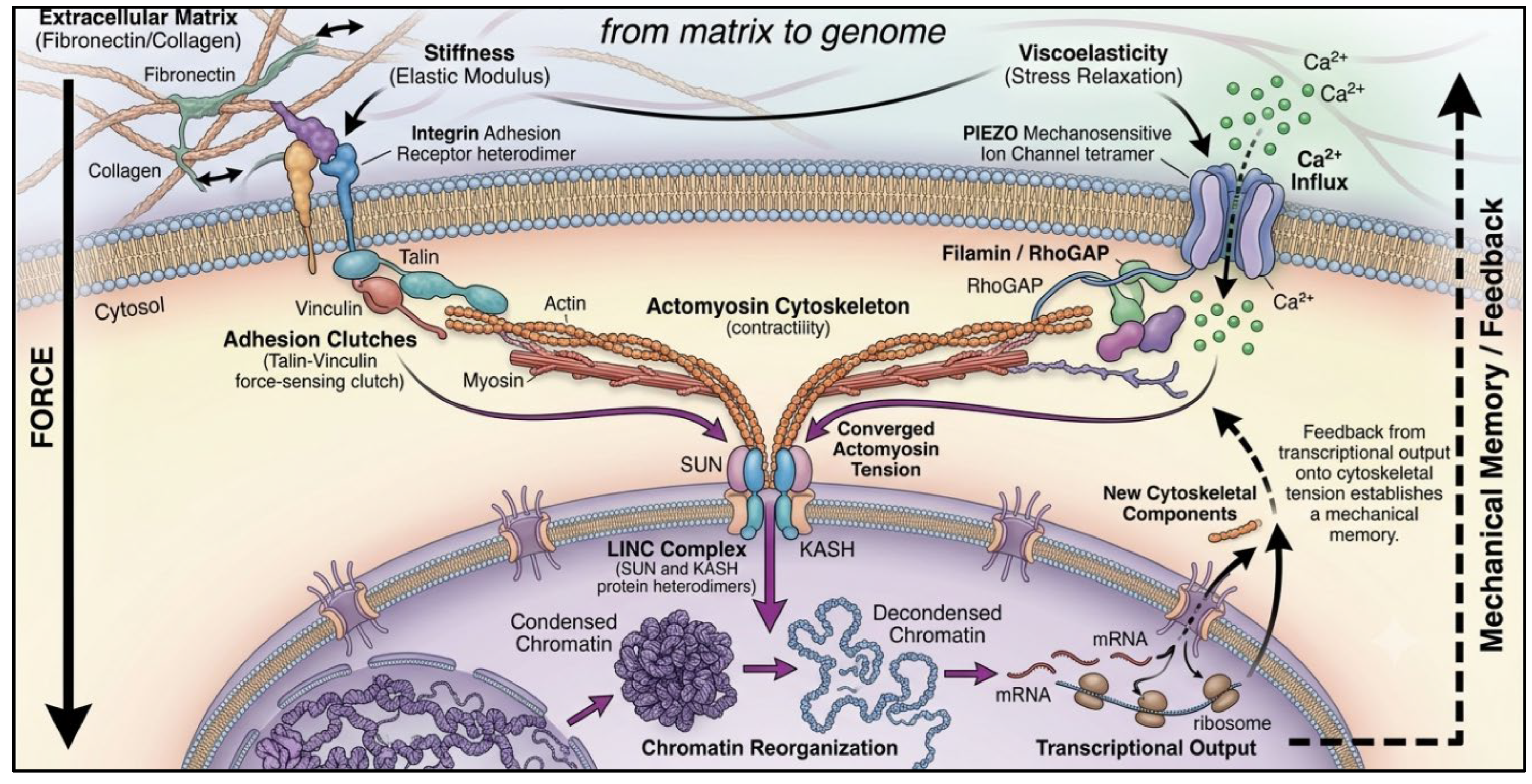
Figure 2.
Force converts talin into a molecular switch. (A) Under low tension the talin rod is folded, and its vinculin-binding sites are buried. (B) Myosin-generated tension from the actin cytoskeleton unfolds the rod, exposing cryptic sites; vinculin then binds and reinforces the integrin–actin linkage — a general principle of adhesion-based mechanosensing.
Figure 2.
Force converts talin into a molecular switch. (A) Under low tension the talin rod is folded, and its vinculin-binding sites are buried. (B) Myosin-generated tension from the actin cytoskeleton unfolds the rod, exposing cryptic sites; vinculin then binds and reinforces the integrin–actin linkage — a general principle of adhesion-based mechanosensing.
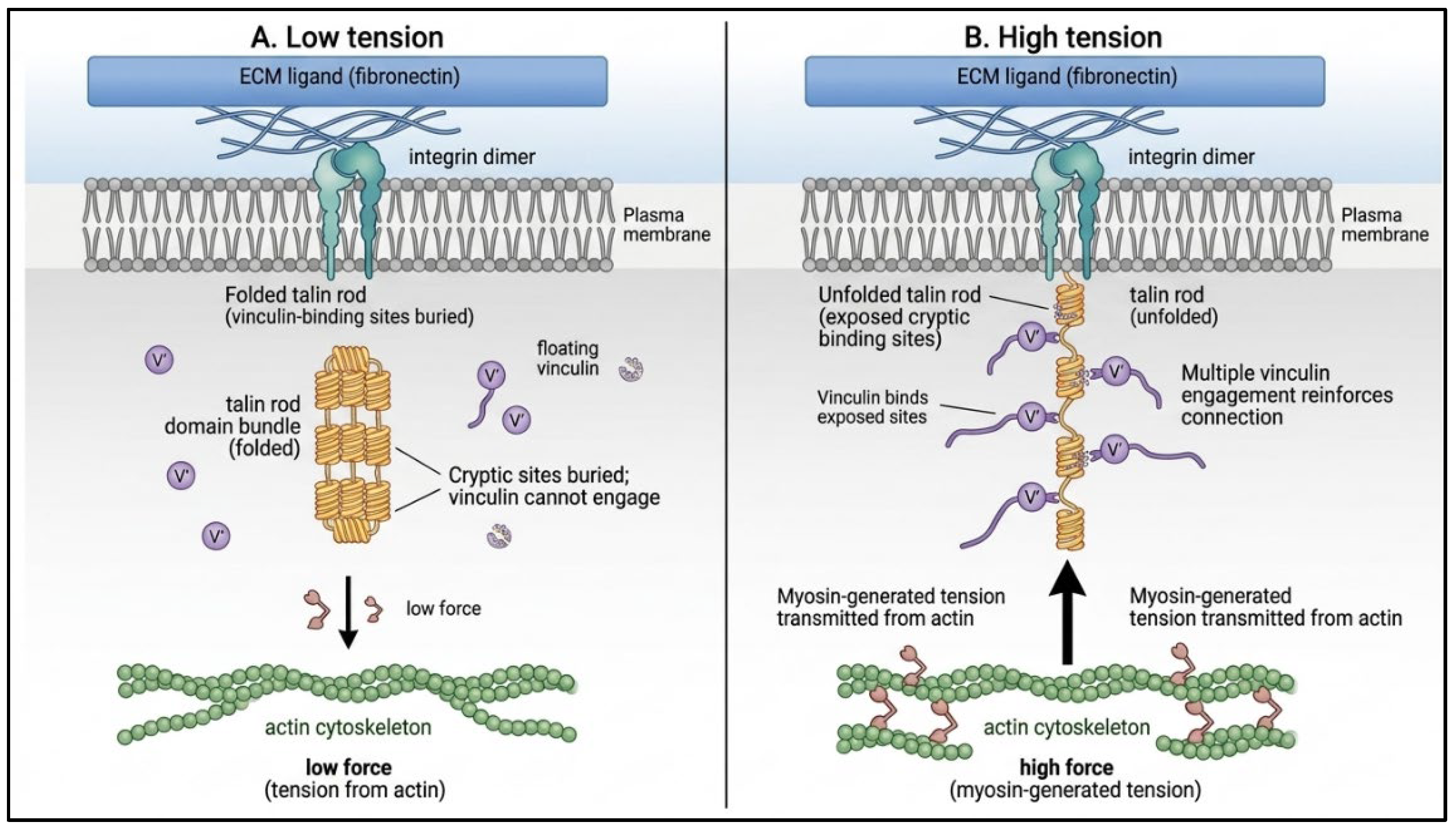
Figure 3.
Force-from-lipid gating. In a relaxed bilayer the pore is closed, and lipids occupy hydrophobic nano-pockets within the channel (left panel). Rising membrane tension thins the bilayer, withdraws pocket lipids and tilts the transmembrane helices to open a cation-conducting pore (right panel). .
Figure 3.
Force-from-lipid gating. In a relaxed bilayer the pore is closed, and lipids occupy hydrophobic nano-pockets within the channel (left panel). Rising membrane tension thins the bilayer, withdraws pocket lipids and tilts the transmembrane helices to open a cation-conducting pore (right panel). .
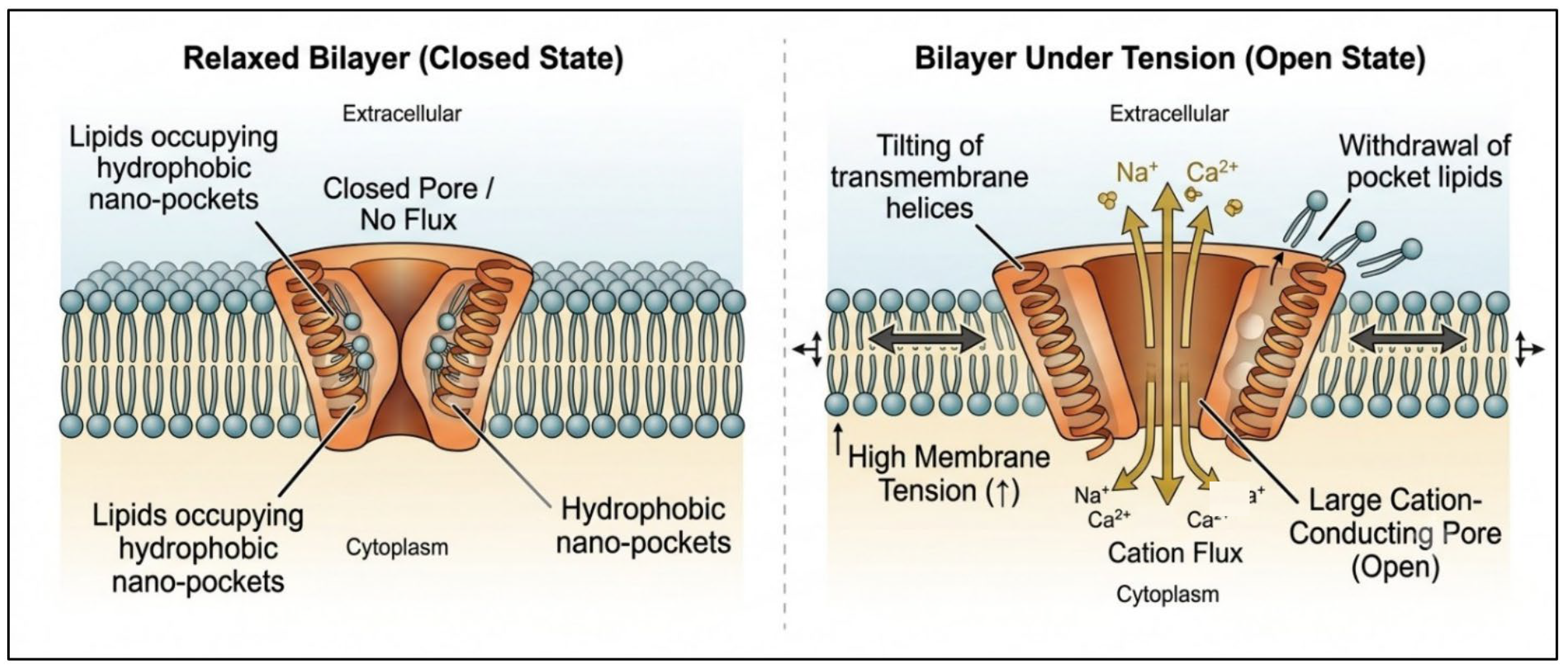
Figure 4.
Matrix mechanics instruct cell fate. Stiffness biases mesenchymal stem-cell lineage along a neurogenic–myogenic–osteogenic axis, while faster stress relaxation (viscoelasticity) enhances spreading and differentiation at a given stiffness. Cell-driven degradation and nascent-protein remodeling locally retune the cue.
Figure 4.
Matrix mechanics instruct cell fate. Stiffness biases mesenchymal stem-cell lineage along a neurogenic–myogenic–osteogenic axis, while faster stress relaxation (viscoelasticity) enhances spreading and differentiation at a given stiffness. Cell-driven degradation and nascent-protein remodeling locally retune the cue.
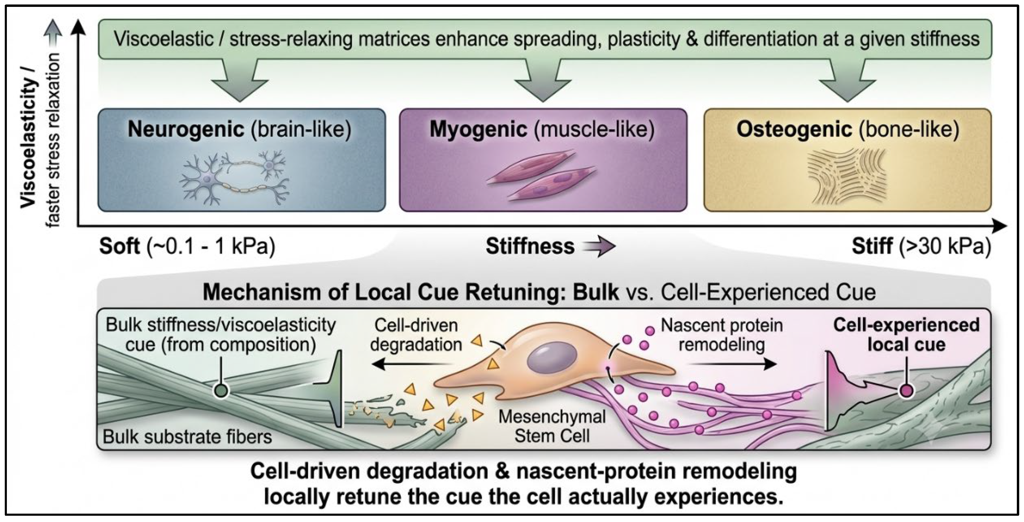
Figure 5.
Nuclear mechanotransduction as a self-reinforcing loop. A stiff or loaded matrix raises actomyosin tension and LINC coupling, deforms the nuclear envelope and triggers chromatin condensation via EZH2, HDACs and histone H1.0. The resulting transcriptional changes feed back on tension, encoding a mechanical memory.
Figure 5.
Nuclear mechanotransduction as a self-reinforcing loop. A stiff or loaded matrix raises actomyosin tension and LINC coupling, deforms the nuclear envelope and triggers chromatin condensation via EZH2, HDACs and histone H1.0. The resulting transcriptional changes feed back on tension, encoding a mechanical memory.
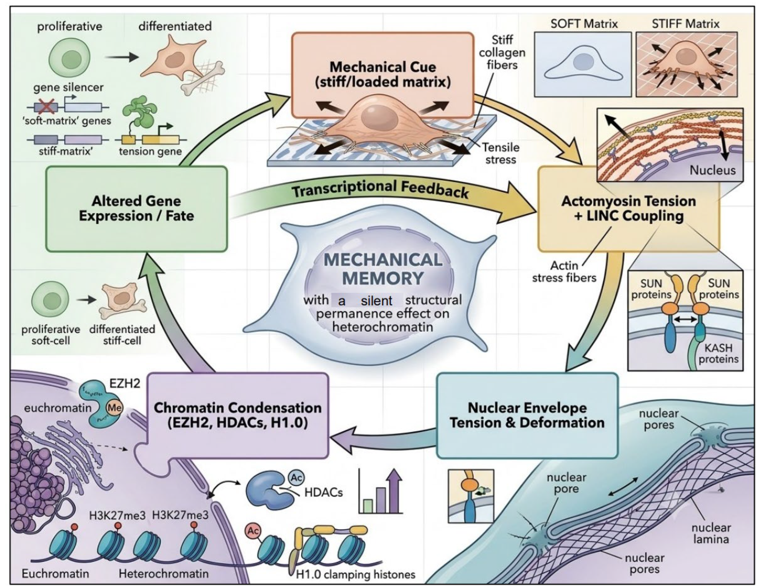
Figure 6.
Mechanotransduction in plants: distinct architectures. (A) In tip-growing moss and Arabidopsis cells, PIEZO homologs localize to the vacuolar — not the plasma — membrane, promoting vacuole tubulation and shaping cytoplasmic calcium oscillations. (B) The receptor kinase FERONIA organizes sterol-dependent plasma-membrane nanoclusters that, activated by the RALF34 peptide and moderate heat, drive heat acclimation.
Figure 6.
Mechanotransduction in plants: distinct architectures. (A) In tip-growing moss and Arabidopsis cells, PIEZO homologs localize to the vacuolar — not the plasma — membrane, promoting vacuole tubulation and shaping cytoplasmic calcium oscillations. (B) The receptor kinase FERONIA organizes sterol-dependent plasma-membrane nanoclusters that, activated by the RALF34 peptide and moderate heat, drive heat acclimation.
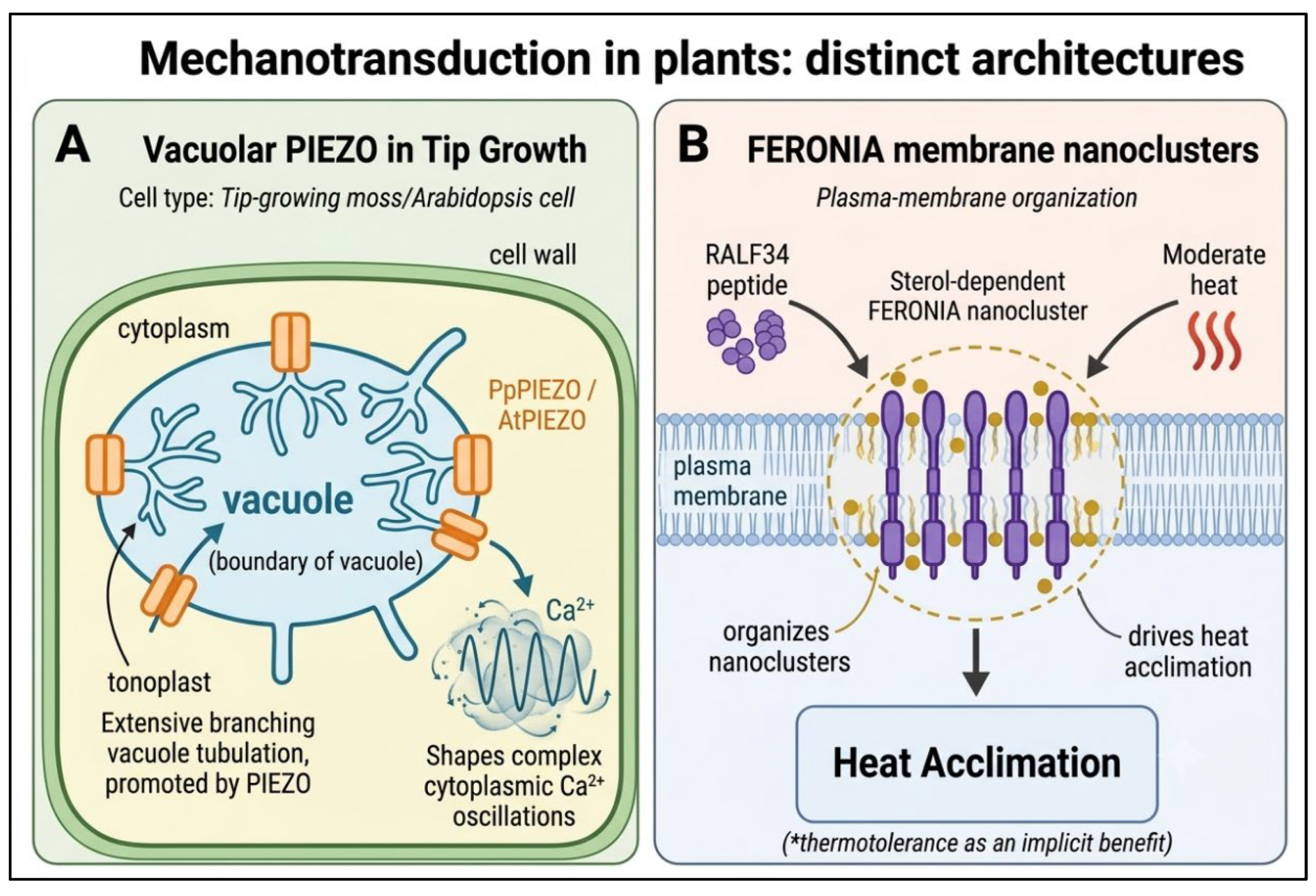

Table 1.
The five mechanistic classes of mechanotransduction.
| Mechanistic class | Principal sensors / effectors | Representative outputs | References |
| Adhesion & cytoskeleton | Integrins, talin, vinculin, filamin, actomyosin | Focal-adhesion maturation, stiffness sensing, TGF-β release | [5,6,7,8,9,10,11] |
| Mechanosensitive channels | PIEZO1/2, TMHS/TMC, MscS, MscL, TRP family | Ca²⁺ influx, fast electrical signaling | [12,13,14,15,16,17,18,19] |
| Extracellular matrix | Collagen/fibronectin networks; stiffness & viscoelasticity | Stem-cell lineage, ECM homeostasis | [20,21,22] |
| Nuclear / chromatin | LINC complex, lamins, EZH2, HDACs, histone H1.0 | Chromatin condensation, gene regulation, memory | [23,24,25,26,27] |
| Engineering & synthetic | Optogenetics, mechanogenetic circuits, biomaterials | Programmable force–gene coupling, regeneration | [28,29,30,31,32,33] |
Table 2.
Mechanosensitive ion channels by model system.
| Channel / complex | Model system | Key structural / mechanistic insight | References |
| PIEZO1 / PIEZO2 | Mammalian cells | Curved three-bladed architecture; curvature-based gating | [16,17,18] |
| TMHS (LHFPL5) | Cochlear hair cells | Integral subunit coupling tip-link tension to gating | [19] |
| DEG/ENaC, TRP | Worm, fly, mammal | Directly gated neurosensory channel families | [12,13,14,15] |
| MscL | Bacteria | Amphipathic N-terminus as tension sensor; helix tilting | [36,39] |
| MscS | Bacteria | Cryo-EM under tension; lipid-pocket control of state | [37,38,40,41] |
Classification of channels organized by model systems, highlighting their principal contributions to the force-from-lipid paradigm.
Table 3.
Matrix mechanics and cell fate. Representative studies linking manipulated matrix properties to specific cellular outcomes.
Table 3.
Matrix mechanics and cell fate. Representative studies linking manipulated matrix properties to specific cellular outcomes.
| Study system | Matrix property manipulated | Cellular outcome | References |
| MSCs in alginate | Stress-relaxation rate (viscoelasticity) | Faster relaxation → spreading, osteogenesis | [21] |
| MSCs in HA gels | Cell-mediated degradation / traction | Traction directs lineage independent of shape | [42] |
| MSCs in HA gels | Nascent pericellular protein layer | Local proteins guide mechanosensing & fate | [43] |
| Cells in 3D networks | Dynamic crosslink dissociation rate | Force-induced remodeling enhances sensing | [44,45] |
| Tissues (general) | ECM synthesis / degradation balance | Homeostatic control of tissue mechanics | [20] |
Table 4.
Tools and strategies for probing and engineering mechanotransduction. Methods grouped by whether they measure, apply, or reprogram cellular force.
Table 4.
Tools and strategies for probing and engineering mechanotransduction. Methods grouped by whether they measure, apply, or reprogram cellular force.
| Tool / strategy | Function | References |
| Optogenetic RhoA control | Spatiotemporal up/down-regulation of contractile force | [29] |
| Laser microcavitation | High-throughput shear-stress screening of mechanosignaling | [30] |
| Micropipette / biomembrane force probe | Single-molecule force–kinetics and conformational assays | [31] |
| Mechanogenetic gene circuits | Force-triggered, programmable therapeutic gene expression | [32,33] |
| Synthetic mechanotransduction modules | Engineered exogenous force–biochemistry coupling | [28] |
| Force-sensor disruption | Pro-regenerative reprogramming of tissue repair | [50] |
Table 5.
Plant mechanotransduction components.
| Component | Role in plant mechano/stress signaling | References |
| PpPIEZO1/2, AtPIEZO | Vacuolar-membrane channels shaping tubulation & Ca²⁺ oscillations in tip growth | [51] |
| FERONIA (FER) | Receptor kinase enabling root penetration via PIF3 | [52] |
| FER + RALF34 | Sterol-dependent nanoclusters acting as a thermal switch for heat acclimation | [53] |
Conserved and plant-specific molecules linking membrane mechanics to growth and stress responses.
Disclaimer/Publisher’s Note: The statements, opinions and data contained in all publications are solely those of the individual author(s) and contributor(s) and not of MDPI and/or the editor(s). MDPI and/or the editor(s) disclaim responsibility for any injury to people or property resulting from any ideas, methods, instructions or products referred to in the content. |
© 2026 by the authors. Licensee MDPI, Basel, Switzerland. This article is an open access article distributed under the terms and conditions of the Creative Commons Attribution (CC BY) license (http://creativecommons.org/licenses/by/4.0/).
Copyright: This open access article is published under a Creative Commons CC BY 4.0 license, which permit the free download, distribution, and reuse, provided that the author and preprint are cited in any reuse.