

Submitted:
27 July 2026
Posted:
29 July 2026
You are already at the latest version
Abstract
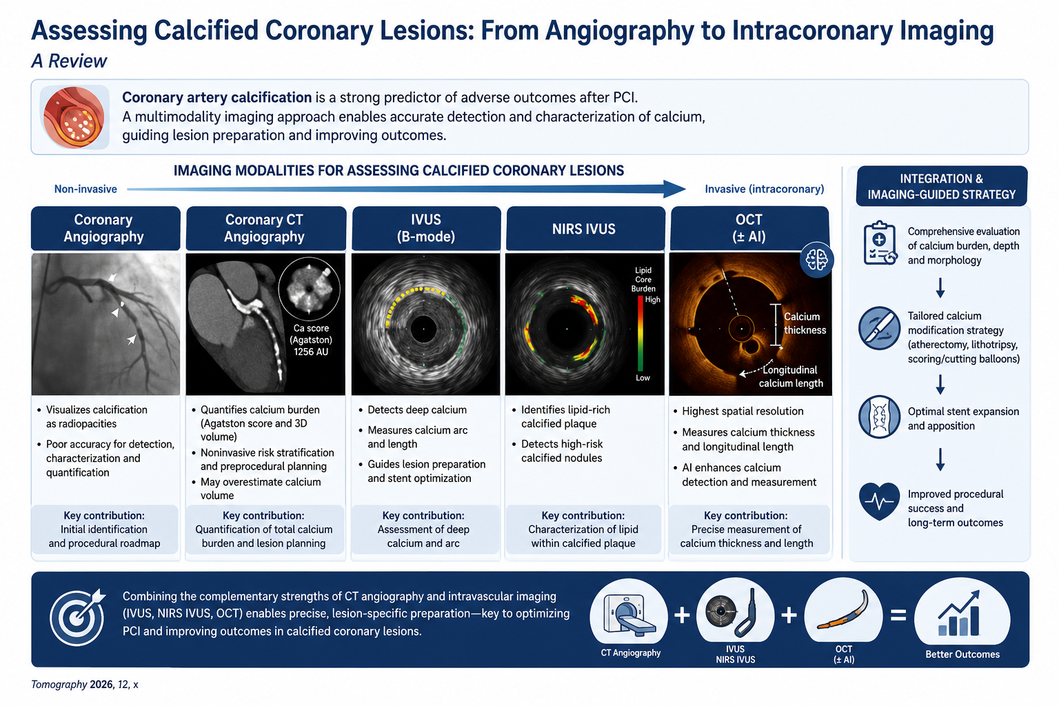
Background/Objectives: Coronary artery calcification is a leading cause of stent failure and a strong predictor of adverse outcomes after percutaneous coronary intervention (PCI). Accurate characterization of calcium burden, depth, and morphology is central to procedural planning, targeted treatment, and better outcomes. This review summarizes the contemporary armamentarium for imaging calcified coronary lesions, from coronary angiography and computed tomography to advanced intracoronary imaging, and how it facilitates treatment. Methods: We synthesized evidence from landmark studies, validated imaging scoring systems, registries, and current guideline and consensus documents addressing the assessment and imaging guided treatment of coronary calcification. Results: Invasive angiography shows poor diagnostic accuracy for coronary calcium detection, characterization, and quantification. Coronary CT angiography noninvasively quantifies calcium (Agatston score and three-dimensional volume) and supports preprocedural planning, although it overestimates calcium volume. Intravascular imaging by means of intravascular ultrasound (IVUS) and optical coherence tomography (OCT) effectively detects coronary calcium and provides the best evidence to avoid stent failure. IVUS accurately measures deep calcium, calcium arc, and length; adding near infrared spectroscopy (NIRS IVUS) characterizes lipid within calcified plaque and high risk calcified nodules. OCT provides the highest spatial resolution and uniquely measures calcium thickness and longitudinal calcium length, while the utilization of artificial intelligence further enhances calcium assessment. Integration of these modalities into a morphology based, imaging guided approach facilitates calcium detection and modification, leading to PCI optimization and improved outcomes. Conclusions: Coronary CT angiography and intravascular imaging have transformed the assessment of calcified coronary lesions. Combining their complementary strengths enables precise, lesion specific preparation and is crucial to improve outcomes in contemporary calcified lesion PCI.
Keywords:
coronary artery calcification
; intracoronary imaging
; intravascular ultrasound
; optical coherence tomography
; NIRS IVUS
; high frequency optical coherence tomography
; coronary computed tomography angiography
; calcium scoring
; percutaneous coronary intervention
; stent underexpansion
1. Introduction
Coronary artery calcification is a hallmark of advanced atherosclerosis and a major determinant of technical difficulty and worse clinical outcomes of percutaneous coronary intervention (PCI). Population aging is sharply increasing the prevalence of severely calcified lesions during PCI. Calcified plaques are rigid and poorly compliant, leading to impaired lesion crossing, balloon expansion, stent delivery, and stent deployment; intravascular imaging shows that stent underexpansion and a smaller minimal stent area are associated with higher rates of stent thrombosis and in stent restenosis [1,2,3], and pooled analyses have demonstrated that moderate to severe calcification is independently associated with higher rates of ischaemic events and major adverse cardiac events following PCI [4,5].
The decision of whether and how to modify calcium depends not just on the presence of calcium but on its burden, circumferential arc, thickness, depth, and morphology. These parameters are captured imperfectly by angiography [6,7]. Over the past two decades, the assessment of calcified coronary lesions has evolved from a purely angiographic sign into a multimodality discipline spanning noninvasive computed tomography and high resolution intracoronary imaging [8]. This review provides a comprehensive overview of contemporary imaging modalities, from coronary angiography to coronary CT angiography (CCTA), intravascular ultrasound (IVUS), near infrared spectroscopy IVUS (NIRS IVUS), and optical coherence tomography (OCT) including high frequency OCT (HF OCT), and describes how their complementary strengths can be orchestrated to guide calcium modification, optimize PCI and improve outcomes.
2. Materials and Methods
This narrative review synthesizes the current evidence on multimodality imaging for the assessment of coronary artery calcification and its role in guiding percutaneous coronary intervention (PCI). We searched PubMed/MEDLINE, Scopus, and the Cochrane Library for English-language articles published from database inception through July 2026, using combinations of the following search terms: "coronary artery calcification", "coronary calcification", "intravascular imaging", "intracoronary imaging", "intravascular ultrasound", "near-infrared spectroscopy", "optical coherence tomography", "high-frequency optical coherence tomography", "coronary CT angiography", "calcium score", "stent underexpansion", and "calcium modification". Priority was given to landmark imaging studies, pivotal clinical trials, high-quality observational studies, large registries, and contemporary guideline and consensus documents. The reference lists of relevant publications were also screened to identify additional eligible studies. As this was a narrative rather than a systematic review, studies were selected on the basis of their clinical relevance, methodological quality, and contribution to the topic. No formal risk-of-bias assessment was performed.
3. Pathophysiology and Morphology of Coronary Calcification
Coronary calcification is an actively regulated process involving osteoblast like transformation of vascular smooth muscle cells under the influence of oxidative stress, inflammation, and disordered mineral metabolism [9,10,11,12,13]. These mechanisms promote the deposition of hydroxyapatite crystals within the arterial wall, contributing to plaque progressions and alterations in vascular biomechanics.
Two morphological patterns are recognized. Microcalcifications (measuring <15 µm) develop within the lipid rich necrotic core of vulnerable plaques and increase local mechanical stress on the fibrous cap, thereby promoting plaque instability and rupture [14,15,16]. These deposits are below the spatial resolution of current intracoronary imaging modalities. In contrast, macrocalcifications form dense larger sheets or nodules and are commonly associated with stable atherosclerotic plaques but present substantial technical challenges during PCI due to reduced vessel compliance [17].
From an interventional perspective, the distinction between intimal (superficial) and medial (deep) calcification is particularly important. Intimal calcification develops within atherosclerotic plaques and represents the principal target of coronary intervention, whereas medial calcification (Mönckeberg sclerosis) primarily affects the medial layer of the vessel wall, contributes to arterial stiffening, and is encountered more frequently in patients with diabetes mellitus and chronic kidney disease [18,19]. Although both patterns may coexist, differentiating them has important procedural implications because superficial calcium is generally more amenable to atherectomy-based plaque modification, whereas deeper medial calcium is more effectively fractured using intravascular lithotripsy [18,19].
A further high-risk morphological entity is the calcified nodule, characterised by eruptive nodular calcium protruding into the coronary lumen following disruption of the fibrous cap [20,21,22]. Calcified nodules are associated with acute coronary syndromes, impaired device delivery, incomplete stent expansion, and increased procedural complexity. Their recognition is therefore important both for procedural planning and for selecting the most appropriate calcium-modification strategy.
Importantly, the procedural behaviour of calcified coronary lesions depends not only on the overall calcium burden but also on its three-dimensional morphology, including calcium thickness, circumferential arc, longitudinal extent, depth, and distribution. These characteristics determine lesion compliance, the likelihood of adequate balloon expansion, the probability of calcium fracture during lesion preparation, and ultimately the risk of stent underexpansion. Consequently, contemporary coronary imaging has evolved beyond simple calcium detection toward comprehensive morphological characterization that directly informs procedural planning and device selection [23].
Because these morphological features cannot be adequately characterized by conventional angiography alone, contemporary assessment increasingly relies on complementary non-invasive and intracoronary imaging modalities. The ability to quantify calcium arc, thickness, depth, longitudinal extent, and plaque composition has become central to selecting the optimal calcium-modification strategy and achieving durable PCI results.
4. Impact of Coronary Calcification on Percutaneous Coronary Intervention
Coronary calcification is not merely a marker of disease burden; it actively impairs every stage of PCI. At the outset, heavily calcified and tortuous segments hinder the advancement of guidewires and may limit the crossing of balloons, intravascular imaging catheters, atherectomy devices and stents [24,25]. Even after successful wire crossing, conventional balloons may fail to dilate the lesion, the so called balloon undilatable lesion, encountered in approximately 5 to 10% of calcified procedures [25,26]. When dilation is achieved, rigid calcium causes asymmetric, incomplete stent expansion, with stent expansion reduced as much as 60 to 70% relative to adjacent noncalcified segments [18,27]. Incomplete lesion expasion may result in inadequate stent apposition, further increasing the risk of adrverse procedural and long-term outcomes. Calcified lesions also carry a higher incidence of procedural complications, including coronary dissection, perforation, material entrapment and slow flow or no reflow from distal embolisation of calcific debris [28,29].
These acute mechanical consequences translate into worse long-term outcomes. Stent underexpansion is the strongest predictor of stent thrombosis, a complication with mortality exceeding 40% in some series [30,31], while stent malapposition and geographic miss promote in stent restenosis and neoatherosclerosis [32].
The adverse prognostic impact of calcification has been quantified across large registries and pooled analyses. A patient level pooled analysis of seven contemporary stent trials, comprising more than 20,000 patients, found that moderate to severe calcification independently increased all cause mortality by approximately 50% and major adverse cardiac events by approximately 40% [4], and a pooled analysis of the HORIZONS AMI and ACUITY trials linked calcification to excess ischaemic events after PCI in acute coronary syndromes [5]. In a prospective registry of more than 10,000 patients, moderate to severe calcification independently predicted target vessel revascularisation and composite events at two years [33], and even with second generation drug eluting stents, patients with significant calcification experienced substantially higher five year rates of target lesion failure [34,35]; a single centre study similarly reported markedly higher one year event rates in severely calcified lesions [36]. In the multicentre COBIS II registry of 2,897 patients, moderate or severe calcification of bifurcation lesions increased the adjusted risk of target lesion failure (hazard ratio 1.31), driven mainly by repeat revascularisation [37].
Despite substantial advances in calcium-modification technologies, patients with severe coronary calcification continue to represent one of the highest risk populations undergoing PCI. Contemporary registries of patients treated with dedicated calcium modification confirm that event rates remain high in this population: in a series of 632 patients undergoing rotational atherectomy for predominantly severe calcification, three year mortality reached 21.7% and target lesion revascularization 13.0%, reflecting the high risk lesion and patient profile rather than a failure of treatment [38]. This continuum, from impaired lesion crossing and preparation to stent underexpansion, procedural complications, and adverse long term outcomes, explains why accurate assessment and modification of calcium before stenting, is crucial during contemporary PCI [39,40].
5. Coronary Angiography: The Starting Point
Coronary angiography remains the most widely used and immediately available method for assessing calcification as it provides real-time visualization of coronary anatomy, lesion severity, coronary flow and device positioning. It allows an initial assessment of lesion complexity and procedural planning.
Unofrtunately, angiogaphy has limited sensitivity for detecting and characterising coronary calcifications. In the classic intravascular ultrasound study by Mintz et al with evaluation of 1,155 lesions, angiography identified calcium in only 38% of lesions in which IVUS detected calcium in 73%, while extensive four-quadrant calcium was recognised substantially less frequently [1]. Similarly, Wang et al, demonstrated that angiography had a sensitivity of approximately 60% for calcium detection compared with 83% for IVUS and 96% for OCT, with consistent underestimation of calcium arc and depth, as well as considerable intrerobserver variability [8].
These limitations are inherent to angiography as a two-dimensional luminographic technique. It detects only sufficiently dense radiopaque calcium and cannot reliably determine calcium thickness, circumferential arc, depth, or plaque composition. Consequently, lesions that appear only moderately calcified angiographically may harbour severe circumferential calcium on intravascular imaging and therefore be at increased risk of inadequate lesion preparation and stent underexpansion.
Severe angiographic calcification, defined as radiopacities visible on both vessel walls without cardiac motion, should therefore be regarded as an indication for further assessment with intracoronary imaging rather than as a complete characterization of calcium severity as recommended by contemporary consensus [41].
Coronary angiography provides the starting point for evaluating calcified coronary lesions but should be complemented by advanced imaging modalities when detailed morphological assessment is required.
6. Computed Tomography: Noninvasive Assessment of Coronary Calcium
6.1. Coronary Artery Calcium Score
Non-contrast, electrocardiogram-gated cardiac computed tomography quantifies calcium using the Agatston scoring system, which weights each calcified voxel above 130 Hounsfield units (HU) according to its peak density. The resulting coronary artery calcium (CAC) score is a well established marker of total coronary calcium burden and one of the most extensively validated tools for cardiovascular risk stratification in asymptomatic individuals. A CAC score of zero is associated with a very low 10-year risk of cardiovascular events, whereas scores above 400 indicate a high atherosclerotic burden, and are strongly associated with obstructive coronary artery disease and adverse clinical outcomes[42,43,44,45].
Although the CAC Score quantifies the overall burden of coronary calcification, it provides limited information regarding individual lesion characteristics. The prognostic significance of coronary calcium is not determined by burden alone. In the Multi Ethnic Study of Atherosclerosis (MESA), higher calcium density was independently associated with a lower risk of cardiovascular events for any given calcium volume, suggesting that dense, mature calcifications represent a more stable stage of plaque evolution than lower-density calcium associated with active atherosclerosis [46].
The CAC score should be regarded as a marker of global atherosclerotic burden rather than a tool for procedural planning. It does not identify the lesion-specific distribution, circumferential arc, thickness or depth of calcium, nor can it determine the morphological characteristics that influence PCI strategy. These features require Contrast enhanced coronary CT angiography (CCTA) or intravascular imaging for accurate assessment [23].
6.2. Plaque and Lesion Characterization on Coronary CT Angiography
CCTA extends beyond calcium quantification to provide three-dimensional characterization of individual coronary plaques, allowing classification as calcified, non-calcified, or mixed and depicting the distribution and pattern of calcification, from discrete spotty deposits to extensive sheet-like calcium. Unlike non-contrast CAC scoring, CCTA simultaneously evaluates both calcified and non-calcified plaque components, providing a more comprehensive assessment of plaque morphology and overall coronary atherosclerotic burden.
Several adverse plaque characteristics can be identified on CCTA and have been consistently associated with an increased risk of future cardiovascular events, including low-attenuation plaque, positive remodelling, the napkin-ring sign, and spotty calcification [47]. The coexistence of multiple high-risk plaque features identifies lesions with greater biological activity and a higher likelihood of subsequent clinical events. In contrast, extensive confluent calcification generally reflects more advanced and stable atherosclerotic disease, although it frequently presents greater technical challenges during PCI.
CCTA provides information that extends beyond cardiovascular risk prediction by identifying lesion morphology before the procedure. Plaque composition, calcium distribution, vessel course, lesion length, and the relationship of calcification to adjacent non-calcified plaque can all be assessed non-invasively, facilitating procedural planning before coronary angiography.
Recent advances in artificial intelligence and deep-learning algorithms have further expanded the capabilities of CCTA. Automated plaque analysis now enables rapid quantification of total, calcified, and non-calcified plaque volumes with good agreement compared with intravascular imaging while providing incremental prognostic information beyond conventional visual assessment [48,49].
6.3. Calcium Quantification and Preprocedural Planning
The principal advantage of CCTA lies in procedural planning before coronary angiography. By providing three-dimensional visualisation of calcium distribution throughout the coronary tree, CCTA allows early identification of heavily calcified lesions and facilitates planning of the interventional strategy. It also enables assessment of vessel size, lesion length, coronary tortuosity, and the anatomical distribution of calcification, all of which may influence vascular access, guide catheter selection, device choice, and the anticipated need for calcium-modification techniques.
In a direct comparison with OCT, CCTA-derived calcium volume overestimated the reference standard by approximately 60%, while maintaining good agreement for minimal lumen area [50]. Accordingly, CCTA should be regarded as a reliable tool for identifying extensive coronary calcification rather than for precise quantification of calcium volume.
The principal limitation of CCTA in heavily calcified vessels remains calcium blooming artefact, which exaggerates calcium dimensions and obscures the residual coronary lumen, potentially leading to overestimation of stenosis severity. Recent advances in scanner technology, including photon-counting CT and ultra-high-resolution image reconstruction, have substantially reduced blooming artefact and improved lumen visualisation, resulting in more accurate assessment of calcified coronary stenoses [51].
Emerging technologies may further enhance the role of CCTA in procedural planning. CT-derived fractional flow reserve (CT-FFR) provides complementary functional assessment of lesion significance, while artificial intelligence-assisted image analysis enables automated plaque quantification and risk stratification. Although these techniques continue to evolve, they have the potential to further refine patient selection and procedural planning for complex calcified coronary disease [52,53].
7. Intravascular Ultrasound and NIRS IVUS
7.1. Intravascular Ultrasound
Intravascular ultrasound (IVUS) uses high-frequency ultrasound (typically 20–60 MHz) to generate cross-sectional tomographic images of the coronary artery, enabling assessment of both the vessel lumen and the arterial wall. Its greater tissue penetration compared with optical coherence tomography allows visualisation of the entire vessel architecture, making IVUS particularly useful in large vessels, ostial lesions, and diffuse coronary disease.
Calcium has a characteristic appearance on IVUS. Because it strongly reflects ultrasound waves, it is visualised as a bright hyperechoic arc accompanied by posterior acoustic shadowing, frequently associated with reverberation artefacts. Although this appearance allows calcium to be identified with high confidence, the acoustic shadow prevents visualisation of tissue behind the calcified plaque, limiting direct measurement of calcium thickness [23,54].
Consequently, IVUS accurately assesses calcium arc, longitudinal length, and overall calcium distribution, but does not directly measure calcium thickness or consistently distinguish superficial from deep calcium beyond its leading edge [23,54]. These measurements are clinically important because increasing calcium arc and longitudinal extent are associated with a greater likelihood of incomplete stent expansion. In particular, a calcium arc >270° has consistently been associated with impaired stent expansion following PCI [23,27].
Building on these observations, Zhang et al developed and validated an IVUS-derived calcium score incorporating four independent predictors of stent underexpansion: (1) a superficial calcium arc >270° extending over >5 mm, (2) circumferential (360°) calcium, (3) the presence of a calcified nodule, and (4) a reference vessel diameter <3.5 mm. A score>2 identifies lesions at high risk of stent underexpansion, with excellent negative predictive value, and may assist in selecting lesions that require calcium-modification techniques before stent implantation [7].
Beyond calcium assessment, IVUS offers important practical advantages during PCI. Because ultrasound penetrates blood and does not require contrast-mediated blood clearance, image quality is maintained in large vessels, ostial lesions, left main disease, and haemodynamically unstable patients. IVUS also permits accurate measurement of vessel dimensions, plaque burden, and stent expansion throughout the procedure. Multiple randomised trials and meta-analyses have demonstrated that IVUS-guided PCI reduces major adverse cardiovascular events, target lesion revascularisation, and stent thrombosis compared with angiography-guided PCI, supporting its routine use in complex coronary intervention [55,56].
7.2. Near Infrared Spectroscopy IVUS
Near-infrared spectroscopy combined with intravascular ultrasound (NIRS-IVUS) integrates structural and compositional plaque assessment within a single imaging catheter. While IVUS defines vessel architecture and calcium morphology, NIRS identifies lipid-rich plaque by measuring the maximum lipid core burden index over 4 mm, thereby complementing morphological assessment with information on plaque composition [57].
This combined approach is particularly valuable in calcified coronary disease, where calcium and lipid frequently coexist within the same lesion. NIRS-IVUS identifies lipid-rich components within or adjacent to calcified plaques ("lipidic calcification"), a feature that cannot be detected by grayscale IVUS alone. Recognition of these lipid-rich calcified lesions may be clinically important, as they appear more susceptible to distal embolisation and microvascular obstruction during PCI [57].
NIRS-IVUS also refines the evaluation of calcified nodules, an established high-risk plaque phenotype. Among patients presenting with acute coronary syndromes attributable to calcified nodules, a maxLCBI ≥400 has been associated with a higher incidence of the no-reflow phenomenon and worse long-term clinical outcomes [58].
Although NIRS does not improve calcium quantification itself, it provides incremental information that complements IVUS-derived morphological assessment. By distinguishing predominantly calcified plaques from those containing a substantial lipid core, NIRS-IVUS enhances lesion characterisation and may help identify lesions at increased risk of distal embolisation during PCI.
8. Optical Coherence Tomography and High Frequency OCT
8.1. Optical Coherence Tomography
Optical coherence tomography (OCT) uses near-infrared light rather than ultrasound to generate high-resolution cross-sectional images of the coronary artery. With an axial resolution of approximately 10–20 µm, OCT provides the highest spatial resolution among currently available coronary imaging modalities, nearly ten times greater than IVUS. Unlike ultrasound, near-infrared light penetrates calcified tissue sufficiently to delineate its borders, allowing OCT to characterize calcium morphology with greater precision. On OCT, calcium appears as a signal-poor or heterogeneous region with sharply defined borders, enabling direct measurement of calcium thickness, in addition to calcium arc and longitudinal extent [59,60].
In a head-to-head comparison, OCT demonstrated the highest sensitivity for calcium detection (96%) compared with IVUS (83%) and angiography (60%) [8]. This superior resolution has enabled the development of OCT-derived calcium scoring systems that incorporate the morphological features most strongly associated with stent underexpansion. The Fujino OCT calcium score assigns points according to maximum calcium angle >180° (2 points), calcium thickness >0.5 mm (1 point), and calcium length >5 mm (1 point). A maximum score of 4 identifies lesions at increased risk of stent underexpansion, with lower stent expansion compared with lesions with lower scores (78% versus 96% expansion) [6]. A modified OCT calcium score incorporating calcified nodules and luminal calcium protrusion has subsequently been proposed to improve risk stratification in the contemporary drug-eluting stent (DES) era [61].
Beyond calcium quantification, OCT provides important procedural information during complex PCI. It is considered the reference intracoronary imaging modality for detailed characterization of calcified nodules [21] and enables direct assessment of calcium fracture following lesion modification, including the circumferential calcium fractures observed after intravascular lithotripsy [18]. OCT also facilitates stent optimization by allowing precise evaluation of stent expansion, apposition, and edge complications, with a low stent expansion index independently associated with adverse clinical outcomes [62].
The main limitations of OCT are related to its optical nature. Light penetration is limited in very thick or lipid-rich calcified plaques, where the deep calcium border may not be fully visualized. In addition, OCT requires transient displacement of blood using contrast injection, which may limit its use in patients with advanced renal dysfunction, haemodynamic instability, or situations where increased contrast volume administration is undesirable.
8.2. High Frequency OCT and Emerging Software Technologies
High-frequency optical coherence tomography (HF-OCT) is a recent technological advancement designed to improve the practicality and applicability of intracoronary OCT imaging. The Gentuity HF-OCT (Gentuity LLC, Sudbury, Massachusetts, USA) system incorporates a substantially smaller imaging catheter and faster pullback speeds than conventional OCT, facilitating image acquisition in complex coronary anatomy, including tortuous, heavily calcified, and distal vessels. These technical refinements aim to improve catheter deliverability while preserving the high spatial resolution that distinguishes OCT.
Early clinical experience has been encouraging. In an initial feasibility study, HF-OCT demonstrated excellent deliverability, successful lesion crossability, and high-quality image acquisition before and after PCI across a broad range of vessel sizes, although severe vessel tortuosity reduced the length of analysable images [63]. Subsequent studies have confirmed the feasibility of HF-OCT in large coronary arteries and other complex coronary anatomies, supporting its potential to expand the use of OCT-guided PCI [64]. However, larger prospective studies are still required to determine whether these technical advantages translate into improved procedural or clinical outcomes.
Advances in software have accompanied developments in imaging hardware. Artificial intelligence (AI)-assisted OCT analysis, exemplified by the Ultreon™ software platform (Abbott, North Chicago, Illinois, USA), automates calcium detection together with lumen and external elastic lamina measurements, improving the efficiency and reproducibility of image interpretation. In a multireader study involving 30 operators, AI-assisted analysis improved the accuracy of calcium severity assessment and vessel preparation strategy selection while reducing interobserver variability and analysis time [65].
At present, AI-assisted analysis should be regarded as a decision-support tool rather than a replacement for expert interpretation. Although these technologies improve workflow and standardise image analysis, evidence demonstrating an impact on procedural or long-term clinical outcomes remains limited. As hardware and software continue to evolve, HF-OCT and AI-assisted analysis have the potential to further facilitate the adoption of OCT-guided PCI in complex calcified coronary disease.
9. Multimodality Comparison and Imaging Guided Device Selection
No single imaging modality provides a complete assessment of calcified coronary lesions. Each technique contributes distinct information across different stages of evaluation: coronary angiography provides initial anatomical assessment and procedural guidance, CCTA enables non-invasive whole-coronary assessment and preprocedural planning, IVUS provides robust evaluation of vessel architecture and calcium distribution, NIRS-IVUS adds information regarding plaque composition, and OCT/HF-OCT offers the highest spatial resolution for detailed calcium morphology and thickness assessment. The complementary strengths and limitations of these modalities are summarized in Table 1, while validated intracoronary calcium scores associated with stent under-expansion are presented in Table 2.
Although these scores reliably identify lesions at increased risk of stent underexpansion, they were developed primarily as risk stratification tools and prospective studies are needed to determine whether score-guided device selection improves clinical outcomes. These imaging parameters provide the basis for a morphology-guided approach to calcium modification (Figure 1). According to the 2024 SCAI Expert Consensus, severe angiographic calcification should prompt further evaluation with intracoronary imaging, while high-risk calcium features identified by IVUS or OCT may indicate lesions that could benefit from upfront calcium modification [41,66]. Importantly, these imaging criteria are currently supported by mechanistic and observational evidence, but prospective trials directly demonstrating improved clinical outcomes with imaging-guided device selection remain limited.
Applied clinically, imaging findings can assist in matching lesion morphology with the most appropriate calcium-modification strategy. Superficial, concentric, or extensive calcification may favour rotational or orbital atherectomy, which primarily modifies superficial intimal calcium through mechanical plaque ablation [68,69,70,71,72]. Deep or circumferential calcium may be more suitable for intravascular lithotripsy, which generates acoustic pressure waves capable of producing fractures within both superficial and deep calcium deposits [73,74,75,76]. Focal, moderate, and non-circumferential calcification may be approached with scoring or cutting balloons or high-pressure non-compliant balloons [77,78]. Finally, lesions that remain balloon-undilatable or demonstrate persistent deep calcium despite initial modification may require combination strategies, including RotaTripsy [79,80].
Current guidelines and consensus documents increasingly support this imaging-informed and morphology-guided approach to calcified lesion preparation. The 2021 ACC/AHA/SCAI guideline assigns rotational atherectomy a Class IIa recommendation, while other calcium-modification devices receive Class IIb recommendations [81]. More recent SCAI, EAPCI, and ESC consensus documents emphasise the importance of intracoronary imaging for calcium characterization before and after lesion modification, although the optimal device-selection strategy remains dependent on lesion morphology, operator expertise, and available technology [41,67,82].
10. Impact of Imaging Guided PCI on Outcomes
The clinical rationale for detailed coronary calcium assessment is that imaging-guided PCI allows more accurate procedural optimization and correction of angiographically occult limitations. The principal mechanism through which coronary calcification adversely affects PCI is inadequate lesion preparation and subsequent stent under-expansion, which are strongly associated with stent thrombosis, restenosis, and adverse clinical outcomes [2]. An OCT-derived stent expansion index below 0.86 has independently been associated with major adverse cardiovascular events [62]. By enabling accurate assessment of vessel dimensions, lesion preparation, stent deployment, and residual mechanical complications, intravascular imaging provides information that cannot be reliably obtained from angiography alone.
A substantial body of randomized evidence supports the use of IVUS-guided PCI. The IVUS-XPL trial demonstrated a sustained reduction in major adverse cardiac events at five years among patients undergoing PCI for long coronary lesions compared with angiography-guided intervention [55]. The ULTIMATE trial further demonstrated a reduction in target vessel failure with IVUS-guided drug-eluting stent implantation in an all-comers population, with the greatest benefit observed when predefined criteria for an optimal IVUS-guided result were achieved [83]. A network meta-analysis of randomized trials confirmed that intravascular imaging-guided drug-eluting stent implantation is associated with reductions in target lesion failure, cardiac death, and stent thrombosis compared with angiography-guided PCI [56].
The major randomised trials and meta-analyses evaluating the clinical impact of intravascular imaging-guided PCI are summarized in Table 3.
Evidence supporting OCT-guided PCI has also expanded. The ILUMIEN IV trial demonstrated greater minimal stent area with OCT guidance and provided further evidence supporting the role of high-resolution imaging in procedural optimisation among high-risk PCI patients [84]. The OCTOBER trial demonstrated improved two-year clinical outcomes with OCT guidance in complex bifurcation lesions, highlighting the value of detailed anatomical assessment in challenging interventions [85]. The OCTIVUS trial subsequently demonstrated that OCT-guided PCI was non-inferior to IVUS-guided PCI for one-year clinical outcomes, suggesting that both imaging modalities can provide effective procedural guidance when appropriately applied [86].
The relevance of intravascular imaging is particularly evident in the complex and calcified lesions that represent the focus of this review. The RENOVATE-COMPLEX-PCI trial demonstrated improved outcomes with intravascular imaging-guided PCI in patients undergoing complex coronary intervention [87], while the ECLIPSE trial specifically evaluated patients with severe coronary calcification and demonstrated a reduction in target vessel failure with imaging-guided PCI compared with angiography-guided intervention [88]. Intravascular imaging should be considered an active component of PCI optimisation rather than solely a diagnostic adjunct.
Despite increasing evidence, real-world adoption of intravascular imaging remains limited. In a large United States analysis, IVUS use increased from approximately 6% of PCI procedures in 2008 to 18% by 2020 [89]. This persistent gap between evidence and clinical implementation highlights the need for continued efforts to integrate intravascular imaging into routine practice, particularly for patients with complex and heavily calcified coronary disease.
11. Barriers to Implementation
The barriers to imaging adoption are structural rather than evidentiary. Economic disincentives, workflow constraints, uneven distribution of expertise, ambiguous selection criteria, and interpretive variability each operate independently, and addressing any one in isolation is unlikely to close the implementation gap. Progress will require coordinated action across reimbursement policy, training structures, technology development, and guideline specificity, alongside the prospective trials needed to validate imaging-guided treatment algorithms.
12. Future Directions
Future advances in the imaging of calcified coronary lesions are likely to arise from continued improvements in both imaging technology and image interpretation. Rather than replacing existing imaging modalities, these developments aim to improve the efficiency, reproducibility, and accessibility of imaging-guided PCI.
AI-assisted image analysis is being developed to automate calcium detection, segmentation, and quantification across OCT, IVUS, and CCTA. These technologies have the potential to standardize image interpretation, reduce interobserver variability, and improve workflow efficiency, although current evidence remains largely limited to feasibility and validation studies [49,53,90,91,92,93,94]. Whether AI-assisted analysis translates into improved procedural decision-making or better clinical outcomes remains to be established.
At the same time, ongoing hardware innovations continue to expand the capabilities of coronary imaging. High-frequency OCT seeks to improve catheter deliverability and image acquisition in complex coronary anatomy, while advances in CT technology, including photon-counting detectors and ultra-high-resolution reconstruction, aim to improve visualisation of heavily calcified vessels by reducing blooming artefact. These developments may further enhance the accuracy and practicality of both invasive and non-invasive coronary imaging.
Future research should focus on prospective studies evaluating whether imaging-derived calcium scores, morphology-guided device selection, and AI-assisted image interpretation improve procedural efficiency and long-term clinical outcomes. Establishing standardized imaging protocols and validating imaging-guided treatment algorithms will be essential before these technologies can be routinely incorporated into clinical practice.
13. Limitations
This review has several limitations. First, it is a narrative rather than a systematic review; consequently, although the literature search was comprehensive, the absence of a predefined protocol, formal study selection process, and quantitative quality assessment introduces the potential for selection bias. Second, the available evidence is heterogeneous, comprising randomised trials, observational studies, registry data, expert consensus documents, and imaging validation studies with differing methodologies and clinical endpoints.
In addition, the evidence supporting newer imaging technologies, including NIRS-IVUS in calcified coronary disease, high-frequency OCT, and AI-assisted image analysis, remains limited and is derived predominantly from feasibility studies, observational cohorts, or early clinical experience rather than large randomised outcome trials.
Finally, although contemporary consensus documents increasingly advocate morphology-guided calcium modification based on intracoronary imaging, many proposed imaging algorithms and device-selection strategies have not yet been prospectively validated in dedicated randomised studies. Further prospective investigations are needed to determine whether imaging-guided treatment algorithms translate into improved long-term clinical outcomes.
14. Conclusions
Population aging is sharply increasing the prevalence of severely calcified lesions during PCI. The assessment of calcified coronary lesions has evolved from a predominantly angiographic evaluation to a comprehensive multimodality imaging approach that enables detailed characterization of calcium burden, morphology, and plaque composition. Rather than competing, contemporary imaging modalities provide complementary information that supports lesion assessment before PCI, optimizes procedural planning, and facilitates evaluation of the final procedural result and improves outcomes.
Among invasive imaging techniques, IVUS and OCT have become central to contemporary calcium assessment, with validated imaging-derived calcium scores allowing identification of lesions at increased risk of stent underexpansion and supporting a more individualised approach to lesion preparation. Emerging technologies, including high-frequency OCT and AI-assisted image analysis, may further improve the efficiency and reproducibility of intracoronary imaging, although prospective clinical validation remains necessary.
Taken together, current evidence supports a shift from angiography-guided intervention toward imaging-informed, morphology-guided PCI. Continued integration of multimodality imaging into routine clinical practice has the potential to guide calcium modification strategies, improve procedural outcomes, and ultimately enhance the care of patients with complex calcified coronary artery disease.
Author Contributions
Conceptualization, S.C.K. and A.S.T.; methodology, S.C.K. and A.S.T.; investigation, S.C.K., A.C.K., K.A., I.A., A.-M.P., N.P., K.P. and N.T.; resources, A.S.T., N.K., P.T. and L.E.P.; writing—original draft preparation, S.C.K.; writing—review and editing, A.S.T., A.C.K., K.A., I.A., A.-M.P., N.P., K.P., N.T., N.K., P.T. and L.E.P.; visualization, S.C.K. and A.C.K.; supervision, A.S.T. and L.E.P.; project administration, A.S.T. All authors have read and agreed to the published version of the manuscript.
Funding
This research received no external funding.
Institutional Review Board Statement
Not applicable.
Data Availability Statement
No new data were created. All data can be found on PubMed.
Conflicts of Interest
The authors declare no conflicts of interest.
Acknowledgments
The authors have reviewed and edited the output and take full responsibility for the content of this publication.
Abbreviations
The following abbreviations are used in this manuscript:
| 2D | Two-dimensional |
| 3D | Three-dimensional |
| ACC | American College of Cardiology |
| AHA | American Heart Association |
| AI | Artificial intelligence |
| CAC | Coronary artery calcification |
| CCTA | Coronary computed tomography angiography |
| CT | Computed tomography |
| CT-FFR | Computed tomography–derived fractional flow reserve |
| DES | Drug-eluting stent |
| EAPCI | European Association of Percutaneous Cardiovascular Interventions |
| ESC | European Society of Cardiology |
| HF-OCT | High-frequency optical coherence tomography |
| HU | Hounsfield units |
| IVUS | Intravascular ultrasound |
| maxLCBI | Maximum lipid core burden index over 4 mm |
| MESA | Multi-Ethnic Study of Atherosclerosis |
| MHz | Megahertz |
| NIRS | Near-infrared spectroscopy |
| NIRS-IVUS | Near-infrared spectroscopy intravascular ultrasound |
| OCT | Optical coherence tomography |
| PCI | Percutaneous coronary intervention |
| SCAI | Society for Cardiovascular Angiography and Interventions |
References
- Mintz, G.S.; Popma, J.J.; Pichard, A.D.; et al. Patterns of calcification in coronary artery disease. A statistical analysis of intravascular ultrasound and coronary angiography in 1155 lesions. Circulation 1995, 91(7), 1959–65. [Google Scholar] [CrossRef] [PubMed]
- Fujii, K.; Carlier, S.G.; Mintz, G.S.; et al. Stent underexpansion and residual reference segment stenosis are related to stent thrombosis after sirolimus-eluting stent implantation: an intravascular ultrasound study. J. Am. Coll. Cardiol. 2005, 45(7), 995–8. [Google Scholar] [PubMed]
- Doi, H.; Maehara, A.; Mintz, G.S.; et al. Impact of post-intervention minimal stent area on 9-month follow-up patency of paclitaxel-eluting stents: an integrated intravascular ultrasound analysis from the TAXUS IV, V, and VI and TAXUS ATLAS Workhorse, Long Lesion, and Direct Stent Trials. JACC Cardiovasc Interv. 2009, 2(12), 1269–75. [Google Scholar] [CrossRef] [PubMed]
- Bourantas, C.V.; Zhang, Y.J.; Garg, S.; et al. Prognostic implications of coronary calcification in patients with obstructive coronary artery disease treated by percutaneous coronary intervention: a patient-level pooled analysis of 7 contemporary stent trials. Heart 2014, 100(15), 1158–64. [Google Scholar] [CrossRef] [PubMed]
- Genereux, P.; Madhavan, M.V.; Mintz, G.S.; et al. Ischemic outcomes after coronary intervention of calcified vessels in acute coronary syndromes. Pooled analysis from the HORIZONS-AMI and ACUITY trials. J. Am. Coll. Cardiol. 2014, 63(18), 1845–54. [Google Scholar] [CrossRef] [PubMed]
- Fujino, A.; Mintz, G.S.; Matsumura, M.; et al. A new optical coherence tomography-based calcium scoring system to predict stent underexpansion. EuroIntervention 2018, 13(18), e2182–e2189. [Google Scholar] [CrossRef] [PubMed]
- Zhang, M.; Matsumura, M.; Usui, E.; et al. Intravascular Ultrasound-Derived Calcium Score to Predict Stent Expansion in Severely Calcified Lesions. Circ. Cardiovasc Interv. 2021, 14(10), e010296. [Google Scholar] [CrossRef] [PubMed]
- Wang, X.; Matsumura, M.; Mintz, G.S.; et al. In Vivo Calcium Detection by Comparing Optical Coherence Tomography, Intravascular Ultrasound, and Angiography. JACC Cardiovasc Imaging 2017, 10(8), 869–879. [Google Scholar] [CrossRef] [PubMed]
- Demer, L.L.; Tintut, Y. Inflammatory, metabolic, and genetic mechanisms of vascular calcification. Arterioscler. Thromb. Vasc. Biol. 2014, 34(4), 715–23. [Google Scholar] [CrossRef] [PubMed]
- Leopold, J.A. Vascular calcification: Mechanisms of vascular smooth muscle cell calcification. Trends Cardiovasc Med. 2015, 25(4), 267–74. [Google Scholar] [CrossRef] [PubMed]
- Johnson, R.C.; Leopold, J.A.; Loscalzo, J. Vascular calcification: pathobiological mechanisms and clinical implications. Circ. Res. 2006, 99(10), 1044–59. [Google Scholar] [CrossRef] [PubMed]
- Giachelli, C.M. Vascular calcification mechanisms. J. Am. Soc. Nephrol. 2004, 15(12), 2959–64. [Google Scholar] [CrossRef] [PubMed]
- Hutcheson, J.D.; Goettsch, C.; Bertazzo, S.; et al. Genesis and growth of extracellular-vesicle-derived microcalcification in atherosclerotic plaques. Nat. Mater. 2016, 15(3), 335–43. [Google Scholar] [CrossRef] [PubMed]
- Vengrenyuk, Y.; Carlier, S.; Xanthos, S.; et al. A hypothesis for vulnerable plaque rupture due to stress-induced debonding around cellular microcalcifications in thin fibrous caps. Proc. Natl. Acad. Sci. U S A 2006, 103(40), 14678–83. [Google Scholar] [CrossRef] [PubMed]
- Maldonado, N.; Kelly-Arnold, A.; Vengrenyuk, Y.; et al. A mechanistic analysis of the role of microcalcifications in atherosclerotic plaque stability: potential implications for plaque rupture. Am. J. Physiol. Heart Circ. Physiol. 2012, 303(5), H619–28. [Google Scholar] [CrossRef] [PubMed]
- Jinnouchi, H.; Sato, Y.; Sakamoto, A.; et al. Calcium deposition within coronary atherosclerotic lesion: Implications for plaque stability. Atherosclerosis 2020, 306, 85–95. [Google Scholar] [CrossRef] [PubMed]
- Mori, H.; Torii, S.; Kutyna, M.; et al. Coronary Artery Calcification and its Progression: What Does it Really Mean? JACC Cardiovasc Imaging 2018, 11(1), 127–142. [Google Scholar] [CrossRef] [PubMed]
- Ali, Z.A.; Brinton, T.J.; Hill, J.M.; et al. Optical Coherence Tomography Characterization of Coronary Lithoplasty for Treatment of Calcified Lesions: First Description. JACC Cardiovasc Imaging 2017, 10(8), 897–906. [Google Scholar] [CrossRef] [PubMed]
- Kini, A.S.; Vengrenyuk, Y.; Pena, J.; et al. Optical coherence tomography assessment of the mechanistic effects of rotational and orbital atherectomy in severely calcified coronary lesions. Catheter Cardiovasc Interv. 2015, 86(6), 1024–32. [Google Scholar] [CrossRef] [PubMed]
- Prati, F.; Gatto, L.; Fabbiocchi, F.; et al. Clinical outcomes of calcified nodules detected by optical coherence tomography: a sub-analysis of the CLIMA study. EuroIntervention 2020, 16(5), 380–386. [Google Scholar] [CrossRef] [PubMed]
- Lee, T.; Mintz, G.S.; Matsumura, M.; et al. Prevalence, Predictors, and Clinical Presentation of a Calcified Nodule as Assessed by Optical Coherence Tomography. JACC Cardiovasc Imaging 2017, 10(8), 883–891. [Google Scholar] [CrossRef] [PubMed]
- Katsaros, O.; Sagris, M.; Karakasis, P.; et al. The Role of Calcified Nodules in Acute Coronary Syndrome: Diagnosis and Management. Int. J. Mol. Sci. 2025, 26(6), 2581. [Google Scholar] [CrossRef] [PubMed]
- Mintz, G.S. Intravascular imaging of coronary calcification and its clinical implications. JACC Cardiovasc Imaging 2015, 8(4), 461–471. [Google Scholar] [CrossRef] [PubMed]
- Tzafriri, A.R.; Garcia-Polite, F.; Zani, B.; et al. Calcified plaque modification alters local drug delivery in the treatment of peripheral atherosclerosis. J. Control Release 2017, 264, 203–210. [Google Scholar] [CrossRef] [PubMed]
- Sharma, S.K.; Tomey, M.I.; Teirstein, P.S.; et al. North American Expert Review of Rotational Atherectomy. Circ. Cardiovasc Interv. 2019, 12(5), e007448. [Google Scholar] [CrossRef] [PubMed]
- Sorini Dini, C.; Nardi, G.; Ristalli, F.; et al. Contemporary Approach to Heavily Calcified Coronary Lesions. Interv. Cardiol. 2019, 14(3), 154–163. [Google Scholar] [CrossRef] [PubMed]
- Kobayashi, Y.; Okura, H.; Kume, T.; et al. Impact of target lesion coronary calcification on stent expansion. Circ. J. 2014, 78(9), 2209–14. [Google Scholar] [CrossRef] [PubMed]
- Madhavan, M.V.; Tarigopula, M.; Mintz, G.S.; et al. Coronary artery calcification: pathogenesis and prognostic implications. J. Am. Coll. Cardiol. 2014, 63(17), 1703–14. [Google Scholar] [CrossRef] [PubMed]
- Shimony, A.; Joseph, L.; Mottillo, S.; et al. Coronary artery perforation during percutaneous coronary intervention: a systematic review and meta-analysis. Can. J. Cardiol. 2011, 27(6), 843–50. [Google Scholar] [CrossRef] [PubMed]
- Iakovou, I.; Schmidt, T.; Bonizzoni, E.; et al. Incidence, predictors, and outcome of thrombosis after successful implantation of drug-eluting stents. JAMA 2005, 293(17), 2126–30. [Google Scholar] [CrossRef] [PubMed]
- Cutlip, D.E.; Windecker, S.; Mehran, R.; et al. Clinical end points in coronary stent trials: a case for standardized definitions. Circulation 2007, 115(17), 2344–51. [Google Scholar] [CrossRef] [PubMed]
- Otsuka, F.; Byrne, R.A.; Yahagi, K.; et al. Neoatherosclerosis: overview of histopathologic findings and implications for intravascular imaging assessment. Eur. Heart J. 2015, 36(32), 2147–59. [Google Scholar] [CrossRef] [PubMed]
- Jia, S.; Li, J.; Zhang, C.; et al. Long-Term Prognosis of Moderate to Severe Coronary Artery Calcification in Patients Undergoing Percutaneous Coronary Intervention. Circ. J. 2020, 85(1), 50–58. [Google Scholar] [CrossRef] [PubMed]
- Guedeney, P.; Claessen, B.E.; Mehran, R.; et al. Coronary Calcification and Long-Term Outcomes According to Drug-Eluting Stent Generation. JACC Cardiovasc Interv. 2020, 13(12), 1417–1428. [Google Scholar] [CrossRef] [PubMed]
- Lee, C.H.; Ahn, J.M.; Lee, K.S.; et al. Prevalence, predictors, prognostic significance, and effect of techniques on outcomes of coronary lesion calcification following implantation of drug-eluting stents: a patient-level pooled analysis of stent-specific, multicenter, prospective IRIS-DES registries. Coron. Artery Dis. 2021, 32(1), 42–50. [Google Scholar] [PubMed]
- Sharma, S.K.; Bolduan, R.W.; Patel, M.R.; et al. Impact of calcification on percutaneous coronary intervention: MACE-Trial 1-year results. Catheter Cardiovasc Interv. 2019, 94(2), 187–194. [Google Scholar] [CrossRef] [PubMed]
- Kim, M.C.; Ahn, Y.; Sim, D.S.; et al. Impact of calcified bifurcation lesions in patients undergoing percutaneous coronary intervention using drug-eluting stents: results from the COronary BIfurcation Stent (COBIS) II registry. EuroIntervention 2017, 13(3), 338–344. [Google Scholar] [CrossRef] [PubMed]
- Koch, T.; Tauchmann, L.; Cassese, S.; et al. Long-term safety and efficacy of rotational atherectomy for lesion preparation of calcified coronary lesions. Int. J. Cardiol. 2025, 440, 133711. [Google Scholar] [CrossRef] [PubMed]
- Ramazani, N.; Abdallah, A.W.; DiCaro, M.V.; et al. Use of Calcium Modification in Percutaneous Coronary Intervention: A Comprehensive Review. J. Clin. Med. 2025, 14(22), 8130. [Google Scholar] [CrossRef] [PubMed]
- Kotoulas, S.C. Calcium-Modifying Devices and Patient Outcomes: Single-Center Experience from “Asklepeion” General Hospital of Voula. Master's Thesis, National and Kapodistrian University of Athens, Athens, Greece, 2026. Available online: https://pergamos.lib.uoa.gr/item/uoadl:5421671.
- Riley, R.F.; Patel, M.P.; Abbott, J.D.; et al. SCAI Expert Consensus Statement on the Management of Calcified Coronary Lesions. J. Soc. Cardiovasc Angiogr. Interv. 2024, 3(2), 101259. [Google Scholar] [CrossRef] [PubMed]
- Agatston, A.S.; Janowitz, W.R.; Hildner, F.J.; et al. Quantification of coronary artery calcium using ultrafast computed tomography. J. Am. Coll. Cardiol. 1990, 15(4), 827–32. [Google Scholar] [CrossRef] [PubMed]
- Greenland, P.; Blaha, M.J.; Budoff, M.J.; et al. Coronary Calcium Score and Cardiovascular Risk. J. Am. Coll. Cardiol. 2018, 72(4), 434–447. [Google Scholar] [CrossRef] [PubMed]
- Blaha, M.J.; Cainzos-Achirica, M.; Greenland, P.; et al. Role of Coronary Artery Calcium Score of Zero and Other Negative Risk Markers for Cardiovascular Disease: The Multi-Ethnic Study of Atherosclerosis (MESA). Circulation 2016, 133(9), 849–58. [Google Scholar] [CrossRef] [PubMed]
- Verma, K.P.; Inouye, M.; Meikle, P.J.; et al. New Cardiovascular Risk Assessment Techniques for Primary Prevention: JACC Review Topic of the Week. J. Am. Coll. Cardiol. 2022, 80(4), 373–387. [Google Scholar] [CrossRef] [PubMed]
- Criqui, M.H.; Denenberg, J.O.; Ix, J.H.; et al. Calcium density of coronary artery plaque and risk of incident cardiovascular events. JAMA 2014, 311(3), 271–278. [Google Scholar] [CrossRef] [PubMed]
- Gu, M.; Mao, Q.; Wang, H.; et al. Coronary computed tomographic angiography-derived anatomic and hemodynamic plaque characteristics in prediction of cardiovascular events. Int. J. Cardiovasc Imaging 2024, 40(8), 1641–1652. [Google Scholar] [CrossRef] [PubMed]
- Lin, A.; Kolossvary, M.; Motwani, M.; et al. Deep learning-enabled coronary CT angiography for plaque and stenosis quantification and cardiac risk prediction: an international multicentre study. Lancet Digit Health 2022, 4(4), e256–e265. [Google Scholar] [CrossRef] [PubMed]
- Schulze, K.; Stantien, A.M.; Williams, M.C.; et al. Coronary CT angiography evaluation with artificial intelligence for individualized medical treatment of atherosclerosis: a Consensus Statement from the QCI Study Group. Nat. Rev. Cardiol. 2026, 23(2), 100–115. [Google Scholar] [CrossRef] [PubMed]
- Monizzi, G.; Sonck, J.; Nagumo, S.; et al. Quantification of calcium burden by coronary CT angiography compared to optical coherence tomography. Int. J. Cardiovasc Imaging 2020, 36(12), 2393–2402. [Google Scholar] [CrossRef] [PubMed]
- Koons, E.K.; Chang, S.; Missert, A.D.; et al. Learned high resolution energy-integrating detector CT angiography: harnessing the power of ultra-high-resolution photon counting detector CT. Med. Phys. 2025, 52(7), e17874. [Google Scholar] [CrossRef] [PubMed]
- Kotoulas, S.C.; Triantafyllis, A.S.; Kontogiannis, N.; et al. Radiation-Free Percutaneous Coronary Intervention: Myth or Reality? J. Cardiovasc Dev. Dis. 2025, 12(9), 339. [Google Scholar] [CrossRef] [PubMed]
- Abdelrahman, K.; Shiyovich, A.; Huck, D.M.; et al. Artificial Intelligence in Coronary Artery Calcium Scoring Detection and Quantification. Diagnostics 2024, 14(2), 125. [Google Scholar] [CrossRef] [PubMed]
- Mintz, G.S.; Nissen, S.E.; Anderson, W.D.; et al. American College of Cardiology Clinical Expert Consensus Document on Standards for Acquisition, Measurement and Reporting of Intravascular Ultrasound Studies (IVUS). J. Am. Coll. Cardiol. 2001, 37(5), 1478–92. [Google Scholar] [CrossRef] [PubMed]
- Hong, S.J.; Mintz, G.S.; Ahn, C.M.; et al. Effect of Intravascular Ultrasound-Guided Drug-Eluting Stent Implantation: 5-Year Follow-Up of the IVUS-XPL Randomized Trial. JACC Cardiovasc Interv. 2020, 13(1), 62–71. [Google Scholar] [PubMed]
- Stone, G.W.; Christiansen, E.H.; Ali, Z.A.; et al. Intravascular imaging-guided coronary drug-eluting stent implantation: an updated network meta-analysis. Lancet 2024, 403(10429), 824–837. [Google Scholar] [CrossRef] [PubMed]
- Nagamine, T.; Usui, E.; Kanaji, Y.; et al. Identification of near-infrared spectroscopy-defined lipid content within or adjacent to calcified coronary plaques using optical coherence tomography. Atherosclerosis 2026, 417, 120786. [Google Scholar] [CrossRef] [PubMed]
- Ozaki, Y.; Kitabata, H.; Hikida, R.; et al. Angiographic No-Reflow in Patients With Acute Coronary Syndrome Caused by Calcified Nodules. Am. J. Cardiol. 2025, 253, 59–67. [Google Scholar] [CrossRef] [PubMed]
- Tearney, G.J.; Regar, E.; Akasaka, T.; et al. Consensus standards for acquisition, measurement, and reporting of intravascular optical coherence tomography studies. J. Am. Coll. Cardiol. 2012, 59(12), 1058–72. [Google Scholar] [CrossRef] [PubMed]
- Kume, T.; Okura, H.; Kawamoto, T.; et al. Assessment of the coronary calcification by optical coherence tomography. EuroIntervention 2011, 6(6), 768–72. [Google Scholar] [CrossRef] [PubMed]
- Sato, T.; Matsumura, M.; Yamamoto, K.; et al. A Revised Optical Coherence Tomography-Derived Calcium Score to Predict Stent Underexpansion in Severely Calcified Lesions. JACC Cardiovasc Interv. 2025, 18(5), 622–633. [Google Scholar] [CrossRef] [PubMed]
- Rai, H.; Harzer, F.; Otsuka, T.; et al. Stent Optimization Using Optical Coherence Tomography and Its Prognostic Implications After Percutaneous Coronary Intervention. J. Am. Heart Assoc. 2022, 11(9), e023493. [Google Scholar] [CrossRef] [PubMed]
- Garin, D.; Cook, S.; Togni, M.; et al. Experience with a novel high frequency optical coherence tomography device for intracoronary imaging: a case series. Front Cardiovasc Med. 2024, 11, 1384222. [Google Scholar] [CrossRef] [PubMed]
- Caron, E.; Ciftcikal, Y.; Cannata, M.; et al. Clinical utilization of high-frequency optical coherence tomography in large coronary arteries. Cardiovasc Revasc Med. 2025, 84, 103–104. [Google Scholar] [CrossRef] [PubMed]
- Sibbald, M.; Mitchell, H.R.; Buccola, J.; Pinilla-Echeverri, N. Impact of Artificial Intelligence-Enhanced Optical Coherence Tomography Software on Percutaneous Coronary Intervention Decisions. J. Soc. Cardiovasc Angiogr. Interv. 2025, 4 3 Pt B, 102438. [Google Scholar] [CrossRef] [PubMed]
- De Maria, G.L.; Scarsini, R.; Banning, A.P. Management of Calcific Coronary Artery Lesions: Is it Time to Change Our Interventional Therapeutic Approach? JACC Cardiovasc Interv. 2019, 12(15), 1465–1478. [Google Scholar] [PubMed]
- Barbato, E.; Gallinoro, E.; Abdel-Wahab, M.; et al. Management strategies for heavily calcified coronary stenoses: an EAPCI clinical consensus statement in collaboration with the EURO4C-PCR group. Eur. Heart J. 2023, 44(41), 4340–4356. [Google Scholar] [CrossRef] [PubMed]
- Abdel-Wahab, M.; Richardt, G.; Joachim Buttner, H.; et al. High-speed rotational atherectomy before paclitaxel-eluting stent implantation in complex calcified coronary lesions: the randomized ROTAXUS trial. JACC Cardiovasc Interv. 2013, 6(1), 10–9. [Google Scholar] [CrossRef] [PubMed]
- Abdel-Wahab, M.; Toelg, R.; Byrne, R.A.; et al. High-Speed Rotational Atherectomy Versus Modified Balloons Prior to Drug-Eluting Stent Implantation in Severely Calcified Coronary Lesions. Circ. Cardiovasc Interv. 2018, 11(10), e007415. [Google Scholar] [CrossRef] [PubMed]
- Chambers, J.W.; Feldman, R.L.; Himmelstein, S.I.; et al. Pivotal trial to evaluate the safety and efficacy of the orbital atherectomy system in treating de novo, severely calcified coronary lesions (ORBIT II). JACC Cardiovasc Interv. 2014, 7(5), 510–8. [Google Scholar] [CrossRef] [PubMed]
- Lee, M.; Genereux, P.; Shlofmitz, R.; et al. Orbital atherectomy for treating de novo, severely calcified coronary lesions: 3-year results of the pivotal ORBIT II trial. Cardiovasc Revasc Med. 2017, 18(4), 261–264. [Google Scholar] [CrossRef] [PubMed]
- Genereux, P.; Lee, A.C.; Kim, C.Y.; et al. Orbital Atherectomy for Treating De Novo Severely Calcified Coronary Narrowing (1-Year Results from the Pivotal ORBIT II Trial). Am. J. Cardiol. 2015, 115(12), 1685–90. [Google Scholar] [CrossRef] [PubMed]
- Brinton, T.J.; Ali, Z.A.; Hill, J.M.; et al. Feasibility of Shockwave Coronary Intravascular Lithotripsy for the Treatment of Calcified Coronary Stenoses. Circulation 2019, 139(6), 834–836. [Google Scholar] [CrossRef] [PubMed]
- Hill, J.M.; Kereiakes, D.J.; Shlofmitz, R.A.; et al. Intravascular Lithotripsy for Treatment of Severely Calcified Coronary Artery Disease. J. Am. Coll. Cardiol. 2020, 76(22), 2635–2646. [Google Scholar] [CrossRef] [PubMed]
- Ali, Z.A.; Nef, H.; Escaned, J.; et al. Safety and Effectiveness of Coronary Intravascular Lithotripsy for Treatment of Severely Calcified Coronary Stenoses: The Disrupt CAD II Study. Circ. Cardiovasc Interv. 2019, 12(10), e008434. [Google Scholar] [PubMed]
- Kereiakes, D.J.; Di Mario, C.; Riley, R.F.; et al. Intravascular Lithotripsy for Treatment of Calcified Coronary Lesions: Patient-Level Pooled Analysis of the Disrupt CAD Studies. JACC Cardiovasc Interv. 2021, 14(12), 1337–1348. [Google Scholar] [PubMed]
- Rheude, T.; Rai, H.; Richardt, G.; et al. Super high-pressure balloon versus scoring balloon to prepare severely calcified coronary lesions: the ISAR-CALC randomised trial. EuroIntervention 2021, 17(6), 481–488. [Google Scholar] [CrossRef] [PubMed]
- Rheude, T.; Fitzgerald, S.; Allali, A.; et al. Rotational Atherectomy or Balloon-Based Techniques to Prepare Severely Calcified Coronary Lesions. JACC Cardiovasc Interv. 2022, 15(18), 1864–1874. [Google Scholar] [CrossRef] [PubMed]
- Jurado-Roman, A.; Gonzalvez, A.; Galeote, G.; et al. RotaTripsy: Combination of Rotational Atherectomy and Intravascular Lithotripsy for the Treatment of Severely Calcified Lesions. JACC Cardiovasc Interv. 2019, 12(15), e127–e129. [Google Scholar] [PubMed]
- Sardella, G.; Stefanini, G.; Leone, P.P.; et al. Coronary Lithotripsy as Elective or Bail-Out Strategy After Rotational Atherectomy in the Rota-Shock Registry. Am. J. Cardiol. 2023, 198, 1–8. [Google Scholar] [CrossRef] [PubMed]
- Lawton, J.S.; Tamis-Holland, J.E.; Bangalore, S.; et al. 2021 ACC/AHA/SCAI Guideline for Coronary Artery Revascularization: Executive Summary. Circulation 2022, 145(3), e4–e17. [Google Scholar] [CrossRef] [PubMed]
- Vrints, C.; Andreotti, F.; Koskinas, K.C.; et al. 2024 ESC Guidelines for the management of chronic coronary syndromes. Eur. Heart J. 2024, 45(36), 3415–3537. [Google Scholar] [CrossRef] [PubMed]
- Zhang, J.; Gao, X.; Kan, J.; et al. Intravascular Ultrasound Versus Angiography-Guided Drug-Eluting Stent Implantation: The ULTIMATE Trial. J. Am. Coll. Cardiol. 2018, 72(24), 3126–3137. [Google Scholar] [CrossRef] [PubMed]
- Ali, Z.A.; Landmesser, U.; Maehara, A.; et al. Optical Coherence Tomography-Guided versus Angiography-Guided PCI. N Engl. J. Med. 2023, 389(16), 1466–1476. [Google Scholar] [CrossRef] [PubMed]
- Holm, N.R.; Andreasen, L.N.; Walsh, S.; et al. OCT or Angiography Guidance for PCI in Complex Bifurcation Lesions. N Engl. J. Med. 2023, 389(16), 1477–1487. [Google Scholar] [CrossRef] [PubMed]
- Kang, D.Y.; Ahn, J.M.; Yun, S.C.; et al. Optical Coherence Tomography-Guided or Intravascular Ultrasound-Guided Percutaneous Coronary Intervention: The OCTIVUS Randomized Clinical Trial. Circulation 2023, 148(16), 1195–1206. [Google Scholar] [CrossRef] [PubMed]
- Lee, J.M.; Choi, K.H.; Song, Y.B.; et al. Intravascular Imaging-Guided or Angiography-Guided Complex PCI. N Engl. J. Med. 2023, 388(18), 1668–1679. [Google Scholar] [CrossRef] [PubMed]
- Stone, G.W.; Ali, Z.A.; Maehara, A.; et al. Intravascular imaging-guided PCI versus angiography-guided PCI in patients with severely calcified coronary lesions (ECLIPSE): a randomised trial. Lancet 2025, 405(10491), 1721–1731. [Google Scholar]
- Park, D.Y.; Hu, J.R.; Kanitsoraphan, C.; et al. Trends and In-hospital Outcomes of Intravascular Imaging on Single-Vessel Coronary Chronic Total Occlusion Treated With Percutaneous Coronary Intervention. Am. J. Cardiol. 2023, 206, 79–85. [Google Scholar] [CrossRef] [PubMed]
- Chatzizisis, Y.S.; Alsharqi, M.; Shlofmitz, E.; et al. Artificial Intelligence in Cardiovascular Imaging and Interventional Cardiology: Emerging Trends and Clinical Implications. J. Soc. Cardiovasc Angiogr. Interv. 2025, 4(3), 102471. [Google Scholar] [CrossRef] [PubMed]
- Shin, D.; Sami, Z.; Cannata, M.; et al. Artificial Intelligence in Intravascular Imaging for Percutaneous Coronary Interventions: A New Era of Precision. J. Soc. Cardiovasc Angiogr. Interv. 2025, 4 3 Pt B, 102506. [Google Scholar] [CrossRef] [PubMed]
- Chandramohan, N.; Hinton, J.; O'Kane, P. Artificial intelligence for the interventional cardiologist: powering and enabling OCT image interpretation. Interv. Cardiol. 2024, 19, e03. [Google Scholar] [CrossRef] [PubMed]
- Seligman, H.; Bandeira, D.; Demuyakor, A.; et al. Can AI Capture and Quantify Clinical Expertise? Implications for Intracoronary Imaging in Percutaneous Coronary Intervention. J. Soc. Cardiovasc Angiogr. Interv. 2025, 4 3 Pt B, 102523. [Google Scholar] [CrossRef] [PubMed]
- Finocchiaro, T.; Bucciarelli-Ducci, C.; Manka, R.; et al. Machine Learning for Coronary Plaque Characterization: A Multimodal Review of OCT, IVUS, and CCTA. Diagnostics 2025, 15(14), 1755. [Google Scholar] [CrossRef] [PubMed]
Figure 1.
Proposed imaging-guided framework for selecting a calcium modification strategy, synthesised from contemporary consensus documents and available evidence. Severe angiographic calcification prompts intracoronary imaging assessment. IVUS and OCT calcium scores identify lesions at increased risk of stent underexpansion. Calcium morphology then guides device selection: atherectomy may be considered for superficial or extensive calcification, intravascular lithotripsy for deep or circumferential calcium, modified balloons for focal non-circumferential lesions, and combination approaches for resistant balloon-undilatable lesions. This framework is consensus-based and has not been prospectively validated against clinical outcomes. IVUS, intravascular ultrasound; OCT, optical coherence tomography.
Figure 1.
Proposed imaging-guided framework for selecting a calcium modification strategy, synthesised from contemporary consensus documents and available evidence. Severe angiographic calcification prompts intracoronary imaging assessment. IVUS and OCT calcium scores identify lesions at increased risk of stent underexpansion. Calcium morphology then guides device selection: atherectomy may be considered for superficial or extensive calcification, intravascular lithotripsy for deep or circumferential calcium, modified balloons for focal non-circumferential lesions, and combination approaches for resistant balloon-undilatable lesions. This framework is consensus-based and has not been prospectively validated against clinical outcomes. IVUS, intravascular ultrasound; OCT, optical coherence tomography.
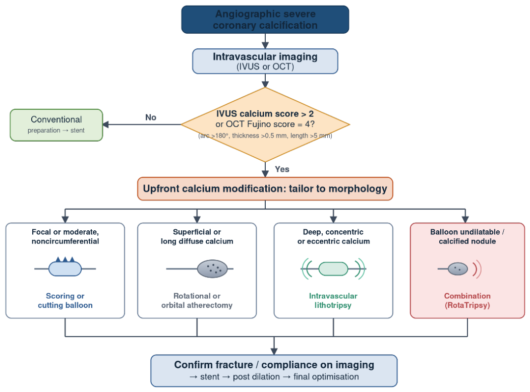

Table 1.
Comparison of imaging modalities for coronary calcium assessment.
| Modality | Resolution | Calcium assessment | Principal strengths | Principal limitations |
| Coronary angiography | ~0.2 mm (2D projection) | Radiopacity; detects ~60% of IVUS calcium | Universal, real time | Underestimates arc and depth; interobserver variability |
| CCTA | ~0.3 to 0.4 mm | Agatston score; 3D volume (overestimates ~60% vs OCT) | Noninvasive, whole tree, preprocedural planning | Blooming artifact; limited lumen detail in severe calcium |
| IVUS | 100 to 200 µm | Arc and length; thickness not measurable (shadowing) | Deep penetration; robust; Zhang score | Cannot measure calcium thickness |
| NIRS IVUS | 100 to 200 µm plus chemical | IVUS metrics plus lipid core (maxLCBI); nodule composition | Adds lipid characterisation; flags high risk nodules | Same thickness limitation as IVUS |
| OCT or HF OCT | 10 to 20 µm | Arc, length, and thickness; Fujino and Sato scores | Highest resolution; direct thickness; HF OCT deliverability | Limited penetration in thick calcium; needs blood clearance |
Table 2.
Validated intravascular imaging calcium scores predicting stent underexpansion.
| Score | Modality | Components | High risk threshold | Predicts |
| Zhang | IVUS | Arc greater than 270° over more than 5 mm; 360° calcium; calcified nodule; vessel smaller than 3.5 mm | Score above 2 | Stent underexpansion |
| Fujino | OCT | Angle greater than 180° (2 pts); thickness greater than 0.5 mm (1 pt); length greater than 5 mm (1 pt) | Score of 4 | Stent underexpansion |
| Sato revised | OCT | Fujino criteria plus calcified nodule and luminal protrusion | Revised contermporary DES-era score | Stent underexpansion |
Table 3.
Major clinical trials evaluating the impact of intravascular imaging-guided PCI.
| Study | Imaging modality | Population | Main finding | Clinical implication |
| IVUS-XPL [55] | IVUS | PCI for long coronary lesions | IVUS-guided PCI reduced major adverse cardiac events with sustained benefit at long-term follow-up | Supports IVUS-guided optimization in complex coronary lesions |
| ULTIMATE [83] | IVUS | All-comers undergoing drug-eluting stent implantation | IVUS-guided PCI reduced target vessel failure compared with angiography guidance | Supports routine IVUS optimization when feasible |
| Network meta-analysis [56] | IVUS/OCT | Randomized trials of intravascular imaging-guided PCI | Imaging-guided PCI reduced target lesion failure, cardiac death, and stent thrombosis compared with angiography guidance | Confirms overall clinical benefit of intravascular imaging |
| ILUMIEN IV [84] | OCT | High-risk PCI patients | OCT guidance resulted in greater minimal stent area and differences in procedural outcomes compared with angiography guidance | Demonstrates the value of high-resolution OCT optimization |
| OCTOBER [85] | OCT | Complex bifurcation lesions | OCT-guided PCI improved two-year clinical outcomes compared with angiography guidance | Supports OCT use in anatomically complex lesions |
| OCTIVUS [86] | OCT vs IVUS | Patients undergoing PCI | OCT-guided PCI was non-inferior to IVUS-guided PCI for one-year clinical outcomes | Suggests both modalities provide effective procedural guidance |
| RENOVATE-COMPLEX-PCI [87] | IVUS/OCT | Complex coronary lesions | Intravascular imaging-guided PCI reduced adverse cardiovascular events compared with angiography guidance | Supports imaging use in complex PCI |
| ECLIPSE [88] | IVUS/OCT | Severely calcified coronary lesions | Imaging-guided PCI reduced target vessel failure in patients undergoing intervention for severe calcification | Provides specific evidence supporting imaging in calcified lesions |
Disclaimer/Publisher’s Note: The statements, opinions and data contained in all publications are solely those of the individual author(s) and contributor(s) and not of MDPI and/or the editor(s). MDPI and/or the editor(s) disclaim responsibility for any injury to people or property resulting from any ideas, methods, instructions or products referred to in the content. |
© 2026 by the authors. Licensee MDPI, Basel, Switzerland. This article is an open access article distributed under the terms and conditions of the Creative Commons Attribution (CC BY) license.
Copyright: This open access article is published under a Creative Commons CC BY 4.0 license, which permit the free download, distribution, and reuse, provided that the author and preprint are cited in any reuse.