

Submitted:
28 July 2026
Posted:
29 July 2026
You are already at the latest version
Abstract
Cognitive decline is closely associated with biological aging and with interconnected processes, including chronic low-grade inflammation, oxidative stress, mitochondrial dysfunction, and cellular senescence. Nutraceutical strategies capable of modulating several of these pathways may represent a complementary approach to supporting cognitive health. This narrative review examines the biological rationale and available evidence regarding palmitoylethanolamide (PEA), luteolin, and their co-ultramicronized formulation (PEA-Lut) in cognitive impairment and dementia. Evidence from mechanistic studies, animal models, observational studies, and clinical trials was considered. PEA is an endogenous N-acylethanolamine with anti-inflammatory and neuroprotective properties, whereas luteolin exerts complementary antioxidant and anti-inflammatory effects. Co-ultramicronization may improve their physicochemical properties and biological activity. Preclinical studies suggest that PEA-Lut may attenuate glial activation, pro-inflammatory signalling, oxidative and nitrosative stress, and amyloid-β-induced cellular injury while supporting neurotrophic signalling and neuronal survival. Preliminary clinical findings have suggested possible benefits in postoperative delirium, stroke rehabilitation, frontotemporal dementia, and other neuroinflammatory conditions. However, human evidence specifically addressing mild cognitive impairment and Alzheimer’s Disease (AD) remains limited, heterogeneous, and largely derived from small or non-randomized studies. PEA-Lut therefore represents a biologically plausible and potentially well-tolerated nutraceutical approach. Adequately powered randomized controlled trials are needed to determine its clinical efficacy, optimal timing and dosage, and long-term safety in individuals at risk of dementia or in the early stages of AD.
Keywords:
aging
; cognitive decline
; cognitive impairment
; luteolin
; neuroinflammation
; palmitoylethanolamide
1. Introduction
Aging is characterized by a complex set of structural and functional changes affecting multiple organs, and physiological systems [1]. Over time, the ability to maintain homeostasis and respond to environmental challenges declines, resulting in increased vulnerability to disease and death. Although some degree of change in physical and cognitive function may accompany aging, clinically relevant cognitive decline is not an inevitable consequence of this process. In some individuals, however, cognitive impairment may progress to dementia, most commonly due to Alzheimer's disease (AD) [2]. Dementia is characterized by a deterioration in memory and other cognitive abilities that interferes with independence in everyday activities, posing substantial challenges for affected individuals, their families, and healthcare system. AD can be conceptualized as a clinical and biological continuum, extending from a preclinical phase to mild cognitive impairment (MCI) due to AD and, subsequently AD dementia of increasing severity [3].
MCI represents an intermediate clinical state between normal cognition and dementia [4]. It is characterized by subjective and objective evidence of cognitive decline beyond that expected for an individual’s age and educational level, without substantial impairment of independence in everyday activities. Individuals with MCI are at increased risk of developing dementia, particularly AD, although progression is not inevitable and cognitive status may remain stable or, in some cases, improve [5]. The clinical diagnosis of AD dementia requires cognitive impairment that interferes with daily functioning and cannot be more appropriately explained by another neurological, psychiatric or systemic condition [6]. From a neuropathological perspective, AD is characterized by the accumulation of amyloid-β-containing plaques and neurofibrillary tangles composed of hyperphosphorylated tau. These alterations are associated with synaptic dysfunction, neuronal loss, and disruption of hippocampal and cortical networks involved in memory and other cognitive functions, leading to poor consolidation of short-term memory into long-term memory [7,8].
Estimates of annual progression from MCI to dementia vary according to the population studied, clinical setting, follow-up duration, and presence of AD biomarkers [9]. Early identification and appropriate management of potentially modifiable factors may nevertheless provide an opportunity to support cognitive health and delay functional decline. Although dementia does not affect all older adults, its incidence and prevalence increase substantially with age [10].
The global number of people living with dementia has been projected to rise from approximately 57.4 million in 2019 to 152.8 million by 2050 [11].
Several interrelated mechanisms may contribute to the association between aging and dementia, including chronic inflammation [12], oxidative stress [13], alterations in neurotransmitter metabolism [14], mitochondrial dysfunction, and cellular senescence. Available symptomatic treatments provide limited clinical benefits, while emerging disease-modifying therapies may slow cognitive and functional decline in selected patients with early AD. However, their eligibility requirements, safety concerns, monitoring burden, and limited applicability to the broader population leave a substantial need for complementary preventive and supportive approaches [14,15].
The identification and modification of dementia risk factors therefore remain important components of prevention and clinical management [15]. In addition to physical activity [16] and cognitive stimulation [17], dietary interventions and nutritional supplementation [18] have attracted interest as possible approaches to support cognitive health. Nutraceuticals are products derived from food sources that may provide physiological benefits beyond their basic nutritional value.
Certain nutraceutical compounds have been investigated for their potential ability to influence inflammation, oxidative stress, and other biological pathways involved in brain aging. However, evidence of biological activity should not be considered equivalent to evidence of dementia prevention or clinical efficacy.
Among the compounds currently under investigation, palmitoylethanolamide (PEA) and luteolin have emerged as biologically plausible candidates. PEA is a naturally occurring fatty acid amide with anti-inflammatory and neuroprotective properties, whereas luteolin is a flavonoid found in several fruits and vegetables that exerts antioxidant and anti-inflammatory effects. Both compounds have demonstrated potentially relevant biological activities in experimental studies, although clinical evidence remains limited.
This narrative review examines the biological rationale and current experimental and clinical evidence supporting the potential role of PEA, luteolin, and their co-ultramicronized formulation in brain aging and cognitive decline. Particular attention is given to their effects on neuroinflammation, oxidative stress, mitochondrial dysfunction, cellular senescence, glial activation, and neurotrophic signalling. Evidence derived from cellular models, animal studies, acute neurological conditions, and neurodegenerative disorders is critically discussed, with emphasis on its relevance and limitations in relation to MCI and AD. Rather than establishing a preventive or therapeutic effect, this review aims to determine whether the available evidence provides a sufficient rationale for future randomized clinical trials in individuals at risk of dementia or in the early stages of cognitive impairment.
2. Methods
A structured literature search was conducted in PubMed/MEDLINE and the Cochrane Library in May 2026. The search included combinations of the following terms: “aging,” “dementia,” “older adults,” “nutrition,” “palmitoylethanolamide,” “PEA,” “luteolin,” “co-ultramicronized palmitoylethanolamide,” “nutraceuticals,” “cognitive impairment,” “mild cognitive impairment,” “Cognitive decline”, “Alzheimer’s disease,” and “neuroinflammation.” The search was designed to identify relevant experimental and clinical evidence, including in vitro studies, animal models, observational studies, and clinical trials. Priority was given to English-language publications directly examining the biological effects of PEA, luteolin, or PEA-Lut and their potential relevance to brain aging, cognitive impairment, and neurodegenerative disorders. Studies conducted in other inflammatory or neurological conditions were considered when they provided mechanistic or translational information relevant to neuroinflammation, oxidative stress, or cognition. Additional publications were identified by screening the reference lists of relevant articles. Given the narrative nature of this review, study selection and evidence synthesis did not follow a formal systematic-review protocol. The review first describes the principal hallmarks of aging that may contribute to cognitive decline and AD. It then examines the relationship between nutrition, nutraceuticals, and brain aging, followed by an overview of ALIAmides, PEA, and luteolin. Finally, the available preclinical and clinical evidence regarding the potential effects of PEA-Lut on chronic inflammation, neuroinflammation, and cognitive outcomes is critically discussed.
3. Cognitive Decline and Hallmarks of Aging
Aging is characterized by progressive molecular and cellular alterations that reduce physiological resilience and increase susceptibility to chronic disease. The original hallmarks of aging included genomic instability, telomere attrition, epigenetic alterations, loss of proteostasis, deregulated nutrient sensing, mitochondrial dysfunction, cellular senescence, stem-cell exhaustion, and altered intercellular communication. More recently, impaired autophagy, chronic inflammation, and dysbiosis have also been incorporated into this conceptual framework [19]. Cognitive decline, shares several biological features with the aging process. Among these, the present review focuses primarily on cellular senescence, mitochondrial dysfunction, oxidative stress, and chronic inflammation, as these interconnected processes have been implicated in AD pathophysiology and are being investigated as potential targets for preventive and therapeutic interventions. Collectively, they may contribute to a chronic neurotoxic environment that promotes synaptic dysfunction, neuronal injury, and cognitive decline [20,21]. Experimental and clinical research has also suggested that some of these mechanisms may be amenable to pharmacological or nutritional modulation, providing a rationale for their consideration in the context of brain aging and cognitive impairment [22].
3.1. Cellular Senescence
Cellular senescence is a state of stable cell-cycle arrest in which cells lose their ability to proliferate while remaining metabolically active. Although senescence has important physiological functions, including tissue repair and tumour suppression, the accumulation of senescent cells during aging may contribute to tissue dysfunction and age-related diseases, including cognitive decline and AD [23,24]. Senescence is not limited to the loss of replicative capacity but involves complex changes in cellular metabolism, gene expression, morphology, and epigenetic regulation [25]. Senescent cell commonly becomes flatter and larger [26] and may exhibit increased senescence-associated β-galactosidase activity [27], increased expression of the cyclin-dependent kinase inhibitor p16 [28], and the formation of senescence-associated heterochromatin foci [29]. Mitochondrial senescence-associated heterochromatin foci [30]. In addition, senescent cells may acquire a senescence-associated secretory phenotype (SASP), characterized by the release of pro-inflammatory cytokines, growth factors, chemokines, and proteases [31].
Evidence suggests that several brain cell populations, including astrocytes, microglia, endothelial cells, oligodendrocyte progenitor cells, and possibly neurons, may acquire senescent or senescence-like features during aging and neurodegenerative disease [32]. Although the overall number of astrocytes in the human brain may not substantially change with aging [33], these cells may accumulate lipofuscin [34] and develop SASP-related alterations [35]. Oxidative metabolism in astrocytes also appears to increase with age, potentially reducing their ability to provide neurons with metabolic support [36]. Consistently, astrocytes exposed to oxidative stress in vitro exhibit several features of cellular senescence [37]. Microglia also undergo age-related structural and functional changes, including cytoplasmic abnormalities and dystrophic morphology [38]. Following repeated exposure to stressful stimuli, such as lipopolysaccharide, cultured microglial cells may develop a senescence-like phenotype characterized by increased β-galactosidase activity [39] and the formation of senescence-associated hetecromatin foci [40].
Neurons are terminally differentiated cells and therefore do not undergo classical replicative senescence [41]. Nevertheless, experimental evidence indicates that post-mitotic neurons may develop a senescence-like phenotype [42,43].
Under oxidative and metabolic stress, neurons may accumulate lipofuscin, misfolded proteins, and other cellular waste products. Mitochondrial dysfunction and oxidative stress have also induced senescence-like features in neuronal cell models, including PC12 and SH-SY5Y cells [44]. Evidence of cellular senescence has also been reported in human AD brain tissue. Increased DNA damage and senescence-associated markers have been observed in astrocytes from brain regions vulnerable to AD [45]. Expression of p16 and SASP-related factors, including matrix metalloproteinase 3, has been reported to be higher in cortical tissue from individuals with AD than in age-matched controls. Microglia from AD brains may similarly exhibit increased senescence-associated markers, shorter telomeres, and a dystrophic phenotype that may precede the development of tau pathology [14]. Telomere shortening has been proposed as a marker of biological aging, although its relationship with AD remains uncertain. Some studies have reported shorter telomeres in peripheral blood mononuclear cells from individuals with AD than in age-matched controls. T-cell telomere length has also been positively associated with cognitive performance and inversely associated with circulating tumor necrosis factor-α, suggesting a possible relationship among telomere attrition, immune dysfunction, and cognitive decline [46]. Overall, cellular senescence may contribute to brain aging through the accumulation of dysfunctional cells, SASP-mediated inflammation, impaired cellular support, and reduced tissue resilience [47,48].
3.2. Mitochondrial Dysfunction and Oxidative Stress
Mitochondria are essential for cellular function. In addition to producing energy through oxidative phosphorylation, they contribute to phospholipid and heme synthesis, calcium homeostasis, cellular signalling, apoptosis, and other forms of regulated cell death [49].
Mitochondrial dysfunction is considered an important feature of biological aging and has been implicated in several age-related conditions, including metabolic, cardiovascular, neoplastic, and neurodegenerative diseases.
During aging, mitochondria may accumulate mitochondrial DNA mutations and develop impaired respiratory function. These alterations can increase the production of reactive oxygen species (ROS) and reduce cellular energy availability, potentially causing oxidative damage to lipids, proteins, and nucleic acids [50]. Oxidative stress occurs when the production of reactive species exceeds the capacity of cellular antioxidant and repair systems. It has been implicated in brain aging and in the pathophysiology of several neurodegenerative disorders, including AD.
The brain is particularly vulnerable to oxidative injury because of its high oxygen consumption, substantial lipid content, abundance of polyunsaturated fatty acids, and relatively limited antioxidant reserves. Increased ROS production, mitochondrial dysfunction, altered metal homeostasis, and reduced antioxidant defenses may impair synaptic activity and neurotransmission, thereby contributing to cognitive dysfunction [51]. Oxidative stress and neuroinflammation are closely interconnected. Activated microglia produce inflammatory and reactive mediators, including nitric oxide (NO) and superoxide. These molecules may react to form peroxynitrite, a highly reactive compound capable of inducing DNA damage, lipid peroxidation, protein modification, mitochondrial dysfunction, and neuronal injury. Reactive astrocytes may also release NO and other inflammatory mediators into the extracellular environment, further contributing to oxidative damage and impaired neuronal survival [52]. Increased concentrations of oxidative damage markers, including advanced oxidation protein products, have been reported in individuals with MCI and AD. The high lipid and iron content of brain tissue may further increase its susceptibility to free-radical-mediated injury [53]. However, many oxidative biomarkers are nonspecific, and their circulating concentrations may not directly reflect oxidative processes within the central nervous system [53]. Glutathione (GSH) is one of the principal endogenous antioxidant systems and plays an essential role in maintaining cellular redox homeostasis in the brain. Redox balance also influences the function of redox-sensitive proteins, including N-methyl-D-aspartate receptors (NMDARs). These glutamate receptors are involved in hippocampal synaptic plasticity and are essential for long-term potentiation, learning, and memory [54]. Overall, mitochondrial dysfunction and oxidative stress appear to contribute to brain aging and AD through interconnected effects on energy metabolism, cellular signalling, neuroinflammation, synaptic function, and neuronal survival [55]. These mechanisms provide a biological rationale for investigating interventions capable of modulating oxidative and mitochondrial pathways. Nevertheless, evidence that changes in oxidative biomarkers translate into clinically meaningful cognitive benefits remains limited.
3.3. Inflammation
Inflammation is a coordinated biological response to infection, tissue damage, toxic compounds, and other harmful stimuli. Its principal functions are to eliminate or contain the source of injury and initiate tissue repair [56]. Although an appropriately regulated inflammatory response is protective, its persistence or inadequate resolution may result in chronic inflammation and contribute to tissue dysfunction.
Aging is associated with a state of persistent, low-grade systemic inflammation, commonly referred to as inflammaging. This condition is characterized by a moderate increase in circulating pro-inflammatory mediators and is thought to result from several age-related processes, including cellular senescence, mitochondrial dysfunction, impaired autophagy, immune dysregulation, and the accumulation of cellular damage. In the context of aging and AD, an imbalance between pro-inflammatory and inflammation-resolving pathways may promote a chronic neuroinflammatory environment characterized primarily by glial activation and sustained release of inflammatory mediators [57]. Neuroinflammation is increasingly recognized as an important component of AD pathophysiology. Reactive microglia and astrocytes are commonly found in proximity to amyloid-β deposits and other areas of neuronal injury. Changes in inflammatory signalling may occur before the onset of overt cognitive symptoms, although their timing, magnitude, and contribution to disease progression remain incompletely understood [58]. Persistent microglial activation may impair amyloid-β clearance, promote synaptic dysfunction, and amplify local inflammatory responses. Conversely, amyloid-β accumulation and neuronal injury may further activate microglia, establishing a self-perpetuating cycle of inflammation and neurodegeneration [59].
Circulating inflammatory mediators have been investigated as possible biomarkers of aging and cognitive decline [60]. Compared with younger adults, older individuals may exhibit higher circulating concentrations of interleukin (IL)-1β, IL-6, tumor necrosis factor-α (TNF-α), and C-reactive protein, together with alterations in anti-inflammatory mediators such as IL-10 and transforming growth factor-β [61]. However, these markers are nonspecific and may be influenced by comorbidities, adiposity, infections, medication use, and other age-related factors. Their circulating levels should therefore not be considered direct measures of neuroinflammation.
Aging is also associated with changes in the morphology, distribution, and function of glial cells [62]. Astrocytes, oligodendrocytes, and microglia contribute to neuronal activity, metabolic support, myelination, immune surveillance, and maintenance of central nervous system homeostasis [63]. When persistently activated or dysfunctional, glial cells may release cytokines, chemokines, reactive oxygen and nitrogen species, eicosanoids, excitatory mediators, and proteases that can damage neurons and oligodendrocytes [64]. This response may sustain and amplify local neuroinflammation.
Astrocytes represent a major component of the glial compartment and participate in aminoacid and lipid metabolism, ion and water homeostasis, antioxidant defense, neurotransmitter recycling, and regulation of inflammatory responses. Disruption of these functions may alter neuronal activity and increase susceptibility to neurological disease [65]. In AD, both microglia and astrocytes interact with amyloid-β. Glial dysfunction may impair amyloid-β clearance, while amyloid accumulation may further stimulate the production of inflammatory mediators and contribute to neurodegeneration [66]. Overall, chronic systemic inflammation and neuroinflammation appear to interact with oxidative stress, mitochondrial dysfunction, and cellular senescence during brain aging and AD. Although inflammatory pathways represent biologically plausible therapeutic targets, their effects may vary according to disease stage, cell type, and local microenvironment. These considerations support the investigation of compounds capable of modulating, rather than completely suppressing, inflammatory responses.
Cellular senescence, oxidative stress, and inflammation are closely interconnected and may reinforce one another. Oxidative stress promotes the generation and accumulation of ROS, which may induce cellular senescence [67]. Inflammatory processes may also increase oxidative stress, which can, in turn, amplify inflammatory signalling through several pathways [68]. Understanding the interactions among these mechanisms may contribute to the identification of new preventive and therapeutic strategies for AD. Given their involvement in brain aging and cognitive decline, interventions capable of modulating inflammation, oxidative balance, and cellular resilience warrant further investigation. Within this framework, nutrition represents a potentially modifiable factor that may influence several of these interconnected biological pathways. The interaction among the principal hallmarkers of aging potentially involved in brain aging are summarized in Figure 1.
The conceptual diagram illustrates three interconnected hallmarks of aging: cellular senescence, chronic inflammation, and oxidative stress. These processes are tightly interconnected, forming a dynamic positive feedback loop that perpetuates and accelerates brain aging. Targeting even a single one of these mechanisms may modulate the others, ultimately influencing the process of brain aging.
4. Alzheimer’s Disease: From Nutrition to Nutraceuticals
The global prevalence of dementia is increasing as the population ages, with AD remaining its most common cause. Although recently introduced disease-modifying therapies may modestly slow cognitive and functional decline in selected patients with early AD, no treatment has been shown to prevent the disease completely. Prevention strategies therefore continue to focus on potentially modifiable risk factors and on the biological mechanisms that contribute to cognitive decline [69].
Diet may influence cognitive health through its effects on cardiometabolic risk, insulin sensitivity, energy metabolism, inflammation, and oxidative stress. Excess adiposity and dietary patterns characterized by high consumption of refined carbohydrates, saturated fats, and highly processed foods have been associated with an increased risk of cognitive decline and dementia [70,71]. However, these associations are influenced by several demographic, clinical, behavioural, and socioeconomic factors, and do not establish a direct causal relationship. Caloric restriction and related metabolic interventions have been associated with increased lifespan and reduced age-related pathology in several experimental models [72]. Nevertheless, their effects on the prevention of AD in humans remain uncertain, and excessive dietary restriction may be harmful in older adults at risk of malnutrition, sarcopenia, or frailty. Dietary composition also influences metabolic homeostasis. High intakes of refined sugars and saturated fats may adversely affect insulin sensitivity [73], whereas dietary patterns with a different macronutrient composition may alter the relative use of glucose and fatty acids as energy substrates [74]. The clinical relevance of these metabolic changes to dementia prevention has not yet been fully established.
Nutritional requirements may change with age, and deficiencies in certain nutrients can adversely affect cognition and neurological function [75]. Dietary patterns rich in fruit, vegetables, legumes, nuts, whole grains, and fish have been associated with better cognitive outcomes and a lower risk of dementia [76,77]. These foods provide polyphenols, unsaturated fatty acids, vitamins, minerals, and other compounds with potential antioxidant and anti-inflammatory properties [78,79]. Adequate concentrations of vitamins such as vitamin B12 and vitamin E, as well as minerals including zinc and magnesium, contribute to normal neurological function [80,81]. However, supplementation in individuals without a documented deficiency has not consistently been shown to prevent cognitive decline. Conversely, dietary patterns high in saturated fats, refined sugars, and highly processed foods have been associated with poorer cognitive outcomes [82].
Research examining the relationship among nutrition, aging, and AD may help identify dietary strategies capable of supporting cognitive health and reducing the burden of modifiable risk factors [83]. Nutritional interventions may also influence cellular senescence, inflammation, oxidative stress, and metabolic dysfunction, although the magnitude and clinical relevance of these effects remain uncertain [84]. In this context, nutraceuticals have attracted increasing scientific interest. A nutraceutical has been broadly defined as “a food or part of a food that provides health benefits in addition to its nutritional content” [85].
The term combines the concepts of nutrition and pharmaceuticals and generally refers to food-derived compounds used with the aim of producing physiological effects beyond basic nutrition. Nutraceuticals have been investigated for potential anti-inflammatory, antioxidant, metabolic, prebiotic, and other biological effects [86].
Nevertheless, the composition, bioavailability, regulatory oversight, and level of supporting evidence vary considerably among products. In the context of brain aging and AD, compounds such as omega-3 fatty acids, vitamins, minerals, carotenoids, and polyphenols have been studied for their possible effects on cognitive function, inflammation, oxidative damage, and neurodegeneration [87]. Although some findings are encouraging, evidence of biological activity or epidemiological association should not be interpreted as proof of clinical efficacy or dementia prevention. Among the dietary patterns investigated in relation to cognitive health, the Mediterranean diet has received particular attention because of its high content of plant-derived bioactive compounds and unsaturated fatty acids [88]. It is characterized by a high intake of fruit, vegetables, legumes, nuts, minimally processed cereals, and olive oil; moderate consumption of fish and dairy products; and limited consumption of red and processed meat [89].
Observational studies have associated greater adherence to this dietary pattern with more favorable cognitive outcomes, although residual confounding and differences in dietary assessment limit causal interpretation.
The potential benefits of the Mediterranean diet are unlikely to depend on a single nutrient. Rather, they may reflect the combined and potentially synergistic effects of fibre, polyphenols, unsaturated fatty acids, vitamins, minerals, and other bioactive compounds [88].
These components may influence vascular and metabolic health, oxidative balance, inflammatory signalling, and gut microbiota, all of which have been implicated in brain aging.
Nutritional compounds have also been investigated for their ability to modulate cellular processes involved in neurogenesis, synaptic plasticity, neurotransmission, neuroinflammation, oxidative stress, and neuronal survival [90]. Nutraceuticals including omega-3 fatty acids [91], B vitamins [92], polyphenols [93], carotenoids [94], and antioxidants compounds [95], have therefore been examined in relation to cognitive decline and dementia.
Among these compounds, PEA represents a biologically plausible candidate for further investigation. PEA is an endogenous lipid mediator synthesized in response to cellular stress and tissue injury. It contributes to the regulation of immune and inflammatory responses and may promote the resolution of inflammation [96]. PEA can also be administered exogenously, either alone or in combination with antioxidant compounds such as luteolin, with the aim of providing complementary biological effects [97]. The following section examines the biological characteristics of PEA and the rationale for its co-administration with luteolin.
4.1. The ALIAmides and Palmitoylethanolamide
The term ALIAmides derives from “Autacoid Local Injury Antagonist amides” and was introduced by Rita Levi-Montalcini and colleagues to describe a group of endogenous bioactive N-acylethanolamines involved in the local regulation of inflammatory and tissue responses. Among these compounds, PEA has been extensively investigated for its anti-inflammatory, analgesic, and neuroprotective properties [97]. PEA is an endogenous N-acylethanolamine and bioactive lipid mediator synthesized in response to cellular stress or tissue injury. It is thought to contribute to the maintenance and restoration of tissue homeostasis by regulating immune-cell activation and inflammatory signalling. Its reported biological activities include anti-inflammatory, analgesic, immunomodulatory, anticonvulsant, and neuroprotective effects [98]. However, the clinical relevance of these effects varies according to the condition studied and the formulation administered.
PEA does not act as a direct antioxidant or free-radical scavenger. Its antioxidant effects, when observed, are therefore likely to be indirect and related to modulation of inflammatory pathways, cellular stress responses, and endogenous defense mechanisms. Native, no micronized PEA also has limited aqueous solubility and variable gastrointestinal absorption, partly because of its lipophilic structure and heterogeneous particle size [97]. Micronized and ultramicronized formulations have been developed to reduce particle size and improve dissolution, dispersion, and potentially oral bioavailability. Experimental studies have reported greater biological activity of micronized or ultramicronized PEA than no micronized PEA in some models of inflammatory and neuropathic pain [99,100]
Nevertheless, evidence directly comparing the pharmacokinetic and clinical performance of different formulations remains limited.
Co-ultramicronization of PEA with luteolin may modify the physicochemical properties of the combined formulation. It has been proposed that interactions between the two molecules, including hydrogen bonding, may reduce PEA crystallization and improve particle stability and dispersion. PEA and luteolin also have potentially complementary biological properties, providing a rationale for their combined administration [99]. However, evidence that the co-ultramicronized formulation is clinically superior to either compound administered alone remains insufficient.
Luteolin (3′,4′,5,7-tetrahydroxyflavone) is a naturally occurring flavone found in several plant-derived foods, including celery, peppers, broccoli, and thyme. Experimental studies have reported antioxidant, anti-inflammatory, analgesic, and neuroprotective activities in both in vitro and in vivo models [97,101,102].
Luteolin may modulate the production of pro-inflammatory cytokines and nitric oxide, activate endogenous antioxidant pathways, and inhibit nuclear factor-κB signalling [103]. These findings provide a mechanistic rationale for its combination with PEA, although their translation into clinically meaningful cognitive benefits has not yet been established.
5. PEA and Luteolin, Chronic Inflammation and Cognitive Impairment: What Evidence?
A growing body of preclinical and clinical evidence suggests that PEA-Lut, may exert neuroprotective effects by modulating chronic inflammation, oxidative stress, and glial activation. Given the central role of these mechanisms in the pathogenesis of chronic inflammation and various neurological disorders, PEA-Lut has been studied in a wide range of conditions characterized by neuroinflammation and cognitive impairment. The following sections summarize the available evidence regarding the potential therapeutic effects of PEA-Lut in major neurodegenerative and cerebrovascular diseases.
5.1. PEA-Lut and Chronic Inflammation
A growing body of experimental evidence suggests that PEA contributes to the regulation of tissue homeostasis under inflammatory conditions. PEA is an endogenous lipid-signalling molecule involved in the control and resolution of inflammatory responses, and its administration has been investigated in several conditions characterized by persistent inflammation [104].
These studies provide indirect support for the anti-inflammatory activity of PEA and PEA-Lut, although findings from peripheral inflammatory disorders cannot be directly extrapolated to cognitive impairment or neurodegenerative disease.
In a murine model of collagen-induced arthritis, Impellizzeri et al. evaluated the effects of PEA and PEA-Lut on clinical and histological manifestations of joint inflammation. Treatment was associated with improvements in clinical signs and tissue histology, together with reductions in oxidative and nitrosative damage and in circulating concentrations of selected pro-inflammatory cytokines and chemokines [105].
These findings support the systemic anti-inflammatory activity of the formulation in an experimental setting but do not provide direct evidence of neuroprotective or cognitive effects.
The potential clinical effects of PEA have also been investigated in individuals with knee osteoarthritis. In a randomized, double-blind, placebo-controlled trial, participants receiving PEA showed greater improvements in Western Ontario and McMaster Universities Osteoarthritis Index scores, pain ratings, and Depression Anxiety Stress Scale scores than those receiving placebo [106].
More directly relevant evidence was provided by Lunardelli et al., who examined PEA-Lut in the context of femoral fracture and postoperative delirium. Following preclinical investigation in mice, the formulation was administered to patients undergoing surgery for femoral fracture. Treatment was associated with a lower occurrence and reduced severity and duration of postoperative delirium [107].
As delirium is an acute neurocognitive disorder closely associated with systemic inflammation, oxidative stress, frailty, and an increased subsequent risk of cognitive decline, these findings may provide a preliminary clinical link between the anti-inflammatory effects of PEA-Lut and cognitive outcomes. Nevertheless, the study design, sample characteristics, and potential confounding factors should be considered when interpreting these results.
Overall, evidence from peripheral inflammatory models supports the biological activity of PEA and PEA-Lut, while the findings in postoperative delirium offer a more direct, although still preliminary, connection with acute cognitive dysfunction. These observations provide a rationale for further investigation but cannot establish efficacy in MCI, AD, or dementia prevention.
5.2. PEA-Lut and Neuroinflammation
In addition to its effects on peripheral inflammatory pathways, PEA and PEA-Lut have been investigated for their potential ability to modulate neuroinflammation [108,109]. Neuroinflammatory mechanisms are involved, to varying degrees, in several neurological and neuropsychiatric conditions, including depression, autism spectrum disorder, traumatic brain injury, stroke, Parkinson’s disease, and AD. Evidence obtained in these conditions may provide useful mechanistic and safety information; however, it should not be interpreted as direct evidence of efficacy in cognitive impairment or dementia.
The potential antidepressant effects of PEA-Lut were investigated in a mouse model of corticosterone-induced anxiety- and depression-like behaviour. The study assessed behavioural outcomes, neurogenesis, neuroplasticity, and the expression of neurotrophic and apoptosis-related proteins. PEA-Lut administration was associated with improvements in depression-like behaviour and with changes in molecular pathways involved in neuronal plasticity and survival [110].
Although these findings support a possible effect on stress-related neurobiological mechanisms, their relevance to age-related cognitive decline remains indirect.
Bertolino et al. investigated the effects of PEA-Lut in a valproic acid-induced mouse model of autism and described its administration in a 10-year-old child with autism spectrum disorder. Improvements in social and repetitive behaviours were reported in the experimental model, while changes in stereotyped behaviour were described in the clinical case [111].
Given the uncontrolled nature of a single case observation, these clinical findings should be considered hypothesis-generating. A subsequent review summarized 10 clinical studies examining PEA signalling or PEA supplementation in autism spectrum disorder. The available findings suggested possible effects on language difficulties, stereotyped behaviour, hyperactivity, and irritability, with no major safety signals identified across the included studies [112].
Nevertheless, the studies were heterogeneous and generally limited by small samples, variable formulations, and incomplete control of confounding factors. Additional controlled trials are therefore required to establish efficacy and longer-term safety.
Taken together, findings from depression and autism models support the possibility that PEA-Lut influences neuroinflammatory, neurotrophic, and behavioural pathways. However, these conditions differ substantially from MCI and AD in their pathophysiology, clinical course, and study populations. Their main contribution to the present review is therefore mechanistic rather than therapeutic.
5.3. PEA-Lut and Traumatic Brain Injury
PEA-Lut has also been investigated in experimental models of traumatic brain injury (TBI), a condition in which secondary neuroinflammatory and oxidative processes contribute to the progression of tissue damage after the initial trauma. Although TBI differs from age-related neurodegenerative disease, these models may provide information on the effects of PEA-Lut on neuroinflammation, neuronal survival, and cognitive recovery.
Cordaro et al. evaluated PEA-Lut in mice subjected to controlled cortical impact. Treatment was associated with improvements in motor and cognitive performance and a reduction in lesion volume. These effects were accompanied by changes in apoptosis-related pathways, cytokine release, reactive oxygen species, chymase and tryptase activity, nitrotyrosine formation, and autophagy-associated signalling [113].
These findings suggest that PEA-Lut may modulate several components of the secondary injury response, although the relative contribution of each mechanism remains uncertain.
The neuroregenerative effects of PEA-Lut were subsequently investigated in an experimental TBI model, together with a preliminary clinical evaluation in patients with moderate TBI. In mice, treatment was associated with increased neurogenesis, restoration of markers of immature and mature neurons, and upregulation of neurotrophic factors. These changes were accompanied by improved performance in behavioural tests of memory [114]. These observations provide further mechanistic support for a possible neuroprotective effect.
Overall, findings from TBI models suggest that PEA-Lut may influence neuroinflammation, oxidative injury, neurotrophic signalling, and cognitive recovery after acute brain injury. Nevertheless, TBI and AD differ substantially in their initiating mechanisms and clinical course. These results should therefore be considered supportive mechanistic evidence rather than direct evidence for the treatment or prevention of age-related cognitive impairment.
5.4. PEA-Lut and Stroke
The effects of PEA-Lut have also been investigated in preclinical models and clinical populations with ischemic stroke, in whom neuroinflammation, oxidative stress, and secondary neuronal injury may influence neurological and cognitive recovery.
Caltagirone et al. evaluated PEA-Lut in both an experimental model of cerebral ischemia and a clinical cohort of patients undergoing post-stroke rehabilitation. In a rat model of middle cerebral artery occlusion, treatment was associated with reduced cerebral edema and infarct volume, improved neurological performance, and decreased expression of selected inflammatory and astrocytic markers. In the clinical component, 250 patients undergoing inpatient or outpatient neurorehabilitation received co-ultramicronized PEA-Lut, consisting of 700 mg of PEA and 70 mg of luteolin, for 60 days. Neurological status, cognitive performance, spasticity, pain, and independence in activities of daily living were assessed at baseline and during follow-up. Improvements were reported across the evaluated outcomes [115]. Although the size of this cohort provides useful preliminary clinical information, the absence of a randomized placebo-controlled comparison limits causal interpretation. Improvements may have been influenced by rehabilitation, spontaneous post-stroke recovery, differences in stroke severity, concomitant treatments, and other clinical factors. The findings should therefore be interpreted as evidence of feasibility and possible clinical benefit rather than proof of efficacy.
A subsequent clinical study evaluated PEA-Lut as an adjunct to thrombolytic treatment in patients with acute ischemic stroke. Over time, improvements were observed in neurological severity, independence in activities of daily living, disability, and cognitive performance assessed using the Mini-Mental State Examination and Montreal Cognitive Assessment. Some outcomes appeared more favourable among patients receiving PEA-Lut than among those receiving standard treatment alone [116]. However, interpretation requires consideration of the study design, sample size, allocation method, baseline comparability, and duration of follow-up. Overall, the available stroke studies suggest that PEA-Lut may have adjunctive effects on neurological and cognitive recovery. However, post-stroke cognitive impairment differs from MCI due to AD in its pathophysiology and clinical trajectory. Moreover, improvements during stroke rehabilitation cannot be assumed to indicate prevention of progressive neurodegenerative cognitive decline. Controlled trials with prespecified cognitive outcomes are required to clarify the clinical relevance of these findings.
5.5. PEA-Lut and Parkinson’s Disease
PEA-Lut has also been investigated in models of Parkinson’s disease (PD), a neurodegenerative disorder in which neuroinflammation, oxidative stress, mitochondrial dysfunction, and impaired protein homeostasis may contribute to dopaminergic neuronal injury.
In an experimental mouse model of PD, PEA-Lut was administered daily for up to 7 days. Treatment was associated with preservation of tyrosine hydroxylase immunoreactivity and with modulation of astrocyte activation, pro-inflammatory cytokines, inducible nitric oxide synthase, and autophagy-related pathways [117]. A clinical case report subsequently described the use of PEA-Lut as an adjunct to carbidopa/levodopa in a patient with PD, with reported improvements in dyskinesia and camptocormia during short- and longer-term observation [118].
Given the uncontrolled nature of a single-patient report, the observed changes cannot be attributed with certainty to PEA-Lut and may have been influenced by fluctuations in PD symptoms, concomitant treatment, placebo effects, or other clinical factors.
Evidence from PD therefore provides additional mechanistic information regarding the possible effects of PEA-Lut on neuroinflammation, oxidative stress, and autophagy. However, the available clinical evidence is insufficient to establish efficacy, and these findings cannot be directly extrapolated to cognitive impairment or AD.
5.6. PEA-Lut and Frontotemporal Dementia
Assogna et al. investigated the effects of PEA-Lut in 17 patients with probable frontotemporal dementia. Participants underwent neuropsychological, behavioural, and neurophysiological assessments at baseline and after 4 weeks of treatment. Changes were reported in Neuropsychiatric Inventory and Frontal Assessment Battery scores following PEA-Lut administration [119]. The investigators also used repetitive and paired-pulse transcranial magnetic stimulation to assess cortical excitability and inhibitory pathways. Treatment was associated with changes in long-interval intracortical inhibition, which were interpreted as possible modulation of GABA-B mediated neurotransmission. These neurophysiological findings may provide preliminary mechanistic information regarding cortical function after PEA-Lut administration.
However, the study was limited by its small sample size, short treatment duration, and absence of a randomized placebo-controlled comparison. Practice effects, fluctuations in behavioural symptoms, concomitant therapies, and expectations related to treatment may have contributed to the observed changes. The findings should therefore be regarded as preliminary and hypothesis-generating.
Although frontotemporal dementia differs from AD in its underlying pathology and clinical presentation, this study is relevant because it directly assessed cognitive, behavioural, and neurophysiological outcomes in a neurodegenerative dementia population. Larger controlled trials are required to determine whether the observed changes are reproducible and clinically meaningful.
5.7. PEA-Lut and Alzheimer’s Disease
In the context of AD, Paterniti et al. investigated the effects of PEA-Lut using in vitro and ex vivo experimental models. Treatment was associated with reduced amyloid-β-induced astrocyte activation and attenuation of cellular injury in glial cells [120]. These findings suggest that PEA-Lut may modulate glial responses to amyloid-β, although they do not establish whether comparable effects occur in the human brain or translate into cognitive benefit.
The effects of PEA-Lut administered during an AD-like prodromal phase were subsequently investigated in rats exposed to amyloid-β. In this model, amyloid-β induced astrocyte and microglial activation together with increased expression of pro-inflammatory mediators. Early PEA-Lut administration was associated with reduced astrogliosis and microgliosis, lower expression of selected pro-inflammatory cytokines and enzymes, and increased expression of neurotrophic factors, including brain-derived neurotrophic factor and glial cell line-derived neurotrophic factor [121].
These findings support a possible neuroprotective effect and suggest that the timing of administration may influence the biological response.
The same research group subsequently evaluated PEA-Lut in cultured astrocytes and oligodendrocytes exposed to amyloid-β1–42. The formulation attenuated some of the morphological and functional changes induced by amyloid-β in both cell types [122]. This study provides additional evidence that the effects of PEA-Lut may extend beyond astrocytes to other glial populations involved in neuronal support and myelin homeostasis.
Collectively, these experimental studies provide a biological rationale for investigating PEA-Lut in AD-related neuroinflammation and amyloid-β-induced cellular injury. Nevertheless, the available evidence is currently limited to cellular, ex vivo, and animal models. These models reproduce only selected aspects of the complex human disease and cannot determine whether PEA-Lut prevents cognitive decline, delays progression from MCI to dementia, or improves clinical outcomes in established AD.
Human studies specifically enrolling individuals with biomarker-confirmed MCI due to AD or AD dementia are lacking. Future randomized controlled trials should therefore evaluate clinically meaningful cognitive and functional outcomes, treatment duration, dose-response relationships, safety, adherence, and potential interactions with standard pharmacological therapies. The inclusion of validated AD biomarkers may also help determine whether any clinical effect is accompanied by changes in disease-related biological pathways.
Overall, the available evidence suggests that PEA-Lut can modulate inflammatory, oxidative, glial, and neurotrophic pathways across several experimental models. Preliminary clinical findings have also been reported in postoperative delirium, stroke rehabilitation, and frontotemporal dementia. However, the human evidence remains heterogeneous and is largely derived from small, uncontrolled, or non-randomized studies conducted in conditions other than MCI and AD. The current evidence therefore supports further clinical investigation but does not establish PEA-Lut as an effective intervention for preventing dementia or treating cognitive impairment. The main effects of PEA-Lut on chronic inflammation, brain aging, and cognitive impairment are summarized in Figure 2.
5. Conclusions
The potential biological effects of co-ultramicronized PEA-Lut appear to extend beyond the modulation of neuroinflammation. The complementary properties of its two components—PEA acting mainly through the regulation of PPAR-α-related and other inflammatory pathways, and luteolin exerting antioxidant and anti-inflammatory effects—provide a rationale for their combined administration. Experimental evidence suggests that PEA-Lut may influence several interconnected mechanisms involved in brain aging and neurodegeneration, including chronic low-grade inflammation, oxidative and nitrosative stress, glial activation, mitochondrial dysfunction, and neurotrophic signalling.
These mechanisms are relevant because their interaction may contribute to the progressive loss of cellular homeostasis and increased vulnerability to age-related neurological disorders. In this context, PEA-Lut represents a biologically plausible strategy for further investigation. Its potential to influence multiple pathways may be of interest in older individuals, who frequently present with multimorbidity and polypharmacy. However, the naturally occurring or endogenous origin of its components should not itself be considered evidence of clinical efficacy or long-term safety.
The available evidence remains predominantly preclinical. Human findings are preliminary and derive mainly from small, heterogeneous, uncontrolled, or non-randomized studies involving postoperative delirium, stroke, frontotemporal dementia, and other neurological conditions. Direct clinical evidence in individuals with MCI due to AD or established AD dementia is currently insufficient. Consequently, no conclusion can yet be drawn regarding the ability of PEA-Lut to prevent dementia, delay progression from MCI to AD, or produce clinically meaningful cognitive or functional benefits.
Future studies should determine whether the timing of treatment influences its biological and clinical effects, particularly when administered during the early stages of cognitive impairment. Adequately powered randomized, placebo-controlled trials should evaluate clearly defined cognitive and functional outcomes, optimal dose and treatment duration, long-term safety, adherence, and interactions with standard therapies. Where possible, the inclusion of validated AD biomarkers would help determine whether any observed clinical effects are accompanied by changes in disease-related biological pathways. Until such evidence becomes available, PEA-Lut should be regarded as an investigational nutraceutical approach rather than an established intervention for cognitive impairment or dementia prevention.
Author Contributions
Conceptualization, F.M. and V.B.; methodology, F.M. and V.B.; investigation, M.V.; M.A.; M.S.; R.F.; V.T.; A.G.G.; M.P.; L.C.; G.N.; C.R.; C.S.; data curation, F.M. and V.B.; writing—original draft preparation, F.M. and V.B.; writing—review and editing, V.B.; visualization, P.M.; supervision, V.B; L.S. and P.M. All authors have read and agreed to the published version of the manuscript.
Funding
This research received no external funding.
Institutional Review Board Statement
Not applicable.
Informed Consent Statement
Not applicable.
Data Availability Statement
No new data were created or analyzed in this study. Data sharing is not applicable to this article.
Conflicts of Interest
The authors declare no conflicts of interest.
References
- Phillip, J.M.; Aifuwa, I.; Walston, J.; Wirtz, D. The Mechanobiology of Aging. Annu. Rev. Biomed. Eng. 2015, 17, 113–141. [Google Scholar] [CrossRef]
- Livingston, G.; Huntley, J.; Sommerlad, A.; Ames, D.; Ballard, C.; Banerjee, S.; Brayne, C.; Burns, A.; Cohen-Mansfield, J.; Cooper, C.; et al. Dementia Prevention, Intervention, and Care: 2020 Report of the Lancet Commission. The Lancet 2020, 396, 413–446. [Google Scholar] [CrossRef]
- Davis, M.; O`Connell, T.; Johnson, S.; Cline, S.; Merikle, E.; Martenyi, F.; Simpson, K. Estimating Alzheimer’s Disease Progression Rates from Normal Cognition Through Mild Cognitive Impairment and Stages of Dementia. Curr. Alzheimer Res. 2018, 15, 777–788. [Google Scholar] [CrossRef]
- Grundman, M.; Petersen, R.C.; Ferris, S.H.; Thomas, R.G.; Aisen, P.S.; Bennett, D.A.; Foster, N.L.; Jack, C.R.; Galasko, D.R.; Doody, R.; et al. Mild Cognitive Impairment Can Be Distinguished from Alzheimer Disease and Normal Aging for Clinical Trials. Arch. Neurol. 2004, 61, 59–66. [Google Scholar] [CrossRef] [PubMed]
- Vega, J.N.; Newhouse, P.A. Mild Cognitive Impairment: Diagnosis, Longitudinal Course, and Emerging Treatments. Curr. Psychiatry Rep. 2014, 16. [Google Scholar] [CrossRef] [PubMed]
- Castellani, R.J.; Rolston, R.K.; Smith, M.A. Alzheimer Disease. Disease-a-Month 2010, 56, 484–546. [Google Scholar] [CrossRef] [PubMed]
- Lamptey, R.N.L.; Chaulagain, B.; Trivedi, R.; Gothwal, A.; Layek, B.; Singh, J. A Review of the Common Neurodegenerative Disorders: Current Therapeutic Approaches and the Potential Role of Nanotherapeutics. Int. J. Mol. Sci. 2022, 23. [Google Scholar] [CrossRef] [PubMed]
- Vasic, V.; Barth, K.; Schmidt, M.H.H. Neurodegeneration and Neuro-Regeneration—Alzheimer’s Disease and Stem Cell Therapy. Int. J. Mol. Sci. 2019, Vol. 20 20. [Google Scholar] [CrossRef] [PubMed]
- Punzi, M.; Sestieri, C.; Picerni, E.; Chiarelli, A.M.; Padulo, C.; Delli Pizzi, A.; Tullo, M.G.; Tosoni, A.; Granzotto, A.; Della Penna, S.; et al. Atrophy of Hippocampal Subfields and Amygdala Nuclei in Subjects with Mild Cognitive Impairment Progressing to Alzheimer’s Disease. Heliyon 2024, 10. [Google Scholar] [CrossRef] [PubMed]
- McDowell, I. Alzheimer’s Disease: Insights from Epidemiology. Aging (Milano) 2001, 13, 143–162. [Google Scholar] [CrossRef] [PubMed]
- Nichols, E.; Steinmetz, J.D.; Vollset, S.E.; Fukutaki, K.; Chalek, J.; Abd-Allah, F.; Abdoli, A.; Abualhasan, A.; Abu-Gharbieh, E.; Akram, T.T.; et al. Estimation of the Global Prevalence of Dementia in 2019 and Forecasted Prevalence in 2050: An Analysis for the Global Burden of Disease Study 2019. Lancet Public Health 2022, 7, e105–e125. [Google Scholar] [CrossRef] [PubMed]
- Ahmad, M.A.; Kareem, O.; Khushtar, M.; Akbar, M.; Haque, M.R.; Iqubal, A.; Haider, M.F.; Pottoo, F.H.; Abdulla, F.S.; Al-haidar, M.B.; et al. Neuroinflammation: A Potential Risk for Dementia. Int. J. Mol. Sci. 2022, 23. [Google Scholar] [CrossRef] [PubMed]
- Luca, M.; Luca, A.; Calandra, C. The Role of Oxidative Damage in the Pathogenesis and Progression of Alzheimer’s Disease and Vascular Dementia. Oxid. Med. Cell. Longev. 2015, 2015, 504678. [Google Scholar] [CrossRef] [PubMed]
- Boccardi, V.; Pelini, L.; Ercolani, S.; Ruggiero, C.; Mecocci, P. From Cellular Senescence to Alzheimer’s Disease: The Role of Telomere Shortening. Ageing Res. Rev. 2015, 22, 1–8. [Google Scholar] [CrossRef] [PubMed]
- Ranson, J.M.; Rittman, T.; Hayat, S.; Brayne, C.; Jessen, F.; Blennow, K.; van Duijn, C.; Barkhof, F.; Tang, E.; Mummery, C.J.; et al. Modifiable Risk Factors for Dementia and Dementia Risk Profiling. A User Manual for Brain Health Services-Part 2 of 6. Alzheimers Res. Ther. 2021, 13. [Google Scholar] [CrossRef] [PubMed]
- Veronese, N.; Soysal, P.; Demurtas, J.; Solmi, M.; Bruyère, O.; Christodoulou, N.; Ramalho, R.; Fusar-Poli, P.; Lappas, A.S.; Pinto, D.; et al. Physical Activity and Exercise for the Prevention and Management of Mild Cognitive Impairment and Dementia: A Collaborative International Guideline. Eur. Geriatr. Med. 2023, 14, 925–952. [Google Scholar] [CrossRef] [PubMed]
- Sherman, D.S.; Mauser, J.; Nuno, M.; Sherzai, D. Evidence Profile: Cognitive Stimulation and Training for Reducing the Risk of Cognitive Decline and/or Dementia. Neuropsychol. Rev. 2019, 27, 440–484. [Google Scholar] [CrossRef] [PubMed]
- Dominguez, L.J.; Barbagallo, M. Nutritional Prevention of Cognitive Decline and Dementia. Acta Biomed. 2018, 89, 276–290. [Google Scholar] [CrossRef] [PubMed]
- López-Otín, C.; Blasco, M.A.; Partridge, L.; Serrano, M.; Kroemer, G. Hallmarks of Aging: An Expanding Universe. Cell 2023, 186, 243–278. [Google Scholar] [CrossRef] [PubMed]
- Palomares, D.; Vanparys, A.A.T.; Jorgji, J.; Paître, E.; Kienlen-Campard, P.; Suelves, N. Telomere-Driven Senescence Accelerates Tau Pathology, Neuroinflammation and Neurodegeneration in a Tauopathy Mouse Model. Acta Neuropathol. Commun. 2025, 13, 206. [Google Scholar] [CrossRef] [PubMed]
- Hudson, H.R.; Sun, X.; Orr, M.E. Senescent Brain Cell Types in Alzheimer’s Disease: Pathological Mechanisms and Therapeutic Opportunities. Neurotherapeutics 2025, 22, e00519. [Google Scholar] [CrossRef] [PubMed]
- Ristori, S.; Bertoni, G.; Bientinesi, E.; Monti, D. The Role of Nutraceuticals and Functional Foods in Mitigating Cellular Senescence and Its Related Aspects: A Key Strategy for Delaying or Preventing Aging and Neurodegenerative Disorders. Nutrients 2025, Vol. 17(2025), 17. [Google Scholar] [CrossRef] [PubMed]
- Hoare, M.; Das, T.; Alexander, G. Ageing, Telomeres, Senescence, and Liver Injury. J. Hepatol. 2010, 53, 950–961. [Google Scholar] [CrossRef] [PubMed]
- Collado, M.; Blasco, M.A.; Serrano, M. Cellular Senescence in Cancer and Aging. Cell 2007, 130, 223–233. [Google Scholar] [CrossRef] [PubMed]
- Roger, L.; Tomas, F.; Gire, V. Mechanisms and Regulation of Cellular Senescence. Int. J. Mol. Sci. 2021, 22, 13173. [Google Scholar] [CrossRef] [PubMed]
- Herranz, N.; Gil, J. Mechanisms and Functions of Cellular Senescence. J. Clin. Invest. 2018, 128, 1238. [Google Scholar] [CrossRef] [PubMed]
- Kurz, D.J.; Decary, S.; Hong, Y.; Erusalimsky, J.D. Senescence-Associated β-Galactosidase Reflects an Increase in Lysosomal Mass during Replicative Ageing of Human Endothelial Cells. J. Cell Sci. 2000, 113, 3613–3622. [Google Scholar] [CrossRef] [PubMed]
- Sweeney, E. Mac; Abate, G.; Bakker, B.R.V.; Mastinu, A.; Lai, Y.; Uberti, D.; Nilsson, P.; Tambaro, S. Cell Type-Specific Expression of P16, P21, and P53 Reveals Age-Dependent Glial Senescence in the AppNL-G-F Mouse Model of Alzheimer’s Disease. Aging Cell JOURNAL:JOURNAL:14749726;PAGE:STRING:ARTICLE/CHAPTER. 2026, 25, e70478. [Google Scholar] [CrossRef] [PubMed]
- Aird, K.M.; Zhang, R. Detection of Senescence-Associated Heterochromatin Foci (SAHF). Methods Mol. Biol. 2013, 965, 185. [Google Scholar] [CrossRef] [PubMed]
- Traa, A.; Keil, A.; AlOkda, A.; Jacob-Tomas, S.; Tamez González, A.A.; Zhu, S.; Rudich, Z.; Van Raamsdonk, J.M. Overexpression of Mitochondrial Fission or Mitochondrial Fusion Genes Enhances Resilience and Extends Longevity. Aging Cell 2024, 23, e14262. [Google Scholar] [CrossRef] [PubMed]
- Lopes-Paciencia, S.; Saint-Germain, E.; Rowell, M.C.; Ruiz, A.F.; Kalegari, P.; Ferbeyre, G. The Senescence-Associated Secretory Phenotype and Its Regulation. Cytokine 2019, 117, 15–22. [Google Scholar] [CrossRef] [PubMed]
- Martínez-Cué, C.; Rueda, N. Cellular Senescence in Neurodegenerative Diseases. Front. Cell. Neurosci. 2020, 14. [Google Scholar] [CrossRef] [PubMed]
- Verkhratsky, A.; Zorec, R.; Rodriguez-Arellano, J.J.; Parpura, V. Neuroglia in Ageing. Adv. Exp. Med. Biol. 2019, 1175, 181. [Google Scholar] [CrossRef] [PubMed]
- Dougnon, G.; Matsui, H. Lipofuscin Accumulation in Aging and Neurodegeneration: A Potential “Timebomb” Overlooked in Alzheimer’s Disease. Transl. Neurodegener. 2025, 14, 67. [Google Scholar] [CrossRef] [PubMed]
- Salminen, A.; Ojala, J.; Kaarniranta, K.; Haapasalo, A.; Hiltunen, M.; Soininen, H. Astrocytes in the Aging Brain Express Characteristics of Senescence-Associated Secretory Phenotype. Eur. J. Neurosci. 2011, 34, 3–11. [Google Scholar] [CrossRef] [PubMed]
- Jiang, T.; Cadenas, E. Astrocytic Metabolic and Inflammatory Changes as a Function of Age. Aging Cell 2014, 13, 1059. [Google Scholar] [CrossRef] [PubMed]
- Lü, L.; Li, J.; Yew, D.T.; Rudd, J.A.; Mak, Y.T. Oxidative Stress on the Astrocytes in Culture Derived from a Senescence Accelerated Mouse Strain. Neurochem. Int. 2008, 52, 282–289. [Google Scholar] [CrossRef] [PubMed]
- Streit, W.J.; Xue, Q.S.; Tischer, J.; Bechmann, I. Microglial Pathology. Acta Neuropathol. Commun. 2014, 2. [Google Scholar] [CrossRef] [PubMed]
- Malvaso, A.; Gatti, A.; Negro, G.; Calatozzolo, C.; Medici, V.; Poloni, T.E. Microglial Senescence and Activation in Healthy Aging and Alzheimer’s Disease: Systematic Review and Neuropathological Scoring. Cells 2023, 12, 2824. [Google Scholar] [CrossRef]
- Narita, M. Cellular Senescence and Chromatin Organisation. Br. J. Cancer 2007 96:5 2007, 96, 686–691. [Google Scholar] [CrossRef] [PubMed]
- Tan, X.; Gao, N. The Emerging Role of Cellular Senescence in Amyotrophic Lateral Sclerosis. Front. Neurosci. 2025, 19, 1599492. [Google Scholar] [CrossRef] [PubMed]
- Baker, D.J.; Petersen, R.C. Cellular Senescence in Brain Aging and Neurodegenerative Diseases: Evidence and Perspectives. J. Clin. Invest. 2018, 128, 1208. [Google Scholar] [CrossRef] [PubMed]
- Suryadevara, V.; Hudgins, A.D.; Rajesh, A.; Pappalardo, A.; Karpova, A.; Dey, A.K.; Hertzel, A.; Agudelo, A.; Rocha, A.; Soygur, B.; et al. SenNet Recommendations for Detecting Senescent Cells in Different Tissues. Nat. Rev. Mol. Cell Biol. 2024, 25, 1001. [Google Scholar] [CrossRef] [PubMed]
- Tan, F.C.C.; Hutchison, E.R.; Eitan, E.; Mattson, M.P. Are There Roles for Brain Cell Senescence in Aging and Neurodegenerative Disorders? Biogerontology 2014, 15, 643. [Google Scholar] [CrossRef] [PubMed]
- Myung, N.H.; Zhu, X.; Kruman, I.I.; Castellani, R.J.; Petersen, R.B.; Siedlak, S.L.; Perry, G.; Smith, M.A.; Lee, H.G. Evidence of DNA Damage in Alzheimer Disease: Phosphorylation of Histone H2AX in Astrocytes. Age (Dordr) 2008, 30, 209–215. [Google Scholar] [CrossRef] [PubMed]
- Panossian, L.A.; Porter, V.R.; Valenzuela, H.F.; Zhu, X.; Reback, E.; Masterman, D.; Cummings, J.L.; Effros, R.B. Telomere Shortening in T Cells Correlates with Alzheimer’s Disease Status. Neurobiol. Aging 2003, 24, 77–84. [Google Scholar] [CrossRef] [PubMed]
- Chinta, S.J.; Woods, G.; Rane, A.; Demaria, M.; Campisi, J.; Andersen, J.K. Cellular Senescence and the Aging Brain. Exp. Gerontol. 2015, 68, 3–7. [Google Scholar] [CrossRef] [PubMed]
- Bronzuoli, M.R.; Facchinetti, R.; Valenza, M.; Cassano, T.; Steardo, L.; Scuderi, C. Astrocyte Function Is Affected by Aging and Not Alzheimer’s Disease: A Preliminary Investigation in Hippocampi of 3xTg-AD Mice. Front. Pharmacol. 2019, 10. [Google Scholar] [CrossRef] [PubMed]
- Osellame, L.D.; Blacker, T.S.; Duchen, M.R. Cellular and Molecular Mechanisms of Mitochondrial Function. Best Pract. Res. Clin. Endocrinol. Metab. 2012, 26, 711–723. [Google Scholar] [CrossRef] [PubMed]
- Srivastava, S. The Mitochondrial Basis of Aging and Age-Related Disorders. In Genes (Basel); 2017. [Google Scholar] [CrossRef] [PubMed]
- Tönnies, E.; Trushina, E. Oxidative Stress, Synaptic Dysfunction, and Alzheimer’s Disease. J. Alzheimers Dis. 2017, 57, 1105–1121. [Google Scholar] [CrossRef] [PubMed]
- Lee, K.H.; Cha, M.; Lee, B.H. Crosstalk between Neuron and Glial Cells in Oxidative Injury and Neuroprotection. Int. J. Mol. Sci. 2021, 22. [Google Scholar] [CrossRef] [PubMed]
- Sun, M.S.; Jin, H.; Sun, X.; Huang, S.; Zhang, F.L.; Guo, Z.N.; Yang, Y. Free Radical Damage in Ischemia-Reperfusion Injury: An Obstacle in Acute Ischemic Stroke after Revascularization Therapy. Oxid. Med. Cell. Longev. 2018, 2018. [Google Scholar] [CrossRef] [PubMed]
- Robillard, J.M.; Gordon, G.R.; Choi, H.B.; Christie, B.R.; MacVicar, B.A. Glutathione Restores the Mechanism of Synaptic Plasticity in Aged Mice to That of the Adult. PLoS ONE 2011, 6. [Google Scholar] [CrossRef] [PubMed]
- Teleanu, D.M.; Niculescu, A.G.; Lungu, I.I.; Radu, C.I.; Vladâcenco, O.; Roza, E.; Costăchescu, B.; Grumezescu, A.M.; Teleanu, R.I. An Overview of Oxidative Stress, Neuroinflammation, and Neurodegenerative Diseases. Int. J. Mol. Sci. 2022, 23. [Google Scholar] [CrossRef] [PubMed]
- Chen, L.; Deng, H.; Cui, H.; Fang, J.; Zuo, Z.; Deng, J.; Li, Y.; Wang, X.; Zhao, L. Inflammatory Responses and Inflammation-Associated Diseases in Organs. Oncotarget 2017, 9, 7204. [Google Scholar] [CrossRef] [PubMed]
- Chuang, K.A.; Li, M.H.; Lin, N.H.; Chang, C.H.; Lu, I.H.; Pan, I.H.; Takahashi, T.; Der Perng, M.; Wen, S.F. Rhinacanthin C Alleviates Amyloid- β Fibrils’ Toxicity on Neurons and Attenuates Neuroinflammation Triggered by LPS, Amyloid- β, and Interferon- γ in Glial Cells. Oxid. Med. Cell. Longev. 2017. [Google Scholar] [CrossRef] [PubMed]
- Cai, Y.; Liu, J.; Wang, B.; Sun, M.; Yang, H. Microglia in the Neuroinflammatory Pathogenesis of Alzheimer’s Disease and Related Therapeutic Targets. Front. Immunol. 2022, 13. [Google Scholar] [CrossRef] [PubMed]
- Fruhwürth, S.; Zetterberg, H.; Paludan, S.R. Microglia and Amyloid Plaque Formation in Alzheimer’s Disease – Evidence, Possible Mechanisms, and Future Challenges. J. Neuroimmunol. 2024, 390. [Google Scholar] [CrossRef] [PubMed]
- Leonardo, S.; Fregni, F. Association of Inflammation and Cognition in the Elderly: A Systematic Review and Meta-Analysis. Front. Aging Neurosci. 2023, 15. [Google Scholar] [CrossRef] [PubMed]
- Bleve, A.; Motta, F.; Durante, B.; Pandolfo, C.; Selmi, C.; Sica, A. Immunosenescence, Inflammaging, and Frailty: Role of Myeloid Cells in Age-Related Diseases. Clin. Rev. Allergy Immunol. 2023, 64, 123–144. [Google Scholar] [CrossRef] [PubMed]
- Latham, A.S.; Moreno, J.A.; Geer, C.E. Biological Agents and the Aging Brain: Glial Inflammation and Neurotoxic Signaling. Front. Aging 2023, 4. [Google Scholar] [CrossRef] [PubMed]
- Kwon, H.S.; Koh, S.H. Neuroinflammation in Neurodegenerative Disorders: The Roles of Microglia and Astrocytes. Transl. Neurodegener. 2020, 9. [Google Scholar] [CrossRef] [PubMed]
- Pawate, S.; Bhat, N.R. Role of Glia in CNS Inflammation. In Handbook of Neurochemistry and Molecular Neurobiology; 2008; pp. 309–330. [Google Scholar] [CrossRef]
- Zhang, Y.M.; Qi, Y.B.; Gao, Y.N.; Chen, W.G.; Zhou, T.; Zang, Y.; Li, J. Astrocyte Metabolism and Signaling Pathways in the CNS. Front. Neurosci. 2023, 17. [Google Scholar] [CrossRef] [PubMed]
- Valenza, M.; Facchinetti, R.; Menegoni, G.; Steardo, L.; Scuderi, C. Alternative Targets to Fight Alzheimer’s Disease: Focus on Astrocytes. Biomolecules 2021, 11. [Google Scholar] [CrossRef] [PubMed]
- Liu, H.M.; Cheng, M.Y.; Xun, M.H.; Zhao, Z.W.; Zhang, Y.; Tang, W.; Cheng, J.; Ni, J.; Wang, W. Possible Mechanisms of Oxidative Stress-Induced Skin Cellular Senescence, Inflammation, and Cancer and the Therapeutic Potential of Plant Polyphenols. Int. J. Mol. Sci. 2023, 24. [Google Scholar] [CrossRef] [PubMed]
- Biswas, S.K. Does the Interdependence between Oxidative Stress and Inflammation Explain the Antioxidant Paradox? Med. Cell. Longev. 2016, 2016. [Google Scholar] [CrossRef] [PubMed]
- Han, M.; Wang, N.; Song, M.; Liu, C.; Wu, Y.; Yin, J.; Jin, L.; Jin, W.; Gao, Z. The Mechanisms and Strategies for Alzheimer’s Disease Prevention: An Update. Acta Pharm. Sin. B 2026, 16, 3582–3602. [Google Scholar] [CrossRef] [PubMed]
- Zhang, R.; Zhang, M.; Wang, P. The Intricate Interplay between Dietary Habits and Cognitive Function: Insights from the Gut-Brain Axis. Front. Nutr. 2025, 12, 1539355. [Google Scholar] [CrossRef]
- Kouvari, M.; D’cunha, N.M.; Travica, N.; Sergi, D.; Zec, M.; Marx, W.; Naumovski, N. Metabolic Syndrome, Cognitive Impairment and the Role of Diet: A Narrative Review. Nutrients 2022, 14, 333. [Google Scholar] [CrossRef] [PubMed]
- Lee, M.B.; Hill, C.M.; Bitto, A.; Kaeberlein, M. Anti-Aging Diets: Separating Fact from Fiction. Science 2021, 374, eabe7365. [Google Scholar] [CrossRef] [PubMed]
- Brøns, C.; Jensen, C.B.; Storgaard, H.; Hiscock, N.J.; White, A.; Appel, J.S.; Jacobsen, S.; Nilsson, E.; Larsen, C.M.; Astrup, A.; et al. Impact of Short-Term High-Fat Feeding on Glucose and Insulin Metabolism in Young Healthy Men. J. Physiol. 2009, 587, 2387. [Google Scholar] [CrossRef] [PubMed]
- Tian, W.; Cao, S.; Guan, Y.; Zhang, Z.; Liu, Q.; Ju, J.; Xi, R.; Bai, R. The Effects of Low-Carbohydrate Diet on Glucose and Lipid Metabolism in Overweight or Obese Patients with T2DM: A Meta-Analysis of Randomized Controlled Trials. Front. Nutr. 2025, 11, 1516086. [Google Scholar] [CrossRef] [PubMed]
- Puri, S.; Shaheen, M.; Grover, B. Nutrition and Cognitive Health: A Life Course Approach. Front. Public Health 2023, 11, 1023907. [Google Scholar] [CrossRef] [PubMed]
- Liao, Z. Bin; Hu, Z.C.; Zeng, G.H.; Chen, J.; Li, X.P.; Liu, Y.H.; Yao, X.Q.; Wang, Y.R. The Association between Omega-3 Supplementation and Cognitive Decline in Older Adults. J. Prev. Alzheimers Dis. 2026, 13, 100569. [Google Scholar] [CrossRef] [PubMed]
- Buckinx, F.; Aubertin-Leheudre, M. Nutrition to Prevent or Treat Cognitive Impairment in Older Adults: A GRADE Recommendation. J. Prev. Alzheimers Dis. 2021, 8, 110–116. [Google Scholar] [CrossRef] [PubMed]
- Feng, J.; Zheng, Y.; Guo, M.; Ares, I.; Martínez, M.; Lopez-Torres, B.; Martínez-Larrañaga, M.R.; Wang, X.; Anadón, A.; Martínez, M.A. Oxidative Stress, the Blood–Brain Barrier and Neurodegenerative Diseases: The Critical Beneficial Role of Dietary Antioxidants. Acta Pharm. Sin. B 2023, 13, 3988–4024. [Google Scholar] [CrossRef] [PubMed]
- Altınsoy, C.; Kahramanoğlu Aksoy, E.; Özgül, S.; Dikmen, D. Nutritional Approaches to Managing Brain Fog: Insights Into Neuroinflammation, the Gut-Brain Axis, and Sleep. Curr. Nutr. Rep. 2026, 15, 33. [Google Scholar] [CrossRef] [PubMed]
- La Fata, G.; Weber, P.; Mohajeri, M.H. Effects of Vitamin E on Cognitive Performance during Ageing and in Alzheimer’s Disease. Nutrients 2014, 6, 5453. [Google Scholar] [CrossRef] [PubMed]
- Fekete, M.; Lehoczki, A.; Tarantini, S.; Fazekas-Pongor, V.; Csípő, T.; Csizmadia, Z.; Varga, J.T. Improving Cognitive Function with Nutritional Supplements in Aging: A Comprehensive Narrative Review of Clinical Studies Investigating the Effects of Vitamins, Minerals, Antioxidants, and Other Dietary Supplements. Nutrients 2023, 15, 5116. [Google Scholar] [CrossRef] [PubMed]
- Beilharz, J.E.; Maniam, J.; Morris, M.J. Diet-Induced Cognitive Deficits: The Role of Fat and Sugar, Potential Mechanisms and Nutritional Interventions. Nutrients 2015, 7, 6719. [Google Scholar] [CrossRef] [PubMed]
- Flanagan, E.; Lamport, D.; Brennan, L.; Burnet, P.; Calabrese, V.; Cunnane, S.C.; de Wilde, M.C.; Dye, L.; Farrimond, J.A.; Emerson Lombardo, N.; et al. Nutrition and the Ageing Brain: Moving towards Clinical Applications. Ageing Res. Rev. 2020, 62. [Google Scholar] [CrossRef] [PubMed]
- Sharma, R.; Padwad, Y. Perspectives of the Potential Implications of Polyphenols in Influencing the Interrelationship between Oxi-Inflammatory Stress, Cellular Senescence and Immunosenescence during Aging. Trends Food Sci. Technol. 2020, 98, 41–52. [Google Scholar] [CrossRef]
- Santini, A.; Tenore, G.C.; Novellino, E. Nutraceuticals: A Paradigm of Proactive Medicine. Eur. J. Pharm. Sci. 2017, 96, 53–61. [Google Scholar] [CrossRef] [PubMed]
- Caponio, G.R.; Lippolis, T.; Tutino, V.; Gigante, I.; De Nunzio, V.; Milella, R.A.; Gasparro, M.; Notarnicola, M. Nutraceuticals: Focus on Anti-Inflammatory, Anti-Cancer, Antioxidant Properties in Gastrointestinal Tract. Antioxidants 2022, 11. [Google Scholar] [CrossRef] [PubMed]
- Nasri, H.; Baradaran, A.; Shirzad, H.; Kopaei, M.R. New Concepts in Nutraceuticals as Alternative for Pharmaceuticals. Int. J. Prev. Med. 2014, 5, 1487. [Google Scholar] [PubMed]
- Mecocci, P.; Tinarelli, C.; Schulz, R.J.; Polidori, M.C. Nutraceuticals in Cognitive Impairment and Alzheimer’s Disease. Front. Pharmacol. 2014, 5. [Google Scholar] [CrossRef] [PubMed]
- Mentella, M.C.; Scaldaferri, F.; Ricci, C.; Gasbarrini, A.; Miggiano, G.A.D. Cancer and Mediterranean Diet: A Review. Nutrients 2019, 11. [Google Scholar] [CrossRef] [PubMed]
- Reza-Zaldívar, E.E.; Jacobo-Velázquez, D.A. Comprehensive Review of Nutraceuticals against Cognitive Decline Associated with Alzheimer’s Disease. ACS Omega 2023, 8, 35499–35522. [Google Scholar] [CrossRef] [PubMed]
- Wei, B.Z.; Li, L.; Dong, C.W.; Tan, C.C.; Xu, W. The Relationship of Omega-3 Fatty Acids with Dementia and Cognitive Decline: Evidence from Prospective Cohort Studies of Supplementation, Dietary Intake, and Blood Markers. Am. J. Clin. Nutr. 2023, 117, 1096–1109. [Google Scholar] [CrossRef] [PubMed]
- Wang, Z.; Zhu, W.; Xing, Y.; Jia, J.; Tang, Y. B Vitamins and Prevention of Cognitive Decline and Incident Dementia: A Systematic Review and Meta-Analysis. Nutr. Rev. 2022, 80, 931–949. [Google Scholar] [CrossRef] [PubMed]
- Albadrani, H.M.; Chauhan, P.; Ashique, S.; Babu, M.A.; Iqbal, D.; Almutary, A.G.; Abomughaid, M.M.; Kamal, M.; Paiva-Santos, A.C.; Alsaweed, M.; et al. Mechanistic Insights into the Potential Role of Dietary Polyphenols and Their Nanoformulation in the Management of Alzheimer’s Disease. Biomed. Pharmacother. 2024, 174. [Google Scholar] [CrossRef] [PubMed]
- Wang, L.; Zhao, T.; Zhu, X.; Jiang, Q. Low Blood Carotenoid Status in Dementia and Mild Cognitive Impairment: A Systematic Review and Meta-Analysis. BMC Geriatr. 2023, 23. [Google Scholar] [CrossRef] [PubMed]
- Zhou, F.; Xie, X.; Zhang, H.; Liu, T. Effect of Antioxidant Intake Patterns on Risks of Dementia and Cognitive Decline. Eur. Geriatr. Med. 2023, 14, 9–17. [Google Scholar] [CrossRef] [PubMed]
- Di Stefano, V.; Steardo, L.; D’Angelo, M.; Monaco, F.; Steardo, L. Palmitoylethanolamide: A Multifunctional Molecule for Neuroprotection, Chronic Pain, and Immune Modulation. Biomedicines 2025, 13, 1271. [Google Scholar] [CrossRef] [PubMed]
- Peritore, A.F.; Siracusa, R.; Crupi, R.; Cuzzocrea, S. Therapeutic Efficacy of Palmitoylethanolamide and Its New Formulations in Synergy with Different Antioxidant Molecules Present in Diets. Nutrients 2019, 11, 2175. [Google Scholar] [CrossRef] [PubMed]
- Clayton, P.; Hill, M.; Bogoda, N.; Subah, S.; Venkatesh, R. Palmitoylethanolamide: A Natural Compound for Health Management. Int. J. Mol. Sci. 2021, 22. [Google Scholar] [CrossRef] [PubMed]
- Impellizzeri, D.; Bruschetta, G.; Cordaro, M.; Crupi, R.; Siracusa, R.; Esposito, E.; Cuzzocrea, S. Micronized/Ultramicronized Palmitoylethanolamide Displays Superior Oral Efficacy Compared to Nonmicronized Palmitoylethanolamide in a Rat Model of Inflammatory Pain. J. Neuroinflammation 2014, 11. [Google Scholar] [CrossRef] [PubMed]
- Cordaro, M.; Cuzzocrea, S.; Crupi, R. An Update of Palmitoylethanolamide and Luteolin Effects in Preclinical and Clinical Studies of Neuroinflammatory Events. Antioxidants 2020, 9, 216. [Google Scholar] [CrossRef] [PubMed]
- Nabavi, S.F.; Braidy, N.; Gortzi, O.; Sobarzo-Sanchez, E.; Daglia, M.; Skalicka-Woźniak, K.; Nabavi, S.M. Luteolin as an Anti-Inflammatory and Neuroprotective Agent: A Brief Review. Brain Res. Bull. 2015, 119, 1–11. [Google Scholar] [CrossRef] [PubMed]
- Valenza, M.; Facchinetti, R.; Steardo, L.; Scuderi, C. Palmitoylethanolamide and White Matter Lesions: Evidence for Therapeutic Implications. Biomolecules 2022, Vol. 12, 2022, 12. [Google Scholar] [CrossRef] [PubMed]
- Ntalouka, F.; Tsirivakou, A. Luteolin: A Promising Natural Agent in Management of Pain in Chronic Conditions. Front. Pain Res. 2023, 4. [Google Scholar] [CrossRef] [PubMed]
- Petrosino, S.; Moriello, A.S. Palmitoylethanolamide: A Nutritional Approach to Keep Neuroinflammation within Physiological Boundaries-A Systematic Review. Int. J. Mol. Sci. 2020, 21, 1–25. [Google Scholar] [CrossRef] [PubMed]
- Impellizzeri, D.; Esposito, E.; Di Paola, R.; Ahmad, A.; Campolo, M.; Peli, A.; Morittu, V.M.; Britti, D.; Cuzzocrea, S. Palmitoylethanolamide and Luteolin Ameliorate Development of Arthritis Caused by Injection of Collagen Type II in Mice. Arthritis Res. Ther. 2013, 15, R192. [Google Scholar] [CrossRef] [PubMed]
- Steels, E.; Venkatesh, R.; Steels, E.; Vitetta, G.; Vitetta, L. A Double-Blind Randomized Placebo Controlled Study Assessing Safety, Tolerability and Efficacy of Palmitoylethanolamide for Symptoms of Knee Osteoarthritis. Inflammopharmacology 2019, 27, 475–485. [Google Scholar] [CrossRef] [PubMed]
- Lunardelli, M.L.; Crupi, R.; Siracusa, R.; Cocuzza, G.; Cordaro, M.; Martini, E.; Impellizzeri, D.; Di Paola, R.; Cuzzocrea, S. Co-UltraPEALut: Role in Preclinical and Clinical Delirium Manifestations. CNS Neurol. Disord. Drug Targets 2019, 18, 530–554. [Google Scholar] [CrossRef] [PubMed]
- Scuderi, C.; Valenza, M.; Stecca, C.; Esposito, G.; Carratù, M.R.; Steardo, L. Palmitoylethanolamide Exerts Neuroprotective Effects in Mixed Neuroglial Cultures and Organotypic Hippocampal Slices via Peroxisome Proliferator-Activated Receptor-α. J. Neuroinflammation 2012, 9. [Google Scholar] [CrossRef] [PubMed]
- Valenza, M.; Facchinetti, R.; Steardo, L.; Scuderi, C. Palmitoylethanolamide and White Matter Lesions: Evidence for Therapeutic Implications. Biomolecules 2022, Vol. 12, 2022, 12. [Google Scholar] [CrossRef] [PubMed]
- Crupi, R.; Paterniti, I.; Ahmad, A.; Campolo, M.; Esposito, E.; Cuzzocrea, S. Effects of Palmitoylethanolamide and Luteolin in an Animal Model of Anxiety/Depression. CNS Neurol. Disord. Drug Targets 2013, 12, 989–1001. [Google Scholar] [CrossRef] [PubMed]
- Bertolino, B.; Crupi, R.; Impellizzeri, D.; Bruschetta, G.; Cordaro, M.; Siracusa, R.; Esposito, E.; Cuzzocrea, S. Beneficial Effects of Co-Ultramicronized Palmitoylethanolamide/Luteolin in a Mouse Model of Autism and in a Case Report of Autism. CNS Neurosci. Ther. 2017, 23, 87–98. [Google Scholar] [CrossRef] [PubMed]
- Colizzi, M.; Bortoletto, R.; Costa, R.; Zoccante, L. Palmitoylethanolamide and Its Biobehavioral Correlates in Autism Spectrum Disorder: A Systematic Review of Human and Animal Evidence. Nutrients 2021, Vol. 13, 2021, 13. [Google Scholar] [CrossRef] [PubMed]
- Cordaro, M.; Impellizzeri, D.; Paterniti, I.; Bruschetta, G.; Siracusa, R.; De Stefano, D.; Cuzzocrea, S.; Esposito, E. Neuroprotective Effects of Co-UltraPEALut on Secondary Inflammatory Process and Autophagy Involved in Traumatic Brain Injury. J. Neurotrauma 2016, 33, 132–146. [Google Scholar] [CrossRef] [PubMed]
- Campolo, M.; Crupi, R.; Cordaro, M.; Cardali, S.M.; Ardizzone, A.; Casili, G.; Scuderi, S.A.; Siracusa, R.; Esposito, E.; Conti, A.; et al. Co-Ultra PEALut Enhances Endogenous Repair Response Following Moderate Traumatic Brain Injury. Int. J. Mol. Sci. 2021, 22. [Google Scholar] [CrossRef] [PubMed]
- Caltagirone, C.; Cisari, C.; Schievano, C.; Di Paola, R.; Cordaro, M.; Bruschetta, G.; Esposito, E.; Cuzzocrea, S.; Ventura, F.; Casaleggio, M.; et al. Co-Ultramicronized Palmitoylethanolamide/Luteolin in the Treatment of Cerebral Ischemia: From Rodent to Man. Transl. Stroke Res. 2016, 7, 54–69. [Google Scholar] [CrossRef] [PubMed]
- Bonzanino, M.; Riolo, M.; Battaglini, I.; Perna, M.; De Mattei, M. PEALut in the Dietary Management of Patients with Acute Ischemic Stroke: A Prospective Randomized Controlled Clinical Trial. J. Clin. Med. 2024, 13. [Google Scholar] [CrossRef] [PubMed]
- Siracusa, R.; Paterniti, I.; Impellizzeri, D.; Cordaro, M.; Crupi, R.; Navarra, M.; Cuzzocrea, S.; Esposito, E. The Association of Palmitoylethanolamide with Luteolin Decreases Neuroinflammation and Stimulates Autophagy in Parkinson’s Disease Model. CNS Neurol. Disord. Drug Targets 2015, 14, 1350–1366. [Google Scholar] [CrossRef] [PubMed]
- Brotini, S. Palmitoylethanolamide/Luteolin as Adjuvant Therapy to Improve an Unusual Case of Camptocormia in a Patient with Parkinson’s Disease: A Case Report. Innov. Clin. Neurosci. 2021, 18, 12. [Google Scholar] [PubMed]
- Assogna, M.; Casula, E.P.; Borghi, I.; Bonnì, S.; Samà, D.; Motta, C.; Di Lorenzo, F.; D’Acunto, A.; Porrazzini, F.; Minei, M.; et al. Effects of Palmitoylethanolamide Combined with Luteoline on Frontal Lobe Functions, High Frequency Oscillations, and GABAergic Transmission in Patients with Frontotemporal Dementia. J. Alzheimers Dis. 2020, 76, 1297–1308. [Google Scholar] [CrossRef] [PubMed]
- Paterniti, I.; Cordaro, M.; Campolo, M.; Siracusa, R.; Cornelius, C.; Navarra, M.; Cuzzocrea, S.; Esposito, E. Neuroprotection by Association of Palmitoylethanolamide with Luteolin in Experimental Alzheimer’s Disease Models: The Control of Neuroinflammation. CNS Neurol. Disord. Drug Targets 2014, 13, 1530–1541. [Google Scholar] [CrossRef] [PubMed]
- Facchinetti, R.; Valenza, M.; Bronzuoli, M.R.; Menegoni, G.; Ratano, P.; Steardo, L.; Campolongo, P.; Scuderi, C. Looking for a Treatment for the Early Stage of Alzheimer’s Disease: Preclinical Evidence with Co-Ultramicronized Palmitoylethanolamide and Luteolin. Int. J. Mol. Sci. 2020, Vol. 21, 2020, 21. [Google Scholar] [CrossRef] [PubMed]
- Facchinetti, R.; Valenza, M.; Gomiero, C.; Mancini, G.F.; Steardo, L.; Campolongo, P.; Scuderi, C. Co-Ultramicronized Palmitoylethanolamide/Luteolin Restores Oligodendrocyte Homeostasis via Peroxisome Proliferator-Activated Receptor-α in an In Vitro Model of Alzheimer’s Disease. Biomedicines 2022, Vol. 10, 2022, 10. [Google Scholar] [CrossRef] [PubMed]
Figure 1.
Interconnected hallmarks of brain aging: cellular senescence, chronic inflammation, and oxidative stress.
Figure 1.
Interconnected hallmarks of brain aging: cellular senescence, chronic inflammation, and oxidative stress.
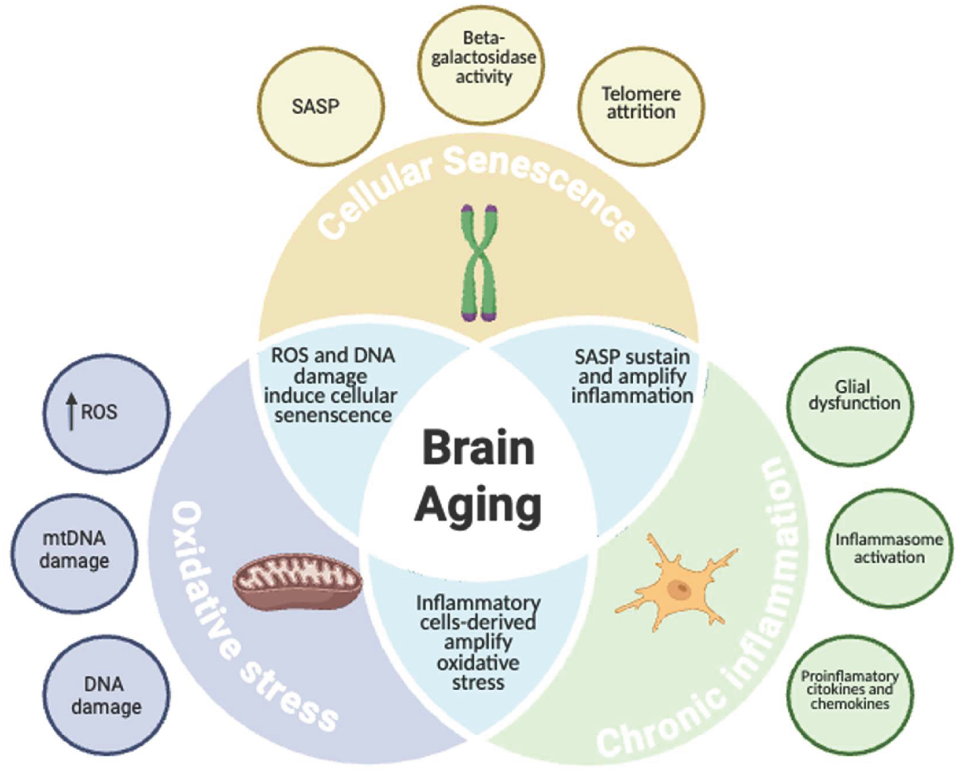
Figure 2.
Proposed biological effects of co-ultramicronized palmitoylethanolamide and luteolin (PEA-Lut) relevant to brain aging and cognitive impairment.Figure 2. The figure summarizes the principal mechanisms through which PEA-Lut has been proposed to influence systemic and central nervous system inflammatory pathways. At the systemic level, PEA-Lut may modulate the production of inflammatory mediators and reduce oxidative and nitrosative stress. Within the central nervous system, experimental findings suggest possible effects on microglial and astrocytic activation, pro-inflammatory signalling, amyloid-β-induced cellular injury, neurotrophic pathways, and neuronal survival. These mechanisms are primarily supported by preclinical evidence and should not be interpreted as demonstrating clinical efficacy in mild cognitive impairment or Alzheimer’s disease.
Figure 2.
Proposed biological effects of co-ultramicronized palmitoylethanolamide and luteolin (PEA-Lut) relevant to brain aging and cognitive impairment.Figure 2. The figure summarizes the principal mechanisms through which PEA-Lut has been proposed to influence systemic and central nervous system inflammatory pathways. At the systemic level, PEA-Lut may modulate the production of inflammatory mediators and reduce oxidative and nitrosative stress. Within the central nervous system, experimental findings suggest possible effects on microglial and astrocytic activation, pro-inflammatory signalling, amyloid-β-induced cellular injury, neurotrophic pathways, and neuronal survival. These mechanisms are primarily supported by preclinical evidence and should not be interpreted as demonstrating clinical efficacy in mild cognitive impairment or Alzheimer’s disease.
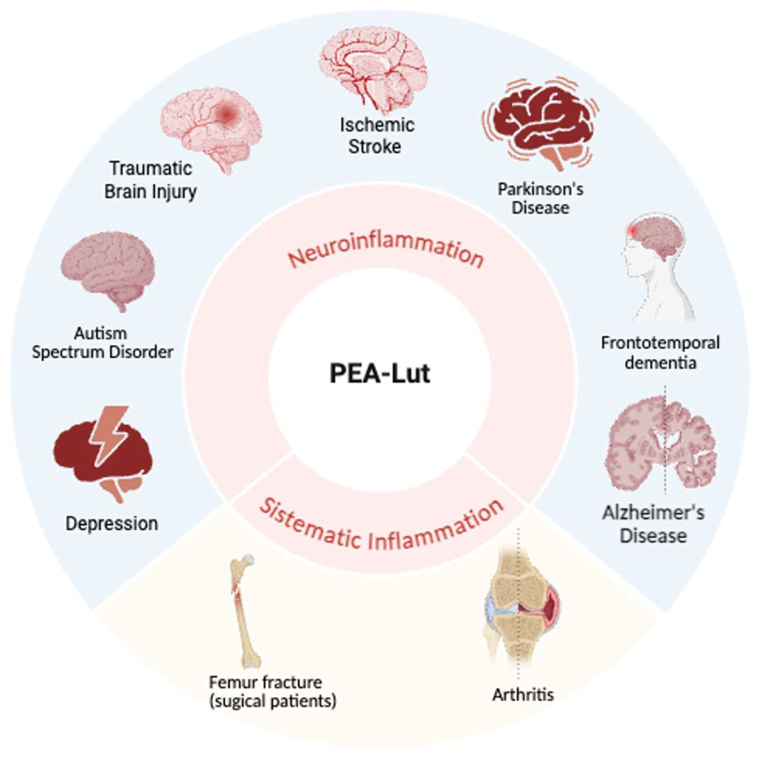
Disclaimer/Publisher’s Note: The statements, opinions and data contained in all publications are solely those of the individual author(s) and contributor(s) and not of MDPI and/or the editor(s). MDPI and/or the editor(s) disclaim responsibility for any injury to people or property resulting from any ideas, methods, instructions or products referred to in the content. |
© 2026 by the authors. Licensee MDPI, Basel, Switzerland. This article is an open access article distributed under the terms and conditions of the Creative Commons Attribution (CC BY) license (http://creativecommons.org/licenses/by/4.0/).
Copyright: This open access article is published under a Creative Commons CC BY 4.0 license, which permit the free download, distribution, and reuse, provided that the author and preprint are cited in any reuse.