
Submitted:
26 July 2026
Posted:
29 July 2026
You are already at the latest version
Abstract
Optical tweezers (OTs) have transformed the ability to manipulate microscopic and nanoscopic matter using light, establishing a non-contact, high-precision platform for probing forces at the picoNewton scale. Since their first realization in the 1980s, OTs have expanded from classical single-beam traps into a diverse set of architectures—including holographic, plasmonic, fiber-based, and integrated photonic platforms—each extending trapping performance across spatial scales and material systems.
This review provides a roadmap that integrates fundamental mechanisms, technological advances, and emerging applications of optical tweezers. We begin with a comprehensive analysis of the optical force landscape, encompassing gradient and scattering forces, Brownian fluctuations, viscous drag, hydrodynamic interactions, and quantum electrodynamic effects such as Casimir–van der Waals interactions. We then discuss experimental techniques for trap calibration and potential reconstruction, including equipartition, power spectral density analysis, Stokes drag, and interferometric detection, which together enable force quantification with sub-piconewton precision.
Architecturally, we classify OTs into single-beam, holographic, fiber-based, plasmonic, and hybrid photonic systems, highlighting how recent advances in metasurfaces, nanophotonic resonators, and AI-assisted control are reshaping optical trapping into compact, intelligent, and scalable platforms. Applications are reviewed across biology, soft matter, and quantum science—from single-molecule mechanics and cellular biomechanics to nanoparticle assembly and quantum simulations.
Finally, we outline current limitations—including diffraction-limited confinement, nanoscale trapping efficiency, and photothermal damage—and discuss future directions toward adaptive beam shaping, cryogenic trapping, and quantum-enhanced tweezers. By bridging theoretical foundations, experimental methodologies, and cross-disciplinary applications, this roadmap aims to provide both a technical reference and a forward-looking vision for the next generation of optical trapping technologies.
By synthesizing the theoretical foundations, experimental methodologies, and future-oriented integrations, this review aims to serve as both a technical guide and a visionary roadmap for advancing the next generation of optical trapping platforms.
Keywords:
optical tweezers
; force spectroscopy
; photonic integration
; holographic trapping
; plasmonic tweezers
1. Introduction
Optical tweezers (OTs), also referred to as optical traps, are advanced scientific instruments that employ highly focused laser beams to capture and manipulate microscopic objects in a noninvasive manner. Since their invention by Arthur Ashkin in the 1980s, OTs have profoundly transformed the study of microscale and nanoscale systems, providing unprecedented control across physics [1,2,3,4,5], chemistry [2,6,7,8,9,10], biology [6,11,12,13,14,15,16], and medicine with highly precise tools to investigate and control matter at these dimensions [1,14,17,18]. The profound significance of this technique was recognized in 2018, when Ashkin was awarded the Nobel Prize in Physics “for the optical tweezers and their application to biological systems,” underscoring the transformative impact of optical trapping technology on modern science .
1.1. Fundamental Principles of Optical Trapping
At the core of OTs lies the principle that light, beyond being an electromagnetic wave, also carries momentum that can be transferred to matter. When a strongly focused laser beam interacts with a dielectric particle, the particle experiences both scattering forces, pushing it along the beam axis, and gradient forces, pulling it toward the high-intensity focal spo [19,20].
In early experiments, Ashkin demonstrated that micrometer-sized transparent beads were stably drawn into the center of a Gaussian beam, where the restoring force created by the gradient overcame outward radiation pressure [21]. The use of high-numerical-aperture objectives enabled the formation of stable three-dimensional traps, establishing the single-beam gradient configuration that remains the foundation of optical tweezers today [22].
1.2. Historical Development from Ashkin’s Experiments to Hybrid Tweezers
The historical trajectory of OTs can be traced back to Ashkin’s pioneering demonstrations in the 1970s [23]. Initially working at Bell Laboratories, he showed that laser radiation could accelerate and confine particles, verifying for the first time that photons exert measurable forces on matter. This insight culminated in 1986 with the realization of a stable single-beam trap for dielectric particles [24,25,26]. TA year later, in a milestone that expanded the scope of OTs into the life sciences, Ashkin used infrared lasers to trap live bacteria without causing damage³². This demonstration established OTs as uniquely suitable for gentle and noninvasive manipulation of biological systems, paving the way for their adoption in biophysics, cellular mechanics, and clinical research.
Over the following decades, continuous innovation diversified the capabilities of OTs. The 1990s and early 2000s witnessed the emergence of holographic optical tweezers, which used spatial light modulators to split a single laser into many independently controllable traps [27,28,29,30,31,32,33,34,35,36,37]. This advance enabled parallel manipulation of particles, large-scale assemblies, and more complex experimental designs. In parallel, hybrid technologies combined optical trapping with magnetic fields, acoustic waves, or plasmonic nanostructure [38], extending the accessible force regimes and allowing the stable trapping of nanoscale objects. Today, the “optical tweezers toolbox” encompasses not only the classical single-beam configuration but also chip-integrated photonic arrays, plasmonic near-field traps, and multimodal platforms capable of handling increasingly complex systems [39,40,41,42].
This historical progression is summarized in Figure 1, which outlines the timeline of optical tweezer development from 1986 to 2024. The figure highlights the expansion from Ashkin’s single-beam trap to holographic arrays, plasmonic tweezers, quantum atom arrays, and AI-driven autonomous manipulation. Milestones such as the integration of OTs with force spectroscopy, the emergence of lab-on-chip systems, and the introduction of AI-enhanced autonomous tweezing illustrate the broadening impact of this technology. Pioneering contributions from Ashkin, Block, Bustamante, Padgett, and others collectively established OTs as a versatile platform that has reshaped our capacity to interrogate matter across scales.
The versatility of OTs has ensured their adoption across disciplines. In biology and medicine, they have become indispensable for probing molecular and cellular mechanics. Experiments involving the stretching of DNA and RNA, unfolding of proteins, and measurement of forces generated by molecular motors such as kinesin and myosin have revealed fundamental details of biomolecular behavior at the piconewton scale [66,67]. Beyond single molecules, OTs enable manipulation of whole cells, measurement of membrane elasticity, and studies of cytoskeletal dynamics under physiological conditions, while their non-contact nature makes them valuable in areas such as in vitro fertilization research and microsurgical tool guidance [54,68]. In soft matter physics, OTs have provided a direct means to assemble colloidal lattices, probe interparticle interactions, and study phase transitions in complex fluids [53,66,67,69,70,71]. Nanotechnology has further expanded the scope of tweezers: plasmonic tweezers exploit field enhancements at nanostructured surfaces to trap nanoparticles and molecules with sub-diffraction precision, enabling single-molecule spectroscopy and nanoscale assembly [18,72]. In atomic and quantum physics, OTs have become essential for laser cooling and trapping of neutral atoms, where defect-free optical arrays serve as qubits in quantum simulators and computing architectures [4,47,73,74]. Industrially, optical tweezers are increasingly deployed in microfluidics, lab-on-chip diagnostics, and semiconductor inspection [75,76,77,78].
Despite these wide-ranging successes, OTs face inherent limitations that constrain their broader application. Their spatial resolution is bound by the diffraction limit, typically a few hundred nanometers, which complicates the trapping of sub-50 nm particles without near-field enhancement [20,79,80,81,82]. Photothermal effects, caused by even slight absorption of trapping light, can lead to local heating, convection, and potential damage to biological specimens [83].
Furthermore, traditional setups typically manipulate only a few objects at once; even holographic arrays divide available laser power and require complex synchronization, limiting throughput and scalability [84,85,86,87]. These challenges also underscore the need for more compact, integrated, and user-friendly instruments.
To overcome these constraints, new directions have emerged that combine physical innovation with technological integration. AI-driven control systems now allow autonomous calibration, feedback stabilization, and adaptive beam shaping [1,88,89]. reducing experimental complexity while enhancing precision. On-chip integration with photonic waveguides, resonators, and metasurfaces enables massively parallel trapping on compact lab-on-chip devices [82,90], while hybrid designs that incorporate acoustic or magnetic fields broaden the force regimes accessible to OTs [91]. The convergence of optical trapping with quantum science has also opened a frontier in precision metrology and quantum simulation, with atom arrays serving as versatile platforms for computing and sensing [92,93].
1.3. Interdisciplinary Expansion and the Scope of This Review
In light of these rapid developments, the present review provides a comprehensive synthesis of the field. We begin by establishing the physical foundations of optical forces and the engineering strategies underlying stable trap design. We then trace the historical trajectory of OTs from Ashkin’s seminal discoveries to state-of-the-art photonic and AI-enhanced systems, highlighting interdisciplinary applications that span from single-molecule biophysics to quantum simulation.[47,67,68,80,94]. We also examine the persistent limitations of diffraction, photothermal damage, and low throughput, and assess the emerging solutions that aim to overcome these obstacles. By emphasizing the convergence of photonic integration, AI-driven automation, and hybrid platforms, we articulate a roadmap for the future evolution of OTs [95].
Scope of This Review:.
The central contribution of this review is to demonstrate that optical tweezers are no longer confined to specialist laboratories but are transitioning into intelligent, scalable, and practical platforms that will be indispensable in next-generation quantum technologies, precision medicine, and nanotechnology. By critically integrating decades of progress with a forward-looking perspective, we aim to provide the community with both a rigorous reference and a conceptual framework to guide the next stage of development. In doing so, this review underscores that optical tweezers are not merely an established tool but a continuously evolving technology at the frontier of science and engineering.
Following this, we assess the current limitations of optical tweezers—such as diffraction limits, photothermal effects, and low throughput—and discuss the technological innovations addressing these issues. Finally, we explore the most promising future directions, including intelligent control via artificial intelligence, chip-scale integration, and hybrid tweezer platforms. Through this structured and detailed review, we aim to not only summarize the transformative progress made in the field of optical tweezers but also to illuminate the expansive opportunities ahead, reinforcing their importance as a dynamic and evolving tool in modern science and engineering.
2. Comprehensive Force Analysis in Optical Tweezers
The stability and precision of optical tweezers arise from a complex interplay of forces acting on trapped particles. While optical gradient and scattering forces provide the fundamental basis of trapping, real experimental conditions inevitably introduce additional contributions that influence performance and reliability. Trapped particles are subject to Brownian fluctuations from thermal motion, hydrodynamic drag in viscous environments, and perturbations from photothermal and photophoretic effects associated with light absorption. At the nanoscale, near-field interactions, Casimir forces, electrostatic contributions, and even specific biomolecular binding can significantly alter trapping stability. A comprehensive force analysis is therefore indispensable, as it defines the operating limits of OTs across different size regimes and experimental conditions, and informs the engineering of advanced trapping platforms that can overcome these challenges.
Figure 2 provides a schematic overview of this multiphysical force landscape, emphasizing the broad spectrum of physical effects that accompany optical trapping. Beyond the dominant optical forces, the diagram highlights how thermal fluctuations, fluid interactions, near-field forces, and nanoscale interactions combine to shape particle dynamics within the trap.
In recent years, the development of engineered optical fields has expanded the functional scope of OTs. By tailoring light–matter interactions through metasurfaces, photonic crystals, and hybrid optoelectronic architectures, researchers have achieved enhanced confinement, parallel manipulation, and improved stability under complex conditions. Figure 3 illustrates representative examples of such strategies, from phase-engineered metastructures and nanopillar arrays to hybrid metalens–hologram systems, each designed to optimize force distribution and extend trapping to regimes beyond conventional diffraction-limited optics [101,102,103,104,105,106,107]. These innovations not only reinforce the need for a rigorous force analysis but also demonstrate how physical understanding translates directly into advanced system design.
2.1. The Force Spectrum from Microscale to Single-Molecule Domains
The force environment in optical tweezers is inherently multiphysical, as depicted in Figure 2, where optical forces coexist with stochastic, hydrodynamic, and surface-mediated interactions. To achieve stable trapping, it is therefore necessary to not only generate sufficiently strong optical gradients but also manage the competing perturbations that arise from the medium, the particle, and the surrounding environment. Modern tweezer platforms address this complexity by integrating field-engineering strategies, as summarized in Figure 3, which illustrates how waveguide metastructures, nanopillar arrays, and holographic beam shaping enable programmable and reconfigurable confinement across scales.
Within this framework, OTs distinguish themselves by providing controlled forces over a remarkably broad range, from the femtonewton (fN) to the nanonewton (nN) regime. At the microscale, dielectric beads several micrometers in diameter can be held with forces spanning tens to hundreds of piconewtons, sufficient to counteract gravity and enable optical levitation [103,104]. These forces support manipulation of entire living cells or organelles in aqueous media, providing a unique capability for biomechanical probing under near-physiological conditions [105].
As particle size decreases, optical force magnitude diminishes due to the cubic dependence of polarizability on radius (αr³) and the reduction of scattering cross-section. Nanoparticles in the range of 50–500 nm typically experience forces in the low-piconewton to sub-piconewton regime, where Brownian fluctuations often dominate and destabilize conventional traps [106]. To counteract this, resonant and near-field platforms such as plasmonic tweezers, photonic-crystal cavities, and dielectric metasurfaces have been developed, confining light below the diffraction limit and enabling manipulation at femtonewton scales [107]. While these enhancements extend trapping capability into the nanoscale, the limited interaction volume ensures that forces remain relatively weak, often requiring advanced detection schemes for quantitative stability analysis.
At the single-molecule level, optical tweezers operate in an optimal force window of ~0.1–50 pN, with sub-pN resolution and nanometer-scale precision. This regime is particularly significant as it matches the forces underlying many fundamental biomolecular processes. Protein unfolding typically requires 5–30 pN, DNA or RNA duplex unzipping occurs around 10–20 pN, and molecular motors such as kinesin or dynein exert step forces of approximately 6–8 pN [108,109,110]. Dual-beam and counter-propagating configurations further improve stiffness calibration and minimize drift, allowing direct observation of conformational transitions, enzymatic stepping, and ligand–receptor binding events with unprecedented accuracy.
Modern OTs leverage advanced readout technologies such as back-focal plane interferometry and quadrant photodiode detection to achieve force sensitivities down to 0.1 pN and spatial resolutions at the nanometer scale. In optimized configurations, the upper force limit can reach ~200 pN, sufficient for probing the mechanics of large biomolecular complexes or entire cell structures. With higher power and high-NA focusing, forces can even surpass 1 nN, though such conditions risk introducing photothermal perturbations or photochemical stress, necessitating careful balancing of trap strength and sample safety [111]. Taken together, this continuum of force magnitudes—ranging from femtonewtons for nanoscale particles to nanonewtons for micrometer-scale objects—positions optical tweezers as a uniquely versatile platform.
Their non-contact operation, tunable stiffness, and compatibility with living systems make them indispensable for probing molecular-scale interactions, cellular mechanics, and nanomaterial assembly. By integrating multiphysical understanding with engineered photonic platforms, as shown in Figure 2 and Figure 3, modern tweezers provide a coherent framework to bridge microscale manipulation with single-molecule precision, thereby extending their impact across physics, biology, and nanotechnology [112,113].
2.2. Theoretical Modeling of Trapping Forces
2.2.1. Momentum Transfer and Radiation Pressure
All optical trapping forces originate from the transfer of momentum from light to matter. When photons scatter from, reflect off, or are absorbed by a particle, they impart momentum and generate a net mechanical force-commonly referred to as radiation pressure [104,114,115]. For micrometer and sub-micrometer-scale particles suspended in liquids, radiation pressure typically overwhelms gravity, buoyancy, and electrostatic forces, thereby setting the dominant scale of light–matter interaction under trapping conditions [116,117,118].
A rigorous calculation of the total optical force F can be obtained via Maxwell’s stress tensor by integrating the electromagnetic momentum flux over a closed surface enclosing the particle [119,120]. Although this tensor formalism captures the full vectorial and near-field physics, it is computationally demanding; accordingly, size-dependent simplified models are routinely employed in practice, contingent on the ratio of particle diameter d to the trapping wavelength .
Ray Optics Model (Mie Regime).
When the particle size is comparable to or larger than the wavelength (), geometric or ray optics provides an accurate and intuitive picture [64]. The incident laser is treated as a bundle of rays that refract and reflect at the particle interface; each change in photon momentum imparts an equal and opposite impulse to the particle [117,121].
Summing the vector momentum changes over all rays yields two principal components that govern trapping dynamics [111]: a gradient force that pulls the particle up the transverse intensity gradient toward the beam center, and a scattering force that pushes it downstream along the propagation axis. If a spherical dielectric is displaced laterally, the more intense side of the beam contributes a larger momentum change, producing a restoring force that returns the particle toward the optical axis [109]. Axially, tight focusing creates a steep upstream gradient that can counterbalance the downstream radiation pressure. Stable three-dimensional trapping therefore arises near, but slightly downstream of, the beam waist where the two forces balance. Crucially, a high-NA objective is required to create the necessary angular spread and intensity gradients; without sufficient NA, the gradient component cannot overcome forward scattering and stable trapping is lost [122]. Within the Mie regime (), the ray model predicts trap stiffness, axial equilibrium positions, and the optical system requirements for robust manipulation with considerable fidelity.
Dipole Approximation (Rayleigh Regime).
For particles much smaller than the wavelength (), the induced-dipole (Rayleigh) approximation applies, in which the dominant, conservative contribution is the gradient force that drives the dipole toward intensity maxima. In this limit, the gradient force can be written [115]:
where is the particle’s polarizability, and is the local electric field amplitude. Since the field intensity , the force is proportional to the intensity gradient, i.e., .
For a spherical dielectric particle of radius rrr and relative refractive index (where and are the refractive indices of the particle and the medium, respectively), the gradient force becomes [123]:
This result shows that the gradient force scales with the particle’s volume () and the local intensity gradient, making it especially significant for small particles in tightly focused beams [111].
The scattering force in the Rayleigh regime originates from isotropic re-radiation, or Rayleigh scattering, and is a non-conservative force. It scales with the scattering cross-section , which for small Rayleigh spheres is given by:
The resulting scattering force can be written as:
This expression reveals the strong dependence of scattering forces and their inverse scaling with , a hallmark of Rayleigh’s law. For very small nanoparticles (e.g., ), the scattering force becomes negligible compared to the gradient force [124,125].
Thus, dielectric particles with diameters below 100 nm can still be optically confined, provided the light field exhibits a sufficiently steep intensity gradient—for instance, via high numerical-aperture (NA) focusing. Although the absolute optical forces acting on such particles are typically on the order of femtonewtons, the gradient force remains dominant and can ensure stable trapping when the appropriate field configuration is employed [126]. In the Rayleigh regime, the optical force components can be summarized as follows: The gradient force draws the dipole toward the intensity maximum [127]; The scattering force causes a weaker forward push along the beam propagation direction [123]; The net result is stable confinement at the beam focus, with applications in trapping nanoparticles, biomolecules, and quantum dots.
Intermediate Regime: Transition to Mie Scattering.
For sizes on the order of the wavelength (d ∼ λ), neither pure ray optics nor the dipole model is quantitatively sufficient [123]. Full-wave solutions of Maxwell’s equations are then required to obtain the time-averaged force [128], using analytical Mie theory for spheres or numerical methods—e.g., finite-difference time-domain (FDTD) and boundary-element methods (BEM)—for arbitrary geometries [129,130,131]. These approaches evaluate the total force by integrating the electromagnetic stress tensor over a closed surface, thereby capturing near-field interactions, retardation, and morphology-dependent resonances [21,112,132]. Physically, the force can still be decomposed into a gradient-like component (in-phase with the incident field, driving particles toward intensity maxima) and a scattering-like component (out-of-phase/radiative, pushing downstream)[133,134,135] . In this regime, multipolar resonances, internal-mode interference, and morphology-dependent field enhancement can all reshape the axial and lateral stability landscape [136,137,138], yet the interpretive core remains: gradient-like terms furnish the conservative well, while scattering terms introduce axial destabilization [139,140]. For resonant or highly polarizable materials—e.g., high-index dielectrics or metallic nanostructures—nonlinearities and mode enhancement may either deepen the well or create new instability channels [108,141].
In all cases, the net optical force can be interpreted as the sum of a conservative trapping force (derivable from a potential function , often approximated as harmonic near the trap center) and a non-conservative radiation pressure force that injects momentum and energy into the system [142,143,144]. This separation underlies many trap modeling frameworks and remains a conceptual tool for understanding light–matter interaction across size scales [109,145].
Figure 4.
Classical force analysis in optical tweezers: ray optics, scattering behavior, and experimental potential mapping. (A) connects directly to the ray-optics (Mie-regime) description above, visualizing how refraction/reflection redistribute photon momentum to yield transverse gradient and axial scattering forces; the 3D vector sketches clarify how stable 3D confinement arises once upstream gradient forces overcome downstream radiation pressure. (B) relates particle size to angular scattering and trap stiffness, spanning Rayleigh to Mie limits; the accompanying power spectral density (PSD) curves illustrate how lateral/axial stiffness constants are extracted experimentally. (C) gathers potential-well reconstruction methods—trajectory statistics, position histograms, and equipartition calibration—together with 3D force-field simulations that reveal spatial stiffness gradients under Gaussian beams and in holographic configurations. (D) highlights boundary-induced modulation: proximity to substrates or nanostructured interfaces modifies near fields via evanescent coupling and image interactions, anisotropically reshaping the force landscape and thereby the stability conditions.
Figure 4.
Classical force analysis in optical tweezers: ray optics, scattering behavior, and experimental potential mapping. (A) connects directly to the ray-optics (Mie-regime) description above, visualizing how refraction/reflection redistribute photon momentum to yield transverse gradient and axial scattering forces; the 3D vector sketches clarify how stable 3D confinement arises once upstream gradient forces overcome downstream radiation pressure. (B) relates particle size to angular scattering and trap stiffness, spanning Rayleigh to Mie limits; the accompanying power spectral density (PSD) curves illustrate how lateral/axial stiffness constants are extracted experimentally. (C) gathers potential-well reconstruction methods—trajectory statistics, position histograms, and equipartition calibration—together with 3D force-field simulations that reveal spatial stiffness gradients under Gaussian beams and in holographic configurations. (D) highlights boundary-induced modulation: proximity to substrates or nanostructured interfaces modifies near fields via evanescent coupling and image interactions, anisotropically reshaping the force landscape and thereby the stability conditions.
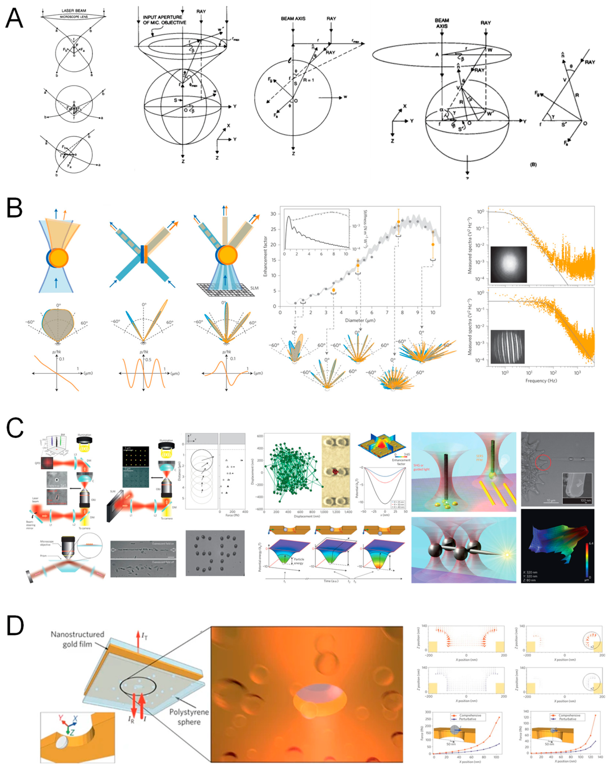
Figure 5.
Decomposition and modeling of gradient and scattering components across optical regimes. (A) organizes efficiency parameters—axial force efficiency (ξ), transverse momentum transfer (Qₚ), trapping efficiency (Qₘₐₓ), and stiffness κ—against the size parameter ka and refractive-index ratios, unifying Rayleigh, Mie, and geometric optics within the GLMT framework and revealing the nonlinear transition from predominantly conservative to increasingly non-conservative force profiles as ka grows. (B) shows experimental reconstructions of 2D/3D intensity and force fields (e.g., by fluorescent tracking and back-focal-plane interferometry), with axial asymmetry consistent with GLMT predictions for forward scattering. (C) quantitatively separates gradient (restoring) and scattering (non-conservative) contributions in force–position curves for radii from a = 5 nm to 100 nm along both axial and lateral axes; symmetry in small-particle potentials (Rayleigh) gives way to scattering-induced asymmetries and reversal points at larger sizes, delineating the thresholds where simplified models fail and full-wave treatments become essential.
Figure 5.
Decomposition and modeling of gradient and scattering components across optical regimes. (A) organizes efficiency parameters—axial force efficiency (ξ), transverse momentum transfer (Qₚ), trapping efficiency (Qₘₐₓ), and stiffness κ—against the size parameter ka and refractive-index ratios, unifying Rayleigh, Mie, and geometric optics within the GLMT framework and revealing the nonlinear transition from predominantly conservative to increasingly non-conservative force profiles as ka grows. (B) shows experimental reconstructions of 2D/3D intensity and force fields (e.g., by fluorescent tracking and back-focal-plane interferometry), with axial asymmetry consistent with GLMT predictions for forward scattering. (C) quantitatively separates gradient (restoring) and scattering (non-conservative) contributions in force–position curves for radii from a = 5 nm to 100 nm along both axial and lateral axes; symmetry in small-particle potentials (Rayleigh) gives way to scattering-induced asymmetries and reversal points at larger sizes, delineating the thresholds where simplified models fail and full-wave treatments become essential.

2.2.2. Optical Gradient Force
The gradient force constitutes the conservative component of optical trapping that attracts particles with a refractive index higher than the surrounding medium, or repels those with a lower index (e.g., air bubbles in water)[146]. Microscopically, the oscillating optical field induces a polarization in the particle—effectively an electric dipole—which interacts with the spatial variation of the field intensity to generate a mechanical force that drives the particle up the intensity gradient [122,147].
An intuitive picture follows from momentum balance: in a focused beam the near-focus side of a dielectric particle encounters a higher local intensity than the far side, producing an imbalance of radiation pressure that biases motion toward the beam center [148]. This force is conservative, as it can be derived from a potential energy function. Near the trap center, the potential is approximately harmonic [149]:
where k is the trap stiffness, which is proportional to the local intensity gradient and the particle’s polarizability . Because polarizability scales with particle volume (), the gradient force itself scales as [150,151]:
This implies that larger particles, or tighter beam focusing (which steepens ), result in stronger gradient forces [152]. Notably, stable three-dimensional trapping requires the gradient force to dominate over the scattering force; otherwise, the particle is pushed downstream and escapes the trap [153]. For this reason, optical tweezers are typically configured with high numerical aperture (NA) objectives, which tightly focus the beam and enhance the spatial intensity gradient [113,154].
The maximum gradient force is realized near the beam waist and acts as a restoring term that returns displaced particles to the focus [155,156]. Its direction inverts with refractive-index contrast—pulling inward for (trapping) and pushing outward for , which explains the difficulty of stably trapping low-index objects such as air bubbles in water with conventional tweezers [110,157,158].
2.2.3. Scattering (Radiation Pressure) Force
By contrast, the scattering force is an axial, non-conservative contribution arising from the linear momentum carried by photons that are reflected, refracted, or absorbed by the particle. Unlike the gradient force, which localizes particles near an intensity maximum [110,157,158,159,160], scattering continuously injects momentum into the system and tends to push the particle downstream along the beam axis [110,157,158,159,160,161].
In single-beam traps this mechanism constitutes the primary destabilizing influence and must be counteracted by a sufficiently strong gradient component to maintain a downstream equilibrium near (but typically slightly beyond) the geometric focus [125,162]. The magnitude of the scattering force can be estimated using the optical momentum flux. For a laser of power P propagating in a medium with refractive index , the rate of momentum flow is [163]:
where is the speed of light in vacuum. If a particle scatters or absorbs a fraction of this momentum, it experiences a force given by:
Here, is a dimensionless scattering efficiency factor that depends on the particle’s optical properties—specifically, its scattering and absorption cross-sections—and typically lies in the range . In the ray optics regime (Mie regime), where particle size is larger than the wavelength, can approach unity for strongly reflecting particles [164]. In contrast, in the Rayleigh regime (), , making the scattering force much weaker relative to the gradient force [165].
The strong size and wavelength dependence is consistent with Rayleigh’s law, whereby scattering efficiency diminishes rapidly for small particles and long wavelengths [166]. The scattering force increases linearly with laser power and is amplified by higher reflectivity or absorptivity of the trapped object [167].
Importantly, as a non-conservative drive, scattering contributes to drift and energy dissipation pathways in liquid-phase trapping [101,168]. In optical trapping experiments performed in liquid media, researchers often employ several strategies to compensate for scattering-induced destabilization [169,170] : Orienting the trap vertically to use gravity to counteract upward scattering ; Adjusting the beam focus slightly below the particle center so that the axial gradient force opposes the scattering force, leading to a stable equilibrium point just downstream of the geometric focus [171,172,173].
Conversely, when axial propulsion is desired, scattering is harnessed intentionally in optical pushing and conveyor implementations that translate particles along waveguides or microchannels [174]. In sum, while scattering is essential to momentum conservation and enables active transport applications, stable trapping demands beam shaping and focal geometry that minimize its destabilizing action or channel it constructively where needed [174].
The conceptual and quantitative distinctions developed in Section 2.2.2 and Section 2.2.3 are synthesized in Figure 6, which links the conservative potential picture of the gradient force to the non-conservative nature of scattering and to the measurable consequences in realistic media. Panel A reconstructs the potential-well geometry from the ray-optics viewpoint, showing how the balance of transverse gradient and axial scattering defines a restoring-paraboloid well whose longitudinal asymmetry reflects downstream radiation pressure; the equilibrium thereby emerges slightly beyond the beam waist, consistent with the stability condition discussed above. Panel B maps vectorial field distributions and Poynting-vector streamlines in a tightly focused beam, visualizing how energy flow organizes around the trapped object and how polarization-induced phase gradients deform the local potential and impart optical torque—an expected signature of non-uniform phase and amplitude profiles. Panel C presents frequency-dependent microrheology, where storage and loss moduli and reveal how viscoelasticity reshapes trap curvature and dissipation; in polymer–water systems, the effective stiffness and energy loss depend sensitively on ω, consistent with the observation that media rheology modifies the conservative well inferred from . Panel D displays experimentally measured two-dimensional trajectories of trapped particles, whose thermally driven, slightly vortical or anisotropic spreads reflect the coexistence of conservative and non-conservative components in the local force field. Panel E refines the geometric-optics construction by explicitly resolving incidence, transmission, and reflection paths and their angular contributions to radial and axial components, thereby connecting the intuitive ray picture to the stiffness anisotropy near focus. Panel F uses generalized Lorenz–Mie theory to produce lateral force maps , normalized to incident power and plotted versus displacement, and demonstrates close agreement with analytic predictions; the axial asymmetry and lateral shifts captured here mirror the scattering-dominated departures from purely harmonic confinement in larger particles. Finally, Panel G quantifies how thermal noise and non-Newtonian rheology can deform a nominally single-well landscape into multi-well effective potentials in heterogeneous media, clarifying why stability margins narrow in biologically relevant environments and why careful power budgeting and beam engineering are necessary to preserve the dominance of the gradient term.
The figure thus operationalizes the conservative gradient term and the non-conservative scattering contribution described in 2.2.2–2.2.3, and shows how medium rheology and noise imprint on measurable force–position relations.
2.2.4. Photophoretic and Photothermal Forces
Beyond the conservative gradient force and the non-conservative scattering force, light can drive particle motion through thermally mediated mechanisms that originate from light-induced temperature gradients in the particle and its surrounding medium [175,176]. In gases, differential surface heating produces photophoretic motion, whereas in liquids, static or optically generated temperature gradients give rise to thermophoresis (the Soret effect). In both cases the light field functions primarily as an energy source that establishes thermal gradients, rather than as a direct carrier of mechanical momentum.
In gaseous media, a strongly illuminated particle develops an asymmetric temperature profile across its surface [177]. Gas molecules that impinge on the hotter side acquire additional kinetic energy and rebound with greater momentum, creating a net recoil on the particle that drives it from hot toward cold regions [178,179,180]. Depending on thermal conductivity, absorption, internal focusing, and geometry, both positive (canonical) and negative (inverse) photophoresis can occur: the former pushes the particle away from the illuminated face, the latter pulls it toward the beam when internal lensing heats the rear surface more strongly [181].
In the free molecular regime (where the particle is much smaller than the mean free path of gas molecules), the photophoretic force on an absorbing spherical particle can be approximated by a simplified scaling law [182]:
Here, is the particle radius, is the optical intensity, and are the particle surface and ambient gas temperatures, and is the thermal velocity of gas molecules. emphasizing its dependence on absorbed power (via the surface–ambient temperature difference), gas properties, and molecular thermal velocity. Under atmospheric conditions, photophoretic forces can reach the nano-Newton range for ∼10 µm absorbing particles, which enables optical manipulation in air even when intensity-gradient forces alone are insufficient for stable confinement [20,183].
In liquids, an allied mechanism—thermophoresis—drives particles along externally imposed or optically generated temperature gradients [184,185]. Local heating by a focused laser can arise from absorption in either the particle or the surrounding fluid; the resulting thermophoretic drift may be directed away from hot regions (common for plasmonic absorbers) or toward them, depending on particle–solvent interactions and interfacial thermodynamics. Although photothermal forces in liquids are generally weaker than the optical gradient force, even modest asymmetries in heating bias trajectories and shift the equilibrium position within an optical trap, thereby creating measurable drift and apparent changes in trap stiffness over time [86,186].
Deliberately engineered optothermal platforms convert this bias into a resource: thermophoretic or opto-thermal tweezers exploit spatially programmable temperature fields to confine low-index or weakly polarizable objects that are difficult to capture with conventional dielectric gradient traps [187,188]. In practice, the magnitude and directionality of these thermally mediated forces are governed by absorption, thermal transport parameters, and the environment (gas versus liquid), and they have become central to trapping and transport strategies for aerosols, nanoparticles, soft matter, and thermally sensitive specimens.
Context for Figure 7 and Figure 8. The phenomenology above is operationalized in Figure 7 and Figure 8. Figure 7 assembles quantitative evidence that trap depth and stability in photothermal and thermophoretic tweezers are tunable by wavelength, optical power, focusing geometry, and electrolyte conditions, while Figure 8 progresses from the basic optothermal trapping concept to system-level implementations, multifunctional manipulation (including CRISPR-enabled SERS readout), and multiphysics modeling and metrology that connect measured trajectories and temperature maps to force landscapes.
2.2.5. Brownian (Thermal) Forces
In any fluid medium—gas or liquid—particles are incessantly bombarded by surrounding molecules, producing random stochastic forces known as Brownian motion. Inside an optical trap, these thermal fluctuations appear as directionless “kicks” that make the trapped particle jitter about its equilibrium position [189,190] . Although the time-averaged Brownian force is zero, its variance is finite and is set by the thermal energy scale:
where is Boltzmann’s constant and is the absolute temperature. For the particle to remain stably confined, the optical trap must provide a potential well depth of at least a few to resist these thermal kicks; otherwise, the particle may escape the trap due to thermal excitation.
Assuming a harmonic trap potential , where is the trap stiffness (i.e., optical spring constant), the equilibrium positional variance of the particle can be derived from the equipartition theorem:
This relation reveals that stronger traps (larger ) confine particles more tightly around the center, while weaker traps allow broader spatial fluctuations.
Brownian motion is inherently time-dependent and stochastic [191]. In modeling frameworks, it is commonly included as a random forcing term in the Langevin equation of motion:
where is the Stokes drag coefficient for a spherical particle of radius in a fluid of viscosity . The spectral density of this force is:
This corresponds to white noise (flat spectrum), implying that force fluctuations occur across all timescales with equal power. For example, in water at room temperature, a 1 μm bead experiences typical force fluctuations on the order of ~1–2 pN, as and trap stiffness , leading to RMS displacement fluctuations of ~10–20 nm.
Importantly, Brownian forces do not favor any direction; they introduce continuous random perturbations that the optical gradient force must balance [192]. The steady-state of a trapped particle thus represents a dynamic equilibrium, with motion characterized by a Boltzmann distribution centered around the trap minimum:
Practically, Brownian motion is both a limitation and a metrological resource: thermal fluctuations set a fundamental precision bound, yet the same fluctuations enable trap-stiffness calibration via equipartition or by fitting the power spectral density (PSD) of recorded trajectories [11,193,194]. In short, thermal (Brownian) forces impose the baseline noise floor for optical tweezers; although no trap can immobilize a particle absolutely, robust gradients maintain nanometer-scale confinement for the vast majority of the time as long as the restoring force exceeds typical thermal kicks [195,196].
Context and integration with prior sections. Section 2.2.2, Section 2.2.3 and Section 2.2.4 established the deterministic force landscape—conservative gradient forces, non-conservative scattering (radiation pressure), and thermally mediated optothermal drives—governing stability and bias in an optical trap. Brownian forcing supplies the universal stochastic term that limits precision and shapes the steady-state distribution through and . The platform-level strategies used to measure, compensate, or even exploit this stochasticity have close analogs beyond optics; Figure 9 highlights acoustic tweezer implementations that realize force-clamp assays, programmable potential landscapes, and single-molecule readouts under pico-Newton loads, thereby complementing optical trapping methodologies for calibration and stability control in the presence of thermal noise.
2.2.6. Viscous Drag Force
When a particle moves through a fluid—either because it is displaced relative to the medium or because the surrounding fluid flows past it—it experiences a viscous drag that opposes the direction of motion [77,197]. In the low–Reynolds number regime typical of micron- and submicron-scale particles in aqueous environments, this resistance is well described by Stokes’ law [87]:
where is the dynamic viscosity of the fluid, is the radius of the spherical particle, and is its velocity relative to the fluid. Within optical tweezers, viscous drag plays two conceptually distinct but tightly linked roles. First, it damps the motion of the trapped particle. Unlike a particle in vacuum that exhibits oscillatory dynamics, a bead in water is strongly overdamped: the viscous force rapidly balances the optical restoring and Brownian forces, and the particle reaches its terminal response on microsecond timescales [198]. Consequently, when the trap position is stepped or scanned (e.g., by moving the laser focus), the bead follows with only a minimal temporal lag, and its motion can be modeled without inertial terms [199]. Second, viscous drag enables force calibration. By translating the trap or the surrounding fluid at a known speed v, one imposes a known drag load that can be balanced against the optical restoring force [200]. This is the principle behind the Stokes drag calibration method:
so that the steady-state displacement from the trap center directly reports the trap stiffness k (via the balance between drag and optical spring), providing an immediate estimate of [55].
Order-of-magnitude values are consistent with single-molecule biophysics: in water at room temperature with , a 1 µm-diameter bead moving at experiences approximately 1 pN of drag force—comparable to the forces involved in single-molecule biophysics experiments [201].
In optical trapping of nanoscale particles, viscous drag becomes smaller due to the size dependence ()[202]. However, such particles also exhibit faster Brownian diffusion, as quantified by the Stokes–Einstein relation:
The interplay between drag and diffusion determines the particle’s dynamic response within the trap.
This behavior is typically modeled by the overdamped Langevin equation:
where inertia is negligible and motion is governed by the balance between optical restoring force, viscous damping, and thermal fluctuations [41]. The response time of the trap—the time it takes for a particle to relax to equilibrium after perturbation—is set by the drag coefficient and stiffness , via the characteristic time constant .
In summary, viscous drag is a velocity-dependent, stabilizing force that damps fluctuations, defines the trap response time, and provides a practical route to apply and measure calibrated forces. It is also routinely exploited for controlled manipulation: by imposing a known flow, one displaces the particle to a steady off-center position where the optical spring balances the hydrodynamic load, enabling continuous, well-calibrated force application in sit [203].
Together with the conservative gradient force (Sec. 2.2.2), the non-conservative scattering force (Sec. 2.2.3), and thermally mediated optothermal drives (Sec. 2.2.4), viscous drag closes the dynamical picture by setting the damping and calibration pathway; in the presence of Brownian forcing (Sec. 2.2.5), the pair () determines both steady-state fluctuations and transient response. Figure 10 operationalizes these relationships by showing how MSD/PSD analyses, imaging-based interferometry, and holographic beam shaping quantify, and mode-dependent sensitivity across axial and radial degrees of freedom.
2.2.7. Near-Field and Evanescent Field Optical Forces
While conventional optical tweezers rely on freely propagating beams, an alternative route to trapping exploits evanescent fields—non-propagating electromagnetic fields that decay exponentially away from a dielectric or metallic interface. Such fields arise naturally under total internal reflection (TIR) or can be engineered in nanophotonic platforms including waveguides, plasmonic resonators, and photonic crystals [204,205,206]. In these architectures, the underlying force components remain the familiar gradient and scattering forces, but they are spatially confined to the near-field region, typically within ~100–200 nm of the surface [85,86]. A canonical case is the TIR-generated evanescent wave at a glass–water interface, which exerts a lateral scattering force that drags particles along the interface while a vertical gradient force attracts them toward the high-intensity near-surface region [207,208]. Because the field decays exponentially, the accessible trapping depth is shallow; however, resonant enhancement can dramatically boost the local intensity and, therefore, the gradient force.
Among near-field implementations, plasmonic optical tweezers—based on surface plasmon polaritons (SPPs) supported by metallic nanostructures—are especially powerful [209]. Carefully designed antennas, nanopillars, or nanogaps concentrate light into nanoscale hot spots, creating intensity gradients that can exceed those of free-space traps by orders of magnitude. In plasmonic gap geometries, gradient forces on the order of 0.01–0.1 pN (10–100 fN) have been reported, sufficient to confine 10–50 nm nanoparticles or even single molecules [67,210]. Relative to standard evanescent traps, this corresponds to an enhancement of roughly several-tens-fold in effective force, yet the absolute forces remain in the femtonewton range for such small objects [10] ; consequently, Brownian agitation must be countered by very steep potential gradients provided by strong field confinement.
Beyond plasmonics, photonic-crystal cavities, whispering-gallery-mode resonators, and waveguide-integrated nanostructures also realize near-field trapping by pinning particles at antinodes of standing waves or other field maxima [211]. In all of these systems, the Maxwell stress tensor remains the rigorous basis for force evaluation; nevertheless, because highly localized resonances dominate the response, analysis is often cast in terms of enhanced polarizability or local-intensity models that capture the effective scaling with geometry and material dispersion [212,213]. A practical consequence is that near-field traps are inherently surface-bound: manipulation is restricted to the immediate vicinity of the device—e.g., a fiber tip, photonic-crystal slab, or metal nanopillar—thus requiring precise nanofabrication and limiting the range of motion compared with free-space tweezers [214,215,216]. Despite this constraint, the benefits are substantial: sub-100 nm spatial resolution, access to weakly polarizable or sub-50 nm objects, and straightforward integration with lab-on-chip architectures. In sum, evanescent and near-field optical forces extend optical tweezing to the true nanoscale, deliberately trading long working distance for extreme field gradients and localization; this enables surface-based control of nanoparticles, biomolecules, and quantum dots at levels unattainable with conventional free-space traps [217,218,219].
The near-field platforms discussed here inherit the force taxonomy established in Secs. 2.2.2–2.2.3 (conservative gradient vs. non-conservative scattering) while operating in regimes where steep, resonance-boosted intensity gradients are essential to overcome Brownian forcing (Sec. 2.2.5) under strong viscous damping (Sec. 2.2.6). Because resonant confinement can also introduce photothermal effects (Sec. 2.2.4), practical designs balance optical enhancement against local heating to preserve trap stability and sample integrity.
Figure 11.
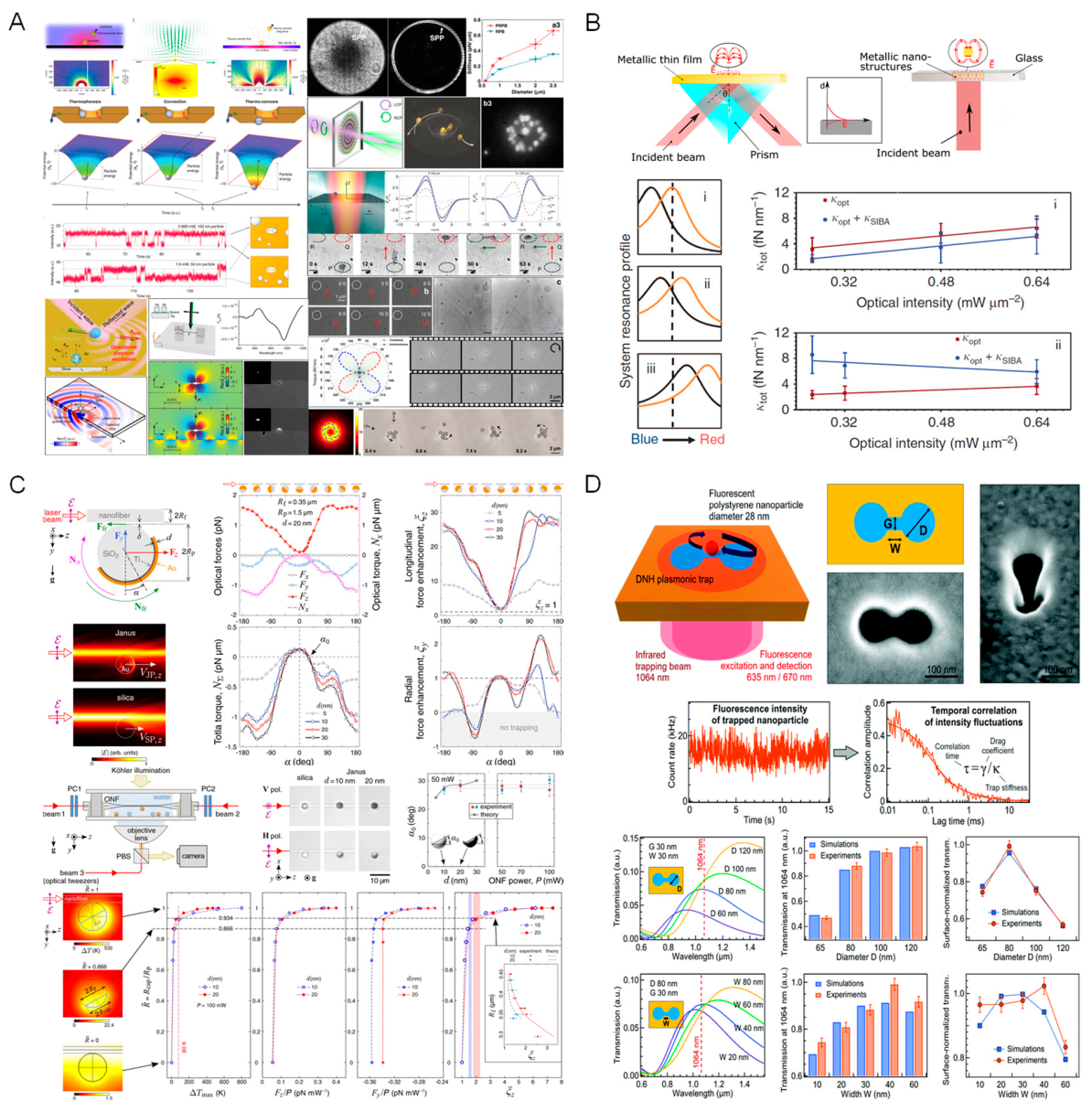
Integration of optical tweezers with plasmonic and thermo-optical platforms. (A) Enhanced Optical Manipulation Using Localized and Propagating Plasmonic Structures. (A) illustrates hybridization with SPPs and localized surface plasmons (LSPs) on nanostructured metals (e.g., gold films, nanopillar arrays), where simulations and experiments visualize near-field intensities and force landscapes that yield asymmetric, high-gradient confinement and demonstrable suppression of Brownian excursions under tailored illumination. (B) contrasts thermo-plasmonic operation across resonance: tuning the excitation wavelength relative to SPP/LSP peaks reshapes the optical-force profile and modulates the optically induced thermal gradients, with measured trap stiffnesses versus intensity revealing resonance-enhanced manipulation at minimal power in absorptive media. (C) examines asymmetric particles (Janus silica–gold half-shells), where differential absorption produces thermophoretic torque; quantitative trends in potential depth, torque-induced lateral shift, and radial force versus intensity demonstrate how particle anisotropy and cross-polarized interference fields enable torque control and confinement optimization. (D) presents plasmon-enhanced fluorescent tweezers based on a nanogap dimer antenna: a 1064 nm trapping beam is co-aligned with a 633 nm excitation path to confine a 28 nm fluorescent polystyrene bead; time-trace fluorescence confirms stable trapping, autocorrelation of intensity fluctuations reports nano-confinement force spectra, and simulations corroborate the measured trap depth and efficiency versus wavelength, gap size, and bead diameter—validating sub-diffraction plasmonic trapping in high-index environments.
Figure 11.
Integration of optical tweezers with plasmonic and thermo-optical platforms. (A) Enhanced Optical Manipulation Using Localized and Propagating Plasmonic Structures. (A) illustrates hybridization with SPPs and localized surface plasmons (LSPs) on nanostructured metals (e.g., gold films, nanopillar arrays), where simulations and experiments visualize near-field intensities and force landscapes that yield asymmetric, high-gradient confinement and demonstrable suppression of Brownian excursions under tailored illumination. (B) contrasts thermo-plasmonic operation across resonance: tuning the excitation wavelength relative to SPP/LSP peaks reshapes the optical-force profile and modulates the optically induced thermal gradients, with measured trap stiffnesses versus intensity revealing resonance-enhanced manipulation at minimal power in absorptive media. (C) examines asymmetric particles (Janus silica–gold half-shells), where differential absorption produces thermophoretic torque; quantitative trends in potential depth, torque-induced lateral shift, and radial force versus intensity demonstrate how particle anisotropy and cross-polarized interference fields enable torque control and confinement optimization. (D) presents plasmon-enhanced fluorescent tweezers based on a nanogap dimer antenna: a 1064 nm trapping beam is co-aligned with a 633 nm excitation path to confine a 28 nm fluorescent polystyrene bead; time-trace fluorescence confirms stable trapping, autocorrelation of intensity fluctuations reports nano-confinement force spectra, and simulations corroborate the measured trap depth and efficiency versus wavelength, gap size, and bead diameter—validating sub-diffraction plasmonic trapping in high-index environments.
2.3. Additional Interaction Forces
2.3.1. Casimir–van der Waals Forces
Beyond the gradient, scattering, and thermal contributions considered above, optical trapping at nanometer-scale separations necessarily involves secondary interactions that become prominent when a particle approaches a substrate or a neighboring object [220]. Chief among these are quantum electrodynamic (QED) forces—van der Waals in the nonretarded, near-field limit and Casimir in the retarded regime—both arising from vacuum electromagnetic fluctuations that induce instantaneous dipoles and correlated polarization fields [221,222]. In optical tweezer experiments, particularly those confining nanoparticles close to interfaces, these interactions bias trajectories toward nearby surfaces and can promote particle–particle aggregation, directly competing with optical restoring forces [223].
Recent theory and measurements emphasize formal analogies between photonic gradient forces and Casimir forces, since both originate in material responses to fluctuating electromagnetic fields; controlled cavities and tunable trap–surface distances have thus emerged as platforms to probe fundamental QED interactions under optically modulated conditions [224]. Because the magnitude grows rapidly as separation decreases and with increasing particle size, Casimir–van der Waals forces set a practical stability boundary near interfaces [225] : a nanoparticle within ~10 nm of a substrate can irreversibly adhere via van der Waals attraction, overwhelming the optical gradient and causing permanent trap loss [226].
Mitigation strategies in nano-optical or surface-bound tweezers include use of index-matched immersion media to reduce contrast, surface passivation to lower adhesion, and nanofabrication of low-adhesion interface chemistries, all of which lessen the effective Casimir/van der Waals bias and preserve reliable manipulation [6,78,227] . In short, these QED interactions represent a fundamental limit for near-surface trapping and must be explicitly accounted for when targeting weakly polarizable particles or precision measurements near dielectric boundaries.
2.3.2. Electrostatic Forces
Electrostatic interactions can also influence particle dynamics in optical tweezers whenever the trapped object carries net charge or when external electric fields are present. The pairwise interaction follows Coulomb’s law:
where and are the interacting charges, is the separation distance, and is the permittivity of the surrounding medium [228,229].
In aqueous suspensions, polystyrene and silica beads often acquire surface charge through ionization of functional groups or adsorption of ions, so two nearby trapped beads may exhibit measurable Coulomb repulsion, and a single bead near a charged substrate or electrode may be attracted or repelled depending on the sign and distribution of charges. In ionic solutions these interactions are screened over the Debye length , typically in biological buffers [230], which renders electrostatic forces short-ranged and often negligible relative to optical gradients in high-salt conditions [231,232].
By contrast, in low ionic strength media, air, or vacuum—where screening is weak or absent—electrostatics can dominate; charged nanoparticles are routinely trapped in vacuum, and even a single elementary charge subject to a modest field (~100 V/cm) produces a detectable force of order ~0.01 pN . Stray static fields from microscope frames, holders, or nearby electrodes can likewise shift trap equilibria and bias measurements [233,234,235].
Conversely, combining tweezers with electrophoresis provides a quantitative route to determine effective charge on cells or colloids, enabling direct surface charge characterization [236]. Because unintended electrostatic forces perturb stiffness calibration and stability, careful experimental design is essential: choose buffers to set ionic strength and , ground and shield opto-electronic components, and manage static buildup through antistatic materials or controlled humidity. Under typical biological conditions electrostatic effects are minor, but in vacuum operation, nanoparticle trapping, or precision electrophoretic assays they can be decisive and must be controlled for accurate, reproducible force measurements [237,238].
2.3.3. Optical Binding Forces
In multi-particle tweezing, light-mediated inter-particle forces can arise even without physical contact. This optical binding occurs when fields scattered by one particle modify the optical landscape experienced by its neighbor, creating a mutual potential that guides relative motion [239]. Classic experiments by Burns and co-workers showed that two microspheres sharing a beam can attract or repel until they settle at a well-defined separation, forming a light-induced dimer [240].
The resultant forces, typically spanning the sub-piconewton to few-piconewton range, depend on particle size and shape, refractive-index contrast, incident polarization and intensity, and center-to-center spacing (often several wavelengths) [241]. Binding is especially pronounced in the Mie regime, where particle diameters approach or exceed the optical wavelength; multiple scattering produces rich interference patterns that drive self-organization into one-dimensional chains along the beam, two-dimensional lattices in structured fields, and even three-dimensional ordered clusters under tailored optical environments [242,243,244,245]: These collective effects are most visible when the global gradient confinement is comparatively weak, allowing particles to explore and reconfigure under the mutual light-induced potential. Modeling commonly employs the coupled-dipole or discrete-dipole approximations, treating each particle as a polarizable element coupled to the incident and multiply scattered fields [246,247,248] . Optical binding has been leveraged for programmed colloidal assembly and the creation of “optical matter,” yet it can complicate multi-object manipulation by introducing unintended couplings, crosstalk, and aggregation in closely spaced systems [249,250]. Recognizing and engineering these interactions is therefore essential both for deliberate, light-driven assembly and for preserving independence in high-precision multi-trap experiments.
Figure 12.
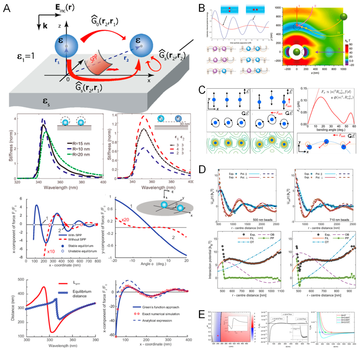
Optical tweezers for colloidal interaction modulation and nanostructure assembly. (A) depicts plasmon-assisted optical binding near metallic substrates, where the scattering Green’s function couples incident and induced fields and SPPs mediate long-range coupling; stiffness spectra versus particle radius and dielectric contrast reveal tunable resonances, and computed binding forces along identify stable and unstable separations under varied illumination, with analytical and numerical models in agreement. (B) presents resonant optical potential wells that template nanoparticle chains: polarization-resolved plasmonic interference patterns create periodic energy minima aligned with field gradients; calculated force vectors and resonance-enhanced potentials explain how counter-rotating vortex fields generate symmetric wells for stable linear assemblies. (C) provides experimental validation of distance-resolved inter-particle potentials for 500 nm and 710 nm colloids under orthogonal polarizations, quantifying well depth and width; comparison among plasmon-enhanced (P), optical-tweezer-only (OT), and control conditions shows that light-induced potentials dominate thermal Brownian agitation at submicron separations. (D) demonstrates programmable colloidal lattices using holographic optical tweezer arrays with real-time thermo-optical feedback; simulations and schematics of thermal fields and convection around trapped colloids show how feedback tunes interaction strength to dynamically reconfigure two-dimensional crystals. (E) illustrates force-landscape modulation by coupled optical and thermophoretic gradients: simulated refractive-index changes reshape the optical potential; experiments overlay the measured landscape with force-field lines to confirm feedback-induced equilibrium shifts; force–distance curves indicate that coupling strength can be reprogrammed by illumination intensity or particle size, enabling dynamic self-organization in colloidal assemblies.
Figure 12.
Optical tweezers for colloidal interaction modulation and nanostructure assembly. (A) depicts plasmon-assisted optical binding near metallic substrates, where the scattering Green’s function couples incident and induced fields and SPPs mediate long-range coupling; stiffness spectra versus particle radius and dielectric contrast reveal tunable resonances, and computed binding forces along identify stable and unstable separations under varied illumination, with analytical and numerical models in agreement. (B) presents resonant optical potential wells that template nanoparticle chains: polarization-resolved plasmonic interference patterns create periodic energy minima aligned with field gradients; calculated force vectors and resonance-enhanced potentials explain how counter-rotating vortex fields generate symmetric wells for stable linear assemblies. (C) provides experimental validation of distance-resolved inter-particle potentials for 500 nm and 710 nm colloids under orthogonal polarizations, quantifying well depth and width; comparison among plasmon-enhanced (P), optical-tweezer-only (OT), and control conditions shows that light-induced potentials dominate thermal Brownian agitation at submicron separations. (D) demonstrates programmable colloidal lattices using holographic optical tweezer arrays with real-time thermo-optical feedback; simulations and schematics of thermal fields and convection around trapped colloids show how feedback tunes interaction strength to dynamically reconfigure two-dimensional crystals. (E) illustrates force-landscape modulation by coupled optical and thermophoretic gradients: simulated refractive-index changes reshape the optical potential; experiments overlay the measured landscape with force-field lines to confirm feedback-induced equilibrium shifts; force–distance curves indicate that coupling strength can be reprogrammed by illumination intensity or particle size, enabling dynamic self-organization in colloidal assemblies.
The additional forces discussed here complete the near-surface and multi-body interaction picture that overlays the single-particle force balance developed in Secs. 2.2.2–2.2.7. Casimir–van der Waals and electrostatic interactions set short-range constraints and bias near interfaces, while optical binding introduces collective, light-mediated organization in ensembles. Practical nano-tweezer design must therefore co-optimize optical gradients with interface chemistry, screening conditions, and illumination geometry to harness or suppress these interactions as required by the experiment.
2.3.4. Hydrodynamic Interactions
In fluid-based optical tweezer experiments, multiple trapped particles interact not only through light but also through the viscous flows they induce in the surrounding medium [251]. These hydrodynamic interactions arise from the fluid’s Stokesian response to particle motion and provide a long-range, velocity-dependent coupling that is unavoidable in multi-particle trapping scenarios [252]. Consider two microspheres held in adjacent traps [253]: if one bead is thermally excited or actively displaced by modulating its trap, it perturbs the fluid and launches a flow that exerts a viscous drag on the neighboring particle—even in the absence of direct optical coupling [254]. In an unbounded fluid the leading-order interaction decays as, producing correlated motion that is readily observed in trajectory cross-correlations and modified fluctuation spectra [255,256]. In dual-trap instruments this coupling is well documented: the power spectral density (PSD) of one bead’s motion is measurably altered by the presence and proximity of the second bead, and traps operated near solid boundaries experience enhanced effective drag due to no-slip wall effects that further reshape the spectra and response functions [256].
Because hydrodynamic coupling is dynamical rather than static, it is naturally described by coupled Langevin equations in which the particle velocities are related to forces through a configuration-dependent mobility matrix. In practice, Oseen or Rotne–Prager-type tensors capture the leading hydrodynamic terms and predict collective modes, cross-correlated Brownian fluctuations, and apparent shifts in trap stiffness or calibration—effects that are especially consequential in precision force spectroscopy and microrheology. The interaction strength scales with fluid viscosity , particle size (radius ), inter-particle distance , and boundary proximity; for two beads separated by in water, the induced hydrodynamic drag on the neighbor can reach 10–30% of the primary drag, large enough to bias parameter extraction if left unaccounted [257,258,259].
Mitigation strategies include increasing inter-bead spacing (typically >10 μm) , incorporating hydrodynamic models to numerically subtract the coupled contribution during analysis, and employing feedback control or flow-resistant chamber geometries to suppress spurious cross-talk. In sum, hydrodynamic interactions are a non-optical yet essential element of the force landscape in fluidic optical tweezers; understanding and compensating for them is critical in multi-particle assays, biophysical measurements, and microrheology, where correlated dynamics could otherwise be mistaken for intrinsic inter-particle forces [260,261].
Together with viscous drag (Sec. 2.2.6) and thermal forcing (Sec. 2.2.5), hydrodynamic interactions complete the fluid-dynamical layer atop the optical force framework (Secs. 2.2.2–2.2.4 and 2.2.7). They set the symmetry and range of velocity correlations, determine how boundary conditions renormalize damping, and define the corrections required to interpret PSDs, response functions, and stiffness measurements in multi-object configurations.
Figure 13.
Environmental and biomedical applications of optical tweezers across plasmonic, optothermal, and nanomechanical platforms. (A) illustrates optical torque and orbital rotation of birefringent or shape-anisotropic probes under circularly polarized light, enabling rotational microrheology: measured angular velocities and orbit radii versus trap asymmetry (), provide viscosity and environmental stiffness readouts useful for microplastic dispersions and biofilms. (B) presents a microfluidic-integrated tweezing platform for single-particle transport, capture, and classification; bright-field tracking with Gaussian fits to position histograms yields trapping stiffness, while flow-induced offsets quantify calibrated hydrodynamic loading for label-free discrimination of vesicles and nanoplastics in complex media. (C) demonstrates photothermal thermophoretic trapping of nanoplastics and environmental vesicles using a 532 nm beam on a gold-coated substrate to impose a vertical temperature gradient; distance-resolved binding potentials, force–distance curves, and escape probabilities versus salt concentration and particle size reveal how electrolytes tune capture in aquatic environments. (D) shows multiscale diffusion analysis and viscoelastic sensing: DNA-tagged vesicles are confined and tracked to obtain displacement histograms, phase-space plots, and PSDs, from which storage and loss moduli ( and) are extracted; MSD crossovers distinguish Brownian, confined, and driven regimes, establishing a workflow extendable to exosomes in diagnostics. (E) highlights active mechanical stimulation and vesicle sorting using circular trap arrays that impose lateral loads; high-speed imaging resolves size- and stiffness-dependent deformation and translation under pulsed modulation (e.g., 1.25 mW vs 2.50 mW), enabling mechanical fingerprinting of extracellular vesicles in biofluids. Collectively, the panels connect optical, hydrodynamic, and thermophoretic drives to quantitative readouts, emphasizing how careful control and modeling of fluid-mediated coupling underpin robust environmental sensing and biomedical assays.
Figure 13.
Environmental and biomedical applications of optical tweezers across plasmonic, optothermal, and nanomechanical platforms. (A) illustrates optical torque and orbital rotation of birefringent or shape-anisotropic probes under circularly polarized light, enabling rotational microrheology: measured angular velocities and orbit radii versus trap asymmetry (), provide viscosity and environmental stiffness readouts useful for microplastic dispersions and biofilms. (B) presents a microfluidic-integrated tweezing platform for single-particle transport, capture, and classification; bright-field tracking with Gaussian fits to position histograms yields trapping stiffness, while flow-induced offsets quantify calibrated hydrodynamic loading for label-free discrimination of vesicles and nanoplastics in complex media. (C) demonstrates photothermal thermophoretic trapping of nanoplastics and environmental vesicles using a 532 nm beam on a gold-coated substrate to impose a vertical temperature gradient; distance-resolved binding potentials, force–distance curves, and escape probabilities versus salt concentration and particle size reveal how electrolytes tune capture in aquatic environments. (D) shows multiscale diffusion analysis and viscoelastic sensing: DNA-tagged vesicles are confined and tracked to obtain displacement histograms, phase-space plots, and PSDs, from which storage and loss moduli ( and) are extracted; MSD crossovers distinguish Brownian, confined, and driven regimes, establishing a workflow extendable to exosomes in diagnostics. (E) highlights active mechanical stimulation and vesicle sorting using circular trap arrays that impose lateral loads; high-speed imaging resolves size- and stiffness-dependent deformation and translation under pulsed modulation (e.g., 1.25 mW vs 2.50 mW), enabling mechanical fingerprinting of extracellular vesicles in biofluids. Collectively, the panels connect optical, hydrodynamic, and thermophoretic drives to quantitative readouts, emphasizing how careful control and modeling of fluid-mediated coupling underpin robust environmental sensing and biomedical assays.

2.4. Trap Stiffness and Trap Depth Analysis
The performance of an optical trap is governed by two intimately related quantities—trap stiffness and trap depth—which together determine how tightly a particle is confined and how robust that confinement is against thermal agitation and external perturbations [106]. Stiffness sets the local restoring force near the equilibrium point, while depth sets the energetic barrier that must be surmounted for escape; optimizing one without regard for the other can yield traps that are either precise yet shallow or deep yet sluggish.
Trap Stiffness.
Trap stiffness () is the proportionality between the restoring force and a small displacement from equilibrium (), and is typically reported in (pN/nm)[262]. In a tightly focused beam and for Rayleigh-to-Mie–size dielectric particles, the scaling can be summarized (to leading order) as
where: is the laser power, is the refractive index of the medium, is the laser wavelength, is the particle radius, is the relative refractive index of particle to medium. This expression highlights several design levers: increasing power, particle size, or refractive-index contrast stiffens the trap, whereas moving to longer wavelengths or relaxing the focus reduces [263].
In practice, stiffness is calibrated in situ by three standard approaches that leverage the stochastic dynamics described in Sec. 2.2.5 and the viscous response of Sec. 2.2.6: the equipartition method ( ) ; frequency-domain fitting of the position power spectral density; and the Stokes-drag method, in which a controlled translation at known velocity imposes a hydrodynamic load balanced by the optical spring. Typical biological traps operate with , sufficient to suppress thermal excursions to the 10–20 nm scale in aqueous media at room temperature. Because high–numerical-aperture focusing creates anisotropic gradients, lateral and axial stiffnesses generally differ, and near surfaces or within structured fields (Secs. 2.2.7 and 2.3) the mode dependence of
must be characterized explicitly.
Trap Depth.
Where stiffness characterizes the local curvature of the potential, the trap depth () quantifies the energy barrier confining the particle and therefore the probability of thermally activated escape [264]. For a harmonic approximation near the minimum, one may write:
where is the effective range over which the optical gradient dominates over Brownian forcing and any axial radiation-pressure bias (Secs. 2.2.3 and 2.2.5). Robust operation requires to exceed the thermal energy by a comfortable margin; a widely used benchmark is , which at (), corresponds to∼. Depending on configuration, single-beam gradient traps typically realize depths of a few tens of , while strongly confined or resonance-enhanced platforms (e.g., plasmonic gaps, photonic-crystal cavities, or evanescent-wave traps) can reach hundreds of [265,266,267,268,269]. The same parameters that raise stiffness—higher power, larger , larger , shorter , and higher NA—also deepen the well, but the relationship can be highly nonlinear in near-field architectures where local intensity and polarizability are resonance-boosted (Sec. 2.2.7). In such cases, stiffness and depth scale superlinearly with the local field, enabling trapping of sub–50 nm objects with adequate despite their weak polarizability [270,271,272].
Factors Influencing Stiffness and Depth
| Parameter | Effect on Stiffness & Depth | Reference |
| Laser power (P) | Increases both linearly (up to damage limits) | [273] |
| Numerical aperture (NA) | Higher NA improves lateral and axial stiffness | [274] |
| Wavelength (λ) | Shorter wavelengths yield stronger gradient force | [275] |
| Particle radius (r) | ; depth depending on regime | [276] |
| Refractive index contrast (m) | Enhances both via increased polarizability | [265,266,267,268,269] |
| Medium viscosity (η) | Affects damping but not stiffness directly | [260,261]. |
Interdependence and experimental trade-offs.
Because and are shaped by the same optical and material parameters, improvements to one often benefit the other; however, dissipative and biological constraints impose practical ceilings. Raising P steepens ∇I and deepens the well but also increases photothermal loading (Sec. 2.2.4), which can alter local rheology, induce thermophoretic drifts, or damage living specimens. Increasing strengthens yet simultaneously increases viscous drag , slowing response dynamics (characteristic time ) and complicating high-bandwidth measurements. The choice of λ trades gradient efficiency against absorption and scattering; shorter wavelengths are more efficient for dielectric trapping but can aggravate absorption in biological media, whereas longer infrared wavelengths reduce photodamage at the cost of weaker gradients. Finally, proximity to surfaces and structured media modifies both stiffness and depth through near-field enhancement and additional short-range interactions (Secs. 2.3.1–2.3.3), which can either stabilize confinement or bias particles toward adhesion and aggregation if not carefully engineered.
Practical implications.
Weak stiffness manifests as large displacement fluctuations and degraded spatial precision, while shallow wells () are vulnerable to thermally driven escape. Applications that demand nanometer-scale positioning, single-molecule mechanochemistry, or high–Q optomechanics generally benefit from the combination of high and deep , provided that optical loading remains below photothermal tolerance thresholds. Accordingly, quantitative reporting of both stiffness and depth—together with the calibration pathway and environmental conditions—serves as a primary benchmark for comparing trap designs and for optimizing experimental performance across particle sizes and platforms.
2.5. Optical Trap Potential Reconstruction Techniques
Quantitatively interpreting optical-tweezers data requires knowledge of the effective potential energy landscape that confines the probe, not merely the local (harmonic) curvature near the minimum [277]. Real traps frequently deviate from ideal parabolas—exhibiting asymmetry, anharmonic shoulders, or even multimodal wells in the presence of near-field enhancement, interference patterns, or multi-beam configurations—so a suite of experimental reconstruction methods has been developed to infer U(r) directly from particle trajectories [272].
Boltzmann Distribution Mapping
The most widely used method for reconstructing trap potentials from particle trajectories is based on the Boltzmann distribution [278]. In thermal equilibrium, the probability of finding a particle at position xxx is related to the potential via:
where: is Boltzmann’s constant, is the absolute temperature, is an arbitrary constant.
By histogramming the position data of the trapped particle (typically from high-resolution video or quadrant photodiode recordings), and applying this logarithmic transformation, one can obtain the one-dimensional potential well shape along any spatial axis. This method is robust, non-invasive, and requires only passive monitoring of thermal fluctuations.
It assumes, however, that the system remains in equilibrium and that sampling is sufficient across the full spatial range of interest. For shallow or asymmetric traps, large data sets may be required to resolve the tails of the distribution accurately.
Power Spectral Density (PSD) Analysis
Another powerful method involves analyzing the frequency-domain signature of particle fluctuations [279]. A particle trapped in a harmonic potential behaves like a damped oscillator in a viscous medium, described by an overdamped Langevin equation. Its position power spectrum takes the Lorentzian form:
where: is the drag coefficient, is the corner frequency, is the trap stiffness.
By fitting the experimentally measured spectrum to this form, one can extract both trap stiffness and drag, indirectly characterizing the trap’s curvature and potential depth. PSD analysis is particularly suited for fast, real-time stiffness monitoring and for detecting deviations from harmonic behavior at high frequencies.
Fluctuation-Dissipation and Response Function Techniques
The Fluctuation–Dissipation Theorem (FDT) provides a deeper link between passive thermal fluctuations and active response to applied perturbations [280]. The linear response function that relates force to displacement in frequency domain is:
By comparing passive fluctuations with the system’s response to a known periodic driving force (e.g., modulating the trap center), one can validate trap linearity or reveal nonlinearities. This approach enables potential mapping in non-equilibrium conditions, extending beyond the assumptions of Boltzmann statistics.
Multidimensional reconstructions
Holographic and dual-beam systems routinely require 2D/3D potential mapping [281,282,283,284]. Practical strategies include joint 2D histogramming with Boltzmann inversion, principal-component analysis (PCA) to identify the trap’s principal axes, and numerical deconvolution of multi-channel detector signals to separate lateral and axial modes. These tools expose anisotropic stiffness matrices, bistable wells, and saddle directions that are central to interpreting protein unfolding, motor stepping, or colloidal interaction landscapes.
Practical considerations.
Accurate reconstruction depends on (i) data rate—higher temporal bandwidth sharpens PSD fits and resolves mode splitting; (ii) measurement noise—instrumental noise inflates ρ(x) tails and contaminates high-frequency PSDs; (iii) detector choice—QPDs offer sub-nanometer, MHz-bandwidth 1D/2D tracking, while cameras furnish full-field 2D trajectories at lower bandwidth; (iv) drift and non-stationarity—slow stage or sample drift violates equilibrium, biasing Boltzmann estimates; and (v) sample size—adequate counts in the tails are essential for reliable U(x) reconstruction. Incorporating hydrodynamic corrections (wall effects, Sec. 2.3.4) and mode anisotropy (Sec. 2.2.7) into the analysis further improves quantitative fidelity.
Practical Considerations
| Factor | Impact on Reconstruction | Reference |
| Data rate | High temporal resolution improves PSD accuracy | [263] |
| Measurement noise | Degrades both PSD and position histograms | [286] |
| Camera vs QPD | QPD offers higher frequency resolution; cameras offer full 2D tracking | [272] |
| Drift/stability | Non-stationarity violates equilibrium assumptions in Boltzmann analysis | [277] |
| Number of samples | Affects histogram quality and potential accuracy in tails | [270] |
Synthesis of the force landscape (link to Secs. 2.2–2.3).
The complete force balance in optical tweezers spans a hierarchy of interactions with distinct physical origins and scalings. At its core are the optical forces: a conservative gradient force that pulls dipoles up intensity gradients, and a non-conservative scattering (radiation-pressure) force along the beam axis (Secs. 2.2.2–2.2.3). Photophoretic/thermophoretic drives emerge from light-induced temperature gradients—especially relevant in air and in plasmonic or absorbing systems (Sec. 2.2.4). Brownian forcing introduces stochastic fluctuations set by , while viscous drag provides overdamped dynamics and a calibration handle via Stokes loading (Secs. 2.2.5–2.2.6). In near-surface or nanophotonic architectures, evanescent and near-field forces deliver sub-wavelength confinement at the expense of range (Sec. 2.2.7). Additional interfacial and environmental interactions—Casimir–van der Waals attractions at nanometer gaps, electrostatic forces from surface charges or fields, optical binding in multi-particle assemblies, and hydrodynamic coupling in viscous flows—enrich or complicate dynamics (Secs. 2.3.1–2.3.4).
These contributions obey different scalings—for instance, gradient forces scale with , scattering with , and Brownian fluctuations with . and their relative magnitudes depend on particle polarizability and absorptivity, geometry (proximity to interfaces, neighbors, or resonators), and medium (water, air, vacuum). Reliable, high-resolution manipulation therefore demands quantifying and modeling the full potential that results from their superposition. The techniques summarized above—Boltzmann mapping, PSD fitting, FDT-based active response, and multidimensional reconstructions—provide the quantitative foundation to calibrate, validate, and interpret optical-tweezer experiments across scales, and they serve as the bridge to the next section, where we detail practical protocols for measuring, modeling, and optimizing these forces for specific applications.
3. Architectures and Calibration of Quantitative Optical Tweezers
Building on the force landscape established in Section 2, this chapter turns to the experimental realizations that make quantitative measurements possible and to the metrology that renders those measurements traceable. Before detailing calibration, we first situate contemporary instruments within a structural taxonomy; different architectures impose distinct optical field geometries, noise pathways, and access to stiffness/depth regimes, all of which shape the attainable precision and bandwidth of force readouts. Figure 14 synthesizes this view by mapping the principal engineering routes from free-space optics to chip-scale and AI-assisted platforms, providing context for the calibration methods that follow.
3.1. Calibration Methods in Optical Tweezers
Precise force calibration is the foundation of all quantitative optical tweezers experiments [320]. Since the optical force is not directly measured but inferred from the particle’s displacement in the trapping potential, reliable calibration methods are essential to convert observed motion into absolute force units (e.g., piconewtons) and to determine key trap parameters such as stiffness, response time, and position sensitivity [321,322,323].
Depending on experimental conditions, particle size, fluid medium, and available instrumentation, several complementary calibration methods have been developed.
Equipartition Theorem Method.
This method leverages the thermal equilibrium between a trapped particle and its environment [324]. According to the equipartition theorem, the average potential energy stored in a harmonic optical trap is related to temperature:
where: is the trap stiffness, is the variance of particle position over time, is Boltzmann’s constant, is the absolute temperature.
This method is non-invasive, easy to implement, and effective for single-axis calibration, especially in aqueous environments [325]. However, it assumes a purely harmonic potential, stationary trap center, and negligible drift. It is less reliable in shallow or anharmonic traps, or under active modulation.
Power Spectral Density (PSD) Analysis.
PSD calibration exploits the frequency spectrum of thermally driven particle motion [326]. The trapped bead acts as an overdamped oscillator, and its positional fluctuations yield a Lorentzian power spectral density:
where: is the corner frequency ( ), is the Stokes drag coefficient, is the fluid viscosity, is the particle radius.
By fitting the experimentally measured spectrum to this form, one can simultaneously determine the trap stiffness and viscous drag. PSD analysis is widely used for real-time calibration, especially in high-bandwidth optical tweezers setups using quadrant photodiodes (QPDs). It also reveals nonlinearities or system noise beyond the trap’s corner frequency.
Stokes Drag Method (Flow Calibration).
This technique applies a known external force on the particle via fluid flow or controlled trap displacement. The Stokes drag force experienced by a particle moving at velocity through a fluid is [327,328]:
By translating the sample stage or the trap at constant speed and observing the steady-state offset of the particle from the trap center, one can infer the trap stiffness from:
This method is highly intuitive and particularly effective for calibrating high-stiffness traps or validating other methods. However, it assumes laminar flow, negligible wall effects, and requires accurate stage control.
Active Modulation & Step Response Calibration.
In this approach, the trap center is actively modulated using an acousto-optic deflector (AOD)[329], spatial light modulator (SLM), or piezo stage, and the particle’s stepwise response is recorded. The bead’s relaxation time constant is related to trap stiffness [298,306,330]:
By analyzing the time-resolved displacement trace, one can extract and . This method is ideal for dynamically characterizing traps in non-equilibrium conditions or under fluctuating laser power, and is often combined with feedback systems.
Comparison of Calibration Methods
| Method | Strengths | Limitations | Reference |
| Equipartition | Simple, passive | Sensitive to drift, assumes harmonicity | [144] |
| PSD | High-frequency resolution | Requires fast detector (QPD) | [328] |
| Stokes Drag | External control, intuitive | Needs flow control, wall correction | [324] |
| Step Response | Dynamic, real-time | More complex instrumentation | [332] |
Method comparison and advanced considerations.
Equipartition is simple and passive but drift-sensitive and harmonicity-dependent; PSD offers high-bandwidth precision but needs fast detectors (QPD) and careful noise handling; Stokes drag is externally controlled and intuitive but requires flow/stage accuracy and wall corrections; step-response is real-time and dynamic but instrumentation-heavier. In non-Newtonian fluids, near surfaces, or with anisotropic particles, calibration demands hydrodynamic corrections, finite-element modeling, or reference beads to account for altered drag and anisotropic stiffness. Best practice is to cross-validate at least two methods to ensure accuracy, reproducibility, and robustness—especially in complex biological or nanoscale regimes.
The structural choices summarized in Figure 14 condition not only what forces can be generated (stiffness, depth, anisotropy) but also how they should be calibrated (available bandwidth, susceptibility to drift, near-field effects). The following sections operationalize these considerations into step-by-step protocols for traceable force spectroscopy across free-space, fiber, and on-chip platforms.
3.2. Force Resolution and Sensitivity Limits
Optical tweezers routinely resolve forces from tens of piconewtons down to the sub-piconewton regime, but their ultimate performance is bounded by a combination of thermal fluctuations, detector precision, trap design, and environmental noise [331,332]. A clear accounting of these limits is essential when designing assays for single-molecule mechanics, weak biomolecular interactions, and nanoscale phenomena.
Thermal (Brownian) force floor.
The fundamental constraint arises from Brownian motion, set by the thermal energy . which drives stochastic force fluctuations on the trapped probe [333,334]. With a white thermal force spectrum (Sec. 2.2.5), the root-mean-square force noise within measurement bandwidth is :
where: is Boltzmann’s constant, is temperature, is the drag coefficient of a spherical particle, is the bandwidth (e.g., 1 Hz to 10 kHz).
For a 1 μm bead in water at room temperature and a bandwidth of 1 kHz, this results in , defining a baseline sensitivity floor.
Spatial and temporal readout.
Sensitivity depends equally on how accurately and how fast the particle position is measured [333,334,335]. Modern back-focal-plane/quadrant-photodiode (QPD) systems reach sub-nanometer spatial precision (< 1 nm) with bandwidths >100kHz; camera-based tracking commonly achieves ∼5–10nm at ≲1kHz in high-resolution modes. The force uncertainty from readout noise obeys :
where is the positional noise floor and is the trap stiffness. For a trap wit, a 1 nm resolution yields 0.1 pN force sensitivity.
Bandwidth–Noise Tradeoff.
Increasing the measurement bandwidth improves time resolution but amplifies thermal and electronic noise [336,337]. There is a tradeoff between: Fast response (high bandwidth) for dynamic processes (e.g., molecular motor stepping). Low noise (narrow bandwidth) for quasi-static force measurements (e.g., equilibrium unbinding forces).
Typical experiments balance this by choosing 1–5 kHz bandwidths, applying signal filtering, and averaging over multiple trials to improve signal-to-noise ratio (SNR).
Detector-Dependent Limits [338,339,340,341,342].
| Detector Type | Spatial Resolution | Temporal Resolution | Force Resolution |
| QPD | ~0.1–1 nm | 10–100 kHz | ~0.1 pN |
| High-speed camera | ~5–10 nm | 0.1–1 kHz | ~1–10 pN |
| Interferometric systems | ~< 0.1 nm | 100 kHz+ | < 0.1 pN |
Advanced techniques, such as back-focal plane interferometry, achieve sub-nanometer displacement sensitivity and enable sub-piconewton force resolution under well-controlled conditions [343,344].
Even in the absence of detector limitations, external factors such as: laser power fluctuations, mechanical vibrations, thermal drift of microscope components or acoustic noise, can introduce significant baseline fluctuations.
Common solutions include: Vibration-isolated tables, Temperature control enclosures, Laser intensity feedback stabilization, Reference channel subtraction, or Differential trapping (comparing two particles in identical traps).
Strategies to Enhance Sensitivity
| Approach | Mechanism |
| Active feedback | Real-time position stabilization via trap modulation |
| Interferometric detection | Enhances displacement readout sensitivity |
| Cooling the environment | Reduces thermal noise floor (limited in biological systems) |
| Stiff trap optimization | Improves force detection range (at cost of photodamage risk) |
| Statistical averaging | Repeated measurements improve SNR and effective resolution |
The force resolution and sensitivity of optical tweezers are ultimately constrained by the interplay between thermal fluctuations, trap stiffness, detection precision, and environmental stability. Sub-piconewton accuracy is routinely achievable with modern systems using QPDs and active stabilization, while new designs continue to push the limits toward femtoNewton and high-bandwidth regimes. Careful optimization of experimental design is essential to match the having outlined the fundamental physical forces in optical trapping systems and examined the theoretical and instrumental factors that determine trap stiffness, resolution, and sensitivity, we now turn to the practical realization of these principles in experimental settings. While theoretical models offer critical insights into how light interacts with matter to generate force, the ability to accurately measure and control these forces in the lab depends on the implementation of robust experimental techniques.
Modern optical tweezers are not merely passive traps but precision tools for quantitative force spectroscopy, capable of detecting sub-piconewton fluctuations and nanometer-scale displacements. However, transforming particle trajectories into meaningful force data requires careful calibration protocols, position detection schemes, and environmental stabilization strategies. Moreover, emerging applications—ranging from single-molecule mechanics to nanomaterial assembly—demand ever-increasing resolution, reliability, and adaptability.
In the following sections, we provide a detailed survey of the experimental methodologies that underlie force calibration and measurement in optical tweezers. These include both classical and advanced approaches, spanning passive thermal-based methods, active feedback systems, and real-time trap modulation. Together, they form the technical foundation that bridges optical physics with experimental biophysics and nanotechnology.
3.3. High-Resolution Position Detection Techniques
Precise force measurements in optical tweezers fundamentally depend on the ability to resolve the position of a trapped particle with high accuracy and temporal fidelity [345]. Since the optical force applied to a particle is typically inferred through its displacement from the center of the optical potential well, the precision of positional tracking directly determines the sensitivity, resolution, and reliability of force quantification [346]. A variety of detection strategies have been developed over the past decades to achieve sub-nanometer spatial resolution and kilohertz-to-megahertz temporal bandwidth, among which interferometric and image-based techniques are most widely adopted [347].
One of the most widely used and experimentally mature approaches is back focal plane interferometry (BFPI)[311]. In this technique, the interference between the unscattered trapping laser light and the forward-scattered light from the trapped particle is detected at the back focal plane of a high numerical aperture microscope objective. A quadrant photodiode (QPD) is typically used as the detector, generating differential voltage signals that linearly correspond to lateral displacements of the particle near the trap center. Owing to its inherently analog readout [348], BFPI offers temporal resolution exceeding 100 kHz and spatial resolution better than 1 nm, making it highly suitable for dynamic force spectroscopy applications.
BFPI systems require careful alignment, particularly in ensuring that the optical axis of the detection path is co-aligned with the trapping beam and that the particle remains near the focal plane [349]. The voltage outputs must be converted into physical units (meters) via calibration procedures, such as the equipartition method, power spectral analysis, or Stokes drag calibration (as will be discussed in later sections). Importantly, the principle of BFPI is fundamentally grounded in conservation of momentum: a change in light deflection direction corresponds to a reactive force on the particle. This forms the basis of more advanced implementations, such as momentum-resolved force detection schemes, where the photon momentum flux is directly measured without requiring positional displacement calibration.
Another category of position detection methods involves video microscopy using high-speed cameras coupled with digital image analysis [350]. Here, the centroid or shape of a trapped particle is tracked frame-by-frame using sub-pixel localization algorithms. This approach, while generally less sensitive than BFPI, provides distinct advantages in flexibility and accessibility. For instance, it allows simultaneous tracking of multiple particles, supports non-spherical or irregular geometries, and facilitates integration with fluorescence imaging [351]. Typical spatial resolutions are on the order of 5–10 nm, and temporal resolution is limited by frame rate, usually ranging from 100 Hz to several kHz depending on sensor specifications. Despite its limitations, video-based tracking remains indispensable in applications where complex or multiplexed imaging configurations preclude interferometric detection.
Beyond BFPI and video microscopy, a range of interferometric and heterodyne detection systems have been developed to achieve even greater sensitivity [352]. These include optical interferometers based on Michelson or Mach–Zehnder geometries, or back-focal plane detection enhanced with modulation techniques. Such systems have demonstrated sub-Ångström resolution in laboratory settings and are instrumental in ultra-sensitive applications such as measuring the energy landscapes of single molecular interactions, characterizing nanomechanical resonances, or tracking thermally activated transitions at near-quantum limits.
In all detection strategies, system stability—including laser coherence, mechanical vibration isolation, and thermal drift compensation—plays a critical role in achieving the theoretical limits of resolution. Real-time feedback, low-noise electronics, and digital filtering are often employed to further suppress background fluctuations [353,354]. As optical tweezers continue to evolve into fully integrated platforms, improvements in position detection will remain foundational to expanding the frontier of force measurement precision.
3.4. Trap Calibration Techniques
In optical tweezers experiments, the central quantity linking particle displacement to mechanical force is the trap stiffness (denoted , with units of pN/nm), which describes the linear restoring force exerted by the optical potential on a trapped particle [355]. Accurate determination of is essential for any quantitative application of optical tweezers, including force spectroscopy, mechanical property mapping, and single-molecule biophysics. Given the inherently nonlinear and often anisotropic nature of real optical traps, multiple experimental techniques have been developed to calibrate stiffness under various conditions, each with distinct advantages and limitations.
Equipartition Method.
The equipartition theorem from statistical mechanics provides a straightforward route to passive stiffness calibration [356]. It states that, at thermal equilibrium, each quadratic degree of freedom contributes to the average energy, where is Boltzmann’s constant and is the absolute temperature. If the trapped particle’s motion along the xxx-axis is modeled as a harmonic oscillator with stiffness , the theorem gives:
Here, is the variance of the particle’s position from the trap center, measured via high-resolution detectors such as BFPI or high-speed cameras.
This method is conceptually simple, requires no active perturbation, and is especially useful as an initial calibration step. However, it assumes a linear (harmonic) trap potential, and thus only yields accurate results when the particle remains near the trap center. Moreover, sufficient sampling statistics and drift correction are necessary to ensure accuracy, and it does not provide information on frequency-dependent dynamics [357].
Power Spectral Density (PSD) Analysis.
A more detailed calibration approach leverages the frequency-domain behavior of a trapped bead undergoing thermal fluctuations [358]. The motion of the bead in a harmonic optical trap immersed in a viscous fluid can be modeled as an overdamped Langevin system, with its positional power spectral density following a Lorentzian form [359,360]:
Here, is the drag coefficient (with fluid viscosity η\etaη and particle radius aaa), and is the corner (or roll-off) frequency, which marks the transition from thermal-dominated to stiffness-dominated motion.
By recording the time series of particle position (using BFPI or equivalent), computing the Fourier transform, and fitting the resulting power spectrum to this model [361], one can extract both and . This method provides access to dynamic stiffness, allows identification of system noise contributions, and is widely adopted in high-performance optical trapping systems.
One practical challenge is the need for detector sensitivity calibration—converting raw voltage to distance. This is often resolved by combining PSD fitting with equipartition-derived variance or with known mechanical displacements.
Stokes Drag Method.
The Stokes drag calibration method involves applying a known viscous force to the trapped particle and observing its displacement within the trap [362]. This is typically done by translating the sample stage at a constant low velocity , generating a hydrodynamic drag force . In the steady state, this is balanced by the optical restoring force:
This active calibration approach does not depend on thermal equilibrium and is independent of detector voltage scaling, making it a valuable cross-validation tool. However, it requires precise control of stage velocity and careful positioning to avoid inducing inertial or convective effects [363]. The method is most effective for traps operating in the linear regime and under low Reynolds number conditions.
Calibration via Known Displacements or Force Standards.
In addition to intrinsic methods, calibration can also be performed using external force standards: displacing the particle using a nanopositioning piezo-stage by a known amount, applying controlled magnetic or electric fields if the particle carries a known charge or magnetic moment or referencing against molecular standards with known force-extension curves (e.g., DNA hairpins, streptavidin-biotin rupture).
These methods are especially useful in hybrid force probes, such as combined optical-magnetic or optical-electric tweezer systems, where cross-calibration between modalities is required.
Conclusion of This Section.
Each calibration technique addresses different experimental needs and assumptions. The equipartition method is elegant in its simplicity but limited in scope. Power spectrum analysis offers high resolution and diagnostic power but requires careful spectral fitting. Stokes drag calibration provides a physical reference point, suitable for absolute validation. In advanced systems, multiple techniques are often applied in combination to ensure internal consistency and reduce systematic error.
In all cases, temperature stability, optical alignment, and medium properties must be carefully controlled to ensure that calibration results are accurate and transferable across experiments [274,355]. The ability to confidently determine trap stiffness is foundational to the conversion of displacement signals into reliable force data—a prerequisite for any quantitative optical tweezer study.
3.5. Active Force Readout and Real-Time Measurement Systems
While passive calibration techniques provide accurate estimations of trap stiffness and force response under equilibrium conditions, modern optical tweezers applications increasingly demand active force control, real-time feedback, and absolute force readout, especially in dynamic or non-equilibrium experiments [313]. This necessitates the development of force measurement systems that go beyond thermal fluctuation analysis and provide direct and instantaneous access to optical forces, ideally without the need for frequent recalibration.
Photon Momentum Detection: Towards Absolute Force Readout.
The fundamental physical basis for all optical trapping forces lies in momentum conservation [364]. When a photon is scattered or absorbed by a particle, it transfers momentum to the particle, and the reaction force can, in principle, be measured by detecting the change in momentum of the light field. This concept underlies a class of methods collectively referred to as photon momentum detection, which aim to directly quantify the force by measuring the deflection or redistribution of the trapping laser beam.
In practical implementations, such as back focal plane interferometry (BFPI), this principle is indirectly utilized: the displacement of the particle alters the interference pattern due to the redistribution of optical momentum, which is then captured as voltage changes in a photodiode array [365]. However, calibration is still required to convert signal into physical force units.
Recent advances have aimed at realizing calibration-free photonic force detectors by directly measuring the optical momentum flux. These include: light momentum sensing using integrating spheres or segmented photodetectors that collect the total angular and intensity distribution of the transmitted or reflected beam. Interferometric force sensors that resolve tiny changes in beam direction or phase associated with particle interactions. Dual-beam balance systems, where two counter-propagating traps allow net force to be inferred from beam displacement differences, canceling out drift and system noise.
Such setups enable absolute force measurements based purely on optical power and beam geometry, offering advantages in scenarios where thermal calibration is compromised (e.g., in non-aqueous media, vacuum, or high-force regimes)[365,366]. The trade-off, however, lies in system complexity: precise optical alignment, high-fidelity beam shaping, and low-noise detection electronics are essential.
Real-Time Force Clamping and Feedback Control.
Another major innovation in modern optical tweezers is the development of real-time feedback mechanisms for active force clamping—analogous to the force-clamp mode in atomic force microscopy (AFM)[367]. In such systems, the trap position (via acousto-optic deflectors or piezo stages) or the laser intensity is dynamically adjusted to maintain a constant force on the particle, as determined from continuous position monitoring.
This method enables experiments where a biological or soft-matter sample is held under a constant mechanical load while it undergoes structural or kinetic transitions, such as [368]: protein unfolding under sustained tension, DNA overstretching at a fixed force threshold, receptor-ligand dissociation studies under force-regulated dynamics.
To implement this, the displacement signal—typically from BFPI—is fed into a digital feedback controller (e.g., PID control), which modifies the trap parameters in real time to maintain a user-defined force setpoint [369]. The accuracy of this method depends on: bandwidth of the feedback loop (typically 1–10 kHz), latency in signal processing and the stiffness of the trap (low-stiffness traps allow more precise force control but reduce spatial confinement).
Advanced implementations have employed field-programmable gate arrays (FPGAs) or real-time operating systems to achieve microsecond-level latency, enabling the capture of rapid biophysical events that would otherwise be averaged out or missed in open-loop systems [370].
Applications in Complex and Non-Equilibrium Systems.
Direct force detection and feedback control are especially valuable in experimental regimes where: the medium has non-Newtonian viscosity (e.g., intracellular environments, polymer gels), the particle undergoes active fluctuations (e.g., molecular motors, living cells) or the thermal equilibrium assumption is invalid (e.g., heating, driven systems).
In such conditions, passive fluctuation-based methods may yield erroneous stiffness estimates or force measurements. Real-time systems that read out and actively regulate force enable precise control over mechanical variables, allowing researchers to decouple force-dependent and time-dependent effects, and to explore energy dissipation, response hysteresis, and nonlinear mechanical landscapes in ways not accessible to equilibrium methods [371].
Conclusion of This Section.
The development of active force readout technologies represents a paradigm shift in optical tweezers instrumentation—from passive probing tools to dynamic, programmable systems for mechanical interrogation and manipulation. By directly measuring or regulating optical forces in real time, these systems enable new classes of experiments in biology, soft matter physics, and nanotechnology.
In the next section, we explore additional calibration and detection strategies based on video tracking, hybrid systems, and environmental compensation, expanding the experimental toolbox available for accurate force quantification under diverse experimental conditions.
3.6. Video Tracking and Hybrid Calibration Methods
While advanced optical sensors such as quadrant photodiodes and interferometric detectors dominate high-resolution force readout in optical tweezers, video-based tracking remains a widely used and accessible technique for position and force calibration [372]. It serves both as a standalone calibration tool in simpler setups and as a complementary method in hybrid systems where multiple detection modalities are integrated. This section discusses the principles, strengths, limitations, and extensions of video tracking for force calibration, and introduces how it combines with other techniques in hybrid schemes to enhance measurement robustness.
Principles of Video Tracking.
In video-based optical tweezers systems, a high-speed camera records the motion of a trapped particle under bright-field, dark-field, or fluorescence illumination [373]. The video frames are then processed to extract the particle’s centroid in each frame, yielding a time series of 2D or 3D position coordinates (x(t),y(t),z(t))(x(t), y(t), z(t))(x(t),y(t),z(t)). Sub-pixel localization algorithms, often based on Gaussian fitting or centroid refinement, allow for position resolution down to ~5–10 nm under favorable conditions [374].
From these trajectories, one can apply the same calibration principles used in interferometric methods:
- Equipartition calibration: by measuring the variance , and applying , assuming a harmonic trap.
- Power spectral analysis: by computing the power spectral density (PSD) of the motion to extract the corner frequency and then deducing trap stiffness via , where is the drag coefficient (calculated using Stokes’ law).
Video tracking also enables visual confirmation of trapping, particle escape, or multi-particle interactions—critical for troubleshooting and interpretation.
Technical Specifications and Constraints.
The temporal and spatial resolution of video tracking is fundamentally limited by camera specifications:
- Temporal resolution is governed by the frame rate . Modern scientific CMOS cameras can reach >1000 fps, but most setups operate between 100–500 fps due to data throughput limits. This limits the upper frequency components of Brownian motion that can be analyzed.
- Spatial resolution is constrained by pixel size, optics magnification, and signal-to-noise ratio. With optimized imaging and sub-pixel localization, ~5–10 nm resolution is achievable, but this remains less sensitive than interferometric detectors (which resolve <1 nm).
These constraints imply that video tracking is best suited for low-bandwidth force measurements or for applications where visual inspection is essential—such as multi-trap arrays, heterogeneous samples, or optically complex environments [375,376,377,378].
Case Study: Dual-Bead Video-Based Calibration.
A common implementation is the dual-trap configuration with video tracking of both trapped beads. By analyzing relative motion, one can cancel common-mode noise (e.g., drift or camera jitter) and extract inter-bead forces [379]. One such example involves tethered DNA molecules stretched between two microspheres, where video tracking reveals force-extension curves under calibrated displacement.
In this configuration: the trap stiffness is first estimated from thermal motion in each trap independently (equipartition). Then, a Stokes drag calibration is performed by moving one trap relative to the other at known velocity and observing steady-state displacement. Finally, force-extension behavior is mapped under controlled stage movement and compared with theoretical worm-like chain (WLC) models for DNA elasticity, verifying calibration fidelity.
This combination demonstrates how hybrid video + mechanical calibration yields robust quantitative force curves with sub-pN precision.
Hybrid Methods and Multimodal Feedback.
As optical tweezers move toward complex in vivo or microfluidic environments, hybrid calibration techniques become crucial [380]. These integrate: BFPI for fast, high-resolution axial force detection. Video tracking for lateral position monitoring or multi-bead detection. Fluorescence imaging for detecting specific biomolecular events (e.g., binding, cleavage).
In such hybrid systems, video tracking acts as a low-frequency verification or auxiliary input to cross-check interferometric signals or to resolve ambiguities in noisy environments.
In recent developments, machine-learning-based video analysis has further extended the capability of video tracking. Neural networks trained on synthetic or empirical datasets now enable real-time, high-accuracy localization even under noisy or low-contrast conditions, expanding the usability of video methods in challenging scenarios.
Summary.
Video tracking in optical tweezers offers a versatile, visual, and quantitative means of position measurement and force calibration. Though limited in spatial and temporal resolution compared to interferometric methods, it provides unique advantages in accessibility, multi-particle handling, and hybrid calibration schemes. By combining video tracking with other force readout methods—such as Stokes drag or PSD analysis—experimentalists gain flexibility and redundancy, ensuring robust calibration across a range of applications from simple single-particle trapping to complex force spectroscopy on multi-component biological systems.
3.7. Advanced Calibration Protocols and Real-Time Feedback Systems
The increasing complexity of optical tweezers experiments—especially those involving live cells [368], dynamic molecular systems, or high-throughput manipulation—demands not only precise force calibration, but also the ability to actively maintain control over force, position, and experimental conditions in real time [381]. To this end, researchers have developed a range of advanced calibration protocols and integrated feedback systems that automate, accelerate, and stabilize force measurements far beyond traditional static calibration schemes.
Real-Time Trap Calibration During Experiments.
Conventional calibration techniques (e.g., equipartition, power spectral analysis) are typically performed prior to data collection and assume constant trap parameters. However, laser power fluctuations, refractive index drift, focus shifts, or biological sample movement can alter trap stiffness or detection sensitivity over time [382]. To maintain accuracy in such dynamic environments, real-time calibration approaches have been developed.
One such method involves real-time spectral analysis of Brownian motion: a continuously recorded trajectory is parsed in short time windows (e.g., 1–2 s), within which the trap is assumed quasi-stationary. Each window yields a local stiffness value based on corner frequency :
where is the drag coefficient. This allows calibration to track time-varying system conditions, and the force readout can be updated dynamically without interrupting the experiment. Such calibration can be integrated into commercial software or FPGA-based hardware systems for real-time implementation.
Automated Calibration Pipelines.
To reduce user dependency and error-prone manual steps, automated calibration routines have become standard in high-end optical trapping platforms [383]. These systems integrate: Built-in stage motion routines to apply known drag forces for Stokes calibration. On-board PSD calculation modules that automatically fit Lorentzian curves and extract . Temperature and viscosity compensation algorithms, which adjust based on real-time environmental sensors.
Such pipelines enable non-expert users to perform force-calibrated experiments with minimal intervention. In research contexts requiring repeated measurements, such as high-throughput single-molecule pulling assays, automation dramatically improves repeatability and statistical power.
Real-Time Force Clamping and Active Positioning.
Beyond passive force measurement, advanced optical tweezers setups incorporate real-time force feedback loops to maintain a constant force on a particle, even as its position changes [368]. This is essential for experiments where biological or mechanical systems respond non-linearly to force, such as: DNA overstretching transitions、 Motor protein stepping under load、Force-induced protein unfolding.
In typical implementations, the displacement of the bead x(t)x(t)x(t) from the trap center is measured (e.g., via BFPI), and the trap position or laser power is modulated in real time to keep the applied force at a preset value. This feedback loop is typically executed by a digital PID controller or implemented in FPGA hardware for sub-millisecond response time.
Experimental demonstrations of force-clamp have achieved <0.1 pN force stability over tens of seconds and Bandwidths >1 kHz, allowing tracking of fast molecular kinetics. Automated adjustment to maintain constant tension during structural transitions.
Multi-Dimensional and Adaptive Calibration.
In modern 3D optical trapping systems, calibration must be extended to multiple spatial axes, and stiffness values may differ along x, y, and z due to beam asymmetry and optical aberrations [382]. Adaptive calibration schemes are employed to handle these anisotropies.
For example, 3D trap stiffness tensor calibration combines: Equipartition or PSD analysis in 、Cross-correlation of positional noise between axes、Z-axis stiffness determination via axial imaging or quadrant photodiode phase-shift detection.
Recent methods also integrate machine learning models to infer force fields in complex environments. Neural networks trained on synthetic Brownian motion data have been shown to accurately predict trap stiffness under noisy or partially observed conditions, enabling blind calibration when traditional models fail.
Summary.
Advanced calibration protocols and real-time control systems are transforming optical tweezers from static instruments into responsive, intelligent tools for biophysical and nanotechnological exploration. These innovations enable stable force control in fluctuating environments, automated force measurements without user intervention, and dynamic adaptation to sample behavior or instrument drift. In conjunction with high-resolution detection and robust modeling, these systems constitute the backbone of modern force spectroscopy experiments—extending the precision, applicability, and reliability of optical tweezers into previously inaccessible regimes.
3.8. Error Sources and Compensation Strategies
Despite the high precision achieved by modern optical tweezers, force calibration and measurement remain vulnerable to several systematic and stochastic error sources. Recognizing, quantifying, and mitigating these sources are essential for ensuring the accuracy, repeatability, and interpretability of optical force experiments—especially when working in the sub-piconewton regime. This section presents the major classes of error sources in optical trapping experiments and outlines commonly adopted correction and compensation strategies.
Optical and Mechanical Drift.
One of the most common experimental limitations arises from slow drifts in optical alignment or mechanical components [356]. Sources include thermal expansion of microscope stages, misalignment of lenses, and long-term focus drift due to temperature fluctuations.
For instance, a typical laser trapping system might experience stage drift at rates of ~10–100 nm/hour, which can be significant when sub-nanometer positional accuracy is required. To mitigate such drifts [384]: Active stabilization techniques are often employed using reference markers and feedback loops (e.g., piezo-actuated stages with interferometric feedback). Temperature regulation of both the sample chamber and optical components (±0.01°C) reduces thermal gradients that cause drift. Reference beads fixed to the coverslip are frequently imaged alongside trapped particles to distinguish real motion from stage-induced artifacts.
Laser Intensity Fluctuations.
Since optical gradient force scales with laser intensity , fluctuations in laser power lead directly to errors in trap stiffness and thus force measurements [357]. Even commercial diode lasers may show intensity noise at the 0.1–1% level, which translates to measurable fluctuations in trap strength.
Power stabilization systems, including acousto-optic modulators (AOMs) controlled by feedback electronics, are often integrated to maintain laser output within ±0.01%. Experiments often include continuous power monitoring, either through a beam-split and photodiode or via the signal from back focal plane interferometry (BFPI) itself.
Nonlinearities and Trap Anharmonicity.
The harmonic approximation of an optical trap potential holds only near the trap center. At displacements >~200–300 nm (depending on beam waist and NA), the potential becomes anharmonic, leading to biased force measurements if the particle samples this region [358].
To quantify trap linearity, researchers often perform force–displacement scans or compute the trap potential directly from the positional probability distribution using the Boltzmann relation:
where deviations from a parabolic profile indicate nonlinearity.
Hydrodynamic Artifacts.
In experiments near surfaces or with multiple beads, hydrodynamic interactions distort drag coefficients and can introduce coupling between particles. For instance, drag on a bead increases by >30% when within one radius of a coverslip. Neglecting this effect leads to underestimation of viscous forces and hence errors in stiffness calibration [383].
To address this: Faxén corrections are applied to Stokes drag calculations when beads are near surfaces:
where a is bead radius and h is distance to surface.
Dual-trap setups use differential measurements to cancel shared hydrodynamic signals.
Electrostatic and Photothermal Disturbances.
Charged surfaces or particles in poorly buffered media can create local electrostatic fields, perturbing force balances unpredictably. Similarly, absorption-induced local heating creates thermophoretic drifts, especially for metallic or pigment-containing particles.
These are countered by careful buffer formulation with high ionic strength to suppress double-layer interactions or use of low-absorption IR lasers (e.g., 1064 nm) and low power densities (<10 mW/µm²) to minimize heating and implementing dual-laser schemes (trapping and imaging) where only one beam is modulated at a time.
Summary.
Errors in optical trapping experiments are multifaceted, arising from both instrumental imperfections and physical interactions in the sample environment. Precision calibration depends not only on using the right method but also on recognizing and correcting for the factors that compromise force readout. Advanced setups today routinely incorporate multiple safeguards—including real-time drift correction, power stabilization, dual-beam trap architectures, and hydrodynamic modeling—to ensure force measurements accurate to within 0.1 pN or better. In combination, these strategies allow optical tweezers to transition from qualitative manipulation tools into true quantitative instruments for probing the mechanics of biological and nanoscale systems.
4. Typology of Optical Tweezers
Anchored in the force landscape and metrology developed in the previous chapters, this section maps how optical tweezers diversify into distinct application arenas and hardware lineages. Figure 15 acts as the narrative bridge: it assembles, at a glance, the dual trajectory of the field—toward biological interrogation and toward physical/material manipulation—while underscoring that a common optical mechanism (gradient and scattering forces sculpted by beam geometry and detection) underlies heterogeneous use cases [78,291,305,385,386,387,388,389,390,391,392,393,394,395,396,397,398,399,400,401,402]. On the biological side, precision stretching of DNA, RNA, and proteins exposes entropic elasticity, base-pair unzipping, and unfolding transitions with piconewton resolution [403] the same trap–detector stack, paired with appropriate probes and readouts, quantifies single-cell mechanics and downstream responses such as calcium influx or immune activation at the single-cell level [404]. Coupling plasmonic enhancement to trapping enables SERS-based biochemical fingerprinting without labels, extending the reach of optical tweezers to molecular diagnostics [405] whereas optothermal concentration of CRISPR complexes in engineered temperature gradients pushes nucleic-acid assays toward ultra-high sensitivity in compact formats [406]. Integration with microfluidic routing and photothermal infrared spectroscopy further allows label-free sorting and classification of cells and subcellular particles in flow [407] and dual-fiber “lab-on-fiber” platforms add in situ force mapping within confined geometries and even in vivo contexts where conventional microscope access is limited [408].
The physical and materials branch is equally broad: tweezer-assembled neutral-atom arrays furnish programmable lattices for quantum simulation of many-body physics [409].; at the mesoscopic scale, traps template colloids and nanoparticles into ordered architectures, including SERS-active substrates that exploit controllable inter-particle gaps for field enhancement [168] Soft condensed-matter studies leverage trapping to extract viscoelastic and poroelastic parameters of hydrogels under well-defined loads [410], while compression and confinement of microcapsules reveal their interfacial and interior mechanics in situ [411]. Near interfaces, tracking the approach and residence of trapped nanoparticles quantifies surface-adsorption kinetics with molecular-scale temporal resolution [412], and in microfluidic chemomechanical environments, tweezers resolve the spatiotemporal release profiles of stimuli-responsive drug carriers [97,413,414,415]. Read together, the two halves of Figure 15 motivate the typology that follows: common optical principles are repeatedly reinterpreted through beam shaping, photonic integration, and measurement strategy to match the mechanical bandwidth and sensitivity demanded by each domain.
Together, these examples highlight the versatility of optical tweezers as a cross-disciplinary platform: in biology, serving as force spectrometers and diagnostic tools; and in physics and materials science, as precision assembly and characterization instruments. The figure underscores how optical tweezers bridge fundamental light–matter interactions with practical applications ranging from single-molecule biophysics to quantum technology and nanomedicine.
4.1. Classical Single-Beam Optical Tweezers
The classical single-beam configuration—first demonstrated by Ashkin in 1986—remains the canonical architecture from which most other platforms depart [416]. A single, tightly focused Gaussian beam, brought to a diffraction-limited waist by a high–numerical-aperture objective, generates a three-dimensional trap via the balance of a conservative gradient force that draws a dielectric particle toward the intensity maximum and a non-conservative scattering force that pushes along the optical axis. The equilibrium typically lies slightly downstream of the geometric focus, where these contributions balance to yield stable confinement. In aqueous or gaseous media this design supports robust manipulation of micrometer-scale dielectric probes, providing a workhorse for piconewton-resolution force spectroscopy [411], single-molecule biophysics [417], colloidal physics, and cell biology [418]. Quantification hinges on the linear spring approximation near the trap minimum and on established in situ calibrations—equipartition, PSD fitting, Stokes drag, and step-response methods—outlined in Chapter 3.
Performance benefits stem from three coupled attributes: the steep intensity gradients produced by high-NA focusing afford high spatial and force resolution; the optical layout is compact and accessible, requiring a stable laser, a high-NA objective, and a position-sensitive detector; and the restoring stiffness is tuneable, approximately proportional to laser power over the operating range, which simplifies force control. The same features delineate the principal limitations. Diffraction and the Rayleigh scaling of polarizability restrict the trap depth for sub-100 nm objects, allowing Brownian agitation to overwhelm confinement unless near-field enhancement or alternative field architectures are introduced [419]; and the elevated intensities required for stronger gradients can induce photothermal loading or photochemical damage, a central concern in live-cell and in vivo applications. Despite these constraints, classical tweezers continue to define the quantitative baseline: improvements in laser stability, objective design, and interferometric detection have progressively pushed displacement noise below the nanometer level and force resolution toward the sub-piconewton regime. As such, single-beam traps serve both as a powerful stand-alone platform and as the reference against which holographic, plasmonic, fiber-integrated, and AI-assisted modalities are benchmarked [420,421,422].
Figure 16 consolidates the operating principles and advanced use cases of the single-beam paradigm within a consistent experimental narrative. The optical layout (A) follows the standard gradient trap: a beam-expansion and steering train feeds a high-NA objective, with dichroics directing fluorescence or imaging paths and a back-focal-plane interferometric module (e.g., QPD) registering sub-nanometer displacements. Within the focal volume, experimental trajectories and simulations co-register the emergence of a harmonic potential well and enable extraction of stiffness and anisotropy (B). A complementary ray-optics picture (C) clarifies momentum transfer via refraction and reflection at the particle interface and situates the single-beam geometry among related embodiments such as confocal and evanescent-field traps. Building on this base, dual-beam arrangements extend single-molecule manipulation to canonical assays—DNA stretching, protein unfolding, and nucleic-acid unzipping—where calibrated force–extension relations and state transitions are resolved in real time (D). Quantitative operability at low optical powers (E) is established by mapping stiffness versus power, waist, and particle size, thus delineating regimes where thermal and radiation-pressure asymmetries remain controlled. Finally, integration with microfluidics and high-resolution imaging (F) illustrates how a compact platform unites fluorescence readout, flow control, and force spectroscopy for biological assays in well-defined chemical and hydrodynamic environments. In sum, Figure 16 ties the theoretical spring model, calibration practice, and experimental implementations into a single workflow that continues to anchor—and to inform the evolution of—modern optical tweezer systems.
4.2. Holographic Optical Tweezers (HOT)
Extending the single-beam paradigm described in Section 4.1, holographic optical tweezers (HOT) transform optical trapping into a problem of computational beam shaping: a single input laser is synthetically decomposed into many tightly focused, diffraction-limited foci that can be positioned, reconfigured, and time-multiplexed in three dimensions from software alone [428,429,430,431]. First realized in the late 1990s, HOT systems employ computer-generated holography to sculpt the complex field at the pupil plane so that the microscope objective reconstructs an engineered constellation of traps in the sample volume. At the heart of this approach is the spatial light modulator (SLM)—typically a phase-only liquid-crystal-on-silicon device, or a digital micromirror device (DMD) for amplitude/temporal modulation—which encodes the hologram calculated by iterative algorithms (e.g., Gerchberg–Saxton, mixed-region amplitude freedom)[432]. Updating the hologram updates the trap geometry in real time, thereby converting mechanical manipulation into a programmable optical operation.
Although the microscopic forces remain those established earlier—conservative gradient forces and non-conservative scattering (radiation-pressure) forces acting on polarizable particles [433]—the HOT architecture introduces distinctive system-level trade-offs and capabilities. Because the laser power is divided among many diffractive orders, per-trap stiffness is lower than in a single-beam trap and must be budgeted against the desired trap count; diffraction efficiency, hologram quality, and optical throughput therefore directly set the usable stiffness envelope. Spatial non-uniformity (from pixelation, phase wrapping, or pupil aberrations) can seed inter-trap cross-talk and stiffness variation across the field; in practice, phase pre-compensation and adaptive optics are used to flatten the wavefront and homogenize the trap array. Axial control is achieved either holographically (adding defocus/astigmatism terms) or by coupling the SLM to a tunable lens, yielding fully three-dimensional arrays with independent lateral and axial placement.
These engineering levers enable capabilities that are difficult or impossible with static optics. Parallel manipulation allows dozens to hundreds of objects to be simultaneously trapped, moved, and assembled, which has been pivotal in soft-matter physics for real-time construction of colloidal crystals, quasicrystals, and controlled defect lattices [434]. In cellular and molecular biophysics, HOT supports coordinated, multi-point manipulation—such as aligning and interrogating many cells at once, corralling organelles within a single living cell, or distributing forces across cytoskeletal networks to quantify cooperative mechanics [429]. The same multi-trap geometry facilitates cooperative force spectroscopy, e.g., forming and stretching multi-handle DNA constructs between independently steered beads or imposing programmable stress patterns within biopolymer gels. Beyond biology, HOT has been applied to nanoparticle and quantum-dot arrays as reconfigurable optical lattices for nano-optomechanical experiments and model quantum simulations, while maintaining the flexibility to pattern vorticity (e.g., Laguerre–Gaussian modes) for optical torque control.
From a computational standpoint, HOT is a fertile area for algorithmic optimization: weighting strategies improve intensity uniformity; regularizers suppress speckle and ghost orders; and closed-loop holography incorporates camera/QPD feedback to iteratively correct trap positions and stiffness in situ. Recent efforts integrate machine learning to accelerate hologram prediction and to adapt trap patterns in heterogeneous or noisy samples, stabilizing assemblies and minimizing sample heating. Instrumentation typically includes a high-resolution SLM (≥ 1024×768), kHz-rate hologram refresh (DMDs excel in update speed), beam-expansion and relay optics to conjugate the SLM to the objective back aperture, and synchronized imaging/position detection for feedback.
A representative biological showcase is the real-time orchestration of immune synapse formation: HOT is used to simultaneously trap and orient multiple T cells and antigen-presenting cells in 3D, standardizing contact geometry and mechanical context so that signaling dynamics can be dissected under precisely controlled loading (Science, 2011)[435]. Comparable workflows extend to microbial communities, neuronal networks, and tissue spheroids, where multi-point, 3D force application reveals emergent mechanics and communication.
Figure 17.
Principles, architectures, and patterning capabilities of holographic optical tweezers (HOT) [34,436,437,438,439,440,441,442]. (A) Foundational configuration of HOT: a spatial light modulator (SLM) encodes phase holograms that are relayed through telescopic optics to project user-defined trap arrays at the sample plane. Diffractive optics and conjugate planes ensure beam shaping and focus control. (B) Optical pathways for dynamic trap generation. A laser beam is expanded and modulated by an SLM, followed by relay optics and dichroic mirrors to direct the beam into a high NA objective. Phase holograms enable simultaneous formation of multiple diffraction-limited traps. (C) Demonstration of multi-trap control and pattern reconfiguration. Vortex beams, optical lattices, and tailored trap geometries are realized via dynamic phase modulation. Both Laguerre-Gaussian and custom-designed patterns are supported. (D) HOT-based precision manipulation and automated control. Real-time feedback enables programmable sorting and error correction in cell/particle assembly. Visual feedback confirms trap success or failure, and adaptive patterns are computed accordingly. (E) Experimental realization of dynamic particle trapping using HOT. Beam shaping with SLM enables spatially addressable manipulation of microspheres, with high-speed feedback and accurate position detection via high-resolution cameras and photodiodes.
Figure 17.
Principles, architectures, and patterning capabilities of holographic optical tweezers (HOT) [34,436,437,438,439,440,441,442]. (A) Foundational configuration of HOT: a spatial light modulator (SLM) encodes phase holograms that are relayed through telescopic optics to project user-defined trap arrays at the sample plane. Diffractive optics and conjugate planes ensure beam shaping and focus control. (B) Optical pathways for dynamic trap generation. A laser beam is expanded and modulated by an SLM, followed by relay optics and dichroic mirrors to direct the beam into a high NA objective. Phase holograms enable simultaneous formation of multiple diffraction-limited traps. (C) Demonstration of multi-trap control and pattern reconfiguration. Vortex beams, optical lattices, and tailored trap geometries are realized via dynamic phase modulation. Both Laguerre-Gaussian and custom-designed patterns are supported. (D) HOT-based precision manipulation and automated control. Real-time feedback enables programmable sorting and error correction in cell/particle assembly. Visual feedback confirms trap success or failure, and adaptive patterns are computed accordingly. (E) Experimental realization of dynamic particle trapping using HOT. Beam shaping with SLM enables spatially addressable manipulation of microspheres, with high-speed feedback and accurate position detection via high-resolution cameras and photodiodes.
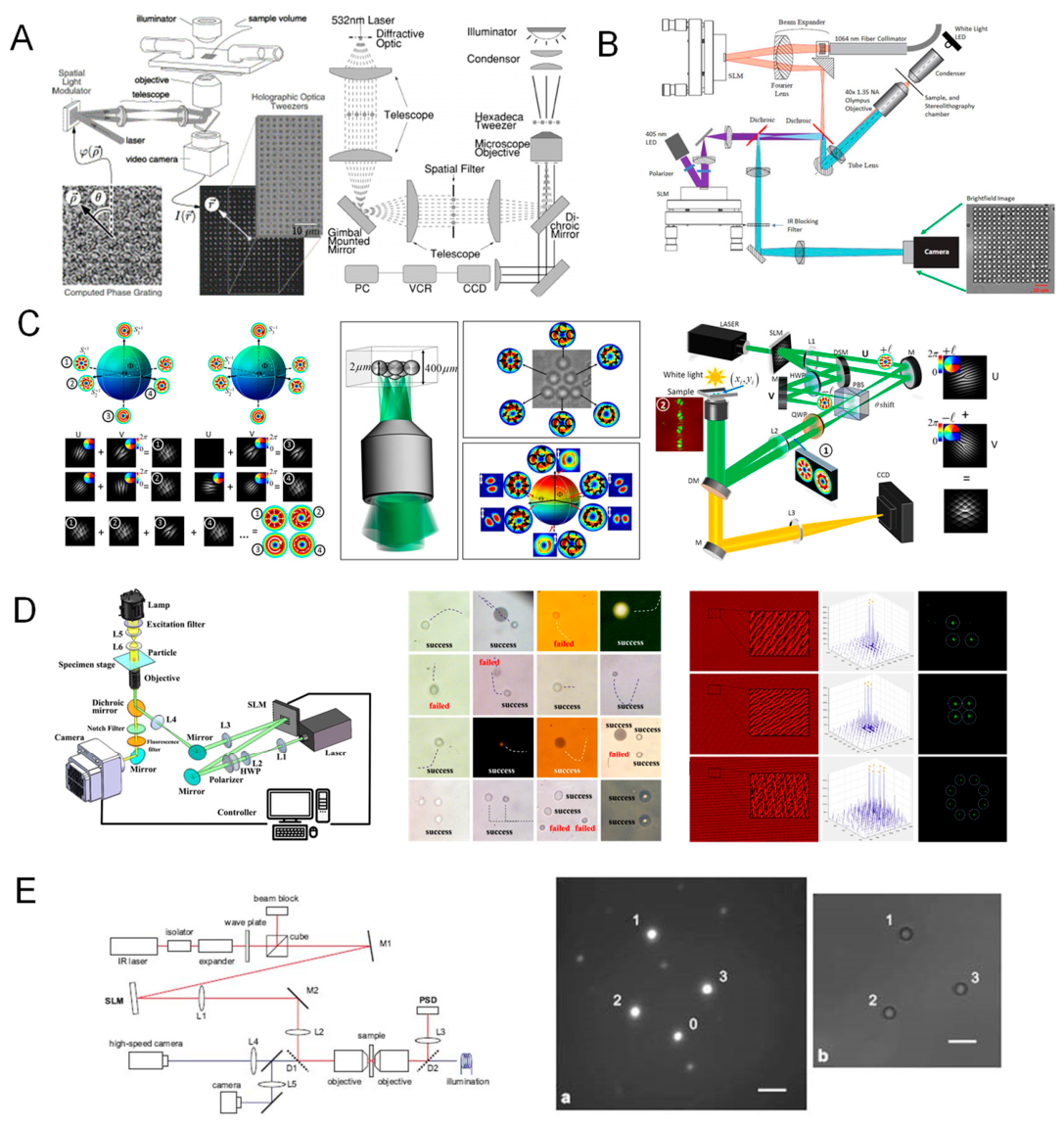
In summary, holographic optical tweezers represent a versatile and scalable platform for high-dimensional optical manipulation. By converting optical trapping into a programmable light field problem, HOT opens a pathway toward automated, parallelized, and intelligent force application in both fundamental research and applied biophotonics.
4.3. Optical Fiber Tweezers and Dual-Fiber Trapping Systems
Optical fiber–based tweezers constitute a compact, easily integrable family of traps in which waveguide-guided light—delivered through single-mode or multimode fibers—generates optical forces directly at the point of use. By dispensing with bulky free-space relay optics, fiber traps operate in geometrically constrained or remote settings (e.g., sealed microfluidic channels, endoscopic probes, or in vivo environments), while preserving the same gradient and scattering force principles established for objective-based tweezers (Secs. 2.2–2.4)[305]. In what follows, we outline the operating principles and canonical geometries, summarize the distinctive advantages and application space, and discuss practical limitations together with current engineering responses, linking each aspect to the calibration and sensitivity considerations developed in Chapter 3.
4.3.1. Principles and Configurations
In the simplest implementation, light emerging from a cleaved fiber end diverges into the sample and exerts both a restoring (gradient) and an axial (scattering) force on a nearby particle. Because the free-space intensity gradient produced by an unmodified fiber facet is relatively shallow, the axial radiation pressure usually dominates, so a single fiber alone rarely provides stable three-dimensional confinement in liquid unless assisted by surface reflections, near-field enhancement, or active feedback [305]. Robust 3D trapping is achieved by dual-fiber optical tweezers (DFOT), in which two opposing fibers launch counter-propagating beams that overlap in space: the opposing scattering forces cancel along the beam axis, while the combined transverse gradients form a potential minimum near the midplane. Precise tip geometry and alignment—flat or angle-cleaved facets, GRIN/ball-lensed ends, or tapered/etched tips—control convergence and field confinement, thereby setting stiffness and depth of the trap.
Several architectures follow from this basic scheme. Core-to-core DFOT aligns two single-mode fibers face-to-face to create a symmetric, thermally stable trap suitable for quantitative force spectroscopy. Tapered fiber traps concentrate light into a sub-wavelength mode volume at the nano-taper apex, increasing local gradients and enabling manipulation of smaller particles near the tip. Lensed fibers fuse micro-lenses to the facet to boost effective numerical aperture, improving axial and lateral stiffness without large external optics. Finally, asymmetric designs—for example, pairing a standard fiber with a waveguide coupler, or employing dual wavelengths—introduce controlled force asymmetries for selective transport or sorting. In all cases, the force balance, stiffness k, and stability criteria map onto the same modeling and calibration tools used for free-space tweezers (equipartition, PSD, Stokes drag; Sec. 3.1), with the added benefit that the fiber itself can serve as an interferometric or power-reflection sensor for on-fiber readout.
4.3.2. Advantages and Applications
Relative to objective-based systems, fiber tweezers emphasize miniaturization, remote access, and mechanical robustness. The slender, alignment-locked geometry allows traps to be positioned where high-NA objectives cannot be brought—inside microcapillaries, organ cavities, or sealed lab-on-a-chip devices—while maintaining quantitative control through the same calibration pathways described earlier. Because the fibers can double as sensing conduits (collecting back-reflected light, fluorescence, or Raman signals; or incorporating fiber Bragg gratings), the platform naturally supports on-fiber force, displacement, and temperature readouts, enabling compact metrology stacks [287].
These features have broadened the application footprint. In in vivo manipulation, DFOT has been used to steer cells or nanoparticles through fluidic conduits with minimal optical access, and to position probes within tissue volumes. Endoscopic trapping integrates DFOT into micro-endoscopes, allowing manipulation during minimally invasive procedures [443]. Within microfluidics, embedded DFOT junctions apply calibrated hydrodynamic loads for size-, refractive-index-, or stiffness-dependent sorting while monitoring trajectories in real time. On-fiber biosensing leverages a trapped bead as a local reporter whose fluctuation spectrum responds to binding events, viscosity changes, or mechanical remodeling in the surrounding medium [444]. A particularly instructive geometry is dual-fiber force spectroscopy, where a dielectric bead held between two opposing fibers is displaced by a nano-positioner on one arm; the resulting extension of a tethered macromolecule (e.g., DNA or protein complexes) is read out interferometrically or via beam deflection [305], achieving piconewton force resolution and sub-nanometer displacement sensitivity with excellent mechanical stability—often in more rugged or portable settings than free-space traps allow
4.3.3. Limitations and Developments
The principal trade-offs trace back to focusing and reconfigurability. Standard fibers offer a lower effective numerical aperture than high-NA microscope objectives, which reduces trap stiffness and depth unless the tip is engineered (lensing, tapering, metasurface coatings) to reinforce local gradients. Trap reconfigurability is typically more limited than in holographic systems (Sec. 4.2) unless fiber outputs are combined with external modulators or multi-port couplers. Finally, fabrication tolerances—especially tip shaping and sub-micrometer alignment in DFOT—govern symmetry and stability, and must be maintained to prevent bias in force measurements.
Current developments address these constraints along several lines. Two-photon 3D-printed micro-optics on fiber facets generate bespoke lenses, axicons, or beam-splitters that raise effective NA and create multi-spot patterns directly at the tip. Active feedback introduced through fiber-coupled acousto-optic or electro-optic devices stabilizes trap position and power, enabling dynamic modulation akin to single-beam or HOT platforms. Hybrid fiber systems integrate electrical (electrophoretic), thermal (opto-thermal), or chemical actuation to expand the manipulation space and to compensate for stiffness limits. As with all optical traps, attention to photothermal loading—especially at metalized tips or in absorbing media—is essential; the thermal and force-noise considerations and mitigation strategies outlined in Sec. 3.2 (laser-power stabilization, drift control, bandwidth management) carry over directly.
Taken together, optical fiber tweezers and dual-fiber traps complement bulk optical architectures by delivering portable, alignment-robust, and sensor-ready manipulation where access, footprint, or environmental constraints dominate. Their natural synergy with lab-on-fiber concepts, microfluidic instrumentation, and minimally invasive biophotonics suggests a continued expansion of roles in applied manipulation and in situ force spectroscopy, while ongoing advances in fiber-tip photonics and feedback control promise steadily improving stiffness, uniformity, and measurement fidelity.
4.4. Near-Field and Plasmonic Optical Tweezers
Near-field optical tweezers represent a transformative class of trapping systems that circumvent the diffraction limit by harnessing evanescent and resonantly enhanced optical fields in the proximity of nanostructured surfaces [445]. Among these, plasmonic optical tweezers have emerged as a leading approach for high-precision trapping and manipulation at the nanometer scale, capable of exerting femtonewton-level forces on particles smaller than 100 nm, including single biomolecules [446].
Figure 18.
Plasmonic optical tweezers: mechanisms, nanostructure designs, and integrated applications [413,447,448,449,450,451,452,453,454]. (A) Basic principles of plasmonic optical trapping. Localized surface plasmon resonance (LSPR) on nanostructured metal films (e.g., gold) creates strong near-field gradients for enhanced optical trapping and Raman signal amplification. Schematic of nanoparticle confinement on gold nanostructures and in situ SERS measurements. (B) Resonant field design and 3D simulation. Optical nanoantennas such as bowtie, disc, and spiral geometries enable electromagnetic field confinement below the diffraction limit. FEM/FDTD simulations illustrate electric field localization. (C) Common plasmonic trapping platforms. Schematics of plasmonic substrates including slot waveguides, tapered trenches, and ridge nanoapertures. Multiphysics simulations show temperature, field gradient, and thermophoretic force distributions. (D) Hybrid trapping with SERS/fluorescence detection. Integration of optical tweezers with photothermal or SERS modules enables simultaneous manipulation and biochemical identification of nanoscale objects such as single molecules or vesicles. (E) Fabrication of plasmonic nanoarrays. Lithography-based methods generate reproducible nanopillar and nanohole arrays, ensuring uniform trapping across large areas. Cross-sectional and topographic views show nanostructure fidelity. (F) Plasmonic manipulation with dual-function probes. Functionalized nanoparticles or nanorods offer optothermal and optical gradient control for enhanced manipulation. Optical torque and polarization-sensitive rotation are illustrated. (G) Application to material science. Optical control of luminescent nanoparticles or OLED-based emitters using patterned plasmonic substrates for light–matter interaction studies. (H) Experimental implementation. Optical setup for plasmonic trapping, combining high-NA objectives, laser sources, and imaging modules. Side illumination and near-field excitation schemes are shown.
Figure 18.
Plasmonic optical tweezers: mechanisms, nanostructure designs, and integrated applications [413,447,448,449,450,451,452,453,454]. (A) Basic principles of plasmonic optical trapping. Localized surface plasmon resonance (LSPR) on nanostructured metal films (e.g., gold) creates strong near-field gradients for enhanced optical trapping and Raman signal amplification. Schematic of nanoparticle confinement on gold nanostructures and in situ SERS measurements. (B) Resonant field design and 3D simulation. Optical nanoantennas such as bowtie, disc, and spiral geometries enable electromagnetic field confinement below the diffraction limit. FEM/FDTD simulations illustrate electric field localization. (C) Common plasmonic trapping platforms. Schematics of plasmonic substrates including slot waveguides, tapered trenches, and ridge nanoapertures. Multiphysics simulations show temperature, field gradient, and thermophoretic force distributions. (D) Hybrid trapping with SERS/fluorescence detection. Integration of optical tweezers with photothermal or SERS modules enables simultaneous manipulation and biochemical identification of nanoscale objects such as single molecules or vesicles. (E) Fabrication of plasmonic nanoarrays. Lithography-based methods generate reproducible nanopillar and nanohole arrays, ensuring uniform trapping across large areas. Cross-sectional and topographic views show nanostructure fidelity. (F) Plasmonic manipulation with dual-function probes. Functionalized nanoparticles or nanorods offer optothermal and optical gradient control for enhanced manipulation. Optical torque and polarization-sensitive rotation are illustrated. (G) Application to material science. Optical control of luminescent nanoparticles or OLED-based emitters using patterned plasmonic substrates for light–matter interaction studies. (H) Experimental implementation. Optical setup for plasmonic trapping, combining high-NA objectives, laser sources, and imaging modules. Side illumination and near-field excitation schemes are shown.

4.4.1. Physical Principles and Mechanism
Conventional optical tweezers are constrained by the diffraction limit, which restricts the smallest achievable trap size to approximately λ/2. For sub-wavelength particles, the optical gradient force becomes insufficient to overcome thermal fluctuations unless very high laser powers are used—risking sample damage. To overcome this, near-field tweezers exploit evanescent waves, which decay exponentially from surfaces (typically within ~100–200 nm) but can exhibit high spatial confinement and intensity gradients [455].
When light interacts with metallic nanostructures, such as gold or silver nanodisks, antennas, or gratings, it can excite surface plasmon resonances (SPRs)—collective oscillations of conduction electrons [456]. These resonances concentrate electromagnetic energy into sub-wavelength volumes, creating localized electromagnetic “hot spots” with field intensities enhanced by orders of magnitude. The resulting gradient forces are significantly stronger than in conventional traps, enabling stable confinement of objects in the 10–50 nm range.
The key mechanism in plasmonic trapping is thus the conversion of far-field light into intense near-fields via resonance, generating localized optical potentials that dominate over Brownian forces even for nanoparticles and macromolecules.
4.4.2. Plasmonic Trap Designs and Architectures
A wide variety of nanostructures have been designed and fabricated to act as plasmonic tweezers [457,458,459]: Bowtie antennas, two triangular gold tips facing each other form a narrow nanoscale gap (~10–20 nm), where the local field is maximized. Particles are drawn into the gap by the intense gradient; Nanohole arrays, arrays of circular or elliptical apertures in metallic films support resonant modes and confine particles within or near the holes; Nanorod dimers and metasurfaces , engineered to support tunable resonances across visible and near-infrared wavelengths, offering design flexibility for specific trapping conditions; Hybrid dielectric-metallic structures, combine high field confinement with reduced heating, optimizing biocompatibility and trap stiffness.
These devices are typically fabricated using focused ion beam milling, electron-beam lithography, or template stripping methods. Increasingly, self-assembled nanostructures and nanoimprint lithography are being employed to scale up production and reduce cost.
4.4.3. Performance and Applications
Plasmonic optical tweezers have emerged as a powerful platform for nanoscale optical manipulation, offering highly localized field enhancement and sub-diffraction confinement capabilities [460,461].
By exploiting localized surface plasmon resonances (LSPRs) in nanostructured metallic substrates, these systems can generate gradient forces in the range of 10–100 femtonewtons, sufficient to stably trap dielectric nanoparticles as small as ~20 nm in diameter under moderate laser powers on the order of a few milliwatts. The resulting optical potential wells exhibit volumes on the zeptoliter scale (~10⁻²¹ m³), enabling selective confinement of single viruses, DNA fragments, or individual protein molecules.
Beyond mechanical trapping, plasmonic architectures inherently facilitate electromagnetic field localization, thereby enhancing spectroscopic signals such as Raman scattering and fluorescence emission. The co-localization of analytes within plasmonic “hot spots” enables surface-enhanced Raman spectroscopy (SERS) and label-free detection of biomolecular species at the single-molecule level, significantly expanding the analytical utility of such platforms.
These unique features have enabled a range of high-impact applications. In single-molecule biophysics, plasmonic tweezers have been used to investigate protein–DNA interactions, conformational transitions, and intramolecular dynamics with unprecedented spatial resolution. In biosensing, they support real-time monitoring of biomolecular binding events via plasmonic resonance shifts or force-based displacement measurements. Additionally, in nanomaterials engineering, these tweezers provide a route for the directed assembly of nanoparticles with nanometer-scale precision, facilitating the bottom-up fabrication of metamaterials and integrated nanophotonic devices.
A representative demonstration of this technology was reported by Krishna et al. (Journal of Biomedical Optics, 2024[462]), where 20 nm polystyrene beads were stably trapped within a gold bowtie antenna using only 1 mW of incident laser power. The system achieved confinement durations on the order of several seconds—sufficient for real-time tracking, spectroscopic interrogation, and localized biochemical manipulation.
4.4.4. Limitations and Considerations
Despite the significant advances in nanoscale confinement and manipulation offered by near-field and plasmonic tweezers, these systems face several technical challenges that limit their broader applicability [463,464]. A principal concern is photothermal heating, which arises from intrinsic absorption of incident light by metallic nanostructures. This localized energy deposition can cause temperature elevations exceeding 10 ℃, potentially leading to thermal denaturation of biological samples or inducing thermally driven convection currents that destabilize the trapping potential. Strategies to mitigate these effects include the incorporation of heat-dissipating substrates, the use of pulsed or time-shared excitation schemes, and the application of low-power illumination optimized for resonance efficiency.
Another limiting factor is surface adhesion, particularly relevant in high-intensity near-field regions where trapped nanoparticles are positioned within nanometers of the substrate. In such configurations, van der Waals attractions and electrostatic interactions can cause the particle to adhere irreversibly to the surface, complicating controlled release or dynamic manipulation. Careful engineering of surface coatings and tuning of interfacial charge properties have been explored to alleviate this constraint.
Furthermore, the fabrication of plasmonic structures remains a major technical hurdle. Achieving reproducible nanoscale geometries with consistent optical responses demands high-resolution lithography and precise material control, which can limit device scalability and throughput. Batch-to-batch variability in plasmonic response also introduces uncertainty in quantitative force measurements or spectroscopic enhancement factors.
The spatial reach of near-field optical forces is inherently constrained by the exponential decay of the evanescent field, typically confining effective trapping to within 10–50 nm from the surface. This narrow working distance necessitates careful alignment and restricts manipulation to surface-adjacent particles, limiting three-dimensional versatility.
To address these limitations, recent advances have proposed alternative architectures that retain the advantages of sub-wavelength localization while improving system robustness. Notable developments include thermoplasmonic tweezers, which leverage controlled thermal gradients for trap stabilization; dielectric near-field tweezers, which utilize low-loss high-index structures to minimize heating; and nanophotonic waveguide-based traps, which offer extended trapping ranges with integrated on-chip control. These emerging platforms represent promising directions toward overcoming the inherent constraints of conventional near-field trapping systems.
Future advancements in plasmonic and near-field optical tweezers are increasingly directed toward enhancing functionality, integration, and quantum-level control. Efforts include the development of actively tunable plasmonic platforms, where reconfigurable nanostructures or electro-optic modulation enables dynamic adjustment of trap strength and position. Simultaneously, the integration of plasmonic surfaces with microfluidic architectures is enabling on-chip capture, sorting, and analysis of biomolecules under controlled flow conditions. At the quantum frontier, near-field tweezers are being explored for the manipulation of quantum emitters and entangled photon sources, opening possibilities for hybrid quantum-plasmonic systems. Collectively, these developments aim to overcome existing limitations in thermal management, reproducibility, and scalability. With their capacity for sub-wavelength confinement, ultra-low power operation, and site-specific control, near-field and plasmonic tweezers are poised to become indispensable tools in nanoscience, biophotonics, and molecular diagnostics.
4.5. Photonic Crystal and Waveguide-Based Tweezers
Photonic crystal and waveguide-based optical tweezers represent a new generation of integrated trapping technologies that leverage engineered photonic structures to control and confine light at the microscale and nanoscale [396]. These systems offer on-chip optical trapping with reduced size, improved mechanical stability, and enhanced scalability, forming the basis for compact, high-throughput manipulation platforms ideal for lab-on-a-chip and biomedical diagnostics applications [397,465,466].
Physical Principles and Photonic Structures.
Photonic crystals (PhCs) are periodic dielectric structures that create photonic bandgaps—frequency ranges where light propagation is forbidden [467]. By introducing defects or waveguide modes into these crystals, light can be localized or guided with high efficiency and minimal loss. Similarly, dielectric waveguides—such as silicon nitride or silicon-on-insulator (SOI) platforms—can confine light within sub-micron cross-sections and produce strong evanescent fields along their surfaces.
In both systems, evanescent field gradients form along the surface or at localized modes (e.g., cavity modes), producing optical gradient forces capable of trapping nanoparticles or cells near the surface. Compared to metallic (plasmonic) systems, photonic crystal tweezers offer lower optical losses and reduced photothermal heating, making them particularly suitable for sensitive biological samples [468].
Several key device architectures have been implemented slotted photonic crystal waveguides: Create intense localized fields in the slot region for particle trapping [469]; Resonant PhC cavities: Provide ultra-high quality factor (Q) modes for localized, low-power trapping [470]; Bragg gratings or ring resonators: Guide and trap particles in defined locations along waveguides [471]; Waveguide arrays: Create 1D or 2D trap lattices for parallel trapping of multiple particles [467].
The optical force arises from the interaction between the particle’s polarizability and the field gradients generated by the guided or localized light modes, governed by:
where is the particle polarizability and is the local electric field.
Waveguide- and photonic crystal (PhC)-based optical tweezers present several distinct advantages over conventional free-space trapping systems, making them highly attractive for integrated nanomanipulation platforms. Their on-chip scalability allows for the fabrication of high-density trapping arrays on silicon-compatible substrates, facilitating massively parallel manipulation [265]. Owing to strong field confinement, these systems operate at significantly lower optical powers, often requiring only a few milliwatts to achieve stable trapping [472]. Additionally, the use of dielectric materials minimizes absorption losses and associated photothermal effects, enabling biocompatible operation at infrared wavelengths [401]. These tweezers can also be readily co-integrated with microfluidic channels, photodetectors, biosensing modules, and electrical interfaces, allowing compact, multifunctional lab-on-chip designs. Furthermore, sub-wavelength positioning precision can be achieved through tailored cavity or waveguide geometries [473]. As a representative example, slotted photonic crystal waveguides with ~100 nm slot widths have demonstrated the ability to trap 50–100 nm polystyrene beads using less than 2 mW of optical power, achieving trap stiffness comparable to that of conventional free-space optical tweezers.
Waveguide- and photonic crystal-based optical tweezers have enabled a diverse range of applications, particularly in integrated and high-throughput contexts. These platforms support parallel manipulation of cells and particles, facilitating high-throughput sorting using waveguide lattices. Their ability to trap nanoscale biological entities, such as exosomes and viruses, makes them well-suited for diagnostic and analytical applications within compact, closed microfluidic systems—an advantage in biosafety-critical or contamination-sensitive environments. Additionally, single-particle tracking is achievable through co-integration with photodiodes or waveguide-coupled detectors, enabling real-time monitoring of displacement and dynamics. Emerging hybrid platforms now combine trapping with optical sensing modalities—such as resonance shifts or interferometric scattering—to allow simultaneous force application and signal transduction on a single chip. A notable example demonstrated sub-piconewton force sensitivity and sub-nanometer displacement resolution by integrating a silicon nitride waveguide trap with an interferometric detection module, underscoring the potential of these systems for precision biophysical measurements.
Limitations andProspect.
Despite their significant potential, photonic crystal and waveguide-based optical tweezers face several technical limitations that constrain their versatility and scalability [474]. The fabrication process demands nanometer-scale precision in lithography and etching, posing challenges for reproducibility and large-scale production. Additionally, the evanescent field profile inherently restricts the trapping volume to within ~100–200 nm of the surface, limiting the capture range. Trapping stability is further influenced by non-optical surface forces, such as electrostatic and van der Waals interactions, which necessitate strategies like surface passivation or electrokinetic control. Moreover, most designs lack intrinsic dynamic reconfigurability, as trap positions and geometries are fixed post-fabrication unless augmented with external tuning mechanisms. To overcome these challenges, emerging approaches include thermo-optic tuning via localized heating, electro-optic modulation using integrated phase shifters, and holographic light injection into waveguide inputs for spatially programmable trap arrays, thereby enhancing flexibility and functional control.
Photonic crystal and waveguide-based optical tweezers mark a pivotal advancement toward fully integrated lab-on-chip platforms for optical manipulation. Future developments are expected to focus on CMOS-compatible large-scale fabrication, facilitating the deployment of compact biomedical diagnostic devices [397]. Additionally, the incorporation of machine learning algorithms for real-time control of trap arrays may enable adaptive and intelligent manipulation of particles with high throughput. In parallel, the emergence of quantum-compatible architectures could allow for the on-chip trapping and control of quantum dots or atom-like systems, bridging the fields of integrated photonics and quantum technologies. By uniting precise optical trapping with scalable photonic integration, these systems hold significant promise for translating optical tweezing from experimental settings into practical clinical and point-of-care applications.
4.6. Optoelectronic, Acousto-Optic, and Hybrid Optical Tweezers
To overcome the limitations of traditional optical tweezers—such as trap stiffness, thermal damage, and scalability—researchers have developed a variety of hybrid trapping platforms that combine optical forces with electronic, acoustic, or plasmonic mechanisms [391,475,476]. These systems exploit multiple types of fields and physical principles to enable multimodal control, enhanced precision, and broadened applicability. In particular, optoelectronic tweezers (OETs) and acousto-optic tweezers (AOTs) have emerged as promising tools for robust and reconfigurable trapping, particularly in high-throughput and biocompatible settings [477,478].
Optoelectronic Tweezers (OET).
Optoelectronic tweezers operate by generating dielectrophoretic (DEP) forces on particles using light-patterned electric fields [479]. In contrast to conventional tweezers that rely solely on gradient optical forces, OET systems apply a low-intensity light pattern to a photoconductive substrate, which in turn modulates an AC electric field across the fluid chamber. The spatially varying electric field gradients induce DEP forces that move and trap particles based on their dielectric properties [480,481].
The key mechanism is as follows:
where is the particle radius, is the permittivity of the medium, is the electric field, and is the Clausius-Mossotti factor depending on frequency and dielectric contrast. By projecting light patterns (e.g., via a digital micromirror device, DMD), one can create reconfigurable electric field landscapes for real-time particle control.
Optoelectronic tweezers (OET) offer a unique platform for large-area, low-power, and highly reconfigurable particle manipulation, particularly suited for applications involving delicate biological specimens. Operating at extremely low optical intensities (typically <1 mW/cm²), OET systems minimize photothermal effects, making them ideal for the label-free trapping and sorting of live cells and other sensitive biomaterials [482]. The use of photosensitive substrates in conjunction with digital micromirror devices (DMDs) or liquid crystal displays (LCDs) enables real-time, reconfigurable electrode patterns, allowing simultaneous manipulation of thousands of particles across areas on the order of square centimeters [483]. This spatial flexibility has supported diverse applications including reconfigurable microfluidics, light-defined cell assembly, and interactive diagnostic platforms where user-defined optical patterns control particle motion.
Despite these advantages, OET also presents several inherent limitations. The dielectrophoretic (DEP) forces that underlie OET operation are strongly dependent on the electrical properties of the particles and the suspending medium—particularly conductivity and ionic strength—which may constrain reproducibility and robustness across varying sample conditions [484]. Furthermore, OET systems primarily support planar (two-dimensional) manipulation, with limited vertical confinement due to the nature of the induced electric fields. In terms of mechanical sensitivity, OET typically achieves lower force resolution compared to optical tweezers, operating in the range of approximately 0.1–10 pN, which may restrict applications requiring high-precision force measurements.
Nevertheless, OET has enabled functionalities that remain inaccessible to conventional optical tweezing approaches. These include large-scale, parallel sorting of heterogeneous cell populations, as well as the programmable assembly of microscale structures over extended areas, highlighting its value in high-throughput biophotonics, lab-on-chip systems, and point-of-care diagnostics [485].
Figure 19.
Design principles, operational architectures, and microfluidic integration of optoelectronic tweezers (OET) [62,486,487,488,489,490,491,492,493,494,495]. (A) Fundamental working mechanism of OET: projected light patterns from a digital micromirror device (DMD) induce localized electric fields across a photoconductive substrate (e.g., a-Si), generating dielectrophoretic (DEP) forces for particle manipulation. (B) Cross-sectional schematic of the light-induced electric field gradient and the corresponding projected light patterns on photoconductive layers, enabling dynamic and large-area particle trapping. (C) Programmable grid-based manipulation system using photoconductive ITO electrodes and real-time control for digital patterning of cell or bead arrays. (D) High-throughput sorting and reconfigurable trapping using LED illumination and DMD projection. Integrated micro-LED light engines can dynamically reconfigure trap geometry and selectively capture target particles. (E) Modular OET system with multiple projection and detection modules. Continuous optical switching allows for target isolation, release, and re-capture within custom-configured patterns. (F) Experimental demonstration of OET-generated trap arrays for parallel micromanipulation. Arrays of optical potential wells are dynamically created, as visualized by real-time fluorescence and bright-field imaging. (G) System integration schematic: key components include the DMD projection unit, light sources, microelectrode arrays, and programmable waveform generators. An optically controlled DEP field is generated on a TiOPc-coated ITO substrate. (H) Structural details of the OET platform: transparent electrodes, photoconductive layers, and microchambers enclosed in a lab-on-chip assembly for biological sample handling. (I) Multiphysics simulations showing electric field intensity and potential distribution across projected image regions. 3D plots demonstrate light-induced potential gradients and DEP well localization. (J) Application workflow: sequential LED and laser exposures enable multi-step manipulation, including illumination-trapping transitions, particle pattern encoding, and hybrid capture modes in complex fluids.
Figure 19.
Design principles, operational architectures, and microfluidic integration of optoelectronic tweezers (OET) [62,486,487,488,489,490,491,492,493,494,495]. (A) Fundamental working mechanism of OET: projected light patterns from a digital micromirror device (DMD) induce localized electric fields across a photoconductive substrate (e.g., a-Si), generating dielectrophoretic (DEP) forces for particle manipulation. (B) Cross-sectional schematic of the light-induced electric field gradient and the corresponding projected light patterns on photoconductive layers, enabling dynamic and large-area particle trapping. (C) Programmable grid-based manipulation system using photoconductive ITO electrodes and real-time control for digital patterning of cell or bead arrays. (D) High-throughput sorting and reconfigurable trapping using LED illumination and DMD projection. Integrated micro-LED light engines can dynamically reconfigure trap geometry and selectively capture target particles. (E) Modular OET system with multiple projection and detection modules. Continuous optical switching allows for target isolation, release, and re-capture within custom-configured patterns. (F) Experimental demonstration of OET-generated trap arrays for parallel micromanipulation. Arrays of optical potential wells are dynamically created, as visualized by real-time fluorescence and bright-field imaging. (G) System integration schematic: key components include the DMD projection unit, light sources, microelectrode arrays, and programmable waveform generators. An optically controlled DEP field is generated on a TiOPc-coated ITO substrate. (H) Structural details of the OET platform: transparent electrodes, photoconductive layers, and microchambers enclosed in a lab-on-chip assembly for biological sample handling. (I) Multiphysics simulations showing electric field intensity and potential distribution across projected image regions. 3D plots demonstrate light-induced potential gradients and DEP well localization. (J) Application workflow: sequential LED and laser exposures enable multi-step manipulation, including illumination-trapping transitions, particle pattern encoding, and hybrid capture modes in complex fluids.
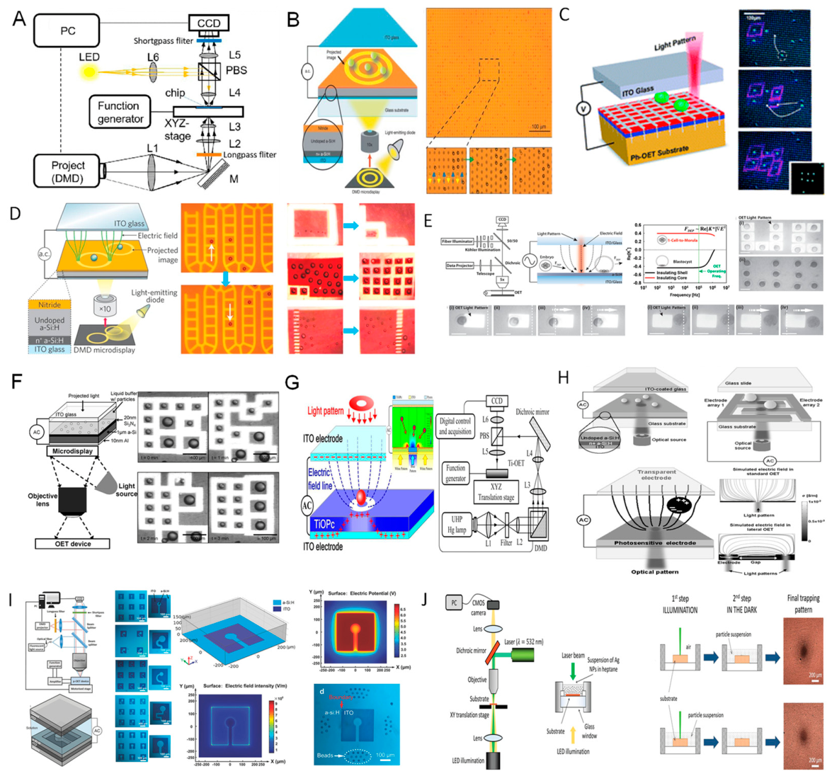
Acousto-Optic and Acoustofluidic Tweezers.
Acousto-optic tweezers utilize surface acoustic waves (SAWs) or bulk acoustic waves to exert pressure gradients in fluid, enabling the manipulation of particles without direct optical interaction [496]. Acoustic fields generate periodic pressure nodes and antinodes; particles with density or compressibility contrast are driven toward specific regions (e.g., nodes for negative contrast, antinodes for positive contrast), enabling non-contact, label-free particle control [497,498].
Acoustic tweezers utilize pressure wave–induced forces to manipulate particles within fluidic environments, offering a non-photothermal alternative to optical trapping. The primary mechanisms include the acoustic radiation force, which acts analogously to the optical gradient force by driving particles toward pressure nodes or antinodes, and acoustic streaming, wherein induced fluid flows facilitate bulk transport and enhanced mixing [499]. These systems are particularly well-suited for biocompatible manipulation, as they avoid photothermal damage and are capable of handling fragile biological entities such as organelles, embryos, or spheroids. The use of standing wave fields enables parallel, large-area trapping, and acoustic transducers can be readily integrated with microfluidic chips to create compact and scalable platforms.
Typical applications span blood cell sorting, organoid patterning for tissue engineering, and directed nanoparticle aggregation for bottom-up material synthesis. However, acoustic tweezers also face limitations, including lower spatial resolution compared to optical tweezers, complex fluid–structure interactions that can complicate system modeling, and challenges in achieving precise single-particle control, particularly at the nanoscale [500]. Despite these constraints, acoustic tweezers remain a powerful modality for contactless, label-free manipulation in biological and materials science contexts.
Figure 20.
Design strategies, physical principles, and functional demonstrations of acoustic tweezers based on surface and bulk acoustic waves [184,501,502,503,504,505,506,507,508]. (A) Core working mechanisms of acoustic tweezers. Interdigitated transducers (IDTs) or piezoelectric arrays generate standing surface acoustic waves (SSAW) or bulk acoustic waves (BAW) that create spatially periodic pressure fields. Particles experience acoustic radiation forces that drive them toward pressure nodes or antinodes, enabling label-free trapping and transport. (B) Simulation and experimental visualization of acoustic pressure and displacement fields in microchannels. Models illustrate wave interference, node formation, and transverse force generation across various frequencies and device geometries. (C) Multiphysics analysis of microchannel-integrated acoustic tweezers. Finite element modeling (FEM) reveals particle behavior under varying acoustic amplitudes and channel widths. Accompanying graphs quantify displacement, force magnitude, and particle response curves. (D) High-throughput acoustic cell sorting. A sheath-flow based microfluidic device steers cells or particles through pressure-modulated channels, directing them into separate outlets based on size, stiffness, or density differences. (E) Reconfigurable acoustic field patterning. 2D programmable IDT arrays enable single-particle trapping, rotation, and pairwise assembly. Electric field distribution and pressure maps confirm local field control. Fluorescence imaging demonstrates dynamic multi-site trapping. (F) Miniaturized and lens-assisted acoustic tweezers. Acoustic lenses and concentric transducer arrays generate focused or vortex acoustic beams. Demonstrations include spiral vortex formation, particle levitation, and on-chip manipulation with compact devices. (G) Acoustic vortex tweezers. Acoustic streaming and angular momentum in vortex beams provide contactless, rotational trapping for levitated particles. Phase maps and intensity profiles reveal the spatial structure of acoustic vortex fields. (H) Structural tunability using sharp-focused IDTs (sFITs). Highly localized acoustic nodes are achieved in microchannels using dense IDT arrays. Experimental trapping of cells or particles in highly confined regions is shown with high efficiency. (I) Experimental demonstration of fluorescent particle trapping using node-based field alignment. Stable and ordered trapping patterns are formed across large areas, confirming platform scalability and biocompatibility.
Figure 20.
Design strategies, physical principles, and functional demonstrations of acoustic tweezers based on surface and bulk acoustic waves [184,501,502,503,504,505,506,507,508]. (A) Core working mechanisms of acoustic tweezers. Interdigitated transducers (IDTs) or piezoelectric arrays generate standing surface acoustic waves (SSAW) or bulk acoustic waves (BAW) that create spatially periodic pressure fields. Particles experience acoustic radiation forces that drive them toward pressure nodes or antinodes, enabling label-free trapping and transport. (B) Simulation and experimental visualization of acoustic pressure and displacement fields in microchannels. Models illustrate wave interference, node formation, and transverse force generation across various frequencies and device geometries. (C) Multiphysics analysis of microchannel-integrated acoustic tweezers. Finite element modeling (FEM) reveals particle behavior under varying acoustic amplitudes and channel widths. Accompanying graphs quantify displacement, force magnitude, and particle response curves. (D) High-throughput acoustic cell sorting. A sheath-flow based microfluidic device steers cells or particles through pressure-modulated channels, directing them into separate outlets based on size, stiffness, or density differences. (E) Reconfigurable acoustic field patterning. 2D programmable IDT arrays enable single-particle trapping, rotation, and pairwise assembly. Electric field distribution and pressure maps confirm local field control. Fluorescence imaging demonstrates dynamic multi-site trapping. (F) Miniaturized and lens-assisted acoustic tweezers. Acoustic lenses and concentric transducer arrays generate focused or vortex acoustic beams. Demonstrations include spiral vortex formation, particle levitation, and on-chip manipulation with compact devices. (G) Acoustic vortex tweezers. Acoustic streaming and angular momentum in vortex beams provide contactless, rotational trapping for levitated particles. Phase maps and intensity profiles reveal the spatial structure of acoustic vortex fields. (H) Structural tunability using sharp-focused IDTs (sFITs). Highly localized acoustic nodes are achieved in microchannels using dense IDT arrays. Experimental trapping of cells or particles in highly confined regions is shown with high efficiency. (I) Experimental demonstration of fluorescent particle trapping using node-based field alignment. Stable and ordered trapping patterns are formed across large areas, confirming platform scalability and biocompatibility.
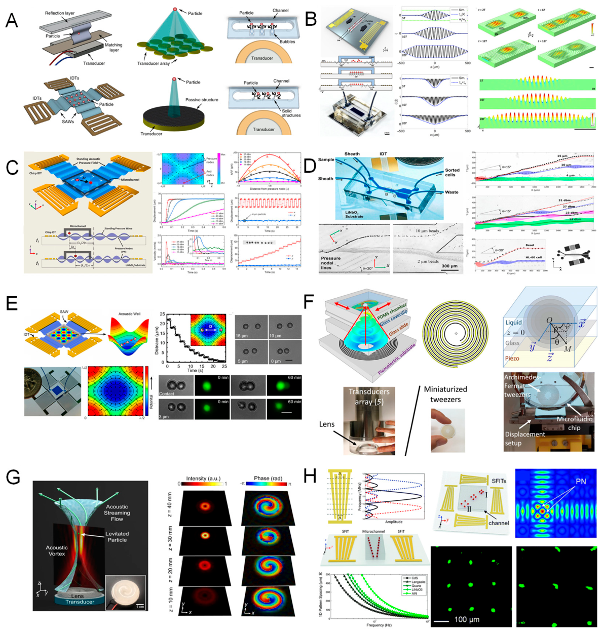
Hybrid Tweezers and Multimodal Systems.
Beyond the capabilities of standalone optoelectronic (OET) and acoustic (AOT) tweezers, a growing class of hybrid optical manipulation platforms has emerged, combining multiple physical forces within a unified architecture to enhance versatility and functional control [509] . Notable examples include magneto-optical tweezers, which integrate magnetic fields with optical trapping to manipulate magnetically labeled particles, offering improved axial stability and long-range transport capabilities [510]. Thermoplasmonic tweezers exploit localized heating from plasmonic nanostructures to generate temperature gradients, enabling thermophoretic trapping and directional control [511]. In parallel, electro-optofluidic tweezers synergistically combine electric fields, optical forces, and hydrodynamic flow fields to achieve selective, programmable particle sorting in complex fluidic environments [512].
These hybrid systems offer several distinct advantages over single-modality tweezers. By combining disparate force fields, they provide expanded manipulation ranges—for instance, employing magnetic forces for coarse positioning and optical forces for nanoscale precision. Furthermore, selectivity can be tuned based on particle properties such as size, refractive index, charge, or magnetic susceptibility, enabling highly specific targeting. The incorporation of multiple control modalities (e.g., light, voltage, fluid flow) also introduces greater operational flexibility, allowing dynamic reconfiguration in response to complex experimental requirements.
A representative implementation involves the integration of OET with conventional optical tweezers, wherein low-intensity light patterns enable bulk cell sorting across large areas, while highly focused laser beams provide precise single-cell manipulation. Such hybrid approaches exemplify the potential for multi-scale, multi-physics control in next-generation optical manipulation systems.
4.7. AI-Enhanced and Intelligent Optical Tweezers Systems
As optical tweezers become increasingly complex and miniaturized, a new frontier has emerged at the intersection of artificial intelligence (AI) and automated optical manipulation [513]. By embedding intelligent control algorithms—especially machine learning (ML) and computer vision—into optical tweezing platforms, researchers aim to overcome traditional limitations such as low throughput, manual calibration, and limited adaptability. This subfield, often referred to as “intelligent optical tweezers”, promises a new paradigm of autonomous, high-precision, and context-aware manipulation systems [514].
Motivation and Scope of AI Integration.
Conventional optical tweezing workflows have traditionally depended on substantial user intervention, including manual calibration, heuristic parameter tuning, and continuous real-time supervision [513]. These requirements inherently constrain experimental reproducibility, throughput, and the system’s responsiveness to dynamic perturbations, such as particle drift, fluidic fluctuations, or sample heterogeneity. Recent advances in artificial intelligence (AI) have introduced new paradigms for automating and enhancing optical tweezer operation. Computer vision algorithms enable real-time detection, tracking, and classification of trapped particles, facilitating closed-loop control. Reinforcement learning frameworks have been employed to dynamically adjust trap position, laser power, and other parameters in response to environmental changes. In addition, supervised learning models trained on historical calibration data can predict and adjust trap stiffness without requiring extensive manual input. Unsupervised learning techniques further support the identification of complex patterns in particle trajectories or biological responses, aiding in the discovery of emergent behaviors or states. By integrating these AI-driven approaches, intelligent optical tweezers can autonomously adapt trapping parameters, enhance force resolution, and maintain stable operation across heterogeneous sample types [515]. This convergence of optical manipulation and machine learning not only improves system robustness and precision but also enables new levels of automation and scalability in complex experimental environments.
AI in Trap Control and Calibration.
One of the earliest and most impactful applications of artificial intelligence in optical tweezing systems lies in automated trap position control and stiffness calibration. Leveraging real-time imaging data, convolutional neural networks (CNNs) have been shown to accurately extract particle displacement from raw microscope video streams with sub-pixel spatial resolution, enabling precise tracking without the need for manual segmentation or thresholding [516]. This positional information can be fed into adaptive control loops, which dynamically modulate beam-steering components such as acousto-optic deflectors (AODs) or galvanometric mirrors to maintain trap stability under conditions of mechanical drift or fluidic perturbation [517].
In parallel, machine learning algorithms have been developed to infer trap stiffness by analyzing Brownian motion characteristics—such as power spectral density or positional variance—and correlating them with theoretical or experimentally derived stiffness models [518]. This approach eliminates the need for labor-intensive calibration routines, offering rapid and consistent parameter estimation that is particularly advantageous in high-throughput settings or biological assays with variable viscosity, temperature, or particle size.
A notable experimental demonstration involved training a deep neural network on approximately 10,000 particle trajectories to classify optical trapping regimes and recommend laser power adjustments accordingly. This data-driven system achieved an order-of-magnitude improvement in calibration speed and accuracy relative to conventional manual techniques, underscoring the potential of AI-enhanced optical tweezers for autonomous, scalable manipulation in complex environments.
AI-Driven High-Throughput Manipulation.
Artificial intelligence further expands the functional capabilities of optical tweezers by enabling real-time, parallel control of multiple particles and traps, a critical requirement for advanced applications in cell sorting, nanoassembly, and synthetic biology. Recent implementations have demonstrated the use of deep learning–based multi-object tracking algorithms to simultaneously monitor and localize over 50 individual particles within microfluidic trap arrays, maintaining high spatial and temporal resolution [519]. In such settings, AI-driven tracking ensures precise feedback for downstream actuation.
Moreover, reinforcement learning agents have been employed to dynamically allocate and reposition optical traps based on particle-specific attributes—such as velocity, fluorescence intensity, or interparticle interactions—allowing context-aware manipulation strategies. Complementing these efforts, machine learning classifiers can be integrated with spatial light modulators (SLMs) to adjust light field distributions in real time according to particle state. For instance, optical traps can be selectively applied or withdrawn based on classification outcomes (e.g., distinguishing live from dead cells), enabling autonomous decision-making within complex biological samples.
Collectively, these AI-enhanced systems transform optical tweezers into intelligent, adaptive manipulation platforms, functioning analogously to interactive robotic systems. By continuously monitoring environmental inputs, interpreting particle behavior, and adjusting trapping conditions in real time, such platforms offer a paradigm shift in the scalability, autonomy, and precision of optical manipulation.
Predictive Modeling and Experimental Automation.
Artificial intelligence also plays an increasingly important role in enhancing the theoretical modeling and predictive control of optical trapping systems. Neural network–based surrogate models have been developed to approximate optical force fields as a function of system parameters—such as laser beam profile, particle size, and refractive index—thus significantly reducing the computational overhead associated with traditional methods like finite-difference time-domain (FDTD) or finite element method (FEM) simulations. These data-driven models enable near-instantaneous estimation of force landscapes, facilitating rapid system design and optimization.
In addition, predictive algorithms trained on experimental and simulated data can anticipate potential trapping instabilities—such as particle escape due to fluidic disturbances or thermal degradation from excessive laser power—and adjust operational parameters proactively to maintain trap integrity. This capability is particularly valuable in dynamic or heterogeneous environments, where real-time adaptability is essential.
More broadly, closed-loop automated systems integrating AI, optical tweezers, and microfluidics have been proposed as a foundation for autonomous experimentation platforms. For example, such systems can screen hundreds of individual cells for mechanical properties (e.g., stiffness or deformability) without human intervention, using AI to guide trap positioning, adjust optical parameters, and interpret measurement outputs.
These integrated platforms effectively function as self-driving laboratories, where optical tweezers serve as the physical manipulation engine, and artificial intelligence acts as the computational and decision-making core—together enabling scalable, adaptive, and intelligent experimentation.
Future Prospects and Challenges.
Although the integration of artificial intelligence with optical tweezers remains in its early stages, the technological trajectory suggests considerable potential for transformative impact across biophysics, diagnostics, and nanoscience [515]. Several key challenges must be addressed to fully realize the capabilities of AI-enhanced optical manipulation. One major hurdle is robustness to biological variability: biological systems are inherently heterogeneous, and training models that generalize across varying cell types, environmental conditions, and experimental configurations remains a nontrivial task. Moreover, effective implementation requires seamless hardware–software integration, where real-time communication between lasers, spatial light modulators (SLMs), detectors, and control algorithms must be precisely synchronized. Another critical limitation is the availability of high-quality, annotated datasets for model training and validation, which remains a bottleneck in many specialized or custom tweezing platforms.
Despite these challenges, the outlook for AI-augmented optical tweezers is promising. Emerging directions include the deployment of edge-AI processors to enable onboard, low-latency decision-making in portable or field-deployable optical manipulation systems; the use of federated learning frameworks, allowing collaborative model training across multiple laboratories while preserving data privacy; and the development of AI-integrated quantum tweezer platforms, capable of atom-scale manipulation informed by contextual feedback and adaptive learning.
Collectively, these advancements are reshaping the paradigm of optical manipulation. By enabling autonomous, intelligent, and context-aware control, AI-enhanced tweezing systems represent a critical step toward fully automated experimentation—bridging fundamental research and translational technologies in biomedicine, materials science, and beyond.
4.8. Comparative Summary and Application Landscape
The rapidly evolving typology of optical tweezers reflects not only technological innovation, but also the growing diversity of applications across disciplines. From the foundational single-beam gradient trap to chip-integrated, AI-enhanced multimodal platforms, each variant has carved out unique strengths and niche domains. This section presents a comparative synthesis of the major optical tweezer types discussed above, along with an overview of their application landscapes.
Comparative Summary of Tweezer Modalities
| Tweezer Type | Principle | Key Advantages | Typical Applications | Limitations |
| Single-Beam Gradient (OT) | Gradient and scattering force balance | Simplicity, high force sensitivity (~0.1–100 pN) | Molecular biophysics, force spectroscopy | Limited to one particle per trap, high laser power needed |
| Dual-Beam OT / Optical Stretcher | Counter-propagating beams | Trap stiff objects, apply mechanical stretch | Cell elasticity, tissue biomechanics | Alignment complexity, limited throughput |
| Holographic OT (HOT) | Dynamic light shaping via SLM/DMD | Parallel manipulation, flexible trap geometry | Colloidal assembly, multi-particle manipulation | Computational load, lower stiffness per trap |
| Plasmonic Tweezers | Near-field plasmon-enhanced gradient force | Trapping below 100 nm, enhanced field gradients | Single-molecule trapping, surface-enhanced spectroscopy | Limited trapping volume, photothermal heating |
| Photonic Crystal/On-Chip OT | On-chip light-matter interaction | Integration, portability, scalable arrays | Optofluidics, cell sorting, lab-on-a-chip | Fabrication complexity, lower per-trap force |
| Optoelectronic Tweezers (OET) | Light-induced DEP force via AC fields | Low power, large area, reconfigurability | Cell patterning, particle sorting in microfluidics | 2D control, limited vertical force, dependent on conductivity |
| Acousto-Optic/AOT | SAW-driven pressure gradients | Non-invasive, biocompatible, large-scale traps | Sorting, organoid assembly, biofabrication | Lower spatial resolution, complex fluid interactions |
| Hybrid and AI-enhanced OT | Multiphysics intelligent control |
High adaptability, feedback control, automation | Real-time tracking, adaptive manipulation, force-clamp | High system complexity, computational and hardware demands |
This tabulated overview underscores that no single tweezer modality is universally superior; rather, optimal selection depends on experimental goals, such as force resolution, particle size, environmental conditions, and desired throughput.
Domain-Specific Application Mapping.
To illustrate the diverse applicability of optical tweezers across scientific disciplines, a functional mapping can be established between specific tweezer modalities and their dominant research domains:
In molecular biophysics, single-beam and dual-beam optical tweezers are widely employed for high-precision manipulation of biomolecules, such as stretching DNA, unfolding proteins, and quantifying the forces exerted by molecular motors. Their sub-piconewton force sensitivity and compatibility with force-clamp and displacement-clamp configurations make them particularly suited for probing mechanochemical processes at the single-molecule level.
In cell biology and microbiology, techniques such as optical stretchers, optoelectronic tweezers (OETs), and acoustofluidic traps are utilized for the non-invasive manipulation of live cells. These platforms enable functional assays including cell elasticity measurements, targeted cell sorting, and microscale surgical interventions without physical contact, thus preserving cellular viability and behavior.
For research in colloidal and soft matter physics, holographic optical tweezers (HOTs) and photonic tweezers provide the flexibility and precision necessary for constructing colloidal crystals, investigating phase transitions, and quantifying interparticle interaction potentials. The ability to generate and dynamically reconfigure multiple traps in parallel is essential for studying emergent collective behaviors.
In the fields of nanotechnology and surface science, plasmonic tweezers and photonic crystal-based traps offer the spatial resolution and field enhancement required to position individual nanoparticles, investigate nanoscale surface forces, and facilitate reactions at the single-particle level, such as in catalytic or sensing applications.
In quantum and atomic physics, optical lattice tweezers and cavity-based trapping systems serve as critical tools for precision control of neutral atoms. These platforms are foundational for quantum simulation, atomic clock development, and cavity optomechanics, where quantum coherence and stability at the nanoscale are paramount.
Lastly, in biomedical engineering and diagnostics, the emergence of AI-integrated optical tweezing platforms, particularly those coupled with microfluidic systems and high-resolution imaging, is enabling scalable and automated assays. Applications include high-throughput cell-based diagnostics, force-based biomarker detection, and the spatial organization of living or synthetic tissues for regenerative medicine and biofabrication.
This domain-specific alignment underscores the versatility of optical tweezers as both a fundamental research tool and a translational technology across the physical and life sciences.
Emerging Convergence and System-Level Integration.
A prominent emerging trend in the field of optical manipulation is the convergence of multiple tweezer modalities into integrated, multifunctional platforms. Future systems are expected to embody on-chip hybrid architectures that combine optical, electrical, and acoustic actuation within a unified photonic framework, thereby enabling multimodal control with enhanced spatial and functional resolution.
These platforms will be further augmented by closed-loop artificial intelligence (AI) control systems, allowing for real-time feedback and adaptive modulation of trapping forces, particle trajectories, and experimental parameters. Such AI-enhanced systems will support autonomous decision-making in complex or dynamically evolving environments.
In parallel, the orchestration of multi-physics mechanisms—including near-field optical gradients, thermophoretic forces, and fluidic interactions—will enable complex manipulation tasks that are inaccessible to single-modality tweezers. This integration of diverse force landscapes opens new possibilities for precision control in heterogeneous and dynamic systems.
Finally, emphasis is increasingly placed on system-level integration, wherein trapping functionalities are co-localized with imaging, spectroscopic, and data acquisition modules within compact, lab-on-chip formats. These fully integrated systems are poised to shift optical tweezers from specialized laboratory instruments to programmable micro-robotic platforms for applications in advanced materials manufacturing, high-throughput biosensing, and autonomous microscale assembly.
In conclusion, the typology of optical tweezers is no longer a taxonomy of beam shapes or trap geometries, but a rich spectrum of functionally specialized manipulation systems. By choosing and combining appropriate modalities, scientists can now tailor optical manipulation to almost any scale—from atoms to organoids—and across diverse environments.
This chapter lays the foundation for the next: recent advances and research trends, which explore how these tweezers are evolving in technical sophistication and application ambition. The versatility of modern optical tweezers ensures they will continue to play a pivotal role in experimental science, engineering, and biomedicine.
5. Recent Advances and Research Trends
5.1. Integration and Miniaturization: Chip-Based Optical Tweezers
Over the past decade, the drive toward miniaturization has profoundly reshaped the landscape of optical tweezer technologies. Chip-based optical tweezers (CBOTs), a class of integrated photonic systems designed to manipulate microscopic objects directly on optical substrates, represent a major breakthrough in this evolution [445,520,521,522]. These platforms combine the force-manipulation capabilities of conventional optical tweezers with the scalability, alignment-free operation, and multifunctionality inherent to nanophotonic circuits. This section reviews the foundational principles, design architectures, and recent technological milestones that define the current state and future prospects of CBOTs.
Conceptual Overview and Motivation for Integration.
Conventional optical tweezers typically rely on bulk optical components, including high-numerical-aperture (NA) objectives, free-space laser delivery systems, and precision motorized stages for trap control. While these setups offer exceptional spatial resolution and force sensitivity, they are inherently limited by their large physical footprint, alignment sensitivity, and the requirement for environmental isolation to maintain stability and precision [523]. In contrast, chip-based optical tweezers (CBOTs) represent a paradigm shift by miniaturizing the core optical functionalities—such as beam confinement, shaping, and force generation—into integrated photonic structures. By leveraging waveguides, meta-surfaces , and diffractive optical elements at the micro- and nanoscale, these platforms enable compact, portable, and parallelizable optical manipulation systems [524].
The integration of optical trapping functionalities on-chip offers several key advantages. First, device miniaturization is achieved by replacing traditional bulk optics with photonic components that can be fabricated within millimeter-scale footprints. Second, mechanical and thermal stability are significantly improved, as integrated platforms eliminate long free-space optical paths and reduce sensitivity to alignment drift and external vibrations. Third, CBOTs inherently support scalability, allowing for the creation of multiplexed trapping arrays in linear or two-dimensional configurations—an essential capability for high-throughput applications such as single-cell analysis, parallel sorting, or arrayed biomolecular assays. Finally, functional co-integration allows for the monolithic incorporation of additional components, including photodetectors, microheaters, electro-optic modulators, and microfluidic channels, within a single substrate.
Collectively, these features unlock novel experimental capabilities, such as force spectroscopy in confined microfluidic environments, in vivo optical manipulation with minimal invasiveness, and quantum-state control of neutral atoms in integrated vacuum-compatible platforms. Chip-based tweezers thus represent a foundational technology for the next generation of compact, robust, and multifunctional optical manipulation systems.
Photonic Architectures in CBOTs.
The realization of chip-based optical tweezers (CBOTs) relies on a diverse set of integrated photonic architectures, each offering distinct mechanisms for generating and controlling optical forces at the micro- and nanoscale. Among these, several configurations have emerged as particularly effective for on-chip particle manipulation.
Waveguide-based optical traps utilize total internal reflection in high-refractive-index dielectric waveguides to produce evanescent fields that extend into the surrounding fluid medium. Particles in close proximity to the waveguide surface experience optical gradient forces that draw them toward regions of maximal field intensity. These structures can range from simple straight waveguides with localized intensity hotspots to more complex geometries—such as curved guides or standing-wave resonators—that enhance field confinement and enable stable particle trapping [525].
Meta-surface tweezers employ planar arrays of subwavelength nanostructures—such as silicon nanopillars or dielectric resonators—to precisely modulate the phase, amplitude, and polarization of incident light. This modulation enables the formation of well-defined optical traps directly above the metasurface. Recent work by Ruiting Huang et al. demonstrated that a single input beam can be transformed into a highly uniform two-dimensional array of optical traps, making metasurface-based tweezers particularly promising for neutral atom confinement and quantum information processing [526].
Optical phased arrays (OPAs) represent a class of actively tunable photonic structures composed of multiple optical emitters with individually controlled phase settings. By dynamically adjusting the relative phase of each emitter, OPAs can steer optical beams in free space without mechanical movement. Tal Sneh et al. recently reported a silicon photonics-based OPA capable of generating a single-beam optical trap located 5 mm above the chip surface, a significant advancement that overcomes the traditional range limitations of near-field devices. Real-time control over beam position via wavelength or phase modulation enables biologically relevant manipulation at the cellular or tissue scale [524].
Finally, hybrid metalens-guided systems integrate planar metasurfaces with guided-wave photonics to transform in-plane optical modes into focused out-of-plane beams. Gang Yu and colleagues developed a dielectric metalens–based platform that converts guided light into discrete optical traps in the far field while maintaining the benefits of on-chip integration. This architecture supports multiplexed trap generation across a planar substrate, facilitating parallel manipulation within a compact photonic framework [527]. Together, these photonic architectures underpin the functional versatility and scalability of CBOTs, enabling precise, reconfigurable, and miniaturized optical trapping suitable for next-generation applications in biophysics, diagnostics, and quantum technologies.
Design Considerations and Performance Metrics.
The design of an effective chip-based optical tweezer (CBOT) system necessitates careful optimization of multiple interdependent parameters, each of which directly influences trapping performance, system scalability, and application suitability.
Trap depth and stiffness are primarily governed by the local optical intensity and the steepness of the field gradient. Achieving strong and stable confinement, particularly for submicron particles, requires either high numerical aperture focusing or enhanced local fields facilitated by nanophotonic structures such as plasmonic antennas, slot waveguides, or photonic crystal resonators [528]. The range of the evanescent field, which typically extends only 100–300 nm from the chip surface, imposes spatial constraints on the trapping region. This necessitates close proximity between the particle and the photonic interface, where non-optical surface forces—including electrostatic interactions and van der Waals attractions—must be carefully accounted for to maintain trap stability and avoid adhesion artifacts.
Biocompatibility and optical power budget are also critical considerations, particularly in biological applications. The use of low-loss dielectric materials and operation at biologically compatible near-infrared wavelengths can significantly reduce photothermal effects, thereby minimizing the risk of heating-induced cellular damage and enabling long-term trapping in aqueous or physiological media.
In parallel, fabrication complexity remains a key determinant of system viability. The realization of precise nanophotonic features requires high-resolution lithographic techniques and process compatibility with CMOS or photonic integrated circuit (PIC) foundries. These considerations influence not only the reproducibility and yield of individual devices but also the scalability to high-density trapping arrays.
Recent experimental advances have demonstrated force resolutions below 1 piconewton and trap positioning accuracies under 100 nanometers, enabling the precise manipulation of microspheres, subcellular organelles, and even intact cells within integrated microfluidic environments. These capabilities highlight the growing potential of CBOT systems in high-throughput bioanalysis, mechanobiology, and lab-on-chip diagnostics.
Case Study: Opto-Hydrodynamic Fiber Tweezers.
A compelling example of CBOT potential is presented in the 2024 work by Shreyas Vasantham et al.,[529] who introduced opto-hydrodynamic fiber tweezers in a lab-on-fiber format. Their device used dual optical fibers aligned in a microfluidic channel, where hydrodynamic flow pre-aligned particles into the trap zone. By carefully tuning laser power and flow speed, they achieved stable trapping of 1–5 µm beads with throughput exceeding 14 particles per minute. The compact footprint and robust operation highlight how CBOTs can be deployed in real-world sensing environments without bulky optics.
5.2. Multiplexed and Programmable Optical Tweezer Arrays
As the complexity and scale of experimental systems grow, the demand for parallel and programmable optical manipulation has surged [438]. Conventional single-beam tweezers, while precise, are inherently limited in throughput. In response, researchers have developed multiplexed optical tweezer arrays, capable of manipulating dozens to hundreds of particles simultaneously in three dimensions [530]. These arrays not only enhance efficiency in large-scale biological and colloidal systems, but also open the door to studying collective phenomena, force networks, and assembly dynamics at the microscale.
Principles of Multiplexing.
Multiplexing in optical tweezer systems refers to the generation and control of multiple, simultaneous optical traps, enabling parallel manipulation of particles or biological specimens. Several optical strategies have been developed to achieve this functionality, each leveraging distinct physical principles and offering trade-offs in spatial resolution, temporal response, and scalability.
One of the most widely adopted approaches is holographic beam shaping, which utilizes spatial light modulators (SLMs) or digital micromirror devices (DMDs) to modulate the phase or amplitude of an incident laser beam. By encoding a computer-generated hologram onto the SLM, the phase front of the beam is tailored to diffract light into multiple user-defined focal points in three-dimensional space. Each focal spot acts as an independent optical trap, and the configuration can be dynamically updated to reconfigure the trapping pattern in real time [531]. Acousto-optic deflectors (AODs) offer an alternative strategy for high-speed beam steering. By modulating acoustic waves through a birefringent crystal, the incident laser beam is deflected at controllable angles. When combined with time-sharing techniques, a single laser beam can be rapidly scanned across multiple trap positions at frequencies in the kilohertz to megahertz range. Due to the temporal integration inherent in particle response and imaging systems, these rapidly alternating traps appear quasi-static to the trapped particles, enabling effective multiplexing with minimal hardware complexity [531].
In the context of integrated photonics, on-chip multiplexing is achieved using components such as waveguide-based beam splitters, optical phased arrays, and multi-mode interferometers (MMIs). These structures divide a single optical input into multiple spatially resolved trapping outputs, which can be arranged in linear or two-dimensional arrays on the chip surface. This approach offers high mechanical and thermal stability, as well as inherent scalability, making it particularly attractive for compact, lab-on-chip manipulation systems [532].
Each multiplexing strategy presents distinct advantages and limitations. Holographic methods offer high spatial flexibility but are limited by SLM refresh rates and computational latency. AOD-based systems provide superior temporal resolution but are constrained by beam scanning geometry. Integrated photonic multiplexers, while highly stable and scalable, typically operate in fixed geometries and require precise nanofabrication. The selection of an appropriate multiplexing method depends on the specific experimental requirements, including trap density, update speed, and integration level.
Applications and Functional Capabilities.
Multiplexed optical tweezers significantly expand the functional landscape of optical manipulation by enabling the simultaneous control of numerous particles or structures within a single experimental platform. This capability has been leveraged across a range of scientific domains to facilitate both high-throughput analysis and dynamic microscale assembly.
In colloidal science and soft matter physics, arrays of optical traps have been employed to assemble and interrogate ordered colloidal lattices with tunable inter-particle spacing. Such configurations allow for real-time studies of phase transitions, defect dynamics, and entropic ordering phenomena, providing valuable insights into the thermodynamic behavior of complex fluids and self-assembled materials.
In biophysical research, multiplexed optical tweezers enable parallel force spectroscopy, allowing the mechanical characterization of multiple biomolecules or cells simultaneously. This parallelization dramatically enhances throughput and statistical power, supporting the rapid acquisition of mechanical response data under controlled force-loading conditions.
In the field of cell sorting and biophysical diagnostics, programmable trap arrays have been used to implement optical logic gates for label-free cell discrimination. Cells can be sorted based on intrinsic properties such as size, refractive index, or deformability, enabling applications in immunophenotyping, cancer diagnostics, and stem cell selection without the need for biochemical markers.
Furthermore, multiplexed trapping architectures support the reconfigurable assembly of microscale structures, including optically controlled micro-robotic elements, modular actuators, or dynamically tunable optical components (e.g., micro-lenses). The ability to rearrange particles on demand facilitates bottom-up construction of complex architectures with applications in microfabrication and opto-mechanics.
A notable demonstration of this technology involved the use of holographic optical tweezers (HOTs) to simultaneously trap and organize over 100 particles into predefined lattice geometries, with real-time reconfiguration of trap positions to dynamically alter the structural arrangement. Such systems exemplify the power of multiplexed tweezing platforms in enabling adaptive, scalable, and high-precision optical manipulation for both fundamental research and translational applications.
Figure 21.
Optical-field-assisted nanostructure assembly and programmable colloidal manipulation [64,533,534,535,536,537,538,539,540,541,542,543,544,545,546]. (A) Schematic of hybrid optical assembly. A focused laser beam interacts with nanoparticles or nanorods in oil–glass systems. Optical, thermophoretic, and electrostatic forces jointly guide anisotropic structures into organized lattices, as shown in field distributions and scanning probe images. (B) Tailored optical fields for directional nanostructure binding. Simulation of optical binding between particles using Laguerre–Gaussian beams or Bessel traps. Stable ring-shaped or linearly arranged clusters are formed and visualized via dark-field microscopy. (C) Dynamic trapping and rotation using vortex beams. Polarization and orbital angular momentum allow for rotational manipulation and asymmetric field tuning. Experimentally reconstructed phase and intensity maps confirm beam shape and assembly effect. (D) Assembly of photonic lattices via HOT platforms. A spatial light modulator (SLM) creates dynamically reconfigurable arrays of optical traps. Colloids are guided into desired patterns, with pitch and spacing controlled by holographic phase masks. Simulated band structures demonstrate application in tunable photonic crystals. (E) Particle–field interaction modes. Various nanoparticle geometries (rods, cubes, spheres) interact differently with Gaussian or plasmonic-enhanced traps. Images show particle orientation, field localization, and assembly dynamics under designed fields. (F) Colloidal memory states and shape encoding. Optical tweezers manipulate microbead arrays into defined geometries, introducing and removing perturbations to study structural memory, reversibility, and collective behavior across the trap array. (G) Optical sorting, residence time analysis, and trap stiffness statistics. Fluorescent bead tracking and temporal confinement measurements quantify trapping strength and allow real-time switching of trap state, optimizing design for specific particle types. (H) Phase transitions and optically induced coalescence. Laser-triggered particle clustering or demixing in binary emulsions enables reversible aggregation. Phase diagrams derived from droplet morphology under varying light conditions support tunable phase control.
Figure 21.
Optical-field-assisted nanostructure assembly and programmable colloidal manipulation [64,533,534,535,536,537,538,539,540,541,542,543,544,545,546]. (A) Schematic of hybrid optical assembly. A focused laser beam interacts with nanoparticles or nanorods in oil–glass systems. Optical, thermophoretic, and electrostatic forces jointly guide anisotropic structures into organized lattices, as shown in field distributions and scanning probe images. (B) Tailored optical fields for directional nanostructure binding. Simulation of optical binding between particles using Laguerre–Gaussian beams or Bessel traps. Stable ring-shaped or linearly arranged clusters are formed and visualized via dark-field microscopy. (C) Dynamic trapping and rotation using vortex beams. Polarization and orbital angular momentum allow for rotational manipulation and asymmetric field tuning. Experimentally reconstructed phase and intensity maps confirm beam shape and assembly effect. (D) Assembly of photonic lattices via HOT platforms. A spatial light modulator (SLM) creates dynamically reconfigurable arrays of optical traps. Colloids are guided into desired patterns, with pitch and spacing controlled by holographic phase masks. Simulated band structures demonstrate application in tunable photonic crystals. (E) Particle–field interaction modes. Various nanoparticle geometries (rods, cubes, spheres) interact differently with Gaussian or plasmonic-enhanced traps. Images show particle orientation, field localization, and assembly dynamics under designed fields. (F) Colloidal memory states and shape encoding. Optical tweezers manipulate microbead arrays into defined geometries, introducing and removing perturbations to study structural memory, reversibility, and collective behavior across the trap array. (G) Optical sorting, residence time analysis, and trap stiffness statistics. Fluorescent bead tracking and temporal confinement measurements quantify trapping strength and allow real-time switching of trap state, optimizing design for specific particle types. (H) Phase transitions and optically induced coalescence. Laser-triggered particle clustering or demixing in binary emulsions enables reversible aggregation. Phase diagrams derived from droplet morphology under varying light conditions support tunable phase control.
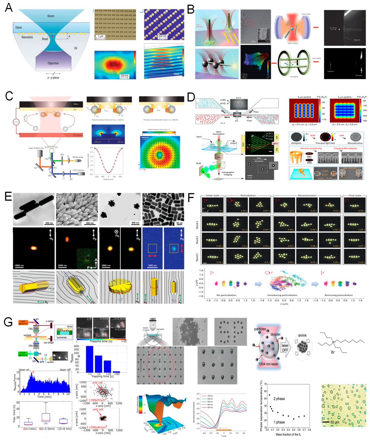
Adaptive Control and AI-Augmented Arrays.
Recent advancements in multiplexed optical tweezing have increasingly incorporated adaptive control algorithms and machine learning techniques to enhance system responsiveness, precision, and autonomy. These AI-augmented platforms are designed to address the inherent variability and complexity of dynamic experimental environments.
Key functionalities enabled by these systems include real-time compensation for positional drift and system noise through closed-loop feedback control, ensuring stable trap performance over extended durations. Additionally, algorithms can dynamically modulate trap stiffness or laser intensity in response to changes in particle behavior—such as displacement fluctuations or changes in scattering signal—thereby optimizing force application on a per-trap basis. Moreover, machine learning models can be used to automate the identification, selection, and retention of target particles within large-scale trap arrays, enabling high-throughput and minimally supervised operation.
A notable example was demonstrated by Zhang et al. in 2022, wherein a reinforcement learning framework was implemented to autonomously adjust trap positions within a microfluidic flow environment. The algorithm continuously evaluated particle trajectories and adjusted beam steering parameters in real time to maintain optimal confinement, illustrating the feasibility of fully autonomous, adaptive optical manipulation systems [547]. These developments signal a broader shift toward intelligent multiplexed tweezers, capable of operating as self-optimizing platforms for complex applications in fluidic diagnostics, dynamic self-assembly, and high-throughput cell mechanics.
5.3. Ultrafast and High-Speed Optical Trapping Systems
Optical trapping systems have traditionally operated at bandwidths limited by mechanical scanning devices, frame rates of video cameras, or data acquisition bottlenecks [548]. However, many emerging biological and physical processes—such as rapid conformational changes in biomolecules, fast cellular deformation, or nanoparticle diffusion in turbulent microenvironments—occur on timescales from microseconds to milliseconds [549,550,551]. To address these challenges, the field has witnessed a rapid development of ultrafast and high-speed optical tweezers, designed to achieve sub-millisecond temporal resolution and kilohertz to megahertz force tracking.
Motivation for High-Speed Optical Manipulation.
The need for high-speed optical manipulation is increasingly driven by the temporal complexity of numerous biophysical and soft-matter processes, many of which unfold on timescales that exceed the operational bandwidth of conventional optical tweezer systems. Accurately capturing these rapid dynamics requires both high temporal resolution and fast feedback control.
For example, the stepping behavior of molecular motors such as kinesin and myosin involves transient displacements and force-generating events occurring on the order of microseconds [552]. Similarly, protein folding and unfolding transitions often proceed within sub-millisecond timescales, necessitating rapid force-clamp and position-tracking capabilities to resolve intermediate conformational states [553].
Within living cells, intracellular cargo transport exhibits stochastic, intermittent motion influenced by cytoskeletal dynamics and local viscosity gradients. Monitoring such behavior demands high-frequency tracking to distinguish active transport from diffusive noise. Furthermore, in active matter systems, where collective particle motion is governed by non-equilibrium hydrodynamics, real-time acquisition of positional and force data across multiple entities is essential for probing inter-particle interactions and emergent flow patterns.
In these scenarios, high-speed force and displacement readout is not merely advantageous but a fundamental requirement for quantitative characterization and mechanistic modeling. Advances in detector technology, signal processing, and control algorithms are therefore critical to enabling the next generation of optical tweezers capable of operating at biologically and physically relevant timescales.
Core Technologies for Ultrafast Tweezers.
Realizing ultrafast optical tweezers necessitates substantial advancements across all major system components, from detection and beam steering to control electronics and data processing. These upgrades are essential for capturing rapid molecular events and implementing real-time manipulation at sub-microsecond timescales.
One foundational requirement is the use of high-bandwidth position detectors, such as quadrant photodiodes (QPDs) and back-focal-plane interferometry (BFPI) systems with bandwidths exceeding 100 kHz [554]. These detectors enable precise tracking of particle displacements with sub-nanometer spatial resolution and sub-microsecond temporal resolution, forming the basis for accurate force measurements and feedback.
Fast beam steering is equally critical for dynamic manipulation. Devices such as acousto-optic deflectors (AODs) and electro-optic modulators (EOMs) provide sub-microsecond response times for modulating beam position or intensity, allowing the optical trap to be reconfigured in real time—either in response to particle motion or as part of a scanning or time-sharing routine [555]. To fully exploit these capabilities, low-latency feedback control systems are required. Field-programmable gate arrays (FPGAs) and dedicated real-time processing units are now commonly employed to implement closed-loop trap stabilization. For example, when a trapped particle is perturbed by thermal fluctuations or flow-induced forces, the system can dynamically adjust the trap location or intensity to restore confinement within microseconds [556]. Finally, high-speed data acquisition and processing pipelines are essential for real-time analysis. Positional signals acquired from QPDs or interferometric sensors are digitized at megahertz sampling rates and subjected to rapid signal processing for spectral analysis, trap stiffness calibration, and noise suppression [557]. These capabilities not only support high-resolution tracking but also facilitate quantitative studies of rapid molecular and cellular processes.
Together, these technological innovations enable a new generation of optical tweezer systems capable of probing biological and physical phenomena on ultrafast timescales, expanding the applicability of optical manipulation into domains previously inaccessible due to temporal resolution constraints.
Applications in Force Spectroscopy and Cellular Dynamics.
Ultrafast optical tweezers have enabled a new class of high-resolution, time-resolved experiments, particularly in the domains of force spectroscopy, cellular mechanics, and microfluidic transport. By combining sub-piconewton force sensitivity with microsecond-scale temporal resolution, these systems facilitate quantitative interrogation of dynamic biological and soft-matter processes previously inaccessible to conventional trapping technologies.
In high-speed force spectroscopy, ultrafast tweezers allow the direct observation of rapid molecular transitions, such as the folding and unfolding of DNA hairpins, protein domains, or RNA secondary structures, with force precision better than 0.1 pN and temporal resolution below 1 ms. These measurements enable the reconstruction of free energy landscapes with high fidelity, capturing intermediate states and transition kinetics that are critical for understanding biomolecular function and stability.
For investigations in cellular biomechanics, rapid modulation of the optical trap—such as with sinusoidal oscillations in the kilohertz regime—enables frequency-domain microrheology. This technique provides access to the complex mechanical modulus of cellular components, including membranes and cytoplasm, over a broad frequency spectrum. As a result, the viscoelastic behavior of live cells can be quantified in real time, offering insights into cytoskeletal dynamics, mechanotransduction, and pathological alterations in mechanical properties.
In the context of microfluidic systems, ultrafast tweezers are used to track and stabilize rapidly moving particles or droplets within flow channels. By implementing dynamic feedback traps that follow targets in real time, it is possible to optically “pin” analytes in high-speed flows. This capability has proven valuable for applications in flow cytometry, on-chip chemical sensing, and biomolecular detection, where precise spatiotemporal control over fast-moving species is essential. Collectively, these applications demonstrate the transformative impact of ultrafast optical tweezers in advancing single-molecule biophysics, cellular mechanobiology, and lab-on-chip diagnostics, bridging the gap between optical manipulation and real-time quantitative measurement at biologically relevant timescales.
Challenges and Outlook.
While ultrafast optical tweezers offer transformative capabilities for high-resolution force spectroscopy and dynamic biological manipulation, their implementation remains accompanied by a number of technical and scientific challenges that must be addressed to fully realize their potential.
One major limitation is the signal-to-noise tradeoff inherent to high-speed detection. As integration times decrease to accommodate microsecond-scale measurements, the relative impact of shot noise, detector nonlinearity, and electronic artifacts becomes more pronounced. To preserve measurement fidelity, advanced signal processing techniques—such as real-time filtering, denoising algorithms, and spectral decomposition—are often required, adding computational complexity.
A second concern relates to photodamage and thermal effects. Ultrafast operation frequently necessitates higher laser powers or tightly focused beams to maintain sufficient trap stiffness at rapid timescales. This intensification increases the risk of local heating and phototoxicity, particularly in live-cell applications or when manipulating temperature-sensitive biomolecules. Careful optimization of wavelength, exposure duration, and beam modulation strategies is essential to mitigate these effects.
Furthermore, the deployment of ultrafast systems involves substantial engineering complexity. Components such as acousto-optic deflectors (AODs), high-speed analog-to-digital converters (ADCs), field-programmable gate arrays (FPGAs), and real-time synchronization hardware must be integrated and finely calibrated. This raises both the technical barrier to entry and the overall cost and maintenance requirements of the system.
Despite these challenges, ongoing advances in photonic integration, miniaturized electronics, and software-defined instrumentation are expected to yield next-generation ultrafast tweezer platforms that are more compact, accessible, and user-friendly. In parallel, the integration of AI-based control systems, machine vision, and real-time decision-making algorithms will further enhance temporal precision and operational autonomy. These developments will likely enable fully automated, microsecond-resolved manipulation in complex biological and soft-matter environments, opening new frontiers in dynamic biophysics, cellular mechanics, and molecular diagnostics.
5.4. Plasmonic and Near-Field Enhanced Optical Tweezers
As optical tweezers strive to manipulate ever smaller particles—from nanoparticles to single molecules—the fundamental limitation imposed by diffraction becomes increasingly significant [383]. Traditional gradient forces scale poorly with particle size, particularly below ~100 nm, where optical forces weaken while Brownian motion dominates. To overcome this, researchers have developed a class of plasmonic and near-field enhanced optical tweezers that leverage localized surface plasmon resonances (LSPR) and evanescent field confinement to achieve trapping with forces orders of magnitude stronger than conventional diffraction-limited systems.
Physical Principles and Nanostructure Designs.
Plasmonic optical tweezers exploit the resonant interaction between incident electromagnetic radiation and the collective oscillations of conduction electrons—known as surface plasmons—in metallic nanostructures, typically composed of noble metals such as gold or silver [295]. These interactions give rise to highly localized electromagnetic fields, particularly concentrated in subwavelength regions termed “hot spots”, which are often found at nanogaps, sharp edges, or junctions between closely spaced nanoparticles. Within these hot spots, the local field intensity can be enhanced by factors ranging from 10³ to 10⁵, significantly amplifying the optical gradient forces, which scale proportionally to the spatial variation of the field intensity(∇|E|²)[558]. This extreme field confinement enables the stable trapping of nanoscale objects, including 10–50 nm particles and even individual biomolecules, using relatively low input optical power. Due to the strong spatial localization of the electromagnetic fields, the resulting trapping volumes are typically on the order of tens of nanometers, offering both high spatial precision and single-particle resolution.
A variety of nanostructure geometries have been engineered to support and enhance plasmonic trapping, including: Bowtie antennas, which concentrate fields at the nanogap between two opposing triangular metal tips [559]; Nanoapertures and nanoholes in metallic films, which act as field concentrators through localized transmission resonances [292]; Metallic nanodisks and nanopillars fabricated on dielectric substrates, offering vertical confinement and field enhancement at the metal–dielectric interface [560]; Tip-enhanced plasmonic probes, often integrated with atomic force microscopy (AFM), which localize the field at a sharp apex for scanning-based manipulation [561].
In addition, hybrid photonic–plasmonic architectures have been developed to improve light delivery efficiency and system integration. These platforms combine dielectric waveguides with embedded or adjacent metallic nanostructures, enabling efficient coupling of guided optical modes into localized plasmonic traps while maintaining compatibility with chip-scale integration. This approach facilitates the construction of compact, highly functional trapping systems for applications in nanomanipulation, single-molecule analysis, and lab-on-chip diagnostics.
Force Magnitudes and Scaling Behavior.
A defining feature of plasmonic optical tweezers is their capacity to generate femtonewton-range gradient forces ( N) for subwavelength particle trapping, all while operating at moderate optical power levels [562,563,564]. This performance contrasts sharply with conventional optical tweezers, which typically require laser powers on the order of 100 mW, focused through a high-numerical-aperture objective, to stably trap a 100 nm dielectric particle. In comparison, plasmonic architectures such as gold bowtie nanoantennas can achieve stable confinement of particles as small as 20 nm using only a few milliwatts of input power, due to the intense local field enhancement associated with plasmonic resonances.
The scaling behavior of the optical gradient force in such systems is governed by the relation:
where polarizability for small dielectric particles, but the field gradient near plasmonic hotspots can increase sharply with resonance tuning.
It is important to note that thermal effects become non-negligible in plasmonic systems due to light absorption in metallic nanostructures. Localized heating can induce thermophoretic forces and convective flows, which may either enhance or disrupt trapping performance depending on the specific geometry, material composition, and thermal management strategies. As such, the interplay between optical forces and thermally induced fluid dynamics must be carefully considered when designing and optimizing plasmonic trapping systems for stable, high-precision manipulation at the nanoscale.
Applications and Demonstrations.
Plasmonic and near-field optical tweezers have enabled a range of advanced applications at the intersection of nano-photonics, biophysics, and materials science, capitalizing on their unique ability to confine light and exert optical forces at deeply subwavelength scales [565].
In single-molecule manipulation and spectroscopy, plasmonic tweezers have been integrated with surface-enhanced Raman scattering (SERS) to facilitate simultaneous trapping and vibrational analysis of individual biomolecules, such as proteins and DNA. The enhanced local electromagnetic fields amplify Raman signals, allowing for label-free molecular identification with single-molecule sensitivity [566]. In the field of biosensing, the trapping of nanoscale analytes—such as viruses, exosomes, or individual proteins—within plasmonic hotspots leads to measurable shifts in the local plasmonic resonance spectrum [567,568]. This phenomenon forms the basis of ultra-sensitive, label-free detection platforms, capable of real-time monitoring at attomolar concentrations without the need for chemical labeling or amplification.
For nanoparticle assembly and nano-lithography, near-field tweezers provide precise spatial control over particle positioning, enabling directed assembly of nanostructures on surfaces or in colloidal suspensions [569]. This capability supports the bottom-up fabrication of complex nanoscale architectures, including metamaterials and photonic devices. Moreover, the integration of plasmonic traps into microfluidic environments has led to the development of chip-based optofluidic platforms that combine continuous particle flow with localized optical trapping. These systems have been used for high-throughput tasks such as virus sorting, particle classification, and single-particle analysis, offering compact and scalable alternatives to traditional bulk optical systems. A representative demonstration highlighted the ability to trap a 20 nm polystyrene bead using a gold bowtie nanoantenna with just 3 mW of input power. The particle was confined within a 30 nm hotspot, and the resulting optical gradient force was estimated at approximately 50 femtonewtons, illustrating the extreme force localization achievable with plasmonic architectures.
Collectively, these advancements underscore the potential of plasmonic and near-field tweezers as powerful tools for nanoscale manipulation, biomolecular interrogation, and integrated lab-on-chip diagnostics.
Limitations and Engineering Challenges.
Despite their significant advantages in nanoscale manipulation and field enhancement, plasmonic optical tweezers are subject to several intrinsic limitations and engineering challenges that constrain their broader applicability and scalability.
One of the most critical issues is localized heating, which arises from absorption losses in metallic nanostructures [297]. Temperature rises in the range of 10–30 °C are common near plasmonic hotspots, potentially inducing thermophoretic drift, convection currents, or even thermal damage to sensitive biological samples. Such thermal artifacts can compromise trapping stability and introduce undesired forces, particularly in aqueous or bio-relevant environments.
A second challenge involves the fabrication complexity associated with plasmonic devices [295]. Achieving reproducible nanoscale features—such as sub-20 nm gaps between metallic structures—typically requires advanced nanofabrication techniques, including electron-beam lithography or focused ion beam (FIB) milling. These methods, while precise, are time-consuming and cost-intensive, limiting throughput and posing obstacles to large-scale or disposable device manufacturing.
Additionally, trap accessibility is inherently constrained by the near-field nature of plasmonic confinement. Optical forces are localized to within ~100 nm of the substrate surface, which restricts manipulation to two-dimensional geometries and precludes full three-dimensional control over particle positioning. This limitation is particularly significant for applications requiring volumetric manipulation or suspension-based transport.
To address these challenges, current research efforts are directed toward the development of all-dielectric nanostructures, such as silicon or titanium dioxide metasurfaces, which can support strong Mie resonances while avoiding plasmonic absorption losses [570]. Hybrid platforms that incorporate heat-dissipating substrates, thermal spreaders, or convection channels have also been proposed to minimize localized heating. Furthermore, machine learning–assisted trap optimization strategies are being explored to identify geometries and excitation conditions that maximize optical trapping efficiency while mitigating thermal effects.
These innovations aim to expand the operational envelope of plasmonic tweezers, enabling their deployment in more diverse experimental contexts, including biocompatible manipulation, high-throughput sensing, and integrated photonic systems.
5.5. Hybrid and Multifunctional Optical Tweezers
To overcome the intrinsic limitations of purely optical manipulation—such as limited force range, restricted selectivity, and thermal effects—researchers have increasingly explored hybrid and multifunctional tweezing platforms that combine optical trapping with other physical modalities [571]. These systems integrate magnetic, acoustic, electrical, or thermal fields to enhance trapping capability, enable new forms of interaction, or broaden the class of manipulatable targets. Hybrid tweezers represent a convergence of micro/nano engineering and interdisciplinary physics, pushing the boundaries of what is possible in microscale manipulation.
Magneto-Optical Tweezers.
Magneto-optical tweezers represent one of the most mature and widely adopted hybrid platforms for precision force spectroscopy, integrating the high spatial resolution and stiffness of optical traps with the long-range, directional, and low-noise force application of magnetic fields [572]. This dual-modality approach offers enhanced experimental flexibility, particularly in contexts requiring stable and tunable force control over biomolecular systems.
In a typical configuration, superparamagnetic beads—such as iron oxide–coated polystyrene microspheres—are confined using optical tweezers while being subjected to external magnetic field gradients [573]. The optical trap provides precise lateral and axial positioning, whereas the magnetic field imparts calibrated forces, often in the tens to hundreds of piconewtons, along a defined spatial axis. This arrangement is especially suited for applications in molecular tethering assays, where DNA or protein constructs are anchored between a functionalized surface and the trapped magnetic bead.
The key advantage of this hybrid system lies in its ability to combine high trap stiffness and spatial resolution from the optical component with low-noise, constant-force application enabled by the magnetic field. As such, magneto-optical tweezers are ideal for force-clamp experiments, in which the applied force is held constant while molecular extension, conformational change, or kinetic response is monitored.
This platform has enabled a range of impactful applications, including:Protein unfolding and refolding studies under well-controlled, constant-force conditions; Stepping assays of molecular motors, such as kinesin or myosin, under axial tension to probe mechanochemical coupling; Tethered particle motion (TPM) experiments, wherein optical imaging is used to analyze fluctuations in the bead’s position to extract mechanical or biochemical information.
By leveraging the complementary strengths of optical and magnetic manipulation, magneto-optical tweezers provide a robust and versatile framework for probing biomolecular mechanics, enzyme kinetics, and polymer dynamics at the single-molecule level.
Acousto-Optic and Thermo-Optic Hybrids.
Acousto-optic and thermo-optic hybrid tweezer systems represent emerging strategies for augmenting the functional range of conventional optical trapping platforms by incorporating additional force modalities—namely, acoustic and thermophoretic effects. These hybrid configurations enable simultaneous control over particle positioning at multiple spatial and temporal scales, offering enhanced capabilities for microfluidic manipulation, bioparticle sorting, and nanomaterial assembly [482].
In acousto-optic hybrid systems, ultrasound fields, including both bulk acoustic waves (BAWs) and surface acoustic waves (SAWs), are employed to generate pressure nodes that interact with suspended particles via acoustic radiation forces. When integrated with optical tweezers, the acoustic field provides long-range, parallel transport and pre-concentration, while the optical trap offers high-resolution capture and sorting. This division of roles is particularly advantageous in microfluidic environments, where acoustically guided particles can be funneled toward localized optical trapping regions. Recent demonstrations include cell alignment and manipulation in opto-acoustic fields, as well as acoustofluidic–optical platforms designed for the selective capture of circulating tumor cells (CTCs) from complex biological samples.
In parallel, thermo-optic tweezers exploit light-induced local heating—often mediated by plasmonic nanostructures—to generate thermophoretic forces, which drive particles along temperature gradients. When combined with optical gradient forces, this approach enables selective transport based on particle properties such as size, refractive index, or surface chemistry. These systems allow non-contact manipulation across substrates, leveraging thermal gradients to steer particles with high directional control. Notable implementations include dual-beam infrared laser configurations coupled with plasmonic heating elements, which facilitate temperature-controlled nanoassembly, directional cargo sorting, and spatially resolved biochemical assays.
Collectively, these hybrid strategies extend the operational versatility of optical tweezers, enabling multiscale, multimodal manipulation schemes well suited for lab-on-chip diagnostics, biophysical investigations, and bottom-up nanofabrication. Their integration into microfluidic and photonic platforms is expected to further enhance system compactness, automation, and throughput in next-generation trapping technologies.
Electro-Optical and Dielectrophoretic Integration.
Electro-optical tweezers (EOT) integrate optical trapping mechanisms with externally applied electric fields, enabling enhanced selectivity and force control within micro- and nanoscale manipulation platforms [473]. In these systems, electric fields are typically applied via patterned microelectrodes or generated through spatial field gradients in optofluidic environments. When particles are suspended in such non-uniform electric fields, dielectrophoretic (DEP) forces arise due to field-induced polarization. These forces act differentially based on the dielectric properties of the particle and the surrounding medium, allowing for selective trapping and manipulation based on polarizability.
A key advantage of DEP-assisted manipulation is its capacity to orient and align anisotropic particles, such as nanorods, DNA strands, or elongated cells, along the local field lines. When combined with optical tweezers, DEP forces can serve complementary roles: for instance, DEP fields may be used for pre-alignment or coarse positioning, thereby facilitating subsequent high-precision optical capture. Conversely, optical tweezers can provide localized control where electric fields alone lack sufficient resolution.
The synergistic interplay between optical and electrical forces has enabled a range of advanced applications. These include real-time cell sorting platforms that discriminate based on both optical characteristics (e.g., scattering, fluorescence) and electrical signatures (e.g., membrane capacitance or cytoplasmic conductivity). Hybrid opto-DEP biosensors leverage this dual-mode manipulation to achieve enhanced signal-to-noise ratios, especially in label-free detection schemes. Additionally, the combined application of optical and electric field stimuli has been employed in the field-assisted assembly of nanomaterials, allowing precise spatial organization of colloidal components or functional nanostructures within engineered architectures.
Such electro-optical integrations offer a powerful route toward high-throughput, selective, and multifunctional trapping platforms, bridging the strengths of photonic precision with electrokinetic versatility for applications in biosensing, nanomanufacturing, and cellular diagnostics.
Multifunctional Platforms and Lab-on-a-Chip Integration.
Recent technological advances have enabled the development of multifunctional, fully integrated optical tweezers platforms, which incorporate diverse functional modules within compact, chip-scale architectures. These systems synergistically combine microfluidics for automated sample delivery and fluid control, on-chip photonics—including waveguides, modulators, and meta-surfaces—for light routing and trapping, electrode arrays for electric and magnetic field generation, and embedded sensors capable of performing real-time fluorescence, Raman, or electrical impedance measurements [468,574].
Such integrated platforms support parallelized trapping, manipulation, and analysis of single particles, nanoparticles, or live cells with high throughput and spatial precision. By consolidating optical, electrical, and fluidic functionalities onto a single substrate, these systems enable a wide range of complex experimental protocols to be executed in a lab-on-a-chip format, with significantly reduced system size and user intervention.
One representative example involves a microfluidic chip that integrates plasmonic optical traps, localized thermal modulators, and electrokinetic control channels. This multifunctional device has demonstrated capabilities such as: Label-free detection and trapping of viral particles via resonance-based sensing; Selective sorting of extracellular vesicles, including exosomes, based on dielectric and thermophoretic signatures; Automated single-particle force spectroscopy, used to probe the mechanical properties of protein aggregates under controlled environmental stimuli.
These multifunctional optical tweezers platforms represent a critical step toward portable, intelligent microsystems for precision manipulation and sensing. By enabling real-time control, analysis, and decision-making within a miniaturized form factor, they hold significant promise for applications in point-of-care diagnostics, single-cell omics, nanorobotics, and integrated material characterization, bridging the gap between benchtop instrumentation and deployable analytical technologies.
5.6. AI-Assisted Optical Trapping and Intelligent Control
As optical tweezers evolve into sophisticated, high-precision instruments for manipulating matter at the micro- and nanoscale, their operation becomes increasingly complex. Precise alignment, dynamic calibration, noise suppression, real-time control, and large data volume interpretation are all challenges in modern tweezing experiments [575]. To address these issues, a new frontier has emerged: the integration of artificial intelligence (AI), machine learning (ML), and intelligent control algorithms into optical tweezing platforms.
AI is now being used not only to automate and stabilize optical traps, but also to optimize trap design, interpret force measurements, and enhance feedback response, thereby improving both usability and throughput across scientific and industrial applications.
Figure 22.
AI-Driven Optical Tweezers: Emerging Architectures and Functionalities Empowered by Machine Intelligence [268,550,576,577,578,579,580,581]. This figure highlights seven cutting-edge directions in which artificial intelligence enhances the performance, automation, and scalability of optical tweezers. By embedding machine learning algorithms—such as convolutional neural networks (CNNs), U-Net++, generative adversarial networks (GANs), and reinforcement learning (RL)—optical trapping platforms evolve into intelligent systems capable of real-time adaptation and decision-making. Applications include: (1) deep-learning-assisted sperm diagnostics using U-Net++ for angular velocity quantification [582]; (2) GAN-based trap design for optimized tweezer arrays in cold atom assembly [583]; (3) SmartTrap platforms integrating feedback control, microfluidics, and 3D tracking for autonomous experimentation [584]; (4) vortex trap inverse design via neural networks for tailored orbital angular momentum confinement [575]; (5) AI-stabilized multi-trap arrays enabling large-scale parallel manipulation [100,585]; (6) RL-driven micromanipulation with haptic feedback for intelligent interaction [586]; and (7) CNN-powered high-throughput screening across 48 optical channels for automated cell classification and drug testing [587]. These advances collectively redefine optical tweezers as adaptive, intelligent platforms for next-generation biophotonics and micro/nanomanipulation.
Figure 22.
AI-Driven Optical Tweezers: Emerging Architectures and Functionalities Empowered by Machine Intelligence [268,550,576,577,578,579,580,581]. This figure highlights seven cutting-edge directions in which artificial intelligence enhances the performance, automation, and scalability of optical tweezers. By embedding machine learning algorithms—such as convolutional neural networks (CNNs), U-Net++, generative adversarial networks (GANs), and reinforcement learning (RL)—optical trapping platforms evolve into intelligent systems capable of real-time adaptation and decision-making. Applications include: (1) deep-learning-assisted sperm diagnostics using U-Net++ for angular velocity quantification [582]; (2) GAN-based trap design for optimized tweezer arrays in cold atom assembly [583]; (3) SmartTrap platforms integrating feedback control, microfluidics, and 3D tracking for autonomous experimentation [584]; (4) vortex trap inverse design via neural networks for tailored orbital angular momentum confinement [575]; (5) AI-stabilized multi-trap arrays enabling large-scale parallel manipulation [100,585]; (6) RL-driven micromanipulation with haptic feedback for intelligent interaction [586]; and (7) CNN-powered high-throughput screening across 48 optical channels for automated cell classification and drug testing [587]. These advances collectively redefine optical tweezers as adaptive, intelligent platforms for next-generation biophotonics and micro/nanomanipulation.
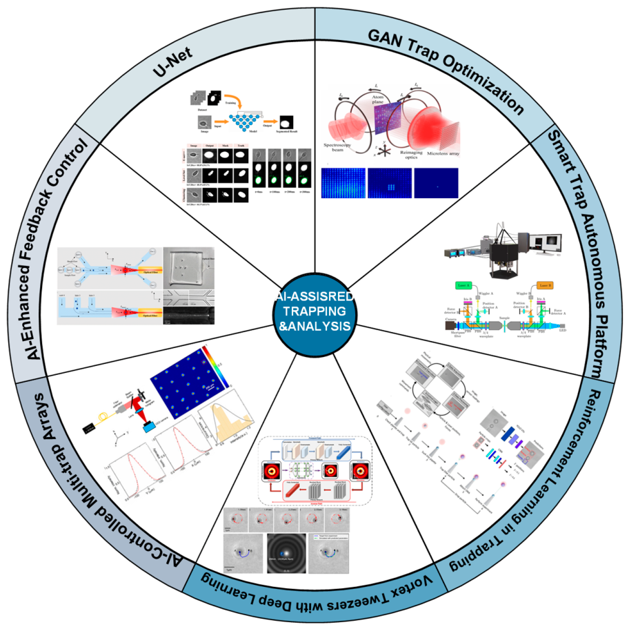
Machine Learning for Trap Optimization.
The operation of optical tweezers involves the optimization of a complex, high-dimensional parameter space, encompassing variables such as laser power, beam shape, numerical aperture, particle size and refractive index, and ambient noise levels. Traditional approaches to trap calibration and tuning often rely on empirical heuristics and manual adjustment, which are time-intensive and frequently suboptimal in terms of performance and adaptability [513].
Recent advances in machine learning (ML)—particularly reinforcement learning (RL) and Bayesian optimization—have begun to transform the way optical trapping systems are configured and controlled. These algorithms enable autonomous exploration and optimization of trapping parameters to achieve robust, high-performance manipulation under diverse experimental conditions. Applications include the automatic identification of optimal laser parameters for trapping specific targets, such as live cells, nanoparticles, or extracellular vesicles, where biological variability or optical heterogeneity may significantly affect trapping dynamics.
ML models are also employed for real-time adaptation of trap position and beam shape, allowing for dynamic target tracking, multi-trap formation, or compensation for perturbations such as drift or flow. Furthermore, generative algorithms, including generative adversarial networks (GANs) and neural network–based field design frameworks, have been used to synthesize custom beam profiles capable of generating non-conventional trap geometries, such as bottle beams, Bessel lattices, or vortex arrays, expanding the functional landscape of optical manipulation.
A notable example involved the use of a convolutional neural network (CNN) trained to classify trapped particles by size and shape using signals obtained from back-focal-plane interferometry (BFPI). The classification output was then utilized to dynamically adjust trap stiffness in real time, enabling adaptive force control tailored to individual particle characteristics.
These machine learning–driven approaches are increasingly positioning optical tweezers as intelligent, self-optimizing platforms, capable of operating efficiently in complex, noisy, and heterogeneous environments without the need for extensive human intervention.
Real-Time Feedback and Adaptive Control.
Real-time feedback and adaptive control are essential components of high-performance optical tweezing systems, particularly in experiments involving long-duration trapping or complex, dynamic environments such as microfluidic channels. While conventional adaptive optics and feedback mechanisms have historically been employed to mitigate mechanical drift, thermal fluctuations, and system instabilities, the integration of artificial intelligence (AI) has significantly advanced these capabilities by introducing predictive and self-correcting control algorithms [518].
One key application area is feedback-controlled tracking, where machine learning algorithms are trained to predict the future trajectories of fast-moving targets, such as motile bacteria, sperm cells, or active colloids. By forecasting particle motion and preemptively adjusting laser position, these systems reduce latency and improve trapping robustness under high-speed or turbulent conditions.
AI-enhanced systems also play a critical role in vibration suppression. By distinguishing between genuine particle displacement and extraneous mechanical noise, AI-based signal filters enhance positional stability and reduce false-positive trap readjustments. This leads to improved force precision and more reliable trajectory analysis, especially in sensitive biophysical assays.
Another significant advancement is in force-clamp control, where reinforcement learning agents are employed to maintain constant mechanical load on single molecules or tethered particles. These agents analyze real-time force readouts—typically derived from back-focal-plane interferometry (BFPI)—and adaptively modulate laser intensity or trap position to counteract fluctuations. This capability is particularly critical in the study of molecular unfolding, motor protein mechanics, or viscoelastic responses, where consistent force application is required for accurate characterization.
Overall, the integration of AI into feedback loops transforms traditional control schemes into intelligent, adaptive systems capable of responding dynamically to environmental perturbations, target behavior, and instrument drift. These advances are paving the way for next-generation optical tweezers with enhanced precision, resilience, and autonomy in increasingly complex experimental settings.
Data Analysis and Signal Enhancement.
Optical tweezers experiments routinely generate large volumes of high-resolution data, encompassing nanometer-scale positional time series, power spectral density (PSD) measurements in the kilohertz to megahertz range, and video streams for real-time tracking of multiple particles. The complexity and density of these datasets present significant challenges for traditional analysis techniques, particularly when extracting subtle biophysical signals from noisy or multidimensional data.
The application of artificial intelligence (AI), particularly machine learning (ML) and deep learning algorithms, has significantly enhanced the efficiency and sensitivity of data processing and signal interpretation in optical tweezing systems. For example, recurrent neural networks (RNNs) and other temporal models have been successfully employed to perform denoising of force and displacement signals, improving signal-to-noise ratios without compromising temporal resolution.
In particle tracking applications, AI-based methods have been used for robust outlier detection and rejection, ensuring the fidelity of extracted trajectories in noisy or crowded environments. Furthermore, multivariate pattern recognition techniques enable the identification of complex behavioral states in experiments involving optical binding, active particle motion, or collective dynamics, which may not be readily detectable through conventional statistical approaches.
In single-molecule force spectroscopy, machine learning has proven particularly effective for event classification and trace segmentation. Algorithms have been trained to detect rupture events, identify hidden intermediate states, and segment noisy unfolding pathways in real time, enabling more accurate and automated interpretation of molecular interactions and conformational transitions.
Collectively, these AI-enhanced analytical tools facilitate real-time, high-throughput, and automated data interpretation, substantially reducing the burden of manual analysis while increasing the depth and reproducibility of insight gained from optical tweezers experiments. These capabilities are critical for advancing the role of optical manipulation in complex, data-rich applications across biophysics, nanotechnology, and mechanobiology.
Smart Optical Tweezers Systems.
The development of smart optical tweezers systems marks a significant step toward fully autonomous, user-friendly platforms for precision optical manipulation. These next-generation systems integrate real-time machine vision, adaptive control hardware, and on-board artificial intelligence (AI) to enable sophisticated functionalities traditionally requiring expert operation.
Key components include machine-vision feedback mechanisms, such as high-speed cameras or quadrant photodiode detectors (QPDs), which provide continuous tracking of particle position and dynamics. These are coupled with high-speed actuators, including piezoelectric stages, acousto-optic deflectors, and spatial light modulators (SLMs), for rapid and precise trap modulation. Crucially, computation is handled by embedded AI processors, such as FPGA-based architectures or edge-computing modules (e.g., Nvidia Jetson), which facilitate real-time data processing and control loop execution.
These integrated platforms enable a suite of autonomous capabilities. Auto-initialization routines allow the system to identify target particles within the field of view and automatically center them into traps without user intervention. Dynamic calibration algorithms continuously estimate trap stiffness and force constants by analyzing the real-time motion of trapped particles, adapting to environmental variations such as temperature, viscosity, or optical aberrations. Moreover, multichannel coordination across large holographic trap arrays allows simultaneous, independent control of dozens of optical traps, enabling complex manipulation protocols such as parallel cell sorting, multi-object assembly, or dynamic reconfiguration of colloidal lattices.
By automating core functions and minimizing the need for manual tuning, smart optical tweezers systems significantly lower the barrier to entry for non-expert users. These platforms thus represent a critical advancement in the democratization of high-end optical manipulation technologies, with broad applications in biophysics, nanomedicine, materials science, and micro-robotics.
5.7. Future Trends and Perspectives in Optical Tweezer Technologies
Over the past three decades, optical tweezers have evolved from a laboratory curiosity into one of the most powerful and precise tools in experimental science. As the field advances, several major trends are shaping the future of optical trapping, including innovations in materials, optics, system design, and interdisciplinary integration. These trends will not only expand the physical capabilities of tweezers—such as force range, spatial resolution, and scalability—but also broaden their scientific and industrial impact in diagnostics, manufacturing, quantum technologies, and beyond.
Toward Ultrafast and Ultraprecise Optical Manipulation.
Ongoing research efforts are increasingly focused on extending the temporal and spatial resolution of optical tweezers to access regimes previously beyond experimental reach. The development of ultrafast optical trapping systems powered by femtosecond to picosecond pulsed lasers has opened new frontiers in nonequilibrium manipulation, allowing researchers to resolve and perturb transient molecular events, such as conformational transitions in biomolecules, chemical reaction intermediates, and rapid gating dynamics in ion channels [588].
Simultaneously, advances in high-bandwidth detection technologies, particularly those based on interferometric schemes and AI-assisted signal reconstruction, now enable the measurement of sub-nanometer displacements with microsecond temporal resolution [290]. These capabilities are critical for dissecting fast stochastic processes and capturing rare or fleeting molecular phenomena.
Looking ahead, the integration of quantum optical techniques—including the use of squeezed states and entangled photon pairs—is anticipated to push the limits of force sensitivity toward the quantum noise limit, offering the prospect of quantum-enhanced optical trapping [589]. Such advances may enable the detection of forces at the sub-femtonewton scale, with potential applications in exploring protein folding pathways, polymer relaxation dynamics, and even interatomic or Casimir-level interactions.
Collectively, these developments position ultrafast and ultraprecise optical tweezers as a transformative platform for interrogating the mechanics of single events and transient processes across a broad spectrum of physical, chemical, and biological systems.
Expansion to New Material Platforms and Spectral Domains.
Advancements in material science are poised to significantly broaden the operational versatility and functional scope of optical tweezers. Emerging material platforms, particularly those based on two-dimensional (2D) materials such as graphene and molybdenum disulfide (MoS₂), are being actively investigated for the development of plasmonic and photonic metasurface traps. These materials exhibit highly tunable optical properties, strong light–matter coupling, and the potential for large local field enhancements, enabling precise and reconfigurable trapping landscapes at the nanoscale [590].
Parallel efforts are extending optical trapping into nontraditional spectral regimes, including both the infrared (IR) and ultraviolet (UV) domains. Infrared tweezers offer the advantage of reduced photodamage, making them particularly suitable for biocompatible manipulation, while UV-based systems enable material-selective trapping and the excitation of specific molecular transitions, broadening the scope of applications in spectroscopy, photochemistry, and nanoassembly [591].
In addition, engineered nanostructures such as optomechanical crystals and metamaterials are being designed to support subwavelength field confinement and tailored dispersion properties. These structures enable not only enhanced optical trapping performance but also open pathways toward quantum-level control of trapped particles through strong optomechanical coupling and coherent light–matter interactions.
Collectively, these developments suggest a transition toward optical trapping platforms that are wavelength-adaptive, material-discriminative, and optically engineered for specific functionalities. Such systems promise to dramatically improve both the fidelity and selectivity of trapping, paving the way for applications in quantum photonics, precision biophysics, and nanostructured material manipulation.
Large-Scale Parallelization and Automation.
A longstanding constraint in the application of classical optical tweezers has been their limited throughput, restricting their utility in high-content or large-scale manipulation tasks. Recent research has begun to address this challenge by pursuing strategies for massive parallelization and system automation, thereby significantly expanding the scalability and applicability of optical trapping technologies.
Holographic optical tweezers, enabled by high-resolution spatial light modulators (SLMs), now allow the generation of hundreds of individually addressable traps within a single optical field. These traps can be dynamically reconfigured in three dimensions, supporting complex, real-time manipulation of multiple particles or biological entities in parallel. Complementing this approach, advances in photonic integrated circuits (PICs) have facilitated the on-chip integration of optical trapping functionalities, offering compact, robust, and scalable solutions with minimal alignment requirements and high mechanical stability.
In parallel, the development of automation frameworks that combine microfluidic sample handling, robotic actuation, and AI-driven control algorithms has led to the emergence of autonomous optical tweezing platforms. These systems are capable of executing sophisticated experimental protocols—including high-throughput mechanical phenotyping, single-particle force spectroscopy, and programmable self-assembly—with minimal user intervention.
Collectively, these innovations are transforming optical tweezers from precision, single-object tools into scalable, programmable platforms suitable for industrial-scale applications. Potential use cases include the assembly of colloidal photonic crystals, deterministic positioning of quantum emitters, and clinical-scale cellular biomechanics assays, underscoring the growing relevance of optical trapping in both fundamental research and translational technologies.
Integration into Cross-Disciplinary Platforms.
Optical tweezers are increasingly being integrated into hybrid and cross-disciplinary platforms, expanding their role from a specialized biophysical tool to a core enabling technology across multiple fields of science and engineering. This convergence is driving the development of multifunctional systems capable of autonomous, intelligent manipulation at microscopic and nanoscopic scales.
In mechanobiology, optical tweezers are employed to apply precisely controlled forces to individual cells and subcellular structures, enabling real-time interrogation of mechanotransduction pathways and cellular responses to mechanical stimuli with submicron spatial and sub-piconewton force resolution. These capabilities are critical for understanding the physical basis of cell behavior, development, and disease progression.
In the domain of quantum technologies, optical tweezers have become essential tools for the trapping, cooling, and entanglement of neutral atoms, where they serve as platforms for scalable quantum computing architectures and quantum simulation. Additionally, the optical levitation of dielectric nanoparticles in high vacuum has opened new avenues for quantum optomechanics, allowing tests of macroscopic quantum coherence and precision force sensing.
Within nanorobotics, optical tweezers enable the construction of light-driven microtransport systems capable of cargo sorting, delivery, and assembly in complex fluidic environments. These optically actuated systems demonstrate the feasibility of remotely controlled nanoscale machinery with applications in targeted delivery, lab-on-chip automation, and reconfigurable material systems.
As optical trapping technologies continue to converge with advancements in synthetic biology, micro- and nanorobotics, materials science, and artificial intelligence, they are poised to underpin the next generation of intelligent microsystems. These systems will be capable of executing complex, context-aware tasks such as environmental sensing, in situ diagnostics, and autonomous construction at the micro- and nanoscale, reshaping the landscape of precision manipulation in both scientific and technological domains.
This chapter has outlined the transformative progress in optical tweezer technologies across six major domains: chip integration, advanced beam shaping, hybrid systems, artificial intelligence, and forward-looking innovations. These advances underscore a paradigm shift: from manual, bench-scale tools to smart, jiexialmodular, and multifunctional platforms that can be embedded into industrial processes, diagnostic systems, and scientific instruments.
As the optical tweezers field moves into the next decade, it is likely to play a pivotal role in the future of precision manipulation, biointerface engineering, and quantum-level control, establishing itself as a cornerstone of microscale science and technology.
Figure 23.
Quantum Tweezers and Frontier Physics: Architectures, Operations, and Quantum Control Pathways [61,592,593,594,595,596,597,598,599,600,601,602,603,604,605,606,607,608]. This layered roadmap illustrates key advances in quantum optical tweezer platforms, spanning from atomic array construction to hybrid quantum systems. Beginning with defect-free cold atom arrays [609], each stage reflects a progressive enhancement in quantum control: Rydberg-mediated entanglement enables two-qubit gates [610,611], while high-fidelity operations surpass 98% gate fidelity in reconfigurable traps [101,612]. Tweezer-based spin simulators emulate many-body Hamiltonians [613], and single-atom tomography reconstructs full quantum states with high resolution [614]. Optical tweezers further support coherent molecular control at the quantum level [615], and integration with other quantum systems yields hybrid architectures for scalable entanglement and cross-platform interfacing [616]. Together, these developments establish optical tweezers as a central tool in building scalable, programmable quantum technologies.
Figure 23.
Quantum Tweezers and Frontier Physics: Architectures, Operations, and Quantum Control Pathways [61,592,593,594,595,596,597,598,599,600,601,602,603,604,605,606,607,608]. This layered roadmap illustrates key advances in quantum optical tweezer platforms, spanning from atomic array construction to hybrid quantum systems. Beginning with defect-free cold atom arrays [609], each stage reflects a progressive enhancement in quantum control: Rydberg-mediated entanglement enables two-qubit gates [610,611], while high-fidelity operations surpass 98% gate fidelity in reconfigurable traps [101,612]. Tweezer-based spin simulators emulate many-body Hamiltonians [613], and single-atom tomography reconstructs full quantum states with high resolution [614]. Optical tweezers further support coherent molecular control at the quantum level [615], and integration with other quantum systems yields hybrid architectures for scalable entanglement and cross-platform interfacing [616]. Together, these developments establish optical tweezers as a central tool in building scalable, programmable quantum technologies.
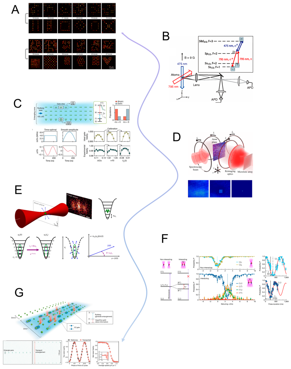
6. Challenges and Limitations
6.1. Diffraction Limitations and Optical Force Constraints
The ability of optical tweezers to manipulate microscopic and mesoscopic objects arises from the momentum transfer of highly focused light beams, with optical gradient forces serving as the primary mechanism for spatial confinement. However, this principle is fundamentally constrained by the diffraction limit, a core restriction of wave optics that determines the minimum spatial scale over which optical energy can be concentrated [617,618]. This limit places intrinsic bounds on the minimum trap size, achievable force strength, and overall spatial resolution of optical tweezing systems.
The diffraction limit can be approximated by the expression: , dictates that the focal spot of a beam cannot be smaller than roughly half the wavelength of the trapping light divided by the numerical aperture (NA) of the focusing lens. For typical near-infrared lasers (λ ≈ 1064 nm) and high-NA objectives (NA ≈ 1.3–1.4), this results in a focal diameter of approximately 500–700 nm. This constraint introduces two major limitations:
First, the minimum trap size imposes a challenge for objects much smaller than the focal volume, such as nanoparticles (<100 nm), biomolecules, or quantum dots. These particles experience significantly reduced gradient forces due to their small polarizability and poor overlap with the intensity gradient, making stable confinement difficult to achieve using traditional far-field optical traps. Second, trap stiffness and force sensitivity diminish rapidly with particle size. For Rayleigh-regime particles, the gradient force scales as:, where r is the particle radius. Since trap stiffness (k) and polarizability are volume-dependent, the manipulation of sub-wavelength particles results in weaker trapping potentials and degraded sensitivity. This limits the capacity of optical tweezers to resolve and quantify nanoscale biophysical interactions or apply forces to low-index dielectric materials.
Even with the implementation of enhanced trapping schemes—such as dual-beam traps, interferometric standing waves, or beam shaping using spatial light modulators (SLMs)—a fundamental trade-off persists between trapping resolution, force magnitude, and the risk of laser-induced photodamage [474]. These trade-offs motivate the exploration of near-field optical trapping techniques, including plasmonic tweezers, structured illumination (e.g., Bessel or Airy beams), and hybrid photonic platforms that aim to circumvent the diffraction limit.
Moreover, the maximum force achievable with optical tweezers, typically ranging from ~0.1 to 200 pN depending on laser power and particle properties, is insufficient for applications requiring higher mechanical loads, such as high-tension single-molecule stretching or mechanical deformation of rigid cellular structures. While increasing the laser power may temporarily enhance trap strength, it introduces significant challenges, including photothermal heating, convection, and sample degradation—issues further elaborated in Section 6.2.
In conclusion, the diffraction-limited trap size and the scaling constraints on gradient forces delineate a central physical boundary for conventional optical tweezers. Although adequate for manipulating microscale entities such as polystyrene beads or whole cells, these limitations necessitate the development of next-generation trapping strategies capable of extending manipulation capabilities into the nanoscale regime, beyond what is achievable through standard far-field optics.
6.2. Photothermal Effects and Sample-Induced Damage
A fundamental limitation of optical tweezers arises from the intrinsic energy deposition associated with the use of tightly focused, high-intensity laser beams. Although optical tweezers are widely regarded as non-contact and minimally invasive tools, the process of focusing coherent light to a diffraction-limited volume inherently produces elevated optical energy densities [592]. When this energy is absorbed—either by the trapped particle or the surrounding medium—it can give rise to photothermal effects, which significantly impair trapping stability, measurement accuracy, and biocompatibility, particularly in sensitive biological or nanoscale systems.
The primary mechanism underlying photothermal heating is photon absorption at the trap site. While dielectric particles such as polystyrene and silica microspheres exhibit minimal absorption in the commonly used near-infrared (NIR) range (e.g., λ = 1064 nm), many biological targets—including cells, proteins, and nucleic acids—as well as metallic or semiconducting nanoparticles possess significant absorption cross-sections at these wavelengths [390]. The absorbed energy is rapidly converted into heat, resulting in localized temperature elevations of approximately 3–5 °C within a timescale of milliseconds.
Even such moderate thermal increases can exert pronounced effects on trapping performance and biological integrity. First, thermal expansion and accompanying refractive index changes in the surrounding medium can distort the optical potential, altering trap geometry and reducing confinement stiffness. Second, thermally induced convection currents in liquid media may generate bulk fluid motion, displacing the trapped particle or introducing positional instability. Third, and most critically, thermal stress can compromise cellular viability, especially in delicate systems where minor deviations in temperature may denature proteins, disrupt membrane structures, or activate cellular stress responses.
Historical evidence underscores these risks: in foundational experiments, Ashkin demonstrated that green laser illumination (λ = 514 nm) resulted in lethal photodamage to E. coli, while the use of infrared light (λ = 1064 nm) preserved cellular viability. Although this observation established the infrared window as a preferred regime for biological trapping, subsequent studies have shown that even NIR beams, when operated at high powers or under extended exposure, can still induce deleterious thermal effects [411]. These findings collectively highlight the thermodynamic constraints inherent to optical tweezing and underscore the necessity of careful wavelength selection, power regulation, and thermal management—particularly when manipulating biological specimens or highly absorptive nanomaterials.
Photodamage Mechanisms in Biological and Nanomaterial Systems.
In addition to photothermal effects, photochemical damage constitutes a significant concern in optical trapping, particularly when operating in aqueous environments or oxygen-rich media. Such damage typically arises from multi-photon absorption processes and the subsequent generation of reactive oxygen species (ROS)[619]. These highly reactive intermediates can interact with surrounding biomolecules or nanomaterials, leading to irreversible structural or functional alterations.
In biophysical applications, photochemical interactions may result in fragmentation of DNA strands or protein backbones, particularly during high-resolution single-molecule force spectroscopy experiments [276]. Similarly, in live-cell trapping or imaging, ROS can perturb subcellular architectures, compromising organelle integrity or triggering stress-related signaling pathways. In the context of nanomaterial systems, such photochemical interactions may induce phase transitions, surface restructuring, or even melting of optically active components, such as the reshaping of gold nanoparticles under prolonged irradiation.
These phenomena collectively underscore a critical operational tradeoff: while increasing laser power is often necessary to enhance trap stiffness, spatial confinement, and measurement sensitivity, doing so inherently elevates the probability of photothermal and photochemical damage. As a result, the optimization of optical trapping conditions must balance mechanical performance against sample integrity, particularly in applications involving fragile biomolecular systems or photoactive nanostructures.
Strategies to Mitigate Photothermal Effects.
To address the challenges posed by photothermal heating in optical tweezers, a variety of mitigation strategies have been developed, targeting both energy deposition mechanisms and thermal dissipation pathways. These approaches aim to preserve trapping efficacy while minimizing damage to biological and nanomaterial systems.
- (1)
- Wavelength optimization remains a foundational strategy, wherein lasers are operated within the optical transparency window of biological media (typically 800–1100 nm). In this regime, absorption by water and most biomolecules is minimized, thus reducing thermal load. Further improvements can be achieved by employing even longer wavelengths (e.g., 1300–1550 nm), although these entail a tradeoff in the form of weakened scattering forces and larger diffraction-limited focal volumes, which reduce trap stiffness for small particles.
- (2)
- Temporal modulation of optical power—such as through pulsed laser excitation or duty-cycled trapping schemes—serves to limit the time-averaged energy deposition without sacrificing effective confinement. These approaches can maintain trapping functionality while reducing cumulative thermal stress, particularly in sensitive biological environments.
- (3)
- Active thermal management strategies have also proven effective. These include the use of thermally conductive substrates, such as sapphire, and the integration of microfluidic cooling channels within lab-on-chip architectures to enhance heat dissipation. Recent advances have introduced embedded thermal sensors for real-time feedback control, enabling adaptive adjustment of trapping parameters based on local temperature conditions.
- (4)
- In certain advanced systems, photothermal force engineering is employed deliberately. For instance, opto-thermophoretic tweezers exploit controlled thermal gradients generated via plasmonic structures or localized heating to induce thermophoretic or thermocapillary forces. These forces can complement weak optical gradients, improving trapping robustness in the sub-wavelength regime. However, such designs introduce additional complexity and demand careful calibration to avoid instability.
- (5)
- Finally, the selection and surface engineering of trapped materials offer a practical means of minimizing photothermal effects. Particles with inherently low optical absorption or those coated with thermally insulating or reflective layers (e.g., silica shells on metallic nanoparticles) demonstrate improved compatibility with high-intensity beams, reducing localized heating and enhancing trap stability.
In summary, photothermal and photochemical constraints remain a critical consideration in the design and application of optical tweezers—especially in the context of prolonged trapping durations, biological viability, and nanostructure integrity. As optical manipulation platforms evolve toward miniaturized, plasmonically enhanced, and chip-integrated configurations, thermal load management becomes a defining factor not only for ensuring sample safety, but also for achieving reproducible and reliable device-level performance across diverse operational environments.
6.3. Environmental and Mechanical Stability Constraints
While optical tweezers offer unparalleled capabilities for non-invasive, high-resolution manipulation of microscale and nanoscale objects, their performance is intimately sensitive to environmental and mechanical factors [620]. Unlike macroscopic tools that benefit from mechanical inertia and physical rigidity, optical tweezers operate in a domain where thermal noise, mechanical drift, and vibrational interference can significantly perturb measurements, especially when operating at sub-picoNewton or nanometer precision levels.
Thermal Fluctuations and Ambient Temperature Control.
Minor environmental temperature fluctuations, even on the order of 0.1–0.5℃, can exert disproportionate influence on the performance and accuracy of high-resolution optical tweezers. Such variations affect not only the optical path length and the refractive index of the trapping medium, but also the Brownian dynamics of trapped particles. These factors are particularly consequential because quantitative force calibration techniques—such as those based on the equipartition theorem or power spectral density (PSD) analysis—assume a stable and well-defined thermal environment governed by the thermal energy term , Deviations from isothermal conditions can introduce systematic errors in force calibration, often in the range of 5–10% [621].
Specifically, (1) the trap stiffness (), which is commonly inferred from thermal position fluctuations of the trapped particle, becomes susceptible to temperature-dependent drift, leading to erroneous estimates of applied forces. (2) In parallel, the viscosity of the suspending fluid—which directly influences the Stokes drag force used in dynamic calibration approaches—is also a temperature-sensitive parameter, further compounding measurement uncertainty. To mitigate these sources of error, high-precision optical tweezing experiments often incorporate active isothermal regulation systems. These include water-jacketed sample stages, thermally stabilized enclosures, and environmental chambers designed to minimize convective air currents and suppress temperature gradients. Such control measures are essential for ensuring calibration fidelity, maintaining trap stability, and enabling accurate quantitative force spectroscopy at nanometer and sub-piconewton resolutions.
Mechanical Drift and Beam Instability.
In addition to thermal fluctuations, mechanical drift in the optical tweezers setup represents a significant, though often underappreciated, source of experimental error. Positional drifts on the scale of 10–100 nm per hour can have substantial consequences for single-molecule force-extension measurements and long-duration particle tracking, particularly in applications requiring nanometer-level spatial resolution [622]. These drifts typically originate from several sources: (1) thermal expansion of microscope components or optical mounts leads to gradual spatial misalignment over time; (2) piezoelectric creep in nanopositioning stages causes nonlinear and time-dependent displacement inaccuracies; and (3) long-term laser pointing instability alters beam alignment, shifting the trap center or modifying the force landscape.
Furthermore, the use of high numerical aperture (NA) objectives, essential for achieving strong optical gradients, exacerbates sensitivity to objective–sample distance variations. Even minimal axial drift can perturb the position of the beam waist, thereby affecting the gradient force profile and the stability of the optical trap.
To mitigate such drift-related artifacts, a variety of active stabilization techniques are employed. These include: (i) feedback-controlled stage stabilization, implemented via capacitive or interferometric displacement sensors that maintain nanometer-scale positional accuracy; (ii) laser beam stabilization systems, which utilize quadrant photodiodes in combination with piezo-driven mirrors to correct angular and positional deviations in real time; and (iii) real-time reference tracking, where immobile fiducial beads are monitored concurrently with the trapped object, enabling post hoc or real-time drift correction.
These measures are essential for maintaining spatial precision, force accuracy, and data reproducibility in optical tweezing experiments, particularly in long-term studies involving molecular mechanics or live-cell systems.
Acoustic and Vibrational Noise.
In high-sensitivity optical tweezers systems, external mechanical and acoustic disturbances can introduce significant sources of error, particularly in experiments requiring nanometer-scale spatial resolution or piconewton-scale force sensitivity. Vibrations originating from HVAC systems, laboratory foot traffic, or ambient acoustic noise can couple into the optical system and manifest as positional jitter or beam misalignment, thereby compromising trap stability and measurement fidelity [623,624,625,626]. This issue is especially pronounced in free-space optical setups with extended beam paths, where acoustic vibrations may couple into optomechanical components such as mirror mounts or optical rails. Such coupling introduces dynamic fluctuations in the optical axis, which can generate spurious forces comparable in magnitude to Brownian noise [627,628]. These disturbances are particularly detrimental in two key contexts: (1) in high-bandwidth force spectroscopy, where transient force fluctuations are used to resolve molecular conformational changes, acoustic-induced noise can obscure true signal dynamics; and (2) in multi-trap holographic tweezers, sub-wavelength positional instabilities can disrupt phase alignment between interfering beams, leading to trap distortion or loss of confinement.
To mitigate these noise sources, several experimental countermeasures are routinely implemented. These include the use of vibration isolation platforms, either pneumatic or actively damped, to attenuate floor- and equipment-borne vibrations; acoustic shielding enclosures surrounding sensitive optical components to suppress airborne pressure waves; and, increasingly, the deployment of fiber-based optical tweezers systems, which minimize the length of free-space optical paths and thus inherently enhance mechanical robustness and passive stability.
Together, these strategies are essential for preserving trap precision, force resolution, and experimental reproducibility in environments subject to unavoidable ambient perturbations.
Refractive Index Variations and Optical Aberrations.
In optical tweezers experiments—particularly those involving long-term measurements, temperature gradients, or deep sample penetration—subtle variations in the refractive index of the trapping medium can lead to optical aberrations that degrade trap performance [306]. Localized heating, for example, may generate thermal refractive index gradients, resulting in spherical aberrations that compromise beam focus, especially in thick specimens or when trapping occurs several micrometers below the coverslip surface. In addition, dynamic changes in buffer composition, flow-induced sample mixing, or perfusion protocols can introduce beam path distortions, thereby reducing trap stiffness and increasing positional uncertainty.
To correct for these optical instabilities, several advanced approaches are employed. These include the use of adaptive optical elements, such as deformable mirrors, to dynamically compensate for wavefront distortion; real-time beam shaping via spatial light modulators (SLMs) to maintain trap symmetry and focal precision; and the selection of immersion objectives matched with refractive index-tuned fluids, which reduce spherical aberration by minimizing refractive discontinuities at the sample–objective interface.
In summary, environmental and mechanical instability imposes significant challenges to the reproducibility and quantitative accuracy of optical trapping experiments. These limitations are not solely technical nuisances but fundamental sources of error that can bias measurements, obscure weak biological interactions, or destabilize long-duration manipulations. Overcoming them demands a combination of robust engineering, active feedback control, and advanced optical design, particularly for force-sensitive applications at the molecular scale.
6.4. Limitations in Throughput and Multiplexing Capability
While traditional single-beam optical tweezers have demonstrated exceptional precision in manipulating individual particles and molecules, their inherent serial nature presents a substantial limitation in terms of throughput and scalability [268]. As modern applications in biology, materials science, and micro/nanomanufacturing increasingly demand parallelized manipulation and high-volume processing, the limitations of conventional optical tweezing become more evident [529,629].
Intrinsic Serial Nature of Optical Trapping.
Conventional optical tweezers architectures rely on a single tightly focused laser beam to trap and manipulate individual particles, operating in a fundamentally serial mode. This configuration is ideally suited for high-precision, single-object investigations, such as quantifying the stepping forces of motor proteins or probing cellular mechanical responses under controlled conditions. However, the same serial nature that enables precise manipulation also imposes significant limitations on scalability.
In experimental contexts that demand high-throughput operation, such as population-level analysis of cells or nanoparticles, construction of multi-component assemblies, or large-scale sorting in lab-on-chip environments, the one-at-a-time trapping paradigm introduces a critical bottleneck. Specifically, it restricts both temporal throughput and spatial parallelism, making it ill-suited for applications requiring the simultaneous manipulation or interrogation of large object ensembles.
Overcoming this intrinsic constraint necessitates the development of multiplexed trapping strategies, including holographic optical tweezers, acousto-optic deflection, and photonic integrated circuits, which collectively aim to extend the capacity of optical tweezers from single-object control to scalable, parallelized platforms capable of meeting the demands of modern high-throughput experimentation.
Challenges in Multi-Trap Generation and Control.
To address the intrinsic serial nature of conventional optical tweezers, a range of strategies has been developed to enable simultaneous multi-particle manipulation. Among the most prominent are Holographic Optical Tweezers (HOT), which employ spatial light modulators (SLMs) to dynamically shape a single laser beam into multiple phase-controlled focal spots [286], and time-shared trapping schemes that utilize acousto-optic or electro-optic deflectors to rapidly cycle a single beam across discrete spatial positions at kilohertz to megahertz frequencies [630].
Despite their effectiveness, these approaches introduce several inherent technical constraints. One major limitation is laser power partitioning: when a fixed laser power is divided among multiple trapping sites, the intensity at each individual trap decreases, leading to reduced trap stiffness and weaker confinement. For example, splitting a 100 mW beam across 10 focal points yields only 10 mW per trap, which may be insufficient for stable trapping of high-refractive-index or thermally sensitive particles.
In addition, real-time control complexity escalates rapidly with the number of traps. Coordinating the spatiotemporal dynamics of large trap arrays requires high-speed computational resources, low-latency beam steering devices, and tight synchronization with high-frame-rate imaging systems. Without such integration, system responsiveness and trapping precision may be compromised.
Another significant challenge is inter-trap interference, particularly in densely packed arrays. Scattered optical fields and hydrodynamic coupling between nearby particles can induce unwanted cross-talk, resulting in trap deformation or particle escape. Moreover, while SLM-based platforms offer substantial flexibility in generating arbitrary trap geometries, their typical refresh rates (~100–200 Hz) impose limitations on applications requiring rapid dynamic reconfiguration, such as high-speed cell sorting, motility assays, or tracking of fast biomolecular events.
These limitations collectively underscore the need for continued innovation in multi-trap generation, emphasizing the importance of optical throughput optimization, control architecture efficiency, and cross-interference mitigation in the design of scalable, high-performance optical tweezing platforms.
Limits in Integration with Microfluidics and Lab-on-Chip Systems.
The integration of optical tweezers with microfluidic and lab-on-chip platforms represents a promising strategy for enhancing throughput, automation, and system miniaturization, with the ultimate goal of enabling compact devices capable of multiplexed manipulation of particles or cells under controlled flow conditions [631]. Despite significant progress in this area, several technical bottlenecks continue to limit the scalability and functional versatility of such integrated systems.
One major challenge lies in achieving precise spatial alignment between the optical trap and the flowing target objects within microchannels. Variations in flow profile, channel geometry, and particle dispersion introduce alignment uncertainties that can compromise trapping efficiency and reproducibility. Additionally, optical access constraints—particularly in buried waveguide geometries, multi-layer chips, or closed polydimethylsiloxane (PDMS) environments—limit the ability to deliver and shape laser beams with the spatial precision necessary for effective trapping.
Another limitation involves detection and signal multiplexing. High-throughput optical tweezing applications often require simultaneous readout of multiple trapping sites, such as in parallel force spectroscopy or dynamic sorting operations. Achieving this typically demands either (1) independent detector channels for each trap—an approach that is optically and electronically complex—or (2) advanced multiplexing schemes that can deconvolve spatial or temporal trap signals. Both approaches remain under active development and are not yet fully optimized for robust, scalable deployment.
As a result, while chip-scale optical tweezers offer attractive benefits in terms of portability, integration, and potential for parallelization, current implementations often fall short of the spatial resolution, temporal responsiveness, and trap configurability achieved by traditional benchtop systems. Overcoming these limitations will require advances in microfabrication precision, optofluidic alignment techniques, and real-time signal processing, all of which are essential for achieving true scalability and automation in lab-on-chip optical manipulation platforms [632].
Emerging Solutions and Future Directions.
To address the scalability and throughput limitations inherent in traditional optical tweezing systems, several emerging strategies have been proposed and experimentally validated. These approaches aim to expand the manipulation capacity of optical tweezers while maintaining or enhancing precision, stability, and integration potential.
- (1)
- Integrated Photonic Tweezer Arrays represent a promising direction for achieving large-scale parallelization. Utilizing on-chip photonic elements—such as waveguide lattices, ring resonators, or photonic crystal cavities—these platforms can generate multiple simultaneous trapping sites without the need for mechanical beam steering. Their inherent compactness, alignment-free operation, and scalability make them well-suited for lab-on-chip integration. However, challenges remain in terms of dynamic tunability, precise trap positioning, and system-level optical alignment, which currently limit their flexibility in complex trapping tasks.
- (2)
- Optoelectronic tweezers (OET) offer an alternative paradigm by employing light-patterned electric fields to generate dielectrophoretic forces, thereby enabling the manipulation of thousands of particles simultaneously over large areas. While OET does not rely on conventional optical gradient forces, it provides low-power, high-throughput manipulation and is inherently compatible with microfluidic integration. This makes it particularly attractive for applications in cell sorting, particle patterning, and lab-on-a-chip automation.
- (3)
- The integration of AI-based control systems, including real-time computer vision, deep learning, and reinforcement learning algorithms, is rapidly enhancing the scalability and autonomy of optical tweezers. These systems enable dynamic trap allocation, automated particle identification, and feedback-based force modulation, significantly reducing user dependency and increasing experimental robustness in complex, multiplexed environments.
- (4)
- Finally, the development of multimodal manipulation platforms, which combine optical tweezers with acoustic, magnetic, or hydrodynamic traps, provides a means to distribute manipulation tasks across complementary physical mechanisms. By offloading coarse transport or confinement to non-optical modalities, the system can reserve optical traps for high-resolution or force-sensitive tasks, thereby alleviating the need for dense optical trap arrays.
Collectively, these emerging technologies point toward a future in which optical tweezers are no longer constrained by serial operation or limited scalability. Instead, through photonic integration, electronic-visual co-control, and multiphysics hybridization, optical tweezing is evolving into a versatile, high-throughput manipulation framework with broad applications across biophysics, materials science, diagnostics, and micro-robotics.
The expansion of optical tweezers into industrial, diagnostic, and high-throughput biomedical applications is currently constrained by fundamental challenges in parallelism, laser power distribution, system complexity, and chip-scale integration. Future progress will likely require synergistic solutions combining novel light delivery architectures, intelligent control algorithms, and hybrid actuation methods. The path toward truly scalable optical manipulation platforms—akin to “optical assembly lines”—is open but demands coordinated advances across photonics, MEMS, and computational engineering.
6.5. Limitations in Force Range and Depth of Penetration
While optical tweezers excel in manipulating microscopic objects with sub-pico-Newton to hundreds of pico-Newtons of precision, their operational force range and penetration depth remain inherently constrained by the physics of light–matter interaction [54]. These limitations restrict the use of optical tweezers in certain biological and materials contexts, especially when dealing with larger or deeper-embedded targets, or when higher forces are required for mechanical perturbation or structural remodeling.
Upper Limits of Optical Force Generation.
The magnitude of the force that optical tweezers can apply is fundamentally limited by two interdependent parameters: laser power and optical gradient. In a Gaussian single-beam trap, the maximum gradient force scales approximately as:
where is the laser power, is the beam waist, is the polarizability, and is the refractive index of the surrounding medium.
However, in practice, increasing the laser power beyond a certain threshold introduces several critical trade-offs that limit the effective force range:
First, photothermal effects become increasingly significant at elevated powers. For absorptive specimens such as metallic nanoparticles, pigmented cells, or biopolymers, localized heating can induce protein denaturation, photobleaching, or cytotoxic effects, thereby compromising sample viability and measurement fidelity.
Second, nonlinear optical phenomena—including two-photon absorption, thermal lensing, and even local ionization—can emerge at high intensities. These effects may disrupt the spatial stability of the trap, degrade the optical quality of beam paths, or damage optical components, particularly in tightly focused, high-NA configurations.
Third, mechanical instabilities may arise when manipulating larger particles or objects with anisotropic geometries. Under high optical forces, such particles can undergo torque-induced rotation, translational escape due to radiation pressure, or trap asymmetry, all of which compromise force control and trap fidelity.
Under typical experimental conditions, conventional optical tweezers operate reliably within a force range of approximately 0.1 to 200 pN, with enhanced configurations—such as dual-beam traps, counter-propagating fiber tweezers, or high-power standing-wave systems—capable of extending this upper limit to ~500 pN under ideal alignment and thermal conditions.
Nonetheless, when experimental requirements demand forces in the nanoNewton regime or higher—for instance, in the mechanical rupture of cytoskeletal filaments, unfolding of rigid protein domains, or deformation of stiff polymer networks—alternative platforms such as magnetic tweezers, atomic force microscopy (AFM), or micropipette aspiration often provide more effective and biocompatible solutions, particularly in terms of force stability, thermal neutrality, and instrumental simplicity.
Limitations in Deep Tissue or High-Scattering Environments.
In biological and biomedical applications requiring deep tissue manipulation—such as optical trapping within organoids, multicellular spheroids, or intact tissue slices—optical tweezers face significant limitations due to the scattering and absorption of light in turbid media. These optical constraints fundamentally limit the trapping depth, spatial precision, and force delivery achievable in such environments.
First, intensity attenuation poses a major challenge. In highly scattering biological media (e.g., brain tissue, collagen matrices, or epithelial layers), the incident laser beam undergoes multiple scattering events that result in rapid exponential loss of intensity with depth. This attenuation leads to a marked reduction in trap stiffness and ultimately compromises the stability and responsiveness of the optical trap.
Second, wavefront distortion becomes increasingly pronounced with depth. Spatial inhomogeneities in the refractive index distribution of biological tissue distort the beam profile, disrupting the formation of a well-defined intensity gradient necessary for effective trapping. These aberrations degrade both spatial resolution and force accuracy, particularly in tightly focused, high-NA systems.
Third, thermal diffusion arising from absorption of near-infrared light by endogenous chromophores or water content in tissues leads to non-localized heating. Unlike the localized thermal effects observed in in vitro systems, tissue absorption can produce broad thermal gradients, potentially affecting cellular function well beyond the trap focal volume and complicating interpretations of localized force application.
As a consequence of these combined effects, the practical trapping depth in biological samples—under typical near-infrared excitation (e.g., λ = 1064 nm) and using high-NA water immersion objectives—is generally limited to ~100–200 µm. Beyond this range, standard optical tweezers become increasingly ineffective, prompting the exploration of adaptive optics, light-sheet-assisted trapping, and wavefront correction algorithms to extend operational depth while preserving force fidelity and biocompatibility.
Mitigation Strategies and Emerging Approaches.
To overcome the inherent limitations of optical trapping in high-scattering environments and to extend the accessible force range and penetration depth, a variety of technological strategies have been proposed and implemented. These approaches aim to preserve trapping efficiency while mitigating thermal effects and optical aberrations in challenging biological contexts.
Dual-trap and counter-propagating beam geometries provide a means of enhancing axial trapping force without increasing local optical intensity. By directing two opposing beams toward the trapped particle, such configurations—commonly referred to as optical stretchers—distribute the energy deposition symmetrically, thereby reducing localized photothermal damage while simultaneously improving trap stiffness and depth penetration. This design is particularly useful for manipulating larger biological specimens or applying calibrated stretching forces.
Wavefront correction techniques, including the use of adaptive optics (AO) systems, have been adapted from astronomical imaging and deep-tissue microscopy for use in optical tweezing. These systems utilize deformable mirrors or spatial light modulators to pre-compensate for aberrations induced by refractive index inhomogeneities, restoring diffraction-limited focusing at greater depths. By maintaining beam symmetry and focus quality, AO enhances both trapping precision and depth performance.
Plasmonic and near-field enhancement strategies leverage the strong localization of optical fields generated by nanostructured metallic surfaces—such as bowtie nanoantennas, nanohole arrays, or photonic crystals—to produce intense evanescent fields. These localized fields can amplify optical forces by several orders of magnitude without requiring high free-space beam power. However, due to their inherently surface-confined nature, such techniques are primarily applicable to shallow trapping regimes or surface-adjacent applications.
Infrared (IR) and multiphoton optical trapping approaches aim to exploit the reduced scattering cross-section at longer wavelengths (e.g., 1300–1550 nm) to increase optical penetration depth. While longer wavelengths afford better transmission through biological tissue, they also result in reduced gradient force efficiency, given the inverse wavelength dependence of optical trapping strength. Multiphoton trapping techniques, using femtosecond laser pulses, are being explored to induce nonlinear absorption-based confinement in turbid media, though such methods introduce greater system complexity and raise concerns regarding pulse-induced photodamage.
Finally, hybrid trapping modalities—which integrate optical tweezers with magnetic, acoustic, or hydrodynamic forces—offer an effective solution for extending manipulation capabilities in deep or optically challenging environments. These multimodal systems allow the optical trap to serve as a high-precision component within a distributed manipulation framework, thereby reducing reliance on light penetration alone while retaining force sensitivity and spatial selectivity.
Collectively, these advancements reflect a concerted effort to expand the operational envelope of optical tweezers, enabling their application in complex biological matrices, deep-tissue systems, and high-force biophysical studies where traditional optical designs fall short.
Despite significant progress, optical tweezers remain most effective for weak-to-moderate force applications in transparent or semi-transparent environments. Their current design limits deep-tissue or high-force operations, posing a challenge for applications in mechanobiology, 3D organoid manipulation, or materials compression. Future advances in light shaping, alternative wavelengths, and hybrid force systems are expected to expand the reach of optical tweezers into domains that are currently inaccessible due to force or depth limitations.
6.6. Photothermal and Biological Compatibility Issues
As optical tweezers increasingly transition from physical laboratories into biological and biomedical domains—such as live-cell manipulation, intracellular force spectroscopy, and microsurgery—their photothermal impact and biological compatibility become critical limiting factors [633,634,635,636]. Even at modest powers, focused laser light can generate localized heating, photochemical reactions, or mechanical perturbations that compromise the integrity of biological specimens or distort experimental results.
Photothermal Effects in Optical Tweezers: Origins, Biological Implications, and Mitigation Strategies.
Photothermal effects in optical tweezers primarily originate from the absorption of focused laser energy by three principal components: the trapped particle, the surrounding medium, and, in some cases, the optical components themselves if coatings or alignments are suboptimal. In particular, metallic nanoparticles, pigmented cellular structures, and even the aqueous medium (e.g., water, cytosol) can absorb near-infrared light and dissipate it as heat through thermal diffusion. This energy conversion gives rise to several secondary physical phenomena, including localized temperature rise, fluid convection, thermophoresis, and in extreme cases, bubble formation or protein denaturation.
Quantitatively, typical optical configurations can lead to temperature increases on the order of ~1 °C per 100 mW of focused power within a diffraction-limited volume in water, though this value is modulated by local absorption coefficients, thermal conductivity, and geometric confinement. The resulting thermal gradients induce microfluidic flow (convection) and thermophoretic drift, both of which destabilize the optical trap and introduce noise into force measurements. These effects are especially detrimental in long-duration experiments, viscous environments, or systems with limited heat dissipation.
Biological specimens—particularly living cells, vesicles, organelles, and single biomolecules—are acutely sensitive to even subtle temperature perturbations. Localized heating may compromise membrane permeability, alter ion transport dynamics, or trigger apoptotic pathways. Cytoskeletal elements, such as actin and microtubules, are known to stiffen or depolymerize in response to small thermal shifts, while molecular motors and nucleic acid processing enzymes exhibit strong temperature dependence, which can affect stepping kinetics, binding affinity, and conformational stability. Empirical studies have demonstrated that even sub-degree fluctuations (<1 °C) can modulate neuronal excitability, gene expression profiles, and protein interaction landscapes, underscoring the necessity of thermal neutrality for accurate and reproducible biophysical measurements.
To mitigate photothermal artifacts and enhance biocompatibility, a suite of optical, thermal, and experimental strategies has been developed:
Infrared excitation wavelengths—such as 1064 nm (Nd:YAG) and 1550 nm—are favored for biological trapping due to their low absorption in water and deeper tissue penetration, in contrast to shorter wavelengths (e.g., 532 nm or 488 nm), which pose greater risks of photochemical damage and localized heating.
Power modulation techniques, including pulse gating, duty cycling, and feedback-based intensity regulation, allow for the maintenance of trapping functionality while reducing average energy deposition, thereby minimizing the thermal load on the sample.
Thermal modeling and real-time sensing are increasingly integrated into experimental workflows. Numerical simulations of heat diffusion and thermophoretic forces help identify safe operational regimes, while experimental platforms equipped with fluorescent temperature indicators, thermocouples, or ratiometric nanosensors provide in situ thermal feedback during trapping.
Figure 24.
Key Limiting Scenarios in Optical Tweezer Systems [48,474,617,618,620,637,638,639,640]. This schematic infographic highlights eight fundamental challenges that constrain the performance and application scope of optical tweezers. Centered on a focused laser interacting with a trapped sample, the surrounding panels illustrate specific limitations: (1) diffraction-limited confinement and weak gradient forces for nanoparticles [641], (2) photothermal heating of trapped probes [642], (3) sample heating and thermal damage [643], (4) mechanical drift and focus instability [644], (5) acoustic and vibrational noise interference [645], (6) radiative and photochemical damage [646], (7) limited multiplexing and low-throughput operation [647], and (8) poor penetration depth in turbid biological media [648]. These scenarios underscore critical engineering, physical, and biological constraints that must be addressed to advance next-generation optical manipulation platforms.
Figure 24.
Key Limiting Scenarios in Optical Tweezer Systems [48,474,617,618,620,637,638,639,640]. This schematic infographic highlights eight fundamental challenges that constrain the performance and application scope of optical tweezers. Centered on a focused laser interacting with a trapped sample, the surrounding panels illustrate specific limitations: (1) diffraction-limited confinement and weak gradient forces for nanoparticles [641], (2) photothermal heating of trapped probes [642], (3) sample heating and thermal damage [643], (4) mechanical drift and focus instability [644], (5) acoustic and vibrational noise interference [645], (6) radiative and photochemical damage [646], (7) limited multiplexing and low-throughput operation [647], and (8) poor penetration depth in turbid biological media [648]. These scenarios underscore critical engineering, physical, and biological constraints that must be addressed to advance next-generation optical manipulation platforms.

Localized cooling strategies—such as microfluidic perfusion, heat-conductive substrates (e.g., sapphire), or embedded Peltier elements—enable thermal isolation and dissipation, helping to maintain environmental stability even during extended trapping intervals.
Trap geometry optimization can also reduce heat deposition in sensitive regions. Advanced beam profiles—such as Bessel beams, hollow-core (donut) traps, or non-diffracting optical fields—displace the intensity maximum away from the particle center, reducing direct absorption while preserving gradient force efficiency.
Material and labeling considerations further contribute to photothermal mitigation. Avoiding highly absorptive materials (e.g., metal-coated microspheres) and employing near-IR transparent fluorophores or labels minimizes unintended thermal interactions within the optical field.
Despite these advancements, several open challenges remain. Measuring temperature distributions at the subcellular scale remains technically demanding, and standardized metrics for photothermal biocompatibility across trapping platforms are still lacking. Moreover, the long-term effects of repeated or prolonged low-level heating—such as those influencing differentiation pathways, epigenetic stability, or immune responses in live organisms—are underexplored. The recent expansion of optical tweezers into in vivo contexts, including applications in small model organisms, organoids, and tissue constructs, further emphasizes the need for comprehensive thermal control strategies.
In summary, photothermal effects represent a critical limitation in the biological application of optical tweezers—not only by posing risks of structural or functional damage to living specimens, but also by introducing non-conservative forces that can bias quantitative measurements. Overcoming these challenges requires a convergence of optical engineering, thermal management, and biological validation. As optical tweezers continue to evolve into multifunctional tools at the interface of physics, life sciences, and medicine, ensuring photothermal compatibility will be central to their broader applicability in diagnostic, therapeutic, and mechanobiological platforms.
7. Conclusions and Future Outlook
7.1. Summary of Key Advances in Optical Tweezer Technology
Over the past four decades, the field of optical tweezers has evolved from a fundamental curiosity in light–matter interaction into a robust, interdisciplinary platform capable of addressing critical scientific questions across scales—from quantum physics to biomedical diagnostics. At the heart of this evolution lies the core mechanism of optical trapping: the precise application of gradient and scattering forces derived from highly focused laser beams. This seemingly simple principle has enabled the manipulation of microscale and nanoscale particles with remarkable precision, often achieving force resolutions down to the femtonewton or even attonewton regime.
In this comprehensive review, we have systematically deconstructed the architecture, mechanics, and applications of optical tweezers through seven thematic chapters. Starting with the underlying physical principles, we traced the origins of optical forces, modeled their behavior under various regimes (Rayleigh and Mie), and detailed the dynamic balance between gradient forces, scattering forces, Brownian motion, viscous drag, and near-field enhancements. These foundational insights laid the groundwork for understanding the diversity of tweezer architectures now available: from traditional single-beam traps to holographic, fiber-integrated, and chip-based designs, each engineered to target specific manipulation needs across disciplines.
We further explored the mechanical calibration strategies essential for translating optical displacement into quantitative force. Techniques such as back focal plane interferometry (BFPI), equipartition analysis, power spectral density methods, and active drag-based calibrations were assessed for their strengths, limitations, and application scope. These methods collectively ensure that optical tweezers are not only qualitative tools for manipulation but also quantitative instruments for precision force spectroscopy.
Moreover, we highlighted how recent innovations, such as AI-assisted adaptive trapping, integrated optoelectronic platforms, and hybrid tweezers (e.g., opto-acoustic and magneto-optical variants), are pushing the boundaries of performance and usability. These advances are complemented by deeper theoretical modeling, real-time feedback control systems, and automated calibration algorithms, marking a transition from handcrafted optical instruments to scalable, intelligent tweezing technologies.
Perhaps most importantly, we emphasized the interdisciplinary impact of optical tweezers: enabling single-molecule biophysics, probing mechanical properties of living cells, constructing colloidal assemblies, and interfacing with quantum information platforms. The convergence of optical trapping with fields such as microfluidics, nanophotonics, and machine learning is not merely additive—it redefines the capabilities and scope of the technology itself.
7.2. Future Research Directions and Emerging Frontiers
Despite the remarkable maturity of optical tweezers as a research tool, the coming decade promises to redefine their capabilities through convergence with emerging technologies, miniaturization platforms, and intelligent systems. Several transformative directions are already beginning to reshape the field, offering a glimpse into what the next generation of optical manipulation may entail.
(1) Full Integration on Photonic Chips.
One of the most active areas of development is the integration of optical tweezers into photonic chips and lab-on-a-chip platforms. Traditional optical tweezers require bulky optical setups, high-NA objectives, and manual alignment, limiting portability and scalability. In contrast, on-chip waveguide tweezers leverage microfabricated photonic structures—such as silicon nitride waveguides, microring resonators, and metasurfaces—to create localized gradient fields capable of trapping nanoparticles, cells, or biomolecules within microfluidic channels. These platforms allow precise control over light–matter interaction at sub-wavelength scales, while enabling parallel manipulation and automated sample delivery.
The ability to embed sensors (electrical, optical, or thermal) into the same chip opens the door for integrated manipulation-analysis pipelines, where optical trapping, real-time imaging, and force readouts co-exist in a seamless microenvironment. Such chip-based systems are particularly promising for point-of-care diagnostics, drug screening, and mechanobiological assays, where miniaturization and high throughput are critical.
(2) Multi-Modal and Hybrid Tweezers.
Another frontier lies in hybrid trapping platforms, which combine optical forces with other physical modalities such as acoustic, magnetic, or electrical fields. For instance, acousto-optical tweezers use ultrasound waves to generate pressure nodes that complement optical gradient forces, improving trap stability in viscous media or within tissues. Similarly, magneto-optical traps can apply torque or rotate magnetic particles while being held optically, allowing studies of viscoelastic or rheological properties in confined geometries.
These multimodal systems are essential for manipulating anisotropic or composite materials, enhancing selectivity, and extending force ranges beyond the limits of pure optical trapping. They also address limitations such as heating, photodamage, and low throughput by distributing the trapping task across different energy channels.
(3) AI-Enhanced Adaptive Trapping Systems.
Perhaps the most transformative trend is the integration of artificial intelligence (AI) and machine learning (ML) into optical tweezing workflows. Intelligent control algorithms can perform real-time object tracking, dynamic trap positioning, force feedback optimization, and even prediction of escape trajectories under stochastic noise. This transition from passive to adaptive optical trapping enables autonomous experiments: for example, detecting a trapped cell’s shape change and instantly adjusting laser power or polarization to maintain optimal confinement.
Recent demonstrations of deep learning-assisted holographic beam shaping, AI-driven calibration routines, and predictive modeling of complex force landscapes point to a future where optical tweezers become more than tools—they become intelligent agents capable of interacting with their targets in a feedback-rich loop. This is especially crucial for high-throughput screening, rare event detection, or single-particle sorting, where human supervision is neither efficient nor scalable.
(4) Nanoscale and Quantum Manipulation.
Optical tweezers are also steadily advancing into the sub-100 nm regime, driven by plasmonic field enhancement, photonic crystal cavities, and evanescent wave engineering. These techniques offer trapping potentials strong enough to hold quantum dots, single viruses, or even individual molecules, despite their weak polarizability and strong Brownian agitation.
At the extreme scale, optical tweezers are being explored as tools for quantum optomechanics, where dielectric particles trapped in vacuum are cooled to their motional ground state and coherently coupled to cavity photons. Such systems provide novel testbeds for macroscopic quantum physics, ultra-sensitive inertial sensing, and gravitational wave analogues at micron scale.
7.3. Cross-Disciplinary Integration and Long-Term Vision
As optical tweezer technology continues to evolve beyond its original physical optics framework, its true potential will be realized through deep cross-disciplinary integration with fields such as biomedicine, quantum technology, materials science, and data-driven automation. These integrations are not peripheral enhancements, but rather synergistic transformations that will redefine both the functional scope and the societal relevance of optical tweezers.
(1) Integration with Biomedicine and Clinical Diagnostics.
Optical tweezers are uniquely positioned to address urgent biomedical challenges, particularly in precision diagnostics, single-cell analysis, and mechanobiology. Their non-contact, gentle trapping capabilities make them ideal for studying delicate biological samples, including circulating tumor cells, stem cells, and intracellular organelles, with single-piconewton sensitivity.
A growing research frontier lies in diagnostic microfluidic platforms, where optically trapped cells can be sorted based on their mechanical responses or visual signatures. Such systems have been used to detect malaria-infected red blood cells, probe cancer cell elasticity, and monitor sperm motility for fertility screening—all without chemical labeling. In the coming years, we expect clinical-grade optical tweezers to emerge, powered by AI algorithms and chip-level integration, facilitating real-time, label-free diagnostics in hospital or point-of-care settings.
In therapeutics, tweezers are also being envisioned as micro-manipulation tools for targeted drug delivery—guiding drug-loaded nanoparticles toward specific cell types—or even performing laser-guided microsurgery in vivo using specialized optothermal platforms. These biomedical applications will benefit from the convergence of optofluidics, biosensing, and robotic manipulation, forming the backbone of next-generation therapeutic devices.
Figure 25.
Optical tweezers-based platforms for single-molecule biophysics and single-cell mechanics [103,266,649,650,651,652,653,654,655]. (A) Schematic representation of typical trapping geometries for biomolecular force spectroscopy. Configurations include single- and dual-trap optical tweezers, bead–surface tethers, evanescent field traps, confocal hybrid systems, and angular optical tweezers capable of torque measurements. Stretching of DNA or protein tethers reveals extension–force relationships and torque-induced transitions. (B) Plasmonic tweezers for sub-diffraction biomolecular trapping. A gold nanohole aperture confines the optical field to ~100 nm. Real-time position fluctuations of single proteins in solution are detected via enhanced photothermal signals. Dual-objective setup with high numerical aperture allows ultra-sensitive axial force readout. (C) Real-time digital feedback systems for molecular force tracking. Field-programmable gate arrays (FPGAs) enable sub-millisecond tracking of bead positions, force calibration, and drift compensation. FPGA-loop diagrams and experimental plots show temporal resolution, signal stability, and positional accuracy in active and passive modes. (D) Single-cell biomechanical manipulation. Trapped cells are subjected to controlled displacements, shear deformation, or adhesion detachment to extract elasticity, stiffness, or activation thresholds. Multicell parallel trapping and asymmetric deformation schemes are demonstrated with high-speed imaging and multi-dimensional trajectory mapping.
Figure 25.
Optical tweezers-based platforms for single-molecule biophysics and single-cell mechanics [103,266,649,650,651,652,653,654,655]. (A) Schematic representation of typical trapping geometries for biomolecular force spectroscopy. Configurations include single- and dual-trap optical tweezers, bead–surface tethers, evanescent field traps, confocal hybrid systems, and angular optical tweezers capable of torque measurements. Stretching of DNA or protein tethers reveals extension–force relationships and torque-induced transitions. (B) Plasmonic tweezers for sub-diffraction biomolecular trapping. A gold nanohole aperture confines the optical field to ~100 nm. Real-time position fluctuations of single proteins in solution are detected via enhanced photothermal signals. Dual-objective setup with high numerical aperture allows ultra-sensitive axial force readout. (C) Real-time digital feedback systems for molecular force tracking. Field-programmable gate arrays (FPGAs) enable sub-millisecond tracking of bead positions, force calibration, and drift compensation. FPGA-loop diagrams and experimental plots show temporal resolution, signal stability, and positional accuracy in active and passive modes. (D) Single-cell biomechanical manipulation. Trapped cells are subjected to controlled displacements, shear deformation, or adhesion detachment to extract elasticity, stiffness, or activation thresholds. Multicell parallel trapping and asymmetric deformation schemes are demonstrated with high-speed imaging and multi-dimensional trajectory mapping.

(2) Applications in Advanced Materials and Soft Matter Physics.
Beyond biology, optical tweezers serve as a central toolkit in materials science, particularly in soft matter and colloidal systems. Using optical traps, researchers have assembled programmable colloidal crystals, mapped inter-particle potentials, and even probed Casimir and depletion forces in real time. Optical tweezers provide the rare capability to apply mechanical perturbations and simultaneously image the structural response with nanometric precision.
In the next decade, optical trapping will be central to active matter research, where self-propelled particles or synthetic swimmers exhibit non-equilibrium dynamics. Tweezers can manipulate, perturb, or track such systems at the single-particle level, enabling real-time control over non-equilibrium phase transitions or emergent collective behavior.
Moreover, tweezers may assist in the fabrication of programmable materials, such as DNA-origami lattices, via optically controlled self-assembly. These applications will benefit from AI-guided beam shaping, automated assembly algorithms, and feedback-controlled force fields, turning optical tweezers into a kind of “nanofactory” engine for customizable microstructures.
(3) Role in Quantum Technology and Fundamental Physics
In quantum science, optical tweezers play an increasingly critical role. Arrays of neutral atoms held in optical traps form the basis of quantum simulators and quantum computing architectures. Precision control over atom positions, entanglement operations, and tunneling dynamics are made possible by ultra-stable optical potentials, often created with high-resolution holographic beam shaping.
In addition to information processing, tweezers are used in quantum metrology. Levitated dielectric spheres in optical cavities serve as sensors for weak forces (gravitational or Casimir) or for testing macroscopic quantum decoherence models. These applications require unprecedented force stability, low optical noise, and the ability to interface with cryogenic or vacuum environments—all frontiers that optical tweezers are actively advancing into.
(4) Vision for “Intelligent Optical Workstations”
Looking forward, the vision for the field converges toward intelligent optical workstations: modular systems that combine tweezing, imaging, AI control, and actuation in a compact, user-friendly interface. Such platforms would allow a non-specialist to trap single molecules, record force-extension curves, identify mechanical signatures of disease, or assemble nanoparticle networks—with no more effort than operating a microscope.
In this paradigm, optical tweezers cease to be niche instruments and become enabling infrastructure, akin to PCR machines in molecular biology or CNC machines in manufacturing. Their role shifts from exploratory tool to standard laboratory infrastructure across disciplines.
7.4. Concluding Remarks: Toward a New Era of Light-Based Precision Tools
Over the past four decades, optical tweezers have grown from a conceptual innovation into a cornerstone technology that bridges the gap between light and matter. What began as a tool for trapping dielectric beads in optical gradients has evolved into a sophisticated platform for biophysical measurements, nanomaterial manipulation, quantum control, and interdisciplinary diagnostics. Their trajectory reflects not only the advancement of laser optics but also the creative fusion of physics, biology, and engineering.
Figure 26.
Environmental sensing and interdisciplinary applications of optical tweezers in biomedical and pollutant detection contexts [656,657,658,659,660,661]. (A) Raman-activated microfluidic trapping platforms for environmental pollutant sensing. Schematics show integration of optical tweezers with surface-enhanced Raman spectroscopy (SERS) to identify nanoplastics and heavy metals in aqueous solutions. Example spectra and calibration curves demonstrate sensitivity to chemical fingerprints and particle size. (B) Label-free extracellular vesicle (EV) profiling for cancer diagnostics. Fluorescence and spectral signatures reveal distinct biomarker expression (e.g., EpCAM, CD9, CD63) in EVs from cancerous and control plasma. Integrated lab-on-chip systems allow early-stage cancer detection with high throughput. (C) Optical identification of nanoplastics using Raman-tweezers. Spectral discrimination between nylon and polypropylene nanoparticles is achieved through single-particle trapping. A particle size-to-wavelength mapping chart illustrates the trapping range from macro- to nanoplastics. (D) Optoelectronic tweezers for extracellular vesicle capture and classification. Gold microelectrode arrays with antibody patterning enable size- and charge-selective trapping of EVs. Finite element simulation reveals electric field distribution and capture zones under active/passive biasing modes. (E) Optical scattering-based biosensing platform for spectral analysis of single particles. A back-scattering detection module with FFT and phase analysis identifies biological versus synthetic particles based on their frequency-domain signatures, demonstrating potential for label-free EV discrimination in complex biofluids.
Figure 26.
Environmental sensing and interdisciplinary applications of optical tweezers in biomedical and pollutant detection contexts [656,657,658,659,660,661]. (A) Raman-activated microfluidic trapping platforms for environmental pollutant sensing. Schematics show integration of optical tweezers with surface-enhanced Raman spectroscopy (SERS) to identify nanoplastics and heavy metals in aqueous solutions. Example spectra and calibration curves demonstrate sensitivity to chemical fingerprints and particle size. (B) Label-free extracellular vesicle (EV) profiling for cancer diagnostics. Fluorescence and spectral signatures reveal distinct biomarker expression (e.g., EpCAM, CD9, CD63) in EVs from cancerous and control plasma. Integrated lab-on-chip systems allow early-stage cancer detection with high throughput. (C) Optical identification of nanoplastics using Raman-tweezers. Spectral discrimination between nylon and polypropylene nanoparticles is achieved through single-particle trapping. A particle size-to-wavelength mapping chart illustrates the trapping range from macro- to nanoplastics. (D) Optoelectronic tweezers for extracellular vesicle capture and classification. Gold microelectrode arrays with antibody patterning enable size- and charge-selective trapping of EVs. Finite element simulation reveals electric field distribution and capture zones under active/passive biasing modes. (E) Optical scattering-based biosensing platform for spectral analysis of single particles. A back-scattering detection module with FFT and phase analysis identifies biological versus synthetic particles based on their frequency-domain signatures, demonstrating potential for label-free EV discrimination in complex biofluids.

This review has charted the foundational principles, design strategies, force models, calibration techniques, and typological diversity of optical tweezers. It has further examined the recent breakthroughs—ranging from on-chip integration and AI-enhanced control to plasmonic confinement and quantum applications—that are pushing the limits of what is experimentally achievable. At each juncture, we have emphasized both the technical sophistication and the scientific insight made possible by optical manipulation at the microscale and nanoscale.
Although optical tweezers have achieved remarkable milestones in precision manipulation and single-molecule biophysics, their evolution remains far from complete. Future developments are expected to be driven by continued innovation in scalability, integration, intelligence, and functional range. A critical direction involves enhancing the scalability and accessibility of tweezing systems, making them more cost-effective, automated, and user-friendly. Such advancements would facilitate broader adoption beyond expert laboratories, enabling their application in clinical diagnostics, educational settings, and decentralized research environments.
Parallel efforts in system integration and miniaturization are paving the way for compact, multifunctional optical trapping platforms. The fusion of optical tweezers with microfluidics, photonic circuits, and lab-on-chip technologies may enable the realization of self-contained “optofluidic laboratories” suitable for point-of-care medical testing, remote environmental monitoring, and even extraterrestrial experimentation, where portability and multifunctionality are paramount.
In addition, the incorporation of artificial intelligence and real-time feedback mechanisms is anticipated to transform optical tweezers from passive instruments into adaptive, autonomous systems. These intelligent platforms could dynamically respond to variations in sample behavior, environmental fluctuations, or experimental objectives—vastly enhancing their applicability in complex, non-equilibrium, and heterogeneous systems.
Finally, expanding both the spatial and force resolution of optical tweezers remains a long-standing goal. Achieving stable trapping at the sub-10 nm scale, while extending the force range into the nano- to micro-Newton regime, would unlock unprecedented opportunities in molecular mechanics, nanofabrication, and intracellular manipulation. These advances would not only refine the analytical capabilities of optical tweezers but also extend their utility into domains currently inaccessible with conventional tools.
In sum, the future of optical tweezing lies in its transformation into intelligent, integrated, and scalable systems that bridge physical precision with biological complexity—heralding a new generation of photonic manipulation technologies across science, engineering, and medicine.
As we move toward a new era of precision photonic instrumentation, optical tweezers will not only continue to illuminate the microscale world but will also increasingly act upon it—with intelligence, accuracy, and purpose. The light trap, once a curiosity of laser physics, now stands as a beacon for interdisciplinary science, guiding us toward deeper understanding and finer control over the physical world.
Author Contributions
Changle Meng: conceptualization, methodology, supervision, formal analysis, investigation, data curation, writing-original draft, and writing-review&editing.
Data Availability Statement
The data that support the findings of this study are available from the corresponding author, upon reasonable request.
Conflicts of Interest
The authors declare no competing interests.
Acknowledgments
Science and Technology Innovation Commission of Shenzhen (JCYJ20240813141317023, KJZD20240903095707010, KCXFZ20230731093259009, JCYJ20220818102618040, GJHZ20220913143207014, JCYJ20241202130558075). Supported by the Graduate Independent Innovation Achievement Cultivation Project of Shenzhen University in 2025 (No. 315-000066010715).
References
- Yang, S.; et al. Acoustic tweezers for high-throughput single-cell analysis. Nat. Protoc. 2023, 18, 2441–2458. [Google Scholar] [CrossRef] [PubMed]
- Wu, M.; et al. Acoustofluidic separation of cells and particles. Microsyst. Nanoeng. 2019, 5. [Google Scholar] [CrossRef] [PubMed]
- Woerdemann, M.; Alpmann, C.; Esseling, M.; Denz, C. Advanced optical trapping by complex beam shaping. LASER PHOTONICS Rev. 2013, 7, 839–854. [Google Scholar] [CrossRef]
- Dufresne, E. R.; Spalding, G. C.; Dearing, M. T.; Sheets, S. A.; Grier, D. G. Computer-generated holographic optical tweezer arrays. Rev. Sci. Instrum. 2001, 72, 1810–1816. [Google Scholar] [CrossRef]
- Delic, U.; et al. Cooling of a levitated nanoparticle to the motional quantum ground state. SCIENCE 2020, 367, 892–+. [Google Scholar] [CrossRef] [PubMed]
- Zhang, T.; et al. All-Optical Chirality-Sensitive Sorting via Reversible Lateral Forces in Interference Fields. ACS NANO 2017, 11, 4292–4300. [Google Scholar] [CrossRef] [PubMed]
- Kobuke, K.; et al. Calneuron 1 Increased Ca2+ in the Endoplasmic Reticulum and Aldosterone Production in Aldosterone-Producing Adenoma. HYPERTENSION 2018, 71, 125–133. [Google Scholar] [CrossRef] [PubMed]
- Mitra, D.; et al. Direct laser cooling of a symmetric top molecule. SCIENCE 2020, 369, 1366–+. [Google Scholar] [CrossRef] [PubMed]
- Goodenough, J. B.; Park, K.-S. The Li-Ion Rechargeable Battery: A Perspective. J. Am. Chem. Soc. 2013, 135, 1167–1176. [Google Scholar] [CrossRef] [PubMed]
- Shvedov, V.; Davoyan, A. R.; Hnatovsky, C.; Engheta, N.; Krolikowski, W. A long-range polarization-controlled optical tractor beam. Nat. PHOTONICS 2014, 8, 846–850. [Google Scholar] [CrossRef]
- Dai, L.; Ge, Z.; Jiao, N.; Liu, L. 2D to 3D Manipulation and Assembly of Microstructures Using Optothermally Generated Surface Bubble Microrobots. SMALL 2019, 15. [Google Scholar] [CrossRef] [PubMed]
- Tang, X.; et al. Achiral and Chiral Optical Force Within Topological Optical Lattices Generated with Plasmonic Metasurfaces and Tunable Incident Beam. Phys. Rev. Appl. 2023, 19. [Google Scholar] [CrossRef]
- Li, P.; et al. Acoustic separation of circulating tumor cells. Proc. Natl. Acad. Sci. USA 2015, 112, 4970–4975. [Google Scholar] [CrossRef] [PubMed]
- Gao, J.; Xu, B. Applications of nanomaterials inside cells. NANO TODAY 2009, 4, 37–51. [Google Scholar] [CrossRef]
- Grover, S. C.; Skirtach, A. G.; Gauthier, R. C.; Grover, C. P. Automated single-cell sorting system based on optical trapping. J. Biomed. Opt. 2001, 6, 14–22. [Google Scholar] [CrossRef] [PubMed]
- Sun, Y.; Nelson, B.J. Biological cell injection using an autonomous microrobotic system. Int. J. Robot. Res. 2002, 21, 861–868. [Google Scholar] [CrossRef]
- Wang, X.; et al. Collecting, transporting and sorting micro-particles via the optical slings generated by a liquid crystal q(φ)-plate. Opt. EXPRESS 2023, 31, 5736–5746. [Google Scholar] [CrossRef] [PubMed]
- Chen, M.; et al. From Conventional to Microfluidic: Progress in Extracellular Vesicle Separation and Individual Characterization. Adv. Healthc. Mater. 2023, 12. [Google Scholar] [CrossRef] [PubMed]
- Alizadeh, M. H.; Reinhard, B. M. Transverse Chiral Optical Forces by Chiral Surface Plasmon Polaritons. ACS PHOTONICS 2015, 2, 1780–1788. [Google Scholar] [CrossRef]
- Markovich, H.; Shishkin, I. I.; Hendler, N.; Ginzburg, P. Optical Manipulation along an Optical Axis with a Polarization Sensitive Meta-Lens. NANO Lett. 2018, 18, 5024–5029. [Google Scholar] [CrossRef] [PubMed]
- Chen, H.; Liang, C.; Liu, S.; Lin, Z. Chirality sorting using two-wave-interference-induced lateral optical force. Phys. Rev. A 2016, 93. [Google Scholar] [CrossRef]
- Bowman, R. W.; Padgett, M. J. Optical trapping and binding. Rep. Prog. Phys. 2013, 76. [Google Scholar] [CrossRef] [PubMed]
- Ashkin, A. ACCELERATION AND TRAPPING OF PARTICLES BY RADIATION PRESSURE. Phys. Rev. Lett. 1970, 24, 156–&. [Google Scholar] [CrossRef]
- Ashkin, A. APPLICATIONS OF LASER-RADIATION PRESSURE. SCIENCE 1980, 210, 1081–1088. [Google Scholar] [CrossRef] [PubMed]
- Ashkin, A.; Gordon, J.P. STABILITY OF RADIATION-PRESSURE PARTICLE TRAPS - AN OPTICAL EARNSHAW THEOREM. Opt. Lett. 1983, 8, 511–513. [Google Scholar] [CrossRef] [PubMed]
- Ashkin, A.; Dziedzic, J. M.; Bjorkholm, J. E.; Chu, S. Observation of a single-beam gradient force optical trap for dielectric particles. Opt. Lett. 1986, 11, 288–290. [Google Scholar] [CrossRef] [PubMed]
- McCoy, J. P.; Chambers, W. H.; Lakomy, R.; Campbell, J. A.; Stewart, C. C. SORTING MINOR SUBPOPULATIONS OF CELLS - USE OF FLUORESCENCE AS THE TRIGGERING SIGNAL. CYTOMETRY 1991, 12, 268–274. [Google Scholar] [CrossRef] [PubMed]
- Seeger, S.; et al. Application of laser optical tweezers in immunology and molecular genetics. Cytometry 1991, 12, 497–504. [Google Scholar] [CrossRef] [PubMed]
- Mehrotra, R.; Namuduri, K. R.; Ranganathan, N. GABOR FILTER-BASED EDGE-DETECTION. PATTERN Recognit. 1992, 25, 1479–1494. [Google Scholar] [CrossRef]
- Imasaka, T.; Kawabata, Y.; Kaneta, T.; Ishidzu, Y. OPTICAL CHROMATOGRAPHY. Anal. Chem. 1995, 67, 1763–1765. [Google Scholar] [CrossRef]
- Gahagan, K. T.; Swartzlander, G. A. Optical vortex trapping of particles. Opt. Lett. 1996, 21, 827. [Google Scholar] [CrossRef] [PubMed]
- Lai, S. L.; Guo, J. Y.; Petrova, V.; Ramanath, G.; Allen, L. H. Size-dependent melting properties of small tin particles: Nanocalorimetric measurements. Phys. Rev. Lett. 1996, 77, 99–102. [Google Scholar] [CrossRef] [PubMed]
- Simpson, N. B.; Allen, L.; Padgett, M. J. Optical tweezers and optical spanners with Laguerre–Gaussian modes. J. Mod. Opt. 1996, 43, 2485–2491. [Google Scholar] [CrossRef]
- Dufresne, E. R.; Grier, D. G. Optical tweezer arrays and optical substrates created with diffractive optics. Rev. Sci. Instrum. 1998, 69, 1974–1977. [Google Scholar] [CrossRef]
- Kneipp, K.; Kneipp, H.; Itzkan, I.; Dasari, R. R.; Feld, M. S. Ultrasensitive chemical analysis by Raman spectroscopy. Chem. Rev. 1999, 99, 2957–+. [Google Scholar] [CrossRef] [PubMed]
- Meiners, J.-C.; Quake, S.R. Direct Measurement of Hydrodynamic Cross Correlations between Two Particles in an External Potential. Phys. Rev. Lett. 1999, 82, 2211–2214. [Google Scholar] [CrossRef]
- Arlt, J.; Padgett, M.J. Generation of a beam with a dark focus surrounded by regions of higher intensity: the optical bottle beam. Opt. Lett. 2000, 25, 191. [Google Scholar] [CrossRef] [PubMed]
- MacDonald, M. P.; Spalding, G. C.; Dholakia, K. Microfluidic sorting in an optical lattice. NATURE 2003, 426, 421–424. [Google Scholar] [CrossRef] [PubMed]
- Karabacak, N. M.; et al. Microfluidic, marker-free isolation of circulating tumor cells from blood samples. Nat. Protoc. 2014, 9, 694–710. [Google Scholar] [CrossRef] [PubMed]
- Kozyryev, I.; Hutzler, N.R. Precision Measurement of Time-Reversal Symmetry Violation with Laser-Cooled Polyatomic Molecules. Phys. Rev. Lett. 2017, 119. [Google Scholar] [CrossRef] [PubMed]
- Yang, Y.; et al. Realization of a three-dimensional photonic topological insulator. NATURE 2019, 565, 622–+. [Google Scholar] [CrossRef] [PubMed]
- Ruttley, D. K.; et al. Formation of Ultracold Molecules by Merging Optical Tweezers. Phys. Rev. Lett. 2023, 130. [Google Scholar] [CrossRef] [PubMed]
- Vilas, N. B.; et al. An optical tweezer array of ultracold polyatomic molecules. Nature 2024, 628, 282–286. [Google Scholar] [CrossRef] [PubMed]
- Lu, Y.; Li, S. J.; Holland, C. M.; Cheuk, L. W. Raman sideband cooling of molecules in an optical tweezer array. Nat. Phys. 2024, 20, 389–394. [Google Scholar] [CrossRef]
- Ciarlo, A.; et al. Deep learning for optical tweezers. Nanophotonics 2024, 13, 3017–3035. [Google Scholar] [CrossRef] [PubMed]
- Shaw, A. L.; et al. Dark-State Enhanced Loading of an Optical Tweezer Array. Phys. Rev. Lett. 2023, 130, 193402. [Google Scholar] [CrossRef] [PubMed]
- Magazzu, A.; Marcuello, C. Investigation of Soft Matter Nanomechanics by Atomic Force Microscopy and Optical Tweezers: A Comprehensive Review. Nanomaterials 2023, 13. [Google Scholar] [CrossRef] [PubMed]
- Kollipara, P. S.; et al. Hypothermal opto-thermophoretic tweezers. Nat. Commun. 2023, 14, 5133. [Google Scholar] [CrossRef] [PubMed]
- Brown, M. O.; et al. Time-of-flight quantum tomography of an atom in an optical tweezer. Nat. Phys. 2023, 19, 569–573. [Google Scholar] [CrossRef]
- Zhang, Y.; et al. Plasmonic tweezers: for nanoscale optical trapping and beyond. Light Sci. Appl. 2021, 10, 59. [Google Scholar] [CrossRef] [PubMed]
- Kaufman, A. M.; Ni, K.-K. Quantum science with optical tweezer arrays of ultracold atoms and molecules. Nat. Phys. 2021, 17, 1324–1333. [Google Scholar] [CrossRef]
- Ge, J.; et al. Stepwise membrane binding of extended synaptotagmins revealed by optical tweezers. Nat. Chem. Biol. 2021, 18, 313–320. [Google Scholar] [CrossRef] [PubMed]
- Bustamante, C. J.; Chemla, Y. R.; Liu, S.; Wang, M. D. Optical tweezers in single-molecule biophysics. Nat. Rev. Methods Prim. 2021, 1. [Google Scholar] [CrossRef] [PubMed]
- Bustamante, C.; Alexander, L.; Maciuba, K.; Kaiser, C.M. Single-Molecule Studies of Protein Folding with Optical Tweezers. Annu Rev. Biochem 2020, 89, 443–470. [Google Scholar] [CrossRef] [PubMed]
- Pettit, R. M.; et al. An optical tweezer phonon laser. Nat. Photonics 2019, 13, 402–405. [Google Scholar] [CrossRef]
- Ozcelik, A.; et al. Acoustic tweezers for the life sciences. Nat. Methods 2018, 15, 1021–1028. [Google Scholar] [CrossRef] [PubMed]
- Kotsifaki, D. G.; Kandyla, M.; Lagoudakis, P. G. Plasmon enhanced optical tweezers with gold-coated black silicon. Sci. Rep. 2016, 6, 26275. [Google Scholar] [CrossRef] [PubMed]
- Kim, H.; et al. In situ single-atom array synthesis using dynamic holographic optical tweezers. Nat. Commun. 2016, 7, 13317. [Google Scholar] [CrossRef] [PubMed]
- Rodrigues Ribeiro, R. S.; Soppera, O.; Oliva, A. G.; Guerreiro, A.; Jorge, P. A. S. New Trends on Optical Fiber Tweezers. J. Light. Technol. 2015, 33, 3394–3405. [Google Scholar] [CrossRef]
- Renaut, C.; et al. On chip shapeable optical tweezers. Sci. Rep. 2013, 3, 2290. [Google Scholar] [CrossRef] [PubMed]
- Marago, O. M.; Jones, P. H.; Gucciardi, P. G.; Volpe, G.; Ferrari, A. C. Optical trapping and manipulation of nanostructures. Nat. Nanotechnol. 2013, 8, 807–819. [Google Scholar] [CrossRef] [PubMed]
- Wu, M.C. Optoelectronic tweezers. Nat. Photonics 2011, 5, 322–324. [Google Scholar] [CrossRef]
- Padgett, M.; Bowman, R. Tweezers with a twist. Nat. Photonics 2011, 5, 343–348. [Google Scholar] [CrossRef]
- Grigorenko, A. N.; Roberts, N. W.; Dickinson, M. R.; Zhang, Y. Nanometric optical tweezers based on nanostructured substrates. Nat. Photonics 2008, 2, 365–370. [Google Scholar] [CrossRef]
- Beugnon, J.; et al. Two-dimensional transport and transfer of a single atomic qubit in optical tweezers. Nat. Phys. 2007, 3, 696–699. [Google Scholar] [CrossRef]
- Conteduca, D.; et al. Multiplexed Near-Field Optical Trapping Exploiting Anapole States. ACS NANO 2023, 17, 16695–16702. [Google Scholar] [CrossRef] [PubMed]
- Pang, Y.; Song, H.; Kim, J. H.; Hout, X.; Cheng, W. Optical trapping of individual human immunodeficiency viruses in culture fluid reveals heterogeneity with single-molecule resolution. Nat. Nanotechnol. 2014, 9, 624–630. [Google Scholar] [CrossRef] [PubMed]
- Chen, J.; et al. CRISPR-powered optothermal nanotweezers: Diverse bio-nanoparticle manipulation and single nucleotide identification. LIGHT-Sci. Appl. 2023, 12. [Google Scholar] [CrossRef] [PubMed]
- Bao, Y. C.; et al. Dipolar spin-exchange and entanglement between molecules in an optical tweezer array. SCIENCE 2023, 382. [Google Scholar] [CrossRef] [PubMed]
- Kaufman, A. M.; Ni, K. K. Quantum science with optical tweezer arrays of ultracold atoms and molecules. Nat. Phys. 2021, 17, 1324–1333. [Google Scholar] [CrossRef]
- Barredo, D.; Lienhard, V.; De Léséleuc, S.; Lahaye, T.; Browaeys, A. Synthetic three-dimensional atomic structures assembled atom by atom. NATURE 2018, 561, 79–82. [Google Scholar] [CrossRef] [PubMed]
- Li, Y.; et al. On the design, functions, and biomedical applications of high-throughput dielectrophoretic micro-/nanoplatforms: a review. NANOSCALE 2021, 13, 4330–4358. [Google Scholar] [CrossRef] [PubMed]
- Jeevanandam, J.; Barhoum, A.; Chan, Y. S.; Dufresne, A.; Danquah, M. K. Review on nanoparticles and nanostructured materials: history, sources, toxicity and regulations. BEILSTEIN J. Nanotechnol. 2018, 9, 1050–1074. [Google Scholar] [CrossRef] [PubMed]
- Wang, M.; et al. Spin-orbit-locked hyperbolic polariton vortices carrying reconfigurable topological charges. ELIGHT 2022, 2. [Google Scholar] [CrossRef]
- Gonzalez-Ballestero, C.; Aspelmeyer, M.; Novotny, L.; Quidant, R.; Romero-Isart, O. Levitodynamics: Levitation and control of microscopic objects in vacuum. SCIENCE 2021, 374, 168–+. [Google Scholar] [CrossRef] [PubMed]
- Taghizadeh, A.; Leffers, U.; Pedersen, T. G.; Thygesen, K. S. A library of ab initio Raman spectra for automated identification of 2D materials. Nat. Commun. 2020, 11. [Google Scholar] [CrossRef] [PubMed]
- Kondov, S. S.; et al. Molecular lattice clock with long vibrational coherence. Nat. Phys. 2019, 15, 1118–+. [Google Scholar] [CrossRef]
- Zhao, Y.; et al. Nanoscopic control and quantification of enantioselective optical forces. Nat. Nanotechnol. 2017, 12, 1055–+. [Google Scholar] [CrossRef] [PubMed]
- Wang, X.; et al. Enhanced cell sorting and manipulation with combined optical tweezer and microfluidic chip technologies. LAB A CHIP 2011, 11, 3656–3662. [Google Scholar] [CrossRef] [PubMed]
- Shen, K.; et al. On-chip optical levitation with a metalens in vacuum. OPTICA 2021, 8, 1359–1362. [Google Scholar] [CrossRef]
- Gan, C.; et al. Optoelectronic Tweezers Micro-Well System for Highly Efficient Single-Cell Trapping, Dynamic Sorting, and Retrieval. SMALL 2024, 20. [Google Scholar] [CrossRef] [PubMed]
- Ren, Y.; et al. Plasmonic Optical Tweezers for Particle Manipulation: Principles, Methods, and Applications. ACS NANO 2021, 15, 6105–6128. [Google Scholar] [CrossRef] [PubMed]
- Fang, T.; et al. Nondestructive Identification and Accurate Isolation of Single Cells through a Chip with Raman Optical Tweezers. Anal. Chem. 2019, 91, 9932–9939. [Google Scholar] [CrossRef] [PubMed]
- Yin, S.; et al. Coherently tunable metalens tweezers for optofluidic particle routing. Opt. EXPRESS 2020, 28, 38949–38959. [Google Scholar] [CrossRef] [PubMed]
- Hansen, P.; Zheng, Y.; Ryan, J.; Hesselink, L. Nano-Optical Conveyor Belt, Part I: Theory. NANO Lett. 2014, 14, 2965–2970. [Google Scholar] [CrossRef] [PubMed]
- Tsesses, S.; et al. Optical skyrmion lattice in evanescent electromagnetic fields. SCIENCE 2018, 361, 993–996. [Google Scholar] [CrossRef] [PubMed]
- Li, S.; Zhou, C.; Liu, T.; Xiao, S. Symmetry-protected bound states in the continuum supported by all-dielectric metasurfaces. Phys. Rev. A 2019, 100. [Google Scholar] [CrossRef]
- Solomon, M. L.; et al. Nanophotonic Platforms for Chiral Sensing and Separation. Acc. Chem. Res. 2020, 53, 588–598. [Google Scholar] [CrossRef] [PubMed]
- Lee, K. S.; et al. Optofluidic Raman-activated cell sorting for targeted genome retrieval or cultivation of microbial cells with specific functions. Nat. Protoc. 2021, 16. [Google Scholar] [CrossRef] [PubMed]
- Wu, W.; et al. Precise Sorting of Gold Nanoparticles in a Flowing System. ACS PHOTONICS 2016, 3, 2497–2504. [Google Scholar] [CrossRef]
- Lin, L.; et al. Bubble-Pen Lithography. NANO Lett. 2016, 16, 701–708. [Google Scholar] [CrossRef] [PubMed]
- Xie, C. G.; Chen, D.; Li, Y. Q. Raman sorting and identification of single living micro-organisms with optical tweezers. Opt. Lett. 2005, 30, 1800–1802. [Google Scholar] [CrossRef] [PubMed]
- Ericsson, M.; Hanstorp, D.; Hagberg, P.; Enger, J.; Nyström, T. Sorting out bacterial viability with optical tweezers. J. Bacteriol. 2000, 182, 5551–5555. [Google Scholar] [CrossRef] [PubMed]
- Chen, J.; et al. Highly-Adaptable Optothermal Nanotweezers for Trapping, Sorting, and Assembling across Diverse Nanoparticles. Adv. Mater. 2024, 36. [Google Scholar] [CrossRef] [PubMed]
- Zhang, S.; et al. Reconfigurable multi-component micromachines driven by optoelectronic tweezers. Nat. Commun. 2021, 12. [Google Scholar] [CrossRef] [PubMed]
- Lee, M.; Hanke, T.; Launer, S.; Hong, S. Modelling optomechanical responses in optical tweezers beyond paraxial limits. Sci. Rep. 2025, 15. [Google Scholar]
- Català-Castro, F.; et al. Measuring age-dependent viscoelasticity of organelles, cells and organisms with time-shared optical tweezer microrheology. Nat. Nanotechnol. 2025. [Google Scholar] [CrossRef] [PubMed]
- Yu, G.; et al. On-chip multi-trap optical tweezers based on a guided wave-driven metalens. Opt. Lett. 2024, 49, 1225–1228. [Google Scholar] [CrossRef] [PubMed]
- Li, L. Y.; Wu, W. G. Bimodal surface lattice resonance sensing based on asymmetric metasurfaces. Appl. Phys. Lett. 2024, 124. [Google Scholar]
- Huang, R. T.; et al. Metasurface optical trap array for single atoms. Opt. Express 2024, 32, 21293–21303. [Google Scholar] [CrossRef] [PubMed]
- Evered, S. J.; et al. High-fidelity parallel entangling gates on a neutral-atom quantum computer. Nature 2023, 622. [Google Scholar] [CrossRef] [PubMed]
- Hsu, T. W.; et al. Single-Atom Trapping in a Metasurface-Lens Optical Tweezer. Prx Quantum 2022, 3. [Google Scholar]
- Ashkin, A.; Dziedzic, J.M. Optical Trapping and Manipulation of Viruses and Bacteria. Science 1987, 235, 1517–1520. [Google Scholar] [CrossRef] [PubMed]
- Chaumet, P. C.; Nieto-Vesperinas, M. Time-averaged total force on a dipolar sphere in an electromagnetic field. Opt. Lett. 2000, 25, 1065–1067. [Google Scholar] [CrossRef] [PubMed]
- Hansen, P. M.; Bhatia, V. K.; Harrit, N.; Oddershede, L. Expanding the optical trapping range of gold nanoparticles. NANO Lett. 2005, 5, 1937–1942. [Google Scholar] [CrossRef] [PubMed]
- Yang, A.H. J.; et al. Optical manipulation of nanoparticles and biomolecules in sub-wavelength slot waveguides. NATURE 2009, 457, 71–75. [Google Scholar] [CrossRef] [PubMed]
- Bowman, R.; et al. Position clamping in a holographic counterpropagating optical trap. Opt. EXPRESS 2011, 19, 9908–9914. [Google Scholar] [CrossRef] [PubMed]
- Jiang, Q.; Rogez, B.; Claude, J.-B.; Baffou, G.; Wenger, J. Temperature Measurement in Plasmonic Nanoapertures Used for Optical Trapping. ACS PHOTONICS 2019, 6, 1763–1773. [Google Scholar] [CrossRef]
- Kotsifaki, D. G.; Truong, V. G.; Chormaic, S. N. Fano-Resonant, Asymmetric, Metamaterial-Assisted Tweezers for Single Nanoparticle Trapping. NANO Lett. 2020, 20, 3388–3395. [Google Scholar] [CrossRef] [PubMed]
- Yang, S.; Hong, C.; Jiang, Y.; Ndukaife, J.C. Nanoparticle Trapping in a Quasi-BIC System. ACS PHOTONICS 2021, 8, 1961–1971. [Google Scholar] [CrossRef]
- Kotnala, A.; Kollipara, P. S.; Li, J.; Zheng, Y. Overcoming Diffusion-Limited Trapping in Nanoaperture Tweezers Using Opto-Thermal-Induced Flow. NANO Lett. 2020, 20, 768–779. [Google Scholar] [CrossRef] [PubMed]
- Ndukaife, J. C.; et al. Long-range and rapid transport of individual nano-objects by a hybrid electrothermoplasmonic nanotweezer. Nat. Nanotechnol. 2016, 11, 53–+. [Google Scholar] [CrossRef] [PubMed]
- Kuo, H. Y.; et al. Cubic-Phase Metasurface for Three-Dimensional Optical Manipulation. NANOMATERIALS 2021, 11. [Google Scholar] [CrossRef] [PubMed]
- Cizmár, T.; Garcés-Chávez, V.; Dholakia, K.; Zemánek, P. Optical conveyor belt for delivery of submicron objects -: art. no. 174101. Appl. Phys. Lett. 2005, 86. [Google Scholar] [CrossRef]
- Kemp, B. A.; Grzegorczyk, T. M.; Kong, J. A. Optical momentum transfer to absorbing mie particles. Phys. Rev. Lett. 2006, 97. [Google Scholar] [CrossRef] [PubMed]
- Mitchem, L.; Reid, J.P. Optical manipulation and characterisation of aerosol particles using a single-beam gradient force optical trap. Chem. Soc. Rev. 2008, 37, 756–769. [Google Scholar] [CrossRef] [PubMed]
- Nieminen, T. A.; Heckenberg, N. R.; Rubinsztein-Dunlop, H. Forces in optical tweezers with radially and azimuthally polarized trapping beams. Opt. Lett. 2008, 33, 122–124. [Google Scholar] [CrossRef] [PubMed]
- Li, T.; Kheifets, S.; Medellin, D.; Raizen, M.G. Measurement of the Instantaneous Velocity of a Brownian Particle. SCIENCE 2010, 328, 1673–1675. [Google Scholar] [CrossRef] [PubMed]
- Liu, Y.; Poon, A.W. Flow-assisted Single-beam Optothermal Manipulation of Microparticles. Opt. EXPRESS 2010, 18, 18483–18491. [Google Scholar] [CrossRef] [PubMed]
- Nieto-Vesperinas, M.; Saenz, J. J.; Gomez-Medina, R.; Chantada, L. Optical forces on small magnetodielectric particles. Opt. EXPRESS 2010, 18, 11428–11443. [Google Scholar] [CrossRef] [PubMed]
- Stilgoe, A. B.; Nieminen, T. A.; Knoener, G.; Heckenberg, N. R.; Rubinsztein-Dunlop, H. The effect of Mie resonances on trapping in optical tweezers. Opt. EXPRESS 2008, 16, 15039–15051. [Google Scholar] [CrossRef] [PubMed]
- Pesce, G.; Jones, P. H.; Marago, O. M.; Volpe, G. Optical tweezers: theory and practice. Eur. Phys. J. PLUS 2020, 135. [Google Scholar] [CrossRef]
- Arosio, P.; Mueller, T.; Mahadevan, L.; Knowles, T.P.J. Density-Gradient-Free Microfluidic Centrifugation for Analytical and Preparative Separation of Nanoparticles. NANO Lett. 2014, 14, 2365–2371. [Google Scholar] [CrossRef] [PubMed]
- Abbassi, M. A.; Mehrany, K. Self-induced backaction in optical waveguides. Opt. EXPRESS 2022, 30, 42967–42981. [Google Scholar] [CrossRef] [PubMed]
- Li, X.; et al. Massive Parallel Sorting of Particles Using Unwound Polygonal Vortex Beams. Front. Phys. 2022, 10. [Google Scholar] [CrossRef]
- Huang, R.; et al. Direct observation of the full transition from ballistic to diffusive Brownian motion in a liquid. Nat. Phys. 2011, 7, 576–580. [Google Scholar] [CrossRef]
- Mazilu, M.; Baumgartl, J.; Kosmeier, S.; Dholakia, K. Optical Eigenmodes; exploiting the quadratic nature of the energy flux and of scattering interactions. Opt. EXPRESS 2011, 19, 933–945. [Google Scholar] [CrossRef] [PubMed]
- Lin, D.; Fan, P.; Hasman, E.; Brongersma, M.L. Dielectric gradient metasurface optical elements. SCIENCE 2014, 345, 298–302. [Google Scholar] [CrossRef] [PubMed]
- Basharin, A. A.; et al. Dielectric Metamaterials with Toroidal Dipolar Response. Phys. Rev. X 2015, 5. [Google Scholar] [CrossRef]
- Chen, H.; Liu, S.; Zi, J.; Lin, Z. Fano Resonance-Induced Negative Optical Scattering Force on Plasmonic Nanoparticles. ACS NANO 9 2015, 1926–1935. [Google Scholar] [CrossRef] [PubMed]
- Hayat, A.; Mueller, J.P. B.; Capasso, F. Lateral chirality-sorting optical forces. Proc. Natl. Acad. Sci. USA 2015, 112, 13190–13194. [Google Scholar] [CrossRef] [PubMed]
- Shilkin, D. A.; Lyubin, E. V.; Shcherbakov, M. R.; Lapine, M.; Fedyanin, A. A. Directional Optical Sorting of Silicon Nanoparticles. ACS PHOTONICS 2017, 4, 2312–2319. [Google Scholar] [CrossRef]
- Bao, F.; Shi, K.; Cao, G.; Evans, J. S.; He, S. Inhomogeneity-Induced Casimir Transport of Nanoparticles. Phys. Rev. Lett. 2018, 121. [Google Scholar] [CrossRef] [PubMed]
- Cao, T.; Qiu, Y. Lateral sorting of chiral nanoparticles using Fano-enhanced chiral force in visible region. NANOSCALE 2018, 10, 566–574. [Google Scholar] [CrossRef] [PubMed]
- Lin, L.; Hill, E. H.; Peng, X.; Zheng, Y. Optothermal Manipulations of Colloidal Particles and Living Cells. Acc. Chem. Res. 2018, 51, 1465–1474. [Google Scholar] [CrossRef] [PubMed]
- Nan, F.; Yan, Z. Creating Multifunctional Optofluidic Potential Wells for Nanoparticle Manipulation. NANO Lett. 2018, 18, 7400–7406. [Google Scholar] [CrossRef] [PubMed]
- Sinjab, F.; et al. Holographic optical trapping Raman micro-spectroscopy for non-invasive measurement and manipulation of live cells. Opt. EXPRESS 2018, 26, 25211–25225. [Google Scholar] [CrossRef] [PubMed]
- Zhu, T.; et al. Self-Induced Backaction Optical Pulling Force. Phys. Rev. Lett. 2018, 120. [Google Scholar] [CrossRef] [PubMed]
- Collard, L.; Sinjab, F.; Notingher, I. Raman Spectroscopy Study of Curvature-Mediated Lipid Packing and Sorting in Single Lipid Vesicles. Biophys. J. 2019, 117, 1589–1598. [Google Scholar] [CrossRef] [PubMed]
- Ilic, O.; Atwater, H.A. Self-stabilizing photonic levitation and propulsion of nanostructured macroscopic objects. Nat. PHOTONICS 2019, 13, 289–+. [Google Scholar] [CrossRef]
- Koshelev, K.; Bogdanov, A.; Kivshar, Y. Meta-optics and bound states in the continuum. Sci. Bull. 2019, 64, 836–842. [Google Scholar] [CrossRef] [PubMed]
- Mage, P. L.; et al. Shape-based separation of synthetic microparticles. Nat. Mater. 2019, 18, 82–+. [Google Scholar] [CrossRef] [PubMed]
- Augenbraun, B. L.; Doyle, J. M.; Zelevinsky, T.; Kozyryev, I. Molecular Asymmetry and Optical Cycling: Laser Cooling Asymmetric Top Molecules. Phys. Rev. X 2020, 10. [Google Scholar] [CrossRef]
- Koshelev, K.; et al. Subwavelength dielectric resonators for nonlinear nanophotonics. SCIENCE 2020, 367, 288–+. [Google Scholar] [CrossRef] [PubMed]
- Lepeshov, S.; Krasnok, A. Virtual optical pulling force. OPTICA 2020, 7, 1024–1030. [Google Scholar] [CrossRef]
- Nan, F.; Yan, Z. Synergy of Intensity, Phase, and Polarization Enables Versatile Optical Nanomanipulation. NANO Lett. 2020, 20, 2778–2783. [Google Scholar] [CrossRef] [PubMed]
- Schwaferts, C.; et al. Nanoplastic Analysis by Online Coupling of Raman Microscopy and Field-Flow Fractionation Enabled by Optical Tweezers. Anal. Chem. 2020, 92, 5813–5820. [Google Scholar] [CrossRef] [PubMed]
- Shen, Z.; Wang, Z.; Liu, H.; Shen, Y. Optical Trapping and Separation of Metal Nanoparticles by Cylindrical Metalenses With Phase Gradients. IEEE PHOTONICS J. 2020, 12. [Google Scholar] [CrossRef]
- Shi, Y.; et al. Chirality-assisted lateral momentum transfer for bidirectional enantioselective separation. LIGHT-Sci. Appl. 2020, 9. [Google Scholar] [CrossRef] [PubMed]
- Valero, A. C.; et al. Nanovortex-Driven All-Dielectric Optical Diffusion Boosting and Sorting Concept for Lab-on-a-Chip Platforms. Adv. Sci. 2020, 7. [Google Scholar] [CrossRef] [PubMed]
- Xu, X.; et al. Kerker-Type Intensity-Gradient Force of Light. LASER PHOTONICS Rev. 2020, 14. [Google Scholar] [CrossRef]
- Zhu, T.; et al. Extraordinary Multipole Modes and Ultra-Enhanced Optical Lateral Force by Chirality. Phys. Rev. Lett. 2020, 125. [Google Scholar] [CrossRef] [PubMed]
- Das, S. C.; Mahdy, M. R. C.; Islam, M. S.; Jani, R.; Bhuiyan, M. S. An Optical Cluster: Near-Field Optical Sorting and Separation of Plasmonic, Dielectric, and Chiral Nanoparticle. Ann. DER Phys. 2021, 533. [Google Scholar] [CrossRef]
- Fang, L.; Wang, J. Optical Trapping Separation of Chiral Nanoparticles by Subwavelength Slot Waveguides. Phys. Rev. Lett. 2021, 127. [Google Scholar] [CrossRef] [PubMed]
- Melik-Gaykazyan, E.; et al. From Fano to Quasi-BIC Resonances in Individual Dielectric Nanoantennas. NANO Lett. 2021, 21, 1765–1771. [Google Scholar] [CrossRef] [PubMed]
- Nandy, A.; et al. Computational Discovery of Transition-metal Complexes: From High-throughput Screening to Machine Learning. Chem. Rev. 2021, 121, 9927–10000. [Google Scholar] [CrossRef] [PubMed]
- Zhang, C.; et al. Optical conveyor belt based on a plasmonic metasurface with polarization dependent hot spot arrays. Opt. Lett. 2021, 46, 1522–1525. [Google Scholar] [CrossRef] [PubMed]
- Zhou, Q.; et al. Microparticle sorting with a virtual optical chip. Rev. Sci. Instrum. 2021, 92. [Google Scholar] [CrossRef] [PubMed]
- Ai, H. GSEA-SDBE: A gene selection method for breast cancer classification based on GSEA and analyzing differences in performance metrics. PLoS ONE 2022, 17. [Google Scholar] [CrossRef] [PubMed]
- Civelekoglu, O.; Frazier, A. B.; Sarioglu, A. F. The Origins and the Current Applications of Microfluidics-Based Magnetic Cell Separation Technologies. MAGNETOCHEMISTRY 2022, 8. [Google Scholar] [CrossRef]
- Hussain, M.; et al. Rapid detection of Pseudomonas aeruginosa based on lab-on-a-chip platform using immunomagnetic separation, light scattering, and machine learning. Anal. Chim. ACTA 2022, 1189. [Google Scholar] [CrossRef] [PubMed]
- Jeon, H.-J.; Lim, H. G.; Shung, K. K.; Lee, O. J.; Kim, M. G. Automated cell-type classification combining dilated convolutional neural networks with label-free acoustic sensing. Sci. Rep. 2022, 12. [Google Scholar] [CrossRef] [PubMed]
- Safkat, K.; Rahim, M.; Mahdy, M.R.C. Optical Sorting of a Plasmonic or Dielectric or Chiral Mie Object Using a Single Metasurface. Ann. DER Phys. 2022, 534. [Google Scholar] [CrossRef]
- Shanei, M. M.; Engay, E.; Kaell, M. Light-driven transport of microparticles with phase-gradient metasurfaces. Opt. Lett. 2022, 47, 6428–6431. [Google Scholar] [CrossRef] [PubMed]
- Xu, F.; et al. Optically levitated conveyor belt based on polarization-dependent metasurface lens arrays. Opt. Lett. 2022, 47, 2194–2197. [Google Scholar] [CrossRef] [PubMed]
- Zeng, L.; et al. High-Resolution Separation of Nanoparticles Using a Negative Magnetophoretic Microfluidic System. MICROMACHINES 2022, 13. [Google Scholar] [CrossRef] [PubMed]
- Zeng, L.; et al. Extraction of small extracellular vesicles by label-free and biocompatible on-chip magnetic separation. LAB A CHIP 2022, 22, 2476–2488. [Google Scholar] [CrossRef] [PubMed]
- Bouloumis, T. D.; Kotsifaki, D. G.; Chormaic, S. N. Enabling Self-Induced Back-Action Trapping of Gold Nanoparticles in Metamaterial Plasmonic Tweezers. NANO Lett. 2023, 23, 4723–4731. [Google Scholar] [CrossRef] [PubMed]
- Lu, C.; et al. Nanoparticle Deep-Subwavelength Dynamics Empowered by Optical Meron-Antimeron Topology. NANO Lett. 2023, 24, 104–113. [Google Scholar] [CrossRef] [PubMed]
- Nan, F.; et al. Creating tunable lateral optical forces through multipolar interplay in single nanowires. Nat. Commun. 2023, 14. [Google Scholar] [CrossRef] [PubMed]
- Shi, Y.; et al. Advances in light transverse momenta and optical lateral forces. Adv. Opt. PHOTONICS 2023, 15, 835–906. [Google Scholar] [CrossRef]
- Tai, Y. P.; Wei, W. J.; Zhang, H.; Ma, H. X.; Li, X. Z. Particle aggregation/disaggregation and sorting using woven spiral beams. Appl. Phys. Lett. 2023, 123. [Google Scholar] [CrossRef]
- Zhang, Y.; Li, T.; Wang, S.; Wang, Z.; Zhu, S. Polarization-Dependent Optical Forces Arising from Fano Interference. Adv. Phys. Res. 2023, 2. [Google Scholar] [CrossRef]
- Huang, C.-H.; et al. Plasmonic Dipole and Quadrupole Scattering Modes Determine Optical Trapping, Optical Binding, and Swarming of Gold Nanoparticles. J. Phys. Chem. C 2024, 128, 5731–5740. [Google Scholar] [CrossRef]
- Sukhia, K. N.; Riaz, M. M.; Ghafoor, A.; Iltaf, N. Overlapping White Blood Cells Detection Based onWatershed Transform and Circle Fitting. RADIOENGINEERING 2017, 26, 1177–1181. [Google Scholar] [CrossRef]
- Truppe, S.; et al. An intense, cold, velocity-controlled molecular beam by frequency-chirped laser slowing. NEW J. Phys. 2017, 19. [Google Scholar] [CrossRef]
- Zhao, J.; Zeng, H.; Kalia, S.; Lui, H. Using Raman Spectroscopy to Detect and Diagnose Skin Cancer In Vivo. Dermatol. Clin. 2017, 35, 495–+. [Google Scholar] [CrossRef] [PubMed]
- Cheuk, L. W.; et al. Λ-Enhanced Imaging of Molecules in an Optical Trap. Phys. Rev. Lett. 2018, 121. [Google Scholar] [CrossRef] [PubMed]
- Krishnan, A.; Wu, S.-H.; Povinelli, M. Tunable size selectivity and nanoparticle immobilization on a photonic crystal optical trap. Opt. Lett. 2018, 43, 5399–5402. [Google Scholar] [CrossRef] [PubMed]
- Lewis, J. M.; et al. Integrated Analysis of Exosomal Protein Biomarkers on Alternating Current Electrokinetic Chips Enables Rapid Detection of Pancreatic Cancer in Patient Blood. ACS NANO 2018, 12, 3311–3320. [Google Scholar] [CrossRef] [PubMed]
- Liu, Y.; et al. Nanoradiator-Mediated Deterministic Opto-Thermoelectric Manipulation. ACS NANO 2018, 12, 10383–10392. [Google Scholar] [CrossRef] [PubMed]
- Magallanes, H.; Brasselet, E. Macroscopic direct observation of optical spin-dependent lateral forces and left-handed torques. Nat. PHOTONICS 2018, 12, 461–464. [Google Scholar] [CrossRef]
- Ota, S.; et al. Ghost cytometry. SCIENCE 2018, 360, 1246–1251. [Google Scholar] [CrossRef] [PubMed]
- Ozcelik, A.; et al. Acoustic tweezers for the life sciences. Nat. METHODS 2018, 15, 1021–1028. [Google Scholar] [CrossRef] [PubMed]
- Shi, Y. Z.; et al. Sculpting nanoparticle dynamics for single-bacteria-level screening and direct binding-efficiency measurement. Nat. Commun. 2018, 9. [Google Scholar] [CrossRef] [PubMed]
- Wang, K.; et al. Quantum metasurface for multiphoton interference and state reconstruction. SCIENCE 2018, 361, 1104–1107. [Google Scholar] [CrossRef] [PubMed]
- Xu, Z.; Song, W.; Crozier, K.B. Optical Trapping of Nanoparticles Using All-Silicon Nanoantennas. ACS Photonics 2018, 5, 4993–5001. [Google Scholar] [CrossRef]
- Abbasi, M. M.; Darbari, S.; Moravvej-Farshi, M. K. Tunable plasmonic force switch based on graphene nano-ring resonator for nanomanipulation. Opt. EXPRESS 2019, 27, 26648–26660. [Google Scholar] [CrossRef] [PubMed]
- Adhikari, S.; Moscatelli, J.; Smith, E. M.; Banerjee, C.; Puchner, E. M. Single-molecule localization microscopy and tracking with red-shifted states of conventional BODIPY conjugates in living cells. Nat. Commun. 2019, 10. [Google Scholar] [CrossRef] [PubMed]
- Anderegg, L.; et al. An optical tweezer array of ultracold molecules. SCIENCE 2019, 365, 1156–+. [Google Scholar] [CrossRef] [PubMed]
- Baumgartner, A. Grating monochromator wavelength calibration using an echelle grating wavelength meter. Opt. Express 2019, 27, 13596. [Google Scholar] [CrossRef] [PubMed]
- Crozier, K.B. Quo vadis, plasmonic optical tweezers? Light Sci. Appl. 2019, 8. [Google Scholar] [CrossRef] [PubMed]
- Ding, W.; Zhu, T.; Zhou, L.-M.; Qiu, C.-W. Photonic tractor beams: a review. Adv. PHOTONICS 2019, 1. [Google Scholar] [CrossRef]
- Du, L.; Yang, A.; Zayats, A. V.; Yuan, X. Deep-subwavelength features of photonic skyrmions in a confined electromagnetic field with orbital angular momentum. Nat. Phys. 2019, 15, 650–+. [Google Scholar] [CrossRef]
- Gupta, R. K.; et al. Label-free optical hemogram of granulocytes enhanced by artificial neural networks. Opt. EXPRESS 2019, 27, 13706–13720. [Google Scholar] [CrossRef] [PubMed]
- Kim, M. E.; Chang, T.-H.; Fields, B. M.; Chen, C.-A.; Hung, C.-L. Trapping single atoms on a nanophotonic circuit with configurable tweezer lattices. Nat. Commun. 2019, 10. [Google Scholar] [CrossRef] [PubMed]
- Li, M.; et al. Optical sorting of small chiral particles by tightly focused vector beams. Phys. Rev. A 2019, 99. [Google Scholar] [CrossRef]
- Liu, C.; et al. Low-cost thermophoretic profiling of extracellular-vesicle surface proteins for the early detection and classification of cancers. Nat. Biomed. Eng. 2019, 3, 183–+. [Google Scholar] [CrossRef] [PubMed]
- Liu, X.; Li, J.; Zhang, Q.; Dirbeba, M.G. Separation of chiral enantiomers by optical force and torque induced by tightly focused vector polarized hollow beams. Phys. Chem. Chem. Phys. 2019, 21, 15339–15345. [Google Scholar] [CrossRef] [PubMed]
- Norrgard, E. B.; et al. Nuclear-spin dependent parity violation in optically trapped polyatomic molecules. Commun. Phys. 2019, 2. [Google Scholar] [CrossRef]
- Shi, Y.; et al. Nanophotonic Array-Induced Dynamic Behavior for Label-Free Shape-Selective Bacteria Sieving. ACS NANO 2019, 13, 12070–12080. [Google Scholar] [CrossRef] [PubMed]
- Song, Y. F.; Shi, X. J.; Wu, C. F.; Tang, D. Y.; Zhang, H. Recent progress of study on optical solitons in fiber lasers. Appl. Phys. Rev. 2019, 6. [Google Scholar] [CrossRef]
- Albert, V. V.; Covey, J. P.; Preskill, J. Robust Encoding of a Qubit in a Molecule. Phys. Rev. X 2020, 10. [Google Scholar] [CrossRef]
- Zhu, T.; et al. Topological optical differentiator. Nat. Commun. 2021, 12. [Google Scholar] [CrossRef] [PubMed]
- Cao, T.; Wang, Z.; Mao, L. Reconfigurable label-free shape-sieving of submicron particles in paired chalcogenide waveguides. NANOSCALE 2022, 14, 2465–2474. [Google Scholar] [CrossRef] [PubMed]
- Yin, S.; Galiffi, E.; Alu, A. Floquet metamaterials. ELIGHT 2022, 2. [Google Scholar] [CrossRef]
- Jakl, P.; et al. Optical sorting of nonspherical and living microobjects in moving interference structures. Opt. EXPRESS 2014, 22, 29746–29760. [Google Scholar] [CrossRef] [PubMed]
- Kumar, R.; Ong, J. R.; Savanier, M.; Mookherjea, S. Controlling the spectrum of photons generated on a silicon nanophotonic chip. Nat. Commun. 2014, 5. [Google Scholar] [CrossRef] [PubMed]
- Lim, J.-M.; et al. Ultra-High Throughput Synthesis of Nanoparticles with Homogeneous Size Distribution Using a Coaxial Turbulent Jet Mixer. ACS NANO 8 2014, 6056–6065. [Google Scholar] [CrossRef] [PubMed]
- Moore, D. C.; Rider, A. D.; Gratta, G. Search for Millicharged Particles Using Optically Levitated Microspheres. Phys. Rev. Lett. 2014, 113. [Google Scholar] [CrossRef] [PubMed]
- Tang, D. Y.; et al. Dark soliton fiber lasers. Opt. EXPRESS 2014, 22, 19831–19837. [Google Scholar] [CrossRef] [PubMed]
- Tkachenko, G.; Brasselet, E. Helicity-dependent three-dimensional optical trapping of chiral microparticles. Nat. Commun. 2014, 5. [Google Scholar] [CrossRef] [PubMed]
- Wang, S. B.; Chan, C. T. Lateral optical force on chiral particles near a surface. Nat. Commun. 2014, 5. [Google Scholar] [CrossRef] [PubMed]
- Zhang, X.; et al. Single-Layered Graphitic-C3N4 Quantum Dots for Two-Photon Fluorescence Imaging of Cellular Nucleus. Adv. Mater. 2014, 26, 4438–+. [Google Scholar] [CrossRef] [PubMed]
- Zheng, Y.; et al. Nano-Optical Conveyor Belt, Part II: Demonstration of Handoff Between Near-Field Optical Traps. NANO Lett. 2014, 14, 2971–2976. [Google Scholar] [CrossRef] [PubMed]
- Leibacher, I.; Reichert, P.; Dual, J. Microfluidic droplet handling by bulk acoustic wave (BAW) acoustophoresis. LAB A CHIP 2015, 15, 2896–2905. [Google Scholar] [CrossRef] [PubMed]
- Lin, J.-R.; Fallahi-Sichani, M.; Sorger, P.K. Highly multiplexed imaging of single cells using a high-throughput cyclic immunofluorescence method. Nat. Commun. 2015, 6. [Google Scholar] [CrossRef] [PubMed]
- Lu, S. B.; et al. Broadband nonlinear optical response in multi-layer black phosphorus: an emerging infrared and mid-infrared optical material. Opt. EXPRESS 2015, 23, 11183–11194. [Google Scholar] [CrossRef] [PubMed]
- Miao, J.; et al. Surface Plasmon-Enhanced Photodetection in Few Layer MoS2 Phototransistors with Au Nanostructure Arrays. SMALL 2015, 11, 2392–2398. [Google Scholar] [CrossRef] [PubMed]
- Qiu, C.-W.; et al. Photon momentum transfer in inhomogeneous dielectric mixtures and induced tractor beams. LIGHT-Sci. Appl. 2015, 4. [Google Scholar] [CrossRef]
- Taylor, M. A.; Waleed, M.; Stilgoe, A. B.; Rubinsztein-Dunlop, H.; Bowen, W. P. Enhanced optical trapping via structured scattering. Nat. Photonics 2015, 9, 669–673. [Google Scholar] [CrossRef]
- Antognozzi, M.; et al. Direct measurements of the extraordinary optical momentum and transverse spin-dependent force using a nano-cantilever. Nat. Phys. 2016, 12, 731–735. [Google Scholar] [CrossRef]
- Baresch, D.; Thomas, J.-L.; Marchiano, R. Observation of a Single-Beam Gradient Force Acoustical Trap for Elastic Particles: Acoustical Tweezers. Phys. Rev. Lett. 2016, 116. [Google Scholar] [CrossRef] [PubMed]
- Chen, Y.; Chu, J.; Xu, X. Plasmonic Multibowtie Aperture Antenna with Fano Resonance for Nanoscale Spectral Sorting. ACS PHOTONICS 2016, 3, 1689–1697. [Google Scholar] [CrossRef]
- Chew, Y. T.; et al. Ultraprecise holographic optical tweezer array. Phys. Rev. A 2024, 110. [Google Scholar] [CrossRef]
- Wu, M.; et al. Acoustic Separation of Nanoparticles in Continuous Flow. Adv. Funct. Mater. 2017, 27. [Google Scholar] [CrossRef] [PubMed]
- Xu, C. W.; et al. Diode-pumped Nd:LuAG ceramic laser on 4F3/2-4I13/2transition. Opt. Mater. 2017, 71, 121–124. [Google Scholar] [CrossRef]
- Huda, S.; et al. Antibacterial Nanoparticle Monolayers Prepared on Chemically Inert Surfaces by Cooperative Electrostatic Adsorption (CELA). ACS Appl. Mater. INTERFACES 2010, 2, 1206–1210. [Google Scholar] [CrossRef] [PubMed]
- Bresme, F.; Oettel, M. Nanoparticles at fluid interfaces. J. Phys.-Condens. MATTER 2007, 19. [Google Scholar] [CrossRef] [PubMed]
- Borra, E. F.; et al. Deposition of metal films on an ionic liquid as a basis for a lunar telescope. NATURE 2007, 447, 979–981. [Google Scholar] [CrossRef] [PubMed]
- Saha, K.; Agasti, S. S.; Kim, C.; Li, X. N.; Rotello, V. M. Gold Nanoparticles in Chemical and Biological Sensing. Chem. Rev. 2012, 112, 2739–2779. [Google Scholar] [CrossRef] [PubMed]
- Kim, K.; et al. Interfacial liquid-state surface-enhanced Raman spectroscopy. Nat. Commun. 2013, 4. [Google Scholar] [CrossRef] [PubMed]
- Duan, H. W.; et al. Magnetic colloidosomes derived from nanoparticle interfacial self-assembly. NANO Lett. 2005, 5, 949–952. [Google Scholar] [CrossRef] [PubMed]
- Borisova, D.; Möhwald, H.; Shchukin, D.G. Mesoporous Silica Nanoparticles for Active Corrosion Protection. ACS NANO 5 2011, 1939–1946. [Google Scholar] [CrossRef] [PubMed]
- Sönnichsen, C.; Reinhard, B. M.; Liphardt, J.; Alivisatos, A. P. A molecular ruler based on plasmon coupling of single gold and silver nanoparticles. Nat. Biotechnol. 2005, 23, 741–745. [Google Scholar] [CrossRef] [PubMed]
- Du, K.; et al. Self-Assembled Electrical Contact to Nanoparticles Using Metallic Droplets. SMALL 2009, 5, 1974–1977. [Google Scholar] [CrossRef] [PubMed]
- Puntes, V. F.; Krishnan, K. M.; Alivisatos, A. P. Colloidal nanocrystal shape and size control: The case of cobalt. SCIENCE 2001, 291, 2115–2117. [Google Scholar] [CrossRef] [PubMed]
- Kowalczyk, B.; Lagzi, I.; Grzybowski, B.A. Nanoarmoured’’ droplets of different shapes formed by interfacial self-assembly and crosslinking of metal nanoparticles. NANOSCALE 2010, 2, 2366–2369. [Google Scholar] [CrossRef] [PubMed]
- Kuhn, S.; et al. Full rotational control of levitated silicon nanorods. OPTICA 2017, 4, 356–360. [Google Scholar] [CrossRef]
- Dholakia, K.; Zemánek, P. Colloquium: Gripped by light: Optical binding. Rev. Mod. Phys. 2010, 82. [Google Scholar] [CrossRef]
- Burns, M. M.; Fournier, J. M.; Golovchenko, J. A. OPTICAL MATTER - CRYSTALLIZATION AND BINDING IN INTENSE OPTICAL-FIELDS. SCIENCE 1990, 249, 749–754. [Google Scholar] [CrossRef] [PubMed]
- Simpson, S. H.; Zemanek, P.; Maragò, O. M.; Jones, P. H.; Hanna, S. Optical Binding of Nanowires. NANO Lett. 2017, 17, 3485–3492. [Google Scholar] [CrossRef] [PubMed]
- Pauzauskie, P. J.; et al. Optical trapping and integration of semiconductor nanowire assemblies in water. Nat. Mater. 2006, 5, 97–101. [Google Scholar] [CrossRef] [PubMed]
- Irrera, A.; et al. Photonic Torque Microscopy of the Nonconservative Force Field for Optically Trapped Silicon Nanowires. NANO Lett. 2016, 16, 4181–4188. [Google Scholar] [CrossRef] [PubMed]
- Yan, Z. J.; Gray, S. K.; Scherer, N. F. Potential energy surfaces and reaction pathways for light-mediated self-organization of metal nanoparticle clusters. Nat. Commun. 2014, 5. [Google Scholar] [CrossRef] [PubMed]
- Simpson, S. H.; Chvátal, L.; Zemánek, P. Synchronization of colloidal rotors through angular optical binding. Phys. Rev. A 2016, 93. [Google Scholar] [CrossRef]
- Nakayama, Y.; et al. Tunable nanowire nonlinear optical probe. NATURE 447 2007, 1098–U1098. [Google Scholar] [CrossRef] [PubMed]
- Yan, Z. J.; Pelton, M.; Vigderman, L.; Zubarev, E. R.; Scherer, N. F. Why Single-Beam Optical Tweezers Trap Gold Nanowires in Three Dimensions. ACS NANO 2013, 7, 8794–8800. [Google Scholar] [CrossRef] [PubMed]
- Donato, M. G.; et al. Optical trapping of nanotubes with cylindrical vector beams. Opt. Lett. 2012, 37, 3381–3383. [Google Scholar] [CrossRef] [PubMed]
- Volpe, G.; Petrov, D. Torque detection using Brownian fluctuations. Phys. Rev. Lett. 2006, 97. [Google Scholar] [CrossRef] [PubMed]
- Kralchevsky, P. A.; Denkov, N. D.; Danov, K. D. Particles with an undulated contact line at a fluid interface: Interaction between capillary quadrupoles and rheology of particulate monolayers. LANGMUIR 2001, 17, 7694–7705. [Google Scholar] [CrossRef]
- Dyer, E. L.; et al. Quantifying Mesoscale \Neuroanatomy Using X-Ray Microtomography. ENEURO 2017, 4. [Google Scholar] [CrossRef] [PubMed]
- Wang, X. W.; Xiong, Z. P.; Liu, Z.; Zhang, T. Exfoliation at the Liquid/Air Interface to Assemble Reduced Graphene Oxide Ultrathin Films for a Flexible Noncontact Sensing Device. Adv. Mater. 2015, 27, 1370–+. [Google Scholar] [CrossRef] [PubMed]
- Domínguez, A.; Oettel, M.; Dietrich, S. Force balance of particles trapped at fluid interfaces. J. Chem. Phys. 2008, 128. [Google Scholar] [CrossRef] [PubMed]
- Eda, G.; Fanchini, G.; Chhowalla, M. Large-area ultrathin films of reduced graphene oxide as a transparent and flexible electronic material. Nat. Nanotechnol. 2008, 3, 270–274. [Google Scholar] [CrossRef] [PubMed]
- Tanyeri, M.; Schroeder, C.M. Manipulation and Confinement of Single Particles Using Fluid Flow. NANO Lett. 2013, 13, 2357–2364. [Google Scholar] [CrossRef] [PubMed]
- Sharifi-Mood, N.; Liu, I. B.; Stebe, K. J. Curvature capillary migration of microspheres. SOFT MATTER 2015, 11, 6768–6779. [Google Scholar] [CrossRef] [PubMed]
- Yao, L.; Sharifi-Mood, N.; Liu, I. B.; Stebe, K. J. Capillary migration of microdisks on curved interfaces. J. COLLOID INTERFACE Sci. 2015, 449, 436–442. [Google Scholar] [CrossRef] [PubMed]
- Luo, G. Z.; et al. Characterization of eukaryotic DNA N6-methyladenine by a highly sensitive restriction enzyme-assisted sequencing. Nat. Commun. 2016, 7. [Google Scholar] [CrossRef] [PubMed]
- Bock, D. D.; et al. Network anatomy and in vivo physiology of visual cortical neurons. NATURE 2011, 471, 177–U159. [Google Scholar] [CrossRef] [PubMed]
- Rogers, J. A.; Lagally, M. G.; Nuzzo, R. G. Synthesis, assembly and applications of semiconductor nanomembranes. NATURE 2011, 477, 45–53. [Google Scholar] [CrossRef] [PubMed]
- Higurashi, E.; Ukita, H.; Tanaka, H.; Ohguchi, O. OPTICALLY INDUCED ROTATION OF ANISOTROPIC MICRO-OBJECTS FABRICATED BY SURFACE MICROMACHINING. Appl. Phys. Lett. 1994, 64, 2209–2210. [Google Scholar] [CrossRef]
- Ye, F.; Soltani, M.; Inman, J. T.; Wang, M. D. Tunable nanophotonic array traps with enhanced force and stability. Opt. EXPRESS 2017, 25, 7907–7918. [Google Scholar] [CrossRef] [PubMed]
- Forth, S.; et al. Abrupt buckling transition observed during the plectoneme formation of individual DNA molecules. Phys. Rev. Lett. 2008, 100. [Google Scholar] [CrossRef] [PubMed]
- Chen, Y. F.; Serey, X.; Sarkar, R.; Chen, P.; Erickson, D. Controlled Photonic Manipulation of Proteins and Other Nanomaterials. Nano Lett. 2012, 12, 1633–1637. [Google Scholar] [CrossRef] [PubMed]
- Killian, J. L.; Inman, J. T.; Wang, M. D. High-Performance Image-Based Measurements of Biological Forces and Interactions in a Dual Optical Trap. ACS NANO 2018, 12, 11963–11974. [Google Scholar] [CrossRef] [PubMed]
- Jannasch, A.; Demirörs, A. F.; van Oostrum, P. D. J.; van Blaaderen, A.; Schäffer, E. Nanonewton optical force trap employing anti-reflection coated, high-refractive-index titania microspheres. Nat. PHOTONICS 2012, 6, 469–473. [Google Scholar] [CrossRef]
- Soltani, M.; et al. Nanophotonic trapping for precise manipulation of biomolecular arrays. Nat. Nanotechnol. 2014, 9, 448–452. [Google Scholar] [CrossRef] [PubMed]
- Ji, X. C.; et al. Ultra-low-loss on-chip resonators with sub-milliwatt parametric oscillation threshold. OPTICA 2017, 4, 619–624. [Google Scholar] [CrossRef]
- Herzog, J. B.; Knight, M. W.; Natelson, D. Thermoplasmonics: Quantifying Plasmonic Heating in Single Nanowires. NANO Lett. 2014, 14, 499–503. [Google Scholar] [CrossRef] [PubMed]
- Ma, J.; Bai, L.; Wang, M.D. Transcription Under Torsion. SCIENCE 2013, 340, 1580–1583. [Google Scholar] [CrossRef] [PubMed]
- Ha, S.; et al. Tunable top-down fabrication and functional surface coating of single-crystal titanium dioxide nanostructures and nanoparticles. NANOSCALE 2016, 8, 10739–10748. [Google Scholar] [CrossRef] [PubMed]
- Shao, B.; Zlatanovic, S.; Ozkan, M.; Birkbeck, A. L.; Esener, S. C. Manipulation of microspheres and biological cells with multiple agile VCSEL traps. Sens. Actuators B-Chem. 2006, 113, 866–874. [Google Scholar] [CrossRef]
- Malekmohammadi, M.; Akhlaghi, E. A.; Soltani, J.; Hajizadeh, F. Escape velocity sorting in optical tweezers system using a home-made piezo mirror. J. Opt. 2020, 22. [Google Scholar]
- Mistry, N. S.; et al. Analysis of optical trap mediated aerosol coalescence. Proc. Spie 2012, 8458. [Google Scholar]
- Fan, G.; Ren, Y.X. Advances in Laser Dual-Trap Optical Tweezers in Single-Molecule Biophysics. Chin. J. Lasers 2023, 50. [Google Scholar]
- Ahluwalia, B. S.; et al. Squeezing red blood cells on an optical waveguide to monitor cell deformability during blood storage. ANALYST 2015, 140, 223–229. [Google Scholar] [CrossRef] [PubMed]
- Nalupurackal, G.; et al. Estimation of the proximal temperature rise of an excited upconversion particle by detecting the wavefront of emission. Opt. EXPRESS 2024, 32, 6011–6024. [Google Scholar] [CrossRef] [PubMed]
- Joshi, V.; Goodwin, P. M.; Bowling, A. Power spectral density analysis of a scaled model simulation of an optical tweezer. Opt. Eng. 2022, 61. [Google Scholar] [CrossRef]
- Jop, P.; Gomez-Solano, J. R.; Petrosyan, A.; Ciliberto, S. Experimental study of out-of-equilibrium fluctuations in a colloidal suspension of Laponite using optical traps. In JOURNAL OF STATISTICAL MECHANICS-THEORY AND EXPERIMENT; 2009. [Google Scholar] [CrossRef]
- Ye, F.; et al. Biocompatible and High Stiffness Nanophotonic Trap Array for Precise and Versatile Manipulation. NANO Lett. 2016, 16, 6661–6667. [Google Scholar] [CrossRef] [PubMed]
- Deufel, C.; Wang, M.D. Detection of forces and displacements along the axial direction in an optical trap. Biophys. J. 2006, 90, 657–667. [Google Scholar] [CrossRef] [PubMed]
- Cardenas, J.; et al. High Coupling Efficiency Etched Facet Tapers in Silicon Waveguides. IEEE PHOTONICS Technol. Lett. 2014, 26, 2380–2382. [Google Scholar] [CrossRef]
- Erickson, D.; Serey, X.; Chen, Y. F.; Mandal, S. Nanomanipulation using near field photonics. LAB A CHIP 2011, 11, 995–1009. [Google Scholar] [CrossRef] [PubMed]
- Sun, Q. A.; et al. Optical vortices with identical topological charges: Interference couplings and optical gradient forces on plasmonic metasurfaces. Opt. Laser Technol. 2025, 181. [Google Scholar]
- Lu, S.; et al. A closed-loop control SLM-based holographic optical tweezers system for manipulating multiple microparticles and cells on a microfluidic chip. Opt. Laser Technol. 2025, 190. [Google Scholar]
- Zhang, T.; Werner, N.; Banerjee, A.G. Toward Automated Formation of Composite Micro-Structures Using Holographic Optical Tweezers. 7th Int. Conf. Manip. Autom. Robot. At. Small Scales Marss 2024 2024, 57–+. [Google Scholar] [CrossRef]
- Xu, X. H.; et al. Metasurfaces-Empowered Optical Micromanipulation (Invited). Acta Opt. Sin. 2024, 44. [Google Scholar]
- Xu, K.; et al. Design and implementation of broadband optical vortices in photonic crystal slabs. Appl. Opt. 2024, 63, 8562–8566. [Google Scholar] [CrossRef]
- Wang, Y.; et al. Dynamic optical tweezers for metallic particle manipulation via tunable plasmonic fields. Photonics Res. 2024, 12, 1840–1845. [Google Scholar] [CrossRef]
- Dai, M. J.; Wang, C. W.; Sun, F. Y.; Wang, Q. J. On-chip photodetection of angular momentums of vortex structured light. Nat. Commun. 2024, 15. [Google Scholar]
- Peng, C.; Dang, Y. T.; Hui, Z.; Jiang, C. L. Single-Fiber Optical Tweezer Based on Coexistence of LP and LP Modes for Multiplexed Capture and Manipulation of Biological Cells. Acta Opt. Sin. 2023, 43. [Google Scholar]
- Marrara, S.; et al. Optical Calibration of Holographic Acoustic Tweezers. Ieee T Instrum. Meas. 2023, 72. [Google Scholar]
- Kingsley-Smith, J. J.; Rodríguez-Fortuño, F. J. Efficient post-processing of electromagnetic plane wave simulations to model arbitrary structured beams incident on axisymmetric structures. New J. Phys. 2023, 25. [Google Scholar]
- Jia, P. X.; Shi, H. Y.; Yan, X. Y.; Pei, Y. B.; Sun, X. D. Dynamic Trapping and Manipulation of Self-Assembled Ag Nanoplates as Efficient Plasmonic Tweezers. Acs Appl. Mater. Interfaces 2023, 15, 28731–28738. [Google Scholar] [CrossRef] [PubMed]
- Jia, P. X.; Shi, H. Y.; Liu, R.; Yan, X. Y.; Sun, X. D. Enhanced trapping properties induced by strong LSPR-exciton coupling in plasmonic tweezers. Opt. Express 2023, 31, 44177–44189. [Google Scholar] [CrossRef] [PubMed]
- Hong, C. C.; Ndukaife, J. C. Scalable trapping of single nanosized extracellular vesicles using plasmonics. Nat. Commun. 2023, 14. [Google Scholar]
- Gupta, D. K.; Ravindran, T. R. Laser intensity mapping across a spatial light modulator to generate uniform and aberration-free focal spots. Opt. Commun. 2023, 530. [Google Scholar] [CrossRef]
- Fu, Y. A.; et al. Controllable plasmonic vortex sequence with on-chip discrete-slit-based metalens. New J. Phys. 2023, 25. [Google Scholar]
- Samadi, M.; Darbari, S.; Moravvej-Farshi, M.K. Thermophoresis suppression by graphene layer in tunable plasmonic tweezers based on hexagonal arrays of gold triangles: numerical study. Opt. Express 2021, 29, 29056–29067. [Google Scholar] [CrossRef] [PubMed]
- Danesh, M.; et al. Monolayer Conveyor for Stably Trapping and Transporting Sub-1 nm Particles. LASER PHOTONICS Rev. 2020, 14. [Google Scholar] [CrossRef]
- Lasnoy, E.; Wagner, O.; Edri, E.; Shpaisman, H. Drag controlled formation of polymeric colloids with optical traps. LAB A CHIP 2019, 19, 3543–3551. [Google Scholar] [CrossRef] [PubMed]
- Ghosh, S.; Ghosh, A. All optical dynamic nanomanipulation with active colloidal tweezers. Nat. Commun. 2019, 10. [Google Scholar]
- Crozier, K.B. Quo vadis, plasmonic optical tweezers? LIGHT-Sci. Appl. 2019, 8. [Google Scholar] [CrossRef] [PubMed]
- Leite, I. T.; et al. Three-dimensional holographic optical manipulation through a high-numerical-aperture soft-glass multimode fibre. Nat. Photonics 2018, 12, 33–+. [Google Scholar] [CrossRef]
- Gupta, D. K.; Tata, B. V. R.; Ravindran, T. R. Optimization of a spatial light modulator driven by digital video interface graphics to generate holographic optical traps. Appl. Opt. 2018, 57, 8374–8384. [Google Scholar] [CrossRef] [PubMed]
- Hu, S. Y.; Chen, S. X.; Chen, S.; Xu, G.; Sun, D. Automated Transportation of Multiple Cell Types Using a Robot-Aided Cell Manipulation System With Holographic Optical Tweezers. IEEE-ASME Trans. Mechatron. 2017, 22, 804–814. [Google Scholar] [CrossRef]
- Català, F.; Marsà, F.; Montes-Usategui, M.; Farrè, A.; Martín-Badosa, E. Extending calibration-free force measurements to optically-trapped rod-shaped samples. Sci. Rep. 2017, 7, 1–10. [Google Scholar] [CrossRef] [PubMed]
- Chen, H. Y.; Wang, C.; Li, X. J.; Sun, D. Transportation of Multiple Biological Cells Through Saturation-Controlled Optical Tweezers In Crowded Microenvironments. IEEE-ASME Trans. Mechatron. 2016, 21, 888–899. [Google Scholar] [CrossRef]
- Bowman, R. W.; et al. “Red Tweezers”: Fast, customisable hologram generation for optical tweezers. Comput. Phys. Commun. 2014, 185, 268–273. [Google Scholar] [CrossRef]
- Marsà, F.; Farré, A.; Martín-Badosa, E.; Montes-Usategui, M. Holographic optical tweezers combined with back-focal-plane displacement detection. Opt. EXPRESS 2013, 21, 30282–30294. [Google Scholar] [CrossRef] [PubMed]
- Lanigan, P.M. P.; et al. Dynamical hologram generation for high speed optical trapping of smart droplet microtools. Biomed. Opt. Express 2012, 3, 1609–1619. [Google Scholar] [PubMed]
- Kannan, M.R. R.; Tata, B. V. R.; Dasgupta, R.; Ahlawat, S.; Gupta, P. K. Optical Trapping of Thermo-responsive Microgel Particles by Holographic Optical Tweezers. Aip Conf. Proc. 2011, 1391. [Google Scholar] [CrossRef]
- Farré, A.; van der Horst, A.; Blab, G. A.; Downing, B. P. B.; Forde, N. R. Stretching single DNA molecules to demonstrate high-force capabilities of holographic optical tweezers. J. BIOPHOTONICS 2010, 3, 224–233. [Google Scholar] [CrossRef] [PubMed]
- van der Horst, A.; Forde, N. R. Calibration of dynamic holographic optical tweezers for force measurements on biomaterials. Opt. EXPRESS 2008, 16, 20987–21003. [Google Scholar] [CrossRef] [PubMed]
- Belloni, F.; Monneret, S.; Monduc, F.; Scordia, M. Multiple holographic optical tweezers parallel calibration with optical potential well characterization. Opt. EXPRESS 2008, 16, 9011–9020. [Google Scholar] [CrossRef] [PubMed]
- Lafong, A.; Hossack, W. J.; Arlt, J.; Nowakowski, T. J.; Read, N. D. Multiple trap Laguerre-Gaussian holographic optical tweezers using a multiplexed ferroelectric SLM. Proc. Spie 2006, 6326. [Google Scholar]
- Lafong, A.; Hossack, W. J.; Arlt, J.; Nowakowski, T. J.; Read, N. D. Time-multiplexed Laguerre-Gaussian holographic optical tweezers for biological applications. Opt. Express 2006, 14, 3065–3072. [Google Scholar] [PubMed]
- Jesacher, A.; Fuerhapter, S.; Maurer, C.; Bernet, S.; Ritsch-Marte, M. Holographic optical tweezers for object manipulations at an air-liquid surface. Opt. EXPRESS 2006, 14, 6342–6352. [Google Scholar] [CrossRef] [PubMed]
- Singer, W.; Bernet, S.; Ritsch-Marte, M. 3D-force calibration of optical tweezers for mechanical stimulation of surfactant-releasing lung cells. LASER Phys. 2001, 11, 1217–1223. [Google Scholar]
- Li, W. Q.; et al. 3D calibration of microsphere position in optical tweezers using the back-focal-plane interferometry method. Opt. EXPRESS 2021, 29, 32271–32284. [Google Scholar] [CrossRef] [PubMed]
- Liu, X. L.; et al. All-optical targeted drug delivery and real time detection of a single cancer cell. NANOPHOTONICS 2020, 9, 611–622. [Google Scholar] [CrossRef]
- Bloch, D.; et al. Anisotropic polarizability of Dy at 532 nm on the intercombination transition. Phys. Rev. A 2024, 110. [Google Scholar] [CrossRef]
- Gong, Z.; Wang, Z.; Li, Y. M.; Lou, L. R.; Xu, S. H. Axial deviation of an optically trapped particle in trapping force calibration using the drag force method. Opt. Commun. 2007, 273, 37–42. [Google Scholar] [CrossRef]
- Vermeulen, K. C.; et al. Calibrating bead displacements in optical tweezers using acousto-optic deflectors. Rev. Sci. Instrum. 2006, 77. [Google Scholar] [CrossRef]
- Sarshar, M.; Wong, W.S. T.; Anvari, B. Comparative study of methods to calibrate the stiffness of a single-beam gradient-force optical tweezers over various laser trapping powers. J. Biomed. Opt. 2014, 19. [Google Scholar] [CrossRef] [PubMed]
- Kashchuk, A. V.; Nieminen, T. A.; Rubinsztein-Dunlop, H.; Stilgoe, A. B. High-speed transverse and axial optical force measurements using amplitude filter masks. Opt. EXPRESS 2019, 27, 10034–10049. [Google Scholar] [CrossRef] [PubMed]
- Phillips, D. B.; et al. Optimizing the optical trapping stiffness of holographically trapped microrods using high-speed video tracking. J. Opt. 2011, 13. [Google Scholar] [CrossRef]
- Song, H. D.; et al. Study of in vitro RBCs membrane elasticity with AOD scanning optical tweezers. Biomed. Opt. EXPRESS 2017, 8, 384–394. [Google Scholar] [CrossRef] [PubMed]
- Engström, D.; Persson, M.; Bengtsson, J.; Goksör, M. Calibration of spatial light modulators suffering from spatially varying phase response. Opt. EXPRESS 2013, 21, 16086–16103. [Google Scholar] [CrossRef] [PubMed]
- Kerssemakers, J.W. J.; et al. Assembly dynamics of microtubules at molecular resolution. NATURE 2006, 442, 709–712. [Google Scholar] [CrossRef] [PubMed]
- Carter, A. R.; King, G. M.; Perkins, T. T. Back-scattered detection provides atomic-scale localization precision, stability, and registration in 3D. Opt. EXPRESS 2007, 15, 13434–13445. [Google Scholar] [CrossRef] [PubMed]
- Abbondanzieri, E. A.; Greenleaf, W. J.; Shaevitz, J. W.; Landick, R.; Block, S. M. Direct observation of base-pair stepping by RNA polymerase. NATURE 2005, 438, 460–465. [Google Scholar] [CrossRef] [PubMed]
- Svoboda, K.; Block, S.M. FORCE AND VELOCITY MEASURED FOR SINGLE KINESIN MOLECULES. CELL 1994, 77, 773–784. [Google Scholar] [CrossRef] [PubMed]
- Nugent-Glandorf, L.; Perkins, T.T. Measuring 0.1-nm motion in 1 ms in an optical microscope with differential back-focal-plane detection. Opt. Lett. 2004, 29, 2611–2613. [Google Scholar] [CrossRef] [PubMed]
- Liphardt, J.; Onoa, B.; Smith, S. B.; Tinoco, I.; Bustamante, C. Reversible unfolding of single RNA molecules by mechanical force. SCIENCE 2001, 292, 733–737. [Google Scholar] [CrossRef] [PubMed]
- Kobayashi, J.; Izumi, Y.; Kumakura, M.; Takahashi, Y. Stable all-optical formation of Bose-Einstein condensate using pointing-stabilized optical trapping beams. Appl. Phys. B-LASERS Opt. 2006, 83, 21–25. [Google Scholar] [CrossRef]
- Li, P.T. X.; Collin, D.; Smith, S. B.; Bustamante, C.; Tinoco, I. Probing the mechanical folding kinetics of TAR RNA by hopping, force-jump, and force-ramp methods. Biophys. J. 2006, 90, 250–260. [Google Scholar] [CrossRef] [PubMed]
- Carter, A. R.; et al. Stabilization of an optical microscope to 0.1 nm in three dimensions. Appl. Opt. 2007, 46, 421–427. [Google Scholar] [CrossRef] [PubMed]
- Marko, J. F.; Siggia, E. D. Stretching DNA. MACROMOLECULES 1995, 28, 8759–8770. [Google Scholar] [CrossRef]
- Yin, H.; et al. TRANSCRIPTION AGAINST AN APPLIED FORCE. SCIENCE 1995, 270, 1653–1657. [Google Scholar] [CrossRef] [PubMed]
- Woodsong, C.; et al. Women’s autonomy and informed consent in microbicides clinical trials. J. Empir. Res. Hum. Res. ETHICS 2006, 1, 11–26. [Google Scholar] [CrossRef] [PubMed]
- Zhang, H.; et al. Molecular Property Prediction with Photonic Chip-Based Machine Learning. LASER PHOTONICS Rev. 2023, 17. [Google Scholar] [CrossRef]
- Ciarlo, A.; et al. Deep learning for optical tweezers. NANOPHOTONICS 2024, 13, 3017–3035. [Google Scholar] [CrossRef] [PubMed]
- Nambiar, R.; Meiners, J.C. Fast position measurements with scanning line optical tweezers. Opt. Lett. 2002, 27, 836–838. [Google Scholar] [CrossRef] [PubMed]
- Allersma, M. W.; Gittes, F.; deCastro, M. J.; Stewart, R. J.; Schmidt, C. F. Two-dimensional tracking of ncd motility by back focal plane interferometry. Biophys. J. 1998, 74, 1074–1085. [Google Scholar] [CrossRef] [PubMed]
- Keyser, U. F.; van der Does, J.; Dekker, C.; Dekker, N. H. Optical tweezers for force measurements on DNA in nanopores. Rev. Sci. Instrum. 2006, 77. [Google Scholar] [CrossRef]
- Praveen, P.; Yogesha Iyengar, S. S.; Bhattacharya, S.; Ananthamurthy, S. Two particle tracking and detection in a single Gaussian beam optical trap. Appl. Opt. 2016, 55, 585–594. [Google Scholar] [CrossRef] [PubMed]
- Kotnala, A.; Zheng, Y.; Fu, J. P.; Cheng, W. Back-focal-plane interferometric detection of nanoparticles in spatially confined microfluidic channels. Rev. Sci. Instrum. 2019, 90. [Google Scholar]
- Pang, Y. J.; Song, H. N.; Kim, J. H.; Hout, X. M.; Cheng, W. Optical trapping of individual human immunodeficiency viruses in culture fluid reveals heterogeneity with single-molecule resolution. Nat. Nanotechnol. 2014, 9, 624–630. [Google Scholar] [CrossRef] [PubMed]
- Heller, I.; et al. STED nanoscopy combined with optical tweezers reveals protein dynamics on densely covered DNA. Nat. METHODS 2013, 10, 910–U132. [Google Scholar] [CrossRef] [PubMed]
- Kotnala, A.; Zheng, Y.; Fu, J. P.; Cheng, W. Microfluidic-based high-throughput optical trapping of nanoparticles. LAB A CHIP 2017, 17, 2125–2134. [Google Scholar] [CrossRef] [PubMed]
- DeSantis, M. C.; Kim, J. H.; Song, H. N.; Klasse, P. J.; Cheng, W. Quantitative Correlation between Infectivity and Gp120 Density on HIV-1 Virions Revealed by Optical Trapping Virometry. J. Biol. Chem. 2016, 291, 13088–13097. [Google Scholar] [CrossRef] [PubMed]
- Schiro, P. G.; Gadd, J. C.; Yen, G. S.; Chiu, D. T. High-Throughput Fluorescence-Activated Nanoscale Subcellular Sorter with Single-Molecule Sensitivity. J. Phys. Chem. B 2012, 116, 10490–10495. [Google Scholar] [CrossRef] [PubMed]
- Dutra, R. S.; Viana, N. B.; Neto, P. A. M.; Nussenzveig, H. M. Absolute calibration of forces in optical tweezers. Phys. Rev. A 2014, 90. [Google Scholar]
- Gillant, F.; et al. Accurate calibration of optical tweezers close to a glass surface using interference rings in backscattered light. J. Eur. Opt. Soc.-Rapid 2023, 19. [Google Scholar]
- Taylor, C. D.; et al. Computer-automated program for calibration of optical tweezers. Proc. Spie 2012, 8498. [Google Scholar]
- Stilgoe, A. B.; Armstrong, D. J.; Rubinsztein-Dunlop, H. Enhanced Signal-to-Noise and Fast Calibration of Optical Tweezers Using Single Trapping Events. Micromachines 2021, 12. [Google Scholar]
- Liu, S.; Qu, Z. L.; Zhao, X.; Wang, J. L. Fast and versatile optical force measurement with digitally modulated stimulus in holographic optical tweezers. Opt. Laser Technol. 2023, 167. [Google Scholar]
- Ribezzi-Crivellari, M.; Ritort, F. Force Spectroscopy with Dual-Trap Optical Tweezers: Molecular Stiffness Measurements and Coupled Fluctuations Analysis. Biophys. J. 2012, 103, 1919–1928. [Google Scholar] [CrossRef] [PubMed]
- Liu, S.; et al. Improving the multi-functionality of optical tweezers with FPGA integration. Appl. Opt. 2024, 63, 255–262. [Google Scholar] [CrossRef] [PubMed]
- Simpson, K.; Bowden, G.; Höök, M.; Anvari, B. Optical measurements of dynamic adhesive forces between bacteria and protein-coated surfaces. P Soc. Photo-Opt. Ins. 2003, 4962, 263–270. [Google Scholar] [PubMed]
- McDonald, C.; McPherson, M.; McDougall, C.; McGloin, D. HoloHands: games console interface for controlling holographic optical manipulation. J. Opt. 2013, 15. [Google Scholar]
- Xu, Z. J.; Li, T. C. Detecting Casimir torque with an optically levitated nanorod. Phys. Rev. A 2017, 96. [Google Scholar]
- Zhang, Y. Z.; Liu, H. J.; Zhu, R. L.; Liu, Y. F.; Gu, F. X. High-Precision Laser Trapping and Intelligent Motion Control of Micro/Nano-Objects on Solid Interfaces. Chin. J. Lasers 2024, 51. [Google Scholar]
- Fu, S. Y.; Gao, C. Q. Generation and Mode Recognition Method of Vectorial Vortex Beams. Acta Opt. Sin. 2023, 43. [Google Scholar]
- Huang, Y. A.; Cheng, P.; Menq, C. H. Dynamic Force Sensing Using an Optically Trapped Probing System. Ieee-Asme Trans. Mechatron. 2011, 16, 1145–1154. [Google Scholar] [CrossRef] [PubMed]
- Roychowdhury, S.; Aggarwal, T.; Salapaka, S.; Salapaka, M.V. High bandwidth optical force clamp for investigation of molecular motor motion. Appl. Phys. Lett. 2013, 103. [Google Scholar]
- Wallin, A. E.; Ojala, H.; Korsback, A.; Haeggstrom, E.; Tuma, R. Real-time control of optical tweezers. Proc. Spie 2007, 6644. [Google Scholar]
- Bhaban, S.; et al. Single Molecule Studies Enabled by Model-Based Controller Design. Ieee-Asme Trans. Mechatron. 2018, 23, 1532–1542. [Google Scholar] [CrossRef] [PubMed]
- Wallin, A. E.; Ojala, H.; Hæggström, E.; Tuma, R. Stiffer optical tweezers through real-time feedback control. Appl. Phys. Lett. 2008, 92. [Google Scholar]
- O’Brien, M. J.; Grier, D. G. Above and beyond: holographic tracking of axial displacements in holographic optical tweezers. Opt. Express 2019, 27, 25375–25383. [Google Scholar] [CrossRef] [PubMed]
- Evans, D. J.; Hollingsworth, A. D.; Grier, D. G. Charge renormalization in nominally apolar colloidal dispersions. Phys. Rev. E 2016, 93. [Google Scholar]
- Yücel, H. Determining errors in force measurements for colloids close to contact. Appl. Phys. A-Mater. Sci. Process. 2025, 131. [Google Scholar]
- Shabaniverki, S.; Juárez, J.J. Characterizing gelatin hydrogel viscoelasticity with diffusing colloidal probe microscopy. J. Colloid Interface Sci. 2017, 497, 73–82. [Google Scholar] [CrossRef] [PubMed]
- Zhong, Z. S.; et al. Chirality and frequency measurement of longitudinal rolling of human sperm using optical trap. Front. Bioeng. Biotechnol. 2022, 10. [Google Scholar]
- Xia, Q. Y.; Xiao, H. N.; Pan, Y. F.; Wang, L. D. Microrheology, advances in methods and insights. Adv. Colloid Interfac. 2018, 257, 71–85. [Google Scholar] [CrossRef] [PubMed]
- Liu, W. Z.; Zhong, Z. S.; Ma, J. Simple way to correct the drift in surface-coupled optical tweezers using the laser reflection pattern. Opt. Express 2021, 29, 18769–18780. [Google Scholar] [CrossRef] [PubMed]
- Kubackova, J.; Kazikova, V.; Zoldak, G.; Tomori, Z.; Bánó, G. Mechanical perturbation of tailor-made elastic micro-structures using optical tweezers. Proc. Spie 2019, 11083. [Google Scholar]
- Tomori, Z.; Kazikova, V.; Kubackova, J.; Bánó, G. Video analysis of polymerized micro-cantilever deflected by the laser trap. Proc. Spie 2019, 11083. [Google Scholar]
- Ni, Z. J.; Yin, M. N.; Pacoret, C.; Benosman, R.; Régnier, S. First High Speed Simultaneous Force Feedback for Multi-trap Optical Tweezers. Ieee Asme Int. C Adv. 2014, 7–12. [Google Scholar] [CrossRef]
- Shaw, A. L.; et al. Dark-State Enhanced Loading of an Optical Tweezer Array. Phys. Rev. Lett. 2023, 130. [Google Scholar]
- Ding, H. R.; et al. Multimodal Optothermal Manipulations along Various Surfaces. Acs Nano 2023, 17, 9280–9289. [Google Scholar] [CrossRef] [PubMed]
- Ma, G. T.; et al. Axial displacement calibration and tracking of optically trapped beads. Opt. Lasers Eng. 2020, 134. [Google Scholar]
- Cai, Y. N.; et al. Rapid tilted-plane Gerchberg-Saxton algorithm for holographic optical tweezers. Opt. Express 2020, 28, 12729–12739. [Google Scholar] [CrossRef] [PubMed]
- Bhebhe, N.; Williams, P.A. C.; Rosales-Guzmán, C.; Rodriguez-Fajardo, V.; Forbes, A. A vector holographic optical trap. Sci. Rep. 2018, 8. [Google Scholar]
- Shaw, L. A.; et al. Holographic optical assembly and photopolymerized joining of planar microspheres. Opt. Lett. 2016, 41, 3571–3574. [Google Scholar] [CrossRef] [PubMed]
- Ushkov, A.; Bezrukov, P.; Kolchanov, D.; Machnev, A.; Ginzburg, P. Golden Vaterite as a Thermo-Optical Agent. In Laser & Photonics Reviews; 2025. [Google Scholar] [CrossRef]
- Qin, J.; Wu, X. F.; Krueger, A.; Hecht, B. Light-driven plasmonic microrobot for nanoparticle manipulation. Nat. Commun. 2025, 16. [Google Scholar]
- Li, Y. X.; Yu, X. N.; Qu, T. T.; Zhang, L.; Chen, J. Physical origin of the repelling gradient force on a dielectric sphere in an evanescent wave. Phys. Rev. A 2025, 111. [Google Scholar]
- Ragisha, C. M.; Habeeb, N. M.; Grace, V. L.; Varanakkottu, S. N. Moving Meniscus-Assisted Template-Free Optothermofluidic Nanoparticle Patterning and Its Application in Optothermoconvective Particle Trapping. Langmuir 2024, 40, 12276–12287. [Google Scholar] [CrossRef] [PubMed]
- Yu, S. L.; et al. On-chip optical tweezers based on freeform optics. Optica 2021, 8, 409–414. [Google Scholar] [CrossRef]
- Spektor, G.; et al. Orbital angular momentum multiplication in plasmonic vortex cavities. Sci. Adv. 2021, 7. [Google Scholar]
- Ghosh, S.; Ghosh, A. Mobile nanotweezers for active colloidal manipulation. Sci. Robot 2018, 3. [Google Scholar]
- Spektor, G.; et al. Revealing the subfemtosecond dynamics of orbital angular momentum in nanoplasmonic vortices. Science 2017, 355, 1187–1191. [Google Scholar] [CrossRef] [PubMed]
- Wang, R.; et al. Optical trapping of mesoscale particles and atoms in hollow-core optical fibers: principle and applications. Light-Sci. Appl. 2025, 14. [Google Scholar]
- Zhang, L.; Cui, C. H.; Chen, P. K.; Fan, L. R. Integrated-waveguide-based acousto-optic modulation with complete optical conversion. Optica 2024, 11, 184–189. [Google Scholar] [CrossRef]
- Yu, L. X.; Liu, J.; Xue, W. R. Surface Plasmon Waveguide Based on Nested Dielectric Parallel Nanowire Pairs Coated with Graphene. Photonics 2024, 11. [Google Scholar]
- Villa, N.; et al. Optical Trapping and Fast Discrimination of Label-Free Bacteriophages at the Single Virion Level. Small 2024, 20. [Google Scholar] [CrossRef] [PubMed]
- Zhang, T. C.; et al. Twisted moire photonic crystal enabled optical vortex generation through bound states in the continuum. Nat. Commun. 2023, 14. [Google Scholar]
- Luan, X. S.; et al. The Integration of Photonic Crystal Waveguides with Atom Arrays in Optical Tweezers. Adv. Quantum Technol. 2020, 3. [Google Scholar]
- Wang, P. Y.; Mason, T. G. A Brownian quasi-crystal of pre-assembled colloidal Penrose tiles. Nature 2018, 561, 94–+. [Google Scholar] [CrossRef] [PubMed]
- Smith, S. B.; Cui, Y. J.; Bustamante, C. Overstretching B-DNA: The elastic response of individual double-stranded and single-stranded DNA molecules. SCIENCE 1996, 271, 795–799. [Google Scholar] [CrossRef] [PubMed]
- Arbore, C.; Perego, L.; Sergides, M.; Capitanio, M. Probing force in living cells with optical tweezers: from single-molecule mechanics to cell mechanotransduction. Biophys. Rev. 2019, 11, 765–782. [Google Scholar] [CrossRef] [PubMed]
- Fu, W.; et al. Efficient optical plasmonic tweezer-controlled single-molecule SERS characterization of pH-dependent amylin species in aqueous milieus. Nat. Commun. 2023, 14. [Google Scholar] [CrossRef] [PubMed]
- Kollipara, P. S.; et al. Hypothermal opto-thermophoretic tweezers. Nat. Commun. 2023, 14. [Google Scholar] [CrossRef] [PubMed]
- Kato, R.; Yano, T.-a.; Tanaka, T. Single-cell infrared vibrational analysis by optical trapping mid-infrared photothermal microscopy. The Analyst 2023, 148, 1285–1290. [Google Scholar] [CrossRef] [PubMed]
- Li, G.; et al. Dual-fiber optical tweezers integrating high-sensitivity structured-light displacement measurement system on fiber end-face. Sci. Rep. 2025, 15. [Google Scholar] [CrossRef] [PubMed]
- Ruttley, D. K.; Hepworth, T. R.; Guttridge, A.; Cornish, S. L. Long-lived entanglement of molecules in magic-wavelength optical tweezers. Nature 2025, 637, 827–832. [Google Scholar] [CrossRef] [PubMed]
- Cecconi, C.; Shank, E. A.; Bustamante, C.; Marqusee, S. Direct observation of the three-state folding of a single protein molecule. Science 2005, 309, 2057–2060. [Google Scholar] [CrossRef] [PubMed]
- Zhong, H.; Chen, J.; Liao, J.S. Progress in Optical Trapping and Spectroscopic Measurements of Airborne Particles. Chin. J. Lasers 2024, 51. [Google Scholar]
- Zhang, C.; et al. Determining intrinsic potentials and validating optical binding forces between colloidal particles using optical tweezers. Nat. Commun. 2024, 15. [Google Scholar] [CrossRef] [PubMed]
- Babaei, E.; Wright, D.; Gordon, R. Fringe Dielectrophoresis Nanoaperture Optical Trapping with Order of Magnitude Speed-Up for Unmodified Proteins. Nano Lett. 2023, 23, 2877–2882. [Google Scholar] [CrossRef] [PubMed]
- Neupane, K.; et al. Structural dynamics of single SARS-CoV-2 pseudoknot molecules reveal topologically distinct conformers. Nat. Commun. 2021, 12. [Google Scholar]
- Hou, J. D.; et al. Characterization of Drug Effect on Leukemia Cells Through Single Cell Assay With Optical Tweezers and Dielectrophoresis. Ieee T Nanobiosci. 2016, 15, 820–827. [Google Scholar] [CrossRef] [PubMed]
- ASHKIN, A.; DZIEDZIC, J. OPTICAL TRAPPING AND MANIPULATION OF VIRUSES AND BACTERIA. In SCIENCE; 1987. [Google Scholar] [CrossRef] [PubMed]
- Devi, A.; Neupane, K.; Jung, H. K.; Neuman, K. C.; Woodside, M. T. Nonlinear effects in optical trapping of titanium dioxide and diamond nanoparticles. Biophys. J. 2023, 122, 3439–3446. [Google Scholar] [CrossRef] [PubMed]
- Yoo, J.; et al. Single-Beam Acoustic Tweezers for Cell Biology: Molecular to In Vivo Level. Ieee Trans. Ultrason. Ferroelectr. Freq. Control 2024, 71, 1269–1288. [Google Scholar] [CrossRef] [PubMed]
- Kazemi, N.; et al. Optical trapping stability of different irregularly shaped microplastic particles. Sci. Rep. 2025, 15. [Google Scholar]
- Wu, J. R.; et al. Generation and Properties of Anomalous Optical Vortex Lattice. Acta Opt. Sin. 2024, 44. [Google Scholar]
- Su, C.; et al. The elimination of polarization-dependent crosstalk between radial displacements of levitated nanospheres in optical tweezers in vacuum. Proc. Spie 2025, 13500. [Google Scholar]
- Zhu, L. H.; et al. Diverse particle manipulation within different 2D planes via orientation-selective Airy vortex beams. J. Appl. Phys. 2025, 137. [Google Scholar]
- Neuman, K. C.; Block, S. M. Optical trapping. Rev. Sci. Instrum. 2004, 75, 2787–2809. [Google Scholar] [CrossRef] [PubMed]
- Guck, J.; et al. Optical Deformability as an Inherent Cell Marker for Testing Malignant Transformation and Metastatic Competence. Biophys. J. 2005, 88, 3689–3698. [Google Scholar] [CrossRef]
- Ermilov, S.; Anvari, B. Dynamic Measurements of Transverse Optical Trapping Force in Biological Applications. Ann. Biomed. Eng. 2004, 32, 1016–1026. [Google Scholar] [CrossRef] [PubMed]
- Shan, X.; et al. Optical tweezers beyond refractive index mismatch using highly doped upconversion nanoparticles. Nat. Nanotechnol. 2021, 16, 531–537. [Google Scholar] [CrossRef] [PubMed]
- Dai, S.; et al. Optical tweezers integrated surface plasmon resonance holographic microscopy for characterizing cell-substrate interactions under noninvasive optical force stimuli. Biosens. Bioelectron. 2022, 206, 114131. [Google Scholar] [CrossRef]
- Miccio, L.; et al. Investigation on cytoskeleton dynamics for non-adherent cells under point-like stimuli. Proc. Spie 2015, 9529. [Google Scholar]
- Xie, M. Y.; et al. Cell Out-of-Plane Rotation Control Using a Cell Surgery Robotic System Equipped with Optical Tweezers Manipulators. 2016 Ieee International Conference on Information and Automation (Icia), 2016; pp. 103–108. [Google Scholar]
- Xie, M. Y.; Mills, J. K.; Wang, Y.; Mahmoodi, M.; Sun, D. Automated Translational and Rotational Control of Biological Cells With a Robot-Aided Optical Tweezers Manipulation System. Ieee T Autom. Sci. Eng. 2016, 13, 543–551. [Google Scholar] [CrossRef]
- Kubacková, J.; Bánó, G.; Tomori, Z. Tool fabrication for automated indirect optical micro-manipulation. Proc. Spie 2018, 10976. [Google Scholar]
- Xie, M. Y.; Mills, J. K.; Li, X. P.; Wang, Y.; Sun, D. Modelling and Control of Optical Manipulation for Cell Rotation. Ieee Int. Conf. Robot 2015, 956–961. [Google Scholar] [CrossRef]
- Tomori, Z.; Kesa, P.; Nikorovic, M.; Kanka, J.; Zemánek, P. Semi-automated sorting using holographic optical tweezers remotely controlled by eye/hand tracking camera. Proc. Spie 2016, 10142. [Google Scholar]
- Porter, M. D.; et al. Experimental characterization and modeling of optical tweezer particle handling dynamics. Appl. Opt. 2018, 57, 6565–6571. [Google Scholar] [CrossRef] [PubMed]
- Cheng, W.; Arunajadai, S. G.; Moffitt, J. R.; Tinoco, I.; Bustamante, C. Single-Base Pair Unwinding and Asynchronous RNA Release by the Hepatitis C Virus NS3 Helicase. SCIENCE 2011, 333, 1746–1749. [Google Scholar] [CrossRef] [PubMed]
- Curtis, J. E.; Koss, B. A.; Grier, D. G. Dynamic holographic optical tweezers. Opt. Commun. 2002, 207, 169–175. [Google Scholar] [CrossRef]
- Bhebhe, N.; Williams, P.A. C.; Rosales-Guzmán, C.; Rodriguez-Fajardo, V.; Forbes, A. A vector holographic optical trap. Sci. Rep. 2018, 8, 17387. [Google Scholar] [CrossRef] [PubMed]
- Hossack, W. J.; Theofanidou, E.; Crain, J.; Heggarty, K.; Birch, M. High-speed holographic optical tweezers using a ferroelectric liquid crystal microdisplay. Opt. Express 2003, 11, 2053–2059. [Google Scholar] [CrossRef] [PubMed]
- Lu, S.; et al. A closed-loop control SLM-based holographic optical tweezers system for manipulating multiple microparticles and cells on a microfluidic chip. Opt. Laser Technol. 2025, 190, 113257. [Google Scholar] [CrossRef]
- Liang, Y.; et al. Holographic Optical Trapping and Manipulation Based on Phase-only Liquid-Crystal Spatial Light Modulator. Acta Opt. Sin. 2016, 36, 0309001–0309001–0309001–0309007. [Google Scholar] [CrossRef]
- Horst, A.; Forde, N. R. Calibration of dynamic holographic optical tweezers for force measurements on biomaterials. Opt. Express 2008, 16, 20987–21003. [Google Scholar] [CrossRef] [PubMed]
- Anna, L.; et al. Proc.SPIE. 88102F. [CrossRef]
- Chizari, S.; Shaw, L. A.; Hopkins, J. B. Simultaneous printing and deformation of microsystems. [CrossRef]
- two-photon lithography and holographic optical tweezers. Mater. Horiz. 2019, 6, 350–355. [CrossRef]
- Cui, C. Y.; et al. Hundredfold surface plasmon enhancement of fluorescence from up-conversion micro-nano particles emissions. Proc. Spie 2020, 11617. [Google Scholar]
- Cheng, W. C.; Cheng, Z.; Wang, X. D.; Tong, X.; Zhang, L. On-chip cascaded tapered waveguides for optical trapping of microparticles. Pro BioMed Opt. Imag. 2024, 12846. [Google Scholar]
- Liu, Z. H.; et al. All-Fiber Near-Field Optical Tweezer: Reducing Thermal Disturbance. J. Light. Technol. 2022, 40, 2511–2515. [Google Scholar] [CrossRef]
- Reece, P.J. Finer optical tweezers. Nat. Photonics 2008, 2, 333–334. [Google Scholar] [CrossRef]
- Zhang, Y.; et al. Plasmonic tweezers: for nanoscale optical trapping and beyond. Light Sci. Appl. 2021, 10, 59. [Google Scholar] [CrossRef] [PubMed]
- Fu, W.; et al. Efficient optical plasmonic tweezer-controlled single-molecule SERS characterization of pH-dependent amylin species in aqueous milieus. Nat. Commun. 2023, 14, 6996. [Google Scholar] [CrossRef] [PubMed]
- Gramotnev, D. K.; Bozhevolnyi, S. I. Plasmonics beyond the diffraction limit. Nat. Photonics 2010, 4, 83–91. [Google Scholar] [CrossRef]
- Wang, K.; Schonbrun, E.; Steinvurzel, P.; Crozier, K.B. Trapping and rotating nanoparticles using a plasmonic nano-tweezer with an integrated heat sink. Nat. Commun. 2011, 2, 469. [Google Scholar] [CrossRef] [PubMed]
- Crozier, K.B. Quo vadis, plasmonic optical tweezers? Light Sci. Appl. 2019, 8, 35. [Google Scholar] [CrossRef] [PubMed]
- Qin, J.; Wu, X.; Krueger, A.; Hecht, B. Light-driven plasmonic microrobot for nanoparticle manipulation. Nat. Commun. 2025, 16, 2570. [Google Scholar] [CrossRef] [PubMed]
- Fusella, M. A.; et al. Plasmonic enhancement of stability and brightness in organic light-emitting devices. Nature 2020, 585, 379–382. [Google Scholar] [CrossRef] [PubMed]
- Minin, I. V.; Geints, Y. E.; Zemlyanov, A. A.; Minin, O. V. Specular-reflection photonic nanojet: physical basis and optical trapping application. Opt. Express 2020, 28, 22690–22704. [Google Scholar] [CrossRef] [PubMed]
- Li, Y.; Wang, J.; Lin, H. Q.; Shao, L. Plasmon-coupling-induced photon scattering torque. J. Opt. Soc. Am. B-Opt. Phys. 2022, 39, 671–676. [Google Scholar] [CrossRef]
- Jia, P. X.; Shi, H. Y.; Niu, Y. F.; Xu, C.; Sun, X. D. Enhanced Optical Forces and Tunable LSPR of Ag Triangular Nanoplates for Plasmonic Tweezers. Plasmonics 2022, 17, 551–558. [Google Scholar] [CrossRef]
- Kotsifaki, D. G.; Chormaic, S. N. The role of temperature-induced effects generated by plasmonic nanostructures on particle delivery and manipulation: a review. Nanophotonics 2022, 11, 2199–2218. [Google Scholar] [CrossRef] [PubMed]
- Pin, C.; Fujiwara, H.; Sasaki, K. Controlled optical manipulation and sorting of nanomaterials enabled by photonic and plasmonic nanodevices. J. Photoch Photobio C 2022, 52. [Google Scholar]
- Jones, S.; Andrén, D.; Karpinski, P.; Käll, M. Photothermal Heating of Plasmonic Nanoantennas: Influence on Trapped Particle Dynamics and Colloid Distribution. Acs Photonics 2018, 5, 2878–2887. [Google Scholar] [CrossRef]
- Fujiwara, H.; et al. Spin-Orbit Angular-Momentum Transfer from a Nanogap Surface Plasmon to a Trapped Nanodiamond. Nano Lett. 2021, 21, 6268–6273. [Google Scholar] [CrossRef] [PubMed]
- Krishna, K.; et al. Femtosecond laser-assisted selective holding with ultra-low power for direct manipulation of biological species. J. Biomed. Opt. 2024, 29. [Google Scholar] [CrossRef]
- Zhu, R. L.; et al. In Situ Dynamic Spatial Reconfiguration of Nanoplasmonics Using Photothermal-Shock Tweezers. In Laser & Photonics Reviews; 2025. [Google Scholar] [CrossRef]
- Yang, F.; et al. Plasmonic metasurfaces: Light-matter interactions, fabrication, applications and future outlooks. Prog. Mater. Sci. 2025, 154. [Google Scholar]
- Chen, S. F.; et al. Vortex solitons in rotating quasi-phase-matched photonic crystals. Opt. Express 2024, 32, 39963–39973. [Google Scholar] [CrossRef] [PubMed]
- Wang, J. H.; et al. Au nanobowtie on a SiO microsphere for optoplasmonic trapping. Appl. Opt. 2021, 60, 7094–7098. [Google Scholar] [CrossRef] [PubMed]
- Ravnik, M.; Zumer, S. Topological Soft Matter for Optics and Photonics. Mol. Cryst. Liq. Cryst. 2014, 594, 2–10. [Google Scholar] [CrossRef]
- Gao, G. Z.; Shao, T. H.; Li, T. Y.; Wang, S. M. Harnessing optical forces with advanced nanophotonic structures: principles and applications. Discov. Nano 2025, 20. [Google Scholar]
- Melzer, J. E.; McLeod, E. 3D Nanophotonic device fabrication using discrete components. Nanophotonics 2020, 9, 1373–1390. [Google Scholar] [CrossRef]
- Lee, P. T.; Lu, T. W.; Lin, P. T.; Lin, C. Y.; Tsai, C. Y. Nanophotonic structures for highly efficient on-chip optical manipulation. Proc. Spie 2018, 10722. [Google Scholar]
- Serey, X.; Mandal, S.; Erickson, D. Comparison of silicon photonic crystal resonator designs for optical trapping of nanomaterials. Nanotechnology 2010, 21. [Google Scholar]
- Sahafi, M.; Habibzadeh-Sharif, A. Robust increase of the optics forces in waveguide-based optical tweezers using V-groove structure. J. Opt. Soc. Am. B-Opt. Phys. 2018, 35, 1905–1909. [Google Scholar] [CrossRef]
- Hu, S.; Liao, Z. W.; Cai, L.; Jiang, X. X. Near-Field Optical Tweezers for Chemistry and Biology. Phys. Status Solidi A 2020, 217. [Google Scholar]
- Hong, C. C.; Yang, S.; Ndukaife, J. C. Stand-off trapping and manipulation of sub-10 nm objects and biomolecules using opto-thermo-electrohydrodynamic tweezers. Nat. Nanotechnol. 2020, 15, 908–+. [Google Scholar] [CrossRef] [PubMed]
- Ussembayev, Y. Y.; et al. Uni- and Bidirectional Rotation and Speed Control in Chiral Photonic Micromotors Powered by Light. Small 2023, 19. [Google Scholar] [CrossRef] [PubMed]
- Hou, S. S.; Liu, Y.; Zhang, W. X.; Zhang, X. D. Separating and trapping of chiral nanoparticles with dielectric photonic crystal slabs. Opt. Express 2021, 29, 15177–15189. [Google Scholar] [CrossRef] [PubMed]
- Conteduca, D.; et al. Monitoring of individual bacteria using electro-photonic traps. Biomed. Opt. Express 2019, 10, 3463–3471. [Google Scholar] [CrossRef] [PubMed]
- Lin, L. H.; Peng, X. L.; Zheng, Y. B. Reconfigurable opto-thermoelectric printing of colloidal particles. Chem. Commun. 2017, 53, 7357–7360. [Google Scholar] [CrossRef] [PubMed]
- Simmons, C. S.; Knouf, E. C.; Tewari, M.; Lin, L. Y. Utilization of Plasmonic and Photonic Crystal Nanostructures for Enhanced Micro- and Nanoparticle Manipulation. Jove-Journal of Visualized Experiments. 2011.
- Lei, M.; Yao, B. L.; Rupp, R. A. Structuring by multi-beam interference using symmetric pyramids. Opt. Express 2006, 14, 5803–5811. [Google Scholar] [PubMed]
- Leach, J.; et al. 3D manipulation of particles into crystal structures using holographic optical tweezers. Opt. Express 2004, 12, 220–226. [Google Scholar] [PubMed]
- Zhu, J. W.; Zhang, Q. Q.; Liang, F.; Feng, Y. X.; Wang, W. H. High-throughput acoustofluidic microchannels for single cell rotation. J. Micromechanics Microengineering 2021, 31. [Google Scholar]
- Carlse, G.; et al. Technique for Rapid Mass Determination of Airborne Microparticles Based on Release and Recapture from an Optical Dipole Force Trap. Phys. Rev. Appl. 2020, 14. [Google Scholar]
- Cui, W. W.; Mu, L. Y.; Duan, X. X.; Pang, W.; Reed, M. A. Trapping of sub-100 nm nanoparticles using gigahertz acoustofluidic tweezers for biosensing applications. Nanoscale 2019, 11, 14625–14634. [Google Scholar] [CrossRef] [PubMed]
- Ozcelik, A.; et al. Acoustofluidic Rotational Manipulation of Cells and Organisms Using Oscillating Solid Structures. Small 2016, 12, 5120–5125. [Google Scholar] [CrossRef] [PubMed]
- Gan, C.; et al. 2021 WRC Symposium on Advanced Robotics and Automation (WRC SARA). pp. 7–12.
- Chiou, P. Y.; Ohta, A. T.; Wu, M. C. Massively parallel manipulation of single cells and microparticles using optical images. Nature 2005, 436, 370–372. [Google Scholar] [CrossRef] [PubMed]
- Yang, S.-M.; et al. Dynamic manipulation and patterning of microparticles and cells by using TiOPc-based optoelectronic dielectrophoresis. Opt. Lett. 2010, 35, 1959–1961. [Google Scholar] [CrossRef] [PubMed]
- Ohta, A. T.; et al. Dynamic Cell and Microparticle Control via Optoelectronic Tweezers. J. Microelectromech S 2007, 16, 491–499. [Google Scholar] [CrossRef]
- Dholakia, K. Optoelectronic tweezers. Nat. Mater. 2005, 4, 579–580. [Google Scholar] [CrossRef] [PubMed]
- Ohta, A. T.; et al. Optically Controlled Cell Discrimination and Trapping Using Optoelectronic Tweezers. IEEE J. Sel. Top. Quantum Electron. 2007, 13, 235–243. [Google Scholar] [CrossRef]
- Hu, S.; Si, G.; Fu, R. Numerical model of the sorting of biological cells based on gravity-driven optoelectronic tweezers. J. Mech. Sci. Technol. 2017, 31, 2451–2457. [Google Scholar] [CrossRef]
- Rogers, J.A. Organizing nanowires. Nat. Photonics 2008, 2, 69–70. [Google Scholar] [CrossRef]
- Zhang, S.; et al. Patterned Optoelectronic Tweezers: A New Scheme for Selecting, Moving, and Storing Dielectric Particles and Cells. Small 2018, 14, 1803342. [Google Scholar] [CrossRef]
- Sebastián-Vicente, C.; Muñoz-Cortés, E.; García-Cabañes, A.; Agulló-López, F.; Carrascosa, M. Real-Time Operation of Photovoltaic Optoelectronic Tweezers: New Strategies for Massive Nano-object Manipulation and Reconfigurable Patterning. Part. Part. Syst. Charact. 2019, 36, 1900233. [Google Scholar] [CrossRef]
- Collins, D. J.; et al. Acoustic tweezers via sub-time-of-flight regime surface acoustic waves. Sci. Adv. 2016, 2. [Google Scholar]
- Wang, X.; Wang, L.; Feng, H. R.; Jin, J. M.; Yin, Q. Design, simulation, and experimental investigation on a novel multi-mode piezoelectric acoustofluidic device for ICF target manipulation. Smart Mater. Struct. 2022, 31. [Google Scholar]
- Chen, Y.; et al. Onset of particle trapping and release acoustic bubbles. Lab A Chip 2016, 16, 3024–3032. [Google Scholar] [CrossRef] [PubMed]
- Ma, J.; et al. Raman spectroscopy of optical-trapped single particle using bull’s eye nanostructure. Opt. Lett. 2023, 48, 1204–1207. [Google Scholar] [CrossRef] [PubMed]
- Huang, T.; et al. Wavelength-Tunable Narrow-Linewidth Laser Diode Based on Self-Injection Locking with a High-Q Lithium Niobate Microring Resonator. Nanomaterials 2023, 13. [Google Scholar]
- Collins, D. J.; et al. Acoustic tweezers via sub–time-of-flight regime surface acoustic waves. Sci. Adv. 2, e1600089. [CrossRef] [PubMed]
- Ding, X.; et al. Tunable patterning of microparticles and cells using standing surface acoustic waves. Lab A Chip 2012, 12, 2491–2497. [Google Scholar] [CrossRef] [PubMed]
- Ding, X.; et al. On-chip manipulation of single microparticles, cells, and organisms using surface acoustic waves. Proc. Natl. Acad. Sci. 2012, 109, 11105–11109. [Google Scholar] [CrossRef] [PubMed]
- Guo, F.; et al. Controlling cell–cell interactions using surface acoustic waves. Proc. Natl. Acad. Sci. 2015, 112, 43–48. [Google Scholar] [CrossRef] [PubMed]
- Ding, X.; et al. Cell separation using tilted-angle standing surface acoustic waves. Proc. Natl. Acad. Sci. 2014, 111, 12992–12997. [Google Scholar] [CrossRef] [PubMed]
- Marzo, A.; et al. Holographic acoustic elements for manipulation of levitated objects. Nat. Commun. 2015, 6, 8661. [Google Scholar] [CrossRef] [PubMed]
- Li, J.; et al. Three dimensional acoustic tweezers with vortex streaming. Commun. Phys. 2021, 4, 113. [Google Scholar] [CrossRef]
- Baudoin, M.; et al. Folding a focalized acoustical vortex on a flat holographic transducer: Miniaturized selective acoustical tweezers. Sci. Adv. 5, eaav1967. [CrossRef] [PubMed]
- Walewska, A.; et al. A hybrid trans-modal liquid crystal optical vortex generator. Opt. Laser Technol. 2025, 181. [Google Scholar]
- Liu, A. P.; et al. Advances in Integrated Photonic-atom Chips (Invited). Acta Photonica Sin. 2022, 51. [Google Scholar]
- Sakamoto, M.; Saitow, K.I. Fast, Economical, and Reproducible Sensing from a 2D Si Wire Array: Accurate Characterization by Single Wire Spectroscopy. Anal. Chem. 2022, 94, 6672–6680. [Google Scholar] [CrossRef] [PubMed]
- Donato, M. G.; et al. Optical force decoration of 3D microstructures with plasmonic particles. Opt. Lett. 2018, 43, 5170–5173. [Google Scholar] [CrossRef] [PubMed]
- Deng, R. P.; et al. AI-assisted cell identification and optical sorting [Invited]. Chin. Opt. Lett. 2023, 21. [Google Scholar]
- Shan, X. Z.; Li, Z. Y.; Guo, J.; Gao, X. M. Arbitrarily adjustable vector perfect optical vortices in free space. J. Opt. Soc. Am. B-Opt. Phys. 2024, 41, 2527–2533. [Google Scholar] [CrossRef]
- Zhang, S. S.; Chen, Y. X.; Bi, S. B.; Cui, B. L.; Wang, C. Structural parameter optimization of a fused all-fiber probe for light manipulation and multi-particle capture. Appl. Opt. 2025, 64, 623–633. [Google Scholar] [CrossRef] [PubMed]
- Niu, C.; et al. High-Sensitivity Bioaerosol Detection via Optical Trapping-Assisted Second Harmonic Generation and Laser-Induced Plasma: Synergistic Surface and Internal Analysis. Acs Photonics 2025, 12, 2475–2483. [Google Scholar] [CrossRef]
- Jiang, J. G.; et al. Collective transport of particles and cells enabled by wavelength-division multiplexing in microcavity cascade optical tweezers. Opt. Lett. 2025, 50, 2167–2170. [Google Scholar] [CrossRef] [PubMed]
- Taha, B. A.; et al. Synergizing Nanomaterials and Artificial Intelligence in Advanced Optical Biosensors for Precision Antimicrobial Resistance Diagnosis. Acs Synth. Biol. 2024. [Google Scholar] [CrossRef] [PubMed]
- Diao, Z. D.; et al. Artificial intelligence-assisted automatic and index-based microbial single-cell sorting system for One-Cell-One-Tube. Mlife 2022, 1, 448–459. [Google Scholar] [CrossRef] [PubMed]
- Cheng, Z.; Guo, Y. J.; Zhang, L. Optical trapping of microparticles with cascaded tapered waveguides. Proc. Spie 2021, 11894. [Google Scholar]
- Kotsifaki, D. G.; et al. Geometrical effect characterization of femtosecond-laser manufactured glass microfluidic chips based on optical manipulation of submicroparticles. Opt. Eng. 2017, 56. [Google Scholar]
- Rinklin, P.; Krause, H. J.; Wolfrum, B. On-chip electromagnetic tweezers-3-dimensional particle actuation using microwire crossbar arrays. Lab A Chip 2016, 16, 4749–4758. [Google Scholar] [CrossRef] [PubMed]
- Tong, L. M.; Righini, M.; Gonzalez, M. U.; Quidant, R.; Käll, M. Optical aggregation of metal nanoparticles in a microfluidic channel for surface-enhanced Raman scattering analysis. Lab A Chip 2009, 9, 193–195. [Google Scholar] [CrossRef] [PubMed]
- Desantis, D. M.; et al. Spiral integrated optical phased arrays for tunable near-field-focusing emission. Opt. Express 2024, 32, 44567–44580. [Google Scholar] [CrossRef]
- Cheng, Z.; Tong, X.; Zhang, L. Low-Loss Tapered Waveguides for Cascaded Trapping of Microparticles. J. Light. Technol. 2022, 40, 728–734. [Google Scholar] [CrossRef]
- Liang, W. F.; Wang, S.; Dong, Z. L.; Lee, G. B.; Li, W. J. Optical Spectrum and Electric Field Waveform Dependent Optically-Induced Dielectrophoretic (ODEP) Micro-Manipulation. Micromachines 2012, 3, 492–508. [Google Scholar] [CrossRef]
- Reichle, C.; et al. Combined laser tweezers and dielectric field cage for the analysis of receptor-ligand interactions on single cells. Electrophoresis 2001, 22, 272–282. [Google Scholar] [PubMed]
- Choi, W.; Kim, S. H.; Jang, J.; Park, J. K. Lab-on-a-display: a new microparticle manipulation platform using a liquid crystal display (LCD). Microfluid. Nanofluidics 2007, 3, 217–225. [Google Scholar] [CrossRef]
- Vasantham, S.; Kotnala, A.; Promovych, Y.; Garstecki, P.; Derzsi, L. Opto-hydrodynamic tweezers. Lab A Chip 2024, 24, 517–527. [Google Scholar] [CrossRef] [PubMed]
- Li, C. Y.; et al. Fluorescence Detection of H5N1 Virus Gene Sequences Based on Optical Tweezers with Two-Photon Excitation Using a Single Near Infrared Nanosecond Pulse Laser. Anal. Chem. 2016, 88, 4432–4439. [Google Scholar] [CrossRef] [PubMed]
- Cizmár, T.; Dalgarno, H.I. C.; Ashok, P. C.; Gunn-Moore, F. J.; Dholakia, K. Optical aberration compensation in a multiplexed optical trapping system. J. Opt. 2011, 13. [Google Scholar]
- Liu, Z. H.; et al. A micro-particle launching apparatus based on mode-division-multiplexing technology. Opt. Commun. 2015, 342, 30–35. [Google Scholar] [CrossRef]
- Yang, M.; et al. Optical sorting: past, present and future. Light Sci. Appl. 2025, 14, 103. [Google Scholar] [CrossRef] [PubMed]
- Brouhard, G. J.; Schek, H. T.; Hunt, A. J. Advanced optical tweezers for the study of cellular and molecular biomechanics. Ieee T Bio-Med. Eng. 2003, 50, 121. [Google Scholar] [CrossRef] [PubMed]
- Lidstrom, M. E.; Meldrum, D. R. Life-on-a-chip. Nat. Rev. Microbiol. 2003, 1, 158. [Google Scholar] [CrossRef] [PubMed]
- Schonbrun, E.; et al. 3D interferometric optical tweezers using a single spatial light modulator. Opt. Express 2005, 13, 3777. [Google Scholar] [CrossRef] [PubMed]
- Lapinski, M. M.; Castro-Forero, A.; Greiner, A. J.; Ofoli, R. Y.; Blanchard, G. J. Comparison of liposomes formed by sonication and extrusion: Rotational and translational diffusion of an embedded chromophore. Langmuir 2007, 23, 11677. [Google Scholar] [CrossRef] [PubMed]
- Pang, Y.; Gordon, R. Optical trapping of a single protein. Nano Lett. 2012, 12, 402. [Google Scholar] [CrossRef] [PubMed]
- Conteduca, D.; Dell’Olio, F.; Krauss, T. F.; Ciminelli, C. Photonic and plasmonic nanotweezing of nano- and microscale particles. Appl. Spectrosc. 2017, 71, 367. [Google Scholar] [CrossRef] [PubMed]
- Hernández-Sarria, J. J.; Oliveira, O. N.; Mejía-Salazar, J. R. Toward Lossless Infrared Optical Trapping of Small Nanoparticles Using Nonradiative Anapole Modes. Phys. Rev. Lett. 2021, 127, 186803. [Google Scholar] [CrossRef] [PubMed]
- Ndukaife, J. C.; et al. Long-range and rapid transport of individual nano-objects by a hybrid electrothermoplasmonic nanotweezer. Nat. Nanotechnol. 2016, 11, 53–59. [Google Scholar] [CrossRef] [PubMed]
- Righini, M.; Zelenina, A. S.; Girard, C.; Quidant, R. Parallel and selective trapping in a patterned plasmonic landscape. Nat. Phys. 2007, 3, 477–480. [Google Scholar] [CrossRef]
- Tanaka, M.; Kuramichi, I.; Tsuboi, Y.; Yuyama, K.-i. Confinement and aggregation of colloidal particles in an ionic liquid microdroplet formed by optical tweezers. Jpn. J. Appl. Phys. 2022, 61, 100901. [Google Scholar] [CrossRef]
- Senyuk, B.; et al. Shape-Dependent Oriented Trapping and Scaffolding of Plasmonic Nanoparticles by Topological Defects for Self-Assembly of Colloidal Dimers in Liquid Crystals. Nano Lett. 2012, 12, 955–963. [Google Scholar] [CrossRef] [PubMed]
- Nan, F.; Yan, Z. Light-Driven Self-Healing of Nanoparticle-Based Metamolecules. Angew. Chem. Int. Ed. 2019, 58, 4917–4922. [Google Scholar] [CrossRef]
- Maragò, O. M.; Jones, P. H.; Gucciardi, P. G.; Volpe, G.; Ferrari, A. C. Optical trapping and manipulation of nanostructures. Nat. Nanotechnol. 2013, 8, 807–819. [Google Scholar] [CrossRef] [PubMed]
- Zhang, Y. X.; et al. Single Fiber Optical Tweezer for Particles Multi-Dimensional Arrangement. J. Light. Technol. 2022, 40, 1144–1149. [Google Scholar] [CrossRef]
- van der Horst, A.; Forde, N. R. Power spectral analysis for optical trap stiffness calibration from high-speed camera position detection with limited bandwidth. Opt. Express 2010, 18, 7670–7677. [Google Scholar] [CrossRef] [PubMed]
- Wu, J. J.; et al. Tight Focusing Holographic Network Enables 3D Real Time and Accurate Light Field Modulation. Laser Photonics Rev. 2025, 19. [Google Scholar] [CrossRef]
- Zhao, Y. N.; et al. Light-Driven Micronavigators for Directional Migration of Cells. Laser Photonics Rev. 2024, 18. [Google Scholar] [CrossRef]
- Sun, M. X.; et al. Monitoring the compaction of single DNA molecules in egg extract in real time. Proc. Natl. Acad. Sci. USA 2023, 120. [Google Scholar]
- Bartsch, T. F.; et al. Measurement of hindered diffusion in complex geometries for high-speed studies of single-molecule forces. Sci. Rep. 2021, 11. [Google Scholar]
- Kudo, N. High-Speed Observation System for Sonoporation of Cells With Size- and Position-Controlled Microbubbles. Ieee Trans. Ultrason. Ferroelectr. Freq. Control 2017, 64, 273–280. [Google Scholar] [CrossRef] [PubMed]
- Li, X. P.; Yang, H.; Huang, H. B.; Sun, D. A switching controller for high speed cell transportation by using a robot-aided optical tweezers system. Automatica 2018, 89, 308–315. [Google Scholar] [CrossRef]
- Mouchel, P.; et al. Graphene-based saturable absorber for high average-power fiber lasers. Proc. Spie 2018, 10684. [Google Scholar]
- Sergides, M.; Arbore, C.; Pavone, F. S.; Capitanio, M. Ultrafast single molecule technique for the study of force dependent kinetics and conformational changes of actin-protein interaction involved in mechanotransduction. Proc. Spie 2018, 10500. [Google Scholar]
- Armstrong, D. J.; et al. Swimming force and behavior of optically trapped micro-organisms. Optica 2020, 7, 989–994. [Google Scholar] [CrossRef]
- Tkachenko, G.; Truong, V. G.; Esporlas, C. L.; Sanskriti, I.; Chormaic, S. N. Evanescent field trapping and propulsion of Janus particles along optical nanofibers. Nat. Commun. 2023, 14. [Google Scholar]
- Zhu, Y. C.; et al. Optofluidic Tweezers: Efficient and Versatile Micro/Nano-Manipulation Tools. Micromachines 2023, 14. [Google Scholar]
- Wu, J. F.; Chen, J. H.; Li, C. Manipulating photons in a way like an optical tweezer. New J. Phys. 2022, 24. [Google Scholar]
- Li, H.; Song, W. Y.; Zhao, Y.; Cao, Q.; Wen, A. H. Optical Trapping, Sensing, and Imaging by Photonic Nanojets. Photonics 2021, 8. [Google Scholar]
- Wang, Y. H.; Li, C. C.; Ji, B. Q.; Wu, J. Z.; Li, M. W. High-efficiency trapping of nanoparticles based on SERS embedded microcavity. Optik 2023, 282. [Google Scholar]
- Shen, Y.; Zeng, B. F.; Zheng, Z. R.; Tang, L. H. Optical Modulation and Cause Analysis of Photocurrent in Nanoscale Junction Devices. Chin. J. Lasers 2023, 50. [Google Scholar]
- Ju, Z. D.; et al. All-optically controlled holographic plasmonic vortex array for multiple metallic particles manipulation. Opt. Lett. 2023, 48, 6577–6580. [Google Scholar] [CrossRef] [PubMed]
- Rahman, M.; et al. A Critical Review on the Sensing, Control, and Manipulation of Single Molecules on Optofluidic Devices. Micromachines 2022, 13. [Google Scholar]
- Xing, H.; et al. Influence of Polarization State on Identification for Topological Charges of Vortex Beam. Acta Opt. Sin. 2022, 42. [Google Scholar]
- Goswami, D. Interplay of thermal and nonlinear processes for stable optical tweezers. Proc. Spie 2022, 12198. [Google Scholar]
- Takahashi, S.; Oshige, M.; Katsura, S. DNA Manipulation and Single-Molecule Imaging. Molecules 2021, 26. [Google Scholar]
- Paul, S.; Kundu, A.; Banerjee, A. Single-shot phase-sensitive wideband active microrheology of viscoelastic fluids using pulse-scanned optical tweezers. J. Phys.-Condens. Matter 2019, 31. [Google Scholar]
- Hong, I. K.; Anyika, T.; Hong, C. C.; Yang, S.; Ndukaife, J. C. Hybrid Optical and Diffusiophoretic Nanomanipulation Using All-Dielectric Anapole-Enhanced Thermonanophotonics. Acs Photonics 2023, 10, 4038–4044. [Google Scholar] [CrossRef]
- Singh, K. K.; Kumar, D.; Singh, A.; Goswami, D. Precise Nanoparticle Manipulation Using Femtosecond Laser Trapping. J. Phys. Chem. Lett. 2024, 15, 10360–10365. [Google Scholar] [CrossRef] [PubMed]
- Colapietro, P.; et al. High Stability and Low Power Nanometric Bio-Objects Trapping through Dielectric-Plasmonic Hybrid Nanobowtie. Biosensors-Basel 2024, 14. [Google Scholar]
- Shehzad, S.; Kim, H. Single-molecule DNA-flow stretching assay as a versatile hybrid tool for investigating DNA-protein interactions. Bmb Rep. 2025, 58, 41–51. [Google Scholar] [CrossRef] [PubMed]
- Cai, W. L.; Xie, X. M.; Yang, Z. Z.; Guo, X. F. Stereochemistry at the Single-Molecule Level: From Monitoring to Regulation. In Angewandte Chemie-International Edition; 2025. [Google Scholar] [CrossRef] [PubMed]
- Shen, Z.; Liu, N. Optical Tweezers with Optical Vortex Based on Deep Learning. Acs Photonics 2025, 12, 2212–2218. [Google Scholar] [CrossRef]
- Kashchuk, A. V.; et al. Particle Localization Using Local Gradients and Its Application to Nanometer Stabilization of a Microscope. Acs Nano 2023, 17, 1344–1354. [Google Scholar] [CrossRef] [PubMed]
- Buzón, P.; et al. Virus self-assembly proceeds through contact-rich energy minima. Sci. Adv. 2021, 7. [Google Scholar]
- Liu, W.; Dong, D. S.; Yang, H.; Gong, Q. H.; Shi, K. B. Robust and high-speed rotation control in optical tweezers by using polarization synthesis based on heterodyne interference. Opto-Electron Adv. 2020, 3. [Google Scholar]
- Melzer, J. E.; McLeod, E. Fundamental Limits of Optical Tweezer Nanoparticle Manipulation Speeds. Acs Nano 2018, 12, 2440–2447. [Google Scholar] [CrossRef] [PubMed]
- Carmona-Sosa, V.; Alba-Arroyo, J. E.; Quinto-Su, P. A. Periodic cavitation in an optical tweezer. Proc. Spie 2015, 9548. [Google Scholar]
- Capitanio, M.; et al. Ultrafast force-clamp spectroscopy of single molecules reveals load dependence of myosin working stroke. Nat. Methods 2012, 9, 1013–+. [Google Scholar] [CrossRef] [PubMed]
- Zhao, J.; Bai, C.; Zhang, Z.; Zhang, Q. Deep learning-based method for analyzing the optically trapped sperm rotation. Sci. Rep. 2023, 13. [Google Scholar] [CrossRef] [PubMed]
- 2401.06014v2.pdf.
- SmartTrap Automated Precision Experiments with Optical Tweezers
- Lim, S.W. D.; et al. Point singularity array with metasurfaces. Nat. Commun. 2023, 14. [Google Scholar]
- Sneh, T.; et al. Optical tweezing of microparticles and cells using silicon-photonics-based optical phased arrays. Nat. Commun. 2024, 15. [Google Scholar]
- Zhao, J. C.; Bai, C. B.; Zhang, Z. G.; Zhang, Q. C. Deep learning-based method for analyzing the optically trapped sperm rotation. Sci. Rep. 2023, 13. [Google Scholar] [CrossRef] [PubMed]
- Mejía-Salazar, J. R.; Oliveira, O. J. r. Plasmonic nanoarchitectured systems for biomedical application☆. Adv. Colloid Interfac. 2025, 342. [Google Scholar]
- Bernatova, S.; et al. Detection of microplastics and nanoplastics: Are Raman tweezers and enhanced Raman methods the solution for sub 20 μm particles? Epj Web Conf. 2024, 309. [Google Scholar]
- Estakhri, N. M.; Zahraii, P.; Kashanchi, S.; Estakhri, N. M. Predictive residual neural networks for optical trapping of small particles. Proc. Spie 2024, 13112. [Google Scholar]
- Crozier, K.B. Plasmonic Nanotweezers: What’s Next? Acs Photonics 2024, 11, 321–333. [Google Scholar] [CrossRef]
- Xu, D. D.; et al. Optical Trapping of a Single Virus in a Host Cell Based on Active-Passive Calibration for Trap Stiffness. Acs Nano 2025, 19, 20144–20152. [Google Scholar] [CrossRef] [PubMed]
- Khosravi, A. A.; et al. Corrugated Gold Tip as Optical Tweezers to Apply Force on Zinc Sulphide Quantum Dot Nanoparticle. Plasmonics 2025, 20, 245–251. [Google Scholar] [CrossRef]
- Landes, T.; Smith, B. J.; Raymer, M. G. Limitations in fluorescence-detected entangled two-photon-absorption experiments: Exploring the low- to high-gain squeezing regimes. Phys. Rev. A 2024, 110. [Google Scholar]
- Li, Y.; Li, C.; Zhang, J. D.; Dong, Y.; Hu, H. Z. Tunable phonon-atom interaction in a hybrid optomechanical system. Chin. Phys. B 2023, 32. [Google Scholar]
- Samadi, M.; et al. Plasmonic tweezers: Towards nanoscale manipulation. Opt. Lasers Eng. 2022, 154. [Google Scholar]
- Jiang, Q. B.; Roy, P.; Claude, J. B.; Wenger, J. Single Photon Source from a Nanoantenna-Trapped Single Quantum Dot. Nano Lett. 2021, 21, 7030–7036. [Google Scholar] [CrossRef] [PubMed]
- Du, G. Q.; Lu, Y.; Lankanath, D.; Hou, X.; Chen, F. Theoretical Study on Symmetry-Broken Plasmonic Optical Tweezers for Heterogeneous Noble-Metal-Based Nano-Bowtie Antennas. Nanomaterials 2021, 11. [Google Scholar]
- Koya, A. N.; et al. Novel Plasmonic Nanocavities for Optical Trapping-Assisted Biosensing Applications. Adv. Opt. Mater. 2020, 8. [Google Scholar]
- Khosravi, M. A.; et al. Gold cauldrons as efficient candidates for plasmonic tweezers. Sci. Rep. 2020, 10. [Google Scholar]
- Samadi, M.; et al. Hexagonal arrays of gold triangles as plasmonic tweezers. Opt. Express 2019, 27, 14754–14766. [Google Scholar] [CrossRef] [PubMed]
- He, Q. H.; et al. Spectral-optical-tweezer-assisted fluorescence multiplexing system for QDs-encoded bead-array bioassay. Biosens. Bioelectron. 2019, 129, 107–117. [Google Scholar] [CrossRef] [PubMed]
- Han, X.; Sun, C.S. Plasmonic Tweezers towards Biomolecular and Biomedical Applications. Appl. Sci.-Basel 2019, 9. [Google Scholar]
- Gordon, R. Nanostructured metals for light-based technologies. Nanotechnology 2019, 30. [Google Scholar]
- Shoji, T.; et al. Optical Trapping of Gold and Semiconductor Nanoparticles at Oil-Water Interfaces with a focused near-infrared laser beam. Proc. Spie 2018, 10712. [Google Scholar]
- Pin, C.; et al. Trapping and Deposition of Dye-Molecule Nanoparticles in the Nanogap of a Plasmonic Antenna. Acs Omega 2018, 3, 4878–4883. [Google Scholar] [CrossRef] [PubMed]
- Endres, M.; et al. Atom-by-atom assembly of defect-free one-dimensional cold atom arrays. Science 2016, 354, 1024–1027. [Google Scholar] [CrossRef] [PubMed]
- Lin, L. Y.; Jing, P. F.; Keeler, E. G.; Wu, J. D. Optical Modulation and Manipulation of Neurons and Cells with High Efficiency through Quantum Dots and Photonic Crystals. In Ieee Nanotechnol Mat; 2015. [Google Scholar]
- Schymik, K. N.; et al. Enhanced atom-by-atom assembly of arbitrary tweezer arrays. Phys. Rev. A 2020, 102. [Google Scholar]
- Wilk, T.; et al. Entanglement of Two Individual Neutral Atoms Using Rydberg Blockade. Phys. Rev. Lett. 2010, 104. [Google Scholar]
- Gaëtan, A.; et al. Observation of collective excitation of two individual atoms in the Rydberg blockade regime. Nat. Phys. 2009, 5, 115–118. [Google Scholar] [CrossRef]
- Muniz, J. A.; et al. High-Fidelity Universal Gates in the 171Yb Ground-State Nuclear-Spin Qubit. Prx Quantum 2025, 6. [Google Scholar]
- Schaeffner, D.; Schreiber, T.; Lenz, F.; Schlosser, M.; Birkl, G. Quantum Sensing in Tweezer Arrays: Optical Magnetometry on an Individual-Atom Sensor Grid. Prx Quantum 2024, 5. [Google Scholar]
- Brown, J.; Paternostro, M.; Ferraro, A. Optimal quantum control via genetic algorithms for quantum state engineering in driven-resonator mediated networks. Quantum Sci. Technol. 2023, 8. [Google Scholar]
- Ruttley, D. K.; Hepworth, T. R.; Guttridge, A.; Cornish, S. L. Long-lived entanglement of molecules in magic-wavelength optical tweezers. Nature 2025, 637. [Google Scholar] [CrossRef] [PubMed]
- Bluvstein, D.; et al. A quantum processor based on coherent transport of entangled atom arrays. Nature 2022, 604, 451–+. [Google Scholar] [CrossRef] [PubMed]
- Krishna, K.; et al. Femtosecond laser-assisted selective holding with ultra-low power for direct manipulation of biological species. J. Biomed. Opt. 2024, 29. [Google Scholar] [CrossRef]
- Anyika, T.; Hong, I. K.; Ndukaife, J. C. Mirror-Enhanced Plasmonic Nanoaperture for Ultrahigh Optical Force Generation with Minimal Heat Generation. Nano Lett. 2023, 23, 11416–11423. [Google Scholar] [CrossRef] [PubMed]
- Du, X.; et al. Integration of Microfluidic Chip and Probe with a Dual Pump System for Measurement of Single Cells Transient Response. Micromachines 2023, 14. [Google Scholar]
- Booth, M.J. Adaptive optics in microscopy. Philos. T R Soc. A 2007, 365, 2829–2843. [Google Scholar] [CrossRef] [PubMed]
- Angelsky, O. V.; et al. Laser controllable generation and manipulation of micro-bubbles in water. Proc. Spie 2017, 10612. [Google Scholar]
- Barik, A.; et al. Graphene-edge dielectrophoretic tweezers for trapping of biomolecules. Nat. Commun. 2017, 8. [Google Scholar]
- Garbin, V.; et al. Time-resolved nanoseconds dynamics of ultrasound contrast agent microbubbles manipulated and controlled by optical tweezers. Proc. Spie 2006, 6326. [Google Scholar]
- Memoli, G.; Fury, C. R.; Baxter, K. O.; Gélat, P. N.; Jones, P. H. Acoustic force measurements on polymer-coated microbubbles in a microfluidic device. J. Acoust. Soc. Am. 2017, 141, 3364–3378. [Google Scholar] [CrossRef] [PubMed]
- Cui, W. W.; et al. Hypersonic-Induced 3D Hydrodynamic Tweezers for Versatile Manipulations of Micro/Nanoscale Objects. Part. Part. Syst. Charact. 2018, 35. [Google Scholar]
- Lee, I. H.; et al. Anisotropic Acoustic Plasmons in Black Phosphorus. Acs Photonics 2018, 5, 2208–2216. [Google Scholar] [CrossRef]
- Marzo, A.; Drinkwater, B.W. Holographic acoustic tweezers. Proc. Natl. Acad. Sci. USA 2019, 116, 84–89. [Google Scholar] [CrossRef] [PubMed]
- Dabbagh, S. R.; et al. 3D-printed microrobots from design to translation. Nat. Commun. 2022, 13. [Google Scholar]
- Yang, J. H.; et al. Research Progress of Opto-thermal Tweezers and Their Applications (Invited). Chin. J. Lasers 2024, 51. [Google Scholar]
- Wang, H. Q.; et al. Recent Advances of Optical Tweezers-Based Dynamic Force Spectroscopy and Mechanical Measurement Assays for Live-Cell Mechanobiology. Front. Phys. 2022, 10. [Google Scholar]
- Sourani, S.; Bayareh, M. From theory to application: Exploring the motion dynamics of microrobots. Microchem J. 2025, 209. [Google Scholar]
- Matthews, K.; Lamoureux, E. S.; Myrand-Lapierre, M. E.; Duffy, S. P.; Ma, H. S. Technologies for measuring red blood cell deformability. Lab A Chip 2022, 22, 1254–1274. [Google Scholar] [CrossRef] [PubMed]
- Zhu, R. L.; et al. Amphibious Hybrid Laser Tweezers for Fluid and Solid Domains. Acs Nano 2024, 18, 23232–23242. [Google Scholar] [CrossRef] [PubMed]
- Ortiz-Rivero, E.; et al. Light-to-Heat Conversion of Optically Trapped Hot Brownian Particles. Acs Nano 2023, 17, 24961–24971. [Google Scholar] [CrossRef] [PubMed]
- Shoji, T.; Tsuboi, Y. Raman Microspectroscopic Studies on Thermo-Responsive Polymer Rich Domains Formed by Optical Tweezers. Kobunshi Ronbunshu 2018, 75, 243–253. [Google Scholar] [CrossRef]
- Andrén, D.; et al. Probing Photothermal Effects on Optically Trapped Gold Nanorods by Simultaneous Plasmon Spectroscopy and Brownian Dynamics Analysis. Acs Nano 2017, 11, 10053–10061. [Google Scholar] [CrossRef] [PubMed]
- Kollipara, P. S.; et al. Hypothermal opto-thermophoretic tweezers. Nat. Commun. 2023, 14. [Google Scholar]
- Hong, C.; Yang, S.; Ndukaife, J.C. Stand-off trapping and manipulation of sub-10 nm objects and biomolecules using opto-thermo-electrohydrodynamic tweezers. Nat. Nanotechnol. 2020, 15, 908–+. [Google Scholar] [CrossRef] [PubMed]
- Gou, F. w.; et al. Submillisecond-response polymer network liquid crystals for mid-infrared applications. Opt. Express 2018, 26, 29735–29743. [Google Scholar] [CrossRef] [PubMed]
- Barik, A.; Chen, X. S.; Oh, S. H. Ultralow-Power Electronic Trapping of Nanoparticles with Sub-10 nm Gold Nanogap Electrodes. Nano Lett. 2016, 16, 6317–6324. [Google Scholar] [CrossRef] [PubMed]
- Wu, M.-y.; Ling, D.-x.; Ling, L.; Li, W.; Li, Y.-q. Stable optical trapping and sensitive characterization of nanostructures using standing-wave Raman tweezers. Sci. Rep. 2017, 7, 42930. [Google Scholar] [CrossRef] [PubMed]
- Li, F.; et al. Thermo-optical tweezers based on photothermal waveguides. Microsyst. Nanoeng. 2024, 10, 123. [Google Scholar] [CrossRef] [PubMed]
- Blázquez-Castro, A. Optical Tweezers: Phototoxicity and Thermal Stress in Cells and Biomolecules. Micromachines 2019, 10, 507. [Google Scholar] [CrossRef] [PubMed]
- Kolbow, J.; Lindquist, N.; Oh, S.-H. Single Molecule Sensing Beyond Fluorescence; Bowen, Vollmer, Frank, Gordon, Reuven, Eds.; Springer International Publishing, 2022; pp. 233–248. [Google Scholar]
- Liu, W.; Zhong, Z.; Ma, J. Simple way to correct the drift in surface-coupled optical tweezers using the laser reflection pattern. Opt. Express 2021, 29, 18769–18780. [Google Scholar] [CrossRef] [PubMed]
- Czerwinski, F.; Richardson, A. C.; Oddershede, L. B. Quantifying noise in optical tweezers by allan variance. Opt. Express 2009, 17, 13255–13269. [Google Scholar] [CrossRef] [PubMed]
- Mejean, C. O.; Schaefer, A. W.; Millman, E. A.; Forscher, P.; Dufresne, E. R. Multiplexed force measurements on live cells with holographic optical tweezers. Opt. Express 2009, 17, 6209–6217. [Google Scholar] [CrossRef] [PubMed]
- Yoon, S.; et al. Recent advances in optical imaging through deep tissue: imaging probes and techniques. Biomater. Res. 2022, 26, 57. [Google Scholar] [CrossRef] [PubMed]
- Bustamante, C. J.; Chemla, Y. R.; Liu, S.; Wang, M. D. Optical tweezers in single-molecule biophysics. Nat. Rev. Methods Prim. 2021, 1, 25. [Google Scholar] [CrossRef] [PubMed]
- Svoboda, K.; Schmidt, C. F.; Schnapp, B. J.; Block, S. M. Direct observation of kinesin stepping by optical trapping interferometry. Nature 1993, 365, 721–727. [Google Scholar] [CrossRef] [PubMed]
- Gittes, F.; Mickey, B.; Nettleton, J.; Howard, J. Flexural rigidity of microtubules and actin filaments measured from thermal fluctuations in shape. J. Cell Biol. 1993, 120, 923–934. [Google Scholar] [CrossRef] [PubMed]
- Pang, Y.; Gordon, R. Optical Trapping of a Single Protein. Nano Lett. 2012, 12, 402–406. [Google Scholar] [CrossRef] [PubMed]
- Gordon, R. [INVITED] Biosensing with nanoaperture optical tweezers. Opt. Laser Technol. 2019, 109, 328–335. [Google Scholar] [CrossRef]
- Sun, Y.-L.; Luo, Z.-P.; Fertala, A.; An, K.-N. Stretching type II collagen with optical tweezers. J. Biomech. 2004, 37, 1665–1669. [Google Scholar] [CrossRef]
- Hu, S.; Ye, J.-y.; Zhao, Y.; Zhu, C.-l. Advanced optical tweezers on cell manipulation and analysis. Eur. Phys. J. Plus 2022, 137, 1024. [Google Scholar] [CrossRef]
- Gillibert, R.; et al. Raman Tweezers for Small Microplastics and Nanoplastics Identification in Seawater. Environ. Sci. Technol. 2019, 53, 9003–9013. [Google Scholar] [CrossRef] [PubMed]
- Magazzù, A.; et al. Biophotonics Congress: Optics in the Life Sciences 2023; OMA: NTM, BODA, OMP, BRAIN; Optica Publishing Group; Volume AW3D.4.
- Barros, B. J.; Cunha, J. P. S. Single-cell and extracellular nano-vesicles biosensing through phase spectral analysis of optical fiber tweezers back-scattering signals. Commun. Eng. 2024, 3, 97. [Google Scholar] [CrossRef]
- Kwak, T. J.; et al. Electrokinetically enhanced label-free plasmonic sensing for rapid detection of tumor-derived extracellular vesicles. Biosens. Bioelectron. 2023, 237, 115422. [Google Scholar] [CrossRef]
- Chin, L. K.; et al. Plasmonic Sensors for Extracellular Vesicle Analysis: From Scientific Development to Translational Research. ACS Nano 2020, 14, 14528–14548. [Google Scholar] [CrossRef] [PubMed]
- Liang, K.; et al. Nanoplasmonic quantification of tumour-derived extracellular vesicles in plasma microsamples for diagnosis and treatment monitoring. Nat. Biomed. Eng. 2017, 1, 0021. [Google Scholar] [CrossRef] [PubMed]
Figure 1.
Historical Timeline of Optical Tweezers: From Ashkin’s Invention to AI-Powered Quantum Systems (1986–2024) [28,43,44,45,46,47,48,49,50,51,52,53,54,55,56,57,58,59,60,61,62,63,64,65]. This timeline illustrates the progressive evolution of optical tweezers over nearly four decades, beginning with Ashkin’s pioneering single-beam gradient trap in 1986 and extending to recent breakthroughs in AI-integrated quantum systems. Key milestones include the expansion from classical single-particle trapping to holographic arrays enabling parallel manipulation, the development of plasmonic tweezers for nanoscale precision, and the integration of OTs with microfluidics and lab-on-chip architectures. The figure also highlights interdisciplinary applications ranging from single-molecule force spectroscopy and cell biomechanics to atomic clocks and quantum simulation, reflecting how successive innovations have continually redefined the scope and impact of optical tweezers across physics, biology, nanotechnology, and emerging industrial platforms. This infographic presents a chronological overview of key milestones, tracing the evolution from single-beam trapping to holographic arrays, plasmonic enhancement, and the integration of quantum simulation, CRISPR-based biosensing, and AI-guided platforms.
Figure 1.
Historical Timeline of Optical Tweezers: From Ashkin’s Invention to AI-Powered Quantum Systems (1986–2024) [28,43,44,45,46,47,48,49,50,51,52,53,54,55,56,57,58,59,60,61,62,63,64,65]. This timeline illustrates the progressive evolution of optical tweezers over nearly four decades, beginning with Ashkin’s pioneering single-beam gradient trap in 1986 and extending to recent breakthroughs in AI-integrated quantum systems. Key milestones include the expansion from classical single-particle trapping to holographic arrays enabling parallel manipulation, the development of plasmonic tweezers for nanoscale precision, and the integration of OTs with microfluidics and lab-on-chip architectures. The figure also highlights interdisciplinary applications ranging from single-molecule force spectroscopy and cell biomechanics to atomic clocks and quantum simulation, reflecting how successive innovations have continually redefined the scope and impact of optical tweezers across physics, biology, nanotechnology, and emerging industrial platforms. This infographic presents a chronological overview of key milestones, tracing the evolution from single-beam trapping to holographic arrays, plasmonic enhancement, and the integration of quantum simulation, CRISPR-based biosensing, and AI-guided platforms.
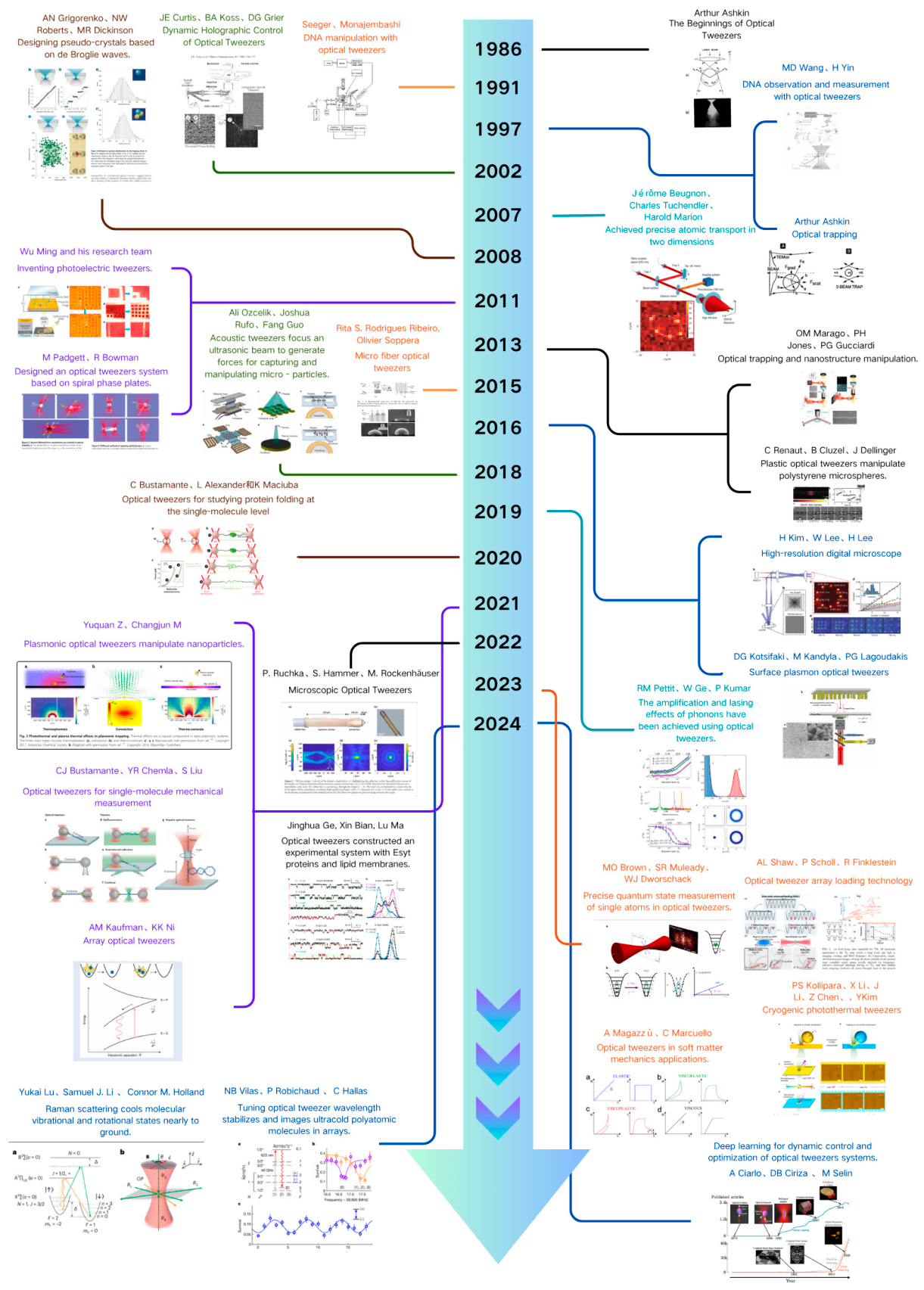
Figure 2.
Multiphysical force landscape in optical tweezers. This schematic summarizes the diverse forces acting on particles in optical traps. In addition to the dominant optical gradient and scattering forces that enable stable confinement, trapped objects are simultaneously influenced by thermal fluctuations (Brownian forces), photophoretic and photothermal forces generated by local heating, and radiation pressure arising from photon momentum transfer. Hydrodynamic drag and fluid interactions contribute viscous resistance in liquid environments, while near-field and evanescent forces become significant in proximity to surfaces. At the nanoscale, Casimir interactions, electrostatic forces, and molecular binding events further modulate trapping stability. A full understanding of this multiphysical landscape is essential for precise and reliable manipulation in modern applications of optical tweezers.
Figure 2.
Multiphysical force landscape in optical tweezers. This schematic summarizes the diverse forces acting on particles in optical traps. In addition to the dominant optical gradient and scattering forces that enable stable confinement, trapped objects are simultaneously influenced by thermal fluctuations (Brownian forces), photophoretic and photothermal forces generated by local heating, and radiation pressure arising from photon momentum transfer. Hydrodynamic drag and fluid interactions contribute viscous resistance in liquid environments, while near-field and evanescent forces become significant in proximity to surfaces. At the nanoscale, Casimir interactions, electrostatic forces, and molecular binding events further modulate trapping stability. A full understanding of this multiphysical landscape is essential for precise and reliable manipulation in modern applications of optical tweezers.
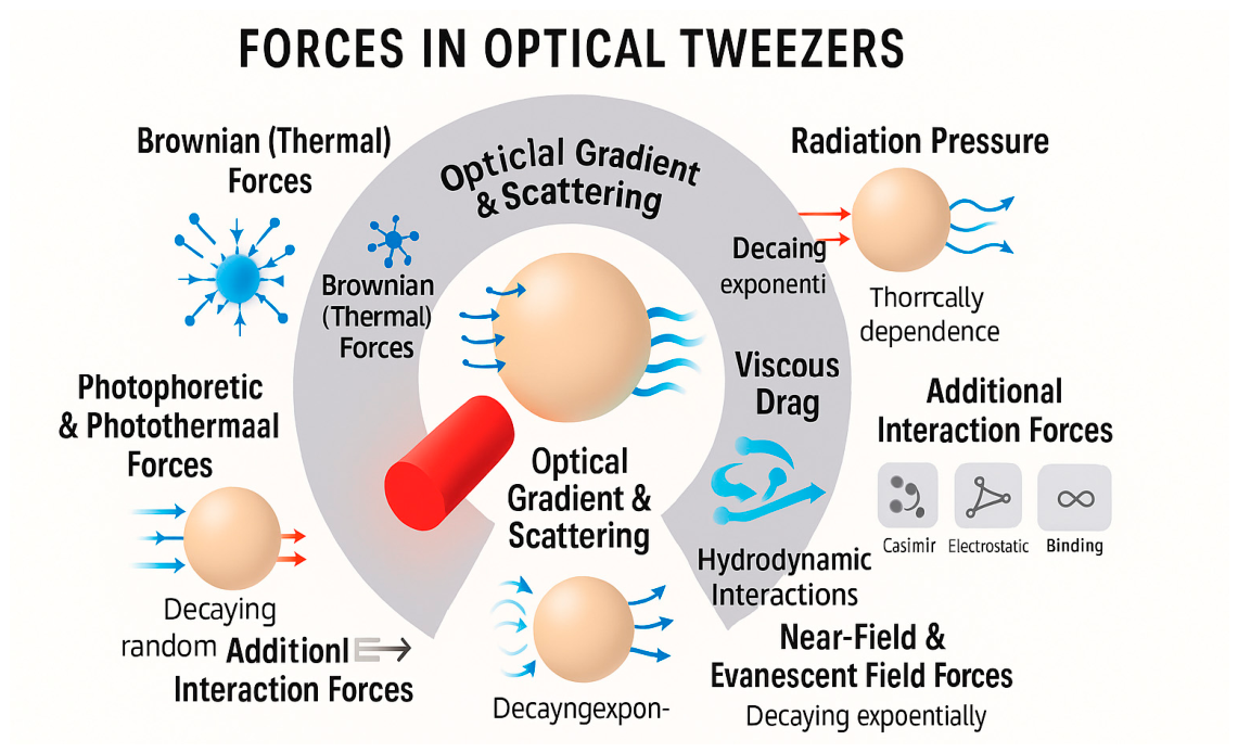
Figure 3.
Physical principles and field-engineering strategies underlying advanced optical tweezers [96,97,98,99,100,101,102]. (A) Vector-resolved force and stiffness distributions along orthogonal axes reveal anisotropic confinement of polystyrene spheres within optical traps. (B) Simulation of a phase-engineered Si₃N₄ metasurface, where discrete meta-atoms holographically shape the beam to create multi-focal optical traps. (C) A TE₀-mode-driven Si nanopillar platform illustrating programmable field generation for tunable particle confinement. (D) Electric field intensity maps of nine-site trapping lattices demonstrate reconfigurable confinement symmetry for parallel manipulation. (E) Comparative analysis of simulated and experimental axial stiffness profiles highlights the correspondence between potential gradients and displacement-tracking data. (F) Frequency-dependent microrheology of soft matter systems (glycerol, PAA, PDMS) reveals characteristic viscoelastic transitions across broad frequency ranges. (G) Canonical mechanical response models captured by OTs, spanning elastic, viscoelastic, viscoplastic, and Newtonian viscous behaviors. (H) Hybrid meta-optical system combining a metalens and meta-hologram for structured atom array confinement in free space. (I) Characterization of TiO₂ nanopillars for phase and reflectivity modulation, confirming the capability of metasurfaces to control light fields in meta-optical tweezers.
Figure 3.
Physical principles and field-engineering strategies underlying advanced optical tweezers [96,97,98,99,100,101,102]. (A) Vector-resolved force and stiffness distributions along orthogonal axes reveal anisotropic confinement of polystyrene spheres within optical traps. (B) Simulation of a phase-engineered Si₃N₄ metasurface, where discrete meta-atoms holographically shape the beam to create multi-focal optical traps. (C) A TE₀-mode-driven Si nanopillar platform illustrating programmable field generation for tunable particle confinement. (D) Electric field intensity maps of nine-site trapping lattices demonstrate reconfigurable confinement symmetry for parallel manipulation. (E) Comparative analysis of simulated and experimental axial stiffness profiles highlights the correspondence between potential gradients and displacement-tracking data. (F) Frequency-dependent microrheology of soft matter systems (glycerol, PAA, PDMS) reveals characteristic viscoelastic transitions across broad frequency ranges. (G) Canonical mechanical response models captured by OTs, spanning elastic, viscoelastic, viscoplastic, and Newtonian viscous behaviors. (H) Hybrid meta-optical system combining a metalens and meta-hologram for structured atom array confinement in free space. (I) Characterization of TiO₂ nanopillars for phase and reflectivity modulation, confirming the capability of metasurfaces to control light fields in meta-optical tweezers.
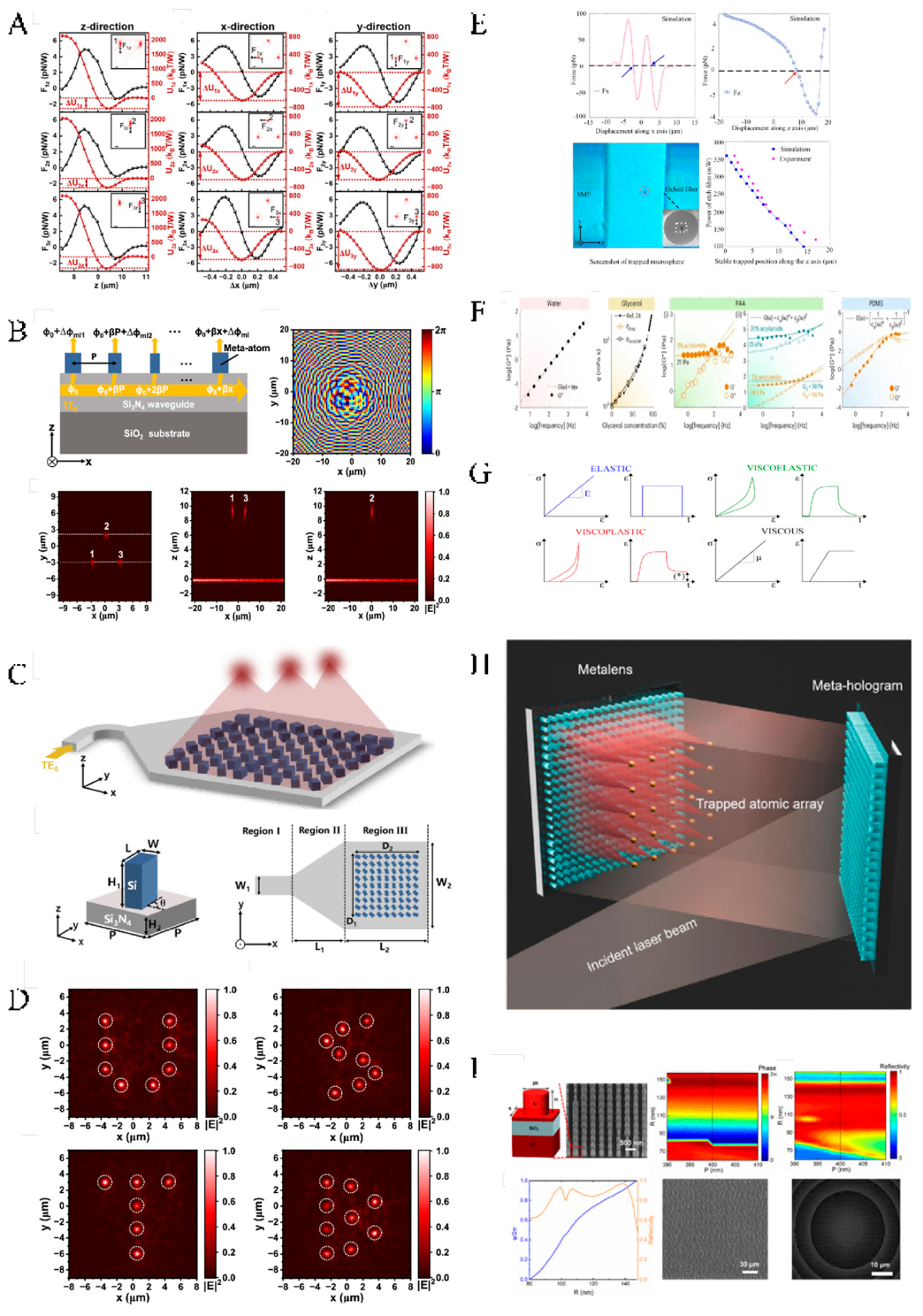
Figure 6.
Potential-well geometry and energy-distribution modeling in optical tweezers. A unified depiction integrates ray-optics derivation of the restoring paraboloid (A), vectorial field simulations with Poynting-vector streamlines and polarization dependence (B), frequency-resolved microrheology linking and to effective stiffness and dissipation (C), experimentally tracked 2D trajectories evidencing anisotropy and rotational modes in non-conservative landscapes (D), a geometric account of angular incidence and path vectors that set radial/axial components and define the trapping envelope (E), GLMT-based lateral force mapping that reproduces scattering-induced asymmetry and validates model predictions (F), and fluctuation-driven reshaping of effective potentials in complex fluids, highlighting multi-well behavior in heterogeneous media (G).
Figure 6.
Potential-well geometry and energy-distribution modeling in optical tweezers. A unified depiction integrates ray-optics derivation of the restoring paraboloid (A), vectorial field simulations with Poynting-vector streamlines and polarization dependence (B), frequency-resolved microrheology linking and to effective stiffness and dissipation (C), experimentally tracked 2D trajectories evidencing anisotropy and rotational modes in non-conservative landscapes (D), a geometric account of angular incidence and path vectors that set radial/axial components and define the trapping envelope (E), GLMT-based lateral force mapping that reproduces scattering-induced asymmetry and validates model predictions (F), and fluctuation-driven reshaping of effective potentials in complex fluids, highlighting multi-well behavior in heterogeneous media (G).

Figure 7.
Trap-depth evolution and stability control in photothermal/thermophoretic tweezer systems under varying wavelength, power, focusing, and electrolyte conditions. (A) Laser-Induced Thermophoretic Writing of Biomolecular Patterns. Top: DNA patterns visualized before and after localized heating (~2 K) demonstrate the ability of optothermal gradients to spatially encode molecular trajectories. Bottom: A schematic of temperature-gradient-induced particle transport (thermophoresis), highlighting how laser focusing generates asymmetric heating regions that drive migration away from the focal point. (B) Size and Debye Length Dependence of the Soret Coefficient (St). Experimental data reveal that the thermophoretic mobility of colloidal particles is highly dependent on particle size () and solution ionic strength (Debye length ), affecting trapping stiffness and trap depth. Larger particles or increased electrostatic screening yield higher St values, enabling more robust trapping at lower laser powers. (C) Multimodal Optical Control of Trap Geometry and Depth. A combination of photothermal, electrokinetic, and electrostatic mechanisms allows dynamic control of trap geometry. Top left: Schematic of field superposition across the axial axis. Center and bottom: Multi-channel trapping configurations enabled by digital micromirror devices (DMD) or spatial light modulators (SLM) produce reconfigurable trap arrays with tailored stiffness profiles. (D) Bubble-Enhanced Optical Trapping via Thermal Convection. Laser-induced microbubbles on gold films create strong Marangoni flows that direct particles toward a thermal ring vortex, stabilizing deep 3D traps. Focal modulation with or without bubble formation demonstrates controllable enhancement of particle aggregation and localized capture. (E) Depth-Resolved Thermophoretic Capture Dynamics. Top: DNA condensation and fluorescence intensities increase with trapping duration. Center: Real-time imaging of particle migration and radial DNA condensation under continuous IR laser exposure (. Bottom: Trap efficiency and depth are modulated by varying ionic conditions (e.g., NaCl, MgCl₂), which affect Debye length and thermophoretic response. (F) Optically Controlled Trap Arrays with Programmable Depth Profiles. Using holographic projection and photonic lattices, a multi-trap platform enables programmable control of trap stiffness and depth. Spatial modulation of thermal fields generates distinct potential landscapes for particle sorting and dynamic reconfiguration. Fluorescence histograms quantify spatial variation in trapping density and depth resolution.
Figure 7.
Trap-depth evolution and stability control in photothermal/thermophoretic tweezer systems under varying wavelength, power, focusing, and electrolyte conditions. (A) Laser-Induced Thermophoretic Writing of Biomolecular Patterns. Top: DNA patterns visualized before and after localized heating (~2 K) demonstrate the ability of optothermal gradients to spatially encode molecular trajectories. Bottom: A schematic of temperature-gradient-induced particle transport (thermophoresis), highlighting how laser focusing generates asymmetric heating regions that drive migration away from the focal point. (B) Size and Debye Length Dependence of the Soret Coefficient (St). Experimental data reveal that the thermophoretic mobility of colloidal particles is highly dependent on particle size () and solution ionic strength (Debye length ), affecting trapping stiffness and trap depth. Larger particles or increased electrostatic screening yield higher St values, enabling more robust trapping at lower laser powers. (C) Multimodal Optical Control of Trap Geometry and Depth. A combination of photothermal, electrokinetic, and electrostatic mechanisms allows dynamic control of trap geometry. Top left: Schematic of field superposition across the axial axis. Center and bottom: Multi-channel trapping configurations enabled by digital micromirror devices (DMD) or spatial light modulators (SLM) produce reconfigurable trap arrays with tailored stiffness profiles. (D) Bubble-Enhanced Optical Trapping via Thermal Convection. Laser-induced microbubbles on gold films create strong Marangoni flows that direct particles toward a thermal ring vortex, stabilizing deep 3D traps. Focal modulation with or without bubble formation demonstrates controllable enhancement of particle aggregation and localized capture. (E) Depth-Resolved Thermophoretic Capture Dynamics. Top: DNA condensation and fluorescence intensities increase with trapping duration. Center: Real-time imaging of particle migration and radial DNA condensation under continuous IR laser exposure (. Bottom: Trap efficiency and depth are modulated by varying ionic conditions (e.g., NaCl, MgCl₂), which affect Debye length and thermophoretic response. (F) Optically Controlled Trap Arrays with Programmable Depth Profiles. Using holographic projection and photonic lattices, a multi-trap platform enables programmable control of trap stiffness and depth. Spatial modulation of thermal fields generates distinct potential landscapes for particle sorting and dynamic reconfiguration. Fluorescence histograms quantify spatial variation in trapping density and depth resolution.

Figure 8.
Thermophoretic trapping and multifunctional manipulation enabled by photothermal optical tweezers. (A) Basic concept of optothermal trapping. A laser beam is focused on an absorptive substrate (e.g., gold or carbon film), producing localized temperature gradients. Thermophoretic forces drive particles toward cooler regions. Experimental and theoretical temperature distributions confirm off-focus trapping behavior. (B) Thermoconvection and concentration gradients in optothermal microfluidics. Temperature and solute profiles across the radial direction generate secondary flows and depletion effects. Microscope images show selective trapping or repulsion of DNA or nanoparticles. (C) System-level implementation: laser scanning over an absorber layer creates addressable thermal traps. Integration with micromirrors and beam modulators enables multiplexed temperature field control. (D) Spatially programmable thermophoretic patterning. Photothermal arrays or pixelated substrates generate dynamic trap geometries for single-cell arrangement and colloidal pattern assembly. (E) Coupling with SERS readout: CRONT (CRISPR–Optothermal Nanotweezers) platform combines optical trapping and Raman sensing. Local field enhancement is enabled via metallic nanostructures, allowing manipulation and biomolecular detection with sub-micron resolution. (F) Multiphysics modeling of thermophoresis. Simulations illustrate temperature gradients and particle trajectories under different geometries, including spheres, rods, and anisotropic substrates. Critical temperature thresholds for efficient trapping are shown. (G) Experimental measurement of thermophoretic forces. Laser-induced temperature maps and corresponding force landscapes are recorded using calibrated probes. Fluorescence tracking confirms motion directionality in designed fields. (H) Advanced applications of optothermal tweezers. Single-molecule localization, force–displacement characterization, and time-resolved trapping dynamics are demonstrated in programmable lab-on-chip environments. Nanoparticles, vesicles, and bacteria are controllably manipulated in 2D/3D geometries.
Figure 8.
Thermophoretic trapping and multifunctional manipulation enabled by photothermal optical tweezers. (A) Basic concept of optothermal trapping. A laser beam is focused on an absorptive substrate (e.g., gold or carbon film), producing localized temperature gradients. Thermophoretic forces drive particles toward cooler regions. Experimental and theoretical temperature distributions confirm off-focus trapping behavior. (B) Thermoconvection and concentration gradients in optothermal microfluidics. Temperature and solute profiles across the radial direction generate secondary flows and depletion effects. Microscope images show selective trapping or repulsion of DNA or nanoparticles. (C) System-level implementation: laser scanning over an absorber layer creates addressable thermal traps. Integration with micromirrors and beam modulators enables multiplexed temperature field control. (D) Spatially programmable thermophoretic patterning. Photothermal arrays or pixelated substrates generate dynamic trap geometries for single-cell arrangement and colloidal pattern assembly. (E) Coupling with SERS readout: CRONT (CRISPR–Optothermal Nanotweezers) platform combines optical trapping and Raman sensing. Local field enhancement is enabled via metallic nanostructures, allowing manipulation and biomolecular detection with sub-micron resolution. (F) Multiphysics modeling of thermophoresis. Simulations illustrate temperature gradients and particle trajectories under different geometries, including spheres, rods, and anisotropic substrates. Critical temperature thresholds for efficient trapping are shown. (G) Experimental measurement of thermophoretic forces. Laser-induced temperature maps and corresponding force landscapes are recorded using calibrated probes. Fluorescence tracking confirms motion directionality in designed fields. (H) Advanced applications of optothermal tweezers. Single-molecule localization, force–displacement characterization, and time-resolved trapping dynamics are demonstrated in programmable lab-on-chip environments. Nanoparticles, vesicles, and bacteria are controllably manipulated in 2D/3D geometries.
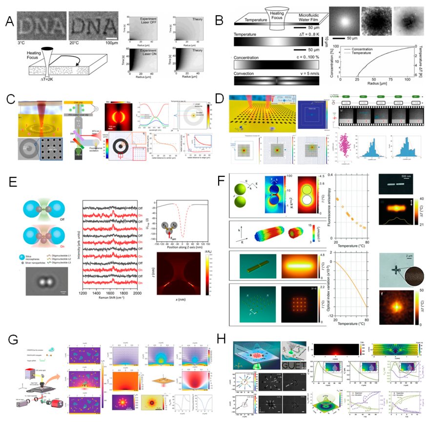
Figure 9.
Force characterization and manipulation principles in acoustic tweezer platforms. (A) Feedback-controlled force-clamp modes illustrate constant-position and constant-force operation: in the first, the acoustic drive is modulated to hold position while monitoring mechanical response; in the second, a fixed load is applied while probe displacement is recorded. Both enable nanoscale assays of biomolecular elasticity and viscoelasticity. (B) Acoustic field mapping shows streamlines converging to a stagnation point—the most stable trapping site where opposing radiation-pressure contributions balance; field symmetry dictates lateral stability and sensitivity. (C) Single-molecule DNA stretching under pico-Newton acoustic stabilization compares dsDNA force–extension data with FJC/WLC/Hookean models and, under varying ionic conditions, reveals ss/dsDNA overstretching, melting, and unzipping transitions; acoustic confinement provides a contactless, stable loading geometry akin to optical force spectroscopy. (D) Dynamic trap reconfiguration via multichannel acoustic flows demonstrates real-time programming of potential-well geometry: a quadrupole transducer forms stagnation-point traps with tunable depth through amplitude modulation, while switching inlet/outlet configurations enables on-demand particle repositioning, merging, and sorting through programmable topologies.
Figure 9.
Force characterization and manipulation principles in acoustic tweezer platforms. (A) Feedback-controlled force-clamp modes illustrate constant-position and constant-force operation: in the first, the acoustic drive is modulated to hold position while monitoring mechanical response; in the second, a fixed load is applied while probe displacement is recorded. Both enable nanoscale assays of biomolecular elasticity and viscoelasticity. (B) Acoustic field mapping shows streamlines converging to a stagnation point—the most stable trapping site where opposing radiation-pressure contributions balance; field symmetry dictates lateral stability and sensitivity. (C) Single-molecule DNA stretching under pico-Newton acoustic stabilization compares dsDNA force–extension data with FJC/WLC/Hookean models and, under varying ionic conditions, reveals ss/dsDNA overstretching, melting, and unzipping transitions; acoustic confinement provides a contactless, stable loading geometry akin to optical force spectroscopy. (D) Dynamic trap reconfiguration via multichannel acoustic flows demonstrates real-time programming of potential-well geometry: a quadrupole transducer forms stagnation-point traps with tunable depth through amplitude modulation, while switching inlet/outlet configurations enables on-demand particle repositioning, merging, and sorting through programmable topologies.

Figure 10.
Experimental strategies for trap-stiffness calibration and force quantification in optical tweezers. ( A ) Mean-square displacement (MSD), autocorrelation, and position power spectral density (PSD) analyses—derived from the overdamped Langevin framework—are used to extract stiffness , viscous drag, and thermal noise levels; contrasting spectra and autocorrelations delineate overdamped versus (hypothetical) underdamped behavior through the response time enabling real-time calibration from sub-pN up to tens of pN. ( B ) MSD trajectories acquired under varying , bead radii (0.22–1.5 µm), and solvents (acetone, water, glycerol) quantify spring constants, corner frequencies, time constants, and potential gradients, thereby comparing hydrodynamic loading with optical response across media and sizes. ( C ) Frequency-resolved PSDs resolve axial and radial modes; imaging of fringe evolution versus axial displacement links field asymmetries and radiation-pressure gradients to mode anisotropy, while Lorentzian fits report radial stiffness and quality factor as functions of radial-to-axial asymmetry. ( D ) SLM-based holographic tweezers generate multifocal traps with programmable geometry; by measuring displacement versus laser power, single-trap stiffnesses are calibrated and trap arrays with tailored stiffness landscapes are realized. ( E ) Back-focal-plane (BFP) interferometry with quadrant photodiodes provides high-bandwidth, phase-sensitive readout of lateral and axial displacements; calibration curves in the Fourier plane define sub-nanometer spatial resolution and piconewton-level force sensitivity, completing a metrology suite that links hydrodynamic drag, thermal fluctuations, and optical restoring forces.
Figure 10.
Experimental strategies for trap-stiffness calibration and force quantification in optical tweezers. ( A ) Mean-square displacement (MSD), autocorrelation, and position power spectral density (PSD) analyses—derived from the overdamped Langevin framework—are used to extract stiffness , viscous drag, and thermal noise levels; contrasting spectra and autocorrelations delineate overdamped versus (hypothetical) underdamped behavior through the response time enabling real-time calibration from sub-pN up to tens of pN. ( B ) MSD trajectories acquired under varying , bead radii (0.22–1.5 µm), and solvents (acetone, water, glycerol) quantify spring constants, corner frequencies, time constants, and potential gradients, thereby comparing hydrodynamic loading with optical response across media and sizes. ( C ) Frequency-resolved PSDs resolve axial and radial modes; imaging of fringe evolution versus axial displacement links field asymmetries and radiation-pressure gradients to mode anisotropy, while Lorentzian fits report radial stiffness and quality factor as functions of radial-to-axial asymmetry. ( D ) SLM-based holographic tweezers generate multifocal traps with programmable geometry; by measuring displacement versus laser power, single-trap stiffnesses are calibrated and trap arrays with tailored stiffness landscapes are realized. ( E ) Back-focal-plane (BFP) interferometry with quadrant photodiodes provides high-bandwidth, phase-sensitive readout of lateral and axial displacements; calibration curves in the Fourier plane define sub-nanometer spatial resolution and piconewton-level force sensitivity, completing a metrology suite that links hydrodynamic drag, thermal fluctuations, and optical restoring forces.

Figure 14.
Structural classification and representative architectures of optical tweezer systems [122,139,285,286,287,288,289,290,291,292,293,294,295,296,297,298,299,300,301,302,303,304,305,306,307,308,309,310,311,312,313,314,315,316,317,318,319]. This circular infographic organizes modern optical tweezers by structural implementation. At the center lies the unifying concept of light–matter force transduction, which expands into five architectural families. (i) Free-space optics includes classic single-beam tweezers, where high-NA objectives generate steep intensity gradients for micron-scale confinement, and multi-beam interference traps, whose periodic potentials (e.g., optical lattices) enable arrayed manipulation. (ii) Holographic and digital modulation employs spatial light modulators (SLMs) and digital micromirror devices (DMDs) to synthesize reconfigurable, multiplexed landscapes with high spatial precision and (for DMDs) elevated update rates suitable for high-throughput work. (iii) On-chip optical tweezers comprise waveguide traps and metasurface-structured fields; guided modes and nanoengineered phase elements create sub-wavelength confinement on photonic substrates, facilitating compact, scalable arrays and co-integration with microfluidics and quantum/atomic platforms. (iv) Fiber-based tweezers include dual-fiber opposed traps, which form stable counter-propagating confinements, and patterned/nanostructured fiber tips that miniaturize trapping for in situ deployment (e.g., inside channels or tissues). (v) Multi-physics and AI-enhanced platforms integrate optofluidic–photonic systems for sorting and analysis with machine-learning feedback (SmartTrap-like) controllers that adaptively optimize parameters in real time. Representative schematics and experimental images in each sector illustrate how structural choices tailor field gradients, trap anisotropy, and multiplexing capacity across the current landscape of optical tweezer technologies.
Figure 14.
Structural classification and representative architectures of optical tweezer systems [122,139,285,286,287,288,289,290,291,292,293,294,295,296,297,298,299,300,301,302,303,304,305,306,307,308,309,310,311,312,313,314,315,316,317,318,319]. This circular infographic organizes modern optical tweezers by structural implementation. At the center lies the unifying concept of light–matter force transduction, which expands into five architectural families. (i) Free-space optics includes classic single-beam tweezers, where high-NA objectives generate steep intensity gradients for micron-scale confinement, and multi-beam interference traps, whose periodic potentials (e.g., optical lattices) enable arrayed manipulation. (ii) Holographic and digital modulation employs spatial light modulators (SLMs) and digital micromirror devices (DMDs) to synthesize reconfigurable, multiplexed landscapes with high spatial precision and (for DMDs) elevated update rates suitable for high-throughput work. (iii) On-chip optical tweezers comprise waveguide traps and metasurface-structured fields; guided modes and nanoengineered phase elements create sub-wavelength confinement on photonic substrates, facilitating compact, scalable arrays and co-integration with microfluidics and quantum/atomic platforms. (iv) Fiber-based tweezers include dual-fiber opposed traps, which form stable counter-propagating confinements, and patterned/nanostructured fiber tips that miniaturize trapping for in situ deployment (e.g., inside channels or tissues). (v) Multi-physics and AI-enhanced platforms integrate optofluidic–photonic systems for sorting and analysis with machine-learning feedback (SmartTrap-like) controllers that adaptively optimize parameters in real time. Representative schematics and experimental images in each sector illustrate how structural choices tailor field gradients, trap anisotropy, and multiplexing capacity across the current landscape of optical tweezer technologies.

Figure 15.
Applications of Optical Tweezers across Biological and Physical/Material Sciences [78,291,305,385,386,387,388,389,390,391,392,393,394,395,396,397,398,399,400,401,402]. This infographic illustrates the breadth of optical tweezer applications by dividing representative examples into two major domains: biological sciences and physical/material sciences. On the biological side, optical tweezers have become indispensable for probing molecular and cellular mechanics, including A DNA/RNA/Protein stretching, where precision force spectroscopy reveals entropic elasticity, unzipping transitions, and protein unfolding dynamics [403]; single-cell mechanics, enabling quantitative analysis of calcium influx, immune activation, and other mechanotransduction processes at single-cell resolution [404]; plasmonic SERS tweezers, where enhanced Raman scattering provides label-free biochemical fingerprinting of pathogenic molecules [405]; CRISPR–optothermal detection, in which thermal-gradient tweezers concentrate CRISPR complexes for ultra-sensitive nucleic acid assays [406]; microfluidic sorting and infrared spectroscopy, integrating tweezers with photothermal IR modules for label-free sorting and classification of cells [407]; and lab-on-fiber systems, where dual-fiber tweezers combined with embedded sensors enable force mapping in confined or in vivo environments [408]. On the physical and material sciences side, optical tweezers extend beyond biology to enable B quantum simulation, where atom arrays assembled by tweezers serve as programmable platforms for many-body physics [409]; nanoparticle assembly, guiding the formation of ordered nanostructures and SERS-active substrates [168]; hydrogel mechanics, quantifying viscoelastic and poroelastic parameters of soft microgels under controlled deformation [410]; microcapsule micromechanics, probing interfacial and internal stresses in synthetic or biological capsules [411]; surface adsorption kinetics, where real-time tracking of nanoparticles interacting with surfaces reveals molecular binding dynamics [412]; and controlled drug release in microfluidics, providing spatiotemporal mapping of release behavior from stimuli-responsive carriers [97,413,414,415].
Figure 15.
Applications of Optical Tweezers across Biological and Physical/Material Sciences [78,291,305,385,386,387,388,389,390,391,392,393,394,395,396,397,398,399,400,401,402]. This infographic illustrates the breadth of optical tweezer applications by dividing representative examples into two major domains: biological sciences and physical/material sciences. On the biological side, optical tweezers have become indispensable for probing molecular and cellular mechanics, including A DNA/RNA/Protein stretching, where precision force spectroscopy reveals entropic elasticity, unzipping transitions, and protein unfolding dynamics [403]; single-cell mechanics, enabling quantitative analysis of calcium influx, immune activation, and other mechanotransduction processes at single-cell resolution [404]; plasmonic SERS tweezers, where enhanced Raman scattering provides label-free biochemical fingerprinting of pathogenic molecules [405]; CRISPR–optothermal detection, in which thermal-gradient tweezers concentrate CRISPR complexes for ultra-sensitive nucleic acid assays [406]; microfluidic sorting and infrared spectroscopy, integrating tweezers with photothermal IR modules for label-free sorting and classification of cells [407]; and lab-on-fiber systems, where dual-fiber tweezers combined with embedded sensors enable force mapping in confined or in vivo environments [408]. On the physical and material sciences side, optical tweezers extend beyond biology to enable B quantum simulation, where atom arrays assembled by tweezers serve as programmable platforms for many-body physics [409]; nanoparticle assembly, guiding the formation of ordered nanostructures and SERS-active substrates [168]; hydrogel mechanics, quantifying viscoelastic and poroelastic parameters of soft microgels under controlled deformation [410]; microcapsule micromechanics, probing interfacial and internal stresses in synthetic or biological capsules [411]; surface adsorption kinetics, where real-time tracking of nanoparticles interacting with surfaces reveals molecular binding dynamics [412]; and controlled drug release in microfluidics, providing spatiotemporal mapping of release behavior from stimuli-responsive carriers [97,413,414,415].
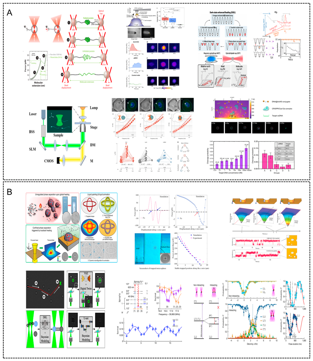
Figure 16.
Schematic principles and advanced applications of single-beam optical tweezers [26,423,424,425,426,427]. (A) Basic optical layout of a single-beam gradient trap system. A tightly focused Gaussian laser beam generates a three-dimensional trapping potential by balancing gradient forces (F₍grad₎) and scattering forces (F₍scat₎). The inset shows the corresponding optical setup involving beam expanders, steering mirrors, dichroic mirrors (DM), and position detection modules. (B) Experimental observation and simulation of particle trapping within the focal volume, illustrating optical stiffness measurement and potential well formation. (C) Ray-optics model of trapping forces: refracted and reflected rays create a net restoring force toward the beam center. Schematic comparisons of optical tweezers with tethered biomolecules and other geometries (e.g., confocal, evanescent-field traps) are shown. (D) Single-molecule manipulation using dual-beam tweezers: stretching of DNA, unfolding of proteins, and unzipping of nucleic acid structures are illustrated. (E) Optical force calibration and trapping stiffness analysis in low-power trapping regimes, highlighting the dependence of trap stability on laser power, beam waist, and particle size. (F) An integrated optical tweezers system coupled with high-resolution detection and microfluidics for biological assays. Fluorescence imaging, flow control, and force spectroscopy modules are embedded into a compact trapping platform.
Figure 16.
Schematic principles and advanced applications of single-beam optical tweezers [26,423,424,425,426,427]. (A) Basic optical layout of a single-beam gradient trap system. A tightly focused Gaussian laser beam generates a three-dimensional trapping potential by balancing gradient forces (F₍grad₎) and scattering forces (F₍scat₎). The inset shows the corresponding optical setup involving beam expanders, steering mirrors, dichroic mirrors (DM), and position detection modules. (B) Experimental observation and simulation of particle trapping within the focal volume, illustrating optical stiffness measurement and potential well formation. (C) Ray-optics model of trapping forces: refracted and reflected rays create a net restoring force toward the beam center. Schematic comparisons of optical tweezers with tethered biomolecules and other geometries (e.g., confocal, evanescent-field traps) are shown. (D) Single-molecule manipulation using dual-beam tweezers: stretching of DNA, unfolding of proteins, and unzipping of nucleic acid structures are illustrated. (E) Optical force calibration and trapping stiffness analysis in low-power trapping regimes, highlighting the dependence of trap stability on laser power, beam waist, and particle size. (F) An integrated optical tweezers system coupled with high-resolution detection and microfluidics for biological assays. Fluorescence imaging, flow control, and force spectroscopy modules are embedded into a compact trapping platform.

Disclaimer/Publisher’s Note: The statements, opinions and data contained in all publications are solely those of the individual author(s) and contributor(s) and not of MDPI and/or the editor(s). MDPI and/or the editor(s) disclaim responsibility for any injury to people or property resulting from any ideas, methods, instructions or products referred to in the content. |
© 2026 by the authors. Licensee MDPI, Basel, Switzerland. This article is an open access article distributed under the terms and conditions of the Creative Commons Attribution (CC BY) license (http://creativecommons.org/licenses/by/4.0/).
Copyright: This open access article is published under a Creative Commons CC BY 4.0 license, which permit the free download, distribution, and reuse, provided that the author and preprint are cited in any reuse.