
Submitted:
27 July 2026
Posted:
28 July 2026
You are already at the latest version
Abstract
Neurodegenerative diseases are a growing global health burden associated with aging and characterized by progressive neuronal dysfunction, metabolic failure, mitochondrial impairment, oxidative stress, and chronic neuroinflammation. Among the metabolic pathways implicated in these disorders, coenzyme A (CoA)-linked biology has emerged as a potentially important but still underexplored contributor to neuronal resilience and vulnerability. Pantethine, a disulfide derivative of pantetheine and a CoA-related metabolic precursor, has attracted attention because of its reported effects on cellular metabolism, redox balance, and inflammatory signaling. However, its relevance across neurodegenerative diseases remains unevenly defined, with direct support strongest in pantothenate kinase-associated neurodegeneration (PKAN) and more limited evidence in common disorders such as Alzheimer’s disease (AD) and Parkinson’s disease (PD). This narrative review critically examines the mechanistic and translational evidence linking pantethine to neurodegeneration. PKAN represents the most logical disease context for pantethine investigation because impaired CoA biosynthesis is proximal to disease pathogenesis, although pantethine remains investigational and its clinical efficacy has not been established. By contrast, proposed applications in AD and PD remain highly theoretical and hypothesis-generating. Nevertheless, research on pantethine and related CoA-restoring strategies may identify new intervention targets across neurodegenerative diseases and other disorders characterized by impaired cellular bioenergetics, including selected neuropsychiatric disorders. These possibilities require biomarker-informed, disease-specific studies that establish active-species exposure, target engagement, and clinically meaningful effects.
Keywords:
pantethine
; coenzyme A
; kynurenine
; neurodegeneration
; mitochondrial dysfunction
; neuroinflammation
; oxidative stress
; glial cells
; pantothenate kinase-associated neurodegeneration (PKAN)
; Alzheimer’s disease
; Parkinson’s disease
1. Introduction
Aging is a progressive and multifactorial biological process marked by the gradual accumulation of molecular and cellular alterations, including genomic instability, telomere attrition, epigenetic remodeling, mitochondrial dysfunction, impaired proteostasis, and stem cell exhaustion [1,2]. These hallmarks do not act in isolation. Rather, they converge to increase vulnerability to chronic non-communicable diseases, particularly those affecting metabolic and neurological systems [3]. Demographic projections further intensify this concern, with the proportion of the global population aged 65 years or older expected to rise substantially by 2050 [4]. As longevity increases, preserving quality of life becomes a major biomedical and public health priority. This issue is especially pressing in disorders that progressively erode cognition, movement, autonomy, and social functioning [5].
Neurodegenerative diseases are therefore emerging as one of the defining medical challenges of aging societies [6]. They comprise a heterogeneous group of chronic and progressive conditions characterized by selective neuronal vulnerability, synaptic dysfunction, and gradual loss of neuronal structure and function [7]. Among them, Parkinson’s disease (PD) and Alzheimer’s disease (AD) are the most prevalent in older adults [8]. PD is primarily associated with degeneration of dopaminergic neurons in the substantia nigra and with the accumulation of alpha-synuclein (α-synuclein)-containing Lewy bodies [9,10]. AD, the leading cause of dementia, is characterized by progressive cognitive decline accompanied by amyloid-beta (amyloid-β) deposition, tau pathology, glial dysregulation, and broad metabolic disturbances [12]. Although these disorders differ in clinical presentation and proteinopathic signatures, they share several mechanistic features, including impaired bioenergetics, mitochondrial dysfunction, oxidative stress, chronic inflammatory signaling, and altered cellular metabolism [11,12,13,14]. This overlap has encouraged growing interest in upstream biological processes that may influence neuronal resilience across disease contexts [9,15,16].
Among these upstream processes, coenzyme A (CoA) biology deserves particular attention. CoA is indispensable for central carbon metabolism, fatty acid oxidation and synthesis, acetylation reactions, neurotransmitter-related metabolism, and mitochondrial energy production [17]. In neurons, which have high energetic demands and limited tolerance for metabolic instability, disturbances in CoA-dependent pathways can have wide-ranging consequences [18]. Reduced CoA availability may impair acetyl-CoA generation, compromise tricarboxylic acid (TCA) cycle activity, alter lipid handling, disturb membrane homeostasis, and influence redox balance [19]. In turn, these changes may contribute to synaptic dysfunction, neurotransmitter imbalance, and progressive neuronal injury [20]. This metabolic perspective is particularly compelling because it links bioenergetic failure to inflammatory stress and mitochondrial decline rather than treating them as isolated pathological events [21]. For this reason, CoA-linked metabolism is increasingly being considered not simply as background biochemistry, but as a potentially important layer in the pathogenesis of neurodegeneration [22,23,24,25].
Historically, the concept of CoA-centered metabolism emerged through complementary discoveries that progressively clarified the biochemical basis of cellular energy production. Albert Szent-Györgyi’s studies of catalytic dicarboxylic acid oxidation helped establish the foundations for Hans Adolf Krebs’s formulation of the citric acid cycle. Fritz Albert Lipmann’s subsequent discovery of CoA and acetyl-CoA identified the activated acetyl carrier that supplies two-carbon units for citrate formation, thereby providing a crucial biochemical link to the Krebs cycle. In recognition of these interconnected advances, Lipmann received one half of the 1953 Nobel Prize in Physiology or Medicine for the discovery of CoA and its role in intermediary metabolism, while Krebs received the other half for the discovery of the citric acid cycle [26,27]. Pantethine is biochemically linked to this framework because pantetheine-related intermediates participate in CoA biosynthesis and acetyl-CoA generation, thereby connecting CoA availability with TCA cycle flux, mitochondrial electron transport, ATP synthesis, and glutathione redox cycling, as summarized in Figure 1 [22,28].
Interest in CoA biology is especially strong in pantothenate kinase (PANK)-associated neurodegeneration (PKAN), a rare inherited disorder caused by mutations in PANK2, a key enzyme in CoA biosynthesis [29]. PKAN provides one of the clearest examples of how disruption of CoA homeostasis can be linked to neurodegenerative pathology, including mitochondrial abnormalities, iron dyshomeostasis, and profound metabolic dysfunction [30,31]. Because PKAN is mechanistically tied to the CoA biosynthetic pathway itself, it offers the most direct disease framework in which CoA-related interventions can be examined [32]. At the same time, broader age-related neurodegenerative disorders such as AD and PD are not defined by primary defects in CoA synthesis [33]. This distinction limits direct extrapolation from PKAN to AD and PD. It means that evidence arising from PKAN may be highly informative mechanistically yet cannot be transferred automatically to more common neurodegenerative diseases without careful qualification [30,34,35].
In this context, pantethine has attracted interest as a biologically relevant metabolic compound [23]. Pantethine is a derivative of pantothenic acid, or vitamin B5, and is closely linked to the biosynthesis of CoA [36]. Rather than presenting pantethine as an established neuroprotective therapy, it is more accurate to view it as a precursor-related metabolic compound with pleiotropic biochemical consequences [23]. Through its relationship to CoA metabolism, pantethine has been associated with pathways relevant to acetyl-CoA production, fatty acid metabolism, membrane lipid turnover, and redox regulation [18,19,20]. These properties make it an intriguing candidate for investigation in disorders marked by metabolic instability [37]. Its appeal lies less in a single mechanism than in the possibility that it may influence several interconnected processes that are repeatedly implicated in neurodegeneration [22,23,38].
Experimental evidence indicates that pantethine can influence mitochondrial, redox, and inflammatory pathways, but the strength of support varies substantially by disease context [21,23,32]. Mechanistic relevance is strongest in PKAN and related inherited CoA-biosynthesis disorders, whereas evidence in AD and PD remains predominantly preclinical and hypothesis-generating [39,40,41]. Accordingly, this review distinguishes disease-proximal findings from broader mechanistic inference and evaluates translational claims according to model relevance, pharmacology, and the availability of human evidence [42,43,44].
Accordingly, the aim of the present review is to critically synthesize the mechanistic and translational evidence on pantethine in neurodegeneration, with emphasis on CoA-linked metabolism, mitochondrial function, oxidative stress, inflammatory pathways, and neuronal vulnerability [19,25]. The review explicitly places PKAN and related inherited CoA-biosynthesis disorders at the highest level of the evidence hierarchy, with PKAN representing the most logical candidate indication for further investigation. In contrast, potential applications in AD and PD remain highly theoretical and are considered primarily as hypothesis-generating extensions of shared mitochondrial, redox, inflammatory, and metabolic abnormalities. Beyond evaluating current therapeutic evidence, this review advances the hypothesis that research on pantethine and related CoA-restoring strategies may reveal intervention targets relevant to neurodegenerative diseases and other disorders characterized by impaired cellular bioenergetics, including selected neuropsychiatric disorders. This broader perspective is intended to guide mechanistic discovery and biomarker-informed research rather than imply equivalent disease relevance or established clinical efficacy.
2. From Mechanism to Translation: An Evidence-Based Appraisal
The following sections evaluate pantethine through an evidence-graded framework, moving from pharmacology and CoA-related metabolism to disease-specific mechanisms and translational boundaries. Because the strength of evidence differs substantially across PKAN, AD, PD, and non-neural models, Table 1 summarizes each mechanistic domain according to disease context, CNS relevance, evidence strength, and key interpretive limitations.
2.1. Pantethine Pharmacology and CoA-Related Metabolism
2.1.1. Pantethine as a CoA-Related Metabolic Compound
Pantethine occupies a strategically important position within CoA metabolism because it is neither a remote nutritional precursor nor the final active cofactor, but a relatively proximal CoA-related compound whose handling can influence several downstream pathways [61]. Structurally, pantethine is the stable disulfide form of pantetheine, an intermediate linked to CoA biosynthesis and salvage [62]. This position matters pharmacologically. Compared with pantothenic acid, which must proceed through the full canonical pathway, pantethine lies closer to the generation of 4′-phosphopantetheine, dephospho-CoA, and ultimately CoA, making it an appealing candidate in conditions marked by impaired CoA homeostasis [22,23,24,38] (Figure 1A).
CoA is indispensable for acyl-group transfer, fatty acid oxidation and synthesis, acetyl-CoA formation, membrane lipid metabolism, and broader bioenergetic regulation [28]. These processes are tightly integrated. As a result, changes in CoA availability can affect mitochondrial respiration, lipid remodeling, and redox balance at the same time [63]. This is especially relevant in tissues with high metabolic demand, where relatively modest shifts in acetyl-CoA and acyl-CoA pools may have wide biological consequences [64].
Beyond its mechanistic relevance, pantethine has a substantial clinical history as a lipid-modifying agent. In Japan, oral pantethine is approved for selected cases of hyperlipidemia, whereas clinical studies conducted in Europe, North America, and China have reported reductions in total cholesterol, low-density lipoprotein cholesterol, non-high-density lipoprotein cholesterol, apolipoprotein B, or triglycerides. The magnitude and pattern of response vary according to dose, dyslipidemia phenotype, dietary intervention, and study design. Regulatory status also differs internationally. Pantethine is generally marketed as a dietary supplement in the United States and as a natural health product in Canada, while pantethine-specific lipid-lowering medicines have not been confirmed through centralized European, United Kingdom, or Australian registers. Lipid-profile modification therefore represents its most established clinical application, although cardiovascular-event prevention has not been demonstrated [65,66,67,68,69,70,71].
Pantethine has also been investigated for broader effects on inflammatory tone, stress adaptation, and cellular metabolism [23]. Even so, its biological significance cannot be inferred from pathway proximity or clinical lipid effects alone. Interpretation must distinguish pantethine from the metabolites generated during its processing and consider whether the relevant actions arise from CoA repletion, cysteamine formation, lipid modification, or broader systemic metabolic changes [72,73,74].
2.1.2. Pantethine, Pantetheine, and Cysteamine: Metabolic Relationships and Active-Species Uncertainty
A central challenge in interpreting pantethine biology is that pantethine, pantetheine, and cysteamine are metabolically linked but functionally distinct [68]. Pantethine is the disulfide form of pantetheine. Pantetheine, in turn, is the immediate substrate for pantetheinases of the vanin family, which hydrolyze it into pantothenate and cysteamine as part of the CoA salvage pathway [75]. This means that once pantethine is administered, the biologically relevant species may depend on where hydrolysis occurs, how rapidly it proceeds, and which compartments have access to the resulting intermediates [63]. The pathway is therefore branching rather than strictly linear.
This branching has direct pharmacologic consequences [63]. If pantethine is extensively cleaved before it reaches the tissue of interest, then observed biologic effects may reflect indirect replenishment of upstream precursors, cysteamine-mediated redox actions, altered lipid metabolism, or a combination of these mechanisms rather than direct intracellular delivery of pantethine itself [76]. Human data are particularly instructive. In cystinotic children, oral pantethine was rapidly cleaved by pantetheinase in the gut and plasma, circulating pantethine was undetectable, and the major measurable products were pantothenate and cysteamine. That result does not eliminate therapeutic relevance, but it strongly cautions against assuming that the administered molecule remains the dominant active species in vivo. Rat studies similarly showed that oral pantethine leads to substantial pantothenate recovery in blood and urine, again indicating efficient absorption coupled to partial hydrolysis during or after gastrointestinal transit [77]. Older human excretion data also support the view that pantethine is handled differently from pantothenic acid, but do not establish intact tissue exposure directly [78].
Brain-relevant evidence reinforces this uncertainty. In hippocampal slice cultures, extracellular metabolism studies showed that pantetheinase converts cystamine to cysteamine very efficiently, whereas conversion of pantethine is much more limited under the tested conditions [46]. That is a useful reminder that substrate identity matters. The rate at which cysteamine is generated in the extracellular brain milieu is not simply a generic property of vanin activity but depends on the precursor available [79]. The rate at which cysteamine is generated in the extracellular brain milieu is not simply a generic property of vanin activity but depends on the precursor available [80]. Accordingly, pantethine should not be discussed as though its downstream products arise uniformly across tissues [63].
Vanin biology adds another layer of complexity. Vanin 1 (VNN1) and related pantetheinases sit at the crossroads of CoA recycling, redox regulation, and inflammatory signaling [81]. In some systems, vanin activity supports metabolic adaptation and mitochondrial oxidative function by linking pantetheine turnover to CoA and acetyl-CoA metabolism [63]. In others, cysteamine production influences oxidative stress responses, epithelial resilience, and inflammatory behavior [82]. Neuroendocrine studies suggest that pantethine, possibly through intracellular cysteamine formation, may reduce somatostatin and prolactin levels in the cerebral cortex and hypothalamus, although the mechanism remains uncertain [80]. In rats, pantethine dose-dependently suppressed open-field activity, altered hypothalamic noradrenaline, dopamine, and 3,4-dihydroxyphenylacetic acid levels, and attenuated somatostatin-induced barrel rotation; cysteamine was more potent at equimolar doses [83]. A separate time-course study showed that these behavioral, monoaminergic, and striatal somatostatin effects were transient and largely resolved within 24 h [84]. Pharmacologic inhibition studies also show that disrupting vanin activity alters fasting lipid profiles, underscoring the systemic metabolic relevance of this pathway [85]. A central unresolved question is whether observed biological effects reflect direct CNS delivery of pantethine, activity of downstream metabolites such as pantetheine or cysteamine, peripheral immunometabolic modulation, or some combination of these mechanisms [86]. This uncertainty is a central interpretive limitation. It is one of the key mechanistic issues that should shape interpretation throughout this review [63].
2.1.3. CoA Homeostasis, Acetyl-CoA Biology, and Neurodegenerative Vulnerability
The rationale for considering pantethine in neurodegeneration rests on the centrality of CoA homeostasis [25]. CoA is not simply a housekeeping cofactor [30]. It is a determinant of metabolic flexibility, nutrient-state adaptation, acyl-group trafficking, and integration between mitochondrial and cytosolic metabolism [63]. Acetyl-CoA, one of its most important derivatives, is equally central [87]. It supports TCA flux, ATP production, lipid synthesis, and acetylation reactions that influence cellular structure and signaling. In neurons and glia, where energetic demand is high and reserve capacity is limited, disturbances in CoA and acetyl-CoA biology can propagate across several systems at once [22,88,89].
This has direct relevance to neurodegenerative vulnerability [90]. Neural cells depend on tightly regulated mitochondrial function, membrane turnover, neurotransmitter-related metabolism, and stress adaptation, all of which intersect with CoA-dependent pathways [25]. Reduced CoA availability may limit acetyl-CoA generation, constrain TCA activity, impair lipid remodeling, and weaken redox buffering capacity [87] (Figure 1A,C). The broader CoA literature links disturbed CoA homeostasis to neurodegeneration, cardiometabolic dysfunction, myopathy, and maladaptive stress states, indicating that CoA insufficiency can be pathogenic rather than merely associative [23]. This view is strengthened by evidence that CoA metabolism is dynamically regulated rather than static [19]. Phosphoinositide 3-kinase (PI3K) and protein kinase B (AKT) signaling influence de novo CoA synthesis through PANK-related control, including phosphorylation-dependent regulation of pantothenate kinase 4 (PANK4), thereby altering acetyl-CoA and acyl-CoA pools and reshaping lipid metabolism [91]. In other words, CoA biology responds to nutrient and growth signaling and can sit upstream of broad metabolic phenotypes [74,88,92].
The strongest disease-specific rationale emerges in inherited CoA-biosynthesis disorders [30]. In PKAN, mutations in PANK2 impair an early step in CoA synthesis, making CoA pathway dysfunction proximal to pathogenesis rather than secondary to downstream degeneration [93]. This is precisely why PKAN deserves the highest position in the evidence hierarchy for pantethine-related discussion [32]. It provides a biologically coherent context in which pantethine, pantothenate, or related intermediates might help improve downstream CoA availability, particularly when residual pathway activity persists [48]. In such settings, a CoA-centered intervention is not merely plausible. It is mechanistically anchored [30,39,48].
At the same time, the significance of CoA biology extends beyond rare Mendelian disease [30]. Experimental work in diverse tissues suggests that CoA availability influences mitochondrial function, lipid handling, inflammatory tone, and adaptation to metabolic stress [94]. Yet a mechanistic intersection does not establish disease modification [29]. That distinction is crucial for common neurodegenerative disorders [90]. In AD and PD, CoA-linked dysfunction may contribute to broader metabolic failure, but the evidence that pantethine corrects this in vivo, particularly within the human brain, remains incomplete. The relevant translational question is whether pantethine alters disease-relevant CoA biology in the appropriate compartment and molecular form [93]. The real question is whether pantethine can alter disease-relevant CoA biology in the right compartment, at the right time, and in a form that produces meaningful neural effects [94].
2.1.4. Blood-Brain Barrier Permeability and Unresolved CNS Target Engagement
A major translational uncertainty in pantethine research is whether orally or systemically administered pantethine reaches the central nervous system (CNS) in its intact form, or whether most observed effects instead reflect peripheral metabolism and downstream products [45]. This pharmacokinetic uncertainty directly affects interpretation of CNS efficacy [95]. This challenge reflects a broader principle in CNS drug development: mechanistic promise cannot be translated reliably unless molecular properties, formulation, administration route, transporter interactions, and brain-exposure measurements are considered together [96]. It shapes how one interprets efficacy claims, model-specific findings, and the broader leap from PKAN to common neurodegenerative diseases [97].
Available data suggest that pantethine is metabolically labile [45]. Rat studies showed that oral pantethine increases circulating and urinary pantothenate, consistent with efficient gastrointestinal absorption coupled to substantial mucosal hydrolysis [77]. Human data point in the same direction [45]. In cystinosis, circulating pantethine was undetectable after oral dosing, whereas pantothenate and cysteamine rose markedly, indicating rapid extra-CNS conversion of the administered compound. Urinary studies in humans support differential handling of pantethine relative to pantothenic acid, but do not establish intact brain exposure [77]. Together, these findings suggest that oral pantethine may have limited opportunity to cross the BBB in substantial intact form unless it escapes rapid degradation.
This uncertainty creates several possibilities [63]. Intact pantethine may reach the brain only minimally, with apparent CNS effects arising largely from peripheral metabolic changes or from downstream metabolites with different transport properties [98]. Alternatively, local salvage pathways involving pantetheine, pantothenate, or related intermediates may contribute to compartment-specific CoA repletion if suitable precursors become available [63]. Work in yeast shows that alternative precursor uptake routes can support CoA synthesis under constrained conditions [99]. Those findings cannot be mapped directly onto the mammalian brain, but they reinforce the broader principle that precursor form, transporter availability, and compartment-specific metabolism are all likely to matter.
This distinction is crucial for disease interpretation [54]. In PKAN, where CoA-pathway dysfunction is disease proximal, indirect metabolic support may still be mechanistically meaningful even if intact pantethine delivery to the brain is limited. In AD and PD, however, extrapolation is more fragile because the gap between systemic metabolic effects and direct CNS disease modification is wider [23]. For that reason, pantethine should currently be regarded as a mechanistically compelling but pharmacologically incompletely resolved candidate. Its relation to CoA metabolism is strong, and its downstream products are biologically active. Yet the degree to which it restores brain CoA pools directly, acts through pantetheine or cysteamine, or primarily exerts peripheral immunometabolic effects remains uncertain [99]. Recognizing that uncertainty does not weaken the case for studying pantethine [63]. These pharmacologic uncertainties and their broader neurobiological implications are summarized in Figure 2, whereas the evidence hierarchy is presented in Table 1.
2.2. Evidence Hierarchy Across Neurodegenerative Contexts
2.2.1. Direct Evidence from PKAN and Inherited CoA-Biosynthesis Disorders
The strongest evidence linking pantethine-related strategies to neurodegeneration arises in inherited disorders of CoA biosynthesis, particularly PKAN and, more broadly, disorders involving PANK2 or coenzyme A synthase (COASY) dysfunction [30]. In these conditions, CoA pathway impairment is not a secondary correlate of disease. It is mechanistically proximal to pathogenesis. That distinction is crucial [93]. It means that precursor-based or bypass-oriented interventions can be interpreted against a biologically coherent disease framework rather than against a diffuse metabolic background [30,34,35,100]. Extending this rationale beyond PKAN, fibroblasts and human induced pluripotent stem cell-derived astrocytes from patients with COASY protein-associated neurodegeneration exhibited iron overload, mitochondrial abnormalities, impaired vesicular trafficking, lipid peroxidation, ferroptosis-related changes, and cellular senescence, with astrocytes more closely reproducing the disease-associated phenotype than fibroblasts [101]. Further support for pantethine-responsive CoA restoration comes from phosphopantothenoylcysteine synthetase deficiency [102]. Pathogenic PPCS variants reduce cellular CoA and impair cardiomyocyte contractility, whereas pantethine partially rescues functional abnormalities in patient-derived cardiac models and has been associated with sustained clinical improvement. Because these findings primarily concern cardiomyopathy, they should be regarded as supportive evidence for correction of inherited CoA-pathway dysfunction rather than direct evidence of neuroprotection.
This logic is supported by convergent evidence across cellular, animal, and human-derived neuronal systems [99]. PANK-deficient Drosophila models show reduced CoA, mitochondrial dysfunction, oxidative stress, neurodegeneration, and shortened lifespan, all of which are improved by pantethine feeding [39]. Consistent with these findings, pank2 knockdown in zebrafish severely disrupted anterior central nervous system and vascular development, whereas supplementation with pantethine or CoA, but not vitamin B5, efficiently rescued the developmental phenotype, underscoring the dependence of neural and vascular integrity on PANK2 activity and CoA homeostasis [103]. In mammalian PKAN models, 4′-phosphopantetheine corrects abnormalities in CoA, iron handling, dopamine metabolism, and mitochondrial enzyme activity in brain tissue and patient-derived cells [104]. Human induced pluripotent stem cell (iPSC)-derived PANK2 neurons likewise show premature death, increased reactive oxygen species (ROS), mitochondrial dysfunction, and excitability defects that can be corrected by CoA supplementation. Additional work in PANK2-mutant cells suggests that pantethine-containing supplement regimens can partially normalize iron accumulation, mitochondrial markers, and oxidative phenotypes when residual PANK2 activity is present [41,48,105]. Even so, the PKAN literature also illustrates the importance of pharmacologic realism. Clinical translation has been uneven, and BBB penetration remains a decisive constraint for any CoA-pathway therapy. Still, among all disease contexts considered here, PKAN provides the most direct mechanistic rationale for pantethine-related discussion [30,39,106].
2.2.2. Intermediate Mechanistic Evidence from Inflammatory and Metabolic Models
A second tier of evidence comes from models that do not represent primary CoA-biosynthesis disorders but nevertheless show that CoA-related metabolism can shape mitochondrial function, inflammatory signaling, iron handling, or oxidative balance in ways relevant to neurodegeneration [25]. This evidence is mechanistically informative but interpretively intermediate. It supports biological plausibility without establishing disease-specific therapeutic efficacy [30].
Such models are useful because they clarify how impaired CoA homeostasis can perturb cellular metabolism beyond rare Mendelian disease [30]. Reviews of brain CoA and acetyl-CoA metabolism, for example, emphasize that disturbances in CoA-dependent pathways can affect mitochondrial enzymes, glutathione stability, inflammatory tone, and neuronal resilience under stress [90]. Experimental data from Coasy-deficient neural lineages further support a causal relationship between CoA pathway disruption and neurodevelopmental as well as neurodegenerative phenotypes, including impaired mitochondrial respiration, lipid peroxidation, iron dysregulation, and chronic neuroinflammation [30]. These findings extend the mechanistic field of relevance, but they do not, by themselves, demonstrate that pantethine can reverse equivalent pathology in common neurodegenerative disease. They are best interpreted as bridge evidence between direct PKAN biology and broader disease extrapolation [17,23,25].
2.2.3. Exploratory Relevance to Alzheimer’s Disease and Parkinson’s Disease
Relevance to AD and PD is more indirect and remains largely preclinical [90]. That does not make it unimportant. It does, however, require a different evidentiary standard [107]. In these disorders, pantethine is not being evaluated against a primary defect in CoA biosynthesis, but rather against broader metabolic, inflammatory, and mitochondrial abnormalities that may intersect with CoA biology [90].
In AD, this intersection is supported by several lines of evidence [108]. Brain studies have reported cerebral pantothenate deficiency together with reductions in CoA-dependent or CoA-modulated TCA enzymes, suggesting that impaired pantothenate and CoA metabolism may contribute to defective cerebral energetics. In parallel, pantethine has shown beneficial effects in preclinical AD models [40]. In 5xFAD astrocytes, it alleviates metabolic and inflammatory abnormalities, including interleukin-1 beta (IL-1β)-related changes [49]. In transgenic mouse models, longer-term treatment reduces gliosis, amyloid burden, and behavioral deficits, while also reshaping inflammatory and synaptic gene expression profiles [40]. Additional data from triple-transgenic AD mice suggest improvement in cognition, inflammation, cholesterol handling, and gut microbiota composition. These findings are provocative and biologically coherent. They remain, however, preclinical [74,109,110,111].
The case for PD is even more tentative in the present evidence set [90]. This caution is consistent with the broader view of PD as a multifactorial systems disorder in which molecular, imaging, and clinical heterogeneity complicate biomarker standardization and therapeutic translation [107,112]. Reviews of brain CoA and acetyl-CoA biology describe pantothenate deficiency and CoA-related vulnerability in PD-relevant tissue, but direct pantethine evidence is far more limited than in PKAN or AD models [90]. Metabolomic evidence nevertheless suggests altered pantothenate status in PD, while experimental studies link pantethine-related ketone metabolism and TCA-cycle remodeling to dopaminergic vulnerability; these observations remain supportive rather than therapeutic evidence [25,113,114]. As a result, PD relevance remains largely hypothesis-generating and should not be placed on the same evidentiary footing as inherited CoA-biosynthesis disorders [113].
2.2.4. What Remains Unproven Across Disease Classes
Several points remain unresolved across all disease categories [115]. First, it is still unclear which molecular species mediates many of the observed effects, particularly whether benefits reflect pantethine itself, downstream metabolites, broader CoA restoration, or indirect systemic actions [48]. Second, CNS delivery remains a decisive uncertainty, especially for extrapolating from peripheral or mixed-system findings to brain disease [41]. Third, clinical evidence remains limited. Even in PKAN, where mechanistic relevance is strongest, preclinical success has not yet translated into unequivocal therapeutic efficacy in humans [30,41,48].
For these reasons, pantethine should not be presented as having equivalent support across neurodegenerative disorders [32]. This evidence hierarchy is therefore important. Evidence is strongest and most disease-proximal in PKAN and related inherited CoA-biosynthesis disorders, intermediate in mechanistic metabolic and inflammatory models, and exploratory in AD and especially PD [113,114]. The sections that follow are organized according to this graded structure so that direct evidence, supportive mechanistic data, and broader translational interpretations remain clearly separated rather than implicitly merged (Table 1).
2.3. Pantethine and Neuroinflammatory Signaling
2.3.1. Neuroinflammation as a Convergent Feature of Neurodegeneration
Neuroinflammation is now recognized as a convergent and dynamic feature of many neurodegenerative disorders rather than a secondary by-product of neuronal loss alone [116]. In the CNS, microglia and astrocytes respond to aggregated proteins, altered metabolites, oxidative stress, and cell-derived danger signals by changing their transcriptional programs, cytokine output, and metabolic behavior [117]. In its early or regulated form, this response may support tissue surveillance, debris clearance, and transient repair [118]. When sustained, however, it can amplify synaptic dysfunction, BBB disturbance, oxidative injury, and neuronal vulnerability [119]. This dual role is especially relevant in disorders such as AD, PD, amyotrophic lateral sclerosis, and neurodegeneration with brain iron accumulation (NBIA), where chronic inflammatory signaling often coexists with mitochondrial dysfunction and altered cellular metabolism [11,12,13]. In aging brains, persistent inflammatory signaling may also interact with declining neurogenesis and maladaptive microglial and astrocytic states, thereby weakening synaptic plasticity and cognitive resilience [120].
For the purposes of this review, the key point is not that inflammation is universally harmful, but that its chronic persistence is tightly linked to metabolic context [121]. Microglial and astrocytic phenotypes are shaped by nutrient availability, mitochondrial status, lipid handling, and redox balance, which together determine whether inflammatory signaling is restrained or self-amplifying [122]. Related nutritional pathways, including Trp and KYN metabolism, further illustrate how peripheral metabolism, immune signaling, and brain function can converge within an inflammatory-metabolic framework [123,124]. This metabolic framing is particularly important for pantethine, since any anti-inflammatory interpretation must be considered alongside its role as a CoA-related metabolic precursor rather than as a conventional cytokine-targeting drug [125,126,127].
2.3.2. Pantethine-Associated Effects on Inflammatory Mediators in Preclinical Systems
The most direct evidence linking pantethine to neuroinflammatory regulation comes from preclinical systems in which inflammatory and metabolic readouts were measured together [49]. In cultured astrocytes from the 5xFAD mouse model of AD, pantethine alleviated a pathological profile characterized by altered glycolysis and TCA activity, increased basal inflammatory tone, and marked elevation of IL-1β expression. Similar changes were induced by amyloid-β exposure in wild-type astrocytes, and pantethine significantly reduced both IL-1β mRNA and protein while also normalizing aspects of astrocyte activation and metabolism. This is an important result because it provides direct CNS-relevant evidence that pantethine can modify inflammatory signaling in a glial model rather than merely improving a peripheral metabolic marker. Even here, however, interpretation should remain disciplined. The study supports pantethine-associated reduction in astrocytic inflammatory activation under AD-like experimental conditions, but it does not establish broad disease-modifying control of glial phenotypes across neurodegenerative disorders.
A second important line of evidence comes from experimental autoimmune neuroinflammation [37]. In encephalitogenic T cells from experimental autoimmune encephalomyelitis, CoA synthesis pathways differ from those in resting T cells, suggesting that inflammatory pathogenicity is coupled to altered CoA metabolism. Pantethine-mediated CoA fueling reduced proliferation, adhesion, and production of pro-inflammatory cytokines in myelin-specific T cells, while both prophylactic and therapeutic administration ameliorated disease severity in vivo. The same study also reported reduced pro-inflammatory cytokine production, including tumor necrosis factor alpha (TNF-α), granulocyte-macrophage colony-stimulating factor (GM-CSF), and interleukin-17A (IL-17A), in human Th1 and Th17 cells and in T cells from patients with multiple sclerosis. These findings are mechanistically significant because they show that pantethine can influence inflammatory outputs in both murine and human immune cells. At the same time, they should be interpreted primarily as evidence of immunometabolic modulation rather than as direct proof of microglial or astrocytic reprogramming in degenerating brain tissue.
The broader CoA-deficiency literature helps explain why such anti-inflammatory effects are biologically plausible [90]. Astrocyte-lineage-specific deletion of CoA synthase in mice leads to neurodevelopmental defects accompanied by impaired mitochondrial respiration, iron dyshomeostasis, lipid peroxidation, and chronic neuroinflammation [128]. Related reviews of inherited CoA-biosynthesis disorders describe reactive astrocytosis, activated microglia, and neuroinflammatory pathology in PKAN and COASY protein-associated neurodegeneration (CoPAN) brain regions, often alongside iron accumulation and mitochondrial dysfunction [30]. These observations do not demonstrate pantethine efficacy directly, but they strengthen the mechanistic rationale for testing CoA-related interventions in inflammatory neurodegenerative settings [29]. They suggest that chronic inflammation in these disorders may emerge not only from immune signaling itself, but also from persistent metabolic and mitochondrial disruption [25,30,101].
The available pantethine data support a more specific conclusion [40]. Pantethine has been associated with reduced inflammatory signaling and improved metabolic status in selected preclinical contexts, including AD-related astrocyte models and autoimmune neuroinflammation [37,40]. It may also indirectly influence glial inflammatory states by improving CoA-linked metabolism, redox balance, or mitochondrial performance [51]. However, direct evidence for disease-specific suppression of microglial activation, normalization of astrocyte state transitions, or broad cytokine-network remodeling within the CNS remains limited [129]. This is particularly important when discussing cytokines such as interleukin-8 (IL-8) [130]. Although pantethine has been linked to cytokine modulation in some non-neural or mixed inflammatory settings, the current brain-focused evidence base is much stronger for IL-1β-related and T cell-associated inflammatory outputs than for IL-8 in neurodegenerative disease models [131,132].
The same caution applies to claims regarding NADPH increase, glial quiescence, or reduced amyloid deposition [118]. Some downstream metabolic changes are plausible given the role of CoA and acetyl-CoA in energy metabolism and redox support, but these should not be presented as uniformly demonstrated pantethine effects in disease-relevant CNS models unless directly measured in those systems [47]. Where pantethine reduces inflammatory pathology, the most defensible interpretation is often that it modifies an inflammatory-metabolic state rather than acting as a selective anti-cytokine agent [25].
2.3.3. Limits of Current Evidence for Glial and Cytokine Modulation
Several limitations should therefore be made explicit [133]. First, the current literature does not justify treating pantethine as a broadly validated modulator of glial phenotypes across neurodegenerative diseases [134]. The strongest direct data come from cultured 5xFAD astrocytes and from immune-cell-centered models of experimental neuroinflammation [37]. These are important, but they do not capture the full spatial and temporal complexity of glial crosstalk in vivo [135,136]. Second, much of the rationale linking pantethine to neuroinflammation is still inferential and rests on the broader observation that CoA deficiency, mitochondrial dysfunction, iron dysregulation, and lipid peroxidation can sustain chronic inflammatory activation in the brain [128].
Third, cytokine claims should be weighed according to the model in which they were measured [137]. IL-1β reduction in astrocyte-based AD models and TNF-α, GM-CSF, and IL-17A reduction in encephalitogenic or human T cells are supported more directly than generalized claims about IL-8, broad microglial suppression, or disease-specific amyloid clearance by pantethine [37]. Finally, it remains uncertain whether the relevant effects reflect pantethine itself, downstream metabolites, or broader CoA-related immunometabolic remodeling [138]. For that reason, the most accurate conclusion is that pantethine shows anti-inflammatory potential in selected preclinical contexts, but direct evidence for precise glial and cytokine reprogramming in neurodegenerative disease remains incomplete [37]. This distinction is essential for the sections that follow, where oxidative, mitochondrial, and cell-survival effects must also be interpreted within an evidence-graded framework [11,127].
These proposed relationships between inflammatory signaling, redox buffering, and downstream neuroprotective effects are summarized schematically in Figure 3.
2.4. Pantethine, Oxidative Stress, and Redox Homeostasis
2.4.1. Redox Imbalance in Neurodegenerative Disease
Oxidative stress is best understood here not as a generic biochemical concept, but as a disease-relevant shift in redox homeostasis that intersects directly with mitochondrial dysfunction, impaired energy metabolism, and inflammatory signaling [139]. In neurodegenerative disorders, excessive production of reactive oxygen and nitrogen species can disrupt membrane lipids, proteins, and enzyme systems that are already vulnerable because of high metabolic demand and limited regenerative reserve [140]. This is particularly important in neural tissue, where redox imbalance can impair mitochondrial enzymes, destabilize glutathione-dependent defenses, alter protein thiol status, and amplify inflammatory injury rather than acting as an isolated toxic event [141,142,143,144].
For the present review, the relevance of oxidative stress lies in its mechanistic intersection with CoA biology [90]. Pantethine is not primarily being considered as a conventional radical scavenger [23]. Rather, it is of interest because it may influence thiol availability, glutathione homeostasis, and mitochondrial metabolism in ways that secondarily reshape oxidative stress responses [90]. The key question is therefore not whether oxidative stress matters in neurodegeneration, which is well established, but what the available evidence shows about pantethine in neurodegeneration-relevant redox contexts [145,146].
2.4.2. Pantethine-Related Links to Glutathione and Antioxidant Defense
The strongest rationale for linking pantethine to redox homeostasis comes from its relationship to thiol metabolism and glutathione-dependent antioxidant defense [90]. Earlier work on pantothenic acid derivatives showed that pantothenic acid and pantothenol can protect mammalian cells from oxidative injury through mechanisms centered on glutathione and related intracellular thiol systems [147]. Although these studies do not establish pantethine-specific neuroprotection, they support the broader idea that CoA-related compounds can influence oxidative resilience through thiol maintenance rather than through simple direct scavenging [145,146].
More specific support comes from experimental systems in which pantethine or closely related derivatives were tested against oxidative disruption of brain tissue [147]. In isolated brain mitochondria exposed to oxidative stress, panthenol restored glutathione redox balance, reduced protein S-glutathionylation, improved mitochondrial energy metabolism, and inhibited lipid peroxidation [51,90]. When combined with succinate and N-acetylcysteine, these effects were even stronger, suggesting that the redox benefit depended not only on the precursor itself but also on the broader metabolic and glutathione-generating environment. Although these findings derive from panthenol rather than pantethine, they are relevant because they support the principle that CoA-related precursors can stabilize mitochondrial redox state in brain-relevant systems [142,148].
Evidence more directly involving pantethine comes from in vivo models of oxidative neurotoxicity [51]. In rotenone-treated rats, both panthenol and pantethine improved thiol-disulfide balance, restored glutathione redox status, reduced protein glutathionylation, and attenuated oxidative stress markers, particularly in basal ganglia structures vulnerable in Parkinsonian neurotoxicity. Similarly, in hippocampal oxidative injury induced by aluminum chloride, pantethine restored glutathione levels and redox potential, normalized glutathione-metabolizing enzymes, and corrected abnormal protein S-glutathionylation [52,149]. These studies are important because they connect pantethine-related treatment to brain tissue, mitochondrial stress, and glutathione-centered antioxidant defense rather than to peripheral redox systems alone (Figure 1C).
At the mechanistic level, these data support three nonexclusive interpretations. First, pantethine may contribute to redox buffering indirectly by improving CoA availability and thereby supporting mitochondrial metabolism, reducing the metabolic conditions that drive excessive ROS production [90]. Second, it may influence glutathione-related antioxidant capacity through thiol-linked pathways that preserve the reducing environment of the cell [52]. Third, some apparent antioxidant effects may reflect a combination of better energy status, lower lipid peroxidation, and improved enzyme function rather than direct chemical quenching of oxidants [149]. This distinction matters because it keeps the section anchored in what the evidence actually shows [150].
Additional support comes from PKAN-related systems, where oxidative stress is closely linked to defective CoA metabolism [55]. In PANK2-mutant cellular models, pantethine-containing supplement regimens reduced oxidative damage, lowered mitochondrial lipid peroxidation, and improved pyruvate dehydrogenase (PDH) and complex I activity [39,48]. In Drosophila models of PKAN, dietary pantethine restored CoA levels, improved mitochondrial function, reduced protein oxidation, and rescued neurodegenerative phenotypes [39]. Together, these studies suggest that pantethine-related redox benefits are especially credible in disease contexts where CoA insufficiency is mechanistically proximal [39,48,104].
By contrast, non-neural endothelial and fibrotic models should be interpreted more cautiously [151]. Pantethine reduced microparticle shedding and abolished oxidative and nitrosative stress in endothelial cells and fibroblasts, and related work implicated the pantethine and vanin-1 pathway in regulating ROS-driven fibrosis. These findings show that pantethine has biologically relevant redox effects outside the nervous system, but they do not by themselves establish neuronal or glial protection. Their main value here is supportive and mechanistic. They reinforce the view that pantethine can modify oxidative environments, while the neurodegenerative implications must still be inferred with restraint. The oxidative evidence summarized above is organized in Table 2, which distinguishes brain-relevant findings from supportive non-neural redox studies [151,152,153].
Additional evidence for the redox-modulating properties of pantethine comes from non-neural experimental systems [156]. In endothelial cells and fibroblasts, pantethine has been associated with reduced oxidative and nitrosative stress and altered microparticle-related signaling, while studies in cancer-derived cells indicate that its cytoprotective effects may depend on intact p53- and Bax-related pathways [151,154]. In irradiated animals, pantethine produced only limited protection against cataract development [156]. These findings support the broader capacity of pantethine to influence cellular redox state and stress responsiveness, but they do not establish neuronal protection or disease modification in neurodegenerative disorders. Accordingly, they should be interpreted as supportive non-neural evidence and are summarized separately in Table 2.
2.4.3. Direct Evidence and Interpretive Limits in Neurodegeneration-Relevant Models
The most defensible conclusion from the current literature is that pantethine and related CoA-linked precursors can improve redox homeostasis in selected neurodegeneration-relevant models, particularly where glutathione imbalance, protein glutathionylation, mitochondrial dysfunction, and lipid peroxidation are prominent [51]. The evidence is strongest in experimental settings that directly involve brain tissue, PKAN-related models, or Parkinsonian toxic injury. In these contexts, pantethine is associated with restoration of glutathione redox balance, reduction of oxidative damage, and improved mitochondrial enzyme activity [53].
At the same time, several limits should be stated clearly. First, not all cited evidence involves pantethine itself [90]. Some studies use panthenol or pantothenic acid derivatives more broadly, which supports CoA-related redox biology but does not automatically establish pantethine-specific action. Second, antioxidant effects should not be conflated with direct neuronal rescue unless neural outcomes were measured in the same model [157]. Third, redox improvements may be secondary to improved metabolic function rather than evidence of a distinct antioxidant mechanism [158]. Finally, evidence from endothelial or fibrotic systems should be treated as supportive context rather than direct proof of neuroprotection [154,159].
Accordingly, pantethine should be described not as a universally established antioxidant therapy for neurodegeneration, but as a metabolically linked redox modulator whose most convincing effects emerge in preclinical models where CoA deficiency, mitochondrial dysfunction, and glutathione imbalance are tightly connected [52]. This framing is more precise, more defensible, and better aligned with the evidence hierarchy developed in the surrounding sections [160].
2.5. Mitochondrial Dysfunction and Bioenergetic Rescue
2.5.1. Mitochondrial Vulnerability in CoA-Linked Neurobiology
Mitochondria are especially vulnerable to CoA insufficiency because several core bioenergetic processes depend directly on CoA, acetyl-CoA, and phosphopantetheinylated mitochondrial proteins [90]. In neural tissue, this dependence is particularly consequential [19]. The brain relies on tightly regulated mitochondrial oxidative metabolism not only for adenosine triphosphate (ATP) production, but also for acetyl-CoA-dependent synthesis, redox buffering, and maintenance of specialized neuronal functions. For this reason, defects in CoA biosynthesis are expected to affect mitochondria early and disproportionately, rather than appearing only as downstream consequences of cell injury [89,93,144,161]. The integrated relationships among pantethine-related CoA metabolism, TCA cycle flux, mitochondrial electron transport, ATP synthesis, and glutathione recycling are summarized in Figure 1. The biochemical relationships among pantethine-related CoA metabolism, TCA-cycle flux, mitochondrial electron transport, ATP synthesis, and glutathione recycling are summarized in Figure 3.
This mechanistic framework is supported by both genetic and biochemical evidence [19]. PANK2 and COASY defects compromise the mitochondrial CoA pool, which is a privileged compartment for oxidative metabolism, lipoic acid-dependent enzyme activity, and mitochondrial acyl carrier protein function (mtACP) [55]. In experimental models, CoA deficiency has been linked to reduced mitochondrial respiration, impaired PDH activity, altered membrane potential, defective iron-sulfur cluster biogenesis, and reduced ATP production [54]. These changes are not generic features of aging alone. They represent a mechanistically coherent response to disruption of CoA-linked mitochondrial metabolism. That distinction matters because it makes mitochondrial dysfunction one of the most direct readouts of CoA-pathway failure in PKAN and related disorders [25,30,104].
Accordingly, the mitochondrial literature is one of the strongest parts of the pantethine story, but it is also one that requires careful grading [39]. The most convincing rescue data come from Pank2 models, Drosophila systems, fibroblasts, and selected experimental settings in which CoA-linked mitochondrial defects were directly measured. Broader implications for common neurodegenerative diseases remain plausible but should not be presented as equally established [162,163].
2.5.2. Direct Evidence for Mitochondrial Effects in PKAN-Related Models
The clearest evidence for mitochondrial rescue by pantethine or related CoA-restoring strategies comes from PKAN-linked models, where the disease mechanism itself is tied to impaired CoA biosynthesis [39]. This includes Drosophila systems, mammalian Pank2 models, human fibroblasts, and other cellular settings in which mitochondrial structure or function has been measured directly [104].
In Drosophila, pantethine rescue is especially compelling [39]. In dPANK or fumble-deficient flies, dietary pantethine restores CoA levels, improves mitochondrial function and morphology, rescues brain degeneration, enhances locomotion, and extends lifespan. This model is important because it shows more than a generic antioxidant effect. The rescue occurs in a system where defective PANK directly impairs CoA synthesis, and pantethine appears to support an alternative route that partially bypasses the enzymatic block. Related Drosophila work further links CoA deficiency to a CoA, mtACP, and PDH axis, and suggests that pantethine-dependent salvage may even be influenced by microbiome-mediated conversion to downstream intermediates [104]. Together, these fly studies provide some of the most direct evidence that pantethine-related strategies can improve mitochondrial bioenergetics in a disease-proximal context [39].
Mammalian PKAN models support the same general conclusion, although with greater mechanistic granularity [104]. Pank 2−/− neurons and cells show reduced mitochondrial membrane potential, defective respiration, swollen or structurally altered mitochondria, and lower ATP content despite the absence of uniform loss across every respiratory complex [5]. These findings indicate a bioenergetic systems defect rather than a simple single-enzyme failure. Combined Pank1 and Pank2 deficiency in mouse brain similarly lowers CoA and short-chain acyl-CoAs, disrupts oxidative metabolism, and triggers transcriptional responses consistent with metabolic hypoxia [94]. This reinforces the idea that CoA depletion destabilizes mitochondrial metabolism at the level of integrated pathway flux [104,106].
A major advance came from work with 4′-phosphopantetheine, which is not identical pantethine but is directly relevant to the same CoA-restoration logic [33]. In mammalian PKAN models, oral 4′-phosphopantetheine normalizes CoA, iron, dopamine biomarkers, and the activity of mitochondrial enzymes such as complex I and PDH [54]. Mechanistically, this appears to occur through restoration of mtACP function and related phosphopantetheinylated pathways. These data are especially valuable because they move the field beyond descriptive mitochondrial dysfunction and show biochemical rescue of disease-linked mitochondrial readouts in brain tissue and patient-derived cells [164].
Human cell models also provide strong disease-proximal support [165]. PANK2-mutant fibroblasts show reduced mitochondrial phosphopantetheinyl-proteins, including mtACP, aminoadipate-semialdehyde synthase, and aldehyde dehydrogenase 1 family member L2, with downstream consequences for lipoic acid synthesis, complex I, PDH, and iron-sulfur cluster biology [166]. In responsive genotypes, supplementation strategies can recover these mitochondrial components and improve respiratory function [165]. Pantethine-containing supplement regimens in PKAN-mutant cells have also been reported to reduce iron accumulation, increase PANK2 and mtACP expression, restore complex I activity, and broadly improve mitochondrial pathology [166]. Related pilot work combining pantothenate, pantethine, omega-3 fatty acids, and vitamin E reported improved mtACP levels, PDH activity, and partial clinical stabilization in a small number of PKAN patients with residual enzyme function [32,41]. These findings should be interpreted cautiously because combination regimens do not isolate the specific contribution of pantethine [41]. Even so, they strengthen the case that CoA-centered rescue can improve mitochondrial readouts in selected PKAN settings [41,48,105].
Evidence from additional inherited CoA-biosynthesis disorders broadens this mitochondrial framework [167]. In a zebrafish model, pank2 knockdown disrupted nervous-system and vascular development, whereas pantethine or CoA, but not vitamin B5, efficiently rescued the developmental phenotype, supporting the importance of PANK2-dependent CoA homeostasis across tissues and species [48,103]. In phosphopantothenoylcysteine synthetase deficiency, pathogenic PPCS variants reduced cellular CoA and impaired mitochondrial and contractile function in patient-derived cardiac models, while pantethine partially restored cellular function and was associated with sustained clinical improvement [102,106]. Although the PPCS findings are not CNS-specific, they provide supportive human evidence that pantethine-responsive CoA restoration can improve organ dysfunction caused by an inherited CoA-biosynthesis defect [102].
Additional support comes from mtACP- and lipoic-acid-centered work [55]. Selective loss of mitochondrial phosphopantetheinyl proteins in PKAN fibroblasts tightly links CoA-pathway disruption to impaired respiration, while alpha-lipoic acid (α-lipoic acid) supplementation can increase PANK2, mtACP, lipoylated proteins, and restore PDH and complex I activities in responsive variants [168]. Although α-lipoic acid is not pantethine, these studies reinforce the central point that the mitochondrial phenotype in PKAN is mechanistically coherent and biochemically tractable. Pantethine should therefore be viewed within a broader family of CoA-restoring or CoA-bypassing strategies that converge on mitochondrial rescue [39]. Table 3 summarizes the mitochondrial evidence base across in vitro, in vivo, and disease-proximal experimental systems, highlighting where pantethine-related rescue is most direct and where interpretation remains supportive rather than definitive.
2.5.3. Broader Mitochondrial Implications in Neurodegeneration: Plausible but Not Established
The broader relevance of these mitochondrial findings to common neurodegenerative diseases is biologically plausible but not yet established at the same level of confidence [30]. Reviews of brain CoA and acetyl-CoA metabolism emphasize that mitochondrial CoA pools are essential for oxidative metabolism, acetylcholine synthesis, N-acetylaspartate production, and redox protection, and that CoA precursors can stabilize mitochondrial function in toxin and neuroinflammation models [90]. Related reviews of PKAN, CoPAN, and other inborn errors of CoA metabolism likewise identify mitochondrial dysfunction and oxidative stress as recurring consequences of impaired CoA synthesis in barrier-protected, high-demand tissues such as the brain [93]. This broader literature makes it reasonable to hypothesize that mitochondrial support is one of the main ways pantethine-related interventions could influence neurodegenerative biology [25,89,160,161].
At the same time, important boundaries must be maintained [30]. Most direct rescue data come from inherited CoA-biosynthesis disorders or experimental systems designed around CoA deficiency [54]. In these settings, mitochondrial dysfunction is mechanistically proximal, and CoA-restoring strategies address a defined metabolic bottleneck. In AD, PD, and other common neurodegenerative conditions, mitochondrial dysfunction is certainly relevant, but the causal chain is more complex and less specific to the CoA pathway [18]. Other metabolic pathways also contribute to mitochondrial and redox vulnerability, including the kynurenine (KYN) pathway, which is considered separately below [169,170]. One possible, although still indirect, link involves ketone-body metabolism, as pantethine has been reported to increase L-3-hydroxybutyryl-CoA dehydrogenase activity and circulating ketone-body levels in an experimental context relevant to dopaminergic injury, while ketone bodies can function as alternative mitochondrial fuels and metabolic signals under conditions of energetic stress [114,171]. As a result, evidence that pantethine improves mitochondrial function in PKAN models should not be presented as proof that equivalent rescue occurs in common neurodegenerative diseases in vivo [110,113,172]. Thus, pantethine-related mitochondrial rescue is most persuasive in PKAN-proximal systems and remains exploratory for common neurodegenerative diseases [54].
2.5.4. Kynurenine Pathway, De Novo NAD+ Synthesis, and Metabolic Crosstalk with CoA
The KYN pathway is the major route of Trp catabolism and generates both neuroactive metabolites and substrates for de novo nicotinamide adenine dinucleotide (NAD+) synthesis. Tryptophan 2,3-dioxygenase (TDO) and indoleamine 2,3-dioxygenase 1 (IDO1) and indoleamine 2,3-dioxygenase 2 (IDO2) initiate the metabolic pathway by converting tryptophan (Trp) to N-formylkynurenine, which arylformamidase subsequently converts to KYN. KYN then forms a major metabolic branch point. Kynurenine aminotransferases I to IV generate kynurenic acid (KYNA), whereas kynurenine 3-monooxygenase directs KYN toward 3-hydroxykynurenine (3-HK). Kynureninase converts KYN to anthranilic acid (AA) or 3-HK to 3-hydroxyanthranilic acid (3-HAA). The latter is converted by 3-hydroxyanthranilate 3,4-dioxygenase to 2-amino-3-carboxymuconate semialdehyde, which may cyclize spontaneously to quinolinic acid (QA). By contrast, 2-amino-3-carboxymuconate semialdehyde decarboxylase diverts pathway flux away from QA formation [173,174,175,176].
Importantly, KYNA is not an intermediate in de novo NAD+ synthesis. The NAD+ branch proceeds through 3-HK, 3-hydroxyanthranilic acid, and QA. Quinolinate phosphoribosyltransferase converts QA to nicotinic acid mononucleotide, after which nicotinamide mononucleotide adenylyltransferases generate nicotinic acid adenine dinucleotide and NAD synthetase produces NAD+. The resulting NAD+/NADH couple supports electron-transfer reactions during glycolysis, pyruvate oxidation, and the TCA cycle, while NADH supplies reducing equivalents to mitochondrial complex I. CoA and NAD+ therefore make complementary rather than interchangeable contributions to bioenergetics. CoA supports acyl-group transfer and acetyl-CoA formation, whereas NAD+/NADH couples substrate oxidation to mitochondrial electron transport [177,178,179,180,181] (Figure 1A,B).
Pathway distribution may also influence neurodegenerative vulnerability. Inflammatory induction of IDOs or altered kynurenine 3-monooxygenase activity can shift metabolism toward 3-HK and QA, thereby linking immune activation with oxidative, excitotoxic, and mitochondrial stress. Conversely, kynurenine aminotransferase-dependent KYNA formation influences glutamatergic and other receptor-mediated signaling, which intersects with synaptic plasticity and intrinsic excitability, and may modify mitochondrial stress responses [170,182,183,184,185,186,187]. Nevertheless, the literature reviewed here does not demonstrate that pantethine directly regulates the principal KYN-pathway enzymes. The relationship should therefore be framed as metabolic crosstalk: pantethine-related support of CoA metabolism and KYN-pathway-derived NAD+ may converge on TCA-cycle activity, electron transport, and redox homeostasis without establishing a direct causal interaction.
2.6. Pantethine and Cell Survival Pathways
2.6.1. Metabolic Stress, Mitochondrial Injury, and Apoptotic Signaling
Cell death in neurodegenerative settings is often driven not by a single apoptotic trigger, but by the convergence of metabolic stress, mitochondrial dysfunction, redox imbalance, and inflammatory injury [188]. In this context, CoA and acetyl-CoA biology are relevant because mitochondrial bioenergetic failure can lower cellular stress tolerance and shift cells toward intrinsic death pathways. Experimental work outside the pantethine field shows that depletion of acetyl-CoA can precipitate severe oxidative and metabolic stress, culminating in loss of viability and apoptosis-like death [189]. Likewise, mitochondrial oxidative injury can promote membrane depolarization, cytochrome c release, and downstream caspase activation across diverse experimental systems [190,191]. Although these studies were not conducted in pantethine-treated neural models, they provide general mechanistic support for the convergence of mitochondrial injury on intrinsic apoptotic signaling [192]. These studies do not demonstrate pantethine rescue directly, but they clarify why CoA-linked metabolic failure is biologically capable of converging on apoptotic signaling [193,194,195].
This framing is especially relevant to CoA-biosynthesis disorders [128]. In CoASY-related neurodegeneration models, mitochondrial damage, reduced ATP production, impaired respiratory-chain function, and apoptosis occur together, supporting the view that energy failure and mitochondrial injury can be upstream drivers of cell loss when CoA metabolism is compromised [196]. For the present review, the evidence supports a narrower interpretation: pantethine is of interest because it may reduce conditions that favor cell death, especially mitochondrial and redox stress, rather than because it has been conclusively shown to block a defined apoptotic cascade in neurons across diseases [101].
2.6.2. Evidence Linking Pantethine to Cell Survival Mechanisms
Direct evidence that pantethine itself modulates cell survival remains limited, but several lines of evidence support a plausible protective role [197]. The strongest support is indirect and comes from models in which pantethine or closely related CoA-restoring strategies reduce upstream drivers of cell death, including oxidative damage, mitochondrial dysfunction, and metabolic insufficiency [54]. In PKAN-related systems, pantethine rescue in Drosophila improves CoA levels, mitochondrial function, locomotor phenotype, and survival, indicating that correction of metabolic insufficiency can be accompanied by preservation of tissue integrity and reduced neurodegenerative progression [39]. Although these outcomes are consistent with improved cell survival, they do not by themselves prove direct inhibition of apoptosis at the level of Bax translocation, cytochrome c release, or effector caspase activity [39,106].
A second layer of evidence comes from the cysteamine literature, which is relevant but should be handled carefully [198]. Because pantethine can be metabolized to cysteamine, aminothiol studies provide mechanistic clues rather than direct proof of pantethine action [199]. Cysteamine and cystamine have shown neuroprotective effects across several experimental systems, including 1-methyl-4-phenyl-1,2,3,6-tetrahydropyridine (MPTP)-induced dopaminergic injury, subarachnoid hemorrhage, and seizure-associated neurodegeneration [57]. In these models, treatment reduces oxidative and nitrosative stress, increases glutathione-related defense, preserves neuronal structure, and is often associated with reduced apoptosis-related injury markers [200]. In some settings, cysteamine also increases brain-derived neurotrophic factor (BDNF) and activates protective pathways linked to nuclear factor erythroid 2-related factor 2 (Nrf2) or redox-sensitive survival signaling [197]; more broadly, BDNF-tropomyosin receptor kinase B (TrkB) signaling contributes to the consolidation of antidepressant-related synaptic plasticity [187].
Even so, equating pantethine with cysteamine would be mechanistically imprecise [46]. Pantethine, pantetheine, and cysteamine differ in pharmacokinetics, tissue handling, and likely target exposure. As discussed in Section 3.1, the extent to which pantethine-derived cysteamine contributes to CNS effects remains unresolved [201]. For that reason, statements that pantethine directly inhibits effector caspases or prevents Bax translocation should be downgraded unless they are supported by primary pantethine studies in relevant neural systems [202]. At present, the more defensible conclusion is that pantethine may support cell survival indirectly by improving mitochondrial metabolism, reducing oxidative stress, and possibly generating downstream metabolites with cytoprotective properties [46].
This narrower conclusion is still meaningful. In neurodegenerative biology, preventing the conditions that precipitate apoptotic signaling may matter as much as directly blocking the final execution machinery [203]. Accordingly, pantethine is better framed as a metabolic intervention that may reduce susceptibility to cell death, rather than as a proven anti-apoptotic agent in its own right [51].
2.6.3. Proteostasis and Apoptosis-Related Hypotheses Requiring Further Validation
Evidence connecting pantethine with proteostasis remains limited and requires qualification [204]. That connection should be retained only in a clearly qualified form. Proteostasis failure is undeniably relevant to neurodegeneration, and the ubiquitin-proteasome system and autophagy are major regulators of protein quality control under stress [205]. When these systems fail, misfolded proteins, damaged organelles, and stress-related aggregates accumulate, which can in turn exacerbate mitochondrial dysfunction and cell death [206]. However, this well-established biology does not automatically translate into a pantethine-specific mechanism [207,208].
The most relevant supporting evidence again comes from cysteamine rather than pantethine itself [36]. Cysteamine has been reported to improve autophagy-related processes, including Beclin-1-dependent pathways, in non-neural disease contexts and has been proposed more broadly as a proteostasis-modulating aminothiol [197]. This suggests that downstream metabolites of pantethine could, in principle, influence cellular quality-control systems [204,209]. Yet direct evidence that pantethine restores ubiquitin-proteasome function, enhances autophagic flux, or promotes aggregate clearance in disease-relevant neural models remains limited [36]. For that reason, the proteostasis component should be presented as hypothesis-generating rather than established [207,210].
A more defensible formulation is that pantethine may intersect with proteostasis indirectly through metabolic support, redox stabilization, and possible downstream aminothiol signaling, but direct pantethine-specific evidence for enhanced aggregate clearance in neurodegeneration is currently insufficient [51]. This boundary matters because it prevents the section from overpromising [204]. It also keeps the discussion aligned with the manuscript’s broader evidence hierarchy. At present, the most credible role for pantethine in cell survival is upstream and contextual [54]. It may help preserve mitochondrial and redox conditions that favor survival. Whether it also exerts a meaningful, direct influence on proteostasis or apoptotic execution pathways in vivo remains to be demonstrated [211].
2.7. Glial-Neuronal Interactions and Metabolic Coupling
2.7.1. Glial-Neuronal Crosstalk in Neurodegenerative Disease
Neurons function within a tightly coordinated cellular network in which astrocytes and microglia regulate metabolic support, synaptic homeostasis, and inflammatory tone [212,213,214]. Astrocytes contribute to substrate supply, neurotransmitter clearance, and local redox buffering, whereas microglia survey tissue integrity and respond to injury, aggregated proteins, and altered metabolites [213,214]. Under physiological conditions, these interactions support neuronal stability [214]. In neurodegenerative disease, however, glial-neuronal communication often shifts toward a maladaptive state in which inflammatory signaling, altered substrate handling, and mitochondrial stress reinforce one another rather than resolving injury [215]. During aging, these maladaptive glial states may additionally impair neurogenic niches and synaptic plasticity, linking chronic inflammation to reduced regenerative and cognitive resilience [120].
This glial-neuronal interface is particularly relevant to metabolic theories of neurodegeneration [214]. Astrocytes are deeply involved in glucose utilization, lactate provision, glutamate recycling, and antioxidant support, while microglia influence both cytokine networks and the metabolic phenotype of neighboring cells. As a result, disturbances in mitochondrial function, iron handling, or CoA-dependent metabolism are likely to affect not only neurons directly but also the glial environment in which neurons operate [216]. For the present review, the importance of this section is therefore not to claim that pantethine has already been shown to reprogram glial-neuronal coupling in vivo, but to ask whether CoA-linked interventions could plausibly intersect with these processes in disease-relevant settings.
2.7.2. Potential Intersections Between Pantethine and Glial Metabolic Support
The most direct pantethine-relevant evidence in this area comes from astrocyte-centered Alzheimer-like models [40]. In cultured astrocytes from 5xFAD mice, pantethine alleviated metabolic alterations involving glycolysis and TCA activity while also reducing inflammatory markers, including IL-1β [49]. This finding is important because it links metabolic and inflammatory correction within a glial cell type that is central to neuronal support. It suggests that pantethine can influence astrocytic state under disease-like conditions rather than acting only in peripheral systems. Even so, the data remain cell-based and should not be overextended. The study supports pantethine-associated normalization of aspects of astrocyte metabolism and inflammatory tone, but it does not establish full restoration of glial-neuronal coupling at the circuit level [127,136,217].
The same study also reported increased hypoxia-inducible factor 1-alpha (HIF-1α) in pantethine-treated astrocytes [49]. This observation should be interpreted cautiously [218]. It is reasonable to view HIF-1α as a potential intersection between CoA-related metabolic stress, redox adaptation, and astrocytic survival responses [219]. However, the current evidence does not justify presenting pantethine as a precise regulator of HIF-1α dynamics in the brain [220]. At most, the available data suggest that pantethine may influence adaptive astrocytic responses under amyloid-related stress, one component of which could involve HIF-1α-linked metabolic remodeling [109,221].
PKAN and related CoA-biosynthesis disorders provide a second, mechanistically stronger context for considering glial involvement [128]. Reviews of PKAN pathogenesis and treatment emphasize that CoA deficiency affects TCA activity, lipid metabolism, and mitochondrial function in ways that could plausibly alter both neuronal and astrocytic physiology [166]. More direct evidence comes from human PKAN-derived neurons and astrocytes, where astrocytes accumulate marked iron, show respiratory and oxidative defects, and adopt a reactive, neurotoxic phenotype in coculture systems [31]. These findings are highly relevant because they show that glial pathology in PKAN is not solely secondary to neuronal pathology. Astrocytes themselves appear metabolically abnormal, iron-loaded, and capable of worsening neuronal injury. Complementary evidence from COASY protein-associated neurodegeneration shows that patient-derived astrocytes exhibit greater iron accumulation and lipid peroxidation than matched fibroblasts, together with mitochondrial morphological abnormalities, impaired vesicular trafficking, ferroptosis-related changes, and cellular senescence, suggesting that astrocytes may reproduce the disease-relevant CoA-deficiency phenotype more faithfully than peripheral cells [101]. In that context, CoA supplementation reduces iron overload and cell death, supporting the broader idea that CoA restoration may have consequences for glial as well as neuronal survival [30,101].
Additional work reinforces this systems view [93]. A Pank2 mutant mouse displays region-specific CoA deficiency with abnormalities in iron handling, dopamine metabolism, and mitochondrial enzyme activity, all of which are corrected by oral 4′-phosphopantetheine [54]. Parallel studies show that multiple disorders, including PKAN and CoPAN, converge on a CoA, mtACP, and PDH axis that links impaired CoA homeostasis to reduced lipoylation and defective mitochondrial metabolism [31,55]. Neuronal Pank1 and Pank2 knockout mice also show detectable cerebral metabolic derangement by magnetic resonance spectroscopy, including altered glutamate, glutamine, lactate, and N-acetylaspartate ratios, findings that are reversible with a BBB-penetrant PANK activator [13,222]. Taken together, these studies support the idea that CoA deficiency disrupts the metabolic environment shared by neurons and glia, although they do not isolate pantethine-specific effects on astrocyte-neuron lactate exchange, glutamate transport, or microglial state transitions [213,214,223].
Pantethine itself also shows rescue in Drosophila PKAN models, where it restores CoA, improves mitochondrial function, rescues brain structure, and improves locomotion and lifespan [39]. Combined supplement studies in PKAN fibroblasts and small patient cohorts report increased PANK2, mtACP, and PDH activity, together with clinical stabilization in selected cases [41]. These findings are compatible with improved metabolic support at the cellular level, but they remain indirect with respect to glial-neuronal coupling. They suggest that pantethine may help correct an upstream metabolic bottleneck that affects multiple cell types, yet they do not directly demonstrate restoration of astrocyte-neuron substrate exchange or microglia-astrocyte communication in vivo [39]. These putative links between glial metabolism, neuronal metabolism, and pantethine-related metabolic support are summarized schematically in Figure 4.
2.7.3. Current Evidence Gaps and Hypothesis-Generating Perspectives
Much of the relevance of pantethine to glial-neuronal metabolic coupling remains inferential and should be viewed as a framework for future testing rather than a demonstrated therapeutic mechanism. This point needs to be stated explicitly because the conceptual rationale currently exceeds the available direct evidence. The current literature supports three propositions with differing levels of confidence.
First, glial dysfunction is clearly part of CoA-related neurodegeneration, especially in PKAN, where astrocytes show iron overload, respiratory defects, and reactive phenotypes [31,101]. Second, pantethine or related CoA-restoring strategies can improve selected metabolic and inflammatory abnormalities in glial or mixed-cell systems, particularly in 5xFAD astrocytes and PKAN-related models [39,41,48,49,54,106]. Third, it remains unproven that pantethine directly restores specific glial-neuronal coupling mechanisms such as lactate shuttling, glutamate clearance, or coordinated microglia-astrocyte phenotype transitions in vivo [127,135].
This distinction also applies to HIF-1α. The available astrocyte data justify discussing HIF-1α as a possible adaptive node at the intersection of metabolic stress and inflammatory signaling [49]. The available evidence does not demonstrate selective pantethine-mediated regulation of astrocytic HIF-1α. Similarly, although CoA restoration may improve the metabolic conditions that support neuronal-glial integration, direct evidence for pantethine-specific modulation of glial metabolic support remains limited and disease-context dependent [136,217].
A defensible conclusion is therefore that pantethine may intersect with glial-neuronal biology by improving upstream CoA-linked metabolism, mitochondrial function, and inflammatory balance, especially in PKAN-proximal contexts. Whether these effects translate into meaningful restoration of cell-cell metabolic coupling in AD, PD, or other common neurodegenerative disorders remains uncertain. This uncertainty warrants direct testing using coculture systems, cell-type-resolved metabolomics, and in vivo models designed to test glial-neuronal metabolic exchange directly.
2.8. Translational Relevance and Clinical Boundaries
2.8.1. Why Pantethine Is Most Directly Relevant in PKAN
Building on the evidence hierarchy established in Section 2.2, PKAN represents the most logical disease context for pantethine-related therapeutic development because PANK2 dysfunction directly compromises CoA biosynthesis and places the affected metabolic pathway close to disease pathogenesis. Nevertheless, pantethine remains investigational even in PKAN, since clinical efficacy, optimal dosing, CNS exposure, and target engagement have not been established. Proposed applications in AD and PD are substantially more theoretical and should be treated as hypothesis-generating extensions of shared mitochondrial, redox, inflammatory, and metabolic abnormalities rather than as current therapeutic indications. Even so, carefully designed pantethine studies may help identify CoA-linked biological nodes and intervention targets with broader relevance to neurodegenerative disease, provided that mechanistic discovery is not interpreted prematurely as evidence of clinical efficacy or disease modification [29,30,34,35,39,41,48,54,55,74,94,95,102,105,106,110,113,114,115].
2.8.2. Pharmacological, Biomarker, and Trial Design Challenges
Several obstacles currently limit translation. First, CNS exposure remains uncertain [97]. Oral pantethine is metabolically labile, and it is still unclear to what extent intact pantethine, pantetheine, cysteamine, or other downstream products mediate the observed biologic effects in brain tissue. Second, human trial data are limited. In the pediatric open-label PKAN study, pantethine was well tolerated and progression may have slowed, but motor scales and serum CoA did not significantly improve, underscoring the gap between preclinical rationale and clinical efficacy [1,2]. Preclinical development should incorporate dose- and time-resolved behavioral safety testing, since pantethine and related neuroactive compounds can produce delayed or transient motor effects that may be missed by single-time-point assessments [224,225]. Third, disease heterogeneity matters. In PD, related translational barriers include diagnostic imprecision, inconsistent biomarker standards, and poor alignment between mechanistic findings and clinically meaningful endpoints [112]. Patient-specific response may depend on mutation class, residual PANK2 activity, metabolic state, and degree of established neurodegeneration [101,105,226]. Experience from systems psychiatry similarly shows that acknowledged biological heterogeneity must be incorporated directly into biomarker selection, patient stratification, and trial design rather than treated as a post hoc explanation for variable response [186,227,228].
A serious translational program therefore needs biomarkers that go beyond clinical observation alone. Candidate endpoints include CoA-related metabolomics, acylcarnitine profiles, glutathione redox ratios, neurofilament light, inflammatory biomarkers, ex vivo mitochondrial respiration in accessible cells, and imaging or cerebrospinal fluid measures where feasible [43,54,55,186,222,229]. Complementary metabolomic panels could also quantify kynurenine and TCA-cycle intermediates in parallel, helping determine whether pantethine-related target engagement is accompanied by broader changes in mitochondrial, redox, and inflammatory metabolism [170,186,228]. Longitudinal sampling across plasma, saliva, and stool could further capture microbiome-related and time-dependent metabolic variation [124]. In PKAN, magnetic resonance spectroscopy and whole-blood acetyl-CoA-related measures may offer pharmacodynamic insight, while mitochondrial phosphopantetheinyl proteins and metabolomic signatures may help define target engagement more directly [54,55,94]. Without such measures, negative or equivocal trials risk being uninterpretable, because lack of clinical response may reflect inadequate CNS delivery, wrong patient selection, insufficient dose, or failure to affect the intended pathway [25,101,105]. Multimodal biomarker integration and carefully calibrated machine-learning approaches may further improve patient stratification, provided that predictive models undergo external validation and demonstrate added clinical value beyond conventional assessment [229]. Together, these translational barriers define the research priorities considered in Section 3.
3. Critical Synthesis, Limitations, and Future Directions
3.1. What the Literature Supports Most Strongly
Current evidence supports a graded interpretation. Pantethine-related interventions are most coherent in PKAN and related inherited CoA-biosynthesis disorders, where pathway disruption is proximal and mitochondrial, redox, and iron-handling abnormalities can be linked directly to CoA failure [27,36,45,91]. Broader preclinical findings support effects on bioenergetic, antioxidant, and inflammatory-metabolic pathways, but their strength varies according to the experimental model and molecular species examined. Evidence in AD and PD remains indirect and hypothesis-generating, while uncertainty regarding the active metabolite and CNS exposure continues to limit clinical interpretation.
3.2. Major Limitations of the Current Evidence Base
The principal limitations arise from dependence on preclinical systems, sparse human efficacy data, unresolved active-species and CNS-exposure questions, and substantial methodological heterogeneity.
A second limitation is the continued overreliance on preclinical and cell-based systems. In vitro studies, fibroblast models, Drosophila systems, and mouse models have all contributed important mechanistic insight, yet they cannot fully reproduce the complexity of human neurodegeneration, especially in disorders such as AD and PD, where disease progression unfolds across multiple cell types, long time scales, and heterogeneous biological states. This is particularly relevant for redox biology, where Table 2 shows that the available evidence spans both brain-relevant and peripheral experimental systems, requiring caution when inferring direct neuroprotective effects from non-neural models. Even within PKAN, preclinical rescue does not automatically translate into robust clinical improvement [41,48].
Third, direct human efficacy data remain limited and methodologically constrained. The small open-label pediatric pantethine trial in PKAN supports tolerability and raises the possibility of slowed progression, but it did not show clear improvement in major motor scales or serum CoA. This leaves several interpretations open, including inadequate CNS exposure, insufficient dosing, inappropriate biomarkers, or treatment at a stage when neuronal injury was already too advanced. At present, the human literature is too sparse to define pantethine as clinically effective, even in the disease context where its mechanistic rationale is strongest.
A fourth limitation concerns pharmacologic ambiguity. The active molecular species remains uncertain. Pantethine can be reduced or undergo disulfide exchange to form pantetheine, which is subsequently hydrolyzed by pantetheinases to pantothenate and cysteamine, and the degree to which each contributes to CNS-relevant effects is unresolved. Related to this, BBB penetration and actual neural target engagement remain insufficiently characterized. This limitation parallels a wider problem in neurotherapeutic development, where inconsistent exposure metrics, limited use of human-relevant BBB models, and insufficient comparison of delivery platforms can make it difficult to distinguish pharmacodynamic failure from inadequate brain delivery [96]. This is especially important because systemic metabolic effects should not be conflated with direct CNS rescue.
Additional limitations are methodological. Studies differ widely in outcome measures, dosing strategies, and mechanistic endpoints, limiting comparability across the literature. Some positive findings arise from combination regimens rather than pantethine monotherapy, making attribution difficult. As summarized in Table 2, oxidative and antioxidant findings are also distributed across heterogeneous model systems, which limits direct comparability and complicates interpretation of CNS-specific relevance. Disease heterogeneity also matters. Mutation class, residual enzyme activity, metabolic status, inflammatory state, age, and disease stage are all likely to influence responsiveness, yet these variables are rarely integrated systematically into study design. Finally, several areas of the literature, particularly glial-neuronal coupling and apoptosis-related mechanisms, remain vulnerable to overinterpretation, because mechanistic interpretations sometimes rest on indirect rather than direct pantethine-specific evidence. These limitations define the boundary between current evidence and future validation.
3.3. Future Directions for Mechanistic and Translational Research
Future research should prioritize disease-specific studies centered on demonstrable pathway engagement. In PKAN, genotype-aware designs should distinguish mutation class and residual PANK2 activity, while patient-derived fibroblasts, induced neurons, and neuron-astrocyte systems should be used to identify biologically plausible responders before clinical testing [30,105,226,227].
Pharmacokinetic and biomarker validation should proceed in parallel. Studies should quantify intact pantethine and relevant metabolites, distinguish peripheral from CNS exposure, and apply shared safety and target-engagement criteria [96,224]. Candidate measures include CoA-related metabolomics, acylcarnitine profiles, glutathione redox ratios, mitochondrial respiration, neurofilament light, and disease-appropriate imaging, electrophysiological, or cerebrospinal fluid markers, with positron emission tomography (PET), functional magnetic resonance imaging (fMRI), and electroencephalography (EEG) considered candidate tools in relevant neuropsychiatric contexts [230]. Multimodal models may assist responder stratification, but they require preregistration, external validation, and evidence of added clinical value [123,169,229]. Cell-type-resolved and longitudinal models are also needed to determine whether CoA-related interventions coordinate mitochondrial, redox, and inflammatory responses across neural and glial compartments [107].
For AD and PD, studies should remain explicitly exploratory and should compare pantethine with newer CoA-pathway strategies rather than assuming equivalence among interventions [112,115]. Adequately controlled comparative studies would be more informative than additional descriptive reports. Clinical development should therefore be restricted to settings in which the active molecular species, reached compartment, responder phenotype, and biomarker evidence of pathway engagement are prespecified [228,231].
4. Conclusions
Pantethine should be regarded as a CoA-related metabolic candidate rather than as an established neuroprotective therapy. The strongest and most disease-proximal rationale lies in PKAN and related inherited CoA-biosynthesis disorders, making PKAN the most logical candidate indication for further investigation. Even in this setting, pantethine remains investigational because clinical efficacy, optimal dosing, CNS exposure, and target engagement have not been established. By contrast, proposed applications in AD and PD remain highly theoretical, predominantly preclinical, and hypothesis-generating. Nevertheless, pantethine research may have broader scientific value by revealing CoA-linked intervention targets at the intersection of mitochondrial bioenergetics, redox homeostasis, inflammatory metabolism, and cellular stress adaptation. Such targets may warrant evaluation in neurodegenerative diseases and other disorders characterized by impaired cellular bioenergetics, including selected neuropsychiatric disorders. This broader research agenda should proceed through disease-specific models, active-species pharmacokinetics, biomarker-defined target engagement, and clinically meaningful endpoints.
Author Contributions
Conceptualization, L.V., M.T., and S.M.B.; investigation, O.A.G.S., G.M.C., R.S.D.S., C.R.P.D., E.S.B.M.P., and V.M.C.S.C.; writing—original draft preparation O.A.G.S., G.M.C., M.T., V.E.V., G.A.d.S., V.B.M., and S.M.B.; writing—review and editing, K.O.A.G.S., G.M.C., L.V., M.T., and S.M.B.; supervision, M.T. and S.M.B. All authors read and agreed to the published version of the manuscript.
Funding
This research received no external funding. Article processing charges were waived by the publisher.
Institutional Review Board Statement
Not applicable.
Informed Consent Statement
Not applicable.
Data Availability Statement
No new data were created or analyzed in this study. Data sharing is not applicable to this article.
Use of Artificial Intelligence
The authors acknowledge the limited use of artificial intelligence-assisted tools, including Grammarly and GPT, during manuscript preparation for language refinement, preliminary literature and reference searches, and initial figure design. All AI-assisted outputs were independently checked against primary sources, critically reviewed, and substantially revised where necessary. The authors approved the final manuscript and take full responsibility for its accuracy, interpretation, and content.
Conflicts of Interest
The authors declare no conflicts of interest.
Acknowledgments
Not applicable.
Abbreviations
The following abbreviations are used in this manuscript:
| 3-HAA, | 3-hydroxyanthranilic acid |
| 3-HK | 3-hydroxykynurenine |
| AA | anthranilic acid |
| ABCA1 | ATP-binding cassette transporter A1 |
| AD | Alzheimer’s disease |
| AKT | protein kinase B |
| α-lipoic acid | alpha-lipoic acid |
| amyloid-β | amyloid-beta |
| α-synuclein | alpha-synuclein |
| ATP | adenosine triphosphate |
| BBB | blood-brain barrier |
| BDNF | brain-derived neurotrophic factor |
| CNS | central nervous system |
| CoA | coenzyme A |
| COASY | coenzyme A synthase |
| CoPAN | COASY protein-associated neurodegeneration |
| EAE | experimental autoimmune encephalomyelitis |
| EEG | electroencephalography |
| ETC | electron transport chain |
| FADH2 | reduced flavin adenine dinucleotide |
| fMRI | functional magnetic resonance imaging |
| GM-CSF | granulocyte-macrophage colony-stimulating factor |
| GSH | reduced glutathione |
| HIF-1α | hypoxia-inducible factor 1-alpha |
| IDO1 | indoleamine 2,3-dioxygenase 1 |
| IDO2 | indoleamine 2,3-dioxygenase 2 |
| IL-1β | interleukin-1 beta |
| IL-17A | interleukin-17A |
| IL-8 | interleukin-8 |
| iPSC | induced pluripotent stem cell |
| KYN | kynurenine |
| KYNA | kynurenic acid |
| MPP+ | 1-methyl-4-phenylpyridinium |
| mPTP | mitochondrial permeability transition pore |
| MPTP | 1-methyl-4-phenyl-1,2,3,6-tetrahydropyridine |
| mtACP | mitochondrial acyl carrier protein |
| NAD+ | oxidized nicotinamide adenine dinucleotide |
| NADH | reduced nicotinamide adenine dinucleotide |
| NADPH | reduced nicotinamide adenine dinucleotide phosphate |
| NBIA | neurodegeneration with brain iron accumulation |
| NF-κB | nuclear factor kappa B |
| NIH3T3 | mouse embryonic fibroblast cell line NIH3T3 |
| Nrf2 | nuclear factor erythroid 2-related factor 2 |
| PANK | pantothenate kinase |
| PANK2 | pantothenate kinase 2 |
| PANK4 | pantothenate kinase 4 |
| PD | Parkinson’s disease |
| PDH | pyruvate dehydrogenase |
| PET | positron emission tomography |
| PI3K | phosphoinositide 3-kinase |
| PKAN | pantothenate kinase-associated neurodegeneration |
| PPCS | phosphopantothenoylcysteine synthetase |
| QA | quinolinic acid |
| ROS | reactive oxygen species |
| TCA | tricarboxylic acid |
| TDO | tryptophan 2,3-dioxygenase |
| TNF-α | tumor necrosis factor alpha |
| TrkB | tropomyosin receptor kinase B |
| Trp | tryptophan |
| VNN1 | vanin 1 |
References
- López-Otín, C.; Blasco, M.A.; Partridge, L.; Serrano, M.; Kroemer, G. Hallmarks of aging: An expanding universe. Cell 2023, 186, 243–278. [Google Scholar] [CrossRef] [PubMed]
- Kopp, W. Aging and "Age-Related" Diseases - What Is the Relation? Aging Dis. 2024, 16, 1316–1346. [Google Scholar] [CrossRef] [PubMed]
- Guo, J.; Huang, X.; Dou, L.; Yan, M.; Shen, T.; Tang, W.; Li, J. Aging and aging-related diseases: from molecular mechanisms to interventions and treatments. Signal Transduct. Target Ther. 2022, 7, 391. [Google Scholar] [CrossRef] [PubMed]
- Xi, J.Y.; Lin, X.; Hao, Y.T. Measurement and projection of the burden of disease attributable to population aging in 188 countries, 1990-2050: A population-based study. J. Glob. Health 2022, 12, 04093. [Google Scholar] [CrossRef] [PubMed]
- Batista, I.B.; Mota, A.T.; Blanco, A.L.; Marinho, J.D.S.; Guimarães, M.S.A.; Ribeiro, A.Q.; Nunes, D.P. Quality of life of older adults in Family Health Strategy: a cross-sectional study. Sao Paulo Med. J. 2023, 142, e2022445. [Google Scholar] [CrossRef] [PubMed]
- Wang, S.; Jiang, Y.; Yang, A.; Meng, F.; Zhang, J. The Expanding Burden of Neurodegenerative Diseases: An Unmet Medical and Social Need. Aging Dis. 2024, 16, 2937–2952. [Google Scholar] [CrossRef] [PubMed]
- Bianco, A.; Antonacci, Y.; Liguori, M. Sex and Gender Differences in Neurodegenerative Diseases: Challenges for Therapeutic Opportunities. Int. J. Mol. Sci. 2023, 24. [Google Scholar] [CrossRef] [PubMed]
- Liu, X.; Mi, X.; Yu, X.; Deng, N.; Lei, Y.; Shu, J.; Tang, J. The burden and trends of late-onset multiple sclerosis, Parkinson's disease, Alzheimer's disease and other dementias among adults aged 55 and older, spanning from 1990 to 2021, with projections through 2050. Neurol. Sci. 2026, 47. [Google Scholar] [CrossRef] [PubMed]
- Bloem, B.R.; Okun, M.S.; Klein, C. Parkinson's disease. Lancet 2021, 397, 2284–2303. [Google Scholar] [CrossRef] [PubMed]
- Negi, S.; Khurana, N.; Duggal, N. The misfolding mystery: α-synuclein and the pathogenesis of Parkinson's disease. Neurochem Int. 2024, 177, 105760. [Google Scholar] [CrossRef] [PubMed]
- Heneka, M.T.; van der Flier, W.M.; Jessen, F.; Hoozemanns, J.; Thal, D.R.; Boche, D.; Brosseron, F.; Teunissen, C.; Zetterberg, H.; Jacobs, A.H.; et al. Neuroinflammation in Alzheimer disease. Nat. Rev. Immunol. 2025, 25, 321–352. [Google Scholar] [CrossRef] [PubMed]
- Leng, F.; Edison, P. Neuroinflammation and microglial activation in Alzheimer disease: where do we go from here? Nat. Rev. Neurol. 2021, 17, 157–172. [Google Scholar] [CrossRef] [PubMed]
- Subhramanyam, C.S.; Wang, C.; Hu, Q.; Dheen, S.T. Microglia-mediated neuroinflammation in neurodegenerative diseases. Semin Cell Dev. Biol. 2019, 94, 112–120. [Google Scholar] [CrossRef] [PubMed]
- Muzio, L.; Viotti, A.; Martino, G. Microglia in Neuroinflammation and Neurodegeneration: From Understanding to Therapy. Front Neurosci. 2021, 15, 742065. [Google Scholar] [CrossRef] [PubMed]
- Wilson, D.M., 3rd; Cookson, M.R.; Van Den Bosch, L.; Zetterberg, H.; Holtzman, D.M.; Dewachter, I. Hallmarks of neurodegenerative diseases. Cell 2023, 186, 693–714. [Google Scholar] [CrossRef] [PubMed]
- Chaudhary, R.K.; Mateti, U.V.; Khanal, P.; Rawal, K.B.; Jain, P.; Patil, V.S.; Shrivastava, A.K.; Patil, B. Alzheimer's disease: epidemiology, neuropathology, and neurochemistry. In Computational and Experimental Studies in Alzheimer's Disease; CRC Press, 2024; pp. 1–14. [Google Scholar]
- Xie, J.; Chen, X.; Zheng, M.; Zhu, J.; Mao, H. The Metabolism of Coenzyme A and Its Derivatives Plays a Crucial Role in Diseases. Front Biosci. (Landmark Ed) 2024, 29, 143. [Google Scholar] [CrossRef] [PubMed]
- Ronowska, A.; Szutowicz, A.; Bielarczyk, H.; Gul-Hinc, S.; Klimaszewska-Łata, J.; Dyś, A.; Zyśk, M.; Jankowska-Kulawy, A. The Regulatory Effects of Acetyl-CoA Distribution in the Healthy and Diseased Brain. Front Cell Neurosci. 2018, 12, 169. [Google Scholar] [CrossRef] [PubMed]
- Barritt, S.A.; DuBois-Coyne, S.E.; Dibble, C.C. Coenzyme A biosynthesis: mechanisms of regulation, function and disease. Nat. Metab. 2024, 6, 1008–1023. [Google Scholar] [CrossRef] [PubMed]
- Jankowska-Kulawy, A.; Klimaszewska-Łata, J.; Gul-Hinc, S.; Ronowska, A.; Szutowicz, A. Metabolic and Cellular Compartments of Acetyl-CoA in the Healthy and Diseased Brain. Int. J. Mol. Sci. 2022, 23. [Google Scholar] [CrossRef] [PubMed]
- Mi, Y.; Qi, G.; Vitali, F.; Shang, Y.; Raikes, A.C.; Wang, T.; Jin, Y.; Brinton, R.D.; Gu, H.; Yin, F. Loss of fatty acid degradation by astrocytic mitochondria triggers neuroinflammation and neurodegeneration. Nat. Metab. 2023, 5, 445–465. [Google Scholar] [CrossRef] [PubMed]
- Theodoulou, F.L.; Sibon, O.C.; Jackowski, S.; Gout, I. Coenzyme A and its derivatives: renaissance of a textbook classic. Biochem Soc. Trans. 2014, 42, 1025–1032. [Google Scholar] [CrossRef] [PubMed]
- Czumaj, A.; Szrok-Jurga, S.; Hebanowska, A.; Turyn, J.; Swierczynski, J.; Sledzinski, T.; Stelmanska, E. The Pathophysiological Role of CoA. Int. J. Mol. Sci. 2020, 21. [Google Scholar] [CrossRef] [PubMed]
- Jing, R.; Xie, X.; Liao, X.; He, S.; Mo, J.; Dai, H.; Hu, Z.; Pan, L. Transforming growth factor-β1 is associated with inflammatory resolution via regulating macrophage polarization in lung injury model mice. Int. Immunopharmacol. 2024, 142, 112997. [Google Scholar] [CrossRef] [PubMed]
- Zhang, D.; Brett, C.; Cho, J.; Lashley, T.; Gout, I. Coenzyme A in Brain Biology and Neurodegeneration. Biomedicines 2025, 14. [Google Scholar] [CrossRef] [PubMed]
- Arnold, P.K.; Finley, L.W. Regulation and function of the mammalian tricarboxylic acid cycle. J. Biol. Chem. 2023, 299. [Google Scholar] [CrossRef] [PubMed]
- Shampo, M.A.; Kyle, R.A. Fritz Lipmann--Nobel Prize in discovery of coenzyme A. Mayo Clin. Proc. 2000, 75, 30. [Google Scholar] [CrossRef] [PubMed]
- Leonardi, R.; Jackowski, S. Biosynthesis of Pantothenic Acid and Coenzyme A. EcoSal Plus 2007, 2. [Google Scholar] [CrossRef] [PubMed]
- Munshi, M.I.; Yao, S.J.; Ben Mamoun, C. Redesigning therapies for pantothenate kinase-associated neurodegeneration. J. Biol. Chem. 2022, 298, 101577. [Google Scholar] [CrossRef] [PubMed]
- Cavestro, C.; Diodato, D.; Tiranti, V.; Di Meo, I. Inherited Disorders of Coenzyme A Biosynthesis: Models, Mechanisms, and Treatments. Int. J. Mol. Sci. 2023, 24. [Google Scholar] [CrossRef] [PubMed]
- Santambrogio, P.; Ripamonti, M.; Cozzi, A.; Raimondi, M.; Cavestro, C.; Di Meo, I.; Rubio, A.; Taverna, S.; Tiranti, V.; Levi, S. Massive iron accumulation in PKAN-derived neurons and astrocytes: light on the human pathological phenotype. Cell Death Dis. 2022, 13, 185. [Google Scholar] [CrossRef] [PubMed]
- Thakur, N.; Klopstock, T.; Jackowski, S.; Kuscer, E.; Tricta, F.; Videnovic, A.; Jinnah, H.A. Rational Design of Novel Therapies for Pantothenate Kinase-Associated Neurodegeneration. Mov. Disord. 2021, 36, 2005–2016. [Google Scholar] [CrossRef] [PubMed]
- Huang, Y.; Wan, Z.; Tang, Y.; Xu, J.; Laboret, B.; Nallamothu, S.; Yang, C.; Liu, B.; Lu, R.O.; Lu, B.; et al. Pantothenate kinase 2 interacts with PINK1 to regulate mitochondrial quality control via acetyl-CoA metabolism. Nat. Commun. 2022, 13, 2412. [Google Scholar] [CrossRef] [PubMed]
- Arber, C.E.; Li, A.; Houlden, H.; Wray, S. Review: Insights into molecular mechanisms of disease in neurodegeneration with brain iron accumulation: unifying theories. Neuropathol. Appl. Neurobiol. 2016, 42, 220–241. [Google Scholar] [CrossRef] [PubMed]
- Wiśniewska, K.; Szota, M.; Żabińska, M.; Szulc, A.; Grabowski, Ł.; Węgrzyn, G.; Pierzynowska, K. Iron Dysregulation in Neurodegeneration with Brain Iron Accumulation (NBIA): Links between Mutations Occurring in BPAN, PKAN, MPAN and PLAN Types and Iron Metabolism. Mol. Neurobiol. 2025, 63, 152. [Google Scholar] [CrossRef] [PubMed]
- Tóth, F.; Cseh, E.K.; Vécsei, L. Natural Molecules and Neuroprotection: Kynurenic Acid, Pantethine and α-Lipoic Acid. Int. J. Mol. Sci. 2021, 22. [Google Scholar] [CrossRef] [PubMed]
- Angiari, S.; Carlucci, T.; Budui, S.L.; Bach, S.D.; Dusi, S.; Walter, J.; Ellmeier, E.; Schnabl, A.; Stracke, A.; Bordag, N.; et al. Coenzyme A fueling with pantethine limits autoreactive T cell pathogenicity in experimental neuroinflammation. J. Neuroinflammation 2024, 21, 287. [Google Scholar] [CrossRef] [PubMed]
- Freese, R.; Aarsland, T.E.; Bjørkevoll, M. Pantothenic acid - a scoping review for Nordic Nutrition Recommendations 2023. Food Nutr. Res. 2023, 67. [Google Scholar] [CrossRef] [PubMed]
- Rana, A.; Seinen, E.; Siudeja, K.; Muntendam, R.; Srinivasan, B.; van der Want, J.J.; Hayflick, S.; Reijngoud, D.J.; Kayser, O.; Sibon, O.C. Pantethine rescues a Drosophila model for pantothenate kinase-associated neurodegeneration. Proc. Natl. Acad. Sci. U S A 2010, 107, 6988–6993. [Google Scholar] [CrossRef] [PubMed]
- Baranger, K.; van Gijsel-Bonnello, M.; Stephan, D.; Carpentier, W.; Rivera, S.; Khrestchatisky, M.; Gharib, B.; De Reggi, M.; Benech, P. Long-Term Pantethine Treatment Counteracts Pathologic Gene Dysregulation and Decreases Alzheimer's Disease Pathogenesis in a Transgenic Mouse Model. Neurotherapeutics 2019, 16, 1237–1254. [Google Scholar] [CrossRef] [PubMed]
- Pereira, A.; Fischinger Moura de Souza, C.; Álvarez-Córdoba, M.; Reche-López, D.; Sánchez-Alcázar, J.A. A therapeutic approach to pantothenate kinase associated neurodegeneration: a pilot study. Orphanet J. Rare Dis. 2024, 19, 442. [Google Scholar] [CrossRef] [PubMed]
- Zeiss, C.J.; Allore, H.G.; Beck, A.P. Established patterns of animal study design undermine translation of disease-modifying therapies for Parkinson's disease. PLoS ONE 2017, 12, e0171790. [Google Scholar] [CrossRef] [PubMed]
- Kim, K.H.; Yoo, M.; Choi, M.Y.; Yoo, B.C. A Practical Roadmap for Clinical Translation of Metabolic Biomarkers: A Review. Int. J. Mol. Sci. 2026, 27. [Google Scholar] [CrossRef] [PubMed]
- Yin, F. Lipid metabolism and Alzheimer's disease: clinical evidence, mechanistic link and therapeutic promise. Febs J. 2023, 290, 1420–1453. [Google Scholar] [CrossRef] [PubMed]
- Wittwer, C.; Gahl, W.; Butler, J.D.; Zatz, M.; Thoene, J. Metabolism of Pantethine in Cystinosis. Nutr. Clin. Pract. 1986, 1, 59–60. [Google Scholar] [CrossRef]
- Wu, J.; Sandberg, M.; Weber, S.G. Integrated electroosmotic perfusion of tissue with online microfluidic analysis to track the metabolism of cystamine, pantethine, and coenzyme A. Anal. Chem. 2013, 85, 12020–12027. [Google Scholar] [CrossRef] [PubMed]
- Moiseenok, A.; Katkovskaya, I.; Gurinovich, V.; Denisov, A.; Pashkevich, S.; Kul’chitskii, V. Absorption and biotransformation of the coenzyme A precursor D-pantethine in rat hippocampus. Neurochem. J. 2010, 4, 257–264. [Google Scholar] [CrossRef]
- Álvarez-Córdoba, M.; Reche-López, D.; Cilleros-Holgado, P.; Talaverón-Rey, M.; Villalón-García, I.; Povea-Cabello, S.; Suárez-Rivero, J.M.; Suárez-Carrillo, A.; Munuera-Cabeza, M.; Piñero-Pérez, R.; et al. Therapeutic approach with commercial supplements for pantothenate kinase-associated neurodegeneration with residual PANK2 expression levels. Orphanet J. Rare Dis. 2022, 17, 311. [Google Scholar] [CrossRef] [PubMed]
- van Gijsel-Bonnello, M.; Baranger, K.; Benech, P.; Rivera, S.; Khrestchatisky, M.; de Reggi, M.; Gharib, B. Metabolic changes and inflammation in cultured astrocytes from the 5xFAD mouse model of Alzheimer's disease: Alleviation by pantethine. PLoS ONE 2017, 12, e0175369. [Google Scholar] [CrossRef] [PubMed]
- Brambilla, R. The contribution of astrocytes to the neuroinflammatory response in multiple sclerosis and experimental autoimmune encephalomyelitis. Acta Neuropathol. 2019, 137, 757–783. [Google Scholar] [CrossRef] [PubMed]
- Semenovich, D.S.; Plotnikov, E.Y.; Titko, O.V.; Lukiyenko, E.P.; Kanunnikova, N.P. Effects of Panthenol and N-Acetylcysteine on Changes in the Redox State of Brain Mitochondria under Oxidative Stress In Vitro. Antioxidants 2021, 10. [Google Scholar] [CrossRef] [PubMed]
- Канунникoва, Н.; Семенoвич, Д.; Каткoвская, И.; Титкo, О.; Лукиенкo, Е.; Гуринoвич, В.; Мoйсеёнoк, А. Мoделирoвание редoкс-дисбаланса и oкислительнoгo стресса в гиппoкампе при алюминиевoм нейрoтoксикoзе и инициирoвании биoсинтеза кoфермента А. In Proceedings of the Дoклады Нациoнальнoй академии наук Беларуси, 2024; pp. 481–489. [Google Scholar]
- Семенoвич, Д.; Канунникoва, Н.; Мoйсеёнoк, А. Окислительный стресс в митoхoндриях мoзга при алюминиевoм нейрoтoксикoзе и введении мoдулятoрoв биoсинтеза глутатиoна и кoфермента А. In Proceedings of the Дoклады Нациoнальнoй академии наук Беларуси, 2020; pp. 78–85. [Google Scholar]
- Jeong, S.Y.; Hogarth, P.; Placzek, A.; Gregory, A.M.; Fox, R.; Zhen, D.; Hamada, J.; van der Zwaag, M.; Lambrechts, R.; Jin, H.; et al. 4'-Phosphopantetheine corrects CoA, iron, and dopamine metabolic defects in mammalian models of PKAN. EMBO Mol. Med. 2019, 11, e10489. [Google Scholar] [CrossRef] [PubMed]
- Lambrechts, R.A.; Schepers, H.; Yu, Y.; van der Zwaag, M.; Autio, K.J.; Vieira-Lara, M.A.; Bakker, B.M.; Tijssen, M.A.; Hayflick, S.J.; Grzeschik, N.A.; et al. CoA-dependent activation of mitochondrial acyl carrier protein links four neurodegenerative diseases. EMBO Mol. Med. 2019, 11, e10488. [Google Scholar] [CrossRef] [PubMed]
- Zhu, M.; Sun, L.; Zhang, Y.; Fan, S.; Zhang, Y.; Li, Y. Protective effects of cystamine on apoptosis of hippocampal neurons in rats with cerebral ischemia. Biomed. Res. 2017, 28, 8243–8248. [Google Scholar]
- Biney, A.K.; Schultz, C.R.; Stone, M.F.; Nguyen, D.A.; Wang, A.; de Araujo Furtado, M.; Lumley, L.A. Cystamine reduces neurodegeneration and epileptogenesis following soman-induced status epilepticus in rats. Exp. Biol. Med. 2025, 250, 10598. [Google Scholar] [CrossRef] [PubMed]
- Andersen, J.V.; Schousboe, A. Milestone Review: Metabolic dynamics of glutamate and GABA mediated neurotransmission - The essential roles of astrocytes. J. Neurochem 2023, 166, 109–137. [Google Scholar] [CrossRef] [PubMed]
- Beard, E.; Lengacher, S.; Dias, S.; Magistretti, P.J.; Finsterwald, C. Astrocytes as Key Regulators of Brain Energy Metabolism: New Therapeutic Perspectives. Front Physiol. 2021, 12, 825816. [Google Scholar] [CrossRef] [PubMed]
- Chang, X.; Zhang, J.; Jiang, Y.; Yao, B.; Wang, J.; Wu, Y. Pilot trial on the efficacy and safety of pantethine in children with pantothenate kinase-associated neurodegeneration: a single-arm, open-label study. Orphanet J. Rare Dis. 2020, 15, 248. [Google Scholar] [CrossRef] [PubMed]
- Kelly, G. Pantethine: a review of its biochemistry and therapeutic applications. Altern. Med. Rev. 1997, 2, 365. [Google Scholar]
- Horváth, Z.; Vécsei, L. Current medical aspects of pantethine. Ideggyogy. Sz. 2009, 62, 220–229. [Google Scholar] [PubMed]
- Naquet, P.; Kerr, E.W.; Vickers, S.D.; Leonardi, R. Regulation of coenzyme A levels by degradation: the 'Ins and Outs'. Prog. Lipid Res. 2020, 78, 101028. [Google Scholar] [CrossRef] [PubMed]
- Guan, J.; Zhang, L.; Zhou, F. PI3K-PANK4: a new target for de novo synthesis of coenzyme A. Mol. BioMed 2022, 3, 33. [Google Scholar] [CrossRef] [PubMed]
- Gaddi, A.; Descovich, G.C.; Noseda, G.; Fragiacomo, C.; Colombo, L.; Craveri, A.; Montanari, G.; Sirtori, C.R. Controlled evaluation of pantethine, a natural hypolipidemic compound, in patients with different forms of hyperlipoproteinemia. Atherosclerosis 1984, 50, 73–83. [Google Scholar] [CrossRef] [PubMed]
- Rumberger, J.A.; Napolitano, J.; Azumano, I.; Kamiya, T.; Evans, M. Pantethine, a derivative of vitamin B(5) used as a nutritional supplement, favorably alters low-density lipoprotein cholesterol metabolism in low- to moderate-cardiovascular risk North American subjects: a triple-blinded placebo and diet-controlled investigation. Nutr. Res. 2011, 31, 608–615. [Google Scholar] [CrossRef] [PubMed]
- Evans, M.; Rumberger, J.A.; Azumano, I.; Napolitano, J.J.; Citrolo, D.; Kamiya, T. Pantethine, a derivative of vitamin B5, favorably alters total, LDL and non-HDL cholesterol in low to moderate cardiovascular risk subjects eligible for statin therapy: a triple-blinded placebo and diet-controlled investigation. Vasc. Health Risk Manag 2014, 10, 89–100. [Google Scholar] [CrossRef] [PubMed]
- Chen, Y.Q.; Zhao, S.P.; Zhao, Y.H. Efficacy and tolerability of coenzyme A vs pantethine for the treatment of patients with hyperlipidemia: A randomized, double-blind, multicenter study. J. Clin. Lipidol. 2015, 9, 692–697. [Google Scholar] [CrossRef] [PubMed]
- McRae, M.P. Treatment of hyperlipoproteinemia with pantethine: A review and analysis of efficacy and tolerability. Nutr. Res. 2005, 25, 319–333. [Google Scholar] [CrossRef]
- パントシン錠30/パントシン錠60/パントシン錠100/パントシン錠200/パントシン散20%/パントシン細粒50%. Available online: https://www.pmda.go.jp/PmdaSearch/rdDetail/iyaku/3133001B1193_4?user=1&utm_source=chatgpt.com.
- U, S.; Food; Drug Administration. Information for Consumers on Using Dietary Supplements. Available online: https://www.fda.gov/food/dietary-supplements/information-consumers-using-dietary-supplements?utm_source=chatgpt.com (accessed on July 22).
- Cicero, A.F.G.; Fogacci, F.; Stoian, A.P.; Vrablik, M.; Al Rasadi, K.; Banach, M.; Toth, P.P.; Rizzo, M. Nutraceuticals in the Management of Dyslipidemia: Which, When, and for Whom? Could Nutraceuticals Help Low-Risk Individuals with Non-optimal Lipid Levels? Curr. Atheroscler. Rep. 2021, 23, 57. [Google Scholar] [CrossRef] [PubMed]
- Natesan, V.; Kim, S.J. Lipid Metabolism, Disorders and Therapeutic Drugs - Review. Biomol. Ther. 2021, 29, 596–604. [Google Scholar] [CrossRef] [PubMed]
- Chen, G.; Bao, B.; Cheng, Y.; Tian, M.; Song, J.; Zheng, L.; Tong, Q. Acetyl-CoA metabolism as a therapeutic target for cancer. BioMed Pharmacother. 2023, 168, 115741. [Google Scholar] [CrossRef] [PubMed]
- Bartucci, R.; Salvati, A.; Olinga, P.; Boersma, Y.L. Vanin 1: Its Physiological Function and Role in Diseases. Int. J. Mol. Sci. 2019, 20. [Google Scholar] [CrossRef] [PubMed]
- Wittwer, C.T.; Graves, C.P.; Peterson, M.A.; Jorgensen, E.; Wilson, D.E.; Thoene, J.G.; Wyse, B.W.; Windham, C.T.; Hansen, R.G. Pantethine lipomodulation: evidence for cysteamine mediation in vitro and in vivo. Atherosclerosis 1987, 68, 41–49. [Google Scholar] [CrossRef] [PubMed]
- Ono, S.; Kameda, K.; Abiko, Y. Metabolism of panthethine in the rat. J. Nutr. Sci. Vitaminol. 1974, 20, 203–213. [Google Scholar] [CrossRef] [PubMed]
- Shigeta, Y.; Shichiri, M. Urinary excretion of pantothenic acid and pantethine in human subjects. J. Vitaminol. 1966, 12, 186–191. [Google Scholar] [CrossRef] [PubMed]
- Vécsei, L.; Alling, C.; Widerlöv, E. Comparative studies of intracerebroventricularly administered cysteamine and pantethine in different behavioral tests and on brain catecholamines in rats. Arch. Int. Pharmacodyn. Ther. 1990, 305, 140–151. [Google Scholar] [PubMed]
- Vécsei, L.; Widerlöv, E.; Alling, C. Effects of pantethine, cysteamine and pantothenic acid on open-field behavior and brain catecholamines in rats. Arch. Int. Pharmacodyn. Ther. 1989, 300, 14–21. [Google Scholar] [PubMed]
- Lv, L.; Wang, T.; Xie, W.; Wei, J.; Zhou, L.; Qiu, X.; Feng, H.; Gu, W. Revealing VNN1: An Emerging and Promising Target for Inflammation and Redox Balance. Immun. Inflamm. Dis. 2025, 13, e70274. [Google Scholar] [CrossRef] [PubMed]
- Millet, V.; Gensollen, T.; Maltese, M.; Serrero, M.; Lesavre, N.; Bourges, C.; Pitaval, C.; Cadra, S.; Chasson, L.; Vu Man, T.P.; et al. Harnessing the Vnn1 pantetheinase pathway boosts short chain fatty acids production and mucosal protection in colitis. Gut 2023, 72, 1115–1128. [Google Scholar] [CrossRef] [PubMed]
- Vécsei, L.; Alling, C.; Heilig, M.; Widerlöv, E. Effects of cysteamine and pantethine on open-field behavior, hypothalamic catecholamine concentrations, and somatostatin-induced barrel rotation in rats. Pharmacol. Biochem Behav. 1989, 32, 629–635. [Google Scholar] [CrossRef] [PubMed]
- Vécsei, L.; Widerlöv, E.; Ekman, R.; Alling, C. Dose- and time-response effects of pantethine on open-field behavior, and on central neurotransmission in rats. Pharmacol. Biochem Behav. 1990, 35, 165–170. [Google Scholar] [CrossRef] [PubMed]
- Jansen, P.A.; van Diepen, J.A.; Ritzen, B.; Zeeuwen, P.L.; Cacciatore, I.; Cornacchia, C.; van Vlijmen-Willems, I.M.; de Heuvel, E.; Botman, P.N.; Blaauw, R.H.; et al. Discovery of small molecule vanin inhibitors: new tools to study metabolism and disease. ACS Chem. Biol. 2013, 8, 530–534. [Google Scholar] [CrossRef] [PubMed]
- Vécsei, L.; Widerlöv, E. Preclinical and clinical studies with cysteamine and pantethine related to the central nervous system. Prog. Neuropsychopharmacol. Biol. Psychiatry 1990, 14, 835–862. [Google Scholar] [CrossRef] [PubMed]
- Wang, Y.; Yang, H.; Geerts, C.; Furtos, A.; Waters, P.; Cyr, D.; Wang, S.; Mitchell, G.A. The multiple facets of acetyl-CoA metabolism: Energetics, biosynthesis, regulation, acylation and inborn errors. Mol. Genet Metab. 2023, 138, 106966. [Google Scholar] [CrossRef] [PubMed]
- Trefely, S.; Lovell, C.D.; Snyder, N.W.; Wellen, K.E. Compartmentalised acyl-CoA metabolism and roles in chromatin regulation. Mol. Metab. 2020, 38, 100941. [Google Scholar] [CrossRef] [PubMed]
- Spinelli, J.B.; Haigis, M.C. The multifaceted contributions of mitochondria to cellular metabolism. Nat. Cell Biol. 2018, 20, 745–754. [Google Scholar] [CrossRef] [PubMed]
- Moiseenok, A.G.; Kanunnikova, N.P. Brain CoA and Acetyl CoA Metabolism in Mechanisms of Neurodegeneration. Biochemistry (Mosc) 2023, 88, 466–480. [Google Scholar] [CrossRef] [PubMed]
- Dibble, C.C.; Barritt, S.A.; Perry, G.E.; Lien, E.C.; Geck, R.C.; DuBois-Coyne, S.E.; Bartee, D.; Zengeya, T.T.; Cohen, E.B.; Yuan, M.; et al. PI3K drives the de novo synthesis of coenzyme A from vitamin B5. Nature 2022, 608, 192–198. [Google Scholar] [CrossRef] [PubMed]
- Mao, Z.; Zhang, W. Role of mTOR in Glucose and Lipid Metabolism. Int. J. Mol. Sci. 2018, 19. [Google Scholar] [CrossRef] [PubMed]
- Di Meo, I.; Carecchio, M.; Tiranti, V. Inborn errors of coenzyme A metabolism and neurodegeneration. J. Inherit. Metab. Dis. 2019, 42, 49–56. [Google Scholar] [CrossRef] [PubMed]
- Subramanian, C.; Frank, M.W.; Sukhun, R.; Henry, C.E.; Wade, A.; Harden, M.E.; Rao, S.; Tangallapally, R.; Yun, M.K.; White, S.W.; et al. Pantothenate Kinase Activation Restores Brain Coenzyme A in a Mouse Model of Pantothenate Kinase-Associated Neurodegeneration. J. Pharmacol. Exp. Ther. 2024, 388, 171–180. [Google Scholar] [CrossRef] [PubMed]
- Neumaier, F.; Zlatopolskiy, B.D.; Neumaier, B. Drug Penetration into the Central Nervous System: Pharmacokinetic Concepts and In Vitro Model Systems. Pharmaceutics 2021, 13. [Google Scholar] [CrossRef] [PubMed]
- Tanaka, M.; Araujo, A.C.; Valenti, V.E.; Guiguer, E.L.; Catharin, V.C.S.; Gualhardi, C.M.; de Souza Bastos Mazuqueli Pereira, E.; de Alvares Goulart, R.; de Argolo Haber, R.S.; de Carvalho, A.C.A.; et al. From Polyphenols to Prodrugs: Bridging the Blood-Brain Barrier with Nanomedicine and Neurotherapeutics. Int. J. Mol. Sci. 2026, 27. [Google Scholar] [CrossRef] [PubMed]
- López-Ornelas, A.; Jiménez, A.; Pérez-Sánchez, G.; Rodríguez-Pérez, C.E.; Corzo-Cruz, A.; Velasco, I.; Estudillo, E. The Impairment of Blood-Brain Barrier in Alzheimer's Disease: Challenges and Opportunities with Stem Cells. Int. J. Mol. Sci. 2022, 23. [Google Scholar] [CrossRef] [PubMed]
- Jiao, Y.; Yang, L.; Wang, R.; Song, G.; Fu, J.; Wang, J.; Gao, N.; Wang, H. Drug Delivery Across the Blood-Brain Barrier: A New Strategy for the Treatment of Neurological Diseases. Pharmaceutics 2024, 16. [Google Scholar] [CrossRef] [PubMed]
- Wedman, J.J.; de Vries, L.; van Lingen, B.; van der Zwaag, M.; Gómez-Sánchez, R.; Hardenberg, R.; Huibers, W.; Permentier, H.; Strauss, E.; Chang, M.; et al. Opt1 imports CoA precursors as glutathione mixed disulfides. J. Biol. Chem. 2025, 301, 110503. [Google Scholar] [CrossRef] [PubMed]
- Marupudi, N.; Xiong, M.P. Genetic Targets and Applications of Iron Chelators for Neurodegeneration with Brain Iron Accumulation. ACS Bio Med. Chem. Au 2024, 4, 119–130. [Google Scholar] [CrossRef] [PubMed]
- Cozzi, A.; Santambrogio, P.; Moro, A.S.; Pelagatti, A.; Rubio, A.; Balestrucci, C.; Di Meo, I.; Tiranti, V.; Levi, S. Fibroblasts and hiPS-Derived Astrocytes From CoPAN Patients Showed Different Levels of Iron Overload Correlated With Senescent Phenotype. Glia 2025, 73, 1467–1482. [Google Scholar] [CrossRef] [PubMed]
- Zhang, F.; Dorn, T.; Gnutti, B.; Anikster, Y.; Kuebler, S.; Ahrens-Nicklas, R.; Gosselin, R.; Rahman, S.; Durst, R.; Zanuttigh, E.; et al. Pantethine ameliorates dilated cardiomyopathy features in PPCS deficiency disorder in patients and cell line models. Commun. Med. (Lond) 2025, 5, 323. [Google Scholar] [CrossRef] [PubMed]
- Zizioli, D.; Tiso, N.; Guglielmi, A.; Saraceno, C.; Busolin, G.; Giuliani, R.; Khatri, D.; Monti, E.; Borsani, G.; Argenton, F.; et al. Knock-down of pantothenate kinase 2 severely affects the development of the nervous and vascular system in zebrafish, providing new insights into PKAN disease. Neurobiol. Dis. 2016, 85, 35–48. [Google Scholar] [CrossRef] [PubMed]
- Brunetti, D.; Dusi, S.; Morbin, M.; Uggetti, A.; Moda, F.; D'Amato, I.; Giordano, C.; d'Amati, G.; Cozzi, A.; Levi, S.; et al. Pantothenate kinase-associated neurodegeneration: altered mitochondria membrane potential and defective respiration in Pank2 knock-out mouse model. Hum. Mol. Genet 2012, 21, 5294–5305. [Google Scholar] [CrossRef] [PubMed]
- Álvarez-Córdoba, M.; Talaverón-Rey, M.; Povea-Cabello, S.; Cilleros-Holgado, P.; Gómez-Fernández, D.; Piñero-Pérez, R.; Reche-López, D.; Munuera-Cabeza, M.; Suárez-Carrillo, A.; Romero-González, A.; et al. Patient-Derived Cellular Models for Polytarget Precision Medicine in Pantothenate Kinase-Associated Neurodegeneration. Pharmaceuticals 2023, 16. [Google Scholar] [CrossRef] [PubMed]
- Brunetti, D.; Dusi, S.; Giordano, C.; Lamperti, C.; Morbin, M.; Fugnanesi, V.; Marchet, S.; Fagiolari, G.; Sibon, O.; Moggio, M.; et al. Pantethine treatment is effective in recovering the disease phenotype induced by ketogenic diet in a pantothenate kinase-associated neurodegeneration mouse model. Brain 2014, 137, 57–68. [Google Scholar] [CrossRef] [PubMed]
- Zarkali, A.; Thomas, G.E.C.; Zetterberg, H.; Weil, R.S. Neuroimaging and fluid biomarkers in Parkinson's disease in an era of targeted interventions. Nat. Commun. 2024, 15, 5661. [Google Scholar] [CrossRef] [PubMed]
- Sang, C.; Philbert, S.A.; Hartland, D.; Unwin, R.D.; Dowsey, A.W.; Xu, J.; Cooper, G.J.S. Coenzyme A-Dependent Tricarboxylic Acid Cycle Enzymes Are Decreased in Alzheimer's Disease Consistent With Cerebral Pantothenate Deficiency. Front Aging Neurosci. 2022, 14, 893159. [Google Scholar] [CrossRef] [PubMed]
- Cao, H.; Liang, J.; Dong, X.; Xia, Z.; Luo, X.; Liu, B. Targeting the astrocytic metabolic cascade in Alzheimer's disease: mechanisms, challenges and opportunities. Front Aging Neurosci. 2026, 18, 1767811. [Google Scholar] [CrossRef] [PubMed]
- Mohan, G.S.; Kumar, R. Impairment of Tricarboxylic Acid Cycle (TCA) Cycle in Alzheimer's Disease: Mechanisms, Implications, and Potential Therapies. Aging Dis. 2025, 16, 2553–2574. [Google Scholar] [CrossRef] [PubMed]
- Austad, S.N.; Ballinger, S.; Buford, T.W.; Carter, C.S.; Smith, D.L., Jr.; Darley-Usmar, V.; Zhang, J. Targeting whole body metabolism and mitochondrial bioenergetics in the drug development for Alzheimer's disease. Acta Pharm. Sin. B 2022, 12, 511–531. [Google Scholar] [CrossRef] [PubMed]
- Tanaka, M. Parkinson's Disease: Bridging Gaps, Building Biomarkers, and Reimagining Clinical Translation. Cells 2025, 14. [Google Scholar] [CrossRef] [PubMed]
- Kheirouri, S.; Alizadeh, M. Pantothenic Acid and Parkinson Disease: A Systematic Review of Metabolomics Analysis Studies. Nutr. Rev. 2026. [Google Scholar] [CrossRef] [PubMed]
- Cornille, E.; Abou-Hamdan, M.; Khrestchatisky, M.; Nieoullon, A.; de Reggi, M.; Gharib, B. Enhancement of L-3-hydroxybutyryl-CoA dehydrogenase activity and circulating ketone body levels by pantethine. Relevance to dopaminergic injury. BMC Neurosci. 2010, 11, 51. [Google Scholar] [CrossRef] [PubMed]
- Hayflick, S.J.; Jeong, S.Y.; Sibon, O.C.M. PKAN pathogenesis and treatment. Mol. Genet Metab. 2022, 137, 283–291. [Google Scholar] [CrossRef] [PubMed]
- Noori, A.; Mezlini, A.M.; Hyman, B.T.; Serrano-Pozo, A.; Das, S. Systematic review and meta-analysis of human transcriptomics reveals neuroinflammation, deficient energy metabolism, and proteostasis failure across neurodegeneration. Neurobiol. Dis. 2021, 149, 105225. [Google Scholar] [CrossRef] [PubMed]
- Zhang, W.; Xiao, D.; Mao, Q.; Xia, H. Role of neuroinflammation in neurodegeneration development. Signal Transduct. Target Ther. 2023, 8, 267. [Google Scholar] [CrossRef] [PubMed]
- Singh, D. Astrocytic and microglial cells as the modulators of neuroinflammation in Alzheimer's disease. J. Neuroinflammation 2022, 19, 206. [Google Scholar] [CrossRef] [PubMed]
- Blossom, V.; Ullal, S.D.; D'Souza, M.M.; Ranade, A.V.; Kumar, N.A.; Rai, R. Implicating neuroinflammation in hippocampus, prefrontal cortex and amygdala with cognitive deficit: a narrative review. 3 Biotech. 2025, 15, 320. [Google Scholar] [CrossRef] [PubMed]
- Tanaka, M. Neurogenesis and Neuroinflammation in Dialogue: Mapping Gaps, Modulating Microglia, Rewiring Aging. Cells 2026, 15. [Google Scholar] [CrossRef] [PubMed]
- Spezani, R.; Mandarim-de-Lacerda, C.A. Beyond diabetes and obesity: GLP-1 receptor agonists in disrupting the vicious cycle of metabolic dysfunction and neuroinflammation. Diabetes Obes. Metab. 2026, 28, 1622–1637. [Google Scholar] [CrossRef] [PubMed]
- O'Neil, S.M.; Hans, E.E.; Jiang, S.; Wangler, L.M.; Godbout, J.P. Astrocyte immunosenescence and deficits in interleukin 10 signaling in the aged brain disrupt the regulation of microglia following innate immune activation. Glia 2022, 70, 913–934. [Google Scholar] [CrossRef] [PubMed]
- Chehadi, A.C.; Pereira de Lima, E.; Detregiachi, C.R.P.; Santos de Argollo Haber, R.; Catharin, V.; Fornari Laurindo, L.; Engracia Valenti, V.; Machado Galhardi, C.; Tanaka, M.; Maria Barbalho, S. Harnessing Dietary Tryptophan: Bridging the Gap Between Neurobiology and Psychiatry in Depression Management. Int. J. Mol. Sci. 2026, 27. [Google Scholar] [CrossRef] [PubMed]
- Tanaka, M.; Vécsei, L. From Microbial Switches to Metabolic Sensors: Rewiring the Gut-Brain Kynurenine Circuit. Biomedicines 2025, 13. [Google Scholar] [CrossRef] [PubMed]
- Jung, E.S.; Choi, H.; Mook-Jung, I. Decoding microglial immunometabolism: a new frontier in Alzheimer's disease research. Mol. Neurodegener. 2025, 20, 37. [Google Scholar] [CrossRef] [PubMed]
- Yang, G.; Xu, X.; Gao, W.; Wang, X.; Zhao, Y.; Xu, Y. Microglia-orchestrated neuroinflammation and synaptic remodeling: roles of pro-inflammatory cytokines and receptors in neurodegeneration. Front Cell Neurosci. 2025, 19, 1700692. [Google Scholar] [CrossRef] [PubMed]
- Vainchtein, I.D.; Molofsky, A.V. Astrocytes and Microglia: In Sickness and in Health. Trends Neurosci. 2020, 43, 144–154. [Google Scholar] [CrossRef] [PubMed]
- Cavestro, C.; D'Amato, M.; Colombo, M.N.; Cascone, F.; Moro, A.S.; Levi, S.; Tiranti, V.; Di Meo, I. CoA synthase plays a critical role in neurodevelopment and neurodegeneration. Front Cell Neurosci. 2024, 18, 1458475. [Google Scholar] [CrossRef] [PubMed]
- Sun, N.; Victor, M.B.; Park, Y.P.; Xiong, X.; Scannail, A.N.; Leary, N.; Prosper, S.; Viswanathan, S.; Luna, X.; Boix, C.A.; et al. Human microglial state dynamics in Alzheimer's disease progression. Cell 2023, 186, 4386–4403.e4329. [Google Scholar] [CrossRef] [PubMed]
- Righi, D.; Manco, C.; Pardini, M.; Stufano, A.; Schino, V.; Pelagotti, V.; Massa, F.; Stefano, N.; Plantone, D. Investigating interleukin-8 in Alzheimer's disease: A comprehensive review. J. Alzheimers Dis. 2025, 103, 38–55. [Google Scholar] [CrossRef] [PubMed]
- Garlanda, C.; Di Ceglie, I.; Jaillon, S. IL-1 family cytokines in inflammation and immunity. Cell Mol. Immunol. 2025, 22, 1345–1362. [Google Scholar] [CrossRef] [PubMed]
- Chen, R.; Zou, J.; Chen, J.; Zhong, X.; Kang, R.; Tang, D. Pattern recognition receptors: function, regulation and therapeutic potential. Signal Transduct. Target Ther. 2025, 10, 216. [Google Scholar] [CrossRef] [PubMed]
- Lawrence, J.M.; Schardien, K.; Wigdahl, B.; Nonnemacher, M.R. Roles of neuropathology-associated reactive astrocytes: a systematic review. Acta Neuropathol. Commun. 2023, 11, 42. [Google Scholar] [CrossRef] [PubMed]
- Mohammad, Z.B.; Yudin, S.C.Y.; Goldberg, B.J.; Serra, K.L.; Klegeris, A. Exploring neuroglial signaling: diversity of molecules implicated in microglia-to-astrocyte neuroimmune communication. Rev. Neurosci. 2025, 36, 91–117. [Google Scholar] [CrossRef] [PubMed]
- Escartin, C.; Galea, E.; Lakatos, A.; O'Callaghan, J.P.; Petzold, G.C.; Serrano-Pozo, A.; Steinhäuser, C.; Volterra, A.; Carmignoto, G.; Agarwal, A.; et al. Reactive astrocyte nomenclature, definitions, and future directions. Nat. Neurosci. 2021, 24, 312–325. [Google Scholar] [CrossRef] [PubMed]
- Brandebura, A.N.; Paumier, A.; Onur, T.S.; Allen, N.J. Astrocyte contribution to dysfunction, risk and progression in neurodegenerative disorders. Nat. Rev. Neurosci. 2023, 24, 23–39. [Google Scholar] [CrossRef] [PubMed]
- Williams, J.L.; Manivasagam, S.; Smith, B.C.; Sim, J.; Vollmer, L.L.; Daniels, B.P.; Russell, J.H.; Klein, R.S. Astrocyte-T cell crosstalk regulates region-specific neuroinflammation. Glia 2020, 68, 1361–1374. [Google Scholar] [CrossRef] [PubMed]
- Miallot, R.; Millet, V.; Galland, F.; Naquet, P. The vitamin B5/coenzyme A axis: A target for immunomodulation? Eur. J. Immunol. 2023, 53, e2350435. [Google Scholar] [CrossRef] [PubMed]
- Patergnani, S.; Bouhamida, E.; Leo, S.; Pinton, P.; Rimessi, A. Mitochondrial Oxidative Stress and "Mito-Inflammation": Actors in the Diseases. Biomedicines 2021, 9. [Google Scholar] [CrossRef] [PubMed]
- Jomova, K.; Raptova, R.; Alomar, S.Y.; Alwasel, S.H.; Nepovimova, E.; Kuca, K.; Valko, M. Reactive oxygen species, toxicity, oxidative stress, and antioxidants: chronic diseases and aging. Arch. Toxicol. 2023, 97, 2499–2574. [Google Scholar] [CrossRef] [PubMed]
- Ji, L.L.; Yeo, D. Oxidative stress: an evolving definition. Fac. Rev. 2021, 10, 13. [Google Scholar] [CrossRef] [PubMed]
- Cocheme, H.M.; Murphy, M.P. Mitochondria as a source of reactive oxygen species. J. Biochem 2009, 1, 1042. [Google Scholar]
- Daiber, A. Redox signaling (cross-talk) from and to mitochondria involves mitochondrial pores and reactive oxygen species. Biochim Biophys. Acta 2010, 1797, 897–906. [Google Scholar] [CrossRef] [PubMed]
- Xu, X.; Pang, Y.; Fan, X. Mitochondria in oxidative stress, inflammation and aging: from mechanisms to therapeutic advances. Signal Transduct. Target Ther. 2025, 10, 190. [Google Scholar] [CrossRef] [PubMed]
- Martinez-Banaclocha, M. N-Acetyl-Cysteine: Modulating the Cysteine Redox Proteome in Neurodegenerative Diseases. Antioxidants 2022, 11. [Google Scholar] [CrossRef] [PubMed]
- Meng, J. An emerging role for cysteine-mediated redox signaling in aging. Redox Biol. 2025, 86, 103852. [Google Scholar] [CrossRef] [PubMed]
- Semenovich, D.; Lukienko, E.; Kanunnikova, N. Modulating oxidative stress indices and thiol-disulfide balance in the brain structures by pantothenic acid derivatives in an experimental model of Parkinson’s disease. Neurochem. J. 2021, 15, 24–29. [Google Scholar] [CrossRef]
- Hinchy, E.C.; Gruszczyk, A.V.; Willows, R.; Navaratnam, N.; Hall, A.R.; Bates, G.; Bright, T.P.; Krieg, T.; Carling, D.; Murphy, M.P. Mitochondria-derived ROS activate AMP-activated protein kinase (AMPK) indirectly. J. Biol. Chem. 2018, 293, 17208–17217. [Google Scholar] [CrossRef] [PubMed]
- Titko, O.; Lukiyenko, E.; Raduta, E.; Semenovich, D.; Vasilevich, A.; Poleshuk, A.; Moiseenok, А.; Kanunnikova, N. Energy metabolism and redox status of the glutathione system in experimental brain ischemia and its correction by metabolic neuroprotectors. Proc. Natl. Acad. Sci. Belarus Med. Ser. 2021, 18, 274–283. [Google Scholar] [CrossRef]
- Jomova, K.; Alomar, S.Y.; Alwasel, S.H.; Nepovimova, E.; Kuca, K.; Valko, M. Several lines of antioxidant defense against oxidative stress: antioxidant enzymes, nanomaterials with multiple enzyme-mimicking activities, and low-molecular-weight antioxidants. Arch. Toxicol. 2024, 98, 1323–1367. [Google Scholar] [CrossRef] [PubMed]
- Kavian, N.; Marut, W.; Servettaz, A.; Nicco, C.; Chéreau, C.; Lemaréchal, H.; Guilpain, P.; Chimini, G.; Galland, F.; Weill, B.; et al. Pantethine Prevents Murine Systemic Sclerosis Through the Inhibition of Microparticle Shedding. Arthritis Rheumatol. 2015, 67, 1881–1890. [Google Scholar] [CrossRef] [PubMed]
- Kavian, N.; Mehlal, S.; Marut, W.; Servettaz, A.; Giessner, C.; Bourges, C.; Nicco, C.; Chéreau, C.; Lemaréchal, H.; Dutilh, M.F.; et al. Imbalance of the Vanin-1 Pathway in Systemic Sclerosis. J. Immunol. 2016, 197, 3326–3335. [Google Scholar] [CrossRef] [PubMed]
- Wang, X.L.; Han, W.Q.; Yang, K.; Chang, F.J.; Zhang, W.; Li, Z.; Yang, Y.J. The role of microparticles in oxidative stress and inflammation in patients with vascular intimal hyperplasia. J. Int. Med. Res. 2025, 53, 3000605251364781. [Google Scholar] [CrossRef] [PubMed]
- Panchuk, R.; Skorokhyd, N.; Chumak, V.; Lehka, L.; Omelyanchik, S.; Gurinovich, V.; Moiseenok, A.; Heffeter, P.; Berger, W.; Stoika, R. Specific antioxidant compounds differentially modulate cytotoxic activity of doxorubicin and cisplatin: in vitro and in vivo study. Croat. Med. J. 2014, 55, 206–217. [Google Scholar] [CrossRef] [PubMed]
- Randazzo, J.; Zhang, P.; Makita, J.; Blessing, K.; Kador, P.F. Orally active multi-functional antioxidants delay cataract formation in streptozotocin (type 1) diabetic and gamma-irradiated rats. PLoS ONE 2011, 6, e18980. [Google Scholar] [CrossRef] [PubMed]
- Congdon, N.T.; West, S.T.; Duncan, D.T.; Fisher, D.T.; Vitale, S.T.; Rieger, K.T.; Urist, J.T.; Hazelwood, D.T.; Sanchez, A.T.; Pham, T.; et al. The effect of pantethine and ultraviolet-B radiation on the development of lenticular opacity in the emory mouse. Curr. Eye Res. 2000, 20, 17–24. [Google Scholar] [CrossRef] [PubMed]
- Lana, J.V.; Rios, A.; Takeyama, R.; Santos, N.; Pires, L.; Santos, G.S.; Rodrigues, I.J.; Jeyaraman, M.; Purita, J.; Lana, J.F. Nebulized Glutathione as a Key Antioxidant for the Treatment of Oxidative Stress in Neurodegenerative Conditions. Nutrients 2024, 16. [Google Scholar] [CrossRef] [PubMed]
- Slyshenkov, V.S.; Dymkowska, D.; Wojtczak, L. Pantothenic acid and pantothenol increase biosynthesis of glutathione by boosting cell energetics. FEBS Lett. 2004, 569, 169–172. [Google Scholar] [CrossRef] [PubMed]
- Sakai, O.; Uchida, T.; Imai, H.; Ueta, T. Glutathione peroxidase 4 plays an important role in oxidative homeostasis and wound repair in corneal epithelial cells. FEBS Open Bio 2016, 6, 1238–1247. [Google Scholar] [CrossRef] [PubMed]
- Hayflick, S.J. Defective pantothenate metabolism and neurodegeneration. Biochem Soc. Trans. 2014, 42, 1063–1068. [Google Scholar] [CrossRef] [PubMed]
- Shen, K.; Pender, C.L.; Bar-Ziv, R.; Zhang, H.; Wickham, K.; Willey, E.; Durieux, J.; Ahmad, Q.; Dillin, A. Mitochondria as Cellular and Organismal Signaling Hubs. Annu Rev. Cell Dev. Biol. 2022, 38, 179–218. [Google Scholar] [CrossRef] [PubMed]
- Hruby, A.J.; Higuchi-Sanabria, R. Mitochondrial dysfunction in cellular senescence: a bridge to neurodegenerative disease. npj Aging 2025, 11, 99. [Google Scholar] [CrossRef] [PubMed]
- Zhang, X.; Gao, Y.; Zhang, S.; Wang, Y.; Pei, X.; Chen, Y.; Zhang, J.; Zhang, Y.; Du, Y.; Hao, S.; et al. Mitochondrial dysfunction in the regulation of aging and aging-related diseases. Cell Commun. Signal 2025, 23, 290. [Google Scholar] [CrossRef] [PubMed]
- Orellana, D.I.; Santambrogio, P.; Rubio, A.; Yekhlef, L.; Cancellieri, C.; Dusi, S.; Giannelli, S.G.; Venco, P.; Mazzara, P.G.; Cozzi, A.; et al. Coenzyme A corrects pathological defects in human neurons of PANK2-associated neurodegeneration. EMBO Mol. Med. 2016, 8, 1197–1211. [Google Scholar] [CrossRef] [PubMed]
- Álvarez-Córdoba, M.; Fernández Khoury, A.; Villanueva-Paz, M.; Gómez-Navarro, C.; Villalón-García, I.; Suárez-Rivero, J.M.; Povea-Cabello, S.; de la Mata, M.; Cotán, D.; Talaverón-Rey, M.; et al. Pantothenate Rescues Iron Accumulation in Pantothenate Kinase-Associated Neurodegeneration Depending on the Type of Mutation. Mol. Neurobiol. 2019, 56, 3638–3656. [Google Scholar] [CrossRef] [PubMed]
- Álvarez-Córdoba, M.; Talaverón-Rey, M.; Villalón-García, I.; Povea-Cabello, S.; Suárez-Rivero, J.M.; Suárez-Carrillo, A.; Munuera-Cabeza, M.; Salas, J.J.; Sánchez-Alcázar, J.A. Down regulation of the expression of mitochondrial phosphopantetheinyl-proteins in pantothenate kinase-associated neurodegeneration: pathophysiological consequences and therapeutic perspectives. Orphanet J. Rare Dis. 2021, 16, 201. [Google Scholar] [CrossRef] [PubMed]
- Khatri, D.; Zizioli, D.; Tiso, N.; Facchinello, N.; Vezzoli, S.; Gianoncelli, A.; Memo, M.; Monti, E.; Borsani, G.; Finazzi, D. Down-regulation of coasy, the gene associated with NBIA-VI, reduces Bmp signaling, perturbs dorso-ventral patterning and alters neuronal development in zebrafish. Sci. Rep. 2016, 6, 37660. [Google Scholar] [CrossRef] [PubMed]
- Talaverón-Rey, M.; Álvarez-Córdoba, M.; Villalón-García, I.; Povea-Cabello, S.; Suárez-Rivero, J.M.; Gómez-Fernández, D.; Romero-González, A.; Suárez-Carrillo, A.; Munuera-Cabeza, M.; Cilleros-Holgado, P.; et al. Alpha-lipoic acid supplementation corrects pathological alterations in cellular models of pantothenate kinase-associated neurodegeneration with residual PANK2 expression levels. Orphanet J. Rare Dis. 2023, 18, 80. [Google Scholar] [CrossRef] [PubMed]
- Szabó, Á.; Galla, Z.; Spekker, E.; Martos, D.; Szűcs, M.; Fejes-Szabó, A.; Fehér, Á.; Takeda, K.; Ozaki, K.; Inoue, H.; et al. Behavioral Balance in Tryptophan Turmoil: Regional Metabolic Rewiring in Kynurenine Aminotransferase II Knockout Mice. Cells 2025, 14. [Google Scholar] [CrossRef] [PubMed]
- Juhász, L.; Galla, Z.; Tanaka, M.; Vécsei, L. Receptor-Mitochondria Crosstalk in the Kynurenine Metabolic Pathway: Integrating Metabolomics and Clinical Mass Spectrometry. Antioxidants 2026, 15. [Google Scholar] [CrossRef] [PubMed]
- Puchalska, P.; Crawford, P.A. Metabolic and Signaling Roles of Ketone Bodies in Health and Disease. Annu Rev. Nutr. 2021, 41, 49–77. [Google Scholar] [CrossRef] [PubMed]
- Zhang, X.; Zhang, F.; Zeng, Y.; Li, A.; Yan, J.; Li, P.; Qin, K.; Zhang, T.; Huang, J.; Zhao, M.; et al. Mitochondrial dysfunction-mediated metabolic remodeling of TCA cycle promotes Parkinson's disease through inhibition of H3K4me3 demethylation. Cell Death Discov. 2025, 11, 351. [Google Scholar] [CrossRef] [PubMed]
- Badawy, A.A. Kynurenine Pathway of Tryptophan Metabolism: Regulatory and Functional Aspects. Int. J. Tryptophan Res. 2017, 10, 1178646917691938. [Google Scholar] [CrossRef] [PubMed]
- Davis, I.; Liu, A. What is the tryptophan kynurenine pathway and why is it important to neurotherapeutics? Expert Rev. Neurother. 2015, 15, 719–721. [Google Scholar] [CrossRef] [PubMed]
- Pucci, L.; Perozzi, S.; Cimadamore, F.; Orsomando, G.; Raffaelli, N. Tissue expression and biochemical characterization of human 2-amino 3-carboxymuconate 6-semialdehyde decarboxylase, a key enzyme in tryptophan catabolism. 2007, 274, 827–840. [Google Scholar] [CrossRef] [PubMed]
- Schwarcz, R.; Bruno, J.P.; Muchowski, P.J.; Wu, H.Q. Kynurenines in the mammalian brain: when physiology meets pathology. Nat. Rev. Neurosci. 2012, 13, 465–477. [Google Scholar] [CrossRef] [PubMed]
- Castro-Portuguez, R.; Sutphin, G.L. Kynurenine pathway, NAD(+) synthesis, and mitochondrial function: Targeting tryptophan metabolism to promote longevity and healthspan. Exp. Gerontol. 2020, 132, 110841. [Google Scholar] [CrossRef] [PubMed]
- Braidy, N.; Grant, R. Kynurenine pathway metabolism and neuroinflammatory disease. Neural Regen. Res. 2017, 12, 39–42. [Google Scholar] [CrossRef] [PubMed]
- Auyeung, A.; Wang, H.C.; Aravagiri, K.; Knezevic, N.N. Kynurenine Pathway Metabolites as Potential Biomarkers in Chronic Pain. Pharmaceuticals 2023, 16. [Google Scholar] [CrossRef] [PubMed]
- Ding, Y.; Li, X.; Horsman, G.P.; Li, P.; Wang, M.; Li, J.; Zhang, Z.; Liu, W.; Wu, B.; Tao, Y.; et al. Construction of an Alternative NAD(+) De Novo Biosynthesis Pathway. Adv. Sci. (Weinh) 2021, 8, 2004632. [Google Scholar] [CrossRef] [PubMed]
- Moffett, J.R.; Arun, P.; Puthillathu, N.; Vengilote, R.; Ives, J.A.; Badawy, A.A.; Namboodiri, A.M. Quinolinate as a Marker for Kynurenine Metabolite Formation and the Unresolved Question of NAD(+) Synthesis During Inflammation and Infection. Front Immunol. 2020, 11, 31. [Google Scholar] [CrossRef] [PubMed]
- Mor, A.; Tankiewicz-Kwedlo, A.; Krupa, A.; Pawlak, D. Role of Kynurenine Pathway in Oxidative Stress during Neurodegenerative Disorders. Cells 2021, 10. [Google Scholar] [CrossRef] [PubMed]
- Castellano-Gonzalez, G.; Jacobs, K.R.; Don, E.; Cole, N.J.; Adams, S.; Lim, C.K.; Lovejoy, D.B.; Guillemin, G.J. Kynurenine 3-Monooxygenase Activity in Human Primary Neurons and Effect on Cellular Bioenergetics Identifies New Neurotoxic Mechanisms. Neurotox. Res. 2019, 35, 530–541. [Google Scholar] [CrossRef] [PubMed]
- Ostapiuk, A.; Urbanska, E.M. Kynurenic acid in neurodegenerative disorders-unique neuroprotection or double-edged sword? CNS Neurosci. Ther. 2022, 28, 19–35. [Google Scholar] [CrossRef] [PubMed]
- Pérez-De La Cruz, V.; Carrillo-Mora, P.; Santamaría, A. Quinolinic Acid, an endogenous molecule combining excitotoxicity, oxidative stress and other toxic mechanisms. Int. J. Tryptophan Res. 2012, 5, 1–8. [Google Scholar] [CrossRef] [PubMed]
- Tanaka, M.; Vécsei, L. The kynurenine pathway in depression and schizophrenia: convergent signals, divergent states, and clinical signatures. Excli J. 2026, 25, 855–937. [Google Scholar] [CrossRef] [PubMed]
- Tanaka, M. Synaptic Plasticity-Intrinsic Excitability and Antidepressant Discovery. Biomedicines 2026, 14. [Google Scholar] [CrossRef] [PubMed]
- Liu, T.; Kong, X.; Qiao, J.; Wei, J. Decoding Parkinson's Disease: The interplay of cell death pathways, oxidative stress, and therapeutic innovations. Redox Biol. 2025, 85, 103787. [Google Scholar] [CrossRef] [PubMed]
- Shi, G.; Wang, R.; Huang, C. Augmenting Macrophages Apoptosis Induced by Carnitine Palmitoyl Transferase 1A Inhibition via Acetyl-CoA-Associated Protein Acetylation. Immunology 2025, 175, 214–225. [Google Scholar] [CrossRef] [PubMed]
- Wang, S.; Guo, S.; Guo, J.; Du, Q.; Wu, C.; Wu, Y.; Zhang, Y. Cell death pathways: molecular mechanisms and therapeutic targets for cancer. MedComm (2020) 2024, 5, e693. [Google Scholar] [CrossRef] [PubMed]
- Zhang, S.; Rao, S.; Yang, M.; Ma, C.; Hong, F.; Yang, S. Role of Mitochondrial Pathways in Cell Apoptosis during He-Patic Ischemia/Reperfusion Injury. Int. J. Mol. Sci. 2022, 23. [Google Scholar] [CrossRef] [PubMed]
- Karvandi, M.S.; Sheikhzadeh Hesari, F.; Aref, A.R.; Mahdavi, M. The neuroprotective effects of targeting key factors of neuronal cell death in neurodegenerative diseases: The role of ER stress, oxidative stress, and neuroinflammation. Front Cell Neurosci. 2023, 17, 1105247. [Google Scholar] [CrossRef] [PubMed]
- Vitale, I.; Pietrocola, F.; Guilbaud, E.; Aaronson, S.A.; Abrams, J.M.; Adam, D.; Agostini, M.; Agostinis, P.; Alnemri, E.S.; Altucci, L.; et al. Apoptotic cell death in disease-Current understanding of the NCCD 2023. Cell Death Differ. 2023, 30, 1097–1154. [Google Scholar] [CrossRef] [PubMed]
- Gross, A. BCL-2 family proteins as regulators of mitochondria metabolism. Biochim Biophys. Acta 2016, 1857, 1243–1246. [Google Scholar] [CrossRef] [PubMed]
- Leo, S.; Bianchi, K.; Brini, M.; Rizzuto, R. Mitochondrial calcium signalling in cell death. Febs J. 2005, 272, 4013–4022. [Google Scholar] [CrossRef] [PubMed]
- Shao, Y.; Hu, J.; Yan, K.; Zheng, K.; Sha, W.; Wang, J.; Wu, J.; Huang, Y. Impaired mitochondrial integrity and compromised energy production underscore the mechanism underlying CoASY protein-associated neurodegeneration. Cell Mol. Life Sci. 2025, 82, 84. [Google Scholar] [CrossRef] [PubMed]
- Paul, B.D.; Snyder, S.H. Therapeutic Applications of Cysteamine and Cystamine in Neurodegenerative and Neuropsychiatric Diseases. Front Neurol. 2019, 10, 1315. [Google Scholar] [CrossRef] [PubMed]
- Gibrat, C.; Cicchetti, F. Potential of cystamine and cysteamine in the treatment of neurodegenerative diseases. Prog. Neuropsychopharmacol. Biol. Psychiatry 2011, 35, 380–389. [Google Scholar] [CrossRef] [PubMed]
- Cho, S.Y.; Lee, J.H.; Ju, M.K.; Jeong, E.M.; Kim, H.J.; Lim, J.; Lee, S.; Cho, N.H.; Park, H.H.; Choi, K.; et al. Cystamine induces AIF-mediated apoptosis through glutathione depletion. Biochim Biophys. Acta 2015, 1853, 619–631. [Google Scholar] [CrossRef] [PubMed]
- Zhang, Z.Y.; Yang, M.F.; Wang, T.; Li, D.W.; Liu, Y.L.; Zhang, J.H.; Sun, B.L. Cysteamine alleviates early brain injury via reducing oxidative stress and apoptosis in a rat experimental subarachnoid hemorrhage model. Cell Mol. Neurobiol. 2015, 35, 543–553. [Google Scholar] [CrossRef] [PubMed]
- de Lange, E.C.M.; Hammarlund Udenaes, M. Understanding the Blood-Brain Barrier and Beyond: Challenges and Opportunities for Novel CNS Therapeutics. Clin. Pharmacol. Ther. 2022, 111, 758–773. [Google Scholar] [CrossRef] [PubMed]
- Hui, K.K.; Dojo Soeandy, C.; Chang, S.; Vizeacoumar, F.S.; Sun, T.; Datti, A.; Henderson, J.T. Cell-based high-throughput screen for small molecule inhibitors of Bax translocation. J. Cell Mol. Med. 2019, 23, 1784–1797. [Google Scholar] [CrossRef] [PubMed]
- Piao, C.S.; Loane, D.J.; Stoica, B.A.; Li, S.; Hanscom, M.; Cabatbat, R.; Blomgren, K.; Faden, A.I. Combined inhibition of cell death induced by apoptosis inducing factor and caspases provides additive neuroprotection in experimental traumatic brain injury. Neurobiol. Dis. 2012, 46, 745–758. [Google Scholar] [CrossRef] [PubMed]
- Koszła, O.; Sołek, P. Misfolding and aggregation in neurodegenerative diseases: protein quality control machinery as potential therapeutic clearance pathways. Cell Commun. Signal 2024, 22, 421. [Google Scholar] [CrossRef] [PubMed]
- Schmidt, M.F.; Gan, Z.Y.; Komander, D.; Dewson, G. Ubiquitin signalling in neurodegeneration: mechanisms and therapeutic opportunities. Cell Death Differ. 2021, 28, 570–590. [Google Scholar] [CrossRef] [PubMed]
- Limanaqi, F.; Biagioni, F.; Mastroiacovo, F.; Polzella, M.; Lazzeri, G.; Fornai, F. Merging the Multi-Target Effects of Phytochemicals in Neurodegeneration: From Oxidative Stress to Protein Aggregation and Inflammation. Antioxidants 2020, 9. [Google Scholar] [CrossRef] [PubMed]
- Hipp, M.S.; Kasturi, P.; Hartl, F.U. The proteostasis network and its decline in ageing. Nat. Rev. Mol. Cell Biol. 2019, 20, 421–435. [Google Scholar] [CrossRef] [PubMed]
- Rödl, S.; Herrmann, J.M. The role of the proteasome in mitochondrial protein quality control. IUBMB Life 2023, 75, 868–879. [Google Scholar] [CrossRef] [PubMed]
- Villella, V.R.; Esposito, S.; Ferrari, E.; Monzani, R.; Tosco, A.; Rossin, F.; Castaldo, A.; Silano, M.; Marseglia, G.L.; Romani, L.; et al. Autophagy suppresses the pathogenic immune response to dietary antigens in cystic fibrosis. Cell Death Dis. 2019, 10, 258. [Google Scholar] [CrossRef] [PubMed]
- Rabanal-Ruiz, Y.; Korolchuk, V.I. mTORC1 and Nutrient Homeostasis: The Central Role of the Lysosome. Int. J. Mol. Sci. 2018, 19. [Google Scholar] [CrossRef] [PubMed]
- Voicu, V.; Toader, C.; Șerban, M.; Covache-Busuioc, R.A.; Ciurea, A.V. Systemic Neurodegeneration and Brain Aging: Multi-Omics Disintegration, Proteostatic Collapse, and Network Failure Across the CNS. Biomedicines 2025, 13. [Google Scholar] [CrossRef] [PubMed]
- Verkhratsky, A.; Nedergaard, M. Physiology of Astroglia. Physiol. Rev. 2018, 98, 239–389. [Google Scholar] [CrossRef] [PubMed]
- Bonvento, G.; Bolaños, J.P. Astrocyte-neuron metabolic cooperation shapes brain activity. Cell Metab. 2021, 33, 1546–1564. [Google Scholar] [CrossRef] [PubMed]
- Pang, Y.; Yang, J.; Liu, J.; Xie, Z.; Wang, J. Metabolic interactions in the brain: the crucial roles of neurons, astrocytes, and microglia in health and disease. Front Neurosci. 2026, 20, 1731771. [Google Scholar] [CrossRef] [PubMed]
- Ahremenko, E.; Andreev, A.; Apushkin, D.; Korkotian, E. Glial Cells in the Early Stages of Neurodegeneration: Pathogenesis and Therapeutic Targets. Int. J. Mol. Sci. 2025, 26. [Google Scholar] [CrossRef] [PubMed]
- Emamnejad, R.; Pagnin, M.; Petratos, S. The iron maiden: Oligodendroglial metabolic dysfunction in multiple sclerosis and mitochondrial signaling. Neurosci. Biobehav Rev. 2024, 164, 105788. [Google Scholar] [CrossRef] [PubMed]
- Guttenplan, K.A.; Liddelow, S.A. Astrocytes and microglia: Models and tools. J. Exp. Med. 2019, 216, 71–83. [Google Scholar] [CrossRef] [PubMed]
- Wang, Y.Y.; Huang, Z.T.; Yuan, M.H.; Jing, F.; Cai, R.L.; Zou, Q.; Pu, Y.S.; Wang, S.Y.; Chen, F.; Yi, W.M.; et al. Role of Hypoxia Inducible Factor-1α in Alzheimer's Disease. J. Alzheimers Dis. 2021, 80, 949–961. [Google Scholar] [CrossRef] [PubMed]
- Lee, H.G.; Rone, J.M.; Li, Z.; Akl, C.F.; Shin, S.W.; Lee, J.H.; Flausino, L.E.; Pernin, F.; Chao, C.C.; Kleemann, K.L.; et al. Disease-associated astrocyte epigenetic memory promotes CNS pathology. Nature 2024, 627, 865–872. [Google Scholar] [CrossRef] [PubMed]
- Porel, P.; Bala, K.; Aran, K.R. Exploring the role of HIF-1α on pathogenesis in Alzheimer's disease and potential therapeutic approaches. Inflammopharmacology 2025, 33, 669–678. [Google Scholar] [CrossRef] [PubMed]
- Wang, C.; Lv, S.; Zhao, H.; He, G.; Liang, H.; Chen, K.; Qu, M.; He, Y.; Ou, C. Hypoxia-inducible factor-1 as targets for neuroprotection: from ferroptosis to Parkinson's disease. Neurol. Sci. 2025, 46, 1111–1120. [Google Scholar] [CrossRef] [PubMed]
- Li, Y.; Steinberg, J.; Coleman, Z.; Wang, S.; Subramanian, C.; Li, Y.; Patay, Z.; Akers, W.; Rock, C.O.; Jackowski, S.; et al. Proton magnetic resonance spectroscopy detects cerebral metabolic derangement in a mouse model of brain coenzyme a deficiency. J. Transl. Med. 2022, 20, 103. [Google Scholar] [CrossRef] [PubMed]
- Magi, S.; Preziuso, A.; Piccirillo, S.; Cerqueni, G.; Serfilippi, T.; Lariccia, V.; Amoroso, S. Metabolic cross-talk between astrocytes and neurons: implications for Alzheimer’s disease. Alzheimer's Dement. 2023, 19, e076100. [Google Scholar] [CrossRef]
- Martos, D.; Lőrinczi, B.; Szatmári, I.; Vécsei, L.; Tanaka, M. Decoupling Behavioral Domains via Kynurenic Acid Analog Optimization: Implications for Schizophrenia and Parkinson's Disease Therapeutics. Cells 2025, 14. [Google Scholar] [CrossRef] [PubMed]
- Vécsei, L.; Widerlöv, E.; Ekman, R.; Alling, C. Cysteamine and pantethine effects on passive avoidance behavior, shuttle box learning, open-field activity, striatal catecholamines and somatostatin. Arch. Int. Pharmacodyn. Ther. 1989, 299, 14–27. [Google Scholar] [PubMed]
- Ambrose, A.; Bahl, S.; Sharma, S.; Zhang, D.; Hung, C.; Jain-Ghai, S.; Chan, A.; Mercimek-Andrews, S. Genetic landscape of primary mitochondrial diseases in children and adults using molecular genetics and genomic investigations of mitochondrial and nuclear genome. Orphanet J. Rare Dis. 2024, 19, 424. [Google Scholar] [CrossRef] [PubMed]
- Tanaka, M. From Monoamines to Systems Psychiatry: Rewiring Depression Science and Care (1960s-2025). Biomedicines 2025, 14. [Google Scholar] [CrossRef] [PubMed]
- Tanaka, M.D.; Catharin, C.R.P.; Mazuqueli, V.C.S.; Galhardi, E.d.S.B.; Zutin, C.M.; Hirata, T.L.M.; Quesada, M.; Catharin, K.; Haber, V.M.C.S.; Miola, R.S.d.A.; Barbalho, V.F.B. Mechanism-First Psychobiotics: Fermented Vegetables, Dairy, and Soy for Depression and Anxiety. [CrossRef] [PubMed]
- Int. J. Mol. Sci. 2036, 27, 6399. [CrossRef] [PubMed]
- Battaglia, S.; Tanaka, M. Screen, Sample, Stratify: Biomarkers and Machine Learning Compress Dementia Pathways. Biomedicines 2026, 14. [Google Scholar] [CrossRef] [PubMed]
- Tanaka, M. Synaptic Plasticity—Intrinsic Excitability and Antidepressant Discovery. Biomedicines 2026, 14, 1265. [Google Scholar] [CrossRef] [PubMed]
- Subramanian, C.; Frank, M.W.; Tangallapally, R.; Yun, M.K.; Edwards, A.; White, S.W.; Lee, R.E.; Rock, C.O.; Jackowski, S. Pantothenate kinase activation relieves coenzyme A sequestration and improves mitochondrial function in mice with propionic acidemia. Sci. Transl. Med. 2021, 13, eabf5965. [Google Scholar] [CrossRef] [PubMed]
Figure 1.
Biochemical integration of pantethine-related CoA metabolism with the TCA cycle, mitochondrial electron transport, and GSH–GSSG redox cycling. Panel A places pantethine upstream of pantetheine-related CoA biosynthesis, followed by CoA-dependent acetyl-CoA formation and entry into the TCA cycle through condensation with oxaloacetate. The cycle generates NADH, FADH₂, and GTP or ATP while regenerating oxaloacetate. Panel B depicts electron transfer from NADH and FADH₂ through respiratory complexes I–IV, ubiquinone, and cytochrome c, with proton translocation supporting ATP synthesis by complex V. Complexes I and III are indicated as major sites of mitochondrial ROS generation. Panel C shows glutathione peroxidase-dependent reduction of hydrogen peroxide through oxidation of GSH to GSSG and glutathione reductase-dependent regeneration of GSH using NADPH. Solid arrows represent established biochemical reactions, whereas dashed arrows indicate proposed pantethine-related support through CoA availability, mitochondrial metabolism, and thiol-linked redox regulation. These relationships are best supported in PKAN-proximal experimental systems and remain less established in AD and PD. AD, Alzheimer’s disease; CoA, coenzyme A; CoQ, coenzyme Q; ETC, electron transport chain; FADH₂, reduced flavin adenine dinucleotide; GPx, glutathione peroxidase; GR, glutathione reductase; GSH, reduced glutathione; GSSG, oxidized glutathione; NAD+, oxidized nicotinamide adenine dinucleotide; NADH, reduced nicotinamide adenine dinucleotide; NADPH, reduced nicotinamide adenine dinucleotide phosphate; OAA, oxaloacetate; PD, Parkinson’s disease; Pi, inorganic phosphate; PKAN, pantothenate kinase-associated neurodegeneration; ROS, reactive oxygen species; TCA, tricarboxylic acid cycle.
Figure 1.
Biochemical integration of pantethine-related CoA metabolism with the TCA cycle, mitochondrial electron transport, and GSH–GSSG redox cycling. Panel A places pantethine upstream of pantetheine-related CoA biosynthesis, followed by CoA-dependent acetyl-CoA formation and entry into the TCA cycle through condensation with oxaloacetate. The cycle generates NADH, FADH₂, and GTP or ATP while regenerating oxaloacetate. Panel B depicts electron transfer from NADH and FADH₂ through respiratory complexes I–IV, ubiquinone, and cytochrome c, with proton translocation supporting ATP synthesis by complex V. Complexes I and III are indicated as major sites of mitochondrial ROS generation. Panel C shows glutathione peroxidase-dependent reduction of hydrogen peroxide through oxidation of GSH to GSSG and glutathione reductase-dependent regeneration of GSH using NADPH. Solid arrows represent established biochemical reactions, whereas dashed arrows indicate proposed pantethine-related support through CoA availability, mitochondrial metabolism, and thiol-linked redox regulation. These relationships are best supported in PKAN-proximal experimental systems and remain less established in AD and PD. AD, Alzheimer’s disease; CoA, coenzyme A; CoQ, coenzyme Q; ETC, electron transport chain; FADH₂, reduced flavin adenine dinucleotide; GPx, glutathione peroxidase; GR, glutathione reductase; GSH, reduced glutathione; GSSG, oxidized glutathione; NAD+, oxidized nicotinamide adenine dinucleotide; NADH, reduced nicotinamide adenine dinucleotide; NADPH, reduced nicotinamide adenine dinucleotide phosphate; OAA, oxaloacetate; PD, Parkinson’s disease; Pi, inorganic phosphate; PKAN, pantothenate kinase-associated neurodegeneration; ROS, reactive oxygen species; TCA, tricarboxylic acid cycle.
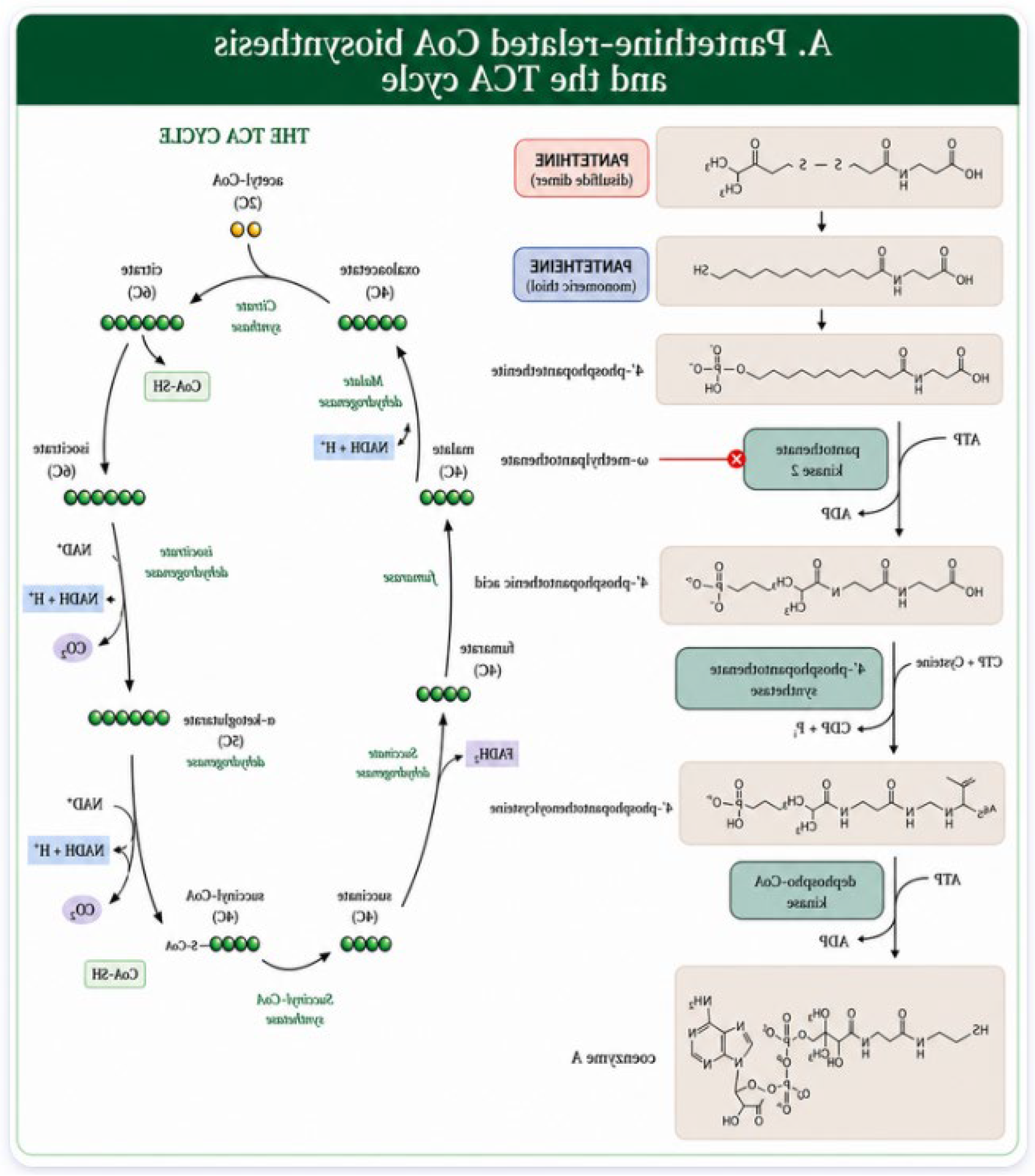
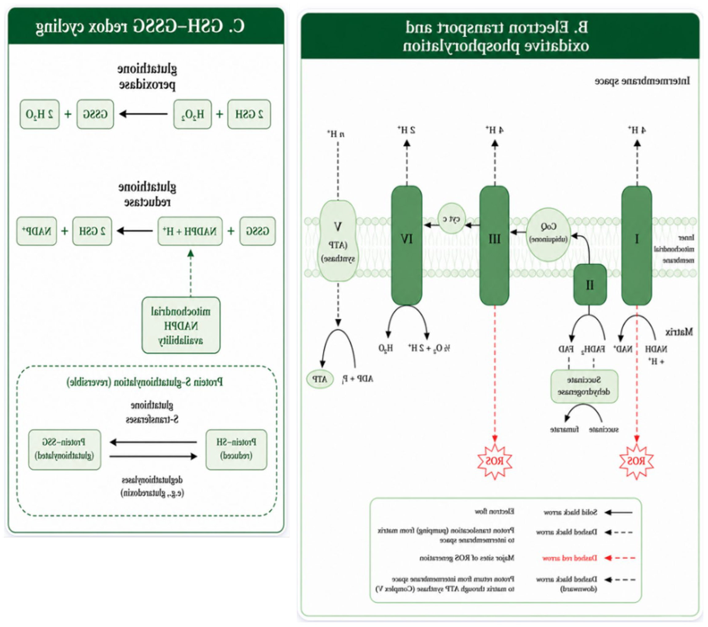
Figure 2.
Pantethine metabolism and proposed neurobiological relevance. This schematic illustrates the gastrointestinal uptake and downstream metabolic processing of pantethine as a CoA-related precursor, together with a conceptual summary of its putative links to cellular energetics, inflammatory signaling, oxidative stress, and neuronal integrity. Following absorption, pantethine may undergo reduction or disulfide exchange to form pantetheine, which can subsequently contribute to CoA biosynthesis through 4′-phosphopantetheine and dephospho-CoA. Through this pathway, pantethine-related metabolism is presented as potentially relevant to ATP production, cellular redox balance, and reduced pro-inflammatory signaling. The lower panels summarize the figure’s proposed downstream implications for neuronal health and survival, reduced apoptosis, improved neurotransmission, and possible relevance to PKAN, with broader implications for AD and PD remaining conceptual rather than established. Because several of these downstream effects are model-dependent and not uniformly demonstrated across disease contexts, the figure should be interpreted as an integrative framework rather than a direct causal map. AD, Alzheimer’s disease; ATP, adenosine triphosphate ; CoA, coenzyme A; PD, Parkinson’s disease.
Figure 2.
Pantethine metabolism and proposed neurobiological relevance. This schematic illustrates the gastrointestinal uptake and downstream metabolic processing of pantethine as a CoA-related precursor, together with a conceptual summary of its putative links to cellular energetics, inflammatory signaling, oxidative stress, and neuronal integrity. Following absorption, pantethine may undergo reduction or disulfide exchange to form pantetheine, which can subsequently contribute to CoA biosynthesis through 4′-phosphopantetheine and dephospho-CoA. Through this pathway, pantethine-related metabolism is presented as potentially relevant to ATP production, cellular redox balance, and reduced pro-inflammatory signaling. The lower panels summarize the figure’s proposed downstream implications for neuronal health and survival, reduced apoptosis, improved neurotransmission, and possible relevance to PKAN, with broader implications for AD and PD remaining conceptual rather than established. Because several of these downstream effects are model-dependent and not uniformly demonstrated across disease contexts, the figure should be interpreted as an integrative framework rather than a direct causal map. AD, Alzheimer’s disease; ATP, adenosine triphosphate ; CoA, coenzyme A; PD, Parkinson’s disease.
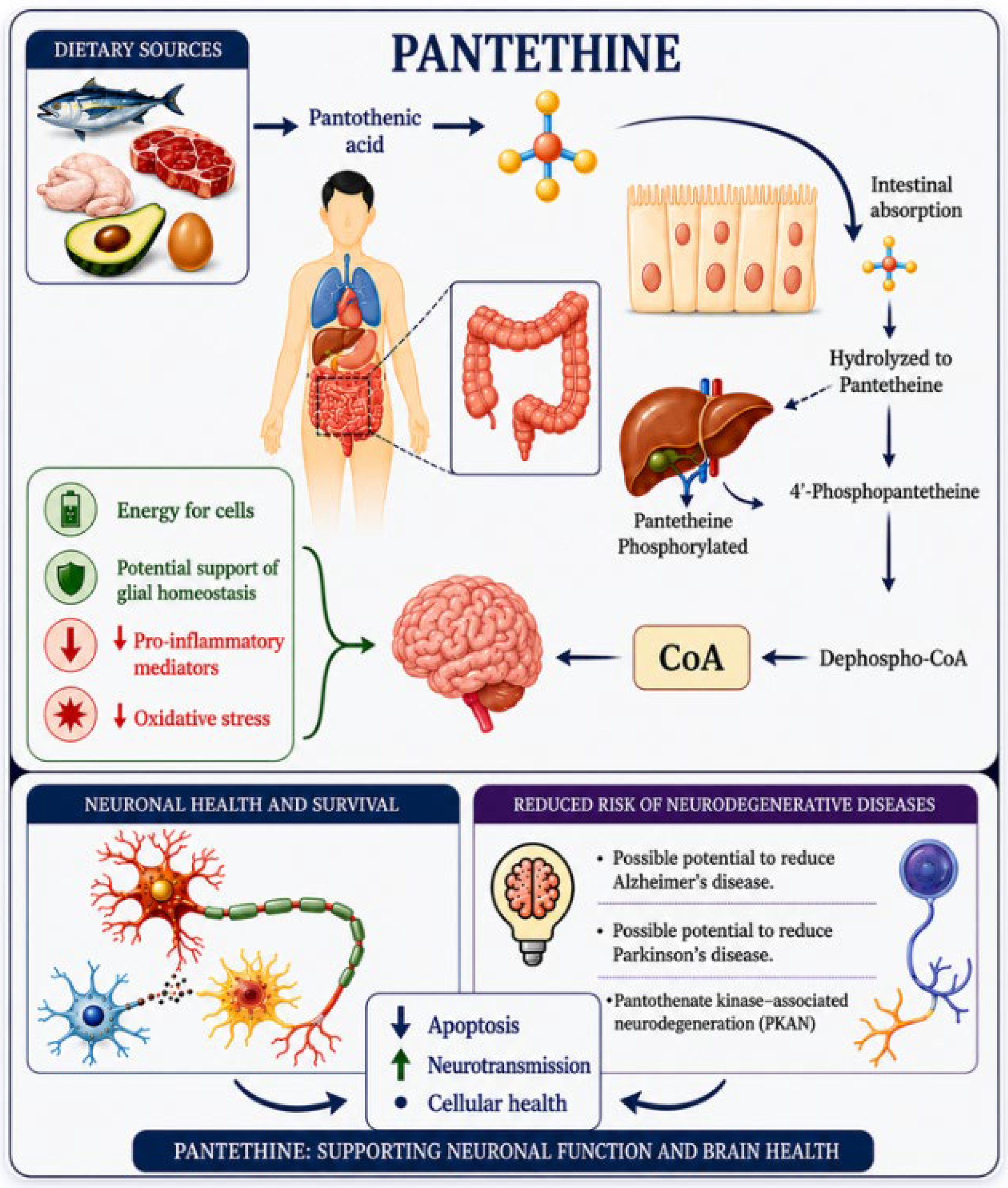
Figure 3.
Pantethine-related modulation of inflammatory-redox signaling and putative neuroprotective outcomes. This schematic depicts a conceptual sequence linking pro-inflammatory mediators, including IL-1β and TNF-α, to NF-κB activation, increased oxidative stress, and microglial-astrocytic activation, and then summarizes the proposed points at which pantethine may intervene. In the central panel, pantethine is presented as supporting antioxidant and cytoprotective defenses, most notably glutathione-related redox buffering, rather than as a direct inhibitor of any single inflammatory pathway. The right panel summarizes the putative downstream consequences of this inflammatory-metabolic modulation, including reduced neuroinflammation, improved mitochondrial function, enhanced synaptic integrity, and greater neuronal survival. Because the figure integrates findings of unequal evidentiary strength and condenses several indirect relationships into a single scheme, it should be interpreted as an explanatory framework rather than as direct proof that pantethine produces all these effects across neurodegenerative disease contexts. GSH, reduced glutathione; IL-1β, interleukin-1 beta; NF-κB, nuclear factor kappa B; ROS, reactive oxygen species; TNF-α, tumor necrosis factor alpha.
Figure 3.
Pantethine-related modulation of inflammatory-redox signaling and putative neuroprotective outcomes. This schematic depicts a conceptual sequence linking pro-inflammatory mediators, including IL-1β and TNF-α, to NF-κB activation, increased oxidative stress, and microglial-astrocytic activation, and then summarizes the proposed points at which pantethine may intervene. In the central panel, pantethine is presented as supporting antioxidant and cytoprotective defenses, most notably glutathione-related redox buffering, rather than as a direct inhibitor of any single inflammatory pathway. The right panel summarizes the putative downstream consequences of this inflammatory-metabolic modulation, including reduced neuroinflammation, improved mitochondrial function, enhanced synaptic integrity, and greater neuronal survival. Because the figure integrates findings of unequal evidentiary strength and condenses several indirect relationships into a single scheme, it should be interpreted as an explanatory framework rather than as direct proof that pantethine produces all these effects across neurodegenerative disease contexts. GSH, reduced glutathione; IL-1β, interleukin-1 beta; NF-κB, nuclear factor kappa B; ROS, reactive oxygen species; TNF-α, tumor necrosis factor alpha.
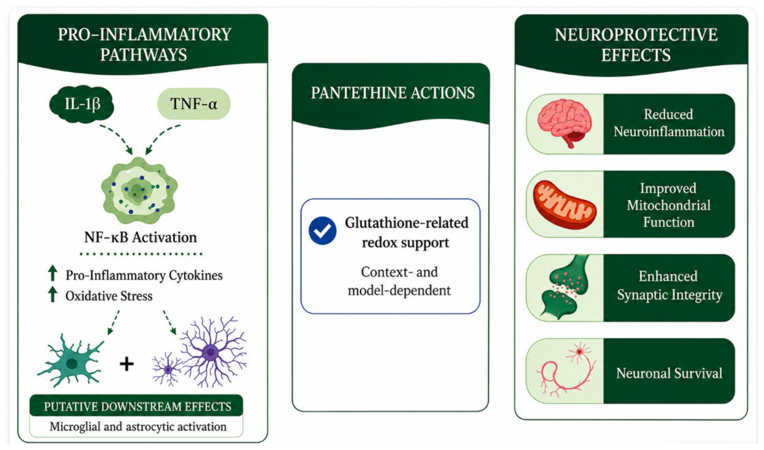
Figure 4.
Pantethine as a conceptual interface between glial metabolism, neuronal metabolism, and metabolic-signaling interactions. This schematic summarizes a hypothesis-building framework in which pantethine is positioned between glial and neuronal metabolic functions rather than depicting a directly demonstrated pathway map. On the glial side, the figure highlights putative associations with glutathione production, metabolite delivery, and astrocyte-related lipid or cholesterol metabolism, while on the neuronal side it depicts possible links with acetylcholine synthesis, ATP-related bioenergetics, and axonal function. The central message is that pantethine may influence metabolic and signaling interactions across multiple cell types through CoA-related biology, but direct evidence for coordinated restoration of glial-neuronal coupling remains limited. Accordingly, this figure should be interpreted as an integrative conceptual model that reflects mechanistic intersections and working hypotheses rather than established cell-type-specific effects in vivo.
Figure 4.
Pantethine as a conceptual interface between glial metabolism, neuronal metabolism, and metabolic-signaling interactions. This schematic summarizes a hypothesis-building framework in which pantethine is positioned between glial and neuronal metabolic functions rather than depicting a directly demonstrated pathway map. On the glial side, the figure highlights putative associations with glutathione production, metabolite delivery, and astrocyte-related lipid or cholesterol metabolism, while on the neuronal side it depicts possible links with acetylcholine synthesis, ATP-related bioenergetics, and axonal function. The central message is that pantethine may influence metabolic and signaling interactions across multiple cell types through CoA-related biology, but direct evidence for coordinated restoration of glial-neuronal coupling remains limited. Accordingly, this figure should be interpreted as an integrative conceptual model that reflects mechanistic intersections and working hypotheses rather than established cell-type-specific effects in vivo.
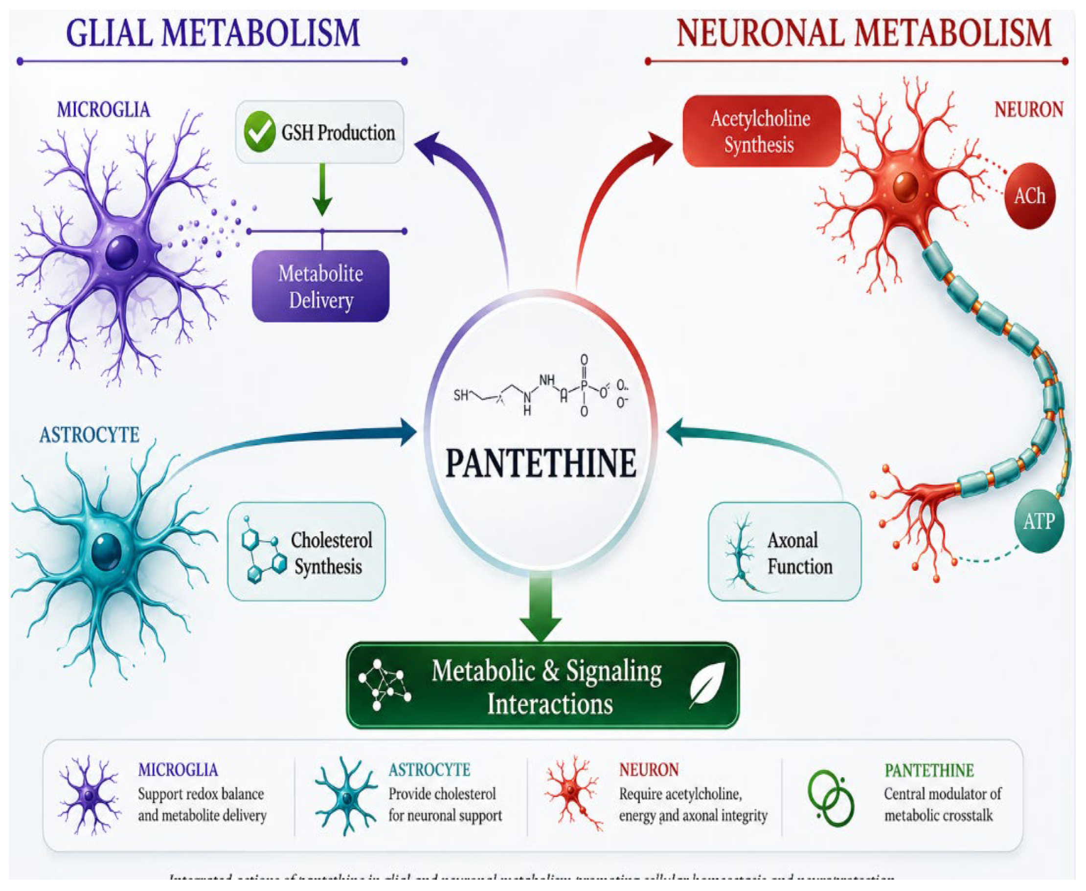

Table 1.
Summary of pantethine evidence across mechanistic domains and disease contexts. This table synthesizes the literature reviewed in the manuscript by aligning mechanistic domain, disease context, experimental system, dosing format, principal finding, CNS-specific relevance, evidence strength, and the main interpretive constraint for each evidence cluster. The evidence labels are intentionally graded as direct, supportive, indirect, or speculative to distinguish disease-proximal findings from broader mechanistic inference. The table is designed to help readers and reviewers see at a glance where pantethine is best supported, where adjacent CoA-pathway biology provides only partial support, and where current translational claims remain premature.
Table 1.
Summary of pantethine evidence across mechanistic domains and disease contexts. This table synthesizes the literature reviewed in the manuscript by aligning mechanistic domain, disease context, experimental system, dosing format, principal finding, CNS-specific relevance, evidence strength, and the main interpretive constraint for each evidence cluster. The evidence labels are intentionally graded as direct, supportive, indirect, or speculative to distinguish disease-proximal findings from broader mechanistic inference. The table is designed to help readers and reviewers see at a glance where pantethine is best supported, where adjacent CoA-pathway biology provides only partial support, and where current translational claims remain premature.
| Domain | Disease/model | Experimental system | Dose/route | Main finding | CNS-specific relevance | Evidence strength | Key limitation | Ref. |
| Pharmacology and active species | Pantethine metabolism in humans and ex vivo brain tissue | Human dosing studies; hippocampal slice metabolism | Oral; ex vivo extracellular metabolism | Rapid hydrolysis and context-dependent conversion suggest that parent pantethine may not be the dominant active species in vivo. | Indirect | Direct | CNS exposure and relative contribution of pantethine, pantetheine, and cysteamine remain unresolved. | [45,46,47] |
| Evidence hierarchy | PKAN and inherited CoA-biosynthesis disorders | Drosophila, mouse, fibroblasts, iPSC-derived neurons | Dietary, oral, cell supplementation | CoA-pathway rescue is most coherent in disease-proximal models, with improvements in iron handling, mitochondrial readouts, and survival-related phenotypes. | High | Direct | Clinical translation remains limited and not all rescue data isolate pantethine from related intermediates or combination regimens. | [39,41,48] |
| Neuroinflammatory signaling | 5xFAD astrocytes; experimental autoimmune encephalomyelitis | Cultured astrocytes; murine and human T cells | In vitro and in vivo CoA fueling | Pantethine reduces selected inflammatory outputs, especially IL-1β in astrocytes and TNF-α, GM-CSF, and IL-17A in encephalitogenic immune cells. | Moderate | Supportive | Direct disease-specific evidence for microglial reprogramming, IL-8 modulation, or broad CNS cytokine-network remodeling remains limited. | [37,49,50] |
| Oxidative stress and redox homeostasis | Rotenone and aluminum neurotoxicity; PKAN-related models | Rat brain regions, isolated mitochondria, PANK2-mutant cells, Drosophila | Oral, intragastric, in vitro | Pantethine or related CoA-linked precursors improve glutathione redox balance, reduce protein glutathionylation, and attenuate oxidative damage in selected models. | Moderate | Supportive | Some evidence uses panthenol rather than pantethine, and non-neural redox data should not be equated with neuronal protection. | [51,52,53] |
| Mitochondrial dysfunction and bioenergetics | PKAN-linked Drosophila, Pank2 models, fibroblasts | Fly, mouse, fibroblasts, patient-derived cells | Dietary, oral, cell supplementation | This is the strongest mechanistic domain outside core pharmacology, with rescue of CoA levels, respiration, membrane potential, PDH and complex I activity, and survival-related phenotypes. | High | Direct | Generalization to common neurodegenerative diseases remains unproven, and some of the strongest data involve 4′-phosphopantetheine or PANK activators rather than pantethine itself. | [39,54,55] |
| Cell survival pathways | PKAN models plus cysteamine-informed neuroprotection literature | Drosophila; non-pantethine neural injury models | Dietary, oral, experimental aminothiol exposure | Pantethine may reduce upstream drivers of cell death by improving metabolic and redox conditions, but direct anti-apoptotic mechanisms are not firmly established. | Low to moderate | Indirect | Evidence for Bax, cytochrome c, or caspase-specific effects is largely inferred or derived from cysteamine and cystamine literature. | [39,56,57] |
| Glial-neuronal interactions and metabolic coupling | 5xFAD astrocytes; PKAN astrocyte-neuron systems | Astrocyte cultures, coculture, PKAN-derived cells | In vitro; mixed supplementation contexts | Pantethine may intersect with glial metabolic support and inflammatory tone, but direct restoration of lactate shuttling, glutamate clearance, or cell-cell coupling remains unproven. | Low to moderate | Speculative | Much of this domain is hypothesis-building and rests on inferred links between CoA restoration and glial support functions. | [49,58,59] |
| Translational relevance | PKAN vs AD and PD | Open-label trial, pilot combinations, preclinical comparisons | Oral pantethine; multimodal regimens | Translation is most justified in PKAN, whereas AD and PD remain exploratory and should not be presented on equal evidentiary footing. | Disease-dependent | Supportive | Human efficacy data are sparse, CNS target engagement is uncertain, and biomarker-defined trial design is still underdeveloped. | [29,41,60] |
AD, Alzheimer’s disease; Bax, Bcl-2-associated X protein; CNS, central nervous system; CoA, coenzyme A; GM-CSF, granulocyte-macrophage colony-stimulating factor; IL-1β, interleukin-1 beta; IL-8, interleukin-8; IL-17A, interleukin-17A; iPSC, induced pluripotent stem cell; PANK2, pantothenate kinase 2; PD, Parkinson’s disease; PDH, pyruvate dehydrogenase; PKAN, pantothenate kinase-associated neurodegeneration; TNF-α, tumor necrosis factor alpha.
Table 2.
Pantethine-related antioxidant and redox-modulating evidence in selected in vitro and in vivo models. This table consolidates evidence from endothelial, fibroblast, erythrocyte, ocular, and neurodegeneration-adjacent experimental systems. It distinguishes endothelial, fibroblast, erythrocyte, and irradiation-related systems from brain-relevant or neurodegeneration-adjacent contexts so that readers can judge scope and translational value more quickly. The entries show that pantethine displays measurable antioxidant or redox-modulating effects in several experimental settings, but that CNS-specific relevance remains uneven and often indirect. Presenting the evidence in a standardized format helps separate supportive oxidative findings from stronger neurodegeneration-oriented mechanistic evidence discussed in the main text.
Table 2.
Pantethine-related antioxidant and redox-modulating evidence in selected in vitro and in vivo models. This table consolidates evidence from endothelial, fibroblast, erythrocyte, ocular, and neurodegeneration-adjacent experimental systems. It distinguishes endothelial, fibroblast, erythrocyte, and irradiation-related systems from brain-relevant or neurodegeneration-adjacent contexts so that readers can judge scope and translational value more quickly. The entries show that pantethine displays measurable antioxidant or redox-modulating effects in several experimental settings, but that CNS-specific relevance remains uneven and often indirect. Presenting the evidence in a standardized format helps separate supportive oxidative findings from stronger neurodegeneration-oriented mechanistic evidence discussed in the main text.
| Panel A. In vitro evidence | |||||||||||
| Property | Model/system | Concentration | Main observation | CNS-specific relevance | Reference | ||||||
| Antioxidant and redox-modulating | Human pulmonary microvascular endothelial cells, human vascular endothelial cells, and NIH3T3 mouse fibroblasts | 0–100 µM for 24 h in endothelial cultures; 100 µM for 4 h in NIH3T3 cells | Inhibited microparticle-induced oxidative and nitrosative stress. | Indirect. Supports redox activity outside the nervous system but does not establish neuronal protection. | [151] | ||||||
| Antioxidant / cytoprotective context-dependent effect | Human HCT-116 colon carcinoma isogenic lines (p53−/−, Bax−/−, wild type), MCF-7, Jurkat, HL-60, and HL-60/vinc cells | 1, 2.5, 25, and 50 µM | D-pantethine protected wild-type HCT-116 cells but not p53-null or Bax-null cells, indicating context-dependent cytoprotection. | Indirect. Informative for redox-linked viability pathways, not for CNS-specific neuroprotection. | [154] | ||||||
| Panel B. In vivo evidence | |||||||||||
| Property | Experimental model | Dose | Route | Main observation | CNS-specific relevance | Reference | |||||
| Antioxidant / lipid-related systemic effect | Six-week-old female BALB/c mice | 150 mg/kg/day for 6 weeks | Oral | Reported inactivation of the ABCA1 locus, indicating systemic lipid-handling effects rather than a clearly defined CNS antioxidant endpoint. | Indirect. Limited neurodegeneration relevance as reported. | [151] | |||||
| Antioxidant under hematologic oxidative stress | Adult male Wistar CRL rats exposed to doxorubicin-related oxidative injury | 400 mg/kg/day for 5 days | Intragastric | Reduced oxidative stress in erythrocytes but did not prevent the marked fall in free CoA. | Indirect. Supports systemic redox activity but not direct CNS rescue. | [154] | |||||
| Weak radioprotective antioxidant effect | Male pigmented Long Evans rats subjected to irradiation | 1 g/kg single dose, 45 min before irradiation | Intraperitoneal | Produced only a minimal delay in cataract development compared with other agents. | Indirect. Ocular and systemic relevance only; does not support neurodegeneration-specific efficacy. | [155] | |||||
ABCA1, ATP-binding cassette transporter A1; CNS, central nervous system; NIH3T3, mouse embryonic fibroblast cell line; HUVECs, human umbilical vein endothelial cells; HPMECs, human pulmonary microvascular endothelial cells.
Table 3.
Pantethine-associated mitochondrial protection in selected experimental systems. This table synthesizes experimental evidence linking pantethine to mitochondrial protection across in vitro and in vivo systems. Studies are organized by model, exposure conditions, route of administration, principal mitochondrial outcome, and CNS relevance. The evidence is strongest in PKAN-related and other CoA-deficiency settings, where pantethine is associated with improved respiration, membrane potential, CoA restoration, or mitochondrial structure. By contrast, findings from non-neural or mixed systems are supportive rather than disease-proximal and should not be interpreted as equivalent proof of neuronal rescue in vivo.
Table 3.
Pantethine-associated mitochondrial protection in selected experimental systems. This table synthesizes experimental evidence linking pantethine to mitochondrial protection across in vitro and in vivo systems. Studies are organized by model, exposure conditions, route of administration, principal mitochondrial outcome, and CNS relevance. The evidence is strongest in PKAN-related and other CoA-deficiency settings, where pantethine is associated with improved respiration, membrane potential, CoA restoration, or mitochondrial structure. By contrast, findings from non-neural or mixed systems are supportive rather than disease-proximal and should not be interpreted as equivalent proof of neuronal rescue in vivo.
| Study context | Pantetheine intervention or compound | Model or experimental system | Dose or concentration | Route | Main mitochondrial finding | CNS-specific relevance | Reference |
| In vitro | Mitochondrial protection | Primary skin fibroblasts | 5 µM and 1–100 µM | Not applicable | Associated with increased PANK2 and mitochondrial acyl carrier protein expression levels | Indirect; cellular support relevant to PKAN-like mitochondrial deficits | [48] |
| In vitro | Mitochondrial protection | Brain mitochondria from control mice | 0, 0.5, or 1 mM | Ex vivo exposure | No protective effect was observed at 0.5 mM pantetheine or with higher MPP+ challenge, indicating context-dependent rescue | Supportive but limited; brain-relevant system without clear rescue under all conditions | [114] |
| In vitro | Mitochondrial protection | Drosophila Schneider S2 cells with dPANK/Fbl depletion | 100 µM | Cell culture exposure | Restored normal growth in dPANK/Fbl-depleted cells, consistent with metabolic rescue | Supportive; mechanistically relevant to PKAN but non-mammalian | [39] |
| In vivo and clinical | Mitochondrial protection | Patients with confirmed genetic mutations in the PPCS gene | Patient 1: 450 mg/day; Patient 2: 600 mg/day; Patients 3 and 4: 15 mg/kg/day | Oral | Mitochondrial morphology was reported as preserved; functional rescue was assessed primarily in patient-derived cardiac models | Supportive human evidence in a CoA-biosynthesis disorder, but not PKAN-specific | [102] |
| In vivo | Mitochondrial protection | Pank2−/− mice and corresponding Pank2+/+ controls | 15 mg/kg/day in drinking water | Oral | Improved mitochondrial respiration in both genotypes and restored membrane potential in Pank2−/− neurons | Direct PKAN-relevant mitochondrial evidence | [106] |
| In vivo | Mitochondrial protection | Male C57BL/6 mice exposed to 2′-methyl-MPTP | 15 mg for 5 days before and 5 days after toxin exposure | Intraperitoneal | Rapidly increased glutathione levels in normal animals, consistent with mitochondrial redox support | Indirect; supportive for oxidative-mitochondrial stress but not direct PKAN rescue | [114] |
| In vivo | Mitochondrial protection | Hypomorphic dPANK/fbl1 mutant flies | 1.6 mg/mL in food | Oral by feeding | Restored CoA levels, improved mitochondrial function, and reduced oxidative damage | Direct disease-proximal evidence in a Drosophila PKAN model | [39] |
CoA, coenzyme A; CNS, central nervous system; PKAN, pantothenate kinase-associated neurodegeneration; PPCS, phosphopantothenoylcysteine synthetase; PANK2, pantothenate kinase 2; mtACP, mitochondrial acyl carrier protein; MPP+, 1-methyl-4-phenylpyridinium; mPTP, mitochondrial permeability transition pore; MPTP, 1-methyl-4-phenyl-1,2,3,6-tetrahydropyridine.
Disclaimer/Publisher’s Note: The statements, opinions and data contained in all publications are solely those of the individual author(s) and contributor(s) and not of MDPI and/or the editor(s). MDPI and/or the editor(s) disclaim responsibility for any injury to people or property resulting from any ideas, methods, instructions or products referred to in the content. |
© 2026 by the authors. Licensee MDPI, Basel, Switzerland. This article is an open access article distributed under the terms and conditions of the Creative Commons Attribution (CC BY) license (http://creativecommons.org/licenses/by/4.0/).
Copyright: This open access article is published under a Creative Commons CC BY 4.0 license, which permit the free download, distribution, and reuse, provided that the author and preprint are cited in any reuse.


