

Submitted:
27 July 2026
Posted:
28 July 2026
You are already at the latest version
Abstract
Background: Endothelial deregulation (ED) manifests as a major secondary complication in clinical Diabetes mellitus (DM) which is established as the preliminary stage of vascular dysfunction. This study evaluates a broader repertoire of biomarkers that are relevant to diabetic - ED and the inflammatory potential of the high mobility group box-1 (HMGB1) nuclear protein in mitigating type-2 diabetes (T2D) -induced ED. Method: A total of 33 biomarkers were evaluated from retrospective metabolic and sociodemographic data in a forest plot following the PICOS study design. A total of 1830 entries published during the past 10 years ending on mid December 2023 from PUBMED, MEDLINE, SCOPUS, SPRINGER-LINK and WOS databases were screened using a variety of MESH terms. The results were generated using the RevMan and GraphPad Prism software. Modified Cochrane Organisation template for systematic reviews and meta-analyses was followed with data validated in PRISMA. The protocol of this study was registered at https://www.crd.york.ac.uk/Prospero/CRD42023493221. Results: Only 16 single case-control studies qualified for data extraction. 18 biomarkers were identified as having a significantly high risk of ED. IL-6 emerged as the biomarker having the highest effect size (SMD 5.20, 95% CI, 3.21, 7.18, p<0.00001, n=209). HMGB1 comprised the sixth highest standardized mean difference (SMD 2.86, 95% CI, 1.91, 3.81, p<0.00001, n=762) out of the eight highest biomarkers calculated. Conclusion: The biomarkers consisting of a mix of traditional and non-traditional markers carried a high risk in developing T2D-induced cardiovascular disease and it was concluded that HMGB1 provides us with a high-risk inflammatory metabolic target.
Keywords:
diabetes mellitus
; endothelial deregulation
; endothelium
; vascular dysfunction
; HMGB1
; systematic review
; meta-analysis
; clinical type-2 diabetes
; inflammation
0. Introduction
1. Background
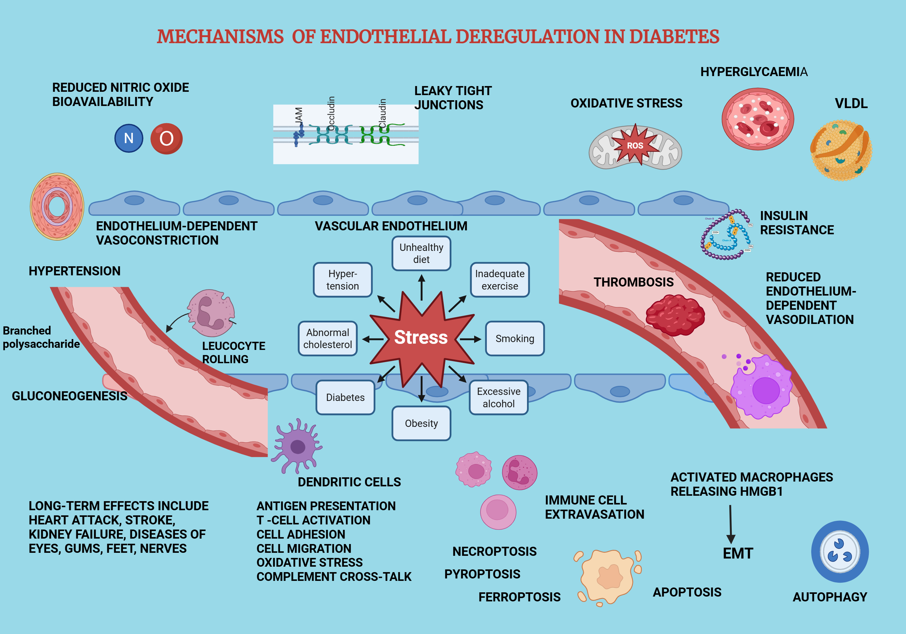
Endothelial deregulation (ED) is the preliminary stage of manifesting progressive cardiovascular defects in Diabetes mellitus (DM) [1]. It is a major adverse outcome of diabetes, which is an endocrine disorder that arises from autoimmunity and as a metabolic disease which develops from uncontrolled hyperglycaemia [2]. The function of the vascular endothelium includes the regulation of blood flow, barrier function, process of blood clotting and immune modulation [3]. This systematic review and meta-analysis focusses upon testing potential immune - mediated diagnostic biomarkers and testing HMGB1 as a specific therapeutic target with its inhibition. The impact of HMGB1 on both traditional and novel biomarkers of diabetes-induced ED as a prospective immune - based inflammatory target [4] was also tested as a means of resolving ED.
1.2. Description of the Condition
Diabetes mellitus comprises of several subtypes out of which type-1 diabetes (T1D) and type-2 diabetes (T2D) are the most common with substantial variation in their aetiology [5]. T2D affects mostly the older age groups such as the geriatric adults [6] and develops from an inability to metabolise the glucose due to insulin resistance [7]. It causes the blood glucose titres to rise or decline rapidly [8]. Thus, the mechanisms of glucose metabolism require regulating the glycaemic and lipid intake in the diet coupled with strenuous exercise to maintain normal blood glucose levels [9]. The major adverse outcome of both subtypes is having a high risk of developing cardiovascular disorders (CVD) [10] which initially develop as vascular dysfunction, specifically manifesting ED as the baseline of CVD morbidity which could progress rapidly into coronary heart disease (CHD) and /or stroke [11]. The other major organs that are seriously debilitated by diabetes are the liver, heart, kidneys, eye, musculature and the limbs [12].
1.3. Description of the Intervention
The high mobility group box 1 (HMGB1) is a polypeptide composed of 215 amino acid residues and has three specific moieties in the structure, named the A Box, the B box, and the acidic carbon-terminal tail, [13] each of which performs different functions and its functions vary depending on its subcellular location [14]. The HMGB1performs dual functions acting as a proinflammatory molecule or an anti-inflammatory molecule based on its three specific structural domains [15]. This study is focused upon investigating HMGB1 for its suitability and sustainability in implementing disease resolution as an immune-mediated therapeutic target. It has been tested for efficacy, low adverse effects and identified as an immune biomarker or a DAMP molecule which upregulates innate immunity in diabetic patients [16].
1.4. How the Intervention Might Work
HMGB1 is known to induce inflammation which precedes immune-mediated resolution of disease [17]. The B box is able to recapitulate proinflammatory activities when it is present within the extracellular environment while the anti-inflammatory action is enhanced when the A box binds with the B box, in an antagonistic manner [18]. These inherent dual mechanisms have conferred the ability to induce inflammation as the first step of immune resolution which mainly occurs to remove the apoptotic and necrotic cells and the debris formed in the extracellular matrix by promoting autophagy following a myocardial infarct in which the tissue remodelling follows the infiltration of immune cells. It prevents further deterioration of the myocardium and promotes anti-inflammatory properties by upregulating favourable cytokines, chemokines and remodelling of the heart tissue and stimulating angiogenesis [17]. The nuclear HMGB1 binds with the DNA and initiates DNA repair, replication, and recombination [19] while the extracellular HMGB1 upregulates innate immunity, cell proliferation, differentiation, migration, tissue regeneration, apoptosis, and autophagy [20]. In diabetes, HMGB1 exerts its proinflammatory actions by binding with the receptors of the RAGE and TLR families which further induces the gene expression of the JNK, p38/MAPK, ERK1/2 and IkB- NF-kB pathways [21]. HMGB1 qualifies as a pathway-specific inflammatory biomarker which could also produce a beneficial effect by suppressing its phosphorylation and blocking its release by binding directly with an inhibitor such as glycyrrhizin, thus attenuating ED given the severity of the disease condition [22].
1.5. Why it is Important to do this Review
Diabetes is a widespread disease in the world and as at year 2024, 240 million persons comprised the undiagnosed cohort of diabetics with almost half of all those adults being unaware of their illness [23]. Yet 537 million persons comprising 10.5% of the global population aged between 20-79 years, have received a diagnosis on diabetes [24]. The total current healthcare expenditure on managing the disease has amounted to US dollars 966 billion and is expected to surpass US dollars 1054 billion in 2045 [25]. It is expected to reach 643 million diagnoses in 2030 rising to a further 783 million cases in 2045 [26]. Up to 80% of the global diabetic population is living in the low and middle income - reporting countries [27]. The people with T2D carry a mortality rate which almost doubles when they are comorbid with CVD [28].
2. Methodology
2.1. The Research Question and PICOS Statement
The research question was based on testing biomarkers of Diabetic-ED which identified a high risk of developing the disease in clinical subjects. According to the PICOS criteria, the population under study consisted of humans. The intervention comprised the presence or absence of the HMGB1 nuclear protein, how the influence of which had further contributed to producing a high or low risk of manifesting Diabetic - ED. The comparators were the age and sex-matched healthy control versus the Diabetic-CVD population, whilst the outcomes were separated into primary and secondary outcomes. For this assessment, the primary outcomes were evaluated using the standardized mean difference (SMD) or the effect size that was generated by the forest plots. The primary outcomes consisted of the biomarker SMDs whilst the secondary outcomes consisted of the non-parametric Mann Whitney U test and the computation of the ecological correlation coefficients, ROC curves, QQ plots and an age analysis. These methods were used to assess the biomarkers of Diabetic – ED as potential therapeutic or diagnostic markers that could help attenuate this disease complex. All the study designs conformed to the baseline case-control studies.
2.2. Bridging the knowledge Gap
The reason for selecting biomarker evaluation in this study was to justify the gap that exists between the quest for searching an effective candidate that will potentially fulfil the dual roles of therapeutic and /or diagnostic agents of Diabetic - ED as opposed to non-diabetic CVD. The reasons for the gap that needs to be filled are the incomplete understanding of the molecular mechanisms of Diabetic - CVD which are complex in nature and not having good glycaemic control methods which can be maintained long-term such as over ten years. The UK Prospective Diabetes Study (UKPDS) has established that T2D could be maintained under control with effective long-term glycaemic control. It is said that prolonging hyperglycaemia of even short duration such as a few weeks could manifest Diabetic - CVD. Nevertheless, novel methods of more effective glycaemic control are needed to be introduced, and the focus of the present study has shifted over to an anti-inflammatory approach of suppressing the chronic inflammation that ensues with the progression of Diabetic-CVD and its implementation to be cost -effective. It is still unclear and lacking the exact mechanism of how hyperglycaemia could manifest cardiovascular disease or cardiomyocyte injury in the heart. Further, the main impediment to resolving this disease is the inability to translate the therapeutic targets of early and/or effective intervention into clinical practice. It is so because there is a gap of knowledge and implementation in resolving this disease group because the standard risk factors and treatment modalities are insufficient to overcome the unique and multifactorial pathologies of Diabetic-CVD. The complex pathophysiology of Diabetic-CVD involves the interplay of hyperglycaemia, dyslipidaemia, insulin resistance, inflammation, endothelial dysfunction and pro- coagulable state whereas the standard CVD risk factors are mostly high cholesterol and hypertension which occur in isolation given the inability to delineate the exact mechanisms so far. Intensive glucose control has shown promise in containing microvascular diseases in T2D, but not the macrovascular diseases. Factually, it is suggested that there are factors beyond hyperglycaemia which are critical to the resolution of Diabetic-CVD. There is disparity between clinical trial efficacy and population-level effectiveness, due to lacking specific guidelines and research on Diabetic -CVD, also which involve age and sex-specific interventions or time-based preventive strategies.
2.3. Types of Included Studies
The types of studies that were selected had patients diagnosed with T2D, comorbid with vascular dysfunction /ED, in comparison to the control group that was healthy. They had to be research studies also known as articles that were peer-reviewed and published in the English language within the specified year range (10 years ending on the 19 December 2023) with accessibility to the full-text version which had at least >6 -10 participants as per the power calculation. The characteristics of the included studies are documented for each study in Table 1.
2.4. Excluded Studies
The exclusion criteria were other CVD except for vascular dysfunction, endothelial stress caused by diseases other than T2D and endothelial stress stimulated by endothelium-independent mechanisms. Reviews, posters, letters, communications, book chapters, conference proceedings, duplicates, in vitro studies, ex-vivo studies, preclinical studies, randomised control trials, cross-over studies, other study designs other than case-control studies, articles in foreign languages, articles published out of the specified time range and those not peer-reviewed, have been excluded.
2.5. Types of Participants
The participants are human subjects of all age groups without any dietary restrictions. The types of studies that were selected had patients diagnosed with T2D comorbid with vascular dysfunction /ED, in comparison to the healthy control.
2.6. Type of Intervention
The intervention is the HMGB1 nuclear protein which possesses both anti-inflammatory and pro-inflammatory characteristics depending on the functions of its structure.
2.7. Outcome Measures (Primary and Secondary outcomes)
2.7.1. Search Methods for Identification of Studies
The PubMed, Medline, Web of Science, SCOPUS and Springer Link databases were electronically searched from 1/1/2013 to 19/12/2023.
2.7.2. Search Strategy
The search strategy consisted of searching the databases with keywords after specifying the year range. The databases were accessed via the Victoria University’s library resources webpage.
2.7.3. Selection of Studies
The full text reports of all the eligible studies were examined. Once satisfied with the eligibility of those selected articles, the data extraction was commenced.
2.7.4. Data Extraction /Collection
The data extraction was carried out by two independent reviewers separately screening the databases. The data was extracted on to a pre-specified template from the Cochrane collaboration that was adapted to suit our investigative study.
2.7.5. Data Management and Analysis
If raw data was unavailable, data was gathered from graphs and charts in the primary studies. Data were extracted into tables using the MS Excel software and for the analysis, RevMan version 5 with GraphPad Prism 10 software were used.
2.8. Assessment of Risk of Bias (ROB) in Included Studies
All the included studies are case-control studies and hence the risk of bias (ROB) in them is comparatively low. The ROB in our study was carried out according to the risk of bias in non-randomized studies -1 or the ROBINS-1 tool introduced by the Cochrane organisation for clinical studies that are included in a systematic review. An assessment of the ROB was performed in each included study, rating the outcome as one of the three levels specified as low risk, unclear risk, or high risk. An assessment of the reporting biases was also carried out as funnel plots. In the meta-analysis, a summary of the overall ROB items is depicted in a risk stratified summary ROB graph. The quality of the body of evidence was maintained with the PRISMA 2020 guidelines, published by the Cochran collaboration, given in the handbook.
2.9. Measures of Treatment Effect
The measures of treatment effect were applied to the data that was processed. Two types of data are encountered in systematic reviews and meta-analysis named: continuous or dichotomous data, which determine whether to adopt a fixed effects model or a random effects model. Continuous variables were reported as the mean + standard deviation and categorical data were reported as percentages.
2.9.1. Unit of Analysis Issues
The unit of analysis issues did not arise with this study which was not a complicated study design.
2.9.2. Assessment of Heterogeneity
The statistical heterogeneity among the included studies was evaluated by the Q statistic, the I squared metric, and the TAU squared value that were significant at 0.1 probability or 99.9% significance or accuracy.
2.9.3. Subgroup Analysis
The subgroup analysis is undertaken if there is a considerable amount of participant data, which is >10 is present in each subgroup. Subgroups are created if the units of measurement varied substantially such as a primary outcome. Descriptive statistics are also separately reported in the forest plots with an overall effect size with the commonly reported Z score, Chi squared distribution, and I squared metric at a probability of p<0.05.
2.9.4. Sensitivity Analysis
This application confirms the sensitivity of the software used for the construction of a forest plot by responding to a change or a deletion made.
2.9.5. Dealing with Missing Data
The missing data was dealt with by contacting the corresponding author of the primary study by email. After two emails were sent with no response, the primary outcome pertaining to the missing data was excluded but not the whole study.
2.10. Data Synthesis
The data synthesis consisted of both quantitative and qualitative syntheses. The quantitative assessment was carried out with a meta-analysis with the RevMan software version 5, and the qualitative study consisted of a narrative synthesis. The secondary outcomes were assessed using the GraphPad Prism version 10 statistical software. This review was completed by fulfilling the PRISMA 2020 guidelines provided by the Cochrane Collaboration for the authors of systematic reviews.
2.11. Meta-Analysis
A meta-analysis was deemed suitable and appropriate when at least two included (eligible) studies reported the same numerical data which could be pooled to construct an effect estimate that would reflect the true impact of the intervention under evaluation. The results were synthesized with an inverse-variance weighted - random-effects model given that there is variability in the intervention effect, the measurement of the outcome and the variability in the instruments used in calculating the numerical outcomes.
2.11.1. Summary Results
We provide three types of tables, (i) the pooled basic attributes or the characteristics of each eligible or included study, (ii) summary of findings calculated in the forest plot results and (iii) a risk of bias summary for each individual study.
2.11.2. Narrative Summary
The narrative summaries will include the following points.
- I.
- A description of the type of intervention in the included studies and how they were implemented.
- II.
- A description of the primary and secondary outcomes in the included studies.
- III.
- A review of findings for the secondary outcomes.
- IV.
- Adverse outcomes or potential threats, harm or losses.
- V.
- Associated financial costs in implementing the study design.
- VI.
- Possible important contextual details pertaining to the study design and /or analysis.
- VII.
- Perceived strengths, weaknesses and contributions made on the studies
2.12. Report on Practical Significance
Identification of high -risk -bearing risk factors aiding in diagnosis, ability to begin treatment early, changes to lifestyle and other epigenetic factors relevant to this study, costs saved from hospitalisations, screening and treatment, formulation of therapies, and new research activities will be reported.
3. Results
3.1. Description of Studies
3.1.1. Search Strategy
The search strategy consisted of searching the databases with keywords after specifying the year range. The databases were accessed via the Victoria University’s library resources webpage. The keywords consisted of:
- Diabetes mellitus AND Type-2D AND /OR Endothelial Deregulation AND HMGB1
- Diabetes AND Endothelium AND clinical research AND high mobility group box-1
- T2D AND clinical models AND Endothelial dysfunction AND HMGB1
- Diabetes mellitus AND Endothelial deregulation AND clinical research AND hmgb1
- Type-2 Diabetes AND clinical studies AND HMGB1
- HMGB1 AND type-2 diabetes AND endothelial dysfunction
These searches were carried out during the year 2023.
3.1.2. Results of the Search
The electronic databases that were searched include the Medline via EBESCO, PubMed, SCOPUS, Springer link and the WOS from which 1830 potentially relevant documents were obtained during the primary search. Among those records were collectively 1368 publications that included duplicates, animal studies and other irrelevant publications. The title and the abstract were screened and the studies which satisfied the pre-determined inclusion and exclusion criteria were then selected. The flowchart depicting the document search that was carried out is presented as Figure 1 adopted from the Cochrane Collaboration template. A total of 16 studies fulfilled the eligibility criteria and were included in this review (Table 1).
3.2. Primary and Secondary outcomes
3.2.1. Primary Outcomes
Each primary outcome model encompasses several biomarkers as shown below.
- Model 1 -The AGES – HMGB1, sRAGE [Supplementary (S)1]
- Model 2 - BP - SBP, DBP (S2)
- Model 3 - Glycaemic - HbA1c%, FBG, HOMA-IR, FINS (S3)
- Model 4 – Insulin Sensitivity – HOMA-IR, FINS (S4)
- Model 5 - Lipid - TC, TG, HDL-c, LDL-c (S5)
- Model 6 – Inflammatory – CRP, IL-6 (S6)
- Model 7 - Renal - SCr, BUN, eGFR, ACR (S7)
- Model 8 – Obesity – BMI, Age (S8)
- Model 9 - Vascular function - sICAM, sVE-cadherin, sVAP-1, sEndoglin, VEGF, HR, LVEF%, LVDV, LVSV (S9)
3.2.2. Secondary Outcomes
- 1)
- Pearson's and Spearman correlation coefficients (S10)
- 2)
- Mann Whitney U-test (S11)
- 3)
- QQ plots (S12)
- 4)
- Receiver Operator Characteristic Curve (ROC curve) (S13)
- 5)
- Age-related analysis (S14)
- 6)
- Funnel Plots (S15)
- 7)
- PRISMA Documentation (S16)
3.3. Risk of Bias in the Included Studies
The risk of bias assessment was carried out as per the Cochrane Organisation guidelines adapted to include the risk of bias in non-randomized studies of interventions -1 (ROBINS-1) tool consisting of 8 classifiers. In the 16 included studies, each study had at least 3 common confounders such as non-randomization, allocation concealment or blinding of the outcome. Almost all the studies did not select the participants using a random sequence generator, although all the studies stated the investigations were carried out in compliance with the Helsinki declaration (Figure 2). Funnel plots were constructed for assessing reporting bias.
Every possible step was taken to minimize the potential biases that may occur during the process of conducting this systematic review and the meta-analysis. The question of randomization does not arise given the fact that all the publications which qualify as eligible studies were based upon a set of inclusion and exclusion criteria. The databases were screened with a selected set of keywords and all the hits that were deemed relevant were individually inspected. Help was sought from an expert when doubts arose during the screening process perusing the title and the abstract of each paper. The full text was read from the selected papers that were identified as potential included studies. These steps served to minimize the bias arising from allocation concealment and performance. An email request was sent out to the authors twice for the missing data before eliminating an eligible study when the authors remained unresponsive. Attrition bias was reduced by not selecting any dropout studies. All positive and negative results of a given eligible study were recorded in order to minimize the reporting bias. The extraction of data was based on a consistent method by calculating the standard deviation from the data given as SEM. The data was included if the number of participants were either 8 or more. The validity of the reported characteristics of HMGB1 could be sometimes questionable due to the small sample size and the outdated techniques utilized in the primary studies. The limitations of the present study are the limited age range of the participants; vascular parameters measured only in a cross-sectional cohort and the lack of dietary and other lifestyle information.
3.4. Effects of the intervention
The HMGB1 nuclear protein was measured in all the included studies as the intervention and evaluated its impact upon various comorbidities of Diabetes mellitus. HMGB1 was associated with autophagy in diabetic nephropathy [35,40,43] and proliferative diabetic retinopathy [30,34,38,40] which involved podocyte apoptosis and the epithelial / endothelial -to- mesenchymal transition (EMT). The importance of this research in the context of the present study is the ensuing increased vascular permeability and the apoptosis of specific vascular cells that accompany the retinal pericytes and the glomerular podocytes, which are prominent vascular defects associated with both DN and DR/PDR respectively. Some of the immune and relevant molecular mechanisms of HMGB1 In DM are highlighted in figure 3. HMGB1 was measured with BDNF that offered neuroprotection when HMGB1 was inhibited [34] and the authors suggested a possible role of neurodegeneration in PDR via reduced BDNF, in conditions where HMGB1 levels were increased [34]. In quantifying the inflammatory paradigm, the biomarkers associated with PDR showed marked elevation including the HMGB1 protein which was 2 to 3-fold higher than in inactive PDR [30]. The HMGB1 expression alongside 8-OHdG, a marker of oxidative DNA damage and vascular adhesion molecule and an oxidative enzyme, VAP-1, were correlated with their concentrations in vitreous fluid with clinical disease activity in persons with PDR [38,45].
The phenomenon that chronic low-grade inflammation promotes angiogenesis in PDR was confirmed by the fact that necrotic immune cells and endothelial cells actively secreted unbound/free HMGB1 extracellularly, thus upregulating inflammatory responses. The vitreous fluid contained the angiogenic markers; VEGF, sEndoglin and sVE-cadherin, titres of which were raised and correlated with increased vascular permeability synonymous with severe PDR. In diabetic retinopathy, these markers characte[30,34,38,40]ristically increased angiogenesis, with VEGF being an endothelial cell mitogen. Shen and team in 2020 reported an association between the serum HMGB1 level and the incidence of PDR, by measuring VEGF, HMGB1 and IL-1β, having higher concentration of HMGB1 and IL-1β in the vitreous fluid but not VEGF in the diabetic group. Serum HMGB1 was conspicuously elevated in the T2D cohort diagnosed with peripheral arterial disease (PAD) together with pro-inflammatory cytokines; IL-6 and TNF-alpha and the molecular marker CRP which were independently associated with PAD being a macrovascular complication of the T2D population. Higher serum HMGB1 levels in association with internal carotid artery stenosis was presented as an independent surrogate marker for carotid plaque buildup in an Italian population of T2D.
There were increases in CRP, IL-6, TNF-alpha and HMGB1 indicating a role in stroke pathophysiology as inflammatory mediators of systemic atherosclerosis in T2D. The plasma C1q/TNF -related protein -3 (CTRP -3) which regulated hepatic glucose and lipid metabolism also had a role in regulating HOMA-IR status when paired with HMGB1. They could also be predictors of emerging insulin resistance and early impairment of insulin secretion in the pre-diabetic individuals [37]. HMGB1 positively correlated with the left ventricular end-diastolic and end-systolic volumes in both diabetic and non-diabetic persons with heart failure, thus demonstrating the influence of HMGB1 in macrovascular CVD among those with confirmed T2D. The HMGB1 levels positively correlated with the vascular adhesion molecules sICAM-1, sVCAM-1, vWF, MMP9 and sRAGE, of which the sRAGE concentration constitutively expressed the level of endothelial dysfunction in the diabetic population [33].
The HMGB1 was associated with haptoglobin, an acute-phase protein which binds with free circulating haemoglobin, particularly released from the rupture of atherosclerotic plaque resulting in erythrocyte extravasation and release of macrophage foam cells. The concentrations of HMGB1, IL-6, IL-17, TNF-alpha, and CRP in the dorsalis pedis artery was assayed in persons having diabetic foot ulceration to determine the vascular endothelial functions which were excessive in the diabetic group. The high inflammation of the ulcers strongly impacted the internal diameter of the arteries, the blood flow volume and the endothelium-dependent/independent dilatation, which were decreased compared to its non-ulcerative controls. The HMGB1 and SIRT1 effects (which regulates glucose metabolism and oxidative stress) in patients of diabetic foot ulceration contributed to the development of vascular pathogenesis in persons with DFU [39]. The HMGB1 levels positively correlated with major cardiovascular events (MACE) and major adverse limb threatening events (MALE) in a population of T2D, assessed by the AUC parameter in patients diagnosed with PAD and chronic limb threatening ischemia (CLT1) from which the CVD and CLTI data were gathered. The HMGB1 and DKK1 which is a proinflammatory glycoprotein released by endothelial cells and platelets were directly related to acute inflammatory responses involving atherosclerosis and other complications of CVD in the diabetic population [44]. The HMGB1 levels in T2D patients comorbid with COPD could be used as a predictor of these two diseases [41]. Ferroptosis, a type of programmed cell death which is mediated by HMGB1 in the presence of high glucose concentration via the Nrf2 pathway occurred in the mesangial cells in patients diagnosed with diabetic kidney disease [43]. A high glucose concentration in blood upregulated the titres of HMGB1, and other pro-inflammatory cytokines: IL-6 and TNF-alpha, conferring a role in the pathogenesis of vascular complications in persons afflicted with DN [35]. An inverse relationship existed between HMGB1 and the endothelium-dependent relaxation which leads to impaired vascular function by the activation of TLR4/NF-kB pathway in persons diagnosed with diabetes [42]. The HMGB1 plays a vital role in insulin resistance, revealing that the adipocyte-derived HMGB1 correlated with inflammatory biomarkers and altered the post-load insulin concentration in blood, also acting as a stimulatory factor which regulates the insulin secretion from the pancreatic beta cells. The expression of HMGB1 positively correlated with body fat, insulin resistance metabolism and inflammation. The plasma concentrations of HbA1C%, FBG, FINS, TG, IL-6, blood pressure, and the WHR, were remarkably elevated in the diabetic group and they upregulated the pathogenesis of obesity and T2D by virtue of their inflammatory status. The HMGB1 levels also positively correlated with serum glucose concentration and a paucity of CD34 cellular markers identified on endothelial progenitor cells and the expression of HMGB1 is a requirement for the thrombi formation in T2D [45]. The HMGB1 protein, IL-6, soluble thrombomodulin, and sRAGE levels in plasma were measured in both diabetic and non-diabetic critically ill persons having hyperglycaemia and were given intense insulin therapy versus normal insulin therapy on certain selected days. The sRAGE inversely correlated with the plasma concentration of HMGB1, while IL-6 and soluble thrombomodulin concentrations were increased in the diabetic group. The expression of sRAGE differed between the hyperglycaemic diabetic and non-diabetic critically ill cohorts, and HMGB1 remained independently associated with T2D [31].
3.5. Adverse Effects
There are many adverse effects that could occur simultaneously with the upregulation of HMGB1 which is considered as a pro-inflammatory cytokine. It upregulates several different immune pathways and therefore, induces multiple pathophysiological effects. These effects too must be addressed or contained notwithstanding complexity when considering HMGB1 to be identified as a therapeutic target. Most of the participants were comorbid with another disease condition other than T2D which indicated that the participants could have developed vascular defects due to the complex nature of their health status. There were eleven comorbidities that were recorded by this study. All these studies confirmed the upregulation of HMGB1 protein which was responsible for the exacerbated vascular defects that showed improvement when its activity was inhibited. Diabetic retinopathy is the most common microvascular disease in the spectrum of comorbidities associated with diabetes and the main adverse effect is the progressive loss of vision in those patients due to neovascularisation and inflammation in the retinal endothelial cells, that progresses into PDR [46].
3.6. Overall Completeness and Applicability of Evidence.
This research was conducted by pooling the data from 16 included studies to identify novel biomarkers and test the effect of those biomarkers on HMGB1 in type 2 – induced ED. The participants were aged 40 to 68 years, and the severity of disease was highest in >60 years in the diabetic group. HMGB1 is widely known as an inflammatory and angiogenic cytokine that is associated with the immune markers including NF-kB which increases its inflammatory properties. Inhibition of inflammatory properties could be utilized in the treatment of diabetic ED [47]. It can translocate from the nucleus to the cytoplasm, or to the extracellular matrix and its metabolic properties vary depending on the location. The HMGB1 are the ligands which enter the cells via binding with either sRAGE, (the soluble receptors of the advanced glycation end products) or TLR (toll-like receptors) [48]. The aim was to investigate its diagnostic and inflammatory properties as a therapeutic target through a systematic synthesis of data. All the primary studies included were case-control models. The total number of participants in this study was 1412 in which there were 573 males and 430 females except for Zhu and team (2020) in which the breakdown of the participant males and females was not given [42]. There was also no great difference between the genders.
A meta-analysis by virtue encompasses variability within the study spectrum which has the advantage of comprising combined data subjected to multiple methodologies [49]. There are factors such as regulations pertaining to GMOs, cultural or religious practices and other biological constraints such as genetic variability and predisposition, epigenetic factors: nutritional levels, obesity, mental health, and exposure to comorbidities that contribute towards high variability in lifestyle and physiological attributes [50]. The clinically relevant socio-demographic factors are likely to present globally validated data which cannot be obtained from remotely conducted single primary studies. Age is a vital factor in diabetes which has the propensity to modulate the metabolic activity of the HMGB1 nuclear protein. The participants were divided into 6 age groups, in which the highest HMGB1 concentration was recorded in the 50-54 age category. From 55 years onwards the HMGB1 titer remained low and steady in the diabetic cohort.
The duration of diabetes in this study population was highly variable. Some studies did not report the disease duration. All the 16 included studies were T2D. The mean duration of disease varied from <1 year to 16 years in the T2D cohort. The diabetes-associated other complications that the diabetic cohort of this study had were of 12 types: PDR, diabetic nephropathy, insulin-resistant obesity, endothelial dysfunction, diabetic foot ulceration (DFU), atherogenicity, coronary thrombosis, hypertension, hyperlipidemia, COPD and smoking. Given that all these diseases were comorbidities of diabetes and had the capability to induce ED, the patient numbers in each included study which reported at least one of these comorbidities was plotted in a bar graph. Even though the total patient number was less, the highest HMGB1 concentration was at the maximum in diabetic foot ulceration initiated by endothelial deregulation (ED). The comorbidity which reportedly had the lowest HMGB1 concentration was hyperlipidemia. Smokers had higher HMGB1 titers than the more conventional disorders that are linked with diabetes.
The metabolic roles played by HMGB1 are wide and varied owing to its inflammatory properties. HMGB1 is primarily an inflammatory biomarker which can influence the immunological mechanisms pertaining to diabetes as well as the other comorbid disorders, in the induction and upregulation of CVD [51]. The early onset of CVD manifests as baseline vascular dysfunction in which the initial phase takes precedence as endothelial deregulation in the blood vessels [52]. The endothelium being the innermost layer of the vessel wall, composed of a very thin and delicate monolayer is continuously exposed to friction arising from the blood flow. The conventional mechanisms of action in HMGB1 are binding with the DNA and regulating gene transcription and initiating DNA repair processes when it is located within the nucleus [44]. Cytoplasmic or extracellular HMGB1 is said to promote autophagy by inducing either the TLR4/NF-kB pathway [53] or the p38-inducible MAPK or the AKT/mTOR and rapamycin pathways which are thought to resolve diabetic nephropathy by reducing apoptosis, EMT and the inflammation in the renal tubules [54]. Autophagy plays dual roles by reducing inflammatory damage to the podocytes which are crucial to sustaining the kidney functions. The podocytes are destroyed by high levels of HMGB1 and the pro-inflammatory cytokines which are activated by HMGB1 in a positive feedback loop [55].
Impaired endothelium-dependent relaxation was observed in a diabetic cohort which had concomitantly reduced left ventricular distal end diameter, high heart rate and decreased ejection fraction, all of which were attributed to a strong association with elevated HMGB1 concentration [56]. The heart rate did not change between the intervention and control groups but had a decreased ejection fraction in our study.
Hypertension is one of the 33 primary outcomes that was assessed in this study as an adverse consequence of diabetes that progresses into CVD. Chronic inflammation plays a key role in diabetes-induced CVD and has been associated with hypertension as a direct outcome of persistent low-grade inflammation [57]. Increasing HMGB1 is associated with elevated SBP and DBP in diabetes although it showed no change in this study. Elevated blood pressure is a cardinal factor which promotes arterial stiffness in diabetics, compared to the non-diabetic controls. HMGB1 is linked with cardiovascular phenotypes through TLR4 activation and there are several potential mechanisms that could explain the association between HMGB1 and high SBP and DBP in clinical diabetes [58]. They include TLR polymorphism in human coronary artery disease, induction of RAGE/ERK 1 /2, through mechanical stress-induced cardiac hypertrophy [59], and angiotensin II- induced vascular smooth muscle cell phenotypic transformation [60]. The activation of persistent hypertensive stimuli that maintains a sustained release of HMGB1 was reported in vascular remodeling, cardiac hypertrophy, and high blood pressure in a model of pulmonary hypertension [61].
Hyperglycemia is the hallmark of Diabetes mellitus in any age group [62]. It is the foremost characteristic feature in diabetes, which is elevated due to impaired glucose homeostasis that arose from insulin deficiency or insulin resistance [63]. Hyperglycemia is closely associated with HMGB1 by augmenting its release extracellularly and particularly assisting its translocation into the nucleus [43]. HMGB1 is secreted by a repertoire of cell types including the endothelial cells, mesangial cells, macrophages, monocytes, dendritic cells, other mononuclear phagocytes, stromal cells, and the osteoblasts when located within the extracellular milieu [64]. The blood vessels are injured in high glucose conditions due to oxidative stress, bacterial pathogens or inflammatory cytokines [65]. The high glucose levels induced HMGB1 to overexpress the mRNA of inflammatory cytokines (IL-6 and TNF-alpha) and mediating their release through lysosomes and autophagy [66]. Anti-HMGB1 antibodies as well as siRNA transfection partially blocked the release of IL-6 and TNF-alpha expression extracellularly [49] and the same effect was produced when antibodies against RAGE and TLR4 were added to culture media of in vitro propagated MG-63 cells. The MG63 were capable of synthesizing HMGB1, under high glucose conditions [50]. Apart from activating the NF-kB pathway, the HMGB1 also activates two established pathways of MAPK, the p38 MAPK and/or ERK1 /2 under high glucose conditions [43].
Hyperlipidemia is a stimulant for the release of nuclear HMGB1 [67] and a reduction in hyperlipidemia positively correlates with a diminished concentration of HMGB1 in blood plasma. Therefore, diabetics co-morbid with impaired lipid metabolism are at high risk of developing vascular diseases such as atherosclerosis [68].
Atherosclerosis was previously known as a lipid accumulation disease with an ongoing inflammatory response [69]. This study reports high total cholesterol, heart rate, high triglycerides, high low-density lipoproteins and decreased high-density cholesterol levels taken together and referred to as hyperlipidemia or dyslipidemia which highlight fluctuations in the level of the blood lipids serving as high risk factors of CVD. Having an abnormally high lipid profile is a common occurrence in diabetes and is a causal factor of diabetes -induced CVD which will gradually lead towards a buildup of atherosclerotic plaque, coronary artery disease and other CVD [70]. Many studies have confirmed that high HMGB1 levels act as a mediator of inflammation which secretes it from vascular smooth muscle tissue indicating vascular dysfunction [71]. The atherogenic indices which provide proof of the robustness of the vasculature and cardiovascular health in the patient cohort of this study are displayed in supplementary section S5B.
HMGB1 upon binding to sRAGE upregulates the expression of cell surface markers such as ICAM-1, VCAM-1 and E-selectin that promote inflammation in the endothelium via the MAPK, JNK and NF-kB pathways which in turn increase the progression of diabetic vascular complications [72]. In DR, increased inflammation results in the accumulation of ICAM-1 [51] which induces the migration of leukocytes to the retinal endothelium, thereby causing the breakdown of the blood-retinal barrier and increased vascular permeability [73].
Sustained hyperglycemia caused a rise simultaneously in both HMGB1 and the ICAM-1 gene expression which was positively correlated with the progression of diabetic retinopathy or DR [54]. An increased ICAM-1 concentration increased the adhesion of leukocytes into the retinal endothelium causing cell death [74]. In the current findings, the ICAM-1 concentration was elevated in the diabetic group, thus re-affirming the facts that were described above.
CRP is also upregulated by IL-6, TNF-alpha and HMGB1 which characteristically induce inflammatory responses in diabetes [55]. The first two pro-inflammatory cytokines stimulate the release of vascular endothelial growth factors that increase the thickness of the basement membrane in the capillaries associated with the peripheral nerves causing restricted blood flow and ischemic necrosis in the nerve tissue [75]. CRP was also positively correlated with glycated hemoglobin, fasting blood glucose and triglycerides [76] and negatively correlated with HDL-C which led to the conclusion of CRP being additionally associated with blood lipid metabolism as well [77]. CRP also serves as a pathogenic agent in the cardiovascular system of the diabetic population [78]. Human CRP is said to induce cardiac dysfunction in the diabetic subjects [14], and it was proven by over-expressed myocardial mRNA isolated from IL-6, TNF-alpha [79], angiotensin receptor 1 [80], plasminogen activator inhibitor-1 [81], angiotensin II [82], glutathione peroxidase [83], NADPH oxidase [84] and connective tissue growth factor [85], in a human CRP-overexpressed rodent model compared to its wildtype diabetic control [86]. Human CRP further induced left ventricular dysfunction and remodeling of cardiac muscle possibly due to increased inflammation, oxidative stress or activating the renin-angiotensin system [83].
The combined overall outcome statistics of this present study have shown that there exist strong biomarkers of ED and indicate the reliability and validity of HMGB1 as a therapeutic option which needs further intensive clinical assessments to be carried out in the future.
T2D is characterized by chronic low-grade inflammation linked to obesity and insulin resistance [87]. Insulin resistance is the hallmark of T2D and runs throughout the diabetes process and involves liver, muscles, and adipose tissue [88]. In insulin resistance, beta cell hypertrophy leading to cell death occurs in the pancreatic islets due to over-functioning to compensate for the lack of proper insulin metabolism [89]. There have been several hypotheses introduced on beta cell remodeling in relation to insulin resistance with a newer theory of islet plasticity which describes cellular trans-differentiation in rodent models [90]. Insulin resistance in human adipocytes is attenuated by reducing the proinflammatory mediators such as IL-6, CXCL-10, and MCP-1 [91]. These facts reiterate that T2D is an inflammatory disease which requires anti-inflammatory action for the amelioration of diabetes. Both insulin resistance and beta cell dysfunction are closely related to inflammation which therefore requires the suppression of pro-inflammatory mediators for anti-diabetic action.
In examining the metabolic role of HMGB1 in micro and macro vasculopathies that lead to ED in diabetic persons, the binding of HMGB1 with sRAGE and subsequent activation of the pro-inflammatory pathways (ERK 1 /2, p38/MAPK, Nrf2, TLR2/4/NF-kB, NLRP3 inflammasome) [43,92,93] is known to develop deleterious effects such as the aggravation of cellular apoptosis and EMT although it was not upregulated in this study. The ERK 1/ 2 activates endoplasmic reticulum (ER) stress that arises from the misfolded proteins within the ER and promotes a cascade of reactions called the unfolded protein response [94]. This pathway disrupts epithelial intercellular contacts, including the impairment of tight junction control that increases vascular permeability and the breakdown of barrier integrity in the vasculature [95]. The activation of p38 MAPK induces the nuclear translocation of the NF-KB pathway and the stimulation of pro-inflammatory cytokines and chemokines which in turn upregulate the release of HMGB1 in a positive feedback loop. Nrf2 pathway is a major regulator of the defense mechanisms related to overcoming oxidative stress reactions and reduced Nrf2 produces ferroptosis which is an iron-mediated type of cell death [96]. When Nrf2 translocate to the nucleus, it activates the target genes of the antioxidant response element, thereby attenuating diabetic complications which tend to develop as comorbidities [97].
The podocyte injuries in DN and the damaged retinal endothelium in DR that manifest due to the increased serum HMGB1 occur with the disruption of DNA transcription, chromatin remodeling and repair process by the nuclear HMGB1 [98] and from the HMGB1 in the cytosol and extracellular locations by augmenting the inflammatory cytokines as a DAMP molecule that upregulates the innate immunity pathways [46]. These harmful effects of HMGB1 could be attenuated by enhancing autophagy which mechanistically removes the injured cells and the necrotic tissue [99]. The autophagy in diabetes performs dual functions in relation to the serum HMGB1 level, where it could either protect or deteriorate tissue. The beneficial autophagy ameliorates the inflammatory reactions by activating phagocytosis in macrophages, blocking the AKT/mTOR pathway and inducing the rapamycin-activated autophagy which reduces the severity of DN and DR and improves ED in the diabetic population [99].
Hyperglycemia stimulates the release of endogenous HMGB1 and its translocation to the cytoplasm. The suppression of HMGB1 induces cell proliferation, reverses ferroptosis, prevents ROS formation, reduces the release of inflammatory cytokines and mediators, and downregulates oxidative stress and acute inflammation, all of which point towards the fact that HMGB1 provides a contextually reliable biomarker and a potential therapeutic target for diabetes and diabetes-induced complications.
Figure 3.
The immune mechanisms/pathways involving HMGB1 in clinical diabetes-induced ED: The functions of HMGB1 in a diabetic setting is broadly displayed here and when HMGB1 binds with its corresponding receptor, sRAGE, it causes ED by inducing innate immunity, acting as a DAMP molecule as well as inducing adaptive immunity by upregulating the signalling pathway molecules: NF-kB, TLR2, TLR4 and STAT-1. It also stimulates the release of pro-inflammatory cytokines: TNF-alpha, IL-6, IL-12, IL-18 and IL-1β which cause high inflammation, release of other cytokines and chemokines, mobilization of immune cells, increasing oxidative stress and ER stress. Once within the nucleus, it activates DNA repair and regeneration mechanisms and induces post-translational modification of proteins. It stimulates inflammation by inducing NLRP3 inflammasomes, LTA, MEKK, TAK, RAS-GTP, VEGF, IRAK1 and IRAK4 signalling cascades which upregulate phosphorylated JNK and p38, MYD88, ERK and phosphorylated NOX pathways inside the nucleus, ER, and mitochondria. HMGB-1 and IL-18 frequently synergize to amplify inflammatory responses although they do not bind to the same receptor. IL-18 upregulates inflammasomes by binding with its IL-18R receptors and HMGB-1 binds with TLRs or RAGE molecules, both of which induce inflammatory signalling pathways. DAMPs are released by necrotic or dying cells. This dual signalling heavily promotes the release of pro-inflammatory cytokines like TNF-alpha and IFN-gamma. DAMPs produce cellular stress by upregulating ROS and potassium ion efflux which activate inflammasomes such as NLRP3. DAMPs are recognized by pattern recognition receptors on immune cells such as the TLRs. The macrophage subtype 1 or the m1 cells consist of TLRs with which the DAMPs bind and activate the downstream pathways such as NF-kB and STAT-1. NF-kB prompts the cell to transcribe the gene for the precursor protein pro-IL-18 which activates inflammasomes and then, the caspase-1 enzyme that cleaves pro-IL-18 to mature IL-18 secreted from the cell and binds to the IL-18 receptor, triggering a signalling cascade that promotes the production of inflammatory cytokines. The TLR signalling mechanism broadly follows after the binding of DAMP, into TLR activation, MYD88 activation, IRAK4 activation, IRAK1/IRAK2 activation, followed by TRAF6 activation, TAK1 activation, IKK complex activation leading to IkB degradation which culminates in activation of the NF-kB complex with the transcription of inflammatory genes. Mox macrophages which are the oxidised macrophages, adopt a pro-inflammatory phenotype by upregulating pro-inflammatory genes such as cyclooxygenase-2 (COX-2) and interleukin-1β (IL-1β), their pro-inflammatory degree is lower compared to conventional M1 macrophages, and their phagocytic capacity is decreased. VEGF binds with its receptor VEGFR in tissues lacking adequate oxygen (hypoxia) or nutrients (due to injury) and stabilizes a transcription factor called HIF-1alpha which then enters the nucleus and activates the gene transcription required to produce and secrete VEGF. Once VEGF binds with its receptor on targeted endothelial cells, dimerization of the receptors occurs activating the tyrosine kinase domains to which phosphate groups are added. The newly added phosphate groups act as docking sites for intracellular molecules such as PLC, PI3K, and MAPK. This initiates the biochemical pathways responsible for cell survival, actin organization, angiogenesis, and increased vascular permeability. The AKT/mTOR pathway in endothelial cells is primarily triggered by extracellular signals like growth factors and nutrients, which initiate cell survival, growth, and angiogenesis. VEGF is the most critical trigger for endothelial cells which display VEGFR, that initiate a cascade of inflammatory pathways, one of which is AKT/mTOR. VEGF variants bind to VEGFR2, initiating a cascade that activates PI3K, which subsequently activates AKT and mTOR to aid in cell survival, vessel adaptation and dysfunction in inflammatory states.
Figure 3.
The immune mechanisms/pathways involving HMGB1 in clinical diabetes-induced ED: The functions of HMGB1 in a diabetic setting is broadly displayed here and when HMGB1 binds with its corresponding receptor, sRAGE, it causes ED by inducing innate immunity, acting as a DAMP molecule as well as inducing adaptive immunity by upregulating the signalling pathway molecules: NF-kB, TLR2, TLR4 and STAT-1. It also stimulates the release of pro-inflammatory cytokines: TNF-alpha, IL-6, IL-12, IL-18 and IL-1β which cause high inflammation, release of other cytokines and chemokines, mobilization of immune cells, increasing oxidative stress and ER stress. Once within the nucleus, it activates DNA repair and regeneration mechanisms and induces post-translational modification of proteins. It stimulates inflammation by inducing NLRP3 inflammasomes, LTA, MEKK, TAK, RAS-GTP, VEGF, IRAK1 and IRAK4 signalling cascades which upregulate phosphorylated JNK and p38, MYD88, ERK and phosphorylated NOX pathways inside the nucleus, ER, and mitochondria. HMGB-1 and IL-18 frequently synergize to amplify inflammatory responses although they do not bind to the same receptor. IL-18 upregulates inflammasomes by binding with its IL-18R receptors and HMGB-1 binds with TLRs or RAGE molecules, both of which induce inflammatory signalling pathways. DAMPs are released by necrotic or dying cells. This dual signalling heavily promotes the release of pro-inflammatory cytokines like TNF-alpha and IFN-gamma. DAMPs produce cellular stress by upregulating ROS and potassium ion efflux which activate inflammasomes such as NLRP3. DAMPs are recognized by pattern recognition receptors on immune cells such as the TLRs. The macrophage subtype 1 or the m1 cells consist of TLRs with which the DAMPs bind and activate the downstream pathways such as NF-kB and STAT-1. NF-kB prompts the cell to transcribe the gene for the precursor protein pro-IL-18 which activates inflammasomes and then, the caspase-1 enzyme that cleaves pro-IL-18 to mature IL-18 secreted from the cell and binds to the IL-18 receptor, triggering a signalling cascade that promotes the production of inflammatory cytokines. The TLR signalling mechanism broadly follows after the binding of DAMP, into TLR activation, MYD88 activation, IRAK4 activation, IRAK1/IRAK2 activation, followed by TRAF6 activation, TAK1 activation, IKK complex activation leading to IkB degradation which culminates in activation of the NF-kB complex with the transcription of inflammatory genes. Mox macrophages which are the oxidised macrophages, adopt a pro-inflammatory phenotype by upregulating pro-inflammatory genes such as cyclooxygenase-2 (COX-2) and interleukin-1β (IL-1β), their pro-inflammatory degree is lower compared to conventional M1 macrophages, and their phagocytic capacity is decreased. VEGF binds with its receptor VEGFR in tissues lacking adequate oxygen (hypoxia) or nutrients (due to injury) and stabilizes a transcription factor called HIF-1alpha which then enters the nucleus and activates the gene transcription required to produce and secrete VEGF. Once VEGF binds with its receptor on targeted endothelial cells, dimerization of the receptors occurs activating the tyrosine kinase domains to which phosphate groups are added. The newly added phosphate groups act as docking sites for intracellular molecules such as PLC, PI3K, and MAPK. This initiates the biochemical pathways responsible for cell survival, actin organization, angiogenesis, and increased vascular permeability. The AKT/mTOR pathway in endothelial cells is primarily triggered by extracellular signals like growth factors and nutrients, which initiate cell survival, growth, and angiogenesis. VEGF is the most critical trigger for endothelial cells which display VEGFR, that initiate a cascade of inflammatory pathways, one of which is AKT/mTOR. VEGF variants bind to VEGFR2, initiating a cascade that activates PI3K, which subsequently activates AKT and mTOR to aid in cell survival, vessel adaptation and dysfunction in inflammatory states.
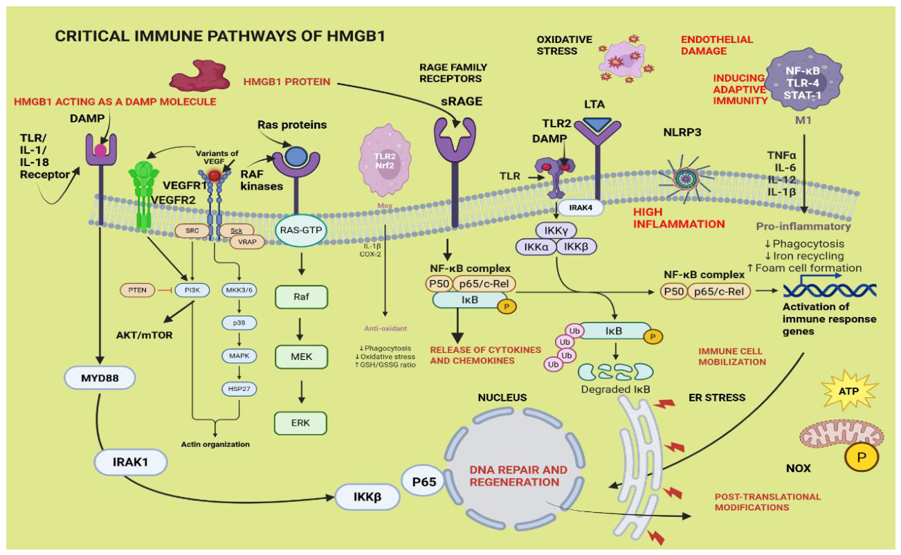
Legend- HMGB-1 -high mobility group box -1, sRAGE - soluble receptor for advanced glycation end products, ED- endothelial deregulation, DAMP – damage associated molecular pattern , NF-kB - nuclear factor kappa beta, TLR-toll-like receptor, TLR2 – toll-like receptor -2, TLR4 -toll-like receptor -4, STAT-1, signal transducer and activator of transcription-1, TNF-alpha – tumour necrosis factor -alpha, IL-6 – interleukin-six, IL-12 – interleukin -twelve, IL-18 – interleukin-eighteen, IL-1β – interleukin-one beta, ER -endoplasmic reticulum, DNA – deoxyribonucleic acid, NLRP3 -Nucleotide-binding oligomerization domain, Leucine-rich Repeat and Pyrin domain containing -3, LTA – lipoteichoic acid, MEKK - Mitogen-Activated Protein Kinase Kinase Kinase or MAPK3, TAK - Transforming growth factor-(beta)-activated kinase, TRAF-6 -Tumor necrosis factor Receptor-Associated Factor 6, RAS-GTP – rat sarcoma virus-guanosine tri phosphate, VEGF – vascular endothelial growth factor, VEGFR – vascular endothelial growth factor receptor, IRAK-1 - Interleukin-1 Receptor-Associated Kinase-1, IRAK-4 - Interleukin-1 Receptor-Associated Kinase -4, JNK – c-Jun N-terminal kinase, p38 - p38 mitogen-activated protein kinase, MYD88 – myeloid differentiation primary response gene 88, ERK - extracellular signal-regulated kinase, NOX- Nicotinamide Adenine Dinucleotide Phosphate oxidase, IL-18R – interleukin eighteen receptor, IFN-gamma – interferon gamma, ROS – reactive oxygen species, HIF-1alpha – hypoxia inducible factor -1 alpha, PLC- phospholipase -C, PI3K -phosphoinositide -3 -kinase, MAPK – mitogen associated protein kinase, Mox – oxidised macrophages, AKT/mTOR - protein kinase -B/ mammalian target of rapamycin, UB - ubiquitin
3.8. Quality of the Evidence
PRISMA 2020 (S16)
3.9. Agreements and Disagreements with other Studies or Reviews.
PDR is a common occurrence in diabetes, and the role of HMGB1 has been demonstrated in uncontrolled angiogenesis, retinal detachment, retinal neurodegeneration, and haemorrhage as per the published results of 3 of the included studies. The same diabetic populations were comorbid with diabetic nephropathy, hyperlipidaemia, and hypertension while this inflammatory phenotype was mediated by the NF-kB pathway. The studies by Abu El-Asar and colleagues 2011-2017 also assayed the common vascular biomarkers; sICAM-1, VCAM-1, sVAP-1, MCP-1, and VEGF, all of which were elevated due to the increased expression of HMGB1 titres in the diabetic group opposed to the non-diabetic controls. These elevated biomarkers promoted leucocyte adhesion to the endothelium, barrier dysfunction and fibroblast translocation which caused fibrosis of the internal organs. In active PDR, the HMGB1 levels were two-to threefold high in the vitreous fluid that was sampled simultaneously with the vascular biomarkers; soluble RAGE, soluble ICAM-1, and MCP-1 compared to the non-diabetic controls.
There was direct upregulation of HMGB1 in diabetic nephropathy, which reported increased inflammation, secretion of pro-inflammatory cytokines, podocyte autophagy and EMT along with ferroptosis respectively in another 3 of the included studies. Elevated levels of HMGB1 were also reported by YM Arabi and co-workers (2011) in a diabetic cohort which had higher BMI, low creatinine clearance and severe illness, and being comorbid with hypertension compared to its non-diabetic controls. Cytoplasmic or extracellular HMGB1 is said to promote autophagy by inducing either the TLR4/NF-kB pathway or the p38-inducible MAPK or the AKT/mTOR and rapamycin pathways which are thought to resolve DN by reducing apoptosis, EMT and the inflammation in the renal tubules. In atherogenicity reported by Al Hakeim and colleagues (2022) the elevated HMGB1 concentration in the diabetic cohort was associated with high glucose toxicity, low pancreatic beta cell function and increased atherogenic indices. The activation of RAGE leads to the biosynthesis of the pro-inflammatory mediators. HMGB1 also binds with TLR9 and activates the NF-kB pathway which generates superoxide anions by the mononuclear phagocytes. HMGB1 is secreted into the extracellular milieu following cellular necrosis which stimulates the secretion of the pro-inflammatory cytokines: TNF-alpha, IL-1beta and IL-6 that upregulate the systemic immune-inflammatory responses. Thus, the glucose metabolism is modulated, and the insulin sensitivity is weakened when HMGB1 is activated with hyperglycaemia and increased HOMA-IR which aggravate diabetes.
Obesity results in an imbalance of the endothelial Vaso-reactive substances that cause vasoconstriction, cell growth and inflammatory activation that progresses into ED. Thus, HMGB1 has been proposed as a surrogate marker of obesity and ED in children. The current findings have indicated a significant increase in the BMI, and HOMA-IR in the diabetic group which are also biomarkers of obesity. There could be a link between the adipocytes and the pancreatic beta cells which is modulated by the HMGB1 protein. High extracellular HMGB1 expression stimulated the macrophages to release pro-inflammatory cytokines while being located within the secretory lysosomes in the macrophages.
HMGB1 titres were profoundly upregulated in diabetic patients diagnosed with coronary thrombosis. The cardiovascular pathophysiology of such patients is characterised by high infiltration of immune cells, larger necrotic cores, larger atherosclerotic plaques, platelet reactivity, coagulability and impaired fibrinolysis. These conditions thus place those patients at very high risk of arterial thrombus formation and importantly, display high levels of the plasma HMGB1 protein.
Hafez and team (2018) examined the relationship between chronic hyperglycaemia, and an epigenetic enzyme of oxidative stress, Sirtuin -1, which is associated with foot ulceration in a diabetic population. The SIRT1 levels are decreased in severe inflammatory conditions caused by oxidative stress factors. With a low SIRT1 level chronic hyperglycaemia induces oxidative stress resulting in generating ROS which releases TNF-alpha and IL-6 levels linking those with the SIRT1 activity in T2D. DFU reportedly had high titres of HMGB1 as ulceration often destroys the blood vessels due to acute inflammation.
Skrha and co-workers (2012), described T2D with a duration of 9 years, which had excessive inflammation and increased levels of HMGB1, sRAGE and EN-RAGE in the T2D group. The RAGE molecules present on the macrophages, and endothelial cells are directly involved in the development of vessel wall abnormalities in diabetic patients. They stimulate the inflammatory activities thus causing endothelial dysfunction by upregulating the gene expression of cell adhesion molecules (ICAM-1, VCAM-1, the selectins), vascular growth factors, pro-inflammatory cytokines, migration of macrophages, the synthesis of fibronectin, proteoglycans, collagen IV, and stimulating cell proliferation and pro-thrombotic pathways.
Serum HMGB1 in comparison to urinary HMGB1 was reportedly more reliable as a biomarker for investigating diabetic kidney disease in which HMGB1 levels were significantly higher in the DKD cohort than the healthy control. Elevated serum HMGB1, serum creatinine and BUN were restored to normalcy when the HMGB1 concentration was decreased, in various kidney disease states. The current findings too indicate that the severity of kidney disease whether it is AKI or CKD or DKD and were attenuated with the knockdown of serum HMGB1 leading to the inhibition of RAGE/TLR4/NF-kB inflammatory pathway in data already published. The tubular epithelial HMGB1 has shown capability in promoting AKI to CKD transition as its latest discovery milestone.
HMGB1 concentration was correlated with the post-load insulin concentration and the HMGB1 in human adipose tissue predominantly displayed an insulin-resistant phenotype. In agreement with these observations, there were other studies that reported positive correlation of HMGB1 with the HbA1c%, FBG, insulin, HOMA-IR, TC, TG, LDL-c concentrations and negative correlation with HDL-c and HOMA-IR -beta concentration. These findings re-asserted that the HMGB1 functions are influenced by both glycaemic and lipid metabolism. The HOMA-IR beta concentration is related to the insulin produced by the beta cells in the pancreas which confirmed low levels of insulin secretion in the diabetic populations.
3.10. Conclusion
3.10.1. Implications for Practice
The SMD calculations have identified HMGB1 as the sixth highest effect size from among 18 of other biomarkers that were evaluated in this study (Figure 4). Therefore, it qualifies as a potential diagnostic marker of clinical type-2 diabetes which has broad implications in terms of early detection of disease and initiation of treatment in the clinic and for the therapists who offer customised lifestyle changes for the management of the disease.
3.10.2. Implications for Research
More research studies are needed to elevate HMGB1 to the level of the cytokine biomarkers which are already in clinical use but offers hope to the drug manufacturers in the pharmaceutical industry. Further, customised treatment strategies could be revised with adjusting the dose and frequency of the insulin intake. One of the latest findings on the impact of HMGB1 on diabetes is the modulation of the gut microbiome to regulate the inflammatory activity of HMGB1 which has been proven successful in a mouse model. The manipulation of the different immune pathways that influence the immune activity of HMGB1 as an immune modulator may provide effective leads that would highlight other prospective therapeutic targets which need to be combined with HMGB1.
4. Meta-Analysis
4.1. Summary Results
There are three types of tables/figures,
4.2. Narrative Summary
(i) Description of the type of intervention in the in cluded studies and how they were implemented.
The type of intervention was the HMGB1 nuclear protein that could perform both proinflammatory functions and anti-inflammatory functions based on its binding site such as the promoter region of the box-A domain or the box-B domain with its acetylation or post-translational modifications which initiate a cascade of molecular reactions that promote inflammation. Extracellular HMGB1 binds with its cell surface receptors, sRAGE for entry into the cells and involve a multitude of biochemical mechanisms which has broad scope for attenuating inflammation associated with T2D-induced ED. HMGB1 also upregulates immunity by its activities as a DAMP molecule which leads to activating both innate and adaptive immunity, initiated by immune cell extravasation, leucocyte rolling on the endothelium, fat deposition leading to thrombosis and plaque formation. As an inflammatory biomarker, HMGB1 was the focus of this study to understand its impact on pre-existing T2D leading to ED.
(ii) Description of the primary and secondary outcomes in the included studies.
The forest plots of each marker, which was significantly upregulated in the experimental group included: HMGB1 (SMD 2.86, p= 0.00001), SBP (SMD 0.53, p=0.04), HbA1c% (SMD 3.68, p=0.00001), FBG (SMD 2.41, p=0.0001), HOMA-IR (SMD 3.59, p=0.0000.1), TC (SMD 0.78, p= 0.02), TG (SMD 1.31, p=0.0002), HDL-c (SMD -0.81, p= 0.0009), sICAM-1(SMD 0.86, p= 0.005), HR (SMD 0.59, p= 0.0008), CRP (SMD 0.49, p= 0.0009), IL-6 (SMD 5.20, p=0.00001), SCr (SMD 0.84, p= 0.03), BUN (SMD 1.96, p=0.02), ACR (SMD 3.03, p= 0.04), eGFR (SMD -3.41, p= 0.04), BMI (SMD 1.04, p= 0.0001), Age (SMD 0.77, p=0.002), LVEF% (SMD -4.06, p<0.05), LVDV (SMD 2.01. p<0.05) and LVSV (SMD 1.88, p<0.05). The secondary outcomes consisted of the non-parametric unpaired Mann-Whitney U test in which the biomarkers that showed a significant outcome included: HMGB1, HbAIc%, FBG, TG, HOMA-IR, HDL-c and BMI (Figure 5).
(iii)A review of findings for the secondary outcomes.
The unpaired non-parametric Mann Whitney U-test results were displayed in bar graphs which highlighted the increases or decreases in the primary outcomes (Figure 5). The HMGB1 (p=0.0002), HbA1c% (p= <0.0001), FBG (p= <0.0001), HOMA-IR (p=0.0159), TG (p=0.0288) and BMI (p=0.0115) exhibited significant increases in the T2D patients compared to the healthy control and a significant corresponding decrease in the HDL-c biomarker (p=0.0056).
(iv) Adverse outcomes
The adverse outcomes that have resulted from the primary outcomes can be listed as the high-risk -bearing biomarkers of the vascular parameters shown by the forest plots of sICAM-1, sVCAM-1, p-Selectin, vWF, sVAP-1, sVE-Cadherin, VEGF, s-Endoglin, and the heart rate in the T2D cohort. Although they were not depicted as significant outcomes in the Mann Whitney U-test, due to being reported mostly by only one or two included studies, basically lacking sufficient study data, the vascular parameters which displayed high risk in developing vascular complications in the T2D subjects, leave those patients exposed and prone to developing microvascular complications. Most of these patients were reportedly co-morbid with diseases such as ED, DR, PDR, DN, DFU, atherogenicity, coronary thrombosis, hypertension, hyperlipidaemia, smoking - related diseases, COPD and obesity.
(v)Associated financial costs in implementing the study design.
Conducting a meta-analysis is possible with a small budget.
(vi) Possible important contextual details pertaining to the study design and /or analysis.
The PICOS criteria pertaining to this study design conforms to the parameters of a reliable research study because they are being case - control baseline studies. They compare the vascular disease data directly with their healthy control participants and are validated by performing several different statistical tests that determined the primary and secondary outcomes.
(vii) Perceived strengths, weaknesses and contributions made on the studies
The strengths in this review are: (i) having the intervention compared with the healthy control, (ii) longer duration of diabetes, (iii) all the studies being T2D, (iv) clinical studies which conformed to the accepted ethical standards and (v) the parameters measured in body fluids (blood or urine). Those attributes increase the reliability of the data extracted compared to animal studies which may not correlate directly with human studies.
IL-6 being the best biomarker that will indicate early signs of diabetes, could stimulate insulin secretion from the pancreas during episodes of insulin deficiency. When hyperglycaemia destabilises the lipid and insulin homeostasis, they combinedly serve to produce a low-level inflammation that warrants pro-inflammatory cytokine upregulation which has a beneficial positive feedback loop in action. The fasting blood glucose level plausibly indicates serious insulin resistance more than insulin deficiency, because this study included data exclusively from the T2D.
There are many limitations to this study such as during screening, many studies not conforming to our PICOS statement. The reliability of the outcomes was not supported by any demographic data pertaining to the lifestyle and socioeconomic status or the educational level in most of the included studies which otherwise exert a considerable influence on the nutritional status and immunity of the diabetics. Having smaller sample size, not having done repeats, adoption of different methodologies to evaluate the same outcome are some weaknesses encountered in the included studies. Some studies were retrospective studies which had a long list of comorbidities. The gap that exists is not having clinical trials putting emphasis on HMGB1 as a noteworthy inflammatory primary outcome, and not considering it as a useful therapeutic target, which researchers may consider evaluating after this study is published. The contribution this study makes is raising awareness among the scientific community to consider HMGB1 as an immunological target and a diagnostic biomarker for the early detection of T2D in persons aged over 40 years. Further, this study including a meta-analysis, gives access to a large amount of data from pooling the values reported for biomarkers which are associated with T2D-induced ED. The data obtained from 16 included studies involving a large number of participants, also was sufficient to validate many ED-related outcomes as the minimum number of participants required by the power calculation is a total of 8. The best contribution made is categorizing the available data into biomarker models that offered a comprehensive and reliable analysis upon which further studies could be based, especially in utilizing HMGB1 as a possible anti-inflammatory therapeutic option for resolving T2D-induced ED.
4.3. Report on Practical Significance
HMGB1 showed a significant increase in the T2D cohort compared to the healthy control. The HbA1c%, FBG and the insulin biomarkers; HOMA-IR except for fasting serum insulin (FINS) also showed significant elevation in the T2D cohort validated by both the forest plots and the Mann Whitney - U test. Among the lipid markers, HDL-c was significantly low in the diabetic group and all the vascular biomarkers indicated high risk in the T2D cohort in the forest plots except for e-Selectin. All the renal, inflammatory and obesity-related biomarkers showed high-risk of developing ED in the forest plots. Any one of these biomarkers could also serve as prognostic and diagnostic markers for screening as well as treatment, particularly for vascular dysfunction that originates from ED. The healthcare burden of treating diabetic patients harbouring complications such as ED and vascular disease is high. In Australia, diabetes medication is offered free but adds on to the economic burden of hospitalisation costs, rehabilitation, physician visits and the productivity losses due to premature mortality or long-term disabilities developing from severe morbidity. The implications of this study in future diabetic research should also involve the suppression of macrophage activation which secretes HMGB1 and promoting relevant anti-inflammatory responses such as anti-IL-6 antibody treatment.
Author Contributions
Ranmali Ranasinghe (RR), Michael Mathai (MM), and Anthony Zulli (AZ) RR created the topic, did the writing, organizing, and referencing of the manuscript. AZ introduced the concept, and MM did the editing and both AZ and MM gave constructive criticism.
Funding
No funding has been received.
Ethics Approval and Consent to Participate
Not applicable.
Consent for Publication
All authors of this review have consented for publication.
Acknowledgments
The first author was a recipient of the RTP scholarship for PhD candidature from the Australian government.
Conflicts of Interest
No competing interests exist.
List of Abbreviations
| AGEs - Advanced glycation end products |
| AI% - Augmentation index percentage |
| AIHW - Australian institute of health and welfare |
| ACR - Albumin creatinine ratio |
| ALT - Alanine amino transferase |
| α7nAchR - Alpha 7 nicotinic acetylcholine receptor |
| AMP - Adenosine mono phosphate |
| AMPK - Adenosine mono phosphate kinase |
| Ang-1 - Angiopoietin-one |
| Ang-2 - Angiopoietin-two |
| ANP - Atrial natriuretic peptide |
| APO-A - Apolipoprotein-A |
| APO-B - Apolipoprotein -B |
| Arf6 - ADP-ribosylation factor -6 |
| AST - Aspartate amino transferase |
| AT2R - Angiotensin receptor two |
| BBB - Blood brain barrier |
| BMI - Body mass index |
| BNP - Brain natriuretic peptide |
| BUN - Blood urea nitrogen |
| CAD - coronary artery disease |
| cm - Centimetre |
| CAM - Cell adhesion molecules |
| CBA - Control before and after |
| CCTA - Coronary computed tomography angiography |
| CK-MB - Creatine kinase in muscle and brain |
| COX - Cyclooxygenase |
| CPG-ODN - Cpg-oligodeoxynucleotides |
| CTP - Cardiac troponin |
| CXCR4 - CXC-chemokine receptor four |
| CXCL12 - CXC-motif- chemokine ligand 12 |
| CVD - Cardiovascular disease |
| DAMP – Damage associated molecular pattern |
| DBP - Diastolic blood pressure |
| DCM - Diabetic cardiomyopathy |
| DM - Diabetes mellitus |
| DNA - Deoxyribonucleic acid |
| DOI - Digital object identifier |
| DPPI-4 - Dipeptidyl peptidase inhibitor 4 |
| ECG/EKG - Electrocardiogram |
| ED - Endothelial dysregulation/ dysfunction/ deregulation |
| EGFR - Estimated glomerular filtration rate |
| EF% - Ejection fraction percentage |
| eNOS - Endothelial nitric oxide synthase |
| ER - Endoplasmic reticulum |
| ERK - Extracellular signal regulated kinases |
| ER - Endoplasmic reticulum |
| ESAM – Endothelial-cell selective adhesion molecule |
| ET-1 - Endothelin-1 |
| ETC - Electron transport chain |
| FBG - Fasting blood glucose |
| FPG – Fasting plasma glucose |
| FMD% -Flow-mediated dilatation percentage |
| FS% - Fractional shortening percentage |
| GGT - Gamma-glutamyl transferase |
| GLP-1 - Glucagon-like peptide-1 |
| GPCR - G protein coupled receptor |
| GRP78 - Glucose regulated protein 78 |
| GSH - Glutathione |
| HbA1c - Glycated haemoglobin |
| HC - Healthy control |
| HDL-C - High density lipoprotein cholesterol |
| HIV - Human immunodeficiency virus |
| HMG - High mobility group molecular family |
| HMGB1 – High mobility group box-1 |
| HO-1 - Haem oxygenase-one |
| HOMA-IR - Homeostatic model assessment of Insulin resistance |
| HR - Heart rate |
| HT - Height |
| HW - Heart weight |
| ICAM - Intercellular cell adhesion molecule |
| IDF - International diabetes federation |
| IL - Interleukin |
| INF-JNK - Interferon JNK |
| INS - Insulin |
| IR - Insulin resistance |
| JNK - Janus kinase |
| KDa - Kilo Dalton |
| LDH - Lactate dehydrogenase |
| LDL-C - Low-density lipoprotein cholesterol |
| LPS - Lipopolysaccharide |
| LVIDd - Left ventricular internal diameter at end-diastole |
| LVIDs - Left ventricular internal diameter at end-systole |
| LVDV - Left ventricular diastolic volume |
| LVSV - Left ventricular systolic volume |
| MAPK - Mitogen-activated protein kinase |
| MASLD - Metabolic -dysfunction associated- steatosis liver disease |
| MCP-1 - Monocyte chemo attractant protein-1 |
| MDA - Malondialdehyde |
| MD2 - Myeloid differentiation protein-2 |
| MFN2 - Mitofusin-2 |
| MIR - Micro RNA |
| MI - Myocardial infarction |
| mm - millimetre |
| MyD88 – Myeloid differentiation factor 88 |
| NADPH – Nicotinamide adenine dinucleotide phosphate |
| NF-KB – Nuclear factor kappa beta |
| NLRP3 – NLR family pyrin binding domain 3 |
| NOD – Nucleotide-binding oligomerization |
| NO - Nitrous oxide |
| NOX – NADPH oxidase |
| Nrf2 – Nuclear factor erythroid 2-relate |
| NT-Pro BNP – Natriuretic peptide test |
| PAI-1 – Plasminogen activator inhibitor -one |
| PAD – Peripheral artery disease |
| PAMP - Pathogen-associated molecular pattern |
| PARP-1 - Poly (ADP ribose) polymerase-one |
| PTM – post-translational modifications |
| PRISMA - Preferred reporting items for systematic reviews and meta-analyses |
| PWV – Pulse wave velocity |
| RAAs – Renin Angiotensin aldosterone system |
| RAGE – Receptor for AGEs |
| RNA – Ribonucleic acid |
| RNS – Reactive nitrogen species |
| ROB – Risk of bias |
| ROC – Receiver operator characteristic curve |
| ROBINS-1 – Risk of bias of non-randomized studies |
| ROS – Reactive oxygen species |
| RR – Risk ratio |
| SARS-Cov-2 – severe acute respiratory syndrome-corona virus-2 |
| SBP – Systolic blood pressure |
| SCr – Serum creatinine |
| SD – Standard deviation |
| SEM – Standard error of the mean |
| SMD- Standardized mean difference |
| SGLT2 – Sodium glucose co-transporter 2 inhibitors |
| SIRT-1 – Sirtuin -1 |
| SMAD-2 – Mothers against decapentaplegic homolog 2 |
| SO -Super oxide |
| SOCE – Store-operated calcium entry |
| SOD – Superoxide dismutase |
| STAT-1 – Signal transducer and activator of transcription-1 |
| TBARS – Thio barbituric acid |
| TC - Total cholesterol |
| T1D/T1DM - Type one diabetes |
| T2D/T2DM - Type two diabetes |
| TG - Triglyceride |
| TGF-β1 - Transforming growth factor -beta one |
| Tie -2 - Tyrosine protein kinase |
| TNF-A - Tumour necrosis factor-alpha |
| TLR - Toll-like receptor |
| TLR4 - Toll-like receptor 4 |
| TIR - Time in range |
| TP53 - Tumour protein 53 |
| TREM-1 - Triggering receptor expressed on myeloid cell group 1 |
| TRPV4 - Transient receptor potential vanilloid family member 4 |
| US/USA - United States of America |
| VAP-1 - Vascular adhesion protein-one |
| VCAM-1 - Vascular cell adhesion molecule – one |
| VE-Cadherin - Vascular endothelial cadherin |
| VEGF - Vascular endothelial growth factor |
| VLDL-c – Very low-density lipoprotein cholesterol |
| vWF - Von Willebrand factor |
| WHO - World Health Organization |
| XO - Xanthine oxidase |
References
- Theofilis, P.; Sagris, M.; Oikonomou, E.; Antonopoulos, A.S.; Siasos, G.; Tsioufis, C.; Tousoulis, D. Inflammatory Mechanisms Contributing to Endothelial Dysfunction. Biomedicines 2021, 9, 781. [Google Scholar] [CrossRef] [PubMed]
- Laakso, M.; Kuusisto, J. Insulin resistance and hyperglycaemia in cardiovascular disease development. Nat. Rev. Endocrinol. 2014, 10, 293–302. [Google Scholar] [CrossRef] [PubMed]
- Pluta, W.; Lubkowska, A.; Dudzińska, W. Vascular Endothelium in Health and Disease: Structure, Function, Assessment and Role in Metabolic Disorders. Vasc. Heal. Risk Manag. 2025, ume 21, 729–747. [Google Scholar] [CrossRef] [PubMed]
- Venereau, E.; De Leo, F.; Mezzapelle, R.; Careccia, G.; Musco, G.; Bianchi, M.E. HMGB1 as biomarker and drug target. Pharmacol. Res. 2016, 111, 534–544. [Google Scholar] [CrossRef] [PubMed]
- Yang, Y.; Chan, L. Monogenic Diabetes: What It Teaches Us on the Common Forms of Type 1 and Type 2 Diabetes. Endocr. Rev. 2016, 37, 190–222. [Google Scholar] [CrossRef] [PubMed]
- Koufakis, T.; Grammatiki, M.; Kotsa, K. Type 2 diabetes management in people aged over seventy-five years: targets and treatment strategies. Maturitas 2021, 143, 118–126. [Google Scholar] [CrossRef] [PubMed]
- Bajaj, M.; A DeFronzo, R. Metabolic and molecular basis of insulin resistance. J. Nucl. Cardiol. 2003, 10, 311–323. [Google Scholar] [CrossRef] [PubMed]
- Raheb, M.A.; Niazmand, V.R.; Eqra, N.; Vatankhah, R. Subcutaneous insulin administration by deep reinforcement learning for blood glucose level control of type-2 diabetic patients. Comput. Biol. Med. 2022, 148, 105860. [Google Scholar] [CrossRef] [PubMed]
- Solomon, T.P.J. Sources of Inter-individual Variability in the Therapeutic Response of Blood Glucose Control to Exercise in Type 2 Diabetes: Going Beyond Exercise Dose. Front. Physiol. 2018, 9, 896. [Google Scholar] [CrossRef] [PubMed]
- Yang, Z.-J.; Liu, J.; Ge, J.-P.; Chen, L.; Zhao, Z.-G.; Yang, W.-Y. Study Group Prevalence of cardiovascular disease risk factor in the Chinese population: the 2007-2008 China National Diabetes and Metabolic Disorders Study. Eur. Heart J. 2012, 33, 213–220. [Google Scholar] [CrossRef] [PubMed]
- Lyu, Y.; Luo, Y.; Li, C.; Guo, X.; Lu, J.; Wu, H.; Huo, X.; Gu, W.; Yang, G.; Ji, L.; et al. Regional Differences in the Prevalence of Coronary Heart Disease and Stroke in Patients With Type 2 Diabetes in China. J. Clin. Endocrinol. Metab. 2018, 103, 3319–3330. [Google Scholar] [CrossRef] [PubMed]
- Yachmaneni, A., Jr.; et al. A comprehensive review of the vascular consequences of diabetes in the lower extremities: current approaches to management and evaluation of clinical outcomes. Cureus 2023, 15(10). [Google Scholar] [CrossRef] [PubMed]
- Tang, D.; Kang, R.; Zeh, H.J.; Lotze, M.T. The multifunctional protein HMGB1: 50 years of discovery. Nat. Rev. Immunol. 2023, 23, 824–841. [Google Scholar] [CrossRef] [PubMed]
- Chen, R.; Kang, R.; Tang, D. The mechanism of HMGB1 secretion and release. Exp. Mol. Med. 2022, 54, 91–102. [Google Scholar] [CrossRef] [PubMed]
- Andersson, U.; Yang, H.; Harris, H. Extracellular HMGB1 as a therapeutic target in inflammatory diseases. Expert Opin. Ther. Targets 2018, 22, 263–277. [Google Scholar] [CrossRef] [PubMed]
- Ren, W.; Zhao, L.; Sun, Y.; Wang, X.; Shi, X. HMGB1 and Toll-like receptors: potential therapeutic targets in autoimmune diseases. Mol. Med. 2023, 29, 1–13. [Google Scholar] [CrossRef] [PubMed]
- Foglio, E.; Pellegrini, L.; Russo, M.A.; Limana, F. HMGB1-Mediated Activation of the Inflammatory-Reparative Response Following Myocardial Infarction. Cells 2022, 11, 216. [Google Scholar] [CrossRef] [PubMed]
- Andersson, U.; Erlandsson-Harris, H.; Yang, H.; Tracey, K.J. HMGB1 as a DNA-binding cytokine. J. Leukoc. Biol. 2002, 72, 1084–1091. [Google Scholar] [CrossRef]
- Mandke, P.; Vasquez, K.M. Interactions of high mobility group box protein 1 (HMGB1) with nucleic acids: Implications in DNA repair and immune responses. DNA Repair 2019, 83, 102701–102701. [Google Scholar] [CrossRef] [PubMed]
- Bianchi, M.E.; Manfredi, A.A. High-mobility group box 1 (HMGB1) protein at the crossroads between innate and adaptive immunity. Immunol. Rev. 2007, 220, 35–46. [Google Scholar] [CrossRef] [PubMed]
- Behl, T.; Sharma, E.; Sehgal, A.; Kaur, I.; Kumar, A.; Arora, R.; Pal, G.; Kakkar, M.; Kumar, R.; Bungau, S. Expatiating the molecular approaches of HMGB1 in diabetes mellitus: Highlighting signalling pathways via RAGE and TLRs. Mol. Biol. Rep. 2021, 48, 1869–1881. [Google Scholar] [CrossRef] [PubMed]
- Kaur, N. Stressed cardiomyocytes in diabetes disrupt intercellular harmony; The University of Manchester (United Kingdom), 2022. [Google Scholar]
- Hossain, M.J.; Al-Mamun, M.; Islam, M.R. Diabetes mellitus, the fastest growing global public health concern: Early detection should be focused. Health Sci. Rep. 2024, 7(3), e2004. [Google Scholar] [CrossRef] [PubMed]
- Soomro, M.H.; Jabbar, A. Diabetes etiopathology, classification, diagnosis, and epidemiology, in BIDE's Diabetes Desk Book; Elsevier, 2024; pp. 19–42. [Google Scholar]
- Peralta, G.L. Hypoglycaemia Prediction on Type 1 Diabetes Patients using Continuous Glucose Monitoring and Health Record Data. 2022. [Google Scholar] [CrossRef] [PubMed]
- Zhu, R.; Zhou, S.; Xia, L.; Bao, X. Incidence, Morbidity and years Lived With Disability due to Type 2 Diabetes Mellitus in 204 Countries and Territories: Trends From 1990 to 2019. Front. Endocrinol. 2022, 13, 905538. [Google Scholar] [CrossRef] [PubMed]
- Smokovski, I. Managing Diabetes in Low Income Countries; Springer Nature: Durham, NC, United States; ISBN, 2021. [Google Scholar]
- Roman, G. A. Pantea Stoian, Cardiovascular risk/disease in type 2 diabetes mellitus. In Type 2 Diabetes: From Pathophysiology to Cyber Systems; 2021. [Google Scholar]
- Dasu, M.R.; Devaraj, S.; Park, S.; Jialal, I. Increased Toll-Like Receptor (TLR) Activation and TLR Ligands in Recently Diagnosed Type 2 Diabetic Subjects. Diabetes Care 2010, 33, 861–868. [Google Scholar] [CrossRef] [PubMed]
- El-Asrar, A.M.A.; et al. High-mobility group box-1 and biomarkers of inflammation in the vitreous from patients with proliferative diabetic retinopathy. Mol. Vis. 2011, 17, 1829. [Google Scholar] [PubMed]
- Arabi, Y.M.; Dehbi, M.; Rishu, A.H.; Baturcam, E.; Kahoul, S.H.; Brits, R.J.; Naidu, B.; Bouchama, A. sRAGE in diabetic and non-diabetic critically ill patients: effects of intensive insulin therapy. Crit. Care 2011, 15, R203–R203. [Google Scholar] [CrossRef] [PubMed]
- Wang, L.J.; Lu, L.; Zhang, F.R.; Chen, Q.J.; De Caterina, R.; Shen, W.F. Increased Serum High-Mobility Group Box-1 and Cleaved Receptor for Advanced Glycation Endproducts Levels and Decreased Endogenous Secretory Receptor for Advanced Glycation Endproducts Levels in Diabetic and Non-Diabetic Patients with Heart Failure. Eur. J. Hear. Fail. 2011, 13, 440–449. [Google Scholar] [CrossRef] [PubMed]
- krha, J., Jr.; et al. Relationship of Soluble RAGE and RAGE Ligands HMGB1 and EN-RAGE to Endothelial Dysfunction in Type 1 and Type 2 Diabetes Mellitus. Exp. Clin. Endocrinol. Diabetes 2012, 120(05), 277–281. [Google Scholar] [CrossRef]
- Abu El-Asrar, A.M.; Nawaz, M.I.; Kangave, D.; Abouammoh, M.; Mohammad, G. High-Mobility Group Box-1 and Endothelial Cell Angiogenic Markers in the Vitreous from Patients with Proliferative Diabetic Retinopathy. Mediat. Inflamm. 2012, 2012, 697489. [Google Scholar] [CrossRef] [PubMed]
- Chen, Y.; Qiao, F.; Zhao, Y.; Wang, Y.; Liu, G. HMGB1 is activated in type 2 diabetes mellitus patients and in mesangial cells in response to high glucose. Int. J. Clin. Exp. Pathol. 2015, 8, 6683–91. [Google Scholar] [PubMed]
- Wang, H.; Qu, H.; Deng, H. Plasma HMGB-1 Levels in Subjects with Obesity and Type 2 Diabetes: A Cross-Sectional Study in China. PLoS ONE 2015, 10, e0136564. [Google Scholar] [CrossRef] [PubMed]
- Wei, H.; Qu, H.; Wang, H.; Deng, H. Plasma C1q/TNF-Related Protein-3 (CTRP-3) and High-Mobility Group Box-1 (HMGB-1) Concentrations in Subjects with Prediabetes and Type 2 Diabetes. J. Diabetes Res. 2016, 2016, 1–8. [Google Scholar] [CrossRef] [PubMed]
- El-Asrar, A.M.A.; et al. Association of HMGB1 with oxidative stress markers and regulators in PDR. Mol. Vis. 2017, 23, 853. [Google Scholar] [PubMed]
- Hafez, Y.M.; El-Deeb, O.S.; Atef, M.M. The emerging role of the epigenetic enzyme Sirtuin-1 and high mobility group Box 1 in patients with diabetic foot ulceration. Diabetes Metab. Syndr. Clin. Res. Rev. 2018, 12, 1065–1070. [Google Scholar] [CrossRef] [PubMed]
- Jin, J.; Gong, J.; Zhao, L.; Zhang, H.; He, Q.; Jiang, X. Inhibition of high mobility group box 1 (HMGB1) attenuates podocyte apoptosis and epithelial-mesenchymal transition by regulating autophagy flux. J. Diabetes 2019, 11, 826–836. [Google Scholar] [CrossRef] [PubMed]
- Huang, J.; Zeng, T.; Tian, Y.; Wu, Y.; Yu, J.; Pei, Z.; Tan, L. Clinical significance of high-mobility group box-1 (HMGB1) in subjects with type 2 diabetes mellitus (T2DM) combined with chronic obstructive pulmonary disease (COPD). J. Clin. Lab. Anal. 2019, 33, e22910. [Google Scholar] [CrossRef] [PubMed]
- Zhu, Z.; Peng, X.; Li, X.; Tu, T.; Yang, H.; Teng, S.; Zhang, W.; Xing, Z.; Tang, J.; Hu, X.; et al. HMGB1 impairs endothelium-dependent relaxation in diabetes through TLR4/eNOS pathway. FASEB J. 2020, 34, 8641–8652. [Google Scholar] [CrossRef] [PubMed]
- Wu, Y.; Zhao, Y.; Yang, H.-Z.; Wang, Y.-J.; Chen, Y. HMGB1 regulates ferroptosis through Nrf2 pathway in mesangial cells in response to high glucose. Biosci. Rep. 2021, 41. [Google Scholar] [CrossRef] [PubMed]
- Al-Hakeim, H.K.; Al-Kaabi, Q.J.; Maes, M. High mobility group box 1 and Dickkopf-related protein 1 as biomarkers of glucose toxicity, atherogenicity, and lower β cell function in patients with type 2 diabetes mellitus. Growth Factors 2022, 40, 240–253. [Google Scholar] [CrossRef] [PubMed]
- Yamashita, A.; Nishihira, K.; Matsuura, Y.; Ito, T.; Kawahara, K.; Hatakeyama, K.; Hashiguchi, T.; Maruyama, I.; Yagi, H.; Matsumoto, M.; et al. Paucity of CD34-positive cells and increased expression of high-mobility group box 1 in coronary thrombus with type 2 diabetes mellitus. Atherosclerosis 2012, 224, 511–514. [Google Scholar] [CrossRef] [PubMed]
- Targher, G.; Lonardo, A.; Byrne, C.D. Nonalcoholic fatty liver disease and chronic vascular complications of diabetes mellitus. Nat. Rev. Endocrinol. 2017, 14, 99–114. [Google Scholar] [CrossRef] [PubMed]
- Joseph, J.J.; Deedwania, P.; Acharya, T.; Aguilar, D.; Bhatt, D.L.; Chyun, D.A.; Di Palo, K.E.; Golden, S.H.; Sperling, L.S. on behalf of the American Heart Association Diabetes Committee of the Council on Lifestyle and Cardiometabolic Health; Council on Arteriosclerosis, Thrombosis and Vascular Biology; Council on Clinical Cardiology; and Council on Hypertension Comprehensive Management of Cardiovascular Risk Factors for Adults With Type 2 Diabetes: A Scientific Statement From the American Heart Association. Circulation 2022, 145, E722–E759. [Google Scholar] [CrossRef] [PubMed]
- Ali, M.K.; Bullard, K.M.; Gregg, E.W.; del Rio, C. A Cascade of Care for Diabetes in the United States: Visualizing the Gaps. Ann. Intern. Med. 2014, 161, 681–689. [Google Scholar] [CrossRef] [PubMed]
- Valenzuela, P.L.; Carrera-Bastos, P.; Gálvez, B.G.; Ruiz-Hurtado, G.; Ordovas, J.M.; Ruilope, L.M.; Lucia, A. Lifestyle interventions for the prevention and treatment of hypertension. Nat. Rev. Cardiol. 2020, 18, 251–275. [Google Scholar] [CrossRef] [PubMed]
- Awuchi, C.G.; Echeta, C.K.; Igwe, V.S. Diabetes and the nutrition and diets for its prevention and treatment: a systematic review and dietetic perspective. Health Sci. Res. 2020, 6(1), 5–19. [Google Scholar]
- Aronow, W.S. Diabetic cardiomyopathy in the elderly. Curr. Cardiovasc. Risk Rep. 2013, 7, 490–494. [Google Scholar] [CrossRef]
- Dong, H.; Zhang, Y.; Huang, Y.; Deng, H. Pathophysiology of RAGE in inflammatory diseases. Front. Immunol. 2022, 13, 931473. [Google Scholar] [CrossRef] [PubMed]
- Jia, G.; Hill, M.A.; Sowers, J.R. Diabetic cardiomyopathy: an update of mechanisms contributing to this clinical entity. Circ. Res. 2018, 122(4), 624–638. [Google Scholar] [CrossRef] [PubMed]
- Adeghate, E.; Singh, J. Structural changes in the myocardium during diabetes-induced cardiomyopathy. Hear. Fail. Rev. 2013, 19, 15–23. [Google Scholar] [CrossRef] [PubMed]
- Lu, F.-H.; Fu, S.-B.; Leng, X.; Zhang, X.; Dong, S.; Zhao, Y.-J.; Ren, H.; Li, H.; Zhong, X.; Xu, C.-Q.; et al. Role of the Calcium-Sensing Receptor in Cardiomyocyte Apoptosis via the Sarcoplasmic Reticulum and Mitochondrial Death Pathway in Cardiac Hypertrophy and Heart Failure. Cell. Physiol. Biochem. 2013, 31, 728–743. [Google Scholar] [CrossRef] [PubMed]
- Mrowicka, M.; Mrowicki, J.; Majsterek, I. Relationship between Biochemical Pathways and Non-Coding RNAs Involved in the Progression of Diabetic Retinopathy. J. Clin. Med. 2024, 13, 292. [Google Scholar] [CrossRef] [PubMed]
- Baurzhan, V. TRENDS IN INCIDENCE AND MORTALITY FROM DISEASES INCLUDED IN DISEASE MANAGEMENT PROGRAMS: DIABETES MELLITUS, ARTERIAL HYPERTENSION, CHRONIC HEART FAILURE. Science 2024, 26, 4. [Google Scholar]
- Mallik, S.; Paria, B.; Firdous, S.M.; Ghazzawy, H.S.; Alqahtani, N.K.; He, Y.; Li, X.; Gouda, M.M. The positive implication of natural antioxidants on oxidative stress-mediated diabetes mellitus complications. J. Genet. Eng. Biotechnol. 2024, 22, 100424. [Google Scholar] [CrossRef] [PubMed]
- Calay, D.; Mason, J.C. The Multifunctional Role and Therapeutic Potential of HO-1 in the Vascular Endothelium. Antioxid. Redox Signal. 2014, 20, 1789–1809. [Google Scholar] [CrossRef] [PubMed]
- Saraswat, N.; Chandra, P.; Sachan, N.; Vyawahare, N. A Detailed Review of Molecular Pathways and Mechanisms Responsible for the Development and Aggravation of Neuropathy and Nephropathy in Diabetes. Curr. Mol. Pharmacol. 2023, 17, 1–1. [Google Scholar] [CrossRef] [PubMed]
- Quintanilha, B.J.; Reis, B.Z.; Duarte, G.B.S.; Cozzolino, S.M.F.; Rogero, M.M. Nutrimiromics: Role of microRNAs and Nutrition in Modulating Inflammation and Chronic Diseases. Nutrients 2017, 9, 1168. [Google Scholar] [CrossRef] [PubMed]
- Bays, H.E.; Taub, P.R.; Epstein, E.; Michos, E.D.; Ferraro, R.A.; Bailey, A.L.; Kelli, H.M.; Ferdinand, K.C.; Echols, M.R.; Weintraub, H.; et al. Ten things to know about ten cardiovascular disease risk factors. Am. J. Prev. Cardiol. 2021, 5, 100149. [Google Scholar] [CrossRef] [PubMed]
- Wong, N.D.; Zhao, Y.; Patel, R.; Patao, C.; Malik, S.; Bertoni, A.G.; Correa, A.; Folsom, A.R.; Kachroo, S.; Mukherjee, J.; et al. Cardiovascular Risk Factor Targets and Cardiovascular Disease Event Risk in Diabetes: A Pooling Project of the Atherosclerosis Risk in Communities Study, Multi-Ethnic Study of Atherosclerosis, and Jackson Heart Study. Diabetes Care 2016, 39, 668–676. [Google Scholar] [CrossRef] [PubMed]
- Lorber, D. Importance of cardiovascular disease risk management in patients with type 2 diabetes mellitus. Diabetes Metab. Syndr. Obes. Targets Ther. 2014, 7, 169–183. [Google Scholar] [CrossRef] [PubMed]
- Wong, N.D.; Sattar, N. Cardiovascular risk in diabetes mellitus: epidemiology, assessment and prevention. Nat. Rev. Cardiol. 2023, 20, 685–695. [Google Scholar] [CrossRef] [PubMed]
- Ahuja, A.; Agrawal, S.; Acharya, S.; Reddy, V.; Batra, N. Strategies for Cardiovascular Disease Prevention in Type 1 Diabetes: A Comprehensive Review. Cureus 2024, 16, e66420. [Google Scholar] [CrossRef] [PubMed]
- Durlach, V.; Vergès, B.; Al-Salameh, A.; Bahougne, T.; Benzerouk, F.; Berlin, I.; Clair, C.; Mansourati, J.; Rouland, A.; Thomas, D.; et al. Smoking and diabetes interplay: A comprehensive review and joint statement. Diabetes Metab. 2022, 48, 101370. [Google Scholar] [CrossRef] [PubMed]
- El-Mahdy, M.A.; Ewees, M.G.; Eid, M.S.; Mahgoup, E.M.; Khaleel, S.A.; Zweier, J.L. Electronic cigarette exposure causes vascular endothelial dysfunction due to NADPH oxidase activation and eNOS uncoupling. Am. J. Physiol. Circ. Physiol. 2022, 322, H549–H567. [Google Scholar] [CrossRef] [PubMed]
- Makievskaya, C.I.; Popkov, V.A.; Andrianova, N.V.; Liao, X.; Zorov, D.B.; Plotnikov, E.Y. Ketogenic Diet and Ketone Bodies against Ischemic Injury: Targets, Mechanisms, and Therapeutic Potential. Int. J. Mol. Sci. 2023, 24, 2576. [Google Scholar] [CrossRef] [PubMed]
- Welsh, A.; et al. Obesity and cardiovascular health. Eur. J. Prev. Cardiol. 2024, 31(8), 1026–1035. [Google Scholar] [CrossRef] [PubMed]
- Gavrilova, J. Role of Metal Ions in Amyloidogenic Properties of Insulin and Superoxide Dismutase. [CrossRef] [PubMed]
- Huang, H.; Liu, J.; Li, Q.; Qiao, L.; Chen, S.; Kang, Y.; Lu, X.; Zhou, Y.; He, Y.; Chen, J.; et al. Relationship between stress hyperglycemia and worsening heart failure in patients with significant secondary mitral regurgitation. Atherosclerosis 2023, 394, 117306. [Google Scholar] [CrossRef] [PubMed]
- Rajbhandari, J.; Fernandez, C.J.; Agarwal, M.; Yeap, B.X.Y.; Pappachan, J.M. Diabetic heart disease: A clinical update. World J. Diabetes 2021, 12, 383–406. [Google Scholar] [CrossRef] [PubMed]
- Mezza, T.; Cinti, F.; Cefalo, C.M.A.; Pontecorvi, A.; Kulkarni, R.N.; Giaccari, A. β-Cell Fate in Human Insulin Resistance and Type 2 Diabetes: A Perspective on Islet Plasticity. Diabetes 2019, 68, 1121–1129. [Google Scholar] [CrossRef] [PubMed]
- Verhaegen, A.A.; Van Gaal, L.F. Drug-induced obesity and its metabolic consequences: a review with a focus on mechanisms and possible therapeutic options. J. Endocrinol. Investig. 2017, 40, 1165–1174. [Google Scholar] [CrossRef]
- American Diabetes Association Professional Practice Committee; ElSayed, N.A.; Aleppo, G.; Bannuru, R.R.; Bruemmer, D.; Collins, B.S.; Das, S.R.; Ekhlaspour, L.; Hilliard, M.E.; Johnson, E.L.; et al. 10. Cardiovascular Disease and Risk Management: Standards of Care in Diabetes—2024. Diabetes Care 2023, 47 (Suppl. 1), S179–S218. [Google Scholar] [CrossRef] [PubMed]
- Datta, S.; Rahman, M.A.; Koka, S.; Boini, K.M. High Mobility Group Box 1 (HMGB1): Molecular Signaling and Potential Therapeutic Strategies. Cells 2024, 13, 1946. [Google Scholar] [CrossRef] [PubMed]
- Chikhirzhina, E.; Starkova, T.; Beljajev, A.; Polyanichko, A.; Tomilin, A. Functional Diversity of Non-Histone Chromosomal Protein HmgB1. Int. J. Mol. Sci. 2020, 21, 7948. [Google Scholar] [CrossRef] [PubMed]
- Pisetsky, D.S.; Erlandsson-Harris, H.; Andersson, U. High-mobility group box protein 1 (HMGB1): an alarmin mediating the pathogenesis of rheumatic disease. Arthritis Res. Ther. 2008, 10, 209–209. [Google Scholar] [CrossRef] [PubMed]
- Bianchi, M.E.; Crippa, M.P.; Manfredi, A.A.; Mezzapelle, R.; Querini, P.R.; Venereau, E. High-mobility group box 1 protein orchestrates responses to tissue damage via inflammation, innate and adaptive immunity, and tissue repair. Immunol. Rev. 2017, 280, 74–82. [Google Scholar] [CrossRef] [PubMed]
- Nawaz, M.I.; Mohammad, G. Role of high-mobility group box-1 protein in disruption of vascular barriers and regulation of leukocyte–endothelial interactions. J. Recept. Signal Transduct. 2014, 35, 340–345. [Google Scholar] [CrossRef] [PubMed]
- Huber, R.; Meier, B.; Otsuka, A.; Fenini, G.; Satoh, T.; Gehrke, S.; Widmer, D.; Levesque, M.P.; Mangana, J.; Kerl, K.; et al. Tumour hypoxia promotes melanoma growth and metastasis via High Mobility Group Box-1 and M2-like macrophages. Sci. Rep. 2016, 6, 29914. [Google Scholar] [CrossRef] [PubMed]
- Ngcobo, N.N.; Sibiya, N.H. The role of high mobility group box-1 on the development of diabetes complications: A plausible pharmacological target. Diabetes Vasc. Dis. Res. 2024, 21. [Google Scholar] [CrossRef] [PubMed]
- Wang, Y.; Zhong, J.; Zhang, X.; Liu, Z.; Yang, Y.; Gong, Q.; Ren, B. The Role of HMGB1 in the Pathogenesis of Type 2 Diabetes. J. Diabetes Res. 2016, 2016, 1–11. [Google Scholar] [CrossRef] [PubMed]
- Kwak, M.S.; Kim, H.S.; Lee, B.; Kim, Y.H.; Son, M.; Shin, J.-S. Immunological Significance of HMGB1 Post-Translational Modification and Redox Biology. Front. Immunol. 2020, 11, 1189. [Google Scholar] [CrossRef] [PubMed]
- Genzor, P. CHARACTERIZATION OF A UNIQUE HIGH MOBILITY GROUP (HMG) BOX DOMAIN OF MOUSE MAELSTROM; Johns Hopkins University, 2015. [Google Scholar]
- Harris, H.E.; Andersson, U.; Pisetsky, D.S. HMGB1: A multifunctional alarmin driving autoimmune and inflammatory disease. Nat. Rev. Rheumatol. 2012, 8, 195–202. [Google Scholar] [CrossRef] [PubMed]
- Chen, R.; Zou, J.; Kang, R.; Tang, D. The Redox Protein High-Mobility Group Box 1 in Cell Death and Cancer. Antioxid. Redox Signal. 2023, 39, 569–590. [Google Scholar] [CrossRef] [PubMed]
- Yang, Z.; Li, L.; Chen, L.; Yuan, W.; Dong, L.; Zhang, Y.; Wu, H.; Wang, C. PARP-1 Mediates LPS-Induced HMGB1 Release by Macrophages through Regulation of HMGB1 Acetylation. J. Immunol. 2014, 193, 6114–6123. [Google Scholar] [CrossRef] [PubMed]
- Yang, H.; Antoine, D.J.; Andersson, U.; Tracey, K.J. The many faces of HMGB1: molecular structure-functional activity in inflammation, apoptosis, and chemotaxis. J. Leukoc. Biol. 2013, 93, 865–873. [Google Scholar] [CrossRef] [PubMed]
- Icardi, L. The importance of acetylation for STAT signaling; Ghent University, 2012. [Google Scholar]
- Tang, Y.; Zhao, X.; Antoine, D.; Xiao, X.; Wang, H.; Andersson, U.; Billiar, T.R.; Tracey, K.J.; Lu, B. Regulation of Posttranslational Modifications of HMGB1 During Immune Responses. Antioxid. Redox Signal. 2016, 24, 620–634. [Google Scholar] [CrossRef] [PubMed]
- Mantonico, M.V. The inhibition of the HMGB1/CXCL12/CXCR4 axis in inflammation-related cancers: a structural and functional study. 2024. [Google Scholar] [CrossRef] [PubMed]
- Magna, M.; Pisetsky, D.S. The Role of HMGB1 in the Pathogenesis of Inflammatory and Autoimmune Diseases. Mol. Med. 2014, 20, 138–146. [Google Scholar] [CrossRef] [PubMed]
- Feng, X.; Sureda, A.; Jafari, S.; Memariani, Z.; Tewari, D.; Annunziata, G.; Barrea, L.; Hassan, S.T.S.; Šmejkal, K.; Malaník, M.; et al. Berberine in Cardiovascular and Metabolic Diseases: From Mechanisms to Therapeutics. Theranostics 2019, 9, 1923–1951. [Google Scholar] [CrossRef] [PubMed]
- Zheng, X.; Lu, J.; Liu, J.; Zhou, L.; He, Y. HMGB family proteins: Potential biomarkers and mechanistic factors in cardiovascular diseases. Biomed. Pharmacother. 2023, 165, 115118. [Google Scholar] [CrossRef] [PubMed]
- Kong, Q.; Li, Y.; Liang, Q.; Xie, J.; Li, X.; Fang, J. SIRT6-PARP1 is involved in HMGB1 polyADP-ribosylation and acetylation and promotes chemotherapy-induced autophagy in leukemia. Cancer Biol. Ther. 2020, 21, 320–331. [Google Scholar] [CrossRef] [PubMed]
- Starkova, T.Y.; Polyanichko, A.M.; Artamonova, T.O.; Tsimokha, A.S.; Tomilin, A.N.; Chikhirzhina, E.V. Structural Characteristics of High-Mobility Group Proteins HMGB1 and HMGB2 and Their Interaction with DNA. Int. J. Mol. Sci. 2023, 24, 3577. [Google Scholar] [CrossRef] [PubMed]
- Yang, H.; Tracey, K.J. Targeting HMGB1 in inflammation. Biochim. Et. Biophys. Acta (BBA) -Gene Regul. Mech. 2010, 1799(1-2), 149–156. [Google Scholar] [CrossRef]
Figure 1.
A modified version of the PRISMA flowchart of year 2020 used for reporting the included studies, (Adopted from the Cochrane Collaboration. Org): This is a flowchart which describes the screening process of the databases with selected keywords to obtain the relevant research articles that fulfill predetermined inclusion and exclusion criteria. The inclusion criteria consisted of only research articles, published within the applied year range in English, and only in clinical models in the age range of 40 to 70 years. Preclinical studies, duplicates, reviews, book chapters, conference proceedings and posters, studies with patients less than 40 years of age, cross-sectional studies and RCTs were excluded.
Figure 1.
A modified version of the PRISMA flowchart of year 2020 used for reporting the included studies, (Adopted from the Cochrane Collaboration. Org): This is a flowchart which describes the screening process of the databases with selected keywords to obtain the relevant research articles that fulfill predetermined inclusion and exclusion criteria. The inclusion criteria consisted of only research articles, published within the applied year range in English, and only in clinical models in the age range of 40 to 70 years. Preclinical studies, duplicates, reviews, book chapters, conference proceedings and posters, studies with patients less than 40 years of age, cross-sectional studies and RCTs were excluded.
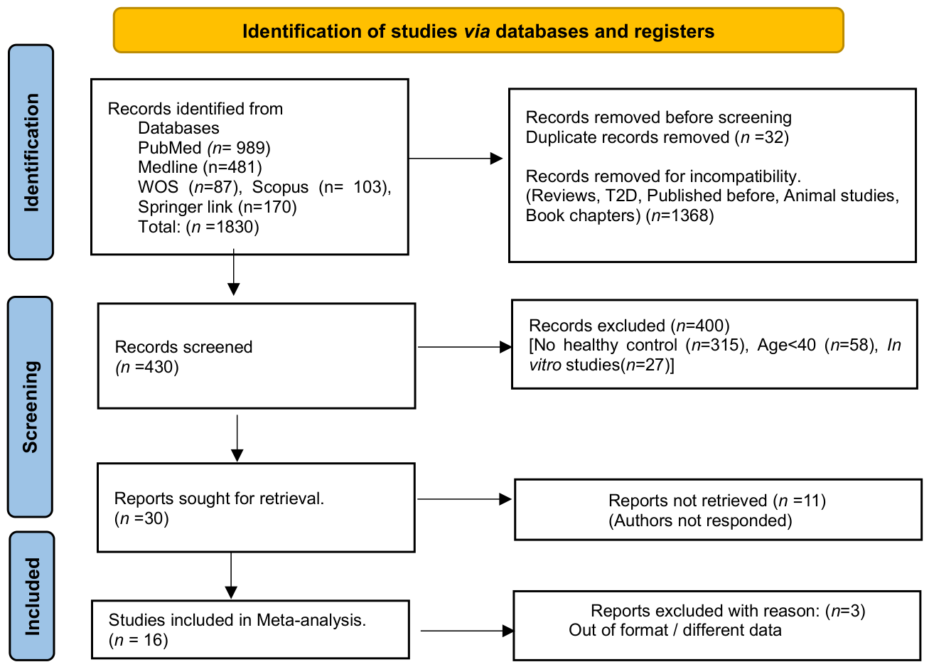
Figure 2.
(a-b): The Risk of Bias (ROB) assessment of the present study as per the ROBINS-1 criteria of the Cochrane Organisation guidelines for clinical studies. These criteria included random sequence generation, blinding of outcome assessment, incomplete outcome data, selective reporting and other biases to be at low risk in all the included studies. Allocation concealment and blinding of participants and personnel, however, was not mentioned and therefore, was deemed to carry high risk in all the included studies. (a) ROB of the individual included studies (b) the summary of the overall ROB.
Figure 2.
(a-b): The Risk of Bias (ROB) assessment of the present study as per the ROBINS-1 criteria of the Cochrane Organisation guidelines for clinical studies. These criteria included random sequence generation, blinding of outcome assessment, incomplete outcome data, selective reporting and other biases to be at low risk in all the included studies. Allocation concealment and blinding of participants and personnel, however, was not mentioned and therefore, was deemed to carry high risk in all the included studies. (a) ROB of the individual included studies (b) the summary of the overall ROB.
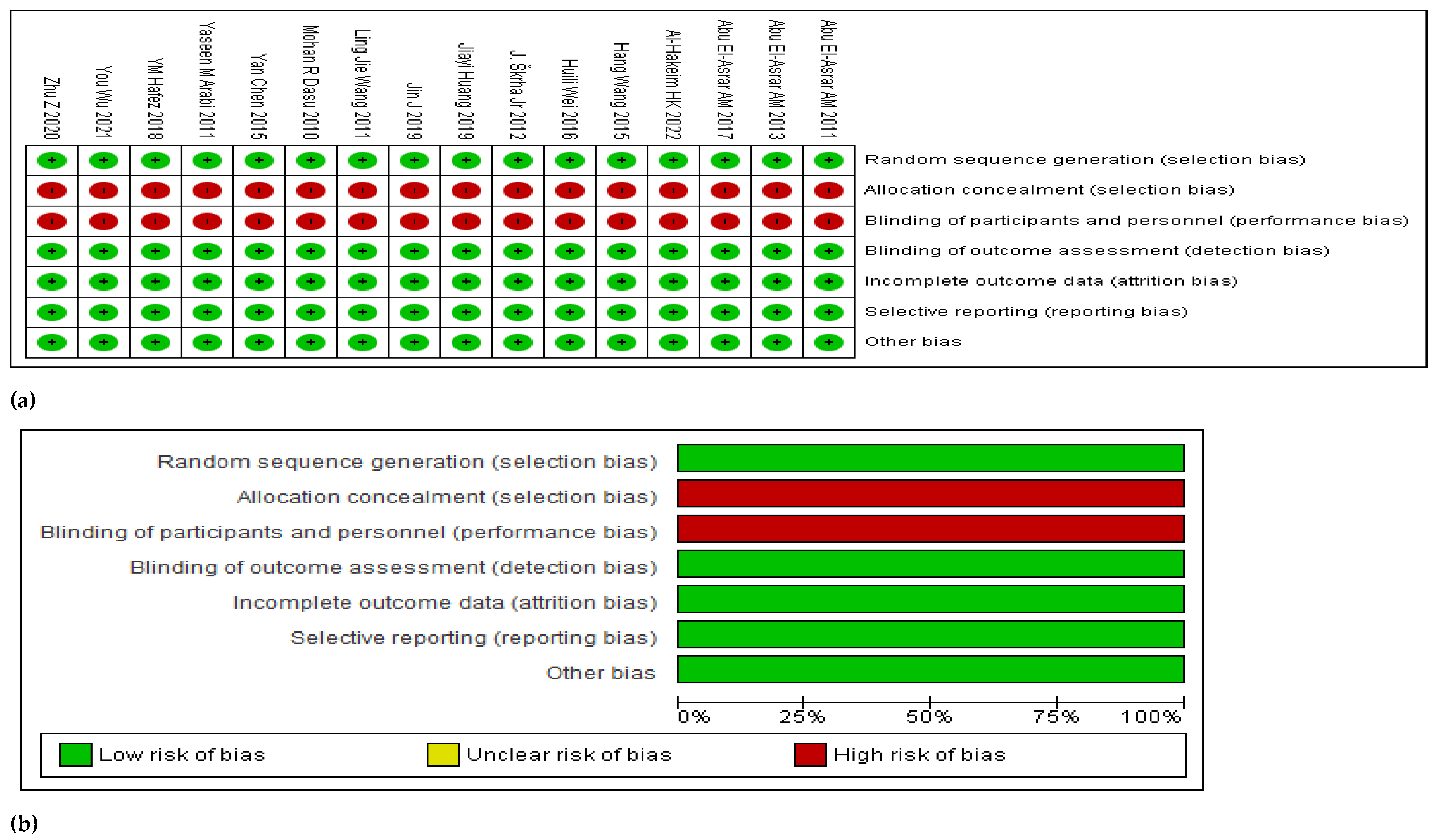
Figure 4.
- A doughnut chart displaying the eight largest effect sizes of the biomarkers under study, in descending order: The highest SMD was IL-6 followed by HbA1c%. HOMA-IR is the third highest biomarker followed by eGFR. HMGB1 comprises the sixth highest effect size (Table 2). This chart indicates that a mix of glycaemic, inflammatory and renal biomarkers have shown higher effect sizes in this analysis. Legend: IL-6 – interleukin 6, HbA1c% - glycated haemoglobin percentage, HOMA-IR- homeostasis model assessment of insulin resistance, EGFR- estimated glomerular filtration rate, ACR- albumin to creatinine ratio, HMGB-1- high mobility group box-1, FBG- fasting blood glucose, BUN- blood urea nitrogen.
Figure 4.
- A doughnut chart displaying the eight largest effect sizes of the biomarkers under study, in descending order: The highest SMD was IL-6 followed by HbA1c%. HOMA-IR is the third highest biomarker followed by eGFR. HMGB1 comprises the sixth highest effect size (Table 2). This chart indicates that a mix of glycaemic, inflammatory and renal biomarkers have shown higher effect sizes in this analysis. Legend: IL-6 – interleukin 6, HbA1c% - glycated haemoglobin percentage, HOMA-IR- homeostasis model assessment of insulin resistance, EGFR- estimated glomerular filtration rate, ACR- albumin to creatinine ratio, HMGB-1- high mobility group box-1, FBG- fasting blood glucose, BUN- blood urea nitrogen.
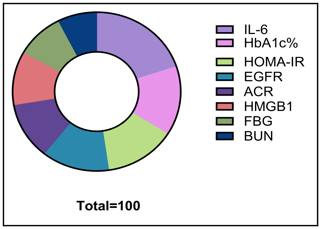
Figure 5.
(a-v) - The non-parametric Mann Whitney U-test bar graphs: 22 biomarkers were tested out of the 33 utilized in this study due to not having sufficient data. These have indicated 7 significant changes in the biomarker levels utilized in this study between the diabetic group and the HC. They included HMGB1, HbA1c%, FBG, HOMA-IR, TG, HDL-c and BMI.
Figure 5.
(a-v) - The non-parametric Mann Whitney U-test bar graphs: 22 biomarkers were tested out of the 33 utilized in this study due to not having sufficient data. These have indicated 7 significant changes in the biomarker levels utilized in this study between the diabetic group and the HC. They included HMGB1, HbA1c%, FBG, HOMA-IR, TG, HDL-c and BMI.
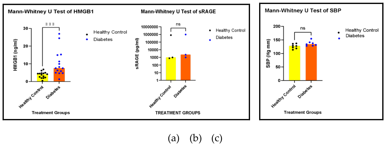
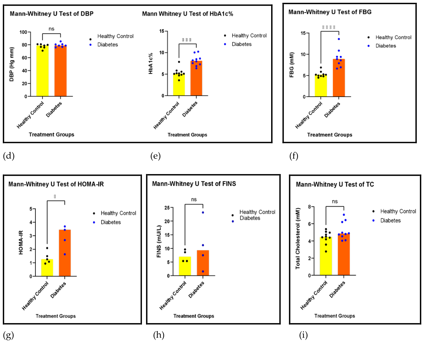
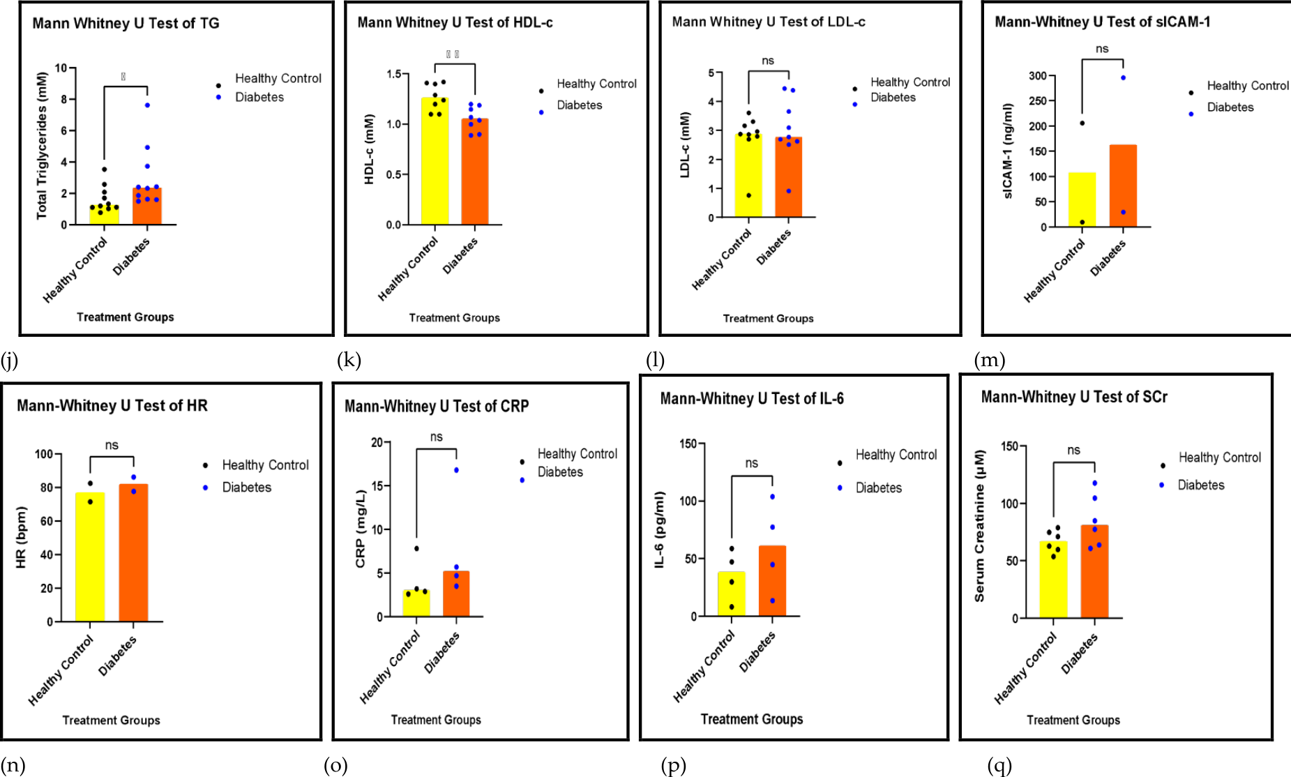
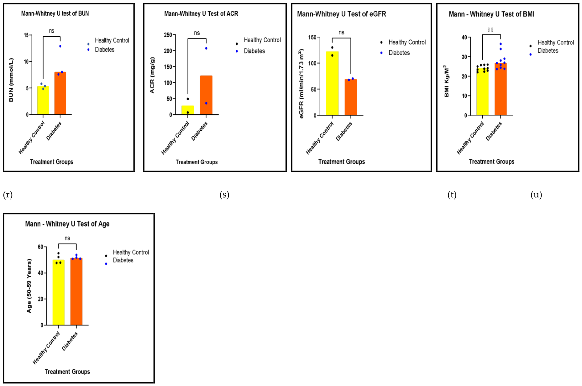

Table 1.
Characteristics of the included studies.
|
STUDY # |
YEAR | FIRST AUTHOR | COUNTRY |
MEAN AGE (YRS) |
SEX |
TOTAL (n) |
DURATION OF DISEASE (YRS) |
DISEASE | OUTCOME | ROLE OF HMGB1 |
VASCULAR BIOMARKERS TESTED |
OTHER PARAMETERS TESTED |
CO-MORBIDITIES | MECHANISM | REF |
|
1 |
2010 | MR Dasu | USA | 51 | M/F | 46 | T2D for 2.41 years | T2D | ↑ HMGB1 | Promoting inflammation as a TLR 2 and 4 ligand | None | HMGB1, FINS, FPG, TC, TG, HDL-C, LDL-C, HOMA-IR, CRP | None | TLR-MyD88-NF-kB | [29] |
| 2 | 2011 |
Abu El-Asrar AM |
Saudi Arabia | 29 | M/F |
46.9 |
16.2 years | T2D and PDR | ↑ HMGB1 | High vitreous HMGB1 concentration with haemorrhage | sICAM-1, MCP-1 | HMGB1, sRAGE, sICAM-1, MCP-1 | Poly diabetic retinopathy | HMGB1/RAGE axis | [30] |
| 3 | 2011 | Yaseen M Arabi | Saudi Arabia | 65 | M/F | 33 | NS | T2D | ↑ HMGB1 | Increased sRAGE and thrombo-modulin | None | HMGB1, sRAGE, IL-6, BMI, thrombomodulin, creatinine | Critically ill diabetes | sRAGE leading to NF-kB | [31] |
| 4 | 2011 |
Ling Jie Wang |
China | 68.3 | M/F | 149 | T2D for 1.5 years | T2D and Ischaemic HF |
↑ HMGB1, cRAGE ↓esRAGE |
Triggering cRAGE severe inflammation |
LVEDV, LVESV, LVEF, Ejection fraction | HMGB1, cRAGE, HsCRP, NT-proBNP, SBP, DBP, TC, TG, BUN, Cr, UA, FBG, HbA1C% |
Hypertension Cigarette smoking |
cRAGE, esRAGE | [32] |
| 5 | 2012 | J Skrha Jr |
Czech Republic |
64 | M/F | 66 | T2D for 9 years | T2D and Endothelial dysfunction | ↑ HMGB1 | Inflammation, ED |
ICAM, VCAM, e-selectin p-selectin, VWF |
HMGB1, SBP, DBP, TC, TG, FBG, CRP, Cr, ALB/CR ratio | NS |
sRAGE EN-RAGE |
[33] |
| 6 | 2013 |
Abu El-Asrar AM |
Saudi Arabia | 53.9 | M/F | 46 | DM for 16.4 years | T2D and Proliferative diabetic retinopathy (PDR) | ↑HMGB1 | Diabetic retinal neurodegeneration | sICAM-1 | BDNF, TBARS, sRAGE, HMGB1, MCP-1 | hypertension, hyperglycaemia | [34] | |
| 7 | 2015 | Yan Chen | China | 55 | M/F | 50 | NS | T2D and DN | ↑ HMGB1 | Regulatory role in inflammation and DN | SBP, DBP | HMGB1, FINS, FPG, TC, TG, HDL-C, LDL-C, HOMA-IR, SBP, DBP | Diabetic nephropathy | NF-kB pathway | [35] |
| 8 | 2015 | Hang Wang | China | 40 | M/F | 64 | Newly diagnosed | T2D and Obesity | ↑ HMGB1 | Increased inflammation | SBP, DBP | HMGB1, BMI, WHR, WC, HOMA-IR, FINS, SBP, DBP, TC, TG, HDL-C, LDL-C, IL-6 | Obesity | NF-kB, TLR2, TLR4 | [36] |
| 9 | 2016 | Huili Wei | China | 56.6 | M/F | 56 | Newly diagnosed | T2D | ↑ HMGB1 | Β cell dysfunction, insulin resistance | SBP, DBP | HMGB1, CTRP-3, SBP, DBP, TC, TG, HDL-c, LDL-c, HOMA-IR, FBG, ALT, AST, GGT, Cr, IL-6, INS, WHR, BMI, HbA1C% | None | RAGE and NF-kB, TLR2, TLR4, MyD88 | [37] |
| 10 | 2017 |
Abu El-Asrar AM |
Saudi Arabia | 47 | M/F | 52.1 | 16.2 years | T2D and PDR | ↑ HMGB1 | Oxidative stress and angiogenesis in the ocular microenvironment | sVAP-1 | HMGB1, 8-OHdG, sVAP-1, HO-1 | Poly diabetic retinopathy | HMGB1, VAP-1, oxidative stress, and HO-1 | [38] |
| 11 | 2018 | YM Hafez | Egypt | 50 | M/F | 30 | 9 years | T2D and DFU | ↑ HMGB1 | Increased inflammation | None | HMGB1, catalase, SIRT1, TNF-a, AGES | DFU | SIRT1 linked to oxidative stress and inflammation | [39] |
| 12 | 2019 | Juan Jin | China | 52 | M/F | 15 | T2D for 10 years | T2D and Diabetic nephropathy (DN) | ↑HMGB1 and TLR4 | Induces podocyte autophagy and EMT | SBP, DBP | SBP, DBP, BMI, HbAIc%, TC, TG, LDL-C, BUN, SCr, HMGB1 | Diabetic retinopathy | Inhibited AKT/mTOR and TGF-β/SMAD-1 | [40] |
| 13 | 2019 | Jiayi Huang | China | 65 | M/F | 112 | T2D for 8 years | T2D and COPD | ↑ HMGB1 | Arterial stenosis, acting as a DAMP | SBP, DBP | HMGB1, FINS, FPG, TC, TG, HDL-C, LDL-C, HOMA-IR, SBP, DBP | COPD | TLR2, TLR4, RAGE | [41] |
| 14 | 2020 | Z Zhu | China | 44 | M/F | 40 | NS | T2D and DN | ↑ HMGB1 | Impaired Endothelium -dependent relaxation | WBC, platelet count, haemoglobin content, HR, LVEDD | HMGB1, FINS, FPG, TC, TG, HDL-C, LDL-C, HOMA-IR, SBP, DBP, albumin, globulin | Chronic kidney disease | TLR4/eNOS pathway | [42] |
| 15 | 2021 | You Wu | China | 55 | M/F | 30 | 3.5 years | T2D and DN | ↑ HMGB1 | Increased ferroptosis | None | HMGB1, ACR, eGFR, HOMA-IR, BUN, creatinine | Diabetic kidney disease/DN | TLR4/NF-κB and Nrf2 signalling pathway | [43] |
| 16 | 2022 | HK Al-Hakeim | Iraq | 34 | M/F | 46.85 years | NS | T2D and atherogenicity | ↑ HMGB1 | Increased inflammation from increased glucose toxicity leading to atherogenicity | None | HMGB1, FINS, HbA1C%, HOMA-IR, TC, TG, HDL-C, LDL-C, DKK, BMI | Atherogenicity |
HMGB1/RAGE/DKK/Wnt pathway |
[44] |
Table 1- Characteristics of the included studies: Consists of the important and prominent characteristics of the sixteen (16) included studies in this systematic review and meta-analysis. The year of publication, name of the first author, the country in which the included studies were conducted, the age and sex of the participants, the disease subtype of Diabetes mellitus (DM), the number of individuals in this study in the control group and the T2D group, the blood sugar cutoff point which confirms the diagnosis of diabetes, the primary outcomes reported by each included study, role of HMGB1, the physiological and immune mechanisms that were studied, comorbidities, vascular biomarkers tested and the study models are described in this table.
Table 2.
Summary of the effect sizes of each biomarker that is calculated in each forest plot based upon the Cohen’s D principle: Out of 25 biomarkers that were assessed according to the Cohen’s D criteria, 18 biomarkers produced large effect sizes/SMDs, while there were 5 biomarkers which showed medium effect size/SMD. There were 2 biomarkers which showed very small effect size/SMD in the diastolic blood pressure and low-density lipoprotein cholesterol. The larger effect sizes signify conspicuous increases or decrease in the biomarker SMDs compared to the healthy control.
Table 2.
Summary of the effect sizes of each biomarker that is calculated in each forest plot based upon the Cohen’s D principle: Out of 25 biomarkers that were assessed according to the Cohen’s D criteria, 18 biomarkers produced large effect sizes/SMDs, while there were 5 biomarkers which showed medium effect size/SMD. There were 2 biomarkers which showed very small effect size/SMD in the diastolic blood pressure and low-density lipoprotein cholesterol. The larger effect sizes signify conspicuous increases or decrease in the biomarker SMDs compared to the healthy control.
| Description of the Model | Biomarkers | Participants (n) |
Small Effect Size (<0.5) |
Medium Effect Size (0.5-0.8) |
Large Effect Size (>0.8) |
| AGEs group and their ligands | |||||
| HMGB1 | 1398 | 2.86 | |||
| sRAGE | 240 | 0.79 | |||
| Blood pressure | |||||
| SBP | 834 | 0.53 | |||
| DBP | 834 | 0.15 | |||
| Glycaemic | |||||
| HbA1C% | 875 | 3.68 | |||
| FBG | 790 | 2.41 | |||
| HOMA-IR | 402 | 3.59 | |||
| FINS | 356 | -0.47 | |||
| Lipids | |||||
| TC | 852 | 0.78 | |||
| TG | 852 | 1.31 | |||
| HDL-C | 713 | -0.81 | |||
| LDL-C | 743 | 0.42 | |||
| Vascular | |||||
| ICAM-1 | 141 | 0.86 | |||
| HR | 407 | 0.59 | |||
| LVEF% | 345 | -4.06 | |||
| LVDV | 345 | 2.01 | |||
| LVSV | 345 | 1.88 | |||
| Inflammatory | |||||
| CRP | 421 | 0.49 | |||
| IL-6 | 392 | 5.20 | |||
| Renal | |||||
| SCr | 703 | 0.84 | |||
| BUN | 356 | 1.96 | |||
| ACR | 214 | 3.03 | |||
| eGFR | 159 | -3.41 | |||
| Obesity-Related | |||||
| BMI | 733 | 1.04 | |||
| AGE | 1249 | 0.77 |
Disclaimer/Publisher’s Note: The statements, opinions and data contained in all publications are solely those of the individual author(s) and contributor(s) and not of MDPI and/or the editor(s). MDPI and/or the editor(s) disclaim responsibility for any injury to people or property resulting from any ideas, methods, instructions or products referred to in the content. |
© 2026 by the authors. Licensee MDPI, Basel, Switzerland. This article is an open access article distributed under the terms and conditions of the Creative Commons Attribution (CC BY) license.
Copyright: This open access article is published under a Creative Commons CC BY 4.0 license, which permit the free download, distribution, and reuse, provided that the author and preprint are cited in any reuse.


