

Submitted:
26 July 2026
Posted:
28 July 2026
You are already at the latest version
Abstract
Purpose: To review how standardized ultrasound-based risk stratification and artificial-intelligence (AI) governance have reshaped early diagnostic pathways for ovarian cancer. Materials and Methods: We performed a critical narrative review of milestones in transvaginal ultrasound, IOTA descriptors, ADNEX, O-RADS US/MRI, population screening trials and AI reporting/governance standards. Priority was given to evidence that used external validation, calibration or explicit links between risk categories and clinical management. Results: Population screening with CA-125 and ultrasound has not reduced mortality in average-risk women; therefore, the highest near-term value lies in triage once an adnexal lesion is detected. IOTA/ADNEX and O-RADS translate sonographic descriptors into reproducible probabilities and management categories; O-RADS MRI is useful for sonographically indeterminate lesions. AI may support segmentation, structured reporting and triage, but high discrimination is insufficient without external validation, calibration, transportability testing, decision-curve analysis and subgroup monitoring. Conclusion: Earlier and safer decisions are more likely to come from living standards - structured ultrasound reporting, calibrated risk models, MRI arbitration, auditable AI updates and equity-aware monitoring - than from population screening alone.
Keywords:
ovarian cancer diagnosis
; adnexal mass risk stratification
; ultrasound
; O-RADS
; IOTA
; ADNEX
; artificial intelligence
; calibration
; decision-curve analysis
1. Introduction
Ovarian cancer remains a disease in which timing strongly determines outcome. Survival is substantially better when disease is localized, yet many patients still enter care with advanced disease; globally, ovarian cancer caused more than 200,000 deaths in 2020 [1]. This gap has repeatedly motivated screening strategies using CA-125 and transvaginal ultrasound. The largest evidence base, however, has been sobering: UKCTOCS showed no ovarian-cancer mortality reduction in average-risk women despite a stage shift [2,3]. The practical question for ultrasound services is therefore not how to screen every asymptomatic woman, but how to make safer and earlier decisions once symptoms or an adnexal mass bring a patient into the diagnostic pathway.
The most durable progress has come from standardization. Ultrasound made adnexal morphology visible; Doppler added vascular information and increased the descriptive range available to the examiner [4,5,6]. The International Ovarian Tumor Analysis (IOTA) group then converted expert sonographic heuristics into shared descriptors and probabilistic models, including Simple Rules, LR2 and ADNEX [7,8]. Ovarian-Adnexal Reporting and Data System (O-RADS) US and O-RADS MRI subsequently connected a common lexicon to ordinal risk categories and management recommendations [9,10,11]. In this sense, early diagnosis became less a property of a single device than of a reproducible imaging ecosystem: the image, the vocabulary, the probability estimate and the clinical action must remain aligned.
The second wave is now algorithmic. Radiomics and deep learning can classify adnexal masses on ultrasound or MRI, while recent studies have synthesized the evidence on ultrasound-based AI and developed calibrated hybrid CNN–Transformer models for multiclass ovarian tumor classification [12,13,14,15]. These studies illustrate genuine promise, but they also show why high accuracy alone is an insufficient endpoint. Dataset shift, shortcut learning and hidden stratification can make apparently strong models fail in new sites [16]. Equity also matters: CA-125 is less frequently elevated at diagnosis in some racial and ethnic groups, so universal thresholds can delay triage [17]. For this reason, TRIPOD+AI and STARD-AI should be treated as implementation requirements, not as optional reporting checklists [18,19]. This review argues that IOTA, O-RADS and AI can improve ovarian-cancer diagnosis only when embedded in calibrated, audited and equity-aware ultrasound pathways. Figure 1 summarizes the milestones that shaped this pathway.
2. Materials and Methods
This article is a critical narrative review rather than a systematic review. We selected evidence around five predefined nodes: adoption of transvaginal ultrasound and Doppler; development and validation of IOTA descriptors, Simple Rules and ADNEX; design and clinical use of O-RADS US/MRI; population screening trials; and AI evaluation standards. We prioritized studies and guidance documents that provided operational definitions, external or temporal validation, calibration, subgroup information or an explicit link between imaging risk and clinical action.
The synthesis followed three steps. First, we reconstructed the transition from expert visual assessment to probabilistic sonographic grammar and category-to-action systems. Second, we contrasted diagnostic triage with population screening, using UKCTOCS and PLCO as boundary-setting trials. Third, we evaluated AI through a governance lens: a clinically credible model must add value over IOTA/O-RADS, generalize across scanners and sites, provide calibrated probabilities, and show net benefit at clinically plausible thresholds.
Data extraction was conceptual but structured. For IOTA/ADNEX we focused on morphological and vascular descriptors, model outputs and validation evidence. For O-RADS we focused on lexicon definitions, category-to-management mapping and the role of MRI after indeterminate ultrasound. For AI we focused on data provenance, external validation, calibration, fairness, drift monitoring and clinical utility. External validations of IOTA strategies and O-RADS informed the benchmark against which algorithmic claims should be judged [20,21].
3. Historical-Technical Periodization
The history of ovarian-cancer diagnosis is not a simple march toward sharper machines; it is the progressive alignment of visibility, vocabulary and action. After Donald's 1958 report, ultrasound entered gynecology as a way to identify pelvic and abdominal masses beyond palpation [5]. Real-time and later transvaginal scanning improved spatial resolution and brought non-palpable adnexal pathology into clinical view. Yet interpretation remained local and operator-dependent: without shared definitions, the same lesion could be described differently across centers.
Color Doppler promised a move beyond morphology by adding resistance indices, flow patterns and vascular distribution [6]. These features were clinically attractive, but performance was fragile outside development settings because equipment, presets, operators and case mix varied. The lesson was that new signals do not automatically become reliable decisions. They require acquisition discipline, shared descriptors and validation in populations resembling routine care.
IOTA provided the first durable grammar for this problem. By defining terms, measurements and follow-up standards, it allowed the same adnexal lesion to be described similarly across institutions [7]. ADNEX then translated those descriptors, with or without CA-125, into individualized risks for benign, borderline, early invasive, advanced invasive and metastatic disease [8]. Independent validation of Simple Rules and two-step strategies confirmed that standardized sonographic assessment could outperform older indices when used by trained examiners [20,21].
O-RADS extended this grammar into routine reporting. O-RADS US provides risk categories and management tables; O-RADS MRI offers an adjudication pathway for indeterminate or solid lesions after ultrasound [9,10,11]. This is particularly relevant for Ultraschall in der Medizin, because ultrasound remains the first-line test and the gatekeeper for referral, surveillance or MRI. The result is not only better terminology; it is a pathway in which a structured report can be audited against downstream action.
Population screening trials sharpened the boundary of this pathway. PLCO and UKCTOCS found no mortality benefit from average-risk screening with CA-125 and/or ultrasound, while documenting false-positive pathways and downstream surgery [2,22]. Screening can shift stage distribution without reducing deaths. The clinical priority is therefore standardized triage of lesions already encountered in care, not revival of population screening without mortality evidence.
4. Diagnostic Standardization
IOTA and O-RADS should be viewed as living standards rather than static checklists. Their value lies in converting image interpretation into reproducible clinical probabilities and in making local performance measurable. A report that states cyst size, wall regularity, septations, papillary projections, solid components, ascites and vascularity in a consistent format is more useful than an impression that simply labels a lesion as "probably benign" or "suspicious". Structured ultrasound reporting also enables teaching, interobserver audit and comparison of local category prevalence with expected ranges.
ADNEX is especially useful because it makes uncertainty explicit. The model does not only distinguish benign from malignant tumors; it provides probabilities across clinically important subtypes. Those probabilities can guide referral to gynecologic oncology, conservative follow-up or MRI arbitration. However, probability is not portable by default. A model calibrated in a tertiary referral population may overestimate risk in a lower-prevalence service, and the same threshold may produce different downstream consequences across hospitals.
O-RADS US addresses the report-to-action problem by linking risk categories to management recommendations. O-RADS MRI provides a second-line, multiparametric framework for sonographically indeterminate lesions, using T1/T2 morphology, diffusion and contrast-enhancement information [11]. This creates a practical sequence: ultrasound first, structured risk assignment, MRI for selected indeterminate cases, and referral when risk and clinical context justify escalation. Such a sequence is more defensible than either universal MRI or unstructured reliance on expert impression.
For quality assurance, services should monitor three levels of performance. The first is descriptive completeness: whether mandatory O-RADS or IOTA fields are present. The second is probabilistic validity: whether observed malignancy rates within O-RADS categories or ADNEX risk bands match expected rates locally. The third is clinical consequence: whether the pathway reduces unnecessary surgery, shortens time to appropriate treatment and improves appropriate referral. These metrics are more relevant than AUC alone because they capture how risk information changes care.
5. AI in Gynecologic Ultrasound
AI can strengthen, but should not replace, the standardized ultrasound pathway. Its near-term roles are pragmatic: lesion segmentation, pre-population of IOTA/O-RADS descriptors, second-reader support, prioritization of equivocal examinations and discordance alerts when image features do not match the assigned category. These uses fit clinical workflow better than stand-alone black-box classification and preserve the interpretability that ultrasound reporting requires.
The evidence is promising but uneven. Deep learning studies report high AUCs for adnexal-mass classification, while recent work underscores both the diagnostic potential of ultrasound-based AI and the need for multicenter, calibrated, and explainable evaluation. Radiomics and machine-learning studies similarly show potential but remain sensitive to acquisition and preprocessing heterogeneity [13,14]. A model trained in a tertiary center may be miscalibrated in a community setting with different prevalence, scanners or operators. Therefore, discrimination must be accompanied by calibration-in-the-large, calibration slope, plots, external validation and subgroup analyses.
Clinical usefulness should be tested with decision-curve analysis (DCA), which quantifies net benefit across thresholds by weighing false positives and false negatives [23]. In adnexal imaging, DCA can ask whether AI-assisted escalation to O-RADS MRI reduces avoidable surgery while preserving sensitivity for invasive disease. If an AI system does not improve net benefit over structured O-RADS/IOTA care, it is not ready for routine use, regardless of AUC.
Transparent reporting is equally important. TRIPOD+AI requires detailed reporting of data provenance, missing-data handling, predictors, model development, internal and external validation and calibration [18]. STARD-AI provides analogous expectations for AI diagnostic-accuracy studies, including reference standards, dataset assembly, reader design, bias and fairness [19]. For an ultrasound department considering procurement or local deployment, these checklists define the minimum dossier that should be requested before pilot testing.
The most credible AI contribution is therefore not a promise to "detect cancer earlier" in the abstract. It is a measurable improvement to an existing pathway: fewer incomplete structured reports, fewer discordant categories, better triage of O-RADS 3-4 lesions, improved selection for MRI, or reduced benign surgery without loss of sensitivity for invasive disease.
6. Equity and Bias
Equity is a calibration problem as much as a social one. If local prevalence, biomarker distributions or access pathways differ across subgroups, the same threshold can produce different clinical consequences. CA-125 illustrates this risk: race- and ethnicity-related differences in elevation at diagnosis can make universal cutoffs less safe for underrepresented populations [17]. Thresholds that appear objective may therefore encode diagnostic delay when transported uncritically.
Imaging AI adds another layer because deep models can infer demographic attributes from medical images and can learn site or device shortcuts [16,24]. A model may therefore perform well overall while failing in a subgroup, scanner type or referral pathway. Harmonization methods can reduce scanner effects, but they do not replace external validation and local calibration [25]. Equity-aware validation requires more than reporting sensitivity and specificity in the whole cohort; it requires subgroup calibration, subgroup net benefit and monitoring after deployment.
Standardization helps but does not eliminate inequity. O-RADS and IOTA create a common grammar, but observed malignancy rates within a category may still vary by menopausal status, referral source, age, ancestry or site. If local O-RADS 4 prevalence is lower than expected, an aggressive threshold could produce disproportionate benign surgery. If a biomarker prior is poorly matched to the local population, an ADNEX- or AI-assisted pathway could under-refer patients whose tumors are biomarker-negative.
A minimum equity program should therefore include local calibration plots, O-RADS stratum prevalence tables, and outcome monitoring by site and clinically relevant subgroups. When divergence appears, the response should be documented: recalibration, threshold adjustment, targeted training, expanded access to MRI arbitration, or review of referral barriers. Table 1 summarizes the minimum implementation actions.
7. Implementation and Governance
Implementation is where methods meet consequences. The safest route is to embed IOTA and O-RADS within quality-assured workflows and to treat any AI layer as a versioned, calibrated component inside an audited system. The first operational step is structured O-RADS US reporting with mandatory lexicon fields and automatic category assignment where possible. The second is a clear rule for when O-RADS MRI is used, especially for sonographically indeterminate lesions or discordant clinical-imaging impressions.
A frequent barrier is the perceived tension between AI-assisted recommendations and clinical autonomy. Deployments should therefore specify that AI is advisory, define escalation pathways for discordant cases, and provide an override mechanism with brief justification. This preserves accountability and creates an audit trail without forcing clinicians to accept an opaque recommendation.
Probability outputs must be trustworthy where they are used. Before routine use, ADNEX, SR-risk or AI probabilities should undergo local calibration with the available case mix. After deployment, services should monitor calibration and category outcomes periodically by site, scanner, menopausal status and relevant demographic groups. A lightweight dashboard can include observed malignancy rates within O-RADS categories, calibration plots for probability models, MRI arbitration rates, referral appropriateness and time from index ultrasound to definitive management.
Change control is essential. A model registry should record model name, version, training and validation datasets, calibration method, DCA results, intended-use statement, current status and update history. Updates should be enabled only after predefined validation, and rollback criteria should be stated before deployment. These requirements need not be expensive: periodic sampling audits, open-source calibration scripts and shared regional registries can provide a minimum viable safety infrastructure.
Two failure modes should be avoided. The first is accuracy myopia: deploying a tool because its AUC is high in a single-center study, without external validation, calibration or DCA in the target environment. The second is frozen standardization: adopting O-RADS terminology but never auditing whether local prevalence, equipment or referral patterns have changed the predictive value of categories. Both failures are preventable if structured reporting, calibration, DCA and version control are treated as routine quality assurance rather than research extras.
8. Research Agenda and Conclusions
The field should move from accuracy demonstrations to patient- and system-level benefit. Future studies should compare new tools against IOTA, ADNEX, O-RADS US and O-RADS MRI, not against unstructured assessment alone. Recommended endpoints include time from index ultrasound to appropriate treatment, rate of unnecessary surgery among benign lesions, appropriate referral to gynecologic oncology, negative predictive value in surveillance strata, report-to-action concordance and quality of life.
Calibration and DCA should be prespecified. Multicenter studies should report apparent and external calibration, recalibration methods and net benefit across clinically plausible thresholds. Prospective pragmatic designs, stepped-wedge trials or interrupted time-series evaluations can test whether structured reporting or AI-assisted workflows improve care in real services [26,27]. Subgroup analyses should be planned rather than exploratory, with publication of O-RADS stratum prevalence and outcomes by site and clinically relevant groups.
The most credible near-term AI research will be workflow-integrated: does segmentation assistance reduce measurement variability; does descriptor auto-completion improve O-RADS completeness; does a discordance flag improve second-review yield; and does AI-guided MRI arbitration reduce benign surgery without missed malignancy? These are narrower questions than population screening, but they are more actionable and safer.
Earlier diagnosis of ovarian cancer is most likely to improve through calibrated diagnostic pathways rather than average-risk population screening alone. Ultrasound provides the entry point; IOTA and ADNEX convert morphology into reproducible probabilities; O-RADS links lexicon, risk and management; and O-RADS MRI adjudicates selected indeterminate lesions. AI should be judged as an extension of this ecosystem: it must add incremental benefit over IOTA/O-RADS, remain calibrated in the target population, generalize across scanners and sites, and be monitored for subgroup performance and drift. The practical standard is exacting but feasible: structured ultrasound reports, local calibration, decision-curve analysis, external validation, equity audits and controlled updates. Under these conditions, algorithms can become safe force multipliers for earlier, accountable decisions in ovarian-cancer care.
Funding
This research received no external funding.
Ethics Approval
Not applicable. This study is a literature review and did not involve human participants or animals.
Declaration of Generative AI and AI-Assisted Technologies in the Writing Process
During preparation, the authors used ChatGPT to improve language and readability. The authors reviewed and edited the content and accepted full responsibility for the publication.
Conflicts of Interest
The authors declare no conflicts of interest.
References
- Sung, H.; Ferlay, J.; Siegel, R.L.; Laversanne, M.; Soerjomataram, I.; Jemal, A.; et al. Global Cancer Statistics 2020. CA Cancer J. Clin. 2021, 71, 209–49. [Google Scholar] [CrossRef] [PubMed]
- Menon, U.; Gentry-Maharaj, A.; Burnell, M.; Singh, N.; Ryan, A.; Karpinskyj, C.; et al. Ovarian cancer population screening and mortality after long-term follow-up in UKCTOCS. Lancet 2021, 397, 2182–93. [Google Scholar] [CrossRef] [PubMed]
- Menon, U.; Gentry-Maharaj, A.; Burnell, M.; Ryan, A.; Singh, N.; Manchanda, R.; et al. Tumour stage, treatment, and survival of women with high-grade serous tubo-ovarian cancer in UKCTOCS. Lancet Oncol. 2023, 24, 1018–28. [Google Scholar] [CrossRef] [PubMed]
- Campbell, S. A Short History of Sonography in Obstetrics and Gynaecology. Facts Views Vis. ObGyn 2013, 5, 213–29. [Google Scholar] [PubMed]
- Donald, I.; Macvicar, J.; Brown, T.G. Investigation of abdominal masses by pulsed ultrasound. Lancet 1958, 271, 1188–95. [Google Scholar] [CrossRef] [PubMed]
- Evans, D.H.; Jensen, J.A.; Nielsen, M.B. Ultrasonic colour Doppler imaging. Interface Focus 2011, 1, 490–502. [Google Scholar] [CrossRef] [PubMed]
- Timmerman, D.; Valentin, L.; Bourne, T.H.; Collins, W.P.; Verrelst, H.; Vergote, I.; et al. Terms, definitions and measurements to describe the sonographic features of adnexal tumors. Ultrasound Obstet. Gynecol. 2000, 16, 500–5. [Google Scholar] [CrossRef] [PubMed]
- Van Calster, B.; Van Hoorde, K.; Valentin, L.; Testa, A.C.; Fischerova, D.; Van Holsbeke, C.; et al. Evaluating the risk of ovarian cancer before surgery using the ADNEX model. BMJ 2014, 349, g5920. [Google Scholar] [CrossRef] [PubMed]
- Andreotti, R.F.; Timmerman, D.; Strachowski, L.M.; Froyman, W.; Benacerraf, B.R.; Bennett, G.L.; et al. O-RADS US Risk Stratification and Management System. Radiology 2020, 294, 168–85. [Google Scholar] [CrossRef] [PubMed]
- Strachowski, L.M.; Jha, P.; Phillips, C.H.; Porter, M.B.; Froyman, W.; Glanc, P.; et al. O-RADS US v2022: An Update. Radiology 2023, 308, e230685. [Google Scholar] [CrossRef] [PubMed]
- Sadowski, E.A.; Thomassin-Naggara, I.; Rockall, A.; Maturen, K.E.; Forstner, R.; Jha, P.; et al. O-RADS MRI Risk Stratification System. Radiology 2022, 303, 35–47. [Google Scholar] [CrossRef] [PubMed]
- Garcia-Atutxa, I.; Villanueva-Flores, F.; Dudagotia Barrio, E.; Sanchez-Villamil, J.I.; Martinez-Mas, J.; Bueno-Crespo, A. Artificial intelligence for ovarian cancer diagnosis via ultrasound. Front Artif. Intell. 2025, 8, 1649746. [Google Scholar] [CrossRef] [PubMed]
- Barcroft, J.F.; Linton-Reid, K.; Landolfo, C.; Al-Memar, M.; Parker, N.; Kyriacou, C.; et al. Machine learning and radiomics for segmentation and classification of adnexal masses on ultrasound. npj Precis Oncol. 2024, 8, 41. [Google Scholar] [CrossRef] [PubMed]
- Chen, H.; Yang, B.-W.; Qian, L.; Meng, Y.-S.; Bai, X.-H.; Hong, X.-W.; et al. Deep Learning Prediction of Ovarian Malignancy at US. Radiology 2022, 304, 106–13. [Google Scholar] [CrossRef] [PubMed]
- Garcia-Atutxa, I.; Martinez-Mas, J.; Bueno-Crespo, A.; Villanueva-Flores, F. Early-fusion hybrid CNN-Transformer models for multiclass ovarian tumor ultrasound classification. Front Artif. Intell. 2025, 8, 1679310. [Google Scholar] [CrossRef] [PubMed]
- Oakden-Rayner, L.; Dunnmon, J.; Carneiro, G.; Re, C. Hidden stratification causes clinically meaningful failures in machine learning for medical imaging. Proc. ACM CHIL. 2020, 151–9. [Google Scholar] [CrossRef] [PubMed]
- Smith, A.J.B.; Gleason, E.; Kadiyala, S.; Wang, X.; Howell, E.A.; McCarthy, A.M. Cancer Antigen 125 Levels at Time of Ovarian Cancer Diagnosis by Race and Ethnicity. JAMA Netw. Open 2025, 8, e251292. [Google Scholar] [CrossRef] [PubMed]
- Collins, G.S.; Moons, K.G.M.; Dhiman, P.; Riley, R.D.; Beam, A.L.; Van Calster, B.; et al. TRIPOD+AI statement. BMJ 2024, 385, e078378. [Google Scholar] [CrossRef] [PubMed]
- Sounderajah, V.; Guni, A.; Liu, X.; Collins, G.S.; Karthikesalingam, A.; Markar, S.R.; et al. The STARD-AI reporting guideline. Nat. Med. 2025. [Google Scholar] [CrossRef] [PubMed]
- Alcazar, J.L.; Pascual, M.A.; Olartecoechea, B.; Graupera, B.; Auba, M.; Ajossa, S.; et al. IOTA simple rules: prospective external validation. Ultrasound Obstet. Gynecol. 2013, 42, 467–71. [Google Scholar] [CrossRef] [PubMed]
- Timmerman, S.; Valentin, L.; Ceusters, J.; Testa, A.C.; Landolfo, C.; Sladkevicius, P.; et al. External Validation of O-RADS and the IOTA 2-Step Strategy. JAMA Oncol. 2023, 9, 225–33. [Google Scholar] [CrossRef] [PubMed]
- Buys, S.S.; Partridge, E.; Black, A.; Johnson, C.C.; Lamerato, L.; Isaacs, C.; et al. Effect of screening on ovarian cancer mortality: PLCO trial. JAMA 2011, 305, 2295–303. [Google Scholar] [CrossRef] [PubMed]
- Vickers, A.J.; Elkin, E.B. Decision curve analysis. Med. Decis. Mak. 2006, 26, 565–74. [Google Scholar] [CrossRef] [PubMed]
- Gichoya, J.W.; Banerjee, I.; Bhimireddy, A.R.; Burns, J.L.; Celi, L.A.; Chen, L.-C.; et al. AI recognition of patient race in medical imaging. Lancet Digit Health 2022, 4, e406-14. [Google Scholar] [CrossRef] [PubMed]
- Orlhac, F.; Eertink, J.J.; Cottereau, A.-S.; Zijlstra, J.M.; Thieblemont, C.; Meignan, M.; et al. A guide to ComBat harmonization of imaging biomarkers. J. Nucl. Med. 2022, 63, 172–9. [Google Scholar] [CrossRef] [PubMed]
- Vickers, A.J.; Van Calster, B.; Steyerberg, E.W. A simple guide to interpreting decision curve analysis. Diagn. Progn. Res. 2019, 3, 18. [Google Scholar] [CrossRef] [PubMed]
- Hemming, K.; Haines, T.P.; Chilton, P.J.; Girling, A.J.; Lilford, R.J. The stepped wedge cluster randomised trial. BMJ 2015, 350, h391. [Google Scholar] [CrossRef] [PubMed]
Figure 1.
Milestones shaping the meaning of early diagnosis in ovarian cancer, from early ultrasound hardware advances to standardized taxonomies and AI governance frameworks.
Figure 1.
Milestones shaping the meaning of early diagnosis in ovarian cancer, from early ultrasound hardware advances to standardized taxonomies and AI governance frameworks.
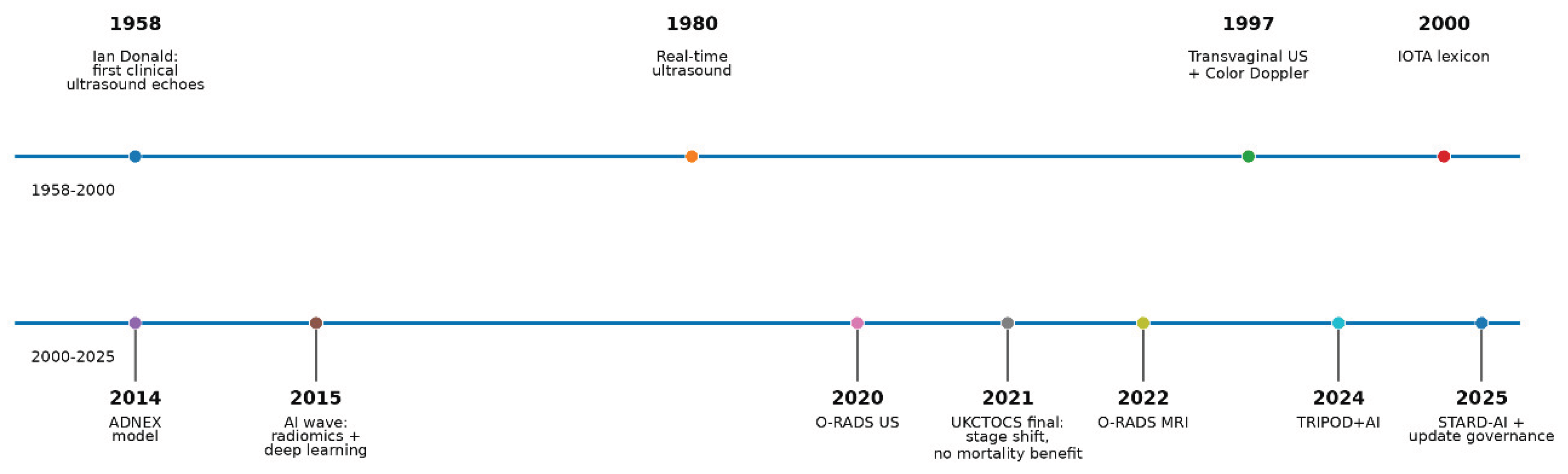

Table 1.
Minimum implementation requirements for ultrasound-based adnexal-mass triage.
| Domain | Main Risk | Minimum Action |
|---|---|---|
| CA-125 and subgroup differences | Universal thresholds can be miscalibrated. | Report biomarker performance by subgroup and recalibrate thresholds when local data justify it. |
| Site, scanner and case-mix shift | PPV, NPV and net benefit can change despite stable AUC. | Perform external validation, local calibration and periodic drift checks. |
| O-RADS/IOTA category transportability | The same category may imply different observed risk. | Publish local prevalence and outcomes by category, site and menopausal status. |
| AI deployment | Models can learn site/device shortcuts or drift after updates. | Use a version registry, subgroup calibration, DCA, override pathway and rollback criteria. |
Disclaimer/Publisher’s Note: The statements, opinions and data contained in all publications are solely those of the individual author(s) and contributor(s) and not of MDPI and/or the editor(s). MDPI and/or the editor(s) disclaim responsibility for any injury to people or property resulting from any ideas, methods, instructions or products referred to in the content. |
© 2026 by the authors. Licensee MDPI, Basel, Switzerland. This article is an open access article distributed under the terms and conditions of the Creative Commons Attribution (CC BY) license (http://creativecommons.org/licenses/by/4.0/).
Copyright: This open access article is published under a Creative Commons CC BY 4.0 license, which permit the free download, distribution, and reuse, provided that the author and preprint are cited in any reuse.