

Submitted:
25 July 2026
Posted:
27 July 2026
You are already at the latest version
Abstract
Encapsulating protein subunit vaccines in biodegradable microparticles (MP) can increase induction of long-term protective mucosal and systemic antibodies after respiratory administration but requires incorporation of a mucosal immunostimulant. We previously found that incorporating complement peptide-derived immunostimulant-02 (CPDI-02) with MP-encapsulated LPS-free OVA through surface modification of the same ~1 µm PLGA 50:50 MP greatly increases mucosal and systemic OVA-specific antibodies in young, naïve female C57BL/6 mice at 14 and 90 days post-intranasal administration. Here, we directly compared effects of incorporating CPDI-02 or inactive scCPDI-02 with MP-encapsulated LPS-free OVA by separate encapsulation (SE), co-encapsulation (CE), or surface modification (SM) on induction of OVA-specific IgA, IgM, and IgG antibody secreting cells (ASCs) in the lungs and spleen, IgA antibodies in nasal lavage fluid, IgA and IgG antibodies in bronchoalveolar lavage fluid, and IgG subclasses in the serum at 14 days post-intranasal administration. We found that SE incorporation induced greater or similar levels of mucosal and systemic OVA-specific ASCs and mucosal IgA and IgG titers but greater titers of systemic IgG subclasses than CE or SM incorporation versus inactive scCPDI-02. Thus, given that surface modification with CPDI-02 induces high titers of OVA-specific systemic and mucosal antibodies at 14 and 90 days post-intranasal immunization under the same experimental conditions, separate encapsulation of CPDI-02 is expected to more broadly induce long-term mucosal and systemic antibodies against MP-encapsulated protein vaccines than co-encapsulation and surface modification after intranasal and possibly other routes of mucosal immunization.
Keywords:
mucosal immunization
; mucosal vaccine
; vaccine delivery
; respiratory-delivered vaccine
; immunostimulant incorporation
; C5a1R
; C5aR1
; CD88
; EP54
; EP67
1. Introduction
The primary function of a vaccine is to safely generate long-term protective adaptive immune responses against the targeted pathogen at levels that significantly reduce or eliminate subsequent infectious disease [1,2,3,4,5,6]. Most licensed vaccines are administered by an intramuscular or subcutaneous route to induce sufficient levels of long-term systemic antibodies that circulate in the bloodstream and tissues (“systemic antibodies”) and increase protection against invasive infections by binding targeted pathogens or toxins with high affinity and specificity to physically block entry or damage to cells [3,4,7]. Approximately 82% of global infectious diseases, however, are caused by upper (~12.8 billion) and lower (~344 million) respiratory infections [8,9,10,11,12,13,14,15,16,17,18] that are largely acquired through mucosal surfaces in the nose, mouth, and airways [19] and responsible for ~19,600 and ~2.2 million deaths, respectively, per year [8,13]. Thus, administering vaccines by an intranasal or inhaled route alone or in combination with systemic vaccines to induce sufficient levels of long-term protective antibodies in both the respiratory mucosae (“mucosal antibodies”) and the bloodstream (“systemic antibodies”) could potentially increase prevention and protection against non-invasive and invasive respiratory infections more effectively than systemic vaccines alone and decrease the incidence and mortality associated with infectious diseases [20,21,22,23,24].
At least 5 respiratory-delivered mucosal vaccines have been approved for clinical use worldwide including four intranasal live attenuated influenza vaccines (FluMist Quadrivalent, Fluenz Tetra, Nasovac-S, and the AstraZeneca H5N1 pandemic vaccine) and one inhaled recombinant adenoviral-vector COVID-19 vaccine (Convidecia Air) [25,26] that take advantage of the intrinsic immunogenicity of the vaccine construct. The continued development of these vaccine types is limited, in part, by the difficulty of balancing attenuation or vector safety with replication and transgene expression to induce durable mucosal immunity, overcoming pre-existing and vaccine-induced anti-vector immunity, achieving consistent respiratory deposition in the presence of mucus and mucociliary clearance, and maintaining viral stability, potency, scalable manufacturing, and compatibility with the administration device [26,27,28,29,30]. Respiratory administration of one or more protective antigens (i.e., subunit vaccines) encapsulated in biodegradable nanoparticles (NP) or microparticles (MP) is being actively developed as an alternative to current respiratory-delivered mucosal vaccines [25,31]. Unlike live-attenuated or recombinant adenoviral-vectors, however, NP/MP-encapsulated subunit vaccines require the incorporation of one or more suitable mucosal immunostimulants to sufficiently activate antigen-presenting cells and generate long-term protective mucosal and systemic adaptive immune responses [25,26,32,33].
Complement peptide-derived immunostimulant-02 (CPDI-02) (formerly “EP67”) is a novel second-generation, host-derived decapeptide agonist of C5a receptor 1 (hC5aR1/hC5a1R/CD88) based on the C-terminal pharmacophore of human C5a (hC5a) ligand [20,34,35]. Unlike hC5a, CPDI-02 selectively activates primary human mononuclear phagocytes (monocytes, monocyte-derived macrophages, and monocyte-derived DC) with ~1000-fold higher potency than primary human neutrophils (NP) (167 nM EC50 in MP vs. 160 μM EC50 in NP) (“MP-selective activation”) to decrease neutrophil-mediated toxicity [20,36]. Directly conjugating CPDI-02 to peptide epitopes, whole proteins, or live pathogens increases the magnitudes of humoral and cellular immune responses in mice after intranasal or intramuscular immunization [20,34,37,38,39,40,41,42]. Combining CPDI-02 with CpG and Montanide also increases B-cell and T-cell responses against a protective APol-solubilized membrane protein after systemic/respiratory immunization and subsequent protection of naïve mice against primary respiratory challenge with C. trachomatis [20,43].
Three common strategies for incorporating mucosal or systemic immunostimulants with NP- or MP-encapsulated subunit vaccines include separate encapsulation in different co-administered nanoscale biodegradable particles, co-encapsulation in the same nanoscale biodegradable particle, or surface modification of the same nanoscale biodegradable particle [31,44,45,46,47] (Figure 1). We recently found that incorporating CPDI-02 with MP-encapsulated LPS-free OVA by surface modification of the same biodegradable PLGA 50:50 MP (~1 µm diam) through 2 kDa PEG linkers significantly increases titers of OVA-specific antibodies at 14 and 90 days post-intranasal immunization [20]. The relative effect of other incorporation strategies on the mucosal adjuvant activity of CPDI-02 with MP-encapsulated protein vaccines, however, remains unclear. In this study, we directly compared the effects of incorporating CPDI-02 with MP-encapsulated LPS-free OVA by separate encapsulation, co-encapsulation, or surface modification on the induction of early mucosal and systemic antibody-secreting cells and antibody titers in naïve female C57BL/6 mice after intranasal immunization and further determined whether increasing the dose of separately encapsulated CPDI-02 could further increase the same humoral responses.
2. Materials and Methods
2.1. LPS Removal from Ovalbumin (OVA)
LPS endotoxin was removed from Grade V hen egg white ovalbumin (OVA: 385 amino acids, MW: 44,287 Da, Sigma) [40 mg] using a Detoxi-Gel™ column (Thermo Scientific) as described [5]. LPS-free OVA (“OVA”) was used in all formulations, assays, and mouse studies.
2.2. Synthesis of CPDI-02, scCPDI-02, CGRR-CPDI-02, and CGRR-scCPDI-02 Peptides
For separate encapsulation and co-encapsulation formulations, CPDI-02 (YSFKDMP[MeL]aR where “MeL” = N-methyl leucine and “a” = D-alanine; formerly “EP67”) or inactive scrambled scCPDI-02 ([MeL]RMYKPaFDS; formerly scEP67) were synthesized, purified (95%), and characterized as described [36,48]. For surface modification of microparticles (MP) surface-activated with maleimide groups through 2 kDa PEG linkers, CPDI-02 and scCPDI-02 were activated with sulfhydryl groups by introducing N-terminal cysteine through a kexin-cleavable glycine double arginine linker (GRR) during solid-phase synthesis (CGRR-CPDI-02 and CGRR-scCPDI-02) then purified (95%) and characterized as described [20].
2.3. Separate Encapsulation (SE) of CPDI-02 and OVA in Biodegradable Microparticles (“SE-CPDI-02”)
OVA was encapsulated in ester-terminated 50:50 poly(D,L-lactic-co-glycolic acid) (PLGA 50:50; research grade; inherent viscosity 0.65 dL g: ~50-75 kDa; Lactel; Pelham, AL) microparticles (MP) (~1 µm in diameter) at a theoretical loading of 10 wt% (mass OVA / mass final formulation) by a water-in-oil-in-water (W1/O/W2) emulsion solvent evaporation method (ESE) but without surface modification as described [5,20].
CPDI-02 or scCPDI-02 were separately encapsulated in PLGA 50:50 MP (~1 µm in diameter) at a theoretical loading of 5 wt% (mass CPDI-02 or scCPDI-02/mass final formulation) by an Oil-in-Oil (O1/O2) emulsion as described with modification [49,50]. Approximately 50 mg of CPDI-02 or scCPDI-02 was dissolved in 100 mL of 100 mM ammonium bicarbonate, transferred to a lyophilizer, and freeze-dried until all solvent was completely evaporated. The resulting deprotonated acidic form CPDI-02 or scCPDI-02 was used in all subsequent formulation steps.
To form the dispersed O1 phase, 150 mg of ester-terminated PLGA 50:50 (inherent viscosity 0.86 dL/g: ~74 kDa; Lactel, Pelham, AL) was dissolved in 1.8 mL of acetonitrile in an 8-mL glass scintillation vial and allowed to dissolve completely (approximately 1 hour). Separately, 8 mg of deprotonated CPDI-02 or scCPDI-02 (theoretical 5 wt% loading) was dissolved in 200 µL of methanol in a 1.5-mL centrifuge tube and vortexed until completely dissolved. The CPDI-02 or scCPDI-02/methanol solution was then added to the PLGA/acetonitrile solution and vortexed at 1000 rpm for 30 seconds to form the co-solvent O1 phase.
The continuous O2 phase was prepared by adding 0.5 mL (0.493 g) of SPAN-80 (Sigma Life Science; density: 0.986 g/mL) to 80 mL of cottonseed oil (Sigma Life Science) in a 150-mL glass beaker and mixing thoroughly on a stir plate to achieve a final surfactant concentration of approximately 0.6% (v/v). To form the crude O1/O2 emulsion, 4 mL of the O2 phase was added to the O1 phase in the scintillation vial and vortexed at 1000 rpm for 30 seconds. The sonicator probe (Hielscher UP200ST ultrasonic homogenizer with an S26d14 sonotrode; Hielscher Ultrasound Technology, Teltow, Germany) was submerged to approximately one-quarter of the total depth of the 6 mL emulsion volume in the scintillation vial, and the crude emulsion was sonicated at full amplitude with a total energy limit of 400 J to achieve a target particle diameter of approximately 1 µm. The fully formed O1/O2 emulsion was then added dropwise to the remaining 75 mL of O2 continuous phase in the 150-mL beaker and stirred at 1000 rpm overnight (16 hours) to evaporate methanol and acetonitrile.
Following solvent evaporation, the emulsion was divided equally between two 40-mL centrifuge tubes and centrifuged at 20,000 x g, 4 °C, for 15 minutes to pellet the MPs. The cottonseed oil supernatant was decanted and the MP pellet was resuspended in approximately 30 mL of anhydrous hexane. The pellet was vortexed to resuspend, sonicated with the Hielscher probe at full amplitude for approximately 20 seconds, and centrifuged again under the same conditions (4 hexane washes total). Washed MPs were recovered by vacuum filtration over a 0.22 µm nylon membrane filter, with additional anhydrous hexane pipetted over the MPs during filtration to complete removal of residual oil. MPs were dried again under vacuum filtration for at least 30 minutes, transferred from the filter membrane into a pre-weighed 20-mL glass scintillation vial using a cell scraper, then stored at -20 °C.
2.4. Co-Encapsulation (CE) of CPDI-02 and OVA in Biodegradable Microparticles (“CE-CPDI-02”)
OVA and either CPDI-02 or inactive scrambled CPDI-02 (scCPDI-02) were co-encapsulated into biodegradable PLGA 50:50 (inherent viscosity 0.65 dL g) microparticles (MP) at a theoretical loading of 10 wt% OVA (mass OVA/mass of final MP formulation) and 5 wt% CPDI-02 or scCPDI-02 (mass of CPDI-02 or scCPDI-02/mass of final MP formulation) by the emulsion solvent evaporation (ESE) method. Ester-terminated PLGA was dissolved at 50 mg/mL in 2 mL dichloromethane (DCM; Fluka, analytical grade ≥ 99.7% pure; 100 mg total) in an 8-mL glass scintillation vial with gentle shaking every 20 to 30 minutes for approximately 2 hours to form the oil (O) phase.
For formation of the inner aqueous (W1) phase, LPS-free OVA and either CPDI-02 or scCPDI-02 were dissolved together in sterile D-PBS (without Ca²⁺ or Mg²⁺; Hyclone) at 10 wt% and 5 wt% theoretical loading, respectively, to a total volume of 250 µL, then vortexed at 1000 RPM for 30 seconds. A primary water-in-oil (W1/O) emulsion was formed by adding 200 µL of the W1 phase dropwise to the O phase while vortexing at 1000 RPM for 30 seconds, followed by sonication on ice for 2 minutes at full amplitude using a Hielscher UP200ST ultrasonic homogenizer with an S26d14 sonotrode (Hielscher Ultrasound Technology, Teltow, Germany; typical power output 21 to 24 W).
A polyvinyl alcohol (PVA; 87 to 90% hydrolyzed, average MW 30 to 70 kDa; Sigma-Aldrich) solution (5% v/v in deionized water; 8 mL) was prepared in a 20-mL glass scintillation vial by stirring at 1000 RPM and 100 °C until fully dissolved, then allowed to cool to room temperature for at least 2 hours before use. The W1/O primary emulsion was transferred dropwise via a glass Pasteur pipette (pipetted up and down 10 to 15 times to saturate with DCM prior to transfer) into the PVA solution while vortexing at 400 RPM for 25 seconds to form the crude secondary water-in-oil-in-water (W1/O/W2) emulsion. The crude secondary emulsion was transferred to a fresh 20-mL glass scintillation vial, leaving foam in the original vial, and sonicated at full amplitude with a total applied energy of 60 W·s (60 J) using the same homogenizer. A magnetic stir bar was added and the emulsion was stirred at 1000 RPM overnight to evaporate DCM and harden the MP.
Hardened MPs were transferred to 40-mL Nalgene centrifuge vials (rated ≥ 30,000 × g), with the glass vial rinsed with deionized water to recover residual particles, and volume adjusted to fill the centrifuge vial with deionized water. MPs were pelleted by centrifugation (23,640 x g, 4 °C, 20 min), supernatants removed, and pellets resuspended in deionized water. This wash was repeated for a total of three centrifugation cycles to remove residual PVA. Washed MPs were resuspended in approximately 10 mL deionized water, transferred to a pre-weighed 50-mL centrifuge tube, flash-frozen in liquid N₂, lyophilized for 48 hours, and stored at −20 °C until use.
2.5. Encapsulation of OVA in Biodegradable Microparticles Surface-Modified with CPDI-02 (“SM-CPDI-02”)
OVA was encapsulated in biodegradable PLGA 50:50 (research grade; inherent viscosity 0.65 dL g: ~50-75 kDa; Lactel; Pelham, AL) microparticles (MP) (~1 µm in diameter) at a theoretical loading of 10 wt% (mass OVA/mass of formulation) and surface modified with CysGlyArgArg-CPDI-02 or inactive CysGlyArgArg-scCPDI-02 through 2 kDa PEG linkers during OVA encapsulation by interfacial activity-assisted surface functionalization (IAASF) as described [20].
2.6. Quantitation of OVA, CPDI-02, and scCPDI-02 Loading in MP by Ultra-Performance Liquid Chromatography (UPLC)
Average loading (n ≥ 3 from at least two independent batches) was determined as described with modifications [20]. Briefly, 2 mg of lyophilized MP were equilibrated to room temperature, dissolved in 125 µL DMSO in an 8-mL borosilicate glass vial, and incubated at room temperature for 1 h with constant shaking. Digestion solution (0.05 M NaOH/0.5% (w/w) SDS in dH₂O, 1.25 mL) was added and the entire solution was stirred (650 RPM) in a capped vial overnight. Undissolved polymer was pelleted (10,000 x g, 10 min) and the supernatant was transferred to a new vial, where 62.5 µL of 10% trifluoroacetic acid (TFA) in dH₂O was added to achieve a pH compatible with the selected UPLC column (pH 2–12). Standards of OVA alone, CPDI-02 alone, or scCPDI-02 alone were prepared under identical digestion conditions and serially diluted in a diluent containing matching concentrations of DMSO, 0.05 M NaOH / 0.5% (w/w) SDS, and 10% TFA.
Samples and standards were run on an ACQUITY UPLC H-Class PLUS System (Waters) using a reversed-phase ACQUITY UPLC Protein BEH C4 Column (Waters, 300 Å, 1.7 µm, 2.1 mm × 150 mm). Solvent A was water containing 0.1% TFA (v/v) and Solvent B was acetonitrile containing 0.1% TFA (v/v). Analytes were eluted by increasing Solvent B from 0% to 100% over 20 minutes, with continuous UV monitoring at 214 nm. AUC for each standard was plotted against concentration and analyte concentrations in digested MP samples were determined from a linear regression of their measured AUC.
2.7. Quantitation of OVA Burst Release from MP
Burst release of OVA (average percent of total OVA released from lyophilized microparticles 24 h after resuspension, n = 3 from at least two independent batches) was determined as previously described with modification [5,20]. Lyophilized MP (10 mg) were equilibrated to room temperature, transferred to a 2.0 mL microcentrifuge tube, and suspended in 1.0 mL of phosphate-buffered saline containing 0.05% Tween-20 (v/v; PBST). Suspensions were vortexed for 20 seconds at 1000 RPM using a Vortex Genie, then incubated at 37 °C with continuous shaking at 200 RPM for 24 h in a Vortemp 56 Shaking Incubator (Labnet International). After 24 h, microparticles were pelleted by centrifugation at 10,000 RCF for 5 min at 4 °C, and supernatants were collected and stored at -20 °C until analysis. OVA concentrations in the supernatants were determined by UPLC as described above (Section 2.6) but with OVA concentrations of 400, 200, and 50 ug/mL for the expected range of released OVA concentrations based on a theoretical encapsulation efficiency between 50 and 90%.
2.8. Quantitation of CPDI-02 or scCPDI-02 Burst Release from MP
Burst release of CPDI-02 or scCPDI-02 (average percent of total peptide released from lyophilized microparticles 24 h after resuspension, n = 3 from at least two independent batches) was determined using the same resuspension, incubation, and centrifugation conditions as described for OVA burst release above (Section 2.7). Concentrations of CPDI-02 or scCPDI-02 in the supernatants were then determined by UPLC as described for loading above (Section 2.6) using serial dilutions of purified CPDI-02 or scCPDI-02 in PBST as calibration standards (approximately 150, 75, and 15 µg/m).
2.9. Quantitation of CPDI-02 and scCPDI-02 Surface Conjugation to MP by Kexin-Mediated Ultra-Performance Liquid Chromatography (UPLC)
Average levels of CPDI-02 or scCPDI-02 conjugated to the surface of MP were determined by kexin-mediated UPLC as described [20].
2.10. Diameters and Zeta Potentials of Microparticles
Average hydrodynamic diameters, PDI, and zeta-potentials (mV; ±SD; n = 3 independent samples from at least two batches) were determined in 10 mM NaCl in deionized H2O (0.5 mg/mL) using a ZetaSizer Nano ZS90 (Malvern Instruments, Malvern, UK) equipped with a He-Ne laser (633 nm) as the incident beam, with samples equilibrated within the instrument at 25 °C for 4 minutes before measuring, as described [20].
2.11. Animals
All animal procedures were approved by the University of Nebraska Medical Center Institutional Animal Care and Use Committee. Naïve female mice (C57BL/6NCrl ~8 weeks old, Charles River Laboratories, Wilmington, MA, USA) were acclimatized in an ABSL-2 facility under pathogen-free conditions at least one week before experiments.
2.12. Intranasal Administration
Vehicle alone (sterile PBS) [50 µL] or vehicle containing an equivalent amount of MP-encapsulated OVA [50 µg] was administered to sedated, supine mice on days −14, −7, and 0 as described [20].
2.13. Isolation of Murine Lung Lymphocytes and Splenocytes
Murine lymphocytes and splenocytes were isolated on the indicated days as described [20].
2.14. IgA, IgG, and IgM ELISpot Assays
Antibody-secreting cells (ASC) (n=3 from 3 mice) were quantitated in the lungs, spleen, and serum on the indicated days using Murine Single-Color ELISpot kits (ImmunoSpot) as described [20].
2.15. Collection of Serum, NLF, and BALF from Mice
Serum, nasal lavage fluid (NLF), and bronchoalveolar lavage fluid (BALF) were isolated as described and stored at −80 °C [20].
2.16. OVA-Specific Antibody Titers in NLF, BALF, and Serum
OVA-specific titers of total IgG in BALF, total IgA in NLF, and IgG1, IgG2b, IgG2c, and IgG3 in serum were determined on the indicated days by indirect ELISA as described [20].
2.17. Statistical Analyses
All statistical analyses were performed using GraphPad Prism (San Diego, CA, USA) for Windows, www.graphpad.com. Sample outliers in all experiments were identified by the ROUT method (Q = 1%) and omitted for statistical comparisons. Data from two treatment groups were compared by two-tailed, nonparametric Mann-Whitney U Test (α = 0.05) and data from three or more treatment groups were compared by nonparametric Kruskal–Wallis one-way ANOVA with uncorrected Dunn’s multiple comparisons post hoc test (α = 0.05). Additional relevant statistical information is provided in the figure legends.
3. Results
3.1. Incorporating CPDI-02 by Separate Encapsulation or Surface Modification Generates Higher Early Mucosal Antibody Secreting Cells Against MP-Encapsulated OVA After IN Administration than Co-Encapsulation
The magnitudes of antigen-specific antibody-secreting cells (ASCs) found within the lungs shortly after mucosal infection or intranasal (IN) immunization potentially correlate with local induction of mucosal humoral immunity and the likelihood of establishing resident plasma cells that can sustain antigen-specific antibody production within the pulmonary compartment [7,8,9,10,11]. Thus, to provide an early indication of how the most common immunostimulant incorporation strategies (Figure 1) affect the ability of CPDI-20 to activate mucosal humoral immunity against MP-encapsulated protein antigen after IN administration, we first encapsulated LPS-free ovalbumin (OVA) in ~1 µM diameter PLGA 50:50 microparticles and incorporated CPDI-02 or inactive, scrambled scCDPI-02 by (i.) separate encapsulation (SE) in different ~1 µM diameter PLGA 50:50 microparticles (SE-CPDI-02) (Figure 1A), (ii.) co-encapsulation (CE) in the same ~1 µm PLGA 50:50 microparticles (CE-CPDI-02) (Figure 1B), or (iii.) surface modification (SM) of the same ~1 µm PLGA 50:50 microparticles with CPDI-02 through a protease-labile N-terminal Cys-Gly-Arg-Arg linker attached to 2 kDa PEG linkers (SM-CPDI-02) (Figure 1C) (Table S1). We then administered SE-CPDI-02, CE-CPDI-02, or SM-CPDI-02 to young, naïve female C57BL/6 mice once every 7 days over 14 days (3 doses total) by the IN route and compared early magnitudes of OVA-specific IgA, IgM, and IgG antibody secreting cells (ASCs) in the lungs 6 days post-treatment (20 days post-prime) by ELISpot (Figure 2). We used a 4.3-fold higher ratio of CPDI-02 to OVA in the CE-CPDI-02 formulations because of unpredictable and variable CPDI-02 loading when co-encapsulating CPDI-02 with OVA.
SE-CPDI-02 (Figure 2, closed red circles) and SM-CPDI-02 (Figure 2, closed blue squares) increased OVA-specific IgA (Figure 2A), IgM (Figure 2B), and IgG ASCs (Figure 2C) vs. inactive SE-scCPDI-02 (Figure 2, open red circles) or inactive SM-scCPDI-02 (Figure 2, open blue squares), respectively, whereas CE-CPDI-02 (Figure 2, closed green triangles) only increased OVA-specific IgG ASCs (Figure 2C) vs. inactive CE-scCPDI-02 (Figure 2C, open green triangles). Furthermore, SE-CDPI-02, CE-CPDI-02, and SM-CPDI-02 (Figure 2, closed symbols) increased all three classes of OVA-specific ASCs vs. vehicle alone (Figure 2, black asterisks), whereas inactive SE-scCPDI-02, CE-scCPDI-02, and SM-scCPDI-02 generated very low to undetectable ASCs (Figure 2, open symbols). Thus, (i.) the increase of OVA-specific ASCs in the lungs against MP-encapsulated OVA is due almost entirely to the activity of incorporated CPDI-02 and (ii.) incorporating CPDI-02 by separate encapsulation or surface modification activates mucosal humoral immunity in the lungs of naïve inbred mice to a greater extent than co-encapsulation of CPDI-02 under the current experimental conditions despite 4.3-fold higher incorporation of co-encapsulated CPDI-02 (Table S1).
3.2. Incorporating CPDI-02 by Separate Encapsulation Generates Higher Early Mucosal Antibodies Against MP-Encapsulated OVA After IN Administration than Co-Encapsulation
To determine the effect of the most common immunostimulant incorporation strategies on the ability of CPDI-02 to induce an early mucosal humoral response against MP-encapsulated protein antigen after IN administration, we administered SE-CPDI-02, CE-CPDI-02, or SM-CPDI-02 (Figure 1) to young, naïve female C57BL/6 mice once every 7 days over 14 days (3 doses total) by the IN route and compared early titers of OVA-specific IgA in nasal lavage fluid (NLF) and titers of OVA-specific IgA and IgG in bronchoalveolar lavage fluid (BALF) normalized to vehicle alone 14 days post-immunization (28 days post-prime) by ELISA (Figure 3). SE-CPDI-02 (Figure 3, closed red circles), CE-CPDI-02 (Figure 3, close green triangles), and SM-CPDI-02 (Figure 3, closed blue squares) greatly increased titers of OVA-specific IgA in the NLF (Figure 3A) and BALF (Figure 3B) and IgG in the BALF (Figure 3C) vs. inactive scCPDI-02 (Figure 3, open symbols), whereas inactive SE-scCPDI-02, CE-CPDI-02, and SM-CPDI-02 generated relatively low to undetectable titers (Figure 3, open symbols). Thus, the increase of OVA-specific IgA antibodies in the nasal cavities and OVA-specific IgA and IgG antibodies in the lungs against MP-encapsulated OVA is due almost entirely to the activity of incorporated CPDI-02.
SE-CPDI-02 (Figure 3, closed red circles) generated higher titers of OVA-specific IgA in the NLF (Figure 3A) and BALF (Figure 3B) and OVA-specific IgG in the BALF (Figure 3C) than CE-CPDI-02 (Figure 3, closed green triangles), whereas SM-CPDI-02 (Figure 3, closed blue squares) generated similar levels of the same OVA-specific antibodies as SE-CPDI-02 (Figure 3, closed red circles) and CE-CPDI-02 (Figure 3, closed green triangles). Thus, separate encapsulation of CPDI-02 increases early mucosal antibodies against MP-encapsulated protein antigen in young, naïve female inbred mice after IN administration to a similar extent as surface modification but to a greater extent than co-encapsulation despite 4.3-fold higher co-encapsulated CPDI-02 (Table S1).
3.3. Incorporating CPDI-02 by Separate Encapsulation Generates Higher Early Systemic Antibody Secreting Cells Against MP-Encapsulated OVA After IN Administration than Co-Encapsulation
The magnitudes of antigen-specific isotype-specific ASCs detected in the spleen shortly after mucosal infection or intranasal immunization potentially correlate with B-cell trafficking to mucosal effector sites and subsequent establishment of secretory IgA responses (IgA ASCs), the earliest antigen-driven B-cell induction preceding class switching (IgM ASCs), and the magnitude and breadth of systemic humoral priming (IgG ASCs) [51,52,53]. Thus, to provide an early indication of how the most common immunostimulant incorporation strategies (Figure 1) affect the ability of CPDI-02 to activate both mucosal and systemic humoral immunity against MP-encapsulated protein antigen after IN administration, we administered SE-CPDI-02, CE-CPDI-02, or SM-CPDI-02 (Figure 1) to young, naïve female C57BL/6 mice once every 7 days over 14 days (3 doses total) by the IN route and compared early magnitudes of OVA-specific IgA, IgM, and IgG ASCs in the spleen 6 days post-treatment (20 days post-prime) by ELISpot (Figure 4).
SE-CPDI-02 (Figure 4, closed red circles) increased OVA-specific IgA (Figure 4A), IgM (Figure 4B), and IgG (Figure 4C) ASCs, SM-CPDI-02 (Figure 4, closed blue squares) increased OVA-specific IgA (Figure 4A) and IgG (Figure 4C) ASCs, and CE-CPDI-02 (Figure 4, closed green triangles) did not increase any OVA-specific ASCs vs. corresponding incorporation of inactive scCPDI-02 (Figure 4, open symbols). Furthermore, SE-CDPI-02 (Figure 4, closed symbols) increased all three classes of OVA-specific ASCs, SM-CPDI-02 (Figure 4, closed blue squares) increased OVA-specific IgA (Figure 4A) and IgG (Figure 4C) ASCs, and CE-CPDI-02 (Figure 4, closed green triangles) increased OVA-specific IgM (Figure 4B) and IgG (Figure 4C), whereas inactive SE-scCPDI-02, SM-scCPDI-02, and CE-scCPDI-02 generated very low to undetectable ASCs (Figure 4, open symbols). vs. vehicle alone (Figure 4, black asterisks). Thus, (i.) the increase of OVA-specific ASCs in the spleen against MP-encapsulated OVA is due almost entirely to the activity of incorporated CPDI-02 and (ii.) incorporating CPDI-02 by separate encapsulation activates mucosal and systemic humoral immunity in naïve inbred female mice after IN administration to a greater extent than surface modification or co-encapsulation under the current experimental conditions despite 4.3-fold higher incorporation of co-encapsulated CPDI-02 (Table S1).
3.4. Incorporating CPDI-02 by Separate Encapsulation Generates Higher Early Systemic IgG Antibody Subclasses Against MP-Encapsulated OVA After IN Administration than Co-Encapsulation and Surface Modification
To determine the extent that the most common immunostimulant incorporation strategies affect the ability of CPDI-02 to increase the generation of systemic antibodies against MP-encapsulated protein antigen after IN administration, we administered SE-CPDI-02, CE-CPDI-02, or SM-CPDI-02 (Figure 1) to young, naïve female C57BL/6 mice once every 7 days over 14 days (3 doses total) by the IN route and compared early titers of OVA-specific IgG1, IgG2b, IgG2c, and IgG3 antibodies in serum normalized to vehicle alone 14 days post-treatment (28 days post-prime) by ELISA (Figure 5). We compared the effects of immunostimulant incorporation strategy on each IgG subclass given that, according to the quartet model of murine IgG function, IgG subclasses work together to clear infections where IgG3 promotes inflammation and IgG2b mediates FcγR-dependent effector functions when T-cell help may be limited during early infection, whereas Th-dependent IgG2a (IgG2c in C57BL/6 mice) increases pathogen clearance and IgG1 limits IgG-driven inflammation during the later stages of infection [54].
SE-CPDI-02 (Figure 5, closed red circles), and SM-CPDI-02 (Figure 5, closed blue squares) increased serum titers of OVA-specific IgG1 (Figure 5A), IgG2b (Figure 5B), IgG2c (Figure 5C), and IgG3 (Figure 5D) vs. inactive SE-scCPDI-02 (Figure 5, open red circles) or inactive SM-scCPDI-02 (Figure 5, open blue squares), respectively, whereas CE-CPDI-02 (Figure 5, closed green triangles) only increased OVA-specific IgG1 (Figure 5A) and IgG2b (Figure 5B) and IgG3 (Figure 5D) vs. inactive CE-scCPDI-02 (Figure 5, open green triangles). Furthermore, unlike mucosal antibodies (Figure 3), inactive SE-scCPDI-02 (Figure 5, open red circles) and SM-scCPDI-02 (Figure 5, open blue squares) generated relatively higher serum titers of antibodies than inactive CE-sCPDI-02. Thus, separate encapsulation and surface modification increase early OVA-specific antibodies through the activity of CPDI-02 with some contribution from the MP formulation, whereas co-encapsulation increases early OVA-specific antibodies primarily through the activity of CPDI-02.
SE-CPDI-02 (Figure 5, closed red circles) generated higher serum titers of OVA-specific IgG1 (Figure 5A), IgG2b (Figure 5B), IgG2c (Figure 5C), and IgG3 (Figure 5D) than CE-CPDI-02 (Figure 5, closed green triangles) and SM-CPDI-02 (Figure 5, closed blue squares). Furthermore, SM-CPDI-02 (Figure 5, closed green triangles) generated higher serum titers of OVA-specific IgG2c (Figure 5C) and IgG3 (Figure 5D) than CE-CPDI-02 (Figure 5, closed green triangles). Thus, separate encapsulation of CPDI-02 increases early systemic antibodies against MP-encapsulated protein antigen in young, naïve inbred female mice after IN administration to a greater extent than co-encapsulation or surface modification and surface modification increases early systemic antibodies against MP-encapsulated protein antigen to a greater extent than co-encapsulation despite 4.3-fold higher co-encapsulated CPDI-02 (Table S1).
3.5. Increasing CPDI-02 Incorporation by Separate Encapsulation Does Not Increase Early Mucosal Antibodies Against MP-Encapsulated OVA After IN Administration
Separate encapsulation of CPDI-02 generally increased the generation of mucosal and systemic antibodies against MP-encapsulated OVA after IN administration to a greater extent than co-encapsulation or surface modification at a relatively low ratio of CPDI-02/µg OVA (64 ng/µg OVA) (Figure 3 and Figure 5). To determine if increasing CPDI-02 incorporation by separate encapsulation could further increase the generation of early mucosal antibodies against MP-encapsulated protein antigen, we administered SE-CPDI-02 (Figure 1A) at 200 ng/µg OVA (Figure 6, closed red squares) or 400 ng/µg OVA (Figure 6, closed red triangles) or inactive SE-scCPDI-02 at 400 ng/µg OVA (Figure 6, open red circles) to young, naïve female C57BL/6 mice once every 7 days over 14 days (3 doses total) by the IN route and compared titers of OVA-specific IgA in nasal lavage fluid (NLF) and titers of OVA-specific IgA and IgG in bronchoalveolar lavage fluid (BALF) normalized to vehicle alone 14 days post-immunization (28 days post-prime) by ELISA (Figure 6).
Like SE-CPDI-02 at 64 ng/µg OVA (Figure 6, closed red circles), SE-CPDI-02 at 200 ng/µg OVA (Figure 6, closed red squares) or 400 ng/µg OVA (Figure 6, closed red triangles) greatly increased titers of OVA-specific IgA in the NLF (Figure 6A) and BALF (Figure 6B) and OVA-specific IgG in the BALF (Figure 6C), whereas inactive SE-scCPDI-02 at 400 ng/µg OVA (Figure 6, open red circles) generated relatively low to undetectable titers in the NLF and BALF (Figure 6, open symbols). This indicates that the increase of OVA-specific IgA antibodies in the nasal cavities and OVA-specific IgA and IgG antibodies in the lungs against MP-encapsulated OVA is due to the activity of incorporated CPDI-02.
SE-CPDI-02 at 200 ng/µg OVA (Figure 6, closed red squares) and 400 ng/µg OVA (Figure 6, closed red triangles) greatly increased titers of OVA-specific IgA in the NLF (Figure 6A) and BALF (Figure 6B) and OVA-specific IgG in the BALF (Figure 6C) to the same extent as SE-CPDI-02 at 64 ng/µg OVA (Figure 6, closed red circles) vs. inactive SE-scCPDI-02 at 400 ng/µg OVA (Figure 6, open red circles). Thus, increasing the amount of separately encapsulated CPDI-02 above 64 ng/µg OVA does not further increase early mucosal antibody responses against MP-encapsulated OVA but suggests that lower CPDI-02 doses may be sufficient to maximize mucosal IgA and IgG production in young, naïve inbred mice after intranasal administration.
3.6. Increasing CPDI-02 Incorporation by Separate Encapsulation Does Not Increase Short-Term Systemic IgG Antibody Subclasses Against MP-Encapsulated OVA After IN Administration
To next determine if increasing CPDI-02 incorporation by separate encapsulation could further increase the generation of early systemic antibodies against MP-encapsulated protein antigen, we administered SE-CPDI-02 (Figure 1A) at 200 ng/µg OVA (Figure 7, closed red squares) or 400 ng/µg OVA (Figure 7, closed red triangles) or inactive SE-scCPDI-02 at 400 ng/µg OVA (Figure 7, open red circles) to young, naïve female C57BL/6 mice once every 7 days over 14 days (3 doses total) by the IN route and compared titers of OVA-specific IgG1, IgG2b, IgG2c, or IgG3 antibodies in serum normalized to vehicle alone 14 days post-treatment (28 days post-prime) by ELISA (Figure 7).
Like SE-CPDI-02 at 64 ng/µg OVA (Figure 7, closed red circles), SE-CPDI-02 at 200 ng/µg OVA (Figure 7, closed red squares) greatly increased serum titers of OVA-specific Th2 IgG1 (Figure 7A) and Th1 IgG2b (Figure 7B), IgG2c (Figure 7C), and IgG3 (Figure 7D). In contrast, 400 ng/µg OVA (Figure 6, closed red triangles) only increased TH2 IgG1 (Figure 7A), Th1 IgG2b (Figure 7B), and IgG3 (Figure 7D) and inactive SE-scCPDI-02 at 400 ng/µg OVA (Figure 7, open red circles) generated relatively low to undetectable titers (Figure 7, open symbols). This indicates that the increase of OVA-specific IgA antibodies in the nasal cavities and OVA-specific IgA and IgG antibodies in the lungs against MP-encapsulated OVA is due to the activity of incorporated CPDI-02.
SE-CPDI-02 at 200 ng/µg OVA (Figure 7, closed red squares) and 400 ng/µg OVA (Figure 7, closed red triangles) greatly increased serum titers of OVA-specific IgG1 (Figure 7A), IgG2b (Figure 7B), IgG2c (Figure 7C), and IgG3 (Figure 7D) to the same extent as SE-CPDI-02 at 64 ng/µg OVA (Figure 7, closed red circles) vs. inactive SE-scCPDI-02 at 400 ng/µg OVA (Figure 6, open red circles). Thus, like mucosal antibodies (Figure 6), increasing the amount of separately encapsulated CPDI-02 above 64 ng/µg OVA does not further increase early systemic IgG antibody responses against MP-encapsulated OVA, suggesting that lower CPDI-02 doses may be sufficient to maximize systemic IgG production in young, naïve inbred mice after intranasal administration.
4. Discussion
We recently found that incorporating CPDI-02 with MP-encapsulated LPS-free OVA by surface modification of the same biodegradable PLGA 50:50 MP (~1 µm diam) through 2 kDa PEG linkers significantly increases titers of OVA-specific antibodies at 14 and 90 days post-intranasal immunization [20]. The relative effect of other common incorporation strategies (Figure 1) on the mucosal adjuvant activity of CPDI-02 with MP-encapsulated protein vaccines, however, remains unclear.
Our study provides evidence that incorporating CPDI-02 by separate encapsulation (Figure 1A) activates mucosal and systemic humoral immunity against MP-encapsulated protein antigen after intranasal immunization more completely than co-encapsulation (Figure 1B) and surface modification (Figure 1C). We found that (i.) separate encapsulation and surface modification of CPDI-02 increased early magnitudes of IgA, IgM, and IgG ASCs in the lungs (Figure 2) and early titers of IgA in the NLF and BALF and IgG in the BALF (Figure 3) vs. inactive scCPDI-02 to a greater extent than co-encapsulation and (ii.) separate encapsulation increased early magnitudes of more ASC subclasses in the spleen (Figure 4) and early titers of all four IgG subclasses in the serum (Figure 5) to a greater extent than both co-encapsulation and surface modification in naïve female inbred mice after IN administration despite 4.3-fold higher co-encapsulated CPDI-02 (Table S1). Given that surface modification with CPDI-02 induces high titers of OVA-specific systemic and mucosal antibodies at 14 and 90 days post-intranasal immunization under the exact same experimental conditions [20], separate encapsulation of CPDI-02 is expected to induce long-term humoral immunity more effectively than co-encapsulation and surface modification after intranasal and possibly other routes of mucosal immunization. Our study also provides evidence that suggests separate encapsulation may require even lower doses of CPDI-02 to maximize induction of mucosal and systemic humoral immunity after intranasal immunization. We found that increasing the amount of separately encapsulated CPDI-02 from 64 ng/µg OVA to 200 ng/µg OVA or 400 ng/µg OVA did not further increase early titers of IgA in the NLF and BALF and IgG in the BALF (Figure 6) or all four IgG subclasses in the serum (Figure 7) in naïve female inbred mice after IN administration, indicating maximum induction at 64 ng/µg OVA.
Our findings are consistent with previous studies of intranasal and pulmonary vaccines that show co-encapsulation does not consistently improve mucosal immunity and that co-administration of particulate or soluble adjuvant systems can be highly effective [55,56,57] and, consequently, challenge the prevailing assumption that co-encapsulation of immunostimulant and antigen is universally optimal for respiratory vaccines [31,44]. Beyond effects on adjuvant activity, separate encapsulation also provides independent control over antigen and immunostimulant loading, particle composition, release kinetics, processing conditions, and dosing vs. co-encapsulation and surface modification [31,45,58].
A key limitation of our study is the variability in CPDI-02 loading, OVA loading, and burst release kinetics between incorporation strategies (Table S1) that further complicate direct comparisons. These differences, however, accurately reflect the inherent strengths and weaknesses of each formulation approach. A second limitation is that our study only uses one route of mucosal administration (intranasal), uses nanoscale biodegradable particles with the same diameter (~1 µm), looks at early induction of humoral immunity against the widely used model protein antigen OVA, does not compare cellular immune responses, and does not compare efficacy in a model of infectious disease. As such, future studies will need to compare the effects of CPDI-02 incorporation strategies and biodegradable particle diameter on long-term antibody effector functions (e.g., neutralizing titers, ADCC titers) and cellular effector functions against various MP-encapsulated protective immunogens and long-term protection against various pathogens in animal models of infectious disease before definitive conclusions can be made about which strategy is the most appropriate for incorporating CPDI-02 with mucosal subunit vaccines encapsulated in nanoscale biodegradable particles.
Supplementary Materials
The following supporting information can be downloaded at the website of this paper posted on Preprints.org.
Author Contributions
Funding acquisition, J.A.V.; Project administration, J.A.V., Supervision, J.A.V., S.M.C., D.D.S. and T.A.W.; Conceptualization, J.A.V., J.A.P., D.D.S., S.M.C., and T.A.W.; Resources, J.A.V.; Methodology, J.A.V., S.M.C., D.D.S., J.E.P., C.D.B., and J.P.S; Validation, J.A.V., S.M.C., D.D.S. and J.E.P.; Investigation, J.E.P., J.P.S., S.M.C., D.D.S. and C.D.B.; Formal analysis, J.A.V., J.E.P., J.P.S., S.M.C., D.D.S. and T.A.W.; Data Curation, J.A.V., J.E.P., S.M.C. and D.D.S.; Visualization, J.A.V. and J.E.P.; Writing—original draft, J.A.V. and J.E.P.; Writing—reviewing and editing, J.A.V., J.E.P., and T.A.W. All authors have read and agreed to the published version of the manuscript.
Funding
This research was funded by NIH/NIAID 5R01AI125137 (J.A.V.), NIH/NIAID 1R01AI121050 (J.A.V.), the Weitz Family Foundation (J.A.V.), the Alcohol Center of Research-Nebraska (ACORN; P50 AA030407), and VA Merit Award (I01 BX005886). T.A.W. is the recipient of a Research Career Scientist Award (IK6 BX005962) from the Department of Veterans Affairs. This research was partially conducted at the Auditory and Vestibular Technology Core (AVT) at Creighton University, Omaha, NE. This facility is supported by the Creighton University School of Medicine and grants GM103427 and GM139762 from the National Institute of General Medical Science (NIGMS), a component of the National Institutes of Health (NIH). This investigation is solely the responsibility of the authors and does not necessarily represent the official views of NIGMS or NIH.
Institutional Review Board Statement
Animal studies were conducted according to the guidelines of the Institutional Animal Care and Use Committee (IACUC) at the University of Nebraska Medical Center (IACUC Protocol 20-026-04-FC, approved 30 April 2020).
Informed Consent Statement
Not applicable.
Data Availability Statement
Data presented in this study are available on request from the corresponding author.
Conflicts of Interest
J.A.V. is listed as an inventor on US patent WO-2013082535-A3 that covers the described technology and approach. Remaining authors declare no conflict of interest.
References
- Plotkin, S.A. Recent updates on correlates of vaccine-induced protection. Front. Immunol. 2022, 13, 1081107. [Google Scholar] [CrossRef]
- Plotkin’s vaccines, 8th ed.; Philadelphia, PA, 2024.
- Nguipdop Djomo, P.; Thomas, S.L.; Fine, P.E.M. Correlates of vaccine-induced protection: methods and implications; WHO Document Production Services: Geneva, Switzerland, 2013; Volume 1, pp. 1–49. [Google Scholar]
- Plotkin, S.A. Complex correlates of protection after vaccination. Clin. Infect. Dis. An. Off. Publ. Infect. Dis. Soc. Am. 2013, 56, 1458–1465. [Google Scholar] [CrossRef] [PubMed]
- Tallapaka, S.B.; Karuturi, B.V.K.; Yeapuri, P.; Curran, S.M.; Sonawane, Y.A.; Phillips, J.A.; David Smith, D.; Sanderson, S.D.; Vetro, J.A. Surface conjugation of EP67 to biodegradable nanoparticles increases the generation of long-lived mucosal and systemic memory T-cells by encapsulated protein vaccine after respiratory immunization and subsequent T-cell-mediated protection against respiratory infection. Int. J. Pharm. 2019, 565, 242–257. [Google Scholar] [CrossRef] [PubMed]
- Siegrist, C.-A. Vaccine Immunology. In Vaccines, 6th ed.; Plotkin, S., Orenstein, W., Offit, P., Eds.; Elsevier: Amsterdam, Netherlands, 2012; pp. 14–33. [Google Scholar]
- Pulendran, B.; Ahmed, R. Immunological mechanisms of vaccination. Nat. Immunol. 2011, 12, 509–517. [Google Scholar] [CrossRef] [PubMed]
- Collaborators, G.B.D.U.R.I.O.M. Global, regional, and national burden of upper respiratory infections and otitis media, 1990-2021: a systematic analysis from the Global Burden of Disease Study 2021. Lancet Infect. Dis. 2025, 25, 36–51. [Google Scholar] [CrossRef] [PubMed]
- Collaborators, G.B.D.D.D. Global, regional, and national age-sex-specific burden of diarrhoeal diseases, their risk factors, and aetiologies, 1990-2021, for 204 countries and territories: a systematic analysis for the Global Burden of Disease Study 2021. Lancet Infect. Dis. 2025, 25, 519–536. [Google Scholar] [CrossRef] [PubMed]
- Li, D.; Fan, S.; Zhao, H.; Song, J.; Guo, L.; Li, W.; Xu, X.; Li, Q. Worldwide trends and future projections of fungal skin disease burden: a comprehensive analysis from the Global Burden of Diseases study 2021. Front Public Health 2025, 13, 1580221. [Google Scholar] [CrossRef] [PubMed]
- Yang, X.; Chen, H.; Zheng, Y.; Qu, S.; Wang, H.; Yi, F. Disease burden and long-term trends of urinary tract infections: A worldwide report. Front Public Health 2022, 10, 888205. [Google Scholar] [CrossRef] [PubMed]
- Global progress report on HIV, viral hepatitis and sexually transmitted infections, 2021; World Health Organization: Geneva, 2021.
- Infections, G.B.D.L.R.; Antimicrobial Resistance, C. Global, regional, and national incidence and mortality burden of non-COVID-19 lower respiratory infections and aetiologies, 1990-2021: a systematic analysis from the Global Burden of Disease Study 2021. Lancet Infect. Dis. 2024, 24, 974–1002. [Google Scholar] [CrossRef] [PubMed]
- World malaria report 2024: addressing inequity in the global malaria response.; World Health Organization: Geneva, 2024.
- Rudd, K.E.; Johnson, S.C.; Agesa, K.M.; Shackelford, K.A.; Tsoi, D.; Kievlan, D.R.; Colombara, D.V.; Ikuta, K.S.; Kissoon, N.; Finfer, S.; et al. Global, regional, and national sepsis incidence and mortality, 1990-2017: analysis for the Global Burden of Disease Study. Lancet 2020, 395, 200–211. [Google Scholar] [CrossRef] [PubMed]
- Estaji, F.; Kamali, A.; Keikha, M. Strengthening the global Response to Tuberculosis: Insights from the 2024 WHO global TB report. J. Clin. Tuberc. Other Mycobact. Dis. 2025, 39, 100522. [Google Scholar] [CrossRef] [PubMed]
- Global hepatitis report 2024: action for access in low- and middle-income countries; World Health Organization: Geneva, 2024.
- Global HIV statistics: FACT SHEET 2025; UNAIDS: Geneva, 2025.
- Leung, N.H.L. Transmissibility and transmission of respiratory viruses. Nat. Rev. Microbiol. 2021, 19, 528–545. [Google Scholar] [CrossRef] [PubMed]
- Parriott, J.E.; Stewart, J.P.; Smith, D.D.; Curran, S.M.; Bauer, C.D.; Wyatt, T.A.; Phillips, J.A.; Lyden, E.; Thiele, G.M.; Vetro, J.A. Surface Modification of Biodegradable Microparticles with the Novel Host-Derived Immunostimulant CPDI-02 Significantly Increases Short-Term and Long-Term Mucosal and Systemic Antibodies against Encapsulated Protein Antigen in Young Naïve Mice after Respiratory Immunization. Pharmaceutics 2022, 14, 1843. [Google Scholar] [CrossRef] [PubMed]
- Park, S.C.; Wiest, M.J.; Yan, V.; Wong, P.T.; Schotsaert, M. Induction of protective immune responses at respiratory mucosal sites. Hum. Vaccin Immunother. 2024, 20, 2368288. [Google Scholar] [CrossRef] [PubMed]
- Mettelman, R.C.; Allen, E.K.; Thomas, P.G. Mucosal immune responses to infection and vaccination in the respiratory tract. Immunity 2022, 55, 749–780. [Google Scholar] [CrossRef] [PubMed]
- Lavelle, E.C.; Ward, R.W. Mucosal vaccines - fortifying the frontiers. Nat. Rev. Immunol. 2022, 22, 236–250. [Google Scholar] [CrossRef] [PubMed]
- Kurono, Y. The mucosal immune system of the upper respiratory tract and recent progress in mucosal vaccines. Auris Nasus Larynx 2022, 49, 1–10. [Google Scholar] [CrossRef] [PubMed]
- Wu, L.; Xu, W.; Jiang, H.; Yang, M.; Cun, D. Respiratory delivered vaccines: Current status and perspectives in rational formulation design. Acta Pharm. Sin. B 2024, 14, 5132–5160. [Google Scholar] [CrossRef] [PubMed]
- Li, J.; Wu, G.; Huang, Z.; Hu, J.; Tie, X.; Wu, H.; Wang, Z.; Chen, K. Advances and prospects of respiratory mucosal vaccines: mechanisms, technologies, and clinical applications. npj Vaccines 2025, 10, 230. [Google Scholar] [CrossRef] [PubMed]
- Minor, P.D. Live attenuated vaccines: Historical successes and current challenges. Virology 2015, 479-480, 379–392. [Google Scholar] [CrossRef] [PubMed]
- Baker, J.R., Jr.; Farazuddin, M.; Wong, P.T.; O’Konek, J.J. The unfulfilled potential of mucosal immunization. J. Allergy Clin. Immunol. 2022, 150, 1–11. [Google Scholar] [CrossRef] [PubMed]
- Lobby, J.L.; Danzy, S.; Holmes, K.E.; Lowen, A.C.; Kohlmeier, J.E. Both Humoral and Cellular Immunity Limit the Ability of Live Attenuated Influenza Vaccines to Promote T Cell Responses. J. Immunol. 2024, 212, 107–116. [Google Scholar] [CrossRef] [PubMed]
- Gardner, J. The immunogenicity and safety of adenoviral-based vaccines. Curr. Opin. Allergy Clin. Immunol. 2026, 26, 264–273. [Google Scholar] [CrossRef] [PubMed]
- Silva, A.L.; Soema, P.C.; Slutter, B.; Ossendorp, F.; Jiskoot, W. PLGA particulate delivery systems for subunit vaccines: Linking particle properties to immunogenicity. Hum. Vaccin Immunother. 2016, 12, 1056–1069. [Google Scholar] [CrossRef] [PubMed]
- Anthi, A.K.; Kolderup, A.; Vaage, E.B.; Bern, M.; Benjakul, S.; Tjarnhage, E.; Ruso-Julve, F.; Jensen, K.R.; Lode, H.E.; Vaysburd, M.; et al. An intranasal subunit vaccine induces protective systemic and mucosal antibody immunity against respiratory viruses in mouse models. Nat. Commun. 2025, 16, 3999. [Google Scholar] [CrossRef] [PubMed]
- Al-Halifa, S.; Gauthier, L.; Arpin, D.; Bourgault, S.; Archambault, D. Nanoparticle-Based Vaccines Against Respiratory Viruses. Front. Immunol. 2019, 10, 22. [Google Scholar] [CrossRef] [PubMed]
- Morgan, E.L.; Morgan, B.N.; Stein, E.A.; Vitrs, E.L.; Thoman, M.L.; Sanderson, S.D.; Phillips, J.A. Enhancement of in vivo and in vitro immune functions by a conformationally biased, response-selective agonist of human C5a: implications for a novel adjuvant in vaccine design. Vaccine 2009, 28, 463–469. [Google Scholar] [CrossRef] [PubMed]
- Sheen, T.R.; Cavaco, C.K.; Ebrahimi, C.M.; Thoman, M.L.; Sanderson, S.D.; Morgan, E.L.; Doran, K.S. Control of methicillin resistant Staphylococcus aureus infection utilizing a novel immunostimulatory peptide. Vaccine 2011, 30, 9–13. [Google Scholar] [CrossRef] [PubMed]
- Alshammari, A.M.; Smith, D.D.; Parriott, J.; Stewart, J.P.; Curran, S.M.; McCulloh, R.J.; Barry, P.A.; Iyer, S.S.; Palermo, N.; Phillips, J.A.; et al. Targeted Amino Acid Substitution Overcomes Scale-Up Challenges with the Human C5a-Derived Decapeptide Immunostimulant EP67. ACS Infect. Dis. 2020, 6, 1169–1181. [Google Scholar] [CrossRef] [PubMed]
- Karuturi, B.V.; Tallapaka, S.B.; Phillips, J.A.; Sanderson, S.D.; Vetro, J.A. Preliminary evidence that the novel host-derived immunostimulant EP67 can act as a mucosal adjuvant. Clin. Immunol. 2015, 161, 251–259. [Google Scholar] [CrossRef] [PubMed]
- Hung, C.Y.; Hurtgen, B.J.; Bellecourt, M.; Sanderson, S.D.; Morgan, E.L.; Cole, G.T. An agonist of human complement fragment C5a enhances vaccine immunity against Coccidioides infection. Vaccine 2012, 30, 4681–4690. [Google Scholar] [CrossRef] [PubMed]
- Kollessery, G.; Nordgren, T.M.; Mittal, A.K.; Joshi, S.S.; Sanderson, S.D. Tumor-specific peptide-based vaccines containing the conformationally biased, response-selective C5a agonists EP54 and EP67 protect against aggressive large B cell lymphoma in a syngeneic murine model. Vaccine 2011, 29, 5904–5910. [Google Scholar] [CrossRef] [PubMed]
- Morgan, E.L.; Thoman, M.L.; Sanderson, S.D.; Phillips, J.A. A novel adjuvant for vaccine development in the aged. Vaccine 2010, 28, 8275–8279. [Google Scholar] [CrossRef] [PubMed]
- Phillips, J.A.; Morgan, E.L.; Dong, Y.; Cole, G.T.; McMahan, C.; Hung, C.Y.; Sanderson, S.D. Single-step conjugation of bioactive peptides to proteins via a self-contained succinimidyl bis-arylhydrazone. Bioconjug Chem. 2009, 20, 1950–1957. [Google Scholar] [CrossRef] [PubMed]
- Karuturi, B.V.K.; Tallapaka, S.B.; Yeapuri, P.; Curran, S.M.; Sanderson, S.D.; Vetro, J.A. Encapsulation of an EP67-Conjugated CTL Peptide Vaccine in Nanoscale Biodegradable Particles Increases the Efficacy of Respiratory Immunization and Affects the Magnitude and Memory Subsets of Vaccine-Generated Mucosal and Systemic CD8(+) T Cells in a Diameter-Dependent Manner. Mol. Pharm. 2017, 14, 1469–1481. [Google Scholar] [CrossRef] [PubMed]
- Tifrea, D.F.; Pal, S.; Le Bon, C.; Giusti, F.; Popot, J.L.; Cocco, M.J.; Zoonens, M.; de la Maza, L.M. Co-delivery of amphipol-conjugated adjuvant with antigen, and adjuvant combinations, enhance immune protection elicited by a membrane protein-based vaccine against a mucosal challenge with Chlamydia. Vaccine 2018, 36, 6640–6649. [Google Scholar] [CrossRef] [PubMed]
- Krishnamachari, Y.; Salem, A.K. Innovative strategies for co-delivering antigens and CpG oligonucleotides. Adv. Drug Deliv. Rev. 2009, 61, 205–217. [Google Scholar] [CrossRef] [PubMed]
- Allahyari, M.; Mohit, E. Peptide/protein vaccine delivery system based on PLGA particles. Hum. Vaccin Immunother. 2016, 12, 806–828. [Google Scholar] [CrossRef] [PubMed]
- Zhao, K.; Xie, Y.; Lin, X.; Xu, W. The Mucoadhesive Nanoparticle-Based Delivery System in the Development of Mucosal Vaccines. Int. J. Nanomed. 2022, 17, 4579–4598. [Google Scholar] [CrossRef] [PubMed]
- Genito, C.J.; Batty, C.J.; Bachelder, E.M.; Ainslie, K.M. Considerations for Size, Surface Charge, Polymer Degradation, Co-Delivery, and Manufacturability in the Development of Polymeric Particle Vaccines for Infectious Diseases. Adv. Nanobiomed Res. 2021, 1, 2000041. [Google Scholar] [CrossRef] [PubMed]
- Stewart, J.P.; Sandall, C.M.; Parriott, J.E.; Curran, S.M.; McCulloh, R.J.; Ronning, D.R.; Phillips, J.A.; Schroeder, R.; Neel, C.; Lechtenberg, K.F.; et al. Early-Stage IM Treatment with the Host-Derived Immunostimulant CPDI-02 Increases Curative Protection of Healthy Outbred Mice Against Subcutaneous Infection with Community-Acquired Methicillin-Resistant Staphylococcus aureus USA300. Pharmaceutics 2024, 16, 1621. [Google Scholar] [CrossRef] [PubMed]
- Sanchez, A.; Villamayor, B.; Guo, Y.; McIver, J.; Alonso, M.J. Formulation strategies for the stabilization of tetanus toxoid in poly(lactide-co-glycolide) microspheres. Int. J. Pharm. 1999, 185, 255–266. [Google Scholar] [CrossRef] [PubMed]
- Herrmann, J.; Bodmeier, R. Biodegradable, somatostatin acetate containing microspheres prepared by various aqueous and non-aqueous solvent evaporation methods. European journal of pharmaceutics and biopharmaceutics: official journal of Arbeitsgemeinschaft fur Pharmazeutische Verfahrenstechnik e.V 1998, 45, 75–82. [Google Scholar] [CrossRef] [PubMed]
- Miao, H.; Sangster, M.Y.; Livingstone, A.M.; Hilchey, S.P.; Zhang, L.; Topham, D.J.; Mosmann, T.R.; Holden-Wiltse, J.; Perelson, A.S.; Wu, H.; et al. Modeling the dynamics and migratory pathways of virus-specific antibody-secreting cell populations in primary influenza infection. PLoS ONE 2014, 9, e104781. [Google Scholar] [CrossRef] [PubMed]
- Oh, J.E.; Song, E.; Moriyama, M.; Wong, P.; Zhang, S.; Jiang, R.; Strohmeier, S.; Kleinstein, S.H.; Krammer, F.; Iwasaki, A. Intranasal priming induces local lung-resident B cell populations that secrete protective mucosal antiviral IgA. Sci. Immunol. 2021, 6, eabj5129. [Google Scholar] [CrossRef] [PubMed]
- Wu, H.Y.; Nikolova, E.B.; Beagley, K.W.; Eldridge, J.H.; Russell, M.W. Development of antibody-secreting cells and antigen-specific T cells in cervical lymph nodes after intranasal immunization. Infect. Immun. 1997, 65, 227–235. [Google Scholar] [CrossRef] [PubMed]
- Collins, A.M. IgG subclass co-expression brings harmony to the quartet model of murine IgG function. Immunol. Cell Biol. 2016, 94, 949–954. [Google Scholar] [CrossRef] [PubMed]
- Dai, X.; He, J.; Zhang, R.; Wu, G.; Xiong, F.; Zhao, B. Co-delivery of polyinosinic:polycytidylic acid and flagellin by poly(lactic-co-glycolic acid) MPs synergistically enhances immune response elicited by intranasally delivered hepatitis B surface antigen. Int. J. Nanomed. 2017, 12, 6617–6632. [Google Scholar] [CrossRef] [PubMed]
- Ebensen, T.; Arntz, A.; Schulze, K.; Hanefeld, A.; Guzman, C.A.; Scherliess, R. Pulmonary Application of Novel Antigen-Loaded Chitosan Nano-Particles Co-Administered with the Mucosal Adjuvant C-Di-AMP Resulted in Enhanced Immune Stimulation and Dose Sparing Capacity. Pharmaceutics 2023, 15, 1238. [Google Scholar] [CrossRef] [PubMed]
- Kingstad-Bakke, B.; Toy, R.; Lee, W.; Pradhan, P.; Vogel, G.; Marinaik, C.B.; Larsen, A.; Gates, D.; Luu, T.; Pandey, B.; et al. Polymeric Pathogen-Like Particles-Based Combination Adjuvants Elicit Potent Mucosal T Cell Immunity to Influenza A Virus. Front. Immunol. 2020, 11, 559382. [Google Scholar] [CrossRef] [PubMed]
- Brusco, S.; Conte, G.; Corteggio, A.; Silvestri, T.; Spitaleri, A.; Brocca, P.; Miro, A.; Quaglia, F.; d’Angelo, I.; D’Apice, L.; et al. PEI-Engineered Lipid@PLGA Hybrid Nanoparticles for Multimodal Delivery of Antigens and Immune Adjuvants to the Respiratory Mucosa. Adv. Healthc. Mater. 2024, 13, e2402688. [Google Scholar] [CrossRef] [PubMed]
Figure 1.
Strategies for incorporating CPDI-02 with a model mucosal protein subunit vaccine encapsulated in ~1 μm PLGA 50:50 biodegradable microparticles. LPS-free ovalbumin (OVA) was encapsulated in ~1 μm biodegradable microparticles (MP) composed of PLGA 50:50 at 10 wt% theoretical loading by the emulsification solvent evaporation (ESE) method. CPDI-02 was then incorporated with MP-encapsulated OVA by (A) separate encapsulation (SE) of CPDI-02 co-administered in different MP at a dose of 64 ng CPDI-02/μg OVA (SE-CPDI-02), (B) co-encapsulation (CE) of CPDI-02 in MP at a dose of 276 ng CPDI-02/μg OVA (CE-CPDI-02), or (C) surface modification (SM) of MP with CPDI-02 through a protease-labile N-terminal CGRR linker attached to PLLA(10 kDa)-PEG(2 kDa)-maleimide linkers by interfacial activity-assisted surface functionalization (IAASF) at a dose of 64 ng CPDI-02/μg OVA (SM-CPDI-02) (Table S1). SE incorporation of CPDI-02/µg OVA was normalized to SM incorporation given the limitations of maximizing SM incorporation by IAASF. CE incorporation of CPDI-02/µg OVA was higher than SM and SE incorporation because of unpredictable and variable co-encapsulation of CPDI-02 with OVA.
Figure 1.
Strategies for incorporating CPDI-02 with a model mucosal protein subunit vaccine encapsulated in ~1 μm PLGA 50:50 biodegradable microparticles. LPS-free ovalbumin (OVA) was encapsulated in ~1 μm biodegradable microparticles (MP) composed of PLGA 50:50 at 10 wt% theoretical loading by the emulsification solvent evaporation (ESE) method. CPDI-02 was then incorporated with MP-encapsulated OVA by (A) separate encapsulation (SE) of CPDI-02 co-administered in different MP at a dose of 64 ng CPDI-02/μg OVA (SE-CPDI-02), (B) co-encapsulation (CE) of CPDI-02 in MP at a dose of 276 ng CPDI-02/μg OVA (CE-CPDI-02), or (C) surface modification (SM) of MP with CPDI-02 through a protease-labile N-terminal CGRR linker attached to PLLA(10 kDa)-PEG(2 kDa)-maleimide linkers by interfacial activity-assisted surface functionalization (IAASF) at a dose of 64 ng CPDI-02/μg OVA (SM-CPDI-02) (Table S1). SE incorporation of CPDI-02/µg OVA was normalized to SM incorporation given the limitations of maximizing SM incorporation by IAASF. CE incorporation of CPDI-02/µg OVA was higher than SM and SE incorporation because of unpredictable and variable co-encapsulation of CPDI-02 with OVA.
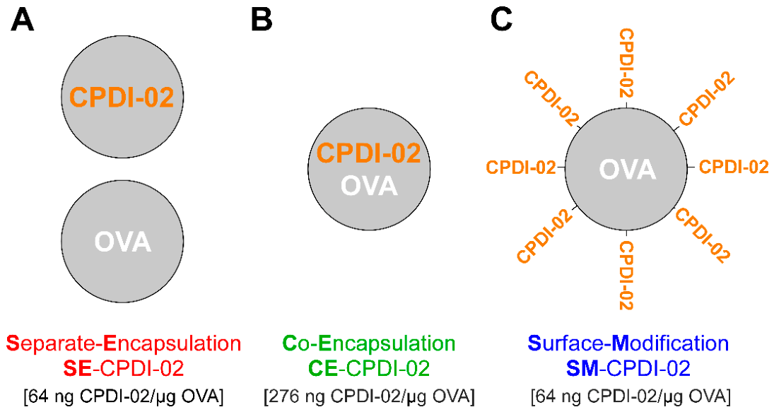
Figure 2.
Effect of incorporating CPDI-02 by separate encapsulation, co-encapsulation, or surface modification on early magnitudes of OVA-specific antibody-secreting cells (ASCs) in the lungs of naïve female C57BL/6 mice generated against microparticle-encapsulated LPS-free OVA after IN administration. Inactive, scrambled scCPDI-02 (open symbols) or CPDI-02 (closed symbols) was incorporated with LPS-free OVA encapsulated in PLGA 50:50 microparticles (~1 µm diam.) by separate encapsulation (SE-scCPDI-02; SE-CPDI-02), co-encapsulation (CE-scCPDI-02; CE-CPDI-02), or surface modification (SM-scCPDI-02; SM-CPDI-02) (Figure 1) (Table S1). Vehicle alone (PBS [50 µL], black asterisks) or vehicle containing SE-scCPDI-02 (open red circles), SE-CPDI-02 (closed red circles), CE-scCPDI-02 (open green triangles), CE-CPDI-02 (closed green triangles), SM-scCPDI-02 (open blue squares), or SM-CPDI-02 (closed blue squares) at 50 µg LPS-free OVA was then administered IN to naive female C57BL/6 mice (n=5 mice for vehicle alone and scCPDI-02 treatment groups and n=10 mice from two independent studies for CPDI-02 treatment groups) on Days -14, -7, and 0. Average OVA-specific (A) IgA, (B) IgM, and (C) IgG antibody secreting cell (ASC) spots / 106 lung lymphocytes ± SD (n=3 replicates per mouse) in the lungs were then determined 6 days post-IN administration by ELISpot. Data were combined from two independent studies, outliers were omitted by the ROUT method (Q = 1%), and mean ranks were compared by Kruskal–Wallis test with uncorrected Dunn’s post-test. Colored P-values indicate immunostimulant incorporation strategy that generated higher average ASCs / 106 lung lymphocytes (SE - Separate encapsulation [Red], CE - Co-Encapsulation [Green]; SM - Surface Modification [Blue]).
Figure 2.
Effect of incorporating CPDI-02 by separate encapsulation, co-encapsulation, or surface modification on early magnitudes of OVA-specific antibody-secreting cells (ASCs) in the lungs of naïve female C57BL/6 mice generated against microparticle-encapsulated LPS-free OVA after IN administration. Inactive, scrambled scCPDI-02 (open symbols) or CPDI-02 (closed symbols) was incorporated with LPS-free OVA encapsulated in PLGA 50:50 microparticles (~1 µm diam.) by separate encapsulation (SE-scCPDI-02; SE-CPDI-02), co-encapsulation (CE-scCPDI-02; CE-CPDI-02), or surface modification (SM-scCPDI-02; SM-CPDI-02) (Figure 1) (Table S1). Vehicle alone (PBS [50 µL], black asterisks) or vehicle containing SE-scCPDI-02 (open red circles), SE-CPDI-02 (closed red circles), CE-scCPDI-02 (open green triangles), CE-CPDI-02 (closed green triangles), SM-scCPDI-02 (open blue squares), or SM-CPDI-02 (closed blue squares) at 50 µg LPS-free OVA was then administered IN to naive female C57BL/6 mice (n=5 mice for vehicle alone and scCPDI-02 treatment groups and n=10 mice from two independent studies for CPDI-02 treatment groups) on Days -14, -7, and 0. Average OVA-specific (A) IgA, (B) IgM, and (C) IgG antibody secreting cell (ASC) spots / 106 lung lymphocytes ± SD (n=3 replicates per mouse) in the lungs were then determined 6 days post-IN administration by ELISpot. Data were combined from two independent studies, outliers were omitted by the ROUT method (Q = 1%), and mean ranks were compared by Kruskal–Wallis test with uncorrected Dunn’s post-test. Colored P-values indicate immunostimulant incorporation strategy that generated higher average ASCs / 106 lung lymphocytes (SE - Separate encapsulation [Red], CE - Co-Encapsulation [Green]; SM - Surface Modification [Blue]).
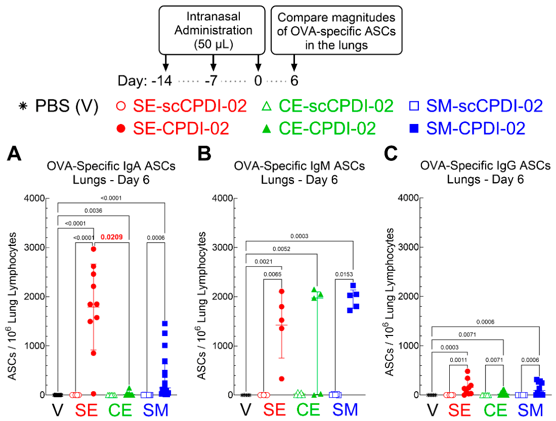
Figure 3.
Effect of incorporating CPDI-02 by separate encapsulation, co-encapsulation, or surface modification on early titers of OVA-specific antibodies in the nasal cavities and lungs of naïve female C57BL/6 mice generated against microparticle-encapsulated LPS-free OVA after IN administration. Inactive, scrambled scCPDI-02 (open symbols) or CPDI-02 (closed symbols) was incorporated with LPS-free OVA encapsulated in PLGA 50:50 microparticles (~1 µm diam.) by separate encapsulation (SE-scCPDI-02; SE-CPDI-02), co-encapsulation (CE-scCPDI-02; CE-CPDI-02), or surface modification (SM-scCPDI-02; SM-CPDI-02) (Figure 1) (Table S1). Vehicle alone (PBS [50 µL], used for normalization) or vehicle containing SE-scCPDI-02 (open red circles), SE-CPDI-02 (closed red circles), CE-scCPDI-02 (open green triangles), CE-CPDI-02 (closed green triangles), SM-scCPDI-02 (open blue squares), or SM-CPDI-02 (closed blue squares) at 50 µg LPS-free OVA was then intranasally administered to naïve female C57BL/6 mice (n=5 mice for vehicle alone and scCPDI-02 treatment groups and n=10 mice from two independent studies for CPDI-02 treatment groups) on Days -14, -7, and 0. Average OVA-specific titers ± SD (n = 3 replicates per mouse) of IgA in the nasal lavage fluid (NLF) (A) and bronchoalveolar lavage fluid (BALF) (B) and total IgG in the BALF (C) 14 days post-treatment were determined by ELISA and normalized to vehicle alone by statistical endpoint titer analysis. Data were combined from two independent studies, outliers were omitted by the ROUT method (Q = 1%), and mean ranks were compared by Kruskal–Wallis test with uncorrected Dunn’s post-test. Titers below the positive titer cutoff threshold are shown as 100. Colored P-values indicate immunostimulant incorporation strategy that generated higher average titers of the indicated class of OVA-specific antibodies (SE - Separate encapsulation [Red], CE - Co-Encapsulation [Green]; SM - Surface Modification [Blue]).
Figure 3.
Effect of incorporating CPDI-02 by separate encapsulation, co-encapsulation, or surface modification on early titers of OVA-specific antibodies in the nasal cavities and lungs of naïve female C57BL/6 mice generated against microparticle-encapsulated LPS-free OVA after IN administration. Inactive, scrambled scCPDI-02 (open symbols) or CPDI-02 (closed symbols) was incorporated with LPS-free OVA encapsulated in PLGA 50:50 microparticles (~1 µm diam.) by separate encapsulation (SE-scCPDI-02; SE-CPDI-02), co-encapsulation (CE-scCPDI-02; CE-CPDI-02), or surface modification (SM-scCPDI-02; SM-CPDI-02) (Figure 1) (Table S1). Vehicle alone (PBS [50 µL], used for normalization) or vehicle containing SE-scCPDI-02 (open red circles), SE-CPDI-02 (closed red circles), CE-scCPDI-02 (open green triangles), CE-CPDI-02 (closed green triangles), SM-scCPDI-02 (open blue squares), or SM-CPDI-02 (closed blue squares) at 50 µg LPS-free OVA was then intranasally administered to naïve female C57BL/6 mice (n=5 mice for vehicle alone and scCPDI-02 treatment groups and n=10 mice from two independent studies for CPDI-02 treatment groups) on Days -14, -7, and 0. Average OVA-specific titers ± SD (n = 3 replicates per mouse) of IgA in the nasal lavage fluid (NLF) (A) and bronchoalveolar lavage fluid (BALF) (B) and total IgG in the BALF (C) 14 days post-treatment were determined by ELISA and normalized to vehicle alone by statistical endpoint titer analysis. Data were combined from two independent studies, outliers were omitted by the ROUT method (Q = 1%), and mean ranks were compared by Kruskal–Wallis test with uncorrected Dunn’s post-test. Titers below the positive titer cutoff threshold are shown as 100. Colored P-values indicate immunostimulant incorporation strategy that generated higher average titers of the indicated class of OVA-specific antibodies (SE - Separate encapsulation [Red], CE - Co-Encapsulation [Green]; SM - Surface Modification [Blue]).
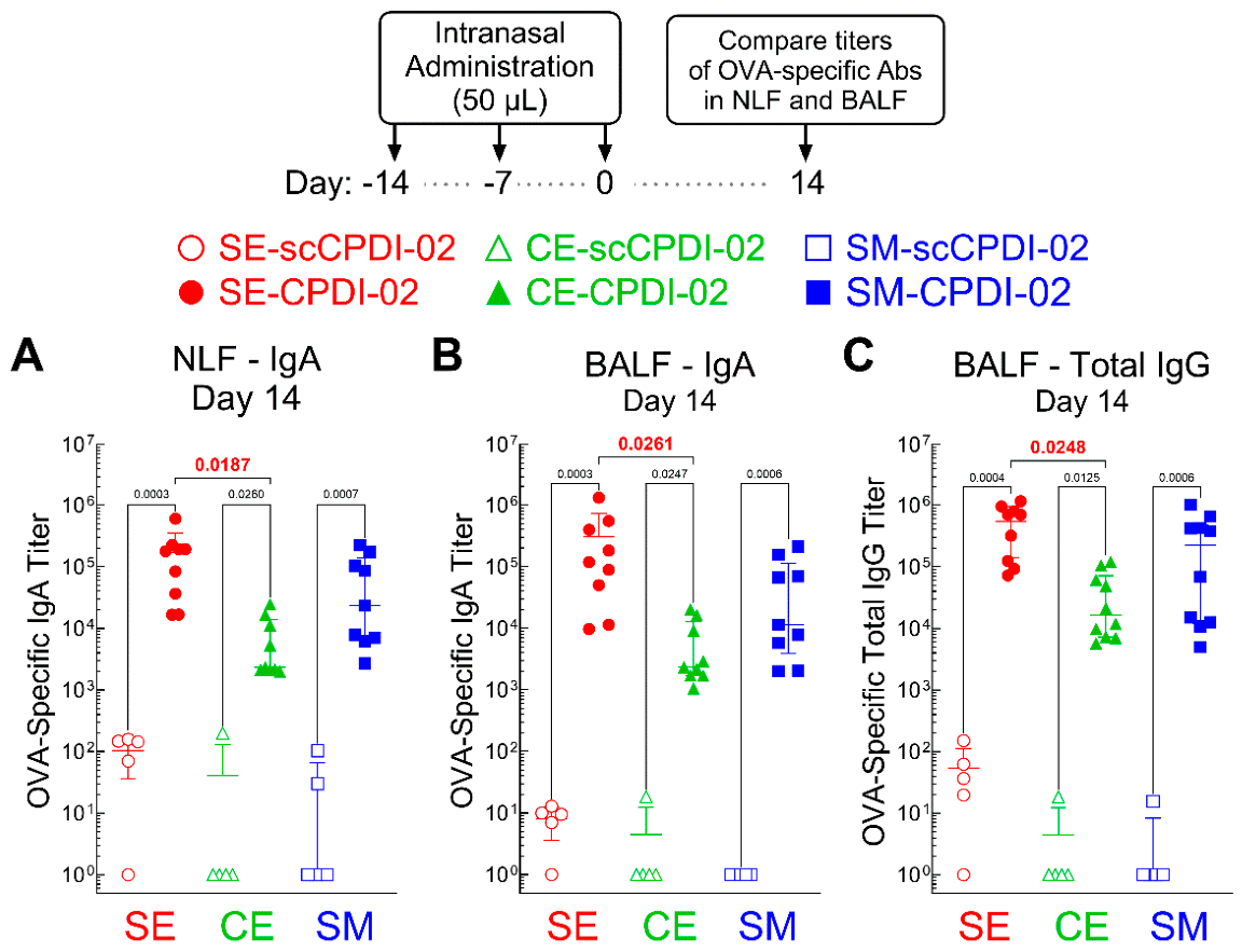
Figure 4.
Effect of incorporating CPDI-02 by separate encapsulation, co-encapsulation, or surface modification on early magnitudes of OVA-specific antibody-secreting cells (ASCs) in the spleens of naïve female C57BL/6 mice generated against microparticle-encapsulated LPS-free OVA after IN administration. Inactive, scrambled scCPDI-02 (open symbols) or CPDI-02 (closed symbols) was incorporated with LPS-free OVA encapsulated in PLGA 50:50 microparticles (~1 µm diam.) by separate encapsulation (SE-scCPDI-02; SE-CPDI-02), co-encapsulation (CE-scCPDI-02; CE-CPDI-02), or surface modification (SM-scCPDI-02; SM-CPDI-02) (Figure 1) (Table S1). Vehicle alone (PBS [50 µL], black asterisks) or vehicle containing SE-scCPDI-02 (open red circles), SE-CPDI-02 (closed red circles), CE-scCPDI-02 (open green triangles), CE-CPDI-02 (closed green triangles), SM-scCPDI-02 (open blue squares), or SM-CPDI-02 (closed blue squares) at 50 µg LPS-free OVA was then intranasally administered to naive female C57BL/6 mice (n=5 mice for vehicle alone and scCPDI-02 treatment groups and n=10 mice from two independent studies for CPDI-02 treatment groups) on Days -14, -7, and 0. Average OVA-specific (A) IgA, (B) IgM, and (C) IgG antibody secreting cell (ASC) spots / 106 splenocytes ± SD (n=3 replicates per mouse) in the spleens were then determined and compared 6 days post-IN administration as described (Figure 2). Colored P-values indicate immunostimulant incorporation strategy that generated higher average ASCs / 106 splenocytes (SE - Separate encapsulation [Red], CE - Co-Encapsulation [Green]; SM - Surface Modification [Blue]).
Figure 4.
Effect of incorporating CPDI-02 by separate encapsulation, co-encapsulation, or surface modification on early magnitudes of OVA-specific antibody-secreting cells (ASCs) in the spleens of naïve female C57BL/6 mice generated against microparticle-encapsulated LPS-free OVA after IN administration. Inactive, scrambled scCPDI-02 (open symbols) or CPDI-02 (closed symbols) was incorporated with LPS-free OVA encapsulated in PLGA 50:50 microparticles (~1 µm diam.) by separate encapsulation (SE-scCPDI-02; SE-CPDI-02), co-encapsulation (CE-scCPDI-02; CE-CPDI-02), or surface modification (SM-scCPDI-02; SM-CPDI-02) (Figure 1) (Table S1). Vehicle alone (PBS [50 µL], black asterisks) or vehicle containing SE-scCPDI-02 (open red circles), SE-CPDI-02 (closed red circles), CE-scCPDI-02 (open green triangles), CE-CPDI-02 (closed green triangles), SM-scCPDI-02 (open blue squares), or SM-CPDI-02 (closed blue squares) at 50 µg LPS-free OVA was then intranasally administered to naive female C57BL/6 mice (n=5 mice for vehicle alone and scCPDI-02 treatment groups and n=10 mice from two independent studies for CPDI-02 treatment groups) on Days -14, -7, and 0. Average OVA-specific (A) IgA, (B) IgM, and (C) IgG antibody secreting cell (ASC) spots / 106 splenocytes ± SD (n=3 replicates per mouse) in the spleens were then determined and compared 6 days post-IN administration as described (Figure 2). Colored P-values indicate immunostimulant incorporation strategy that generated higher average ASCs / 106 splenocytes (SE - Separate encapsulation [Red], CE - Co-Encapsulation [Green]; SM - Surface Modification [Blue]).
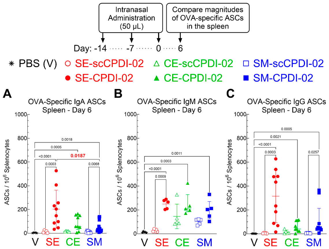
Figure 5.
Effect of incorporating CPDI-02 by separate encapsulation, co-encapsulation, or surface modification on early titers of OVA-specific subclasses of IgG antibodies in the serum of naïve female C57BL/6 mice generated against microparticle-encapsulated LPS-free OVA after IN administration. Inactive, scrambled scCPDI-02 (open symbols) or CPDI-02 (closed symbols) was incorporated with LPS-free OVA encapsulated in PLGA 50:50 microparticles (~1 µm diam.) by separate encapsulation (SE-scCPDI-02; SE-CPDI-02), co-encapsulation (CE-scCPDI-02; CE-CPDI-02), or surface modification (SM-scCPDI-02; SM-CPDI-02) (Figure 1) (Table S1). Vehicle alone (PBS [50 µL], used for normalization) or vehicle containing SE-scCPDI-02 (open red circles), SE-CPDI-02 (closed red circles), CE-scCPDI-02 (open green triangles), CE-CPDI-02 (closed green triangles), SM-scCPDI-02 (open blue squares), or SM-CPDI-02 (closed blue squares) at 50 µg LPS-free OVA was then intranasally administered to naïve female C57BL/6 mice (n=5 mice for vehicle alone and scCPDI-02 treatment groups and n=10 mice from two independent studies for CPDI-02 treatment groups) on Days -14, -7, and 0. Average OVA-specific titers ± SD (n = 3 replicates per mouse) of (A) IgG1, (B) IgG2b, (C) IgG2c, or (D) IgG3 antibodies in the serum were determined 14 days post-treatment by ELISA and normalized to vehicle alone by statistical endpoint titer analysis. Data were combined from two independent studies, outliers were omitted by the ROUT method (Q = 1%), and mean ranks were compared by Kruskal–Wallis test with uncorrected Dunn’s post-test. Titers below the positive titer cutoff threshold are shown as 100. Colored P-values indicate immunostimulant incorporation strategy that generated higher average titers of the indicated subclass of OVA-specific antibodies (SE - Separate encapsulation [Red], CE - Co-Encapsulation [Green]; SM - Surface Modification [Blue]).
Figure 5.
Effect of incorporating CPDI-02 by separate encapsulation, co-encapsulation, or surface modification on early titers of OVA-specific subclasses of IgG antibodies in the serum of naïve female C57BL/6 mice generated against microparticle-encapsulated LPS-free OVA after IN administration. Inactive, scrambled scCPDI-02 (open symbols) or CPDI-02 (closed symbols) was incorporated with LPS-free OVA encapsulated in PLGA 50:50 microparticles (~1 µm diam.) by separate encapsulation (SE-scCPDI-02; SE-CPDI-02), co-encapsulation (CE-scCPDI-02; CE-CPDI-02), or surface modification (SM-scCPDI-02; SM-CPDI-02) (Figure 1) (Table S1). Vehicle alone (PBS [50 µL], used for normalization) or vehicle containing SE-scCPDI-02 (open red circles), SE-CPDI-02 (closed red circles), CE-scCPDI-02 (open green triangles), CE-CPDI-02 (closed green triangles), SM-scCPDI-02 (open blue squares), or SM-CPDI-02 (closed blue squares) at 50 µg LPS-free OVA was then intranasally administered to naïve female C57BL/6 mice (n=5 mice for vehicle alone and scCPDI-02 treatment groups and n=10 mice from two independent studies for CPDI-02 treatment groups) on Days -14, -7, and 0. Average OVA-specific titers ± SD (n = 3 replicates per mouse) of (A) IgG1, (B) IgG2b, (C) IgG2c, or (D) IgG3 antibodies in the serum were determined 14 days post-treatment by ELISA and normalized to vehicle alone by statistical endpoint titer analysis. Data were combined from two independent studies, outliers were omitted by the ROUT method (Q = 1%), and mean ranks were compared by Kruskal–Wallis test with uncorrected Dunn’s post-test. Titers below the positive titer cutoff threshold are shown as 100. Colored P-values indicate immunostimulant incorporation strategy that generated higher average titers of the indicated subclass of OVA-specific antibodies (SE - Separate encapsulation [Red], CE - Co-Encapsulation [Green]; SM - Surface Modification [Blue]).
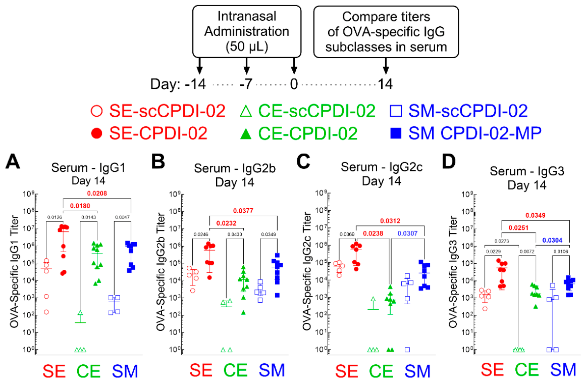
Figure 6.
Dose response of separately encapsulated CPDI-02 on titers of OVA-specific antibodies in the nasal cavities and lungs of naïve female C57BL/6 mice generated against microparticle-encapsulated LPS-free OVA after IN administration. Inactive, scrambled scCPDI-02 (open symbols) or CPDI-02 (closed symbols) was incorporated with LPS-free OVA encapsulated in PLGA 50:50 microparticles (~1 µm diam.) by separate encapsulation (SE-scCPDI-02; SE-CPDI-02) (Figure 1) (Table S1). Vehicle alone (PBS [50 µL], used for normalization) or vehicle containing inactive SE-scCPDI-02 at 400 ng/µg OVA (open red circles), SE-CPDI-02 at 64 ng/µg OVA (closed red circles) (data from Figure 3), SE-CPDI-02 at 200 ng/µg OVA (closed red squares), or SE-CPDI-02 at 400 ng/µg OVA (closed red triangles) with 50 µg LPS-free OVA was then intranasally administered to naïve female C57BL/6 mice (n=5 mice for vehicle alone, scCPDI-02, and SE-CPDI-02 at 200 and 400 ng/µg OVA and n=10 mice from two independent studies for SE-CPDI-02 at 64 ng/µg OVA) on Days -14, -7, and 0. Average OVA-specific titers ± SD (n = 3 replicates per mouse) of IgA in the nasal lavage fluid (NLF) (A) and bronchoalveolar lavage fluid (BALF) (B) and total IgG in the BALF (C) 14 days post-treatment were determined by ELISA and normalized to vehicle alone by statistical endpoint titer analysis. Data were combined from two independent studies, outliers were omitted by the ROUT method (Q = 1%), and mean ranks were compared by Kruskal–Wallis test with uncorrected Dunn’s post-test. Titers below the positive titer cutoff threshold are shown as 100.
Figure 6.
Dose response of separately encapsulated CPDI-02 on titers of OVA-specific antibodies in the nasal cavities and lungs of naïve female C57BL/6 mice generated against microparticle-encapsulated LPS-free OVA after IN administration. Inactive, scrambled scCPDI-02 (open symbols) or CPDI-02 (closed symbols) was incorporated with LPS-free OVA encapsulated in PLGA 50:50 microparticles (~1 µm diam.) by separate encapsulation (SE-scCPDI-02; SE-CPDI-02) (Figure 1) (Table S1). Vehicle alone (PBS [50 µL], used for normalization) or vehicle containing inactive SE-scCPDI-02 at 400 ng/µg OVA (open red circles), SE-CPDI-02 at 64 ng/µg OVA (closed red circles) (data from Figure 3), SE-CPDI-02 at 200 ng/µg OVA (closed red squares), or SE-CPDI-02 at 400 ng/µg OVA (closed red triangles) with 50 µg LPS-free OVA was then intranasally administered to naïve female C57BL/6 mice (n=5 mice for vehicle alone, scCPDI-02, and SE-CPDI-02 at 200 and 400 ng/µg OVA and n=10 mice from two independent studies for SE-CPDI-02 at 64 ng/µg OVA) on Days -14, -7, and 0. Average OVA-specific titers ± SD (n = 3 replicates per mouse) of IgA in the nasal lavage fluid (NLF) (A) and bronchoalveolar lavage fluid (BALF) (B) and total IgG in the BALF (C) 14 days post-treatment were determined by ELISA and normalized to vehicle alone by statistical endpoint titer analysis. Data were combined from two independent studies, outliers were omitted by the ROUT method (Q = 1%), and mean ranks were compared by Kruskal–Wallis test with uncorrected Dunn’s post-test. Titers below the positive titer cutoff threshold are shown as 100.
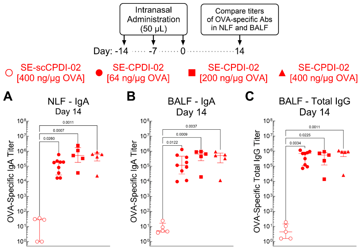
Figure 7.
Dose response of separately encapsulated CPDI-02 on early titers of OVA-specific IgG antibody subclasses in the serum of naïve female C57BL/6 mice generated against microparticle-encapsulated LPS-free OVA after IN administration. Inactive, scrambled scCPDI-02 (open symbols) or CPDI-02 (closed symbols) was incorporated with LPS-free OVA encapsulated in PLGA 50:50 microparticles (~1 µm diam.) by separate encapsulation (SE-scCPDI-02; SE-CPDI-02) (Figure 1) (Table S1). Vehicle alone (PBS [50 µL], used for normalization) or vehicle containing SE-scCPDI-02 at 400 ng/µg OVA (open red circles), SE-CPDI-02 at 64 ng/ µg OVA (closed red circles) (data from Figure 5), SE-CPDI-02 at 200 ng/µg OVA (closed red squares), or SE-CPDI-02 at 400 ng/µg OVA (close red triangles) with 50 µg MP-encapsulated LPS-free OVA was then intranasally administered to naïve female C57BL/6 mice (n=5 mice for vehicle alone, scCPDI-02, and SE-CPDI-02 at 200 and 400 ng/µg OVA and n=10 mice from two independent studies for SE-CPDI-02 at 64 ng/µg OVA) on Days -14, -7, and 0. Average OVA-specific titers ± SD (n = 3 replicates per mouse) of Th2 (A) IgG1 and Th1 (B) IgG2b, (C) IgG2c, or (D) IgG3 antibodies in the serum were determined 14 days post-treatment by ELISA and normalized to vehicle alone by statistical endpoint titer analysis. Data were combined from two independent studies, outliers were omitted by the ROUT method (Q = 1%), and mean ranks were compared by Kruskal–Wallis test with uncorrected Dunn’s post-test. Titers below the positive titer cutoff threshold are shown as 100.
Figure 7.
Dose response of separately encapsulated CPDI-02 on early titers of OVA-specific IgG antibody subclasses in the serum of naïve female C57BL/6 mice generated against microparticle-encapsulated LPS-free OVA after IN administration. Inactive, scrambled scCPDI-02 (open symbols) or CPDI-02 (closed symbols) was incorporated with LPS-free OVA encapsulated in PLGA 50:50 microparticles (~1 µm diam.) by separate encapsulation (SE-scCPDI-02; SE-CPDI-02) (Figure 1) (Table S1). Vehicle alone (PBS [50 µL], used for normalization) or vehicle containing SE-scCPDI-02 at 400 ng/µg OVA (open red circles), SE-CPDI-02 at 64 ng/ µg OVA (closed red circles) (data from Figure 5), SE-CPDI-02 at 200 ng/µg OVA (closed red squares), or SE-CPDI-02 at 400 ng/µg OVA (close red triangles) with 50 µg MP-encapsulated LPS-free OVA was then intranasally administered to naïve female C57BL/6 mice (n=5 mice for vehicle alone, scCPDI-02, and SE-CPDI-02 at 200 and 400 ng/µg OVA and n=10 mice from two independent studies for SE-CPDI-02 at 64 ng/µg OVA) on Days -14, -7, and 0. Average OVA-specific titers ± SD (n = 3 replicates per mouse) of Th2 (A) IgG1 and Th1 (B) IgG2b, (C) IgG2c, or (D) IgG3 antibodies in the serum were determined 14 days post-treatment by ELISA and normalized to vehicle alone by statistical endpoint titer analysis. Data were combined from two independent studies, outliers were omitted by the ROUT method (Q = 1%), and mean ranks were compared by Kruskal–Wallis test with uncorrected Dunn’s post-test. Titers below the positive titer cutoff threshold are shown as 100.
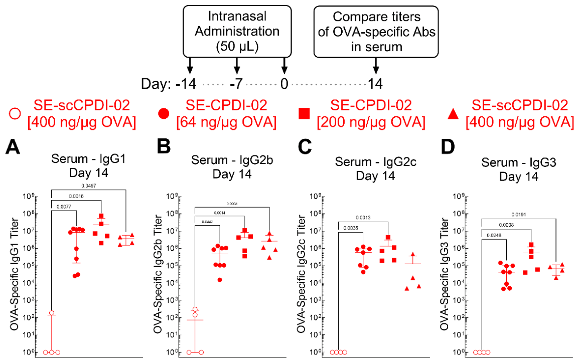
Disclaimer/Publisher’s Note: The statements, opinions and data contained in all publications are solely those of the individual author(s) and contributor(s) and not of MDPI and/or the editor(s). MDPI and/or the editor(s) disclaim responsibility for any injury to people or property resulting from any ideas, methods, instructions or products referred to in the content. |
© 2026 by the authors. Licensee MDPI, Basel, Switzerland. This article is an open access article distributed under the terms and conditions of the Creative Commons Attribution (CC BY) license.
Copyright: This open access article is published under a Creative Commons CC BY 4.0 license, which permit the free download, distribution, and reuse, provided that the author and preprint are cited in any reuse.