

Submitted:
08 July 2026
Posted:
24 July 2026
You are already at the latest version
Abstract
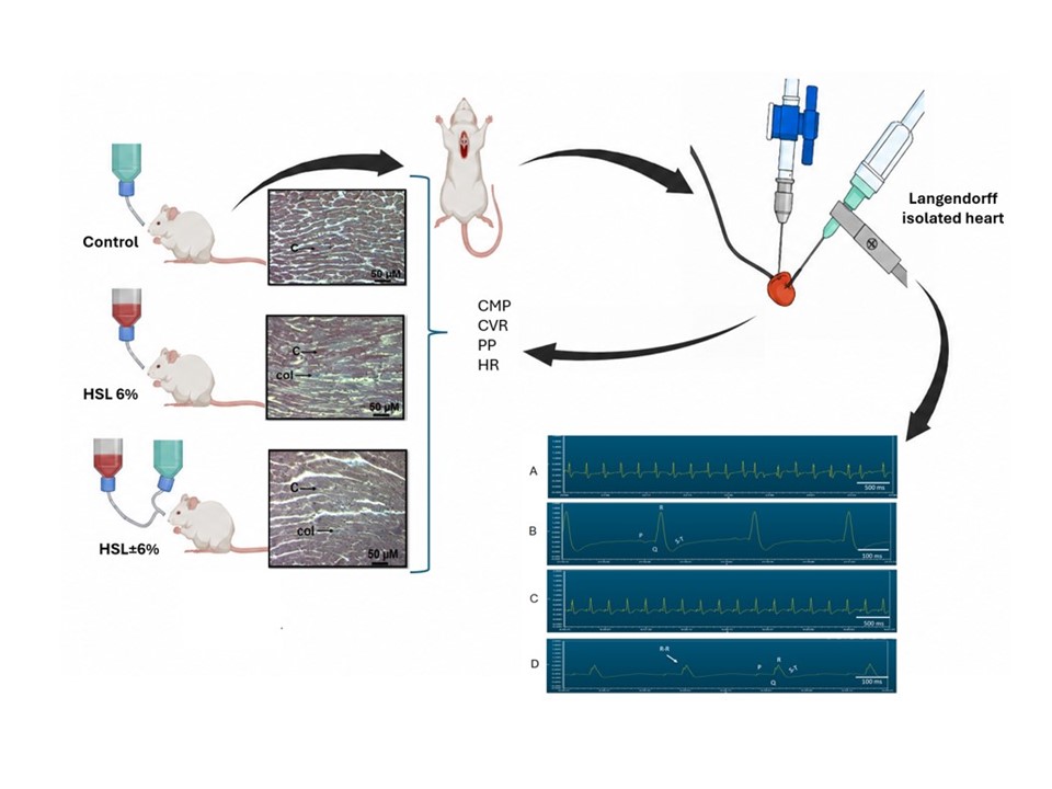
Ingestion of Hibiscus sabdariffa L. (HSL) provides antioxidants with beneficial effects for several pathologies, with underlying oxidative stress. However excessive, chronic consumption of the antioxidant-rich diet with HSL in healthy rats induces reductive stress (RS) with damaging effects. The aim of this study was to evaluate if the consumption of an infusion of 6% HSL for two months alters the function and structure of the myocardium. 24 male Wistar rats were divided into three groups: Control (C), HLS infusion at 6% for two months (HSL 6%), and a washout group that received HSL 6 % for two months followed by two months of natural water (HSL ± 6%). Myocardial performance during ischemia-reperfusion (I/R) was evaluated by using the isolated Langendorff heart model, accompanied by surface electrocardiography and histopathological analysis. Chronic 6% HSL intake increased systolic pressure (p≤0.03), elevated coronary vascular resistance, depressed mechanical performance, led to bradycardia, and critical, extended asystolic/sinus pauses. Histopathology showed dense, permanent networks of interstitial and perivascular collagen encapsulating the hypertrophied cardiomyocytes. Therefore, the overload of antioxidant molecules provided by the HSL infusion leads to RS that increases myocardial interstitial fibrosis and disrupts coronary compliance and intercellular electrical coupling, leaving the heart vulnerable to ischemic stress.
Keywords:
Hibiscus sabdariffa L
; reductive stress
; ischemia-reperfusion
; heart
1. Introduction
The therapeutic value of Hibiscus sabdariffa Linnaeus (HSL), owed to its rich concentration of bioactive phytochemicals including polyphenols, anthocyanins, flavonoids, ascorbic acid, amino acids and minerals among others is extensively documented [1]. Under pathological conditions characterized by oxidative stress (OS), such as metabolic syndrome and essential hypertension, the administration of HSL infusions at a concentration of 2-3 % has consistently demonstrated high efficacy in restoring the cellular redox balance [2]. In healthy organisms, these exogenous antioxidants collaborate with endogenous enzymatic systems to neutralize reactive oxygen species (ROS) and support physiological homeostasis [3]. However, a paradigm in ROS biology that challenges the conventional assumption that a supplementation with a high-dose of antioxidant is safe has arisen.
Prolonged and excessive ingestion of antioxidant-rich diets in healthy states can oversaturate counter-regulatory networks of OS, forcing the cellular environment to a paradoxical condition known as reductive stress (RS) and to a loss of the redox homeostasis [4]. RS is fundamentally defined by an overactivation of enzymatic/non-enzymatic antioxidant systems, an increase in total antioxidant capacity (TAC) and the accumulation of reducing equivalents such as NADH/NADPH, GSH/GSSG and oxidized/reduced thiols [5]. This results in a critical depletion of physiological ROS that triggers systemic maladaptive cascades, damaging the indispensable role of the ROS signaling as second messengers thereby regulating cellular proliferation, mechanical vascular contractility, syncytial communication, spermatogenesis and ovulation among another cellular pathway [6].
Our research group has systematically explored the multi-organic consequences of this phenomenon using a model of chronic consumption of a 6 % HSL infusion in healthy Wistar rats. We previously demonstrated that the excessive intake of this infusion causes systemic alterations, characterized by an upregulated inflammatory profile in plasma alongside with an unexpected elevation in systolic blood pressure (SBP) [7]. At the vascular level, we reported an altered aortic vascular reactivity, which is associated with a decrease in glutathione-S-transferase activity and an increase in TAC [8]. Furthermore, our recent work in renal pathology revealed that prolonged consumption of the 6 % infusion induces severe kidney damage [9]. This renal impairment is driven by an over- production of hydrogen sulfide which elevates cellular reducing power, induces mitochondrial dysfunction via decreased oxidative phosphorylation in complexes I and IV, and results in structural remodeling indicating retraction of the glomerular tuft and tubulointerstitial fibrosis [10].
On the other hand, the myocardium operates as a highly coordinated, functional syncytium in which mechanical contractility and electrical conduction depend on the integrity of the extracellular matrix (ECM), ionic homeostasis, and an uninterrupted supply of ATP. This mechanical syncytium functions to maintain systemic perfusion, making it uniquely sensitive to structural and metabolic remodeling, at the physiological level [11]. While the detrimental effects of RS caused by the ingestion of 6 % HSL on plasma biomarkers, vascular smooth muscle reactivity, and renal architecture have been established by our group, its specific impact on the heart remains unknown. The question arises whether the systemic inflammatory and hypertensive state induced by the chronic ingestion of 6 % HSL could extend to the cardiac ECM, or if the effect of this sustained antioxidant overload may affect coronary compliance and electrical coupling. In normal conditions, the collagen of the ECM provides the necessary structural support to withstand systolic stress; however, chronic pro-inflammatory profiles alter its turnover, promoting fibrosis and ventricular stiffness [12] Cardiac electro-mechanical coupling also requires the precise synchronization of gap junctions, where connexin such as the 43 (Cx43), which is the predominant pore-forming protein is of importance. The assembly and opening of Cx43 is modulated by the redox state with its cysteine residues being highly susceptible to post-translational modifications [13]. Under normal physiological conditions, basal ROS maintain the appropriate phosphorylation of Cx43, ensuring low-resistance electrical coupling. Conversely, excessive accumulation of reducing equivalents could disrupts this balance, causing dephosphorylation or lateralization of Cx43, affecting longitudinal conduction velocity [14]. Since maintaining the electrical ionic gradients of the cardiac syncytium and ECM homeostasis are highly energy-dependent processes, an energy deficit deprives the tissue of the metabolic substrates necessary to sustain its function [15]. Consequently, when faced with hemodynamic challenges such as I/R, a pre-existing bioenergetic collapse, and electrical uncoupling could alter coronary capacitance, leaving the heart vulnerable to lethal arrhythmias and acute failure.
In this sense, the onset of cardiac arrhythmias is associated with tissue excitability and changes in conduction velocity (CV), which are linked by their biophysical dependence on similar membrane proteins, electrochemical forces, and cellular structures [16]. The relationship between uncoupling of gap junctions and tissue excitability and CV underscore the complex interplay of mechanisms that exist in cardiac damage, as CV does not change drastically during gap junction uncoupling [17] Cell uncoupling reduces the electronic charge while decreasing the propensity for conduction failure, because the charge accumulates in the intracellular space of individual cardiomyocytes [18]. However, this increase in intracellular charge leads to faster movement of action potentials across cells, but slower movement between them in the CV due to cell decoupling [19]. However, other local, genetic, or environmental factors may exist that are integrated within the mechanisms of damage and could lead to structural or hemodynamic functional alterations in the heart. This is further compromised if there is inflammation and/or oxidative or RS, which could have a direct effect on ventricular dysfunction and arrhythmias [20]. Therefore, the aim of the present study was to determine whether the chronic and excessive ingestion of a 6 % HSL infusion for two months compromises the structural, mechanical, and hemodynamic performance of the myocardium in hearts from healthy Wistar rats subjected al I/R and whether the effects can be reversed if the treatment is suspended.
2. Materials and Methods
2.1. Preparation of the HSL 6 % Infusion
60 g the HSL calyces were added to 1 liter of boiling water, which was kept boiling for 10 min, allowed to cool and then filtered. It was then stored at 4 °C until consumption. The HSL infusion was prepared and replaced each week to avoid fermentation. This solution was provided ad libitum to the rats.
2.2. Determinations of Polyphenols, Total Flavonoids, Total Anthocyanins and C Vitamin in the HSL 6 % Infusion
The total vitamin C in the 6 % infusion was determined by the method of Jagota. 200 μL of Folin-Ciocalteu reagent 0.20 mM were added to 100 μL of the HSL infusion. The mixture was shaken vigorously and incubated for 10 min. The absorbance was measured at 760 nm. The calibration curve was obtained using an ascorbic acid as standard solution [21]. The total flavonoid content in the 6 % infusion was determined by the Jia method. This technique determines total flavanols and flavones such as apigenin, chrysin, luteonin, morin, quercetin, kaempferol, myricetin, and galangin which can be analyzed spectrophotometrically to 510 nm. The calibration curve was achieved using quercetin as standard [22]. The determination of total anthocyanins was performed by Lee's method and measured at 520 and 700 nm. The values are presented as cyaniding-3-glucoside [23].
2.3. Calculation for the Sample Size
The mean systolic blood pressure (SBP) in healthy rats was reported to be between 116 mmHg and 112.8 mmHg, with a variance of 19, in rats from the National Institute of Cardiology Ignacio Chavez. Based on this, the sample size per group was estimated by μ (SBP of Wistar rats), with 95% confidence and a maximum error (ME) of 3.2 mmHg, according to the following formula: ME: |μ − x|=|116 − 112.8|=3.2 mmHg, Confidence: |μ−x|=|116−112.8|=3.2, SS: n=(σ̅x)2xσ2/(EM2)=(22)×(22)/(3.2)2=(4 × 19)/(10.24)=76/10.24= 7.42, where ME= maximum error, σ̅x= # of standard deviations of the mean estimator, σ2x= variance in the SBP of the rats in our institute, and SS= sample size.
2.4. Experimental Design
Twenty-four male Wistar rats were used to form 3 groups with 8 animals each. Group 1: Rats that received plain tap water ad libitum for two months (C). Group 2: Rats that received a 6 % infusion of HSL ad libitum to for two months (HSL 6 %). Group 3: Rats that received a 6 % infusion of HSL ad libitum for two months, after which they were given tap water for another two months (HSL ± 6 %). The animals were housed for 8 weeks under the following conditions: a 12 h light/12 h dark cycle, room temperature from 18 to 26 °C, and relative humidity ranging from 40 to 70%. The commercial food the rodents consumed was solid rodent kibble supplemented with 23 % crude protein, 4.5 % crude fat, 6 % crude fiber, 8 % ash, and 2.5 % minerals (Lab diet 5008; PMI Nutrition International, Richmond, IN Indiana EE. UU.) ad libitum. This study was designed and carried out in compliance with the Laboratory Animal Care Committee of the National Institute of Cardiology Ignacio Chávez (INC/CICUAL/009/2023).
At the end of treatment and before euthanizing the animals, the SBP was measured using a plethysmograph (Narco Bio-system Houston, Texas, EE. UU) that sends the signals to a computer equipped with a program for data capture and processing (SIEVART version 0.1). Five measurements per rat were performed. The experimental animals were anesthetized with sodium pentobarbital (60 mg/Kg of body weight) and heparin (1000 U/mL/Kg of body weight) was administered before being euthanized.
2.5. Isolated Langendorff Heart Model Under I/R
The heart was exposed by a thoracotomy and removed. It was then placed in ice-cold Krebs–Henseleit solution to be arrested and to prevent ischemic preconditioning. Immediately, the heart was connected to the perfusion system through the ascending aorta. The heart was maintained with mechanical activity with Krebs–Henseleit solution with the following composition (mM): 120 NaCl, 23.4 NaHCO3, 4.8 KCl, 1.2 KH2PO4, 0.86 MgSO4, 1.25 CaCl2 and 11.0 of glucose (pH 7.4 and temperature 37 °C and oxygenated with 95% O2 and 5% CO2) through a constant retrograde perfusion (13 mL/min). The perfusion started with an adaptation period of 30 min (5 min with a flow (F) of 25 mL/min and 25 min with F of 14 mL/min). Heart rate (HR) was maintained at 312-324 beats/min, using a Grass stimulator of quadratic pulses (Grass Model S44F, Grass Instruments Co., Quincy, MA, USA). Coronary flow was regulated with a peristaltic pump (Master Flex Easy-load II, no. 77200-50; Cole-Parmer Instrument Co., Vermon Hills, IL, USA). Parameters including left intraventricular pressure (LIVP) were recorded by means of a Grass hydropneumatics pressure transducer, to which a catheter with a latex balloon was connected. The balloon was introduced through the mitral valve into the left ventricle and once inside the cavity, an internal pressure of 5–10 mmHg (diastolic pressure) was applied, with another Grass hydropneumatics pressure transducer, the perfusion pressure (PP) was recorded and a range of 55–70 mmHg at the beginning of the experiment was considered as an inclusion criterion.
All parameters were recorded using a computer acquisition data system (Grass Telefactor, Grass Technologies, Astro Med, West Warwick, RI, USA) coupled with a Grass model 79D polygraph and online software (Grass PolyView, West Warwick, RI, USA).
Cardiac mechanical performance (CMP) was calculated as HR × LIVP = CMP.
2.5. Heart Homogenization
After completing the experiments on the isolated heart, it was removed from the Langendorff system and segmented into three parts where the superior and apical part were homogenized under liquid nitrogen after adding KH2PO4 (1 mL) 0.05 mM, pH 7.3, in the presence of 20 µL antiprotease inhibitors (2 μM leupeptin, 2 μM pepstatin A, 0.1% aprotinin and 1 mM PMSF).
2.6. Anatomical Changes in the Heart by a Histological Process
To demonstrate the anatomical and structural changes in the heart tissue, after completing the experiments on the isolated heart, the central part was used to perform the histology procedures. The tissue was fixed in 10% formalin solution for 24 h, gradually dehydrated in ethanol, cleared in xylene, embedded in paraffin, and cut into 5 µM thick slices using a microtome (Leica RM212RT, Wetzlar, Germany). The paraffin sections were stained with Masson’s trichrome and Sirio red technique. The histological sections were analyzed at 12.5× magnification using a model 63300 optical microscope (Carl Zeiss, Overcoaches, Germany) equipped with a Tucsen digital camera (18 megapixels) coupled with TSview 7.3.1 software. Blinded evaluation was used to analyze histological and immunohistochemistry sections and it was validated with the scoring system Allred Score. The intensity of light in the microscope was adjusted and remained constant. The heart area was analyzed by densitometry using Sigma Scan Pro 5 Image Analysis software (Systat Software Inc., San Jose, CA, USA).
2.7. Evaluation of Total Antioxidant Capacity (TAC)
100 μg of homogenized heart were added a mixture that contained C2H3O2 at 300 mM, FeCl3·6H2O at 20 mM, 2,4,6-tris-2-pyridyl-s-triazine at 10 mM and HCl at 40 mM, at pH 3.6 (1.5 mL, at a ratio of 10:1:1 v/v), and incubated at 37 °C for 15 min, and the absorbance was measured at 593 nm [24]
2.8. Statistical Analysis
Statistical analysis and graphs were performed using Sigma Plot software (Sigma Plot® version 15.0, Jandel Corporation, Palo Alto, CA, USA). Data are presented as mean ± standard error. Statistical significance was determined using one-way ANOVA and Tukey’s post hoc test. p≤0.05 was considered significant.
3. Results
3.1. Content of Some Antioxidants Provide by HSL 6 % Infusion and General in General Groups
Table 1 shows that the HSL 6 % infusion provides diverse antioxidants such as flavanols, polyphenols, anthocyanins and C vitamin.
Table 2 shows the physiological variables of the animals before they were euthanized, where only in the SBP of the C and HSL ± 6 % groups in comparison with HSL 6 % group showed a significant decreased (p≤0.03).
3.2. Structural Changes in the Heart
In the C group the sections showed preserved myocardial tissue architecture, with cardiomyocyte bundles arranged in compact, parallel sheets and homogeneous cell density. Severe damage, classic to ischemic stroke, is observed. Visible disorganization of the myofibrils is present, along with ample clear interstitial spaces. Specifically, Masson's staining showed compact and homogeneous myocardial tissue (Figure 1A). The muscle fibers were well-organized and aligned, with minimal areas of light blue staining between them, indicating a basal or normal level of interstitial collagen and thick collagen bands. Syrian red staining revealed a regular myocardial structure with thin reddish bands in the normal interstitial spaces (confirming the absence of pathological fibrosis), Figure 1D). However, marked post-ischemic reactive fibrosis, tissue edema, and detachment of muscle bundles were evident. The tissue lost its usual elastic and compact continuity.
In the HSL 6 % group the histopathology showed increased structural damage and remodeling. Specifically, Masson's stain revealed a loss of compact tissue continuity, i.e., areas of separation or elongation between fibers. Lighter, bluish bands or patches appeared in the interstitial space, suggesting cellular disorganization and increased collagen deposition (interstitial fibrosis), (Figure 1B). Syrian red staining showed increased thickness and intensity of the red lines between muscle fascicles. Muscle fibers appeared slightly more separated due to the accumulation of extracellular matrix, resulting in perivascular or interstitial fibrosis, where bright red collagen surrounds and separates groups of cardiomyocytes (Figure 1E).
Finally, in the HSL ± 6 %, group the morphological description of the tissue shows an intermediate structural organization. Although subtle separations between muscle fascicles persist, the architecture of the muscle fibers appears slightly more compact, with the fibers partially regaining the linear arrangement and the lost compact packing (Figure 1C). Sirius red staining, which is specific for type I and III collagen, staining it a bright red or deep pink, while the muscle cells take on a yellowish/ochre hue, shown marked tissue fragmentation and critical widening of the interfascicular spaces, revealing a significantly thinner and more restricted collagen banding pattern compared to the massive and dense infiltration observed in the HSL 6 % group (Figure 1F).
In addition, the desitophotometric analysis of the fibrosis area that were evidenced in the experimental groups by the histological process, show that groups C and HSL ± 6% showed a significant reduction (p=0.01 and p=0.03, Figure 2) in comparison with HSL 6% group.
3.3. Myocardial Function and Hemodynamics
3.3.1. Cardiac Mechanical Performance (CMP)
During the pre-ischemic baseline period (0–30 min), cardiac parameters remained stable in all experimental groups. Global ischemia (31–60 min) induced a complete cessation of mechanical and hemodynamic activity. Distinct variations emerged exclusively during the reperfusion phase (61–90 min). During the reperfusion phase, the C group exhibited a standard physiological recovery (~8,000 mmHg/beats/min). Chronic intake of HSL significant increase in CMP in the HSL 6 % group compared to the C group (p=0.03, Figure 3A). The HSL ± 6 % group exhibited no significant difference from the C group. This indicates a notable attenuation of the mechanically altered state upon treatment cessation.
3.3.2. Coronary Vascular Resistance (CVR)
The C group maintained stable and healthy CVR values throughout reperfusion (~3 mmHg/mL/min). In contrast, the HSL 6 % group displayed a severe, sustained, and statistically significant elevation in CVR (p=0.001, Figure 3B), indicating a prominent vasoconstrictive effect or vascular remodeling. The HSL ± 6 % group demonstrated a substantial decrease in resistance. This group lost statistical significance relative to the C group, demonstrating that the vascular alterations can be partially reversible.
3.3.3. Perfusion Pressure (PP)
PP was significantly elevated in the HSL 6 % group compared to the C group (p=0.001, Figure 3C), peaking near (~60 mmHg). The HSL ± 6 % group showed an intermediate response; PP was noticeably reduced compared to the treatment group and converged toward control baselines (~42 mmHg), showing no statistically significant difference from C group.
3.3.4. Heart Rate (HT)
Upon reperfusion, the C group exhibited a typical chronotropic recovery (~250 beats/min). The HSL 6 % group showed a marked, statistically significant bradycardia (~200 beats/min; p=0.005, Figure 3D). Notably, the washout group (HSL ± 6 %) demonstrated a substantial restoration of sinus rhythm, rising to (~220 beats/min). This partial chronotropic recovery eliminated the statistical discrepancy observed during administration HSL 6 % infusion.
3.4. Electrocardiograms (ECG)
The group C presented normal ECG obtained using the Langendorff method. Panel A in Figure 4 show cyclic complexes with constant intervals and homogeneous amplitudes at 500 ms. In panel B, the tracing magnified to 100 ms detailed well-defined waves where the P wave (atrial depolarization) and the QR complex with a narrow, tall R wave (ventricular depolarization) and the S-T segment showed no significant alterations. The panels after the I/R test (C and D) showed an arrhythmia at 500 ms, as the regularity was lost, evidenced by variations in HR and complex amplitude. The 100 ms magnification highlighted with an arrow shows a premature and widened complex (marked R-R), consistent with a ventricular extrasystole or a doublet.
In the ECG of rat hearts that received the HSL 6 % infusion, baseline electrical instability was observed in panels E and F. Panel E at 500 ms, showed notable variations in amplitude and an elevated HR before the damage was induced. Similarly, in panel F at 100 ms, a premature complex marked R-R, representing an isolated ventricular extrasystole, is clearly visible. This suggests that excessive consumption of antioxidants provided by HSL at 6 % alters cellular electrical homeostasis at baseline, predisposing the tissue to arrhythmias even without ischemia. Furthermore, after the I/R infusion, panel G at 500 ms, showed a complete loss of cyclic regularity and a drastic drop in the amplitude of the QRS complexes. Meanwhile, in panel H at 100 ms, the white arrow points to completely distorted and widened ventricular complexes. This means that consuming the infusion for two months causes lethal ventricular arrhythmias, reflecting a severely damaged myocardium.
On the other hand, panel I shows a representative ECG of the HSL ± 6 % group at 500 ms, where a markedly more homogeneous, cyclical tracing with stable R wave amplitudes was observed, similar to the C group. In the magnification of panel J at 100 ms, the PQR waves, as well as the S-T segment, appear clear and well-defined; unlike the ECG in panel F, no basal ventricular extrasystoles (R-R intervals) were observed. During the reperfusion period, panel K (500 ms) shows that, despite the 31-minute ischemic insult, the heart maintains well-defined QRS complexes and a coordinated rhythm for most of the tracing, unlike the chaotic collapse observed in panel G. Finally, panel L at 100 ms, shows notable pauses and delays in electrical conduction (a flat line or transient asystole between complexes), and the aberrant polymorphic, wide, and fragmented complexes recorded in panel H were not observed. This means that the heart suffers from reperfusion but does not enter polymorphic ventricular tachycardia or exhibit ventricular premature beats (R-R intervals). These results suggest that, while there is no complete restoration to healthy tissue conditions, the myocardium exhibits partial compensation under baseline conditions but develops a failure of automaticity during reperfusion. In other words, the two months of washing with tap water were enough to normalize the resting membrane potential and suppress basal ectopia, but were insufficient to recover the required cell density or function before the reperfusion period.
4. Discussion
In the present study we evaluated whether the chronic and excessive ingestion of a 6 % HSL infusion, which provides a large amount of antioxidant molecules, for two months, as demonstrated by the increase in TAC in the heart homogenate in our study, compromised the structural, mechanical, and hemodynamic performance of the myocardium during I/R in hearts from healthy Wistar rats. We also evaluated if the removal of the infusion could revert the damaging effects of RS associated to the ingestion of the infusion.
In experimental models of I/R, the heart is subjected to a period of restricted flow (ischemia) followed by the restoration of flow (reperfusion). While restoring flow is essential to save ischemic tissue, there is a reperfusion paradox that states that it triggers a secondary wave of tissue damage, microvascular dysfunction, and arrhythmias that are caused in part by the large amount of ROS that are generated during this period [25]. Each of the parameters chosen for evaluation of damages caused by I/R in this study including CTM, CVR, PP and HR provide a window into the distinct I/R pathological mechanisms [26]. The parameters do not operate in isolation; they are deeply intertwined components of a dynamic physiological loop [27].
CMP is an indicator of myocardial viability and functional recovery. Ischemia depletes ATP levels, impairs Ca2+ handling, and damages myofilaments. Assessing CMP indicates in what degree the myocardium has suffered irreversible necrosis versus reversible stunning [28]. A sharp rise during reperfusion indicates Ca2+ overload and hyper-contracture, causing the tissue to stiffen. Monitoring CMP allows to track whether an intervention such as the ingestion of the infusion prevents compliance loss [27,28]. Our results showed that in both the HSL 6 % and the HSL ± 6 % groups the CMP were ~12,000 mmHg/beats/min. It was maintained despite the reperfusion period. In contrast, in the C group there was a decrease of nearly ~7000. This suggests that the antioxidant overload provided by the HSL infusion is able to inhibit or neutralize the overproduction of ROS generated during the reperfusion period. Restoring flow and the abrupt return of oxygen that normally trigger the ROS overproduction [8], which immediately saturates the basal antioxidant system is not observed after the ingestion of the infusion. Therefore, despite the overload of the antioxidant systems in the HSL groups, when ROS levels increase at the time of reperfusion, the tissue already has a massive and abnormally high reserve of antioxidants that were accumulated over two months of treatment [9]. These antioxidants can immediately neutralize the ROS by reacting with the excess accumulated reducing equivalents [8,9]. However, in the HSL ± 6 % the washout period was not long enough and the heart still shows alterations.
The CVR measures the resistance to flow within the coronary circulation. Even when a major epicardial artery is reopened, microvascular damage, endothelial cell swelling, and leukocyte plugging can prevent perfusion. A rising CVR during reperfusion is a direct hallmark of the "no-reflow" phenomenon. Tracking CVR also determines the protection of the coronary vasculature [27]. Our results in C group show that the CVR was constant which suggest that the intramyocardial coronary arteries were free of external mechanical compression before ischemia; however, after reperfusion, a massive contractile effect occurred, suggesting that the myocardium no longer contracts with force and it ceases to compress the blood vessels during systole, eliminating internal mechanical resistance and maintaining low VR [29]. However, in the 6 % HSL group, the results showed a high CRV. This suggests that the antioxidant saturation altered basal cell signaling in myocardial tissue, suggesting chronic structural re-modeling of the cardiac muscle. This structural alteration can cause extrinsic mechanical compression of the coronary microvasculature, physically reducing the diameter of intra-mural blood vessels. As the caliber of the flow conduction pathways decreases, hydrodynamic resistance is chronically elevated, which is why this group had a higher CVR even before the recording of the ischemic period. However, once the reperfusion condition is established, the antioxidant molecules provided by the 6 % HSL infusion can overexpress the antioxidant system and deplete ROS [9,10]. Consequently, the membranes of endothelial cells and vascular smooth muscle are protected from the ROS generated during reperfusion, resulting in the CVR remaining at high levels since the chronic structural compression factor dominates and prevents resistance from decreasing [30].
In addition, the PP is a direct reflection of the hydrostatic force required to push fluid through the arteries of the heart. Furthermore, PP and CVR are directly connected by Ohm's hydrodynamic law, since the flow is constant, PP behaves exactly like CVR [31]. Therefore, a low PP in the C group indicates that in this group vessels are passively and pathologically relaxed, lacking active myogenic regulation due to acute cellular stunning from ischemia-reperfusion. A dramatic spike in PP during reperfusion indicates severe vasoconstriction or microvascular plugging, induced primarily by the burst of ROS and cytosolic Ca2+ overload [32]. Our results show that the 6% HSL group persistently showed that chronic intake of high doses of antioxidants for two months induce a RS state that alters the basal redox signaling pathways necessary for extracellular matrix homeostasis and that the modified structural alteration generated constant extrinsic mechanical compression on the intramural coronary arteries, physically narrowing their internal diameter (vascular lumen) [10]. With the reduced caliber of the arteries, the system requires significantly more force to push perfusion fluid through them. This structural compression is the direct reason why the PP is elevated at the beginning of the experiment and remains elevated during it. In hemodynamic terms, the presence of the perivascular fibrosis exerts extrinsic mechanical compression on the intramural coronary arteries, decreasing the diameter of the vascular lumen and increasing resistance to hydrostatic flow [33]. Finally, the results from the washout group (HSL ± 6%) showed that removal and reintroduction of tap water, partially eliminates the antioxidant overload provided by the HSL infusion reducing the extrinsic mechanical compression in the intramural coronary arteries. As a result, the blood vessels recovered their original internal diameter.
HR is a critical variable that dictates both the energy supply and demand of the cardiac muscle. A higher HR increases the metabolic demands of the heart while simultaneously shortening the duration of diastole (the phase when the coronary arteries actually perfuse the myocardium). Reperfusion is highly prone to trigger ventricular fibrillation, tachycardia, or severe bradycardia due to electrolyte shifts and altered gap junction signaling [34]. Tracking HR and the ECG is vital for quantifying the incidence and severity of reperfusion arrhythmias. Because mechanical performance is intrinsically dependent on HR (due to the force-frequency relationship), monitoring, the HR ensures that changes in contractility are genuinely due to tissue recovery rather than a fluctuating heart rate [27]. In this sense, the results in the C group reflect comprehensive post-ischemic failure, caused by the metabolic inability of the myocardium to sustain sinus automaticity after reperfusion. The resulting severe bradycardia and electrical instability, stemming from a lack of antioxidant protection, directly correlate with the CMP loss. Stabilization and preservation of heart rhythm were observed in the groups treated with 6 % HSL and HSL ± 6%, compared to the C group. This result could be considered contradictory, since the excess ROS and the lack of basal oxidants could act as a shield. That is, the ROS generated after reperfusion increase, and may cause damage, as in the C group. However, instead of causing damage, the first thing they do is to react with the enormous reserve of antioxidants and reducing equivalents accumulated in the tissue. Nevertheless, the HR stabilization is not a direct beneficial property, but rather the functional consequence of having used this excess accumulated reducing power to neutralize the ROS burst of reperfusion, thus saving cellular automaticity from the arrhythmogenic collapse experienced by the C group, and keeping the HR elevated [35].
The combined analysis of representative ECG recordings provides electrophysiological evidence validating the mechanical (CMP and HR) and hemodynamic (CVR and PP) alterations in each of the experimental groups. For example, in C group, the adaptation period (0-30 min) causes widening and distortion of the QRS complex, loss of P wave definition, and critical alterations in the S-T segment [36]. These, combined with the morphological abnormalities observed in this group, confirm a severe delay in conduction velocity and a complete loss of ventricular synchrony. From the perspective of the force-frequency relationship, an electrically chaotic myocardium is unable to coordinate excitation-contraction coupling, thus eliminating the force of cellular ejection.
This functional catastrophe coincides with the low CVR and PP values in this group. During reperfusion, acute ROS damage that disrupts the ECG simultaneously destroys the viability of the endothelium and vascular smooth muscle. Intramural coronary arteries lose their myogenic tone and enter a state of vasoplegia or post-ischemic vaso-flaccidity. A mechanically flaccid heart ceases to exert extrinsic systolic pressure on its own vessels, maintaining CVR hydrodynamic resistance and PP at minimal levels due to widespread cellular failure [36].
Contrary to the results in the C group, the results in the ECG of the 6% HSL group, suggest that the RS state acted as a metabolic sacrificial shield. The massive reserve of accumulated antioxidants in the TAC, stoichiometrically neutralized the acute ROS burst [9,10], and consequently, double deflections or notches were observed in the activation complex. This suggests chronic structural remodeling induced during the two months of treatment. Although myocytes may be protected against ROS, the electrical depolarization front is forced to advance through structurally altered tissue. This locally slows conduction without compromising the overall strength of the organ.
In the HSL ± 6% group, the ECG suggests that the PQR and S-T complexes showed a cleaner, sharper tracing, completely free of the notches or double deflections present in the HSL 6% group. This indicates that the two months of prior HSL treatment induced a lasting electrical and metabolic preconditioning. However, upon the onset of I/R, the tissue remained partially protected against ROS damage, preserving the automaticity of the sinoatrial node.
On the other hand, regarding the potential damage caused by excessive antioxidants in human health, it has been described that there is drug toxicity due to overdoses or drug interactions in healthy subjects and in subjects with comorbidities who chronically consume medications or supplements, whether prescribed by a physician or administered through self-medication. Although the goal of treatment is to improve physical conditions or alleviate discomfort, the use of some medications, due to drug interactions, can synergize or reduce the kinetic effect of other drugs. Studies in the literature have reported no association between antioxidant use and mortality. However, these studies did not analyze the oxidative or reducing status of the subjects who consumed them [36,37]. Therefore, the inclusion in future, randomized clinical trials of the use of antioxidants by patients should be considered to allow for the analysis of oxidative or RS as causes of morbidity and mortality worldwide [37]. Many patients exhibit cardiac remodeling, and medications, along with the individual's genetic involvement, may modulate the erythroid nuclear factor Nrf2 [37]. In this regard, the activation of Nrf2 in the hearts in transgenic mouse models increases endogenous antioxidants and induces RS associated with the development of hypertrophic cardiomyopathy [10]. Over time, this progresses to diastolic dysfunction and heart failure, and the damage is attributed to excessive antioxidant signaling. Therefore, in human patients with heart failure receiving antioxidant supplementation therapy, a thorough assessment of redox homeostasis is suggested and should be performed prior to treatment [32].
5. Conclusions
Our study demonstrates damage in critical mechanical and hemodynamical functioning of the heart when RS is present, which is further evidenced during I/R. It also shows the permanence of intra-cardiac structural remodeling induced by chronic RS. These findings provide a crucial cautionary warning regarding the long-term structural cardiotoxicity of high-dose antioxidant diets. Furthermore, the partial recovery observed during the washout period (HSL ± 6%) highlights a critical mechanistic dichotomy between functional vasomotor tone and fixed structural remodeling within the heart. The withdrawal of the antioxidant overload successfully halts, in part, the heart damage, allowing the re-establishment of basal ROS signaling and the subsequent normalization of in vivo SBP.
Author Contributions
Conceptualization and methodology, L.M.-P.; made the guaranteed analysis determination, M.E.S. and F.L.R.-F.; revised and structured the manuscript, V.G.-L.; cared for experimental animals V.C.-T.; methodology, J.C.T-N.; S.C.-C.; performed the experiments of antioxidant total capacity, R.M.-M.; software analysis L.I.-L.; made systolic blood pressure, I.P.-T.; designed the study, wrote the manuscript, and then performed the statistical study. All authors have read and agreed to the published version of the manuscript.
Funding
This research received no external funding.
Institutional Review Board Statement
The Laboratory Animal Care Committee of the National Institute of Cardiology “Ignacio Chávez” in México (protocol # INC/CICUAL/009/2023) approved the experiments on animals, which were conducted in compliance with the Guide for the Care and Use of Laboratory Animals of the National Institutes of Health (NIH).
Informed Consent Statement
Not applicable.
Data Availability Statement
The datasets generated and analyzed during the current study are available from the corresponding author on reasonable request.
Acknowledgments
We thank to Postgraduate Section of the Universidad Nacional Autónoma México Posdoctoral Program (POSDOC) for a scholarship to Linaloe Manzano Pech. We thank Benito Chávez Rentería for his histology technical support.
Conflicts of Interest
The authors declare no conflicts of interest.
Abbreviations
| HSL | Hibiscus sabdariffa Linnaeus |
| OS | Oxidative stress |
| ROS | Reactive oxygen species |
| RS | reductive stress |
| TAC | total antioxidant capacity |
| SBP | systolic blood pressure |
| ECM | extracellular matrix |
| I/R | ischemia-reperfusion |
| CV | conduction velocity |
| LIVP | left intraventricular pressure |
| PP | perfusion pressure |
| CMP | Cardiac Mechanical Performance |
| CVR | Coronary Vascular Resistance |
| HT | Heart Rate |
References
- Sapian, S.; Ibrahim Mze, A.A.; Jubaidi, F.F.; Mohd Nor, N.A.; Taib, I.S.; Abd Hamid, Z.; Zainalabidin, S.; Mohamad Anuar, N.N.; Katas, H.; Latip, J.; et al. Therapeutic Potential of Hibiscus sabdariffa Linn. in Attenuating Cardiovascular Risk Factors. Pharmaceuticals 2023, 16, 807. [Google Scholar] [CrossRef] [PubMed]
- Ellis, L.R.; Zulfiqar, S.; Holmes, M.; Marshall, L.; Dye, L.; Boesch, C. A systematic review and meta-analysis of the effects of Hibiscus sabdariffa on blood pressure and cardiometabolic markers. Nutr. Rev. 2022, 80, 1723–1737. [Google Scholar] [CrossRef] [PubMed]
- Manful, C.F.; Fordjour, E.; Subramaniam, D.; Sey, A.A.; Abbey, L.; Thomas, R. Antioxidants and Reactive Oxygen Species: Shaping Human Health and Disease Outcomes. Int. J. Mol. Sci. 2025, 26, 7520. [Google Scholar] [CrossRef] [PubMed]
- Yang, R.; Guo, Z.; Li, B. NADH reductive stress drives metabolic reprogramming. Trends Cell Biol. 2026, 36, 177–189. [Google Scholar] [CrossRef] [PubMed]
- Xiao, W.; Loscalzo, J. Metabolic Responses to Reductive Stress. Antioxid. Redox Signal. 2020, 32, 1330–1347. [Google Scholar] [CrossRef] [PubMed]
- Biswas, K.; Alexander, K.; Francis, M.M. Reactive Oxygen Species: Angels and Demons in the Life of a Neuron. Neuroscience 2022, 3, 130–145. [Google Scholar] [CrossRef] [PubMed]
- Manzano-Pech, L.; Soto, M.E.; Guarner-Lans, V.; Pérez-Torres, I. Excessive Consumption Hibiscus sabdariffa L. Increases Inflammation and Blood Pressure in Male Wistar Rats via High Antioxidant Capacity: The Preliminary Findings. Cells 2022, 11, 2774. [Google Scholar] [CrossRef] [PubMed]
- Manzano-Pech, L.; Guarner-Lans, V.; Elena Soto, M.; Díaz-Díaz, E.; Pérez-Torres, I. Alteration of the aortic vascular reactivity associated to excessive consumption of Hibiscus sabdariffa Linnaeus: Preliminary findings. Heliyon 2023, 9, e20020. [Google Scholar] [CrossRef] [PubMed]
- Manzano-Pech, L.; Soto, M.E.; Castrejón-Tellez, V.; Guarner-Lans, V.; Pérez-Flores, A.A.; Caballero-Chacón, S.; Martínez-Memije, R.; Rubio-Ruiz, M.E.; Rodríguez-Fierros, F.L.; Torres-Narváez, J.C.; et al. Impaired Renal Function Due to Reductive Stress Is Associated with Excessive Consumption of Hibiscus sabdariffa Linnaeus. Antioxidants 2025, 14, 984. [Google Scholar] [CrossRef] [PubMed]
- Manzano-Pech, L.; Soto, M.E.; Castrejón-Tellez, V.; Soria-Castro, E.; Guarner-Lans, V.; Caballero-Chacón, S.; Martínez-Memije, R.; Torres-Narváez, J.C.; El-Hafidi, M.; Pérez-Torres, I. The Chronic Elevated Consumption of Hibiscus sabdariffa Linnaeus Results in Kidney Damage Associated with Excess H2S. Int. J. Mol. Sci. 2026, 27, 2190. [Google Scholar] [CrossRef] [PubMed]
- Spach, M.S.; Heidlage, J.F.; Darken, E.R.; Hofer, E.; Raines, K.H.; Starmer, C.F. Cellular Vmax Reflects Both Membrane Properties and the Load Presented by Adjoining Cells. Am. J. Physiol. Heart Circ. Physiol. 1992, 263, H1855–H1863. [Google Scholar] [CrossRef] [PubMed]
- Frangogiannis, N.G. The Extracellular Matrix in Ischemic and Nonischemic Heart Failure. Circ. Res. 2019, 125, 117–146. [Google Scholar] [CrossRef] [PubMed]
- Spach, M.S.; Dolber, P.C.; Heidlage, J.F.; Kootsey, J.M.; Johnson, E.A. Propagating Depolarization in Anisotropic Human and Canine Cardiac Muscle: Apparent Directional Differences in Membrane Capacitance. Circ. Res. 1987, 60, 206–219. [Google Scholar] [CrossRef] [PubMed]
- Lillo, M.A.; Muñoz, M.; Rhana, P.; Gaul-Muller, K.; Quan, J.; Shirokova, N.; Xie, L.H.; Santana, L.F.; Fraidenraich, D.; Contreras, J.E. Remodeled connexin 43 hemichannels alter cardiac excitability and promote arrhythmias. J. Gen. Physiol. 2023, 155, e202213150. [Google Scholar] [CrossRef] [PubMed]
- Lopaschuk, G.D.; Karwi, Q.G.; Tian, R.; Wende, A.R.; Abel, E.D. Cardiac Energy Metabolism in Heart Failure. Circ. Res. 2021, 128, 1487–1513. [Google Scholar] [CrossRef] [PubMed]
- Shaw, R.M.; Rudy, Y. Ionic Mechanisms of Propagation in Cardiac Tissue. Circ. Res. 1997, 81, 727–741. [Google Scholar] [CrossRef] [PubMed]
- Stein, M.; Boulaksil, M.; Engelen, M.A.; van Veen, T.A.; Hauer, R.N.; de Bakker, J.M.; van der Nagel, R.; Verkerk, A.O.; Bierhuizen, M.F.; Rook, M.B.; et al. Conduction reserve and Arrhythmias. Neth. Heart J. 2006, 14, 113–116. [Google Scholar] [PubMed]
- Spach, M.S.; Dolber, P.C.; Heidlage, J.F. Influence of the Passive Anisotropic Properties on Directional Differences in Propagation Following Modification of the Sodium Conductance in Human Atrial Muscle. Circ. Res. 1988, 62, 811–832. [Google Scholar] [CrossRef] [PubMed]
- Diaz, P.J.; Rudy, Y.; Plonsey, R. A Model Study of the Effect of the Intercalated Discs on Discontinuous Propagation in Cardiac Muscle. Adv. Exp. Med. Biol. 1983, 161, 79–89. [Google Scholar] [CrossRef] [PubMed]
- Liu, W.; Wu, X.; Zeng, W.; Chandy, M.; Wu, J.C. Cardiac fibrosis: from mechanisms and models to medicines. Trends Pharmacol. Sci. 2025, 46, 1072–1090. [Google Scholar] [CrossRef] [PubMed]
- Jagota, S.; Dani, H. A new colorimetric technique for the estimation of vitamin C using Folin phenol reagent. Anal. Biochem. 1982, 127, 178–182. [Google Scholar] [CrossRef] [PubMed]
- Jia, Z.; Tang, M.; Wu, J. The determination of flavonoid contents in mulberry and their scavenging effects on superoxide radicals. Food Chem. 1999, 64, 555–559. [Google Scholar] [CrossRef]
- Lee, J.; Durst, R.; Wrolstad, R. Determination of total monomeric anthocyanin pigment content of fruit juices, beverages, natural colorants, and wines by the pH differential method: Collaborative study. J. AOAC Int. 2005, 88, 1269–1278. [Google Scholar] [CrossRef] [PubMed]
- Benzie, I.F.; Strain, J.J. The ferric reducing ability of plasma (FRAP) as a measure of antioxidant power: the FRAP assay. Anal. Biochem. 1996, 239, 70–76. [Google Scholar] [CrossRef] [PubMed]
- Shanmugam, G.; Wang, D.; Gounder, S.S.; Fernandes, J.; Litovsky, S.H.; Whitehead, K.; Radhakrishnan, R.K.; Franklin, S.; Hoidal, John R.; Kensler, Thomas W.; et al. Reductive Stress Causes Pathological Cardiac Remodeling and Diastolic Dysfunction. Antioxid. Redox Signal. 2020, 32, 1293–1312. [Google Scholar] [CrossRef] [PubMed]
- Nwabuo, C.C.; Vasan, R.S. Pathophysiology of Hypertensive Heart Disease: Beyond Left Ventricular Hypertrophy. Curr. Hypertens. Rep. 2020, 22, 11. [Google Scholar] [CrossRef] [PubMed]
- Deflers, C.; Doñate Puertas, R.; Lechene, P.; Mateo, P.; Lefebvre, F.; Fischmeister, R.; Pidoux, G. A Closed Circulation Langendorff Heart Perfusion Method for Cardiac Drug Screening. Physiol. Res. 2024, 73, 951–961. [Google Scholar] [CrossRef] [PubMed]
- Bellicini, M.G. The theory of hypertensive heart disease and heart failure: revisiting the evidence and pathophysiology. J. Hypertens. 2026, 44, 911–917. [Google Scholar] [CrossRef] [PubMed]
- Zhang, M.; Liu, Q.; Meng, H.; Duan, H.; Liu, X.; Wu, J.; Gao, F.; Wang, S.; Tan, R.; Yuan, J. Ischemia-reperfusion injury: molecular mechanisms and therapeutic targets. Signal Transduct. Target. Ther. 2024, 9, 12. [Google Scholar] [CrossRef] [PubMed]
- Wen, W.; Zhang, Y.; Jia, G.; Chi, Y. Exploring coronary microvascular dysfunction from functional impairment and structural damage. Front. Cardiovasc. Med. 2026, 12, 1600947. [Google Scholar] [CrossRef] [PubMed]
- Granger, D.N.; Kvietys, P.R. Reperfusion injury and reactive oxygen species: The evolution of a concept. Redox Biol. 2015, 6, 524–551. [Google Scholar] [CrossRef] [PubMed]
- Shanmugam, G.; Wang, D.; Gounder, S.S.; Fernandes, J.; Litovsky, S.H.; Whitehead, K.; Radhakrishnan, R.K.; Franklin, S.; Hoidal, J.R.; Kensler, T.W.; et al. Reductive Stress Causes Pathological Cardiac Remodeling and Diastolic Dysfunction. Antioxid. Redox Signal. 2020, 32, 1293–1312. [Google Scholar] [CrossRef] [PubMed]
- Lindsey, M.L.; Bolli, R.; Canty, J.M.; Du, X.J.; Frangogiannis, N.G.; Frantz, S.; Gourdie, R.G.; Holmes, J.W.; Jones, S.P.; Kloner, R.A.; et al. Guidelines for experimental models of myocardial ischemia and infarction. Am. J. Physiol. Heart Circ. Physiol. 2018, 314, H812–H838. [Google Scholar] [CrossRef] [PubMed]
- Li, Y.; Gao, Y.; Li, G. Preclinical multi-target strategies for myocardial ischemia-reperfusion injury. Front. Cardiovasc. Med. 2022, 9, 967115. [Google Scholar] [CrossRef] [PubMed]
- Bers, D.M. Cardiac excitation-contraction coupling. Nature 2002, 415, 198–205. [Google Scholar] [CrossRef] [PubMed]
- Hayden, K.M.; Welsh-Bohmer, K.A.; Wengreen, H.J.; Zandi, P.P.; Lyketsos, C.G.; Breitner, J.C.; Cache County Investigators. Risk of mortality with vitamin E supplements: the Cache County study. Am. J. Med. 2007, 120, 180–184. [Google Scholar] [CrossRef] [PubMed]
- Chan, J.Y.; Chan, S.H. Activation of endogenous antioxidants as a common therapeutic strategy against cancer, neurodegeneration and cardiovascular diseases: a lesson learnt from DJ-1. Pharmacol. Ther. 2015, 156, 69–74. [Google Scholar] [CrossRef] [PubMed]
Figure 1.
Histological evaluation of myocardial tissue using specific collagen stains. The upper panels A-C show sections stained with Masson's Trichrome of the 3 experimental groups; A= C, B= HSL 6 % and C= HSL ± 6 % groups respectively, where the structural organization of cardiomyocytes and interstitial connective tissue is evaluated. Panels D-F correspond to myocardial tissue sections stained with Sirius red in the same order of the experimental groups, which highlights the collagen fibers. The arrows indicate areas of collagen deposition/intermyofibrillar interstitial fibrosis, showing the differences in extracellular matrix accumulation between the experimental groups. Abbreviations: C= Cardiomyocyte, col= Collagen.
Figure 1.
Histological evaluation of myocardial tissue using specific collagen stains. The upper panels A-C show sections stained with Masson's Trichrome of the 3 experimental groups; A= C, B= HSL 6 % and C= HSL ± 6 % groups respectively, where the structural organization of cardiomyocytes and interstitial connective tissue is evaluated. Panels D-F correspond to myocardial tissue sections stained with Sirius red in the same order of the experimental groups, which highlights the collagen fibers. The arrows indicate areas of collagen deposition/intermyofibrillar interstitial fibrosis, showing the differences in extracellular matrix accumulation between the experimental groups. Abbreviations: C= Cardiomyocyte, col= Collagen.
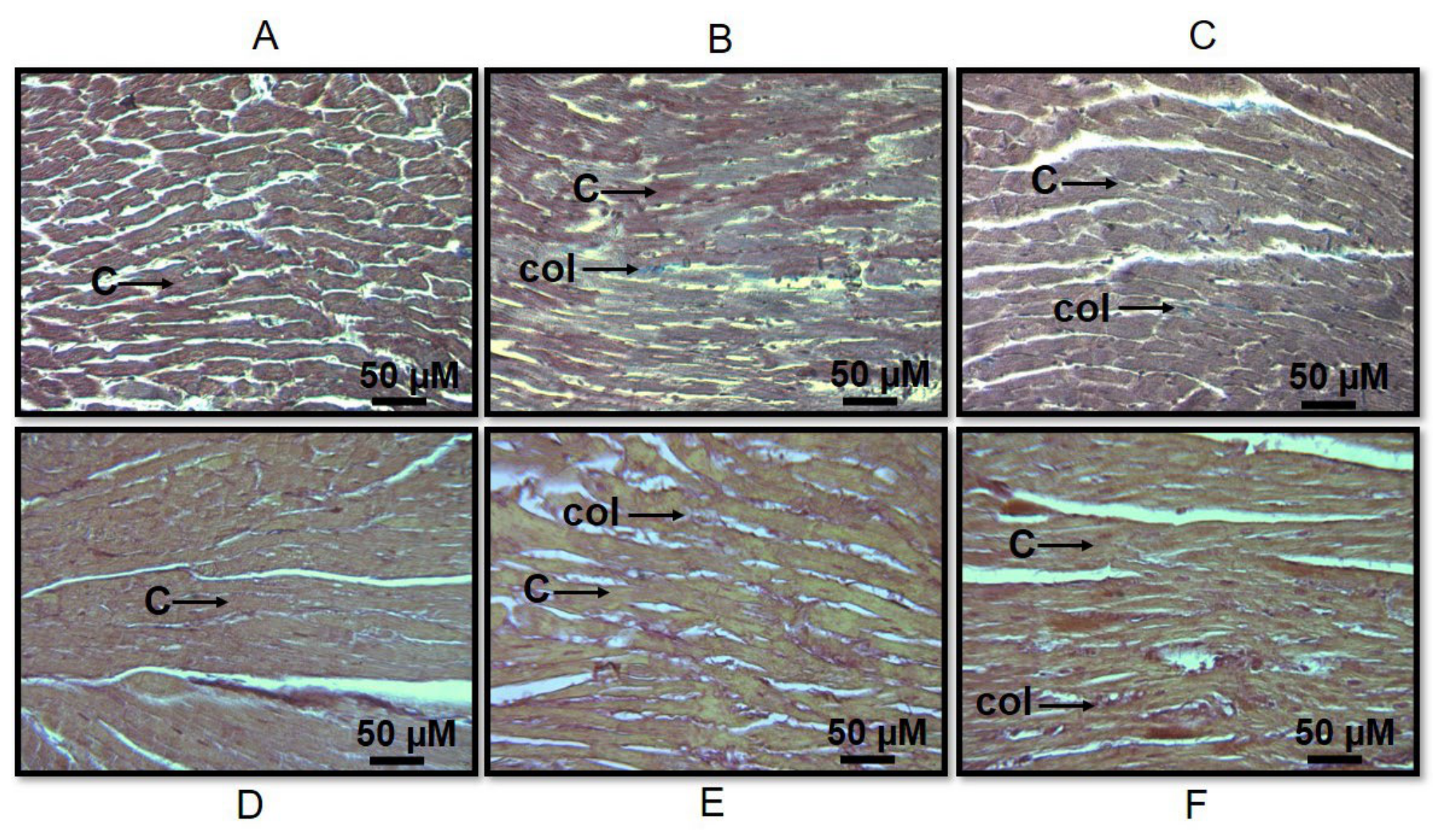
Figure 2.
Quantification of the fibrosis area in the different experimental groups. The fibrosis area is expressed in arbitrary units (AU).
Figure 2.
Quantification of the fibrosis area in the different experimental groups. The fibrosis area is expressed in arbitrary units (AU).
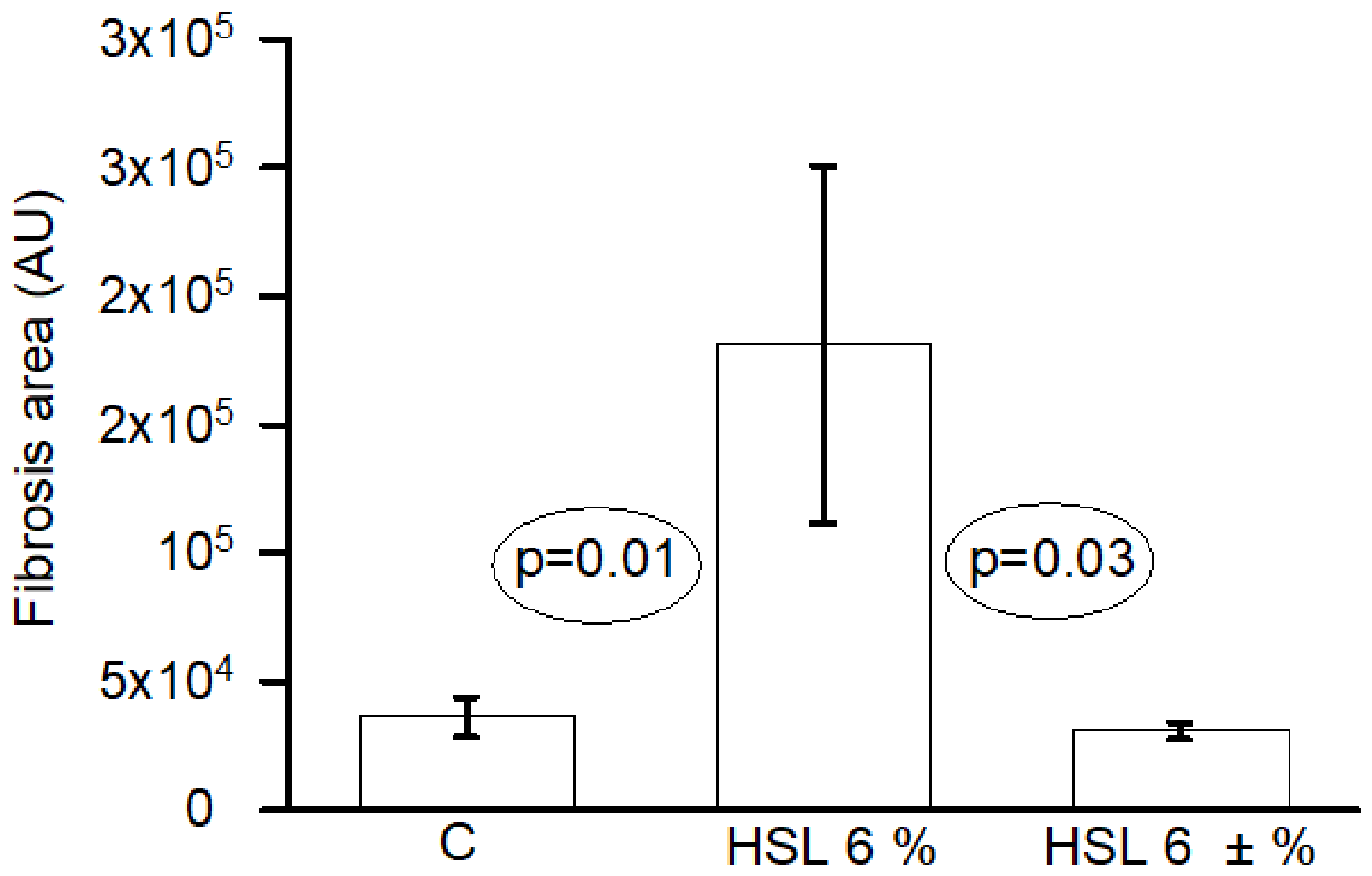
Figure 3.
Evaluation of cardiac hemodynamic and mechanical function during the stabilization, I/R periods. The curves show the temporal behavior in C, HSL 6 %, and HSL ± 6 % groups (A) CMP, (B) CVR, (C) PP and (D) HR respectively. The experimental phases correspond to 0–30 min (stabilization), 31–60 min (ischemia), and 61–90 min (reperfusion). Values are expressed as mean ± standard error (n=8). Statistical analysis indicates significant differences during the reperfusion phase (*p≤0.03 C vs. HSL 6%,). Abbreviations: C= Control; HSL= Hibiscus sabdariffa Linnaeus.
Figure 3.
Evaluation of cardiac hemodynamic and mechanical function during the stabilization, I/R periods. The curves show the temporal behavior in C, HSL 6 %, and HSL ± 6 % groups (A) CMP, (B) CVR, (C) PP and (D) HR respectively. The experimental phases correspond to 0–30 min (stabilization), 31–60 min (ischemia), and 61–90 min (reperfusion). Values are expressed as mean ± standard error (n=8). Statistical analysis indicates significant differences during the reperfusion phase (*p≤0.03 C vs. HSL 6%,). Abbreviations: C= Control; HSL= Hibiscus sabdariffa Linnaeus.
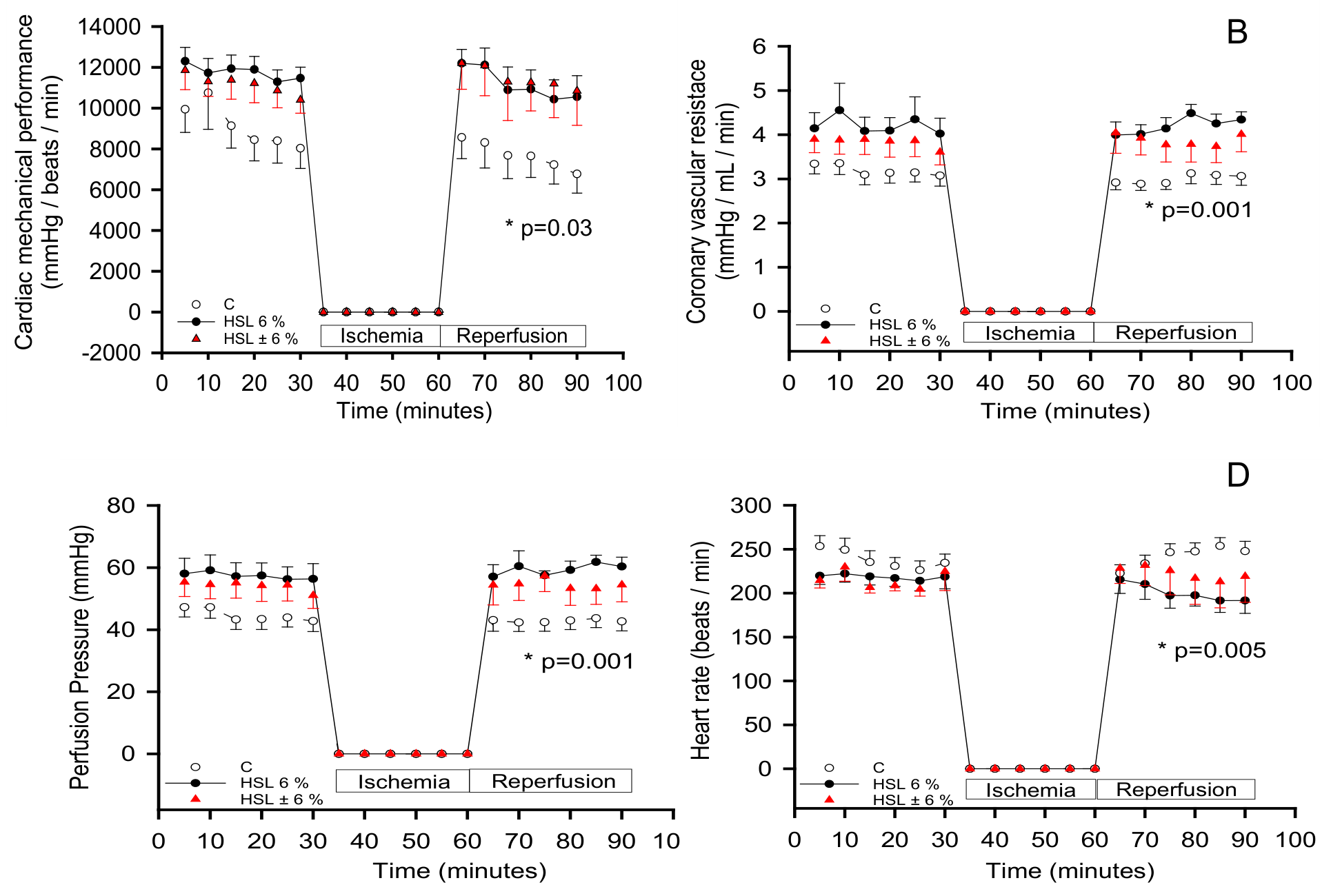
Figure 4.
Representative ECG recordings of isolated rat hearts under baseline conditions and after I/R. Tracings at 500 ms and 100 ms (enlarged view) of C group showing regular sinus rhythm during stabilization (A, B) and ventricular premature beats (R-R) post-I/R (C, D). The HSL 6 % group exhibited baseline electrical instability with spontaneous premature beats (E, F) and ventricular premature beats (R-R) and chaotic and distorted ventricular complexes after I/R with continuous (R-R), (G, H)). The HSL ± 6 % group shows a restored homogeneous sinus rhythm under baseline conditions (I, J) but develops transient asystole and conduction delays without polymorphic arrhythmias during reperfusion (K, L). Arrows indicate premature ectopic complexes (D, F) or fragmented ventricular complexes (H).
Figure 4.
Representative ECG recordings of isolated rat hearts under baseline conditions and after I/R. Tracings at 500 ms and 100 ms (enlarged view) of C group showing regular sinus rhythm during stabilization (A, B) and ventricular premature beats (R-R) post-I/R (C, D). The HSL 6 % group exhibited baseline electrical instability with spontaneous premature beats (E, F) and ventricular premature beats (R-R) and chaotic and distorted ventricular complexes after I/R with continuous (R-R), (G, H)). The HSL ± 6 % group shows a restored homogeneous sinus rhythm under baseline conditions (I, J) but develops transient asystole and conduction delays without polymorphic arrhythmias during reperfusion (K, L). Arrows indicate premature ectopic complexes (D, F) or fragmented ventricular complexes (H).
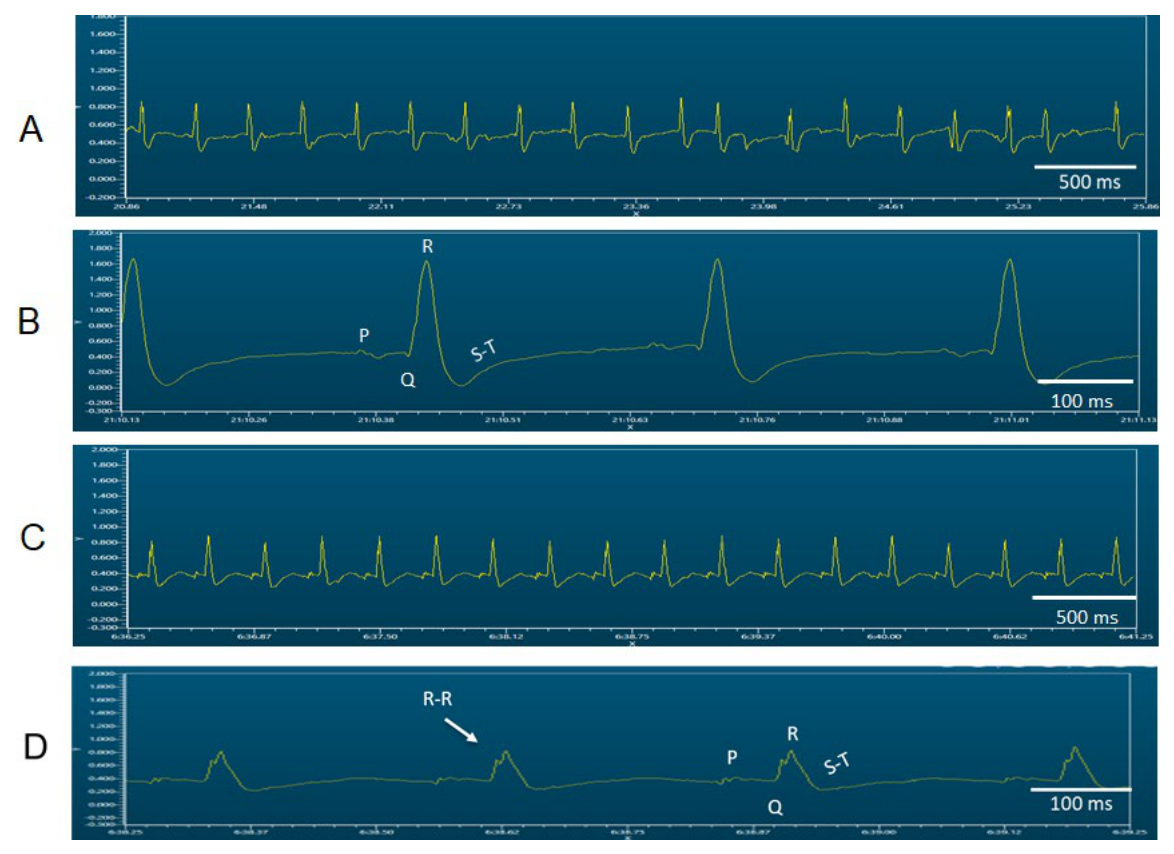
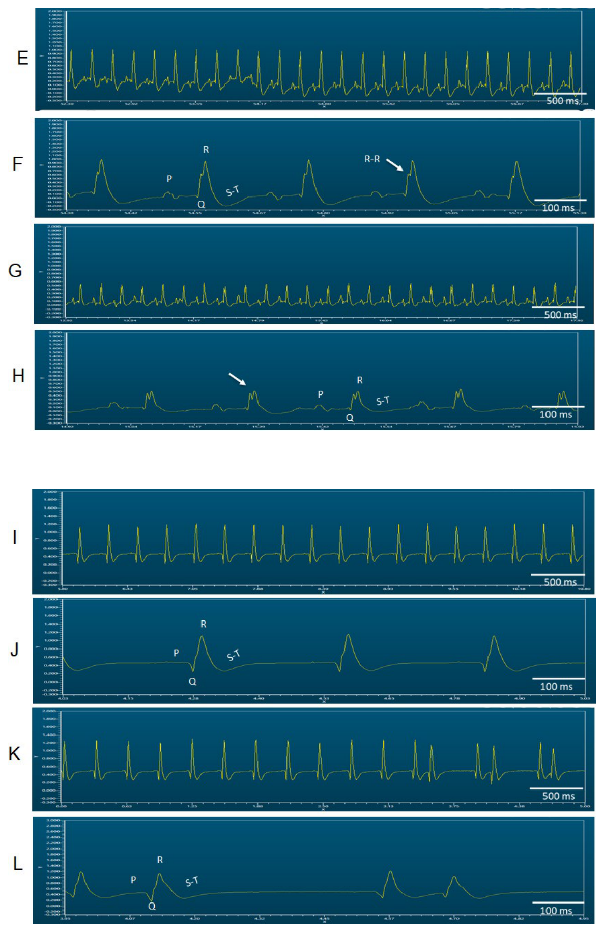

Table 1.
Antioxidants molecules provide by HSL 6 % infusion.
| Analytes of the HSL 6 % infusion | |
|---|---|
| Cyanidin-3-glucoside (mg/L) | 466.30 ± 58.02 |
| Quercetin (mg/L) | 108.73 ± 31.76 |
| Polyphenols (mmol/L) | 60.65 ± 21.67 |
| Vitamin C (mM/L) | 4.31 ± 0.11 |
Table 2.
General characteristics in experimental groups.
| Variables | C | HSL % | HSL ± 6% |
|---|---|---|---|
| Water consumption (mL/24 h) | 17.5 ± 1.5 | 23.5 ± 2.2 | 24.8 ± 2.9 |
| Body weight (g) | 456.5 ± 10.3 | 441.5 ± 11.3 | 422.4. ± 20.4 |
| SBP (mmHg) | 135 ± 1.6 | 153.6 ± 3.7* | 139.7 ± 2.7* |
| TAC (nM) | 83.07 ± 5.52 | 97.40 ± 2.07** | 89.67 ± 2.53* |
*C and HSL ± 6 % vs. HSL 6 % p≤0.03, Abbreviations; SBP= Systolic blood pressure, TAC= Total antioxidant capacity.
Disclaimer/Publisher’s Note: The statements, opinions and data contained in all publications are solely those of the individual author(s) and contributor(s) and not of MDPI and/or the editor(s). MDPI and/or the editor(s) disclaim responsibility for any injury to people or property resulting from any ideas, methods, instructions or products referred to in the content. |
© 2026 by the authors. Licensee MDPI, Basel, Switzerland. This article is an open access article distributed under the terms and conditions of the Creative Commons Attribution (CC BY) license (http://creativecommons.org/licenses/by/4.0/).
Copyright: This open access article is published under a Creative Commons CC BY 4.0 license, which permit the free download, distribution, and reuse, provided that the author and preprint are cited in any reuse.