

Submitted:
23 July 2026
Posted:
24 July 2026
You are already at the latest version
Abstract
Background: Acute bronchiolitis is among the most common causes of lower respiratory tract infection in infancy and remains a clinically heterogeneous syndrome. Current international guidelines recommend supportive management and generally discourage routine bronchodilator administration; however, physiological responsiveness may exist in a subset of patients. Tidal breathing analysis (TBA) provides a non-invasive, bedside method for objectively assessing respiratory mechanics and may objectively quantify physiological changes following bronchodilator administration. Case Presentation: We report a 23-month-old boy presenting with severe RSV bronchiolitis-like wheezing illness complicated by acute respiratory failure. Bronchodilator responsiveness was evaluated using predefined TBA parameters before and after inhaled bronchodilator administration. Following treatment, time to peak tidal expiratory flow (tPTEF) increased from 0.17 to 0.33 s (+94%), while the tPTEF/TE ratio increased from 0.12 to 0.27 (+125%). Expiratory time (TE) and total respiratory cycle duration (Ttot) decreased by 12.6% and 5.1%, respectively, whereas tidal volume (VT) and minute ventilation (MV) increased by 12.1% and 18.3%. Peak expiratory flow remained unchanged (0.14 L/s before and after bronchodilator administration). Discussion: To our knowledge, this is among the first reports using bedside TBA to characterize acute bronchodilator responsiveness in severe bronchiolitis-like illness. The improvement in expiratory timing indices, accompanied by shorter expiratory time without changes in peak expiratory flow, is consistent with improved expiratory flow dynamics following bronchodilator administration. These findings support the hypothesis that a bronchodilator-responsive physiological phenotype may exist. Previous studies have demonstrated an association between severe bronchiolitis during infancy and subsequent recurrent wheezing or asthma; however, whether acute bronchodilator responsiveness, measured objectively by TBA, represents a testable hypothesis for future longitudinal studies. Conclusions: This case illustrates the feasibility of bedside TBA for identifying physiological bronchodilator responsiveness in severe bronchiolitis-like illness. Prospective longitudinal studies are warranted to determine whether TBA-defined responsiveness represents an early physiological marker requiring prospective validation and whether such physiological stratification may support individualized follow-up strategies.
Keywords:
bronchiolitis
; salbutamol response
; tidal breathing analysis
; tPTEF/TE
; asthma risk
1. Introduction
Bronchiolitis is the most frequent lower respiratory tract infection in infants, characterized by inflammation of the small airways that generates tachypnoea, wheeze, increased work of breathing, and sometimes hospitalization. Respiratory syncytial virus (RSV) predominates, with rhinovirus (RV) and other viruses contributing substantially [1,2]. Airway interactions between viruses, especially rhinoviruses, and potentially pathogenic bacteria (Streptococcus pneumoniae, Haemophilus influenzae, and Moraxella catarrhalis) in early infancy may increase the risk of subsequent wheezing and asthma [3]. While most cases resolve, a certain subset of children materialize recurrent wheeze or asthma, in some prospective-followed cohorts, up to 4 years, being as high as 12% [4]. Hospitalization for severe bronchiolitis is associated with an increased risk of subsequent asthma (commonly reported a two- to three-fold increase across cohorts) [5,6,7,8,9]. Severe bronchiolitis in infancy may trigger development of asthma in genetically susceptible individuals, or it could be a marker of predisposition for future asthma. Genetic variants in CDHR3 and GSDMB modulate susceptibility to bronchiolitis, especially when caused by viruses other than RSV [10,11] Host and environment early-life risk factors are associated with progression of wheezing symptoms over time and several groups of researchers have speculated that dynamic interactions of these factors with an infant’s developing respiratory system are the dominant factor for subsequent wheeze and asthma [12,13,14].
A key unresolved question is whether bronchiolitis causes asthma inception or unmasks pre-existing susceptibility [15,16,17] in certain children.
Current guidelines emphasize supportive care and discourage routine bronchodilator use, in typical bronchiolitis cases, due to inconsistent clinical benefit at general population level [18,19,20]. Nevertheless, subgroups appear bronchodilator or steroid-responsive, potentially signalling airway hyperreactivity akin to early asthma [21,22]. In adults, bronchodilator responsiveness, even in the presence of lung function within normal limits, might be a risk factor for incident chronic airflow obstruction [23]. In young paediatric asthma patients, small airway disfunction (SAD) could be present in spite of normal FEV1 values [24], so exploring SAD, via tidal breathing analysis (TBA), could unmask future asthma trajectories in infants with severe bronchiolitis. Evidence of SAD was found in almost one-third of subjects who have been ventilated for life-threatening RSV disease, although not always accompanied by respiratory symptoms [25].
TBA is a non-invasive pulmonary function technique that quantifies timing and flow indices from tidal breathing loops (e.g., tPTEF, tPTEF/TE, VT, MV), being validated in various ethnic groups of infants [26,27]. Changes in timing indices after bronchodilator therapy can objectively demonstrate responsiveness, although mild bronchiolitis cases have a rather poor responsiveness in sedated infants [28].
Here we present a wheezing bronchiolitis-like LRTI case with documented salbutamol responsiveness via TBA, synthesize evidence on bronchiolitis–asthma links and treatment response heterogeneity, and propose a testable hypothesis for a research of physiology-defined responsiveness trajectories in these young children.
Case Presentation
A 23-month-old boy presented to the emergency department with a 2-day history of seromucous rhinorrhoea, productive cough, and dyspnoea. Family history was notable for paternal asthma and a sibling with severe bronchiolitis in infancy. Term born (39 weeks’ gestation; birth weight 2,800 g; Apgar score 9) child, was breastfed in early infancy, and had immunizations up to date. Past medical history included a febrile seizure at 9 months, measles at 12 months complicated with pneumonia, and one severe episode of bronchiolitis, with respiratory failure, at 19 months of age, requiring hospitalization. Environmental risk factors include household tobacco smoke exposure, wood-stove heating, and contact with domestic animals (dogs and backyard chicken).
On admission, he weighed 10.3 kg, afebrile, normal appearance. Respiratory findings: nasal obstruction with seromucous rhinorrhoea and productive cough, tachypnoea (54 breaths/min) with intercostal and subcostal retractions and SpO2 92% in room air, improving to 99% with low-flow nasal cannula. Chest auscultation revealed diminished vesicular breath sounds with bilateral wheezes and fine crackles. Cardiac examination documented a regular rhythm with tachycardia (150 bpm). The abdomen was non-tender, oral intake was reduced, but bowel movements were normal and urine output was preserved. No meningeal signs were present.
Laboratory evaluation documented leucocytosis with neutrophilia. C-reactive protein was 2 mg/dL. Capillary blood gas analysis evoked respiratory acidosis (pH 7.20, pCO2 63 mmHg). The working diagnosis was acute RSV bronchiolitis (RADT documented in Emergency Department) complicated by acute respiratory failure and mild acute dehydration, secondary to reduced intake. Patient underwent non-invasive ventilatory support via HFNC for a total of 7 days.
Tidal-breathing analysis (TBA) was performed with a SpiroScout (Ganshorn Medizin Electronic GmbH, Schiller Group, Germany) pneumotachograph-linked spirometer (see Figure 1) immediately before and ~20 minutes after nebulized salbutamol (0.15 mg/kg). Recordings were gained in non-sedated, quiet-awake state, using a well-fitted facemask with an optimized seal to minimize leak and upper-airway artifacts. At each time point, tidal breathing was recorded for a stable period and ≥3 technically acceptable segments were obtained, in accordance with ERS/ATS infant PFT standards (see Figure 2). Primary indices were derived from a prespecified, artifact-free segment using predefined quality criteria (stable baseline, minimal leak, no cough/swallow/sigh). In cases of uncertainty, segment eligibility was adjudicated by two independent reviewers blinded to test order.
Pre-salbutamol values were: VT 107 mL, RR 28/min, MV 3.0 L/min, TI 0.72 s, TE 1.43 s, Ttot 2.15 s, PEF 0.14 L/s, tPTEF 0.17 s, and tPTEF/TE 0.12.
Post- salbutamol, tidal-breathing indices improved markedly: tPTEF increased from 0.17 to 0.33 s (+94%) and tPTEF/TE from 0.12 to 0.27 (+125%), while TE shortened from 1.43 to 1.25 s (-12.6%) and Ttot from 2.15 to 2.04 s (-5.1%). Minute ventilation rose from 3.0 to 3.55 L/min (+18.3%), mainly through a higher VT (107→120 mL, +12.1%) with a small RR increase (28→30/min, +7.1%). PEF remained unchanged (0.14 L/s). This overall pattern supports bronchodilator responsiveness and is consistent with an improved tidal expiratory profile. Key parameters are summarized in Table 1 and Figure 3.
Management comprised intravenous rehydration for 2 days; high-flow nasal cannula (HFNC) support for 7 days; systemic corticosteroid therapy with methylprednisolone at 2 mg/kg/day for 2 days followed by tapering; inhaled bronchodilator therapy with nebulized salbutamol for 9 days, with adjunctive ipratropium bromide for first 5 days, and inhaled fluticasone propionate. The clinical course was favourable, with progressive improvement in respiratory status and normalization of blood gas measurements.
2. Literature Data
2.1. Aetiology and Epidemiology
Bronchiolitis primarily affects children under 2 years, with RSV responsible for the majority of cases; rhinovirus, parainfluenza, human metapneumovirus, and adenovirus are also involved, and co-infections are common [1,2]. The global burden is substantial, with RSV a leading cause of lower respiratory tract infection in young children [1]. New prevention tools, like long-acting RSV monoclonal antibodies (nirsevimab, clesrovimab) and RSV prefusion F protein-based maternal vaccine during pregnancy had impacted real life burden on hospitalizations or Emergency Department visits in infants [29,30,31,32,33] and will probably generate a more tailored approach of RSV wheezy infants in the foreseeable future, because there is still present a heterogenous approach of these patients [34].
2.2. Association with Asthma
Multiple studies link severe bronchiolitis to increased asthma risk, and rhinovirus-associated bronchiolitis generally carries a higher subsequent risk than RSV [35,36,37,38,39]. One research [35], including 408 children hospitalized for bronchiolitis into a prospective, 3-center, 4-year follow-up study, in Finland, documented at study entry, a median age of 7.5 months. 42% had RSV, 29% RV, 2% both RSV and RV, and 27% had non-RSV/-RV aetiology. The children with RV-A (adjusted hazard ratio, 2.3; P = .01), RV-C (adjusted hazard ratio, 3.5; P < .001), and non-RSV/-RV (adjusted hazard ratio, 2.0; P = .004) bronchiolitis started the asthma control medication earlier than did children with RSV bronchiolitis.
INSPIRE is a large, population-based, birth cohort of healthy infants with non-low birthweight born at term in Tennessee, USA. Children were followed up prospectively for the primary outcome of 5-year current asthma. Not being infected with RSV during infancy was associated with a 26% lower risk of 5-year current asthma than being infected with RSV during infancy (adjusted RR 0·74, 95% CI 0·58–0·94, p=0·014). The estimated proportion of 5-year current asthma cases that could be prevented by avoiding RSV infection during infancy was 15% [40].
Several studies on non-invasive biomarkers for assessing asthma in preschool children have been published [41,42,43]. Specimens that can be easily obtained by non-invasive methods are blood, exhaled breath and urine. Eosinophils, eosinophil cationic protein and eosinophil-derived neurotoxin (EDN) in blood are helpful in evaluating eosinophilic inflammation of the airways. Exhaled breath contains nitric oxide, volatile organic compounds, various cytokines and mediators as analytical components. Fraction of exhaled nitric oxide has been used to assess the degree of eosinophil inflammation and has been standardized in school-age children and adults, but not yet in preschool children. Exhaled breath condensate (EBC) pH and various cytokines/mediators that are detected in EBC seem to be promising biomarkers for assessing asthma, but need more standardization and validation.
Dysregulated interferon responses (changes in gene transcription during cold-associated asthma exacerbations) are important contributors to this risk in children. Low baseline interferon expression followed by greater upregulation of interferon pathways in airway and blood during respiratory illnesses increased exacerbation risk [44].
Bronchiolitis cohort studies have identified early-life environmental exposure [45], genetic [46,47,48,49] and immune risk factors [50] for childhood asthma development by carrying out analysis at single level (e.g., associations with respiratory virus types, host immune response or the microbiome composition of the host) [51]. Multi-omics technologies have elucidated certain mechanisms underlying the gut-lung axis, numerous pathways remain to be fully delineated [52]. Severe bronchiolitis pathogenesis involves interaction of factors at multiple levels (e.g., genome, epigenome, transcriptome, metabolome, microbiome) [53,54,55]. Proposed mechanisms through which early-life RSV infection might contribute to asthma include epithelial injury, immune dysregulation with Th2-skewed responses, and airway/microbiome remodelling, superimposed on host susceptibility [56].
Bronchodilator Response in Bronchiolitis
Guidelines discourage routine β2-agonists in typical bronchiolitis due to absent or inconsistent clinical benefit [18,19]. Nonetheless, subgroup patients may respond, potentially indicating asthma-like airway hyperreactivity [21]. An old meta-analysis [57] is documenting no overall clinical benefits in infants treated with bronchodilators: they do not improve oxygen saturation, do not reduce hospital admission after outpatient treatment, do not shorten the duration of hospitalization and do not reduce the time to resolution of illness at home. Some trials suggest that epinephrine may transiently reduce early admissions in emergency settings, especially in combination with dexamethasone, as in BIPED study [58].
2.3. Tidal Breathing Analysis
TBA can be used to identify potential responders via meaningful improvements in timing indices (e.g., tPTEF/TE), supporting physiology-based phenotyping. TBA is feasible in non-sedated children and quantifies airflow obstruction through timing and flow indices [59,60]. In wheezing infants, lower tPTEF/TE has been reported in those with more persistent symptoms, although variability necessitates standardized acquisition and loop selection [61].
2.4. Treatment Reviews for Acute Bronchiolitis
Hypertonic saline may modestly reduce hospital length of stay and may slightly improve clinical severity score (CSS). Treatment with nebulised hypertonic saline may also reduce the risk of hospitalisation amongst outpatients and ED patients, although a randomized clinical trial of 777 healthy infants, the hospital admission rate in the hypertonic saline group was 48.1% compared with 52.2% in the normal saline group [64]. Nebulised hypertonic saline seems to be a safe treatment in infants with bronchiolitis with only minor and spontaneously resolved adverse events, especially when administered in conjunction with a bronchodilator as documented in a recent review; total number of included trials was 34, involving 5205 infants with acute bronchiolitis, of whom 2727 infants received hypertonic saline [65]. In studies that explored combination of medication, low-quality evidence suggests that nebulized epinephrine plus hypertonic saline may be considered as a safe and efficient therapy for decreasing length of stay and CSS in infants with acute bronchiolitis, especially in those who require hospitalization for more than 48 h [66].
Corticosteroids generally show no consistent benefit in typical bronchiolitis; evidence for benefit in combination regimens (e.g., with epinephrine) has been mixed across trials [67], but in children with severe bronchiolitis, the duration of clinician-managed pressure support in intensive care (defined as high-flow nasal-prong oxygen, nasopharyngeal continuous positive airway pressure, or mechanical ventilation), was reduced by regular treatment with systemic corticosteroids and inhaled epinephrine compared with standard care [68].
Antibiotics are ineffective in absence of bacterial co-infection [69], excepting azithromycin that may reduce hospitalization time in acute bronchiolitis and wheezing episodes among children aged less than two. Azithromycin administrated during the acute wheezing period, does not have preventive effect on wheezing recurrence [70].
DNase has been explored for mucus clearance but evidence is limited [71].
3. Future Research
3.1. Study Question
Does acute bronchodilator responsiveness during an acute bronchiolitis episode, defined by prespecified TBA criteria, predict higher 5-year asthma risk compared with non-responsiveness?
3.2. Objectives
Primary: Compare physician-diagnosed asthma at 5 years (as per GINA 2026 criteria for asthma in children 5 years and younger) [73] among salbutamol-responders and non-responders (TBA-defined).
4. Discussion
The majority of infants with wheezing have transient conditions associated with diminished airway function at birth and do not have increased risks of asthma later in life. In a substantial minority of infants, however, wheezing episodes are probably related to a predisposition to asthma [4,5,6].
Although recent guidelines recommend a minimalist approach to bronchiolitis, there are some issues with this approach. First, there are concerns about the precise definition of the disease, the quality and uniformity of the guidelines, the method of administration of bronchodilators, and the availability of tools to evaluate the response to therapies. Second, for decades it has been assumed that all cases of viral bronchiolitis are similar, but recent evidence has shown that this is not the case. Distinct bronchiolitis phenotypes have been described, with heterogeneity in clinical presentation, molecular immune signatures and clinically relevant outcomes such as respiratory failure and recurrent wheezing [77]. All these limitations are more stringent in primary care settings, even in developed countries [78].
The physiological changes observed after salbutamol are compatible with bronchodilator responsiveness, objectively assessed by TBA, in a child presenting with a second episode of recurrent wheezing triggered by respiratory syncytial virus lower respiratory tract infection. During the initial episode three months earlier, which required hospitalization in our unit for severe bronchiolitis (Paediatric Respiratory Severity Score [PRESS] = 5), the patient exhibited a clinically significant response to inhaled salbutamol. However, TBA was not performed at that time to objectively quantify changes in lung function parameters.
During this second major broncho-obstructive episode, with hospitalization, nebulized salbutamol administration resulted in a marked increase in respiratory timing indices (tPTEF and tPTEF/TE), shortening of expiratory duration, and increased minute ventilation, while peak expiratory flow (PEF) remained unchanged. The observed alterations in TBA waveform morphology were consistent with an improved tidal expiratory pattern in the absence of an increase in peak flow, supporting the presence of pharmacologically mediated bronchodilation. In this patient, who appears to follow a recurrent wheezing trajectory compatible with a viral-induced asthma phenotype, responsiveness to short-acting β2-agonists was objectively documented. This interpretation accounts for potential state-dependent physiological variability and measurement-related fluctuations that may influence respiratory timing indices in young children.
This pattern seems similar to findings of a group of researchers that monitored 76 children presenting with their first wheezing episode at the ages of 3 to 23 months. At study entry, viral aetiology, rhinovirus genome load, atopic and clinical characteristics, and standardized questionnaire were analysed. At 4-year follow-up visit, impulse oscillometry with exercise challenge was performed. Bronchial reactivity (≥35% change in mean crude values of resistance) after exercise challenge or bronchodilation was present in nine (12%) children [79].
This paper has several limitations: it presents only one case of TBA usage for description of bronchodilator responsiveness in a 23 old months child with previous wheezing episodes. This scenario could be used as hypothesis generating and we have evoked potential future research, as strength of this case, we can state that to our knowledge, this is among the first reports using bedside TBA to characterize acute bronchodilator responsiveness in severe bronchiolitis-like illness.
Future precision-medicine strategies integrating validated asthma prediction models and pulmonary function tests with artificial intelligence–based platforms that incorporate electronic health records, genomic data, and molecular biomarkers may further optimize individualized management and improve long-term quality of life in some high-risk populations.
5. Conclusions
Bedside physiological assessment can reveal clinically relevant heterogeneity within phenotypically typical bronchiolitis presentations.
Although routine bronchodilator therapy is not recommended at the population level, physiology-guided bedside trials may provide a pragmatic and evidence-based approach for identifying selected potential responders without reliance on empirical trial-and-error management strategies.
TBA-based phenotyping by future research could refine acute therapeutic decision-making, enable risk-stratified surveillance during early childhood, and potentially will be incorporated into artificial intelligence–driven personalized treatment algorithms.
Funding
No external funding.
Institutional Review Board Statement
The single-patient case report and a prospective BABIES cohort protocol was approved by the Ethics Committee of the National Institute for Mother and Child Health “Alessandrescu-Rusescu”, Bucharest, Romania (Protocol No. 25152/05.11.2025).
Data Availability Statement
Not applicable.
Conflicts of Interest
The authors declare no conflict of interest.
References
- Angurana, S.K.; Takia, L.; Sarkar, S.; et al. Clinico-virological Profile, Intensive Care Needs, and Outcome of Infants with Acute Viral Bronchiolitis: A Prospective Observational Study. Indian J. Crit. Care Med. 2021, 25(11), 1301–1307. [Google Scholar] [CrossRef] [PubMed]
- Tsou, P.; Vadivelan, A.; Kovvuri, M.; et al. Association between multiple respiratory viral infections and pediatric intensive care unit admission among infants with bronchiolitis. Arch. Pediatr. 2020, 27(1), 39–44. [Google Scholar] [CrossRef] [PubMed]
- Takashima, M.D.; Grimwood, K.; Sly, P.D.; et al. Association of rhinovirus and potentially pathogenic bacterial detections in the first 3 months of life with subsequent wheezing in childhood. Pediatr. Pulmonol. 2023, 58(12), 3428–3436. [Google Scholar] [CrossRef] [PubMed]
- Leino, A.; Lukkarinen, M.; Turunen, R.; et al. Pulmonary function and bronchial reactivity 4 years after the first virus-induced wheezing. Allergy 2019, 74(3), 518–526. [Google Scholar] [PubMed]
- Heikkilä, P.; Korppi, M.; Ruotsalainen, M.; et al. Viral wheezing in early childhood as a risk factor for asthma in young adulthood: A prospective long-term cohort study. Health Sci. Rep. 2022, 5(2), e538. [Google Scholar] [CrossRef] [PubMed]
- Sørensen, K.G.; Øymar, K.; Dalen, I.; et al. Bronchial hyper-reactivity in young adults after hospitalisation for bronchiolitis in infancy. BMJ Open Respir. Res. 2025, 12(1), e002881. [Google Scholar] [CrossRef] [PubMed]
- Laitinen, S.; Lauhkonen, E.; Saarikallio, S.; et al. Asthma and health-related quality of life at 16-20 years of age in a prospectively followed post-bronchiolitis cohort. Eur. J. Pediatr. 2024, 183(11), 4877–4883. [Google Scholar] [CrossRef] [PubMed]
- Be’er, M.; Bushmitz, S.; Cahal, M.; et al. Asthma risk after a pediatric intensive care unit admission for respiratory syncytial virus bronchiolitis. Pediatr. Pulmonol. 2022, 57(7), 1677–1683. [Google Scholar] [CrossRef] [PubMed]
- Jia, X.Q.; Lu, L.; Lou, S.Y.; et al. Association between early-life respiratory syncytial virus infection and the risk of asthma: a meta-analysis. J. Thorac. Dis. 2025, 17(11), 9980–9996. [Google Scholar] [CrossRef] [PubMed]
- Pasanen, A.; Karjalainen, M.K.; Korppi, M.; et al. Research Project FinnGen. Genetic Susceptibility to Acute Viral Bronchiolitis. J. Infect. Dis. 2025, 232(2), e193–e202. [Google Scholar] [PubMed]
- Eliasen, A.U.; Pedersen, C.E.T.; Rasmussen, M.A.; et al. Genome-wide study of early and severe childhood asthma identifies interaction between CDHR3 and GSDMB. J. Allergy Clin. Immunol. 2022, 150(3), 622–630. [Google Scholar] [CrossRef] [PubMed]
- Nahum, U.; Gorlanova, O.; Decrue, F.; et al. BILD study group; PASTURE study group. Symptom trajectories in infancy for the prediction of subsequent wheeze and asthma in the BILD and PASTURE cohorts: a dynamic network analysis. Lancet Digit Heal. 2024, 6(10), e718–e728. [Google Scholar] [CrossRef]
- Owora, A.H.; Zhang, Y. Childhood wheeze trajectory-specific risk factors: A systematic review and meta-analysis. Pediatr. Allergy Immunol. 2021, 32(1), 34–50. [Google Scholar] [CrossRef] [PubMed]
- Warden, D.E.; Zhang, H.; Jiang, Y.; et al. The role of wheezing subtypes in the development of early childhood asthma. Respir. Res. 2025, 26(1), 79. [Google Scholar] [CrossRef] [PubMed]
- Fjaerli, H.O.; Farstad, T.; Rød, G.; et al. Acute bronchiolitis in infancy as risk factor for wheezing and reduced pulmonary function by seven years but not allergy. Arch. Dis. Child. 2005, 90(10), 1049–1055. [Google Scholar]
- Brunwasser, S.M.; Snyder, B.M.; Driscoll, A.J.; et al. Assessing the strength of evidence for a causal effect of respiratory syncytial virus lower respiratory tract infections on subsequent wheezing illness: a systematic review and meta-analysis. Lancet Respir. Med. 2020, 8(8), 795–806. [Google Scholar] [CrossRef] [PubMed]
- Driscoll, A.J.; Arshad, S.H.; Bont, L.; et al. Does respiratory syncytial virus lower respiratory illness in early life cause recurrent wheeze of early childhood and asthma? Critical review of the evidence and guidance for future studies from a World Health Organization-sponsored meeting. Vaccine. 2020, 38(11), 2435–2448. [Google Scholar] [CrossRef] [PubMed]
- Borland, M.L.; Loveys, K.; Babl, F.E.; et al. Paediatric Research in Emergency Departments International Collaborative (PREDICT) Network, Australasia. Australasian Bronchiolitis Guideline: 2025 Update. J. Paediatr. Child Heal. 2025, 61(8), 1197–1215. [Google Scholar] [CrossRef]
- Ralston, S.L.; Lieberthal, A.S.; Meissner, H.C.; et al. Clinical practice guideline: the diagnosis, management, and prevention of bronchiolitis. In Pediatrics.; American Academy of Pediatrics, 2014; Volume 134, 5, p. e1474-502. [Google Scholar]
- House, S.A.; Marin, J.R.; Hall, M.; et al. Trends Over Time in Use of Nonrecommended Tests and Treatments Since Publication of the American Academy of Pediatrics Bronchiolitis Guideline. JAMA Netw. Open. 2021, 4(2), e2037356. [Google Scholar] [CrossRef] [PubMed]
- Greenky, D.; Goldman, R.D. Use of β2-agonists for viral bronchiolitis. Can. Fam. Physician 2022, 68(6), 429–430. [Google Scholar] [CrossRef] [PubMed]
- Fernandes, R.M.; Bialy, L.M.; Vandermeer, B.; et al. Glucocorticoids for acute viral bronchiolitis in infants and young children. Cochrane Database Syst. Rev. 2013, 2013(6), CD004878. [Google Scholar] [CrossRef] [PubMed]
- Knox-Brown, B.; Algharbi, F.; Mulhern, O.; BOLD Collaborative Research Group; et al. Bronchodilator responsiveness and future chronic airflow obstruction: a multinational longitudinal study. EClinicalMedicine 2025, 81:103123, 1–10. [Google Scholar]
- Chereches-Panta, P.; Marica, I.; Sas, V.; et al. The Role of Spirometry and MMEF in Pediatric Asthma Monitoring and Prediction of Exacerbations. Children . 2025, 12(10), 1398. [Google Scholar] [CrossRef] [PubMed]
- Lodrup Carlsen, K.C.; Carlsen, K.H. Lung function in awake healthy infants: The first five years of life. Respir. Med. 1997, 91(6), 322–330. [Google Scholar]
- Lai, S.H.; Liao, S.L.; Yao, T.C.; et al. Respiratory Function in Healthy Taiwanese Infants: Tidal Breathing Analysis, Passive Mechanics, and Tidal Forced Expiration. PLoS ONE 2015, 10(11), e0142797. [Google Scholar] [CrossRef] [PubMed]
- Hmeidi, H.; Motamedi-Fakhr, S.; Chadwick, E.; et al. Tidal breathing parameters measured using structured light plethysmography in healthy children and those with asthma before and after bronchodilator. Physiol. Rep. 2017, 5(5), e13168. [Google Scholar] [CrossRef] [PubMed]
- Totapally, B.R.; Demerci, C.; Zureikat, G.; et al. Tidal breathing flow-volume loops in bronchiolitis in infancy: The effect of albuterol. Crit. Care. 2002, 6(2), 160–165. [Google Scholar] [CrossRef] [PubMed]
- Andina Martínez, D.; Claret Teruel, G.; Gijón Mediavilla, M.; et al. Impact of universal Respiratory Syncytial Virus prophylaxis in Spain Working Group. Nirsevimab and Acute Bronchiolitis Episodes in Pediatric Emergency Departments. Pediatrics. 2024, 154(4), e2024066584. [Google Scholar] [CrossRef] [PubMed]
- Touati, S.; Debs, A.; Morin, L.; et al. PANDORA study group. Nirsevimab prophylaxis on pediatric intensive care hospitalization for severe acute bronchiolitis: a clinical and economic analysis. Ann. Intensive Care 2025, 15(1), 56, 1–6. [Google Scholar] [CrossRef]
- Perramon-Malavez, A.; Chiaretti, A.; Coma, E.; et al. Real-world impact of nirsevimab immunisation and maternal RSV vaccination against respiratory disease on emergency department attendances and admissions: a multinational retrospective analysis. Lancet Reg. Health Eur. 2026, 63, 101597. [Google Scholar] [CrossRef] [PubMed]
- Dieussaert, I.; Hyung Kim, J.; Luik, S.; et al. RSV Prefusion F Protein-Based Maternal Vaccine—Preterm Birth and Other Outcomes. N Engl. J. Med. 2024, 390(11), 1009–1021. [Google Scholar] [CrossRef] [PubMed]
- Zar, H.J.; Bont, L.J.; Manzoni, P.; et al. SMART (MK-1654-007) Study Group. Clesrovimab in Infants and Children at Increased Risk for Severe RSV Disease. N Engl. J. Med. 2025, 393(13), 1343–1345. [Google Scholar] [CrossRef] [PubMed]
- Brüssow, H. Respiratory syncytial virus: health burden, disease prevention, and treatment-recent progress and lessons learned. Microlife 2025, 6, uqaf003. [Google Scholar] [CrossRef] [PubMed]
- Bergroth, E.; Aakula, M.; Elenius, V.; et al. Rhinovirus Type in Severe Bronchiolitis and the Development of Asthma. J. Allergy Clin. Immunol. Pract. 2020, 8(2), 588–595.e4. [Google Scholar] [CrossRef] [PubMed]
- Makrinioti, H.; Hasegawa, K.; Lakoumentas, J.; et al. The role of respiratory syncytial virus- and rhinovirus-induced bronchiolitis in recurrent wheeze and asthma-A systematic review and meta-analysis. Pediatr. Allergy Immunol. 2022, 33(3), e13741. [Google Scholar] [CrossRef] [PubMed]
- Fujiogi, M.; Dumas, O.; Hasegawa, K.; et al. Identifying and predicting severe bronchiolitis profiles at high risk for developing asthma: Analysis of three prospective cohorts. EClinicalMedicine. 2022, 43, 101257. [Google Scholar] [CrossRef] [PubMed]
- Muñoz-Quiles, C.; López-Lacort, M.; Díez-Domingo, J.; et al. Bronchiolitis, Regardless of Its Etiology and Severity, is Associated with Increased Risk of Asthma: A Population-Based Study. J. Infect. Dis. 2023, 228(7), 840–850. [Google Scholar] [CrossRef] [PubMed]
- Mansbach, J.M.; Ying, Q.S.; Espinola, J.A.; et al. Recurrent wheezing phenotypes after severe bronchiolitis and progression to asthma. J. Allergy Clin. Immunol. Glob. 2023, 2(1), 84–87. [Google Scholar] [CrossRef] [PubMed]
- Rosas-Salazar, C.; Chirkova, T.; Gebretsadik, T.; et al. Respiratory syncytial virus infection during infancy and asthma during childhood in the USA (INSPIRE): a population-based, prospective birth cohort study. Lancet. 2023, 401(10389), 1669–1680. [Google Scholar] [CrossRef] [PubMed]
- Lee, Y.J.; Fujisawa, T.; Kim, C.K. Biomarkers for Recurrent Wheezing and Asthma in Preschool Children. Allergy Asthma Immunol. Res. 2019, 11(1), 16–28. [Google Scholar] [CrossRef] [PubMed]
- Kang, Y.R.; Kim, H.; Lee, C.E.; et al. Serum and urine eosinophil-derived neurotoxin (EDN) levels predict biologic response in severe asthma. World Allergy Organ J. 2025, 18(1), 100990. [Google Scholar] [CrossRef] [PubMed]
- Rodríguez-Fernández, R.; González-Martínez, F.; González-Sánchez, M.I.; et al. Longitudinal plasma cytokine concentrations and recurrent wheezing after RSV bronchiolitis. Cytokine. 2021, 140, 155434. [Google Scholar] [CrossRef] [PubMed]
- Gaberino, C.L.; Altman, M.C.; Gill, M.A.; et al. Dysregulation of airway and systemic interferon responses promotes asthma exacerbations in urban children. J. Allergy Clin. Immunol. 2025, 155(5), 1499–1509. [Google Scholar] [CrossRef] [PubMed]
- Bradley, J.P.; Bacharier, L.B.; Bonfiglio, J.; et al. Severity of respiratory syncytial virus bronchiolitis is affected by cigarette smoke exposure and atopy. Pediatrics. 2005, 115(1), e7–e14. [Google Scholar] [CrossRef] [PubMed]
- Illi, S.; Depner, M.; Pfefferle, P.I.; et al. PASTURE Study Group. Immune Responsiveness to LPS Determines Risk of Childhood Wheeze and Asthma in 17q21 Risk Allele Carriers. Am. J. Respir. Crit. Care Med. 2022, 205(6), 641–650. [Google Scholar] [CrossRef] [PubMed]
- Jakwerth, C.A.; Weckmann, M.; Illi, S.; et al. ALLIANCE Study Group as part of the German Center for Lung Research. 17q21 Variants Disturb Mucosal Host Defense in Childhood Asthma. Am. J. Respir. Crit. Care Med. 2024, 209(8), 947–959. [Google Scholar] [CrossRef] [PubMed]
- Wolters, A.A.B.; Kersten, E.T.G.; Koppelman, G.H. Genetics of preschool wheeze and its progression to childhood asthma. Pediatr. Allergy Immunol. 2024, 35(1), e14067. [Google Scholar] [CrossRef] [PubMed]
- Turi, K.N.; McKennan, C.; Rosas-Salazar, C.; et al. Association of 17q12-q21 Asthma Risk Locus with Clinical Severity of Infant Respiratory Syncytial Virus Infection. Biomolecules. 2025, 15(8), 1056. [Google Scholar] [CrossRef] [PubMed]
- Kloepfer, K.M.; Jackson, D.J.; Jartti, T.; et al. How Infections and Immune Development Relate to Preschool Recurrent Wheezing and Asthma. J. Allergy Clin. Immunol. Pract. 2025, 10, 2553–2561. [Google Scholar] [CrossRef]
- Cabrera-Rubio, R.; Calvo, C.; Alcolea, S.; et al. Gut and respiratory tract microbiota in children younger than 12 months hospitalized for bronchiolitis compared with healthy children: can we predict the severity and medium-term respiratory outcome? Microbiol. Spectr. 2024, 12(7), e0255623. [Google Scholar] [CrossRef] [PubMed]
- Yu, X.; Yu, X.; Wang, Y.; et al. Respiratory diseases and the gut microbiota: an updated review. Front Cell Infect. Microbiol. 2025, 15, 1629005. [Google Scholar] [CrossRef] [PubMed]
- Wang, Y.; Shi, Y.; Wang, L. Early risk identification for recurrent wheezing in children with respiratory syncytial virus infections. Am. J. Transl. Res. 2025, 17(7), 5411–5422. [Google Scholar] [CrossRef] [PubMed]
- Feng, Q.; Feng, Z.; Yang, B.; et al. Metatranscriptome Reveals Specific Immune and Microbial Signatures of Respiratory Syncytial Virus Infection in Children. Microbiol. Spectr. 2023, 11(2), e0410722. [Google Scholar] [CrossRef] [PubMed]
- Pischedda, S.; Gómez-Carballa, A.; Pardo-Seco, J.; GENDRES Consortium; et al. DNA methylation signatures of severe RSV infection in infants: evidence from non-invasive saliva samples. Epigenet. Chromatin. 2025, 18(1), 25. [Google Scholar] [CrossRef]
- Öner, D.; Drysdale, S.B.; McPherson, C.; et al. RESCEU Investigators. Biomarkers for Disease Severity in Children Infected With Respiratory Syncytial Virus: A Systematic Literature Review. J. Infect. Dis. 2020, 222 (Suppl 7), S648–S657. [Google Scholar] [CrossRef] [PubMed]
- Gadomski, A.M.; Scribani, M.B. Bronchodilators for bronchiolitis. Cochrane Database Syst. Rev. 2014, 2014(6), CD001266. [Google Scholar] [CrossRef] [PubMed]
- Heath, A.; Rios, D.; Vogel, K.I.; et al. KidsCan Pediatric Emergency Research Canada (PERC) Innovative Pediatric Clinical Trials and Pediatric Research in Emergency Department International Collaborative (PREDICT) BIPED Study Team. A randomised controlled trial comparing epinephrine and dexamethasone to placebo in the treatment of infants with bronchiolitis (BIPED study): a statistical analysis plan. Trials 2025, 26(1), 496. [Google Scholar] [CrossRef] [PubMed]
- Qin, J.; Liu, F.; Wang, T.; et al. Correlation between tidal breathing pulmonary function, exhaled nitric oxide and airway hyperresponsiveness in children aged 0-3 years with suspected asthma. Front Pediatr. 2025, 13, 1388951. [Google Scholar] [CrossRef] [PubMed]
- Qi, Y.Y.; Jiang, G.L.; Wang, L.B.; et al. Lung Function in Wheezing Infants after Acute Lower Respiratory Tract Infection and Its Association with Respiratory Outcome. Chin. Med. J. (Engl) . 2017, 130(1), 4–10. [Google Scholar] [CrossRef] [PubMed]
- Bates, J.H.T.; Schmalisch, G.; Filbrun, D.; et al. Tidal breath analysis for infant pulmonary function testing. ERS/ATS Task Force on Standards for Infant Respiratory Function Testing. Eur. Respir. J. 2000, 16(6), 1180–1192. [Google Scholar] [CrossRef] [PubMed]
- ***—Chapter 1.4 Management of bronchiolitis in NICE Guidelines NG9 Bronchiolitis in children: diagnosis and management. 09 August 2021. Available online: https://www.nice.org.uk/guidance/ng9/chapter/Recommendations#management-of-bronchiolitis (accessed on 10th May 2026).
- ***—Australasian Bronchiolitis Guideline—Medication. 14th April 2025. Available online: https://www.predict.org.au/bronchiolitis-guideline/medication/ (accessed on 10th May 2026).
- Angoulvant, F.; Bellêttre, X.; Milcent, K.; et al. Efficacy of 3% Hypertonic Saline in Acute Viral Bronchiolitis (GUERANDE) Study Group. Effect of Nebulized Hypertonic Saline Treatment in Emergency Departments on the Hospitalization Rate for Acute Bronchiolitis: A Randomized Clinical Trial. JAMA Pediatr. 2017, 171(8), e171333. [Google Scholar] [CrossRef] [PubMed]
- Zhang, L.; Mendoza-Sassi, R.A.; Wainwright, C.E.; Aregbesola, A.; Klassen, T.P. Nebulised hypertonic saline solution for acute bronchiolitis in infants. Cochrane Database Syst. Rev. 2023, 4(4), CD006458. [Google Scholar] [CrossRef] [PubMed]
- Pereira, R.A.; Oliveira de Almeida, V.; Zambrano, M.; et al. Effects of nebulized epinephrine in association with hypertonic saline for infants with acute bronchiolitis: A systematic review and meta-analysis. Health Sci. Rep. 2022, 5(3), e598. [Google Scholar] [CrossRef] [PubMed]
- Alansari, K.; Sakran, M.; Davidson, B.L.; et al. Dexamethasone for treatment of acute bronchiolitis in infants: A randomized clinical trial. JAMA Pediatr. 2013, 167(8), 691–698. [Google Scholar]
- Gelbart, B.; McSharry, B.; Delzoppo, C.; et al. Pragmatic Randomized Trial of Corticosteroids and Inhaled Epinephrine for Bronchiolitis in Children in Intensive Care. J. Pediatr. 2022, 244, 17–23. e1. [Google Scholar] [CrossRef] [PubMed]
- Farley, R.; Spurling, G.K.; Eriksson, L.; et al. Antibiotics for bronchiolitis in children under two years of age. Cochrane Database Syst. Rev. 2014, 2014(10), CD005189. [Google Scholar] [CrossRef] [PubMed]
- Ukkonen, R.M.; Renko, M.; Kuitunen, I. Azithromycin for acute bronchiolitis and wheezing episodes in children—a systematic review with meta-analysis. Pediatr. Res. 2024, 95(6), 1441–1447. [Google Scholar] [PubMed]
- Enriquez, A.; Chu, I.W.; Mellis, C.; et al. Nebulised deoxyribonuclease for viral bronchiolitis in children younger than 24 months. Cochrane Database Syst. Rev. 2012, 11(11), CD008395. [Google Scholar] [CrossRef] [PubMed]
- Martinez, F.D.; Wright, A.L.; Taussig, L.M.; et al. Asthma and wheezing in the first six years of life. Group Health Med. Assoc. N Engl. J. Med. 1995, 332(3), 133–8. [Google Scholar] [CrossRef]
- ***—Chapter 10 Diagnosis of asthma in children 5 years and younger. Global Initiative for Asthma; pp. 190–201. Available online: https://ginasthma.org/wp-content/uploads/2026/05/GINA-2026-Strategy-Report-WMS.pdf (accessed on 10th May 2026).
- Stocks, J.; Godfrey, S.; Beardsmore, C.; et al. Plethysmographic measurements of lung volume and airway resistance. ERS/ATS Task Force on Standards for Infant Respiratory Function Testing. Eur. Respir. J. 2001, 17(2), 302–312. [Google Scholar] [CrossRef] [PubMed]
- Frey, U.; Stocks, J.; Coates, A.; et al. Specifications for equipment used for infant pulmonary function testing. ERS/ATS Task Force on Standards for Infant Respiratory Function Testing. Eur. Respir. J. 2000, 16(4), 731–740. [Google Scholar] [CrossRef] [PubMed]
- Ciolino, J.D.; Kaizer, A.M.; Bonner, L.B. Guidance on interim analysis methods in clinical trials. J. Clin. Transl. Sci. 2023, 7(1), e124. [Google Scholar] [CrossRef] [PubMed]
- Rodríguez-Martínez, C.E.; Castro-Rodriguez, J.A.; Nino, G.; Midulla, F. The impact of viral bronchiolitis phenotyping: Is it time to consider phenotype-specific responses to individualize pharmacological management? Paediatr. Respir. Rev. 2020, 34, 53–58. [Google Scholar] [CrossRef] [PubMed]
- Porcaro, F.; Cutrera, R.; Vittucci, A.C.; et al. Bronchiolitis guidelines: what about the Italian situation in a primary care setting? Ital. J. Pediatr. 2023, 49(1), 123. [Google Scholar] [CrossRef] [PubMed]
- Turunen, R.; Jartti, T.; Bochkov, Y.A.; et al. Rhinovirus species and clinical characteristics in the first wheezing episode in children. J. Med. Virol. 2016, 88(12), 2059–2068. [Google Scholar] [CrossRef] [PubMed]
Figure 1.
Bedside tidal-breathing analysis in bronchiolitis. Non-sedated patient undergoes tidal-breathing flow–volume loop (TBVL) acquisition using a paediatric facemask connected to a pneumotachograph (Ganshorn). The operator supports the head/neck and maintains a gentle mask seal to minimize leak and upper-airway artifacts.
Figure 1.
Bedside tidal-breathing analysis in bronchiolitis. Non-sedated patient undergoes tidal-breathing flow–volume loop (TBVL) acquisition using a paediatric facemask connected to a pneumotachograph (Ganshorn). The operator supports the head/neck and maintains a gentle mask seal to minimize leak and upper-airway artifacts.
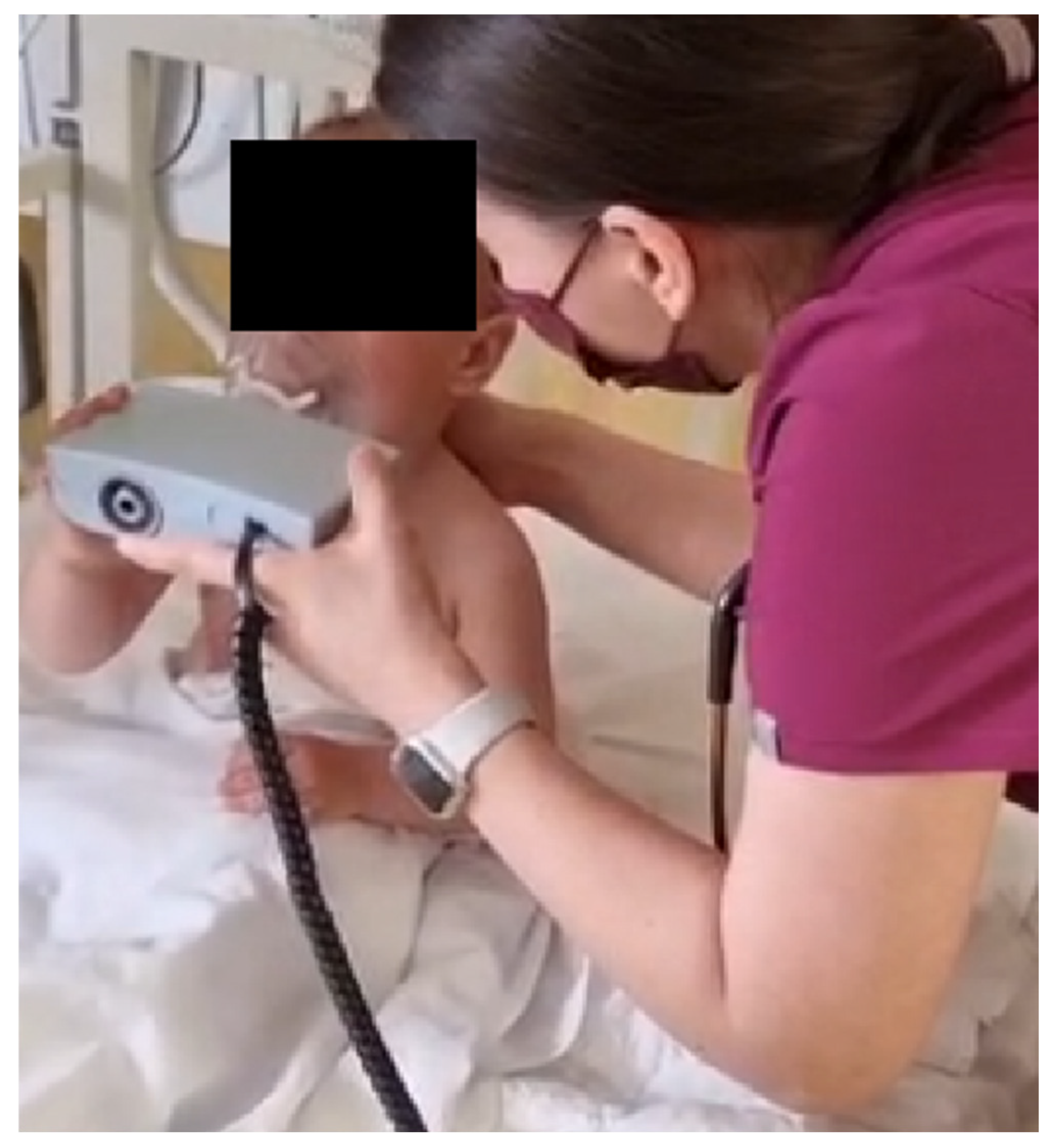
Figure 2.
Tidal breathing flow–volume loops pre- and post-salbutamol. (A) Pre: scooped expiratory limb with delayed time-to-peak expiratory flow (tPTEF) and prolonged expiration, consistent with expiratory flow limitation. (B) Post: larger loop with later peak timing indices (tPTEF 0.17→0.33 s; tPTEF/TE 0.12→0.27) and shorter expiration (TE 1.43→1.25 s; Ttot 2.15→2.04 s); PEF remains 0.14 L/s. Axes: Flow (L/s) vs Volume (L). Morphology indicates clear bronchodilator responsiveness.
Figure 2.
Tidal breathing flow–volume loops pre- and post-salbutamol. (A) Pre: scooped expiratory limb with delayed time-to-peak expiratory flow (tPTEF) and prolonged expiration, consistent with expiratory flow limitation. (B) Post: larger loop with later peak timing indices (tPTEF 0.17→0.33 s; tPTEF/TE 0.12→0.27) and shorter expiration (TE 1.43→1.25 s; Ttot 2.15→2.04 s); PEF remains 0.14 L/s. Axes: Flow (L/s) vs Volume (L). Morphology indicates clear bronchodilator responsiveness.
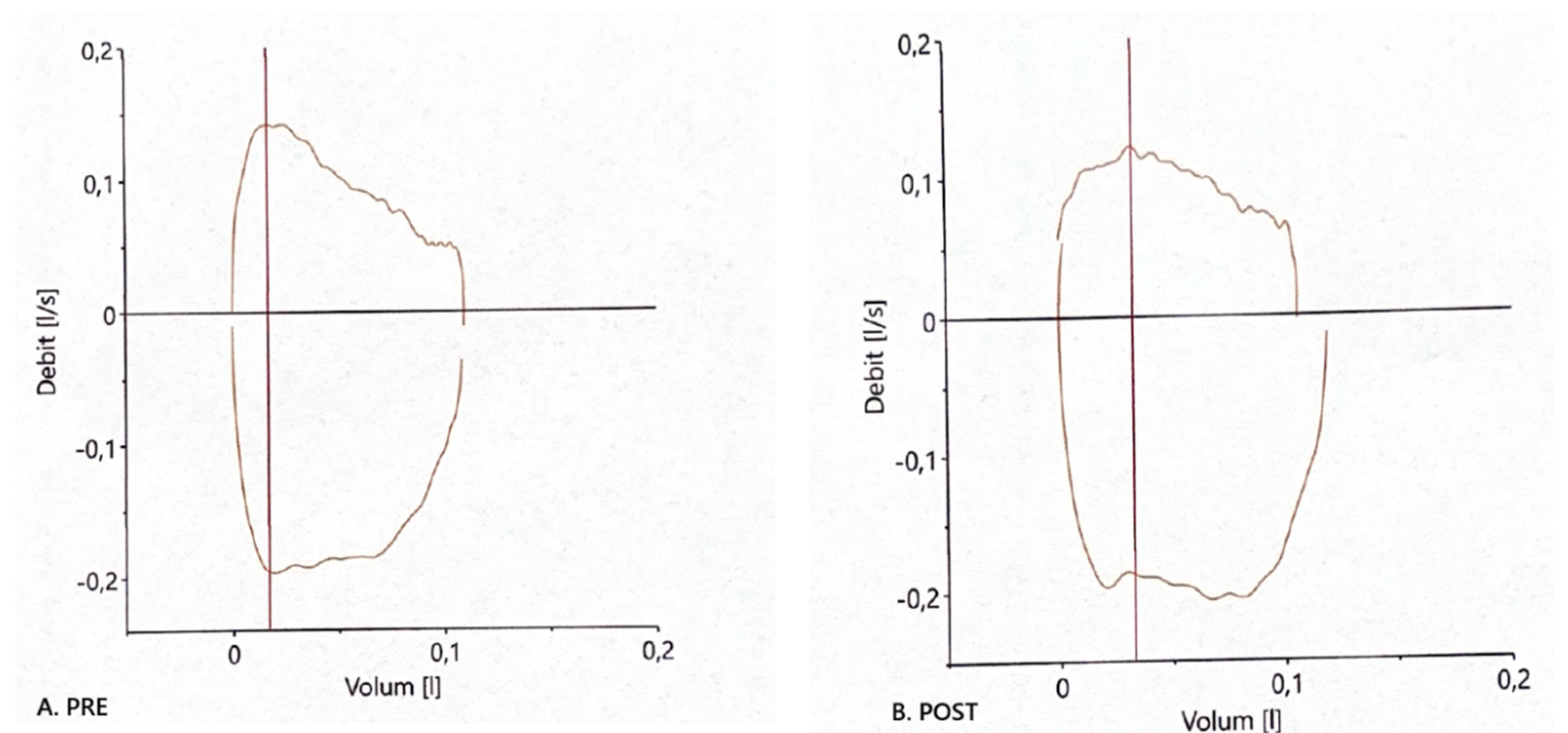
Figure 3.
Percent change in tidal-breathing parameters after salbutamol challenge.
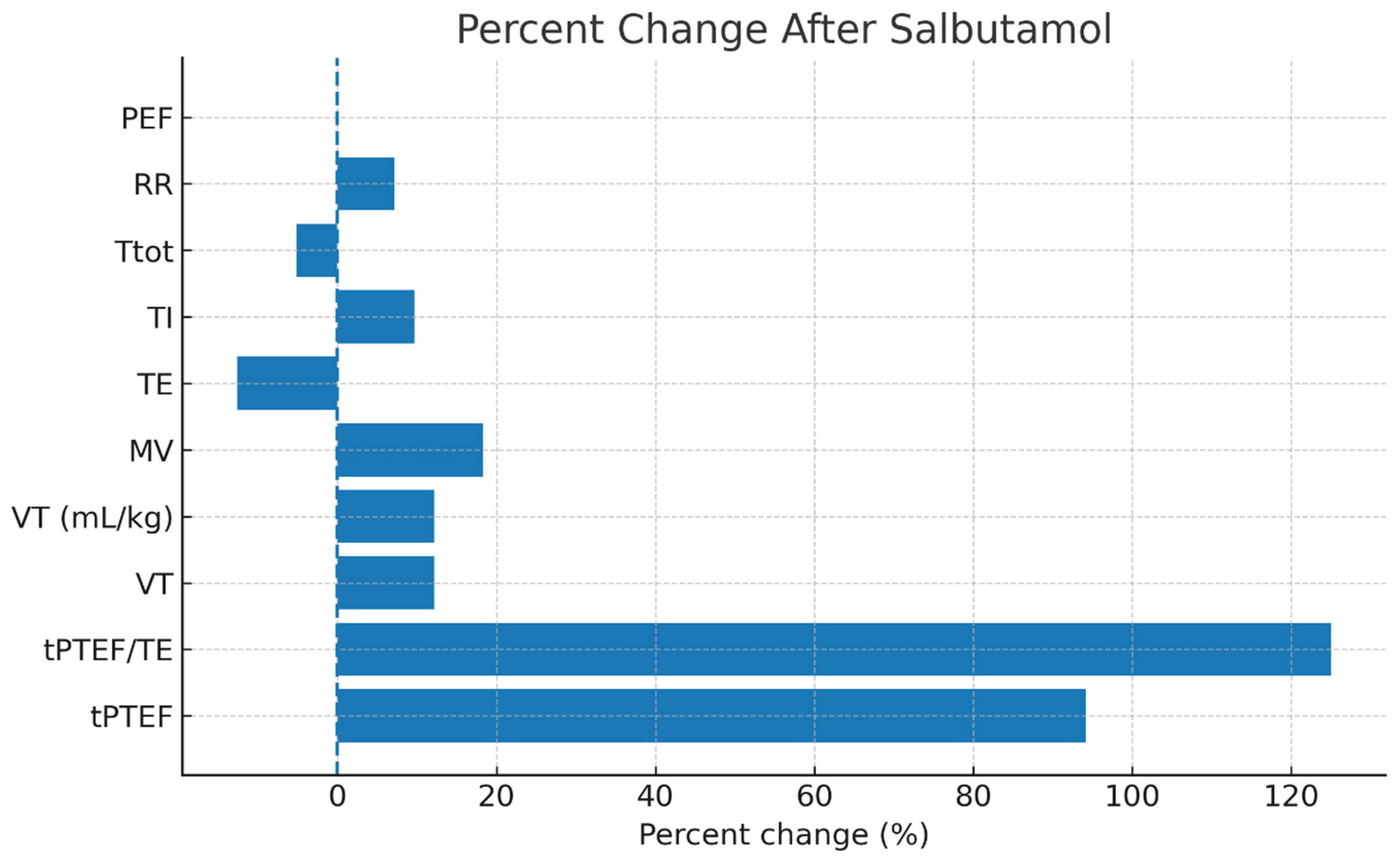

Table 1.
Tidal-breathing parameters before and after SABA challenge.
| Parameter | Unit | Pre | Post | Absolute change | Percent change (%) |
| PEF | L/s | 0.14 | 0.14 | 0 | 0 |
| tPTEF | s | 0.17 | 0.33 | 0.16 | 94.1 |
| Ttot | s | 2.15 | 2.04 | -0.11 | -5.1 |
| TI | s | 0.72 | 0.79 | 0.07 | 9.7 |
| TE | s | 1.43 | 1.25 | -0.18 | -12.6 |
| tPTEF/TE | ratio | 0.12 | 0.27 | 0.15 | 125 |
| MV | L/min | 3 | 3.55 | 0.55 | 18.3 |
| RR | 1/min | 28 | 30 | 2 | 7.1 |
| VT | mL | 107 | 120 | 13 | 12.1 |
Disclaimer/Publisher’s Note: The statements, opinions and data contained in all publications are solely those of the individual author(s) and contributor(s) and not of MDPI and/or the editor(s). MDPI and/or the editor(s) disclaim responsibility for any injury to people or property resulting from any ideas, methods, instructions or products referred to in the content. |
© 2026 by the authors. Licensee MDPI, Basel, Switzerland. This article is an open access article distributed under the terms and conditions of the Creative Commons Attribution (CC BY) license (http://creativecommons.org/licenses/by/4.0/).
Copyright: This open access article is published under a Creative Commons CC BY 4.0 license, which permit the free download, distribution, and reuse, provided that the author and preprint are cited in any reuse.


