

Submitted:
22 July 2026
Posted:
23 July 2026
You are already at the latest version
Abstract
Background: Hair loss is one of the most common reasons patients seek dermatologic evaluation. Although clinical examination and trichoscopy establish the diagnosis in most cases, scalp biopsy remains essential when findings are inconclusive, when cicatricial alopecia is suspected, or when histopathologic documentation is required before systemic therapy. Methods: This narrative review synthesizes evidence from PubMed/MEDLINE and Embase on the indications, biopsy site selection, sampling technique, specimen processing, and histopathologic interpretation of scalp biopsy in alopecia. Results: Diagnostic yield depends on appropriate patient selection, trichoscopy-guided site selection, meticulous sampling, and optimal processing. A 4-mm punch oriented parallel to the hair shafts and extending into the subcutaneous fat, combined with horizontal and vertical sectioning (HoVert or Tyler technique), allows the most comprehensive assessment of follicular architecture. Histopathology reliably separates scarring from nonscarring alopecia and, in cicatricial forms, classifies disease by the predominant inflammatory infiltrate following the NAHRS scheme. Ancillary studies, including direct immunofluorescence, periodic acid-Schiff staining, elastic fiber stains, and microbiologic cultures, are applied selectively when clinically indicated. Conclusions: Integrating clinical, trichoscopic, and histopathologic findings improves diagnostic accuracy and guides therapeutic decisions. Close collaboration between the dermatologist and dermatopathologist maximizes the diagnostic value of scalp biopsy.
Keywords:
alopecia
; biopsy
; scalp
; histopathology
; trichoscopy
; scarring alopecia
; cicatricial alopecia
; review
1. Introduction
Scalp disorders are among the most common reasons for consultation in dermatologic practice. They comprise a heterogeneous group of conditions with distinct pathogenetic mechanisms, clinical presentations, and natural histories. Androgenetic alopecia (AGA), the most common form of hair loss, affects approximately 50% of men by the age of 50 and 40% of women by the age of 70, often resulting in a significant impairment of quality of life [1,2,3]. Although clinical examination and trichoscopy are sufficient to establish the diagnosis in many cases, histopathologic evaluation remains indispensable when findings are inconclusive, when a scarring process is suspected, or when objective histopathologic confirmation is required before initiating long-term systemic therapy [3,4,5]. The diagnostic value of a scalp biopsy depends largely on the integration of clinical, trichoscopic, and histopathologic findings. This multidisciplinary approach is particularly important in primary cicatricial alopecia, a heterogeneous group of inflammatory disorders characterized by irreversible destruction of hair follicles and their replacement by fibrous tissue [6,7]. Early diagnosis is essential because timely recognition of active disease may prevent further irreversible hair loss. Providing the dermatopathologist with detailed clinical information is therefore crucial. Diagnostic accuracy is significantly improved when biopsy specimens are accompanied by a comprehensive clinical history, trichoscopic findings, and photographs documenting the biopsy site [4]. Primary cicatricial alopecia include lichen planopilaris (LPP), frontal fibrosing alopecia (FFA), discoid lupus erythematosus (DLE), folliculitis decalvans (FD), central centrifugal cicatricial alopecia (CCCA), and dissecting cellulitis (DC) of the scalp. According to the North American Hair Research Society (NAHRS) classification, these disorders are categorized as lymphocytic, neutrophilic, mixed, or nonspecific based on the predominant inflammatory infiltrate [8,9,10,11,12,13,14]. Nevertheless, scarring alopecia may also occur secondary to other cutaneous disorders, including scalp sarcoidosis and, rarely, severe psoriatic involvement of the scalp [15,16]. By contrast, scalp involvement in dermatomyositis generally shows a nonscarring histopathologic pattern [17]. Because prognosis and therapeutic management differ substantially among these subtypes, accurate histopathologic classification has direct clinical implications [9]. In contrast, nonscarring alopecia, including AGA, alopecia areata (AA), telogen effluvium (TE), and trichotillomania, are usually diagnosed on the basis of clinical examination and trichoscopy, although scalp biopsy may be valuable in selected or diagnostically challenging cases [18,19]. Histopathologic examination is particularly useful when diffuse AA, TE, and female pattern hair loss (FPHL) present with overlapping clinical and trichoscopic features and cannot be reliably distinguished by noninvasive assessment alone [20,21]. The diagnostic yield of scalp biopsy depends on appropriate patient selection, accurate biopsy site selection, meticulous sampling technique, and optimal specimen processing. Biopsies obtained from inactive lesions or performed using suboptimal technique may provide limited or misleading information [4,21]. Conversely, a properly performed biopsy substantially improves diagnostic accuracy beyond that achieved by clinical evaluation alone and changes the working diagnosis often enough to influence both prognosis and therapeutic decision-making [4,22]. Two major technical advances have significantly enhanced the diagnostic performance of scalp biopsy. Horizontal sectioning, introduced in 1984, allows simultaneous evaluation of multiple hair follicles within a single section and quantitative assessment of follicular density, morphology, and hair-cycle distribution [23,24]. A later systematic review and meta-analysis found no significant difference in overall diagnostic rates between horizontal and vertical sectioning, although the available studies were heterogeneous and generally methodologically limited [25]. A subsequent advance was the introduction of trichoscopy-guided biopsy site selection, which addressed the equally important challenge of identifying the most informative area for sampling. In inflammatory alopecia, trichoscopic features such as perifollicular erythema, perifollicular scaling, broken hairs, black dots, and follicular tufting identify sites of active disease and therefore represent the optimal targets for biopsy [26,27,28,29].
2. Materials and Methods
This is a narrative review of current evidence on the indications, technique, and diagnostic value of scalp biopsy in alopecia.
A comprehensive search of PubMed/MEDLINE and Embase was performed from inception to February 2026. Keywords and MeSH terms included scalp biopsy, alopecia, cicatricial alopecia, hair loss, horizontal sections, transverse sections, trichoscopy, hair follicle histopathology, direct immunofluorescence (DIF), and dermatopathology. These terms were used individually and in combination with Boolean operators (AND, OR). The reference lists of key articles were screened manually to identify additional relevant publications.
Articles were screened by title and abstract, and those considered potentially relevant underwent full-text review. To be eligible, articles had to be published in peer-reviewed journals indexed in PubMed/MEDLINE or Embase, written in English, and focused on scalp biopsy or the histopathologic evaluation of alopecia. They also had to address at least one of the following topics: indications, site selection, technique, specimen processing, histopathologic patterns, ancillary diagnostic methods, or diagnostic pitfalls. Both original studies and reviews were eligible. Priority was given to seminal dermatopathology studies, systematic reviews, meta-analyses, and methodologically focused articles. Conference abstracts without a full-text version, non-English publications, and studies lacking adequate methodological detail were excluded.
The schematic illustration presented in Figure 1 was generated with the assistance of a generative artificial intelligence tool (ChatGPT, version 5.6; OpenAI, San Francisco, CA, USA). All authors reviewed and verified the accuracy of the illustration and take full responsibility for its content.
Study design, biopsy technique, histopathologic criteria, and diagnostic recommendations were synthesized narratively. Special emphasis was placed on biopsy site selection, technical considerations, and the interpretation of histopathological findings.
2.1. Indications for Scalp Biopsy
Biopsy is warranted when alopecia remains diagnostically uncertain after clinical examination and trichoscopy, when a scarring process must be confirmed or excluded, or when objective documentation is required before starting systemic therapy [4]. Its principal indication is the evaluation of suspected primary cicatricial alopecia, in which histopathologic examination can confirm the diagnosis, classify the inflammatory subtype, and guide treatment [6,7].
In diagnostically challenging nonscarring alopecia, horizontal sections permit quantitative assessment of follicular parameters that can aid differentiation of chronic between TE, FPHL, AGA and AA [18,20,30].
In AA, biopsy shows the characteristic peribulbar lymphocytic infiltrate (the “swarm of bees” appearance) together with increased catagen and telogen counts [19,31].
Alopecia areata incognita (AAI) is a condition in which clinicopathologic and trichoscopic correlation is essential. As a diffuse variant of AA, it can mimic TE, producing acute diffuse shedding without the classic patches; its trichoscopic hallmarks are diffuse yellow dots and short regrown hairs [32]. The histopathologic changes, often subtle peribulbar infiltrates, can be interpreted correctly only when the pathologist is guided by trichoscopic suspicion [30,31]. In a histopathologic study of 46 cases, horizontal sections demonstrated preserved follicular units, reduced numbers of terminal follicles, increased telogen structures, a reduced terminal-to-vellus ratio, and dilated infundibular openings [31]. A five-year study of AAI and diffuse AA likewise found trichoscopy to be indispensable for directing the biopsy to the most informative area and for distinguishing these two entities [33]. These observations indicate that clinical experience, but especially the trichoscopic ability and connection to the dermatopathologist, are essential.
Biopsy is also informative in suspected trichotillomania when the clinical history is unclear. Histopathologic clues include increased catagen follicles, pigment casts, traumatized hair bulbs, and trichomalacia [34,35]. In diagnostically challenging diffuse nonscarring alopecia, horizontal sections provide quantitative assessment of follicular density, terminal-to-vellus ratio, and hair-cycle distribution, which may aid differentiation among AGA, chronic TE, and diffuse AA [18,20,24,36].
Scalp biopsy has also become relevant in alopecia associated with immune checkpoint inhibitors, which can cause both nonscarring and scarring hair loss. In a systematic review of alopecia associated with these agents, AA was the most frequently reported pattern, and hair loss generally developed within the first year of treatment [37]. Drug-induced diffuse AA has likewise been reported with other agents, including the anti-CD30 antibody-drug conjugate brentuximab vedotin, in which trichoscopic and histopathologic confirmation was critical to perform the correct diagnosis [38].
Biopsy should be considered whenever the result is likely to resolve diagnostic uncertainty or influence treatment decisions. Careful patient selection is therefore central to maximizing the diagnostic yield of the procedure.
2.2. Selection of the Biopsy Site
Appropriate site selection is essential for diagnostic accuracy. Even a technically adequate biopsy may be uninformative if it is taken from an area of advanced follicular destruction, where only nonspecific fibrosis and follicular dropout remain (Table 1) [4,6]. In scarring alopecia, completely bald areas should be avoided, as they usually show end-stage changes of little diagnostic value [4].
Trichoscopy has markedly improved site selection. In cicatricial alopecia, findings such as perifollicular concentric white scales, follicular tufting, peripilar grey-white halos, follicular red dots, and keratotic plugs can guide sampling toward the most informative area [26,28]. Pruritus, burning, or a positive hair pull test offer additional guidance.
In scarring alopecia, the optimal site is the margin of the alopecic patch, the transition zone between affected and relatively preserved scalp, where inflammatory activity is greatest [4,7]. A well-chosen specimen should contain both scarred follicular units and follicles still undergoing active inflammation, since this combination maximizes diagnostic information [4,7]. In LPP and FFA, areas with perifollicular erythema or scaling should be targeted; in FD, zones with tufted hairs, perifollicular scaling, or follicular pustules; and in CCCA, the active peripheral edge of the vertex patch with peripilar white or grey halo [8,26,39,40,41,42,43]
For AA, an area showing trichoscopic evidence of active disease should be selected; histopathologic inflammation may be sparse or absent in chronic lesions, in which follicular miniaturization and hair-cycle changes may predominate [44]. The vertex or mid-scalp is preferred for diffuse alopecia such as AGA, chronic TE and FPHL [18]. Whatever the diagnosis, the biopsy site should be documented photographically, and the image should be sent to the dermatopathologist [4].
2.3. Biopsy Technique
The 4-mm punch is the standard instrument. Its cross-sectional area (~13 mm²) is nearly twice that of the 3-mm punch (~7 mm²), and this difference translates directly into the number of follicular units available for reliable quantitative analysis [4,5,45]. The punch should be oriented parallel to the direction of hair growth to minimize follicular transection, and it must reach the subcutaneous fat; otherwise the anagen bulbs are amputated rather than sampled (Table 2) [45].
The St John’s protocol uses two 4-mm punch biopsies. In nonscarring alopecia, one biopsy is obtained from the clinically affected scalp, usually the vertex, and the other from an uninvolved control site; both are processed horizontally [46]. In cicatricial alopecia, both biopsies are obtained from the affected area: one is sectioned horizontally, whereas the other is bisected vertically, with one half processed in hematoxylin–eosin and the other reserved for DIF when indicated [46]. When trichoscopy-guided biopsy is routine, however, a single well-targeted specimen usually suffices.
In the two-biopsy approach vertical sections are useful for assessing interface changes and deeper inflammatory processes, whereas horizontal sections facilitate the detection of focal follicular involvement and the evaluation of follicular abnormalities in scalp lupus [47,48]. Horizontal sections also permit the simultaneous assessment of multiple follicles and quantitative analysis of follicular parameters [23].
Available evidence generally supports combined approaches. One pilot study reported 100% sensitivity for transverse sections in nonscarring alopecia, whereas vertical sections performed better for LPP [49]. A meta-analysis found no significant overall difference in diagnostic yield between the two planes, whereas a retrospective series of 228 biopsies concluded that their combination maximized diagnostic yield [25,50]. Techniques that obtain both planes from a single specimen, such as the horizontal-vertical combined sectioning (HoVert) technique and the Tyler technique, offer the most efficient approach (Figure 1). In our experience, the Tyler technique is practical and yields both horizontal and vertical information from a single specimen without compromising histologic quality, and its simplicity makes it well suited to routine use [51].
Local anesthesia is achieved with lidocaine, with or without epinephrine (2–3 ml), injected into the dermis and superficial fat. After injection, several minutes should elapse before the incision is performed, allowing vasoconstriction to provide adequate hemostasis. Infiltration directly into the biopsy site should be kept to a minimum, because distortion of follicular architecture is a real and underappreciated source of interpretive error [4]. The wound is closed with simple interrupted sutures, which are removed at approximately 10 days.
2.4. Specimen Processing and Histopathologic Evaluation
Orientation of the specimen before sectioning is as important as the excision itself. Horizontal sections are obtained by cutting parallel to the skin surface and permit the simultaneous evaluation of multiple follicles [48]. This approach quantifies the parameters needed to differentiate nonscarring alopecia: follicular density, terminal-to-vellus hair ratio, hair-cycle distribution and the degree of perifollicular fibrosis (Figure 2) [18,23,24,52]
In normal scalp, approximately 85–90% of follicles are in anagen, and telogen hairs account for less than 10–15%. On horizontal sections, a terminal-to-vellus ratio below 4:1 at the isthmus is generally regarded as supportive of AGA. An increased proportion of telogen follicles, in the setting of a preserved terminal-to-vellus ratio, supports TE [18,24,50,53]. Because these changes may be subtle, quantitative findings should be interpreted together with the clinical presentation.
Proper grossing is a prerequisite for reliable histopathologic evaluation. In one large series, 22 of 228 biopsies (9.6%) were inconclusive because of faulty grossing, which indicates the need for trained laboratory personnel [50].
A further consideration in quantitative histopathologic assessment is that follicular density is not uniform across the normal scalp. A cadaveric study of 240 specimens from four anatomic zones found that the temporoparietal area showed the lowest total hair and follicular-unit counts, with approximately 18 total hairs per 4-mm punch compared with approximately 24 at the vertex [54]. The anagen-to-telogen ratio was comparable across scalp regions, and no significant associations with sex or increasing age were identified [54]. These findings indicate that biopsy location should be considered when interpreting quantitative follicular parameters.
3. Results and Discussion
3.1. Histopathologic Patterns of Alopecia
The fundamental distinction on biopsy is between nonscarring and scarring alopecia. In scarring forms, destruction of follicular stem cells in the bulge region causes irreversible hair loss, so histopathologic classification is critical for management [6,55,56].
Nonscarring alopecia
AGA is defined histopathologically by progressive follicular miniaturization and a shift toward vellus-like hairs. Horizontal sections outperform vertical ones both for diagnosis and for predicting therapeutic response, including the response to minoxidil [18,24]. Acute TE shows a preserved follicular count with a disproportionate increase in telogen hairs [18,53,57,58].
The histopathologic features of AA vary according to disease stage. Peribulbar lymphocytic inflammation (the “swarm of bees”) is characteristic of acute lesions but may be sparse or absent in later stages. Subacute lesions show increased catagen and telogen follicles, whereas chronic lesions are characterized predominantly by follicular miniaturization; nanogen follicles may provide an additional diagnostic clue [18,44,59,60,61].
On horizontal sections, AAI presents a more subtle picture: preserved follicular units, reduced numbers of terminal follicles, increased telogen structures with a mean count of 37%, a reduced terminal-to-vellus ratio averaging 3.3:1, telogen germinal units or small telogen follicles, and dilated infundibular openings [31,62]. Diffuse AA may show more intense peribulbar inflammation than patchy AA, including eosinophils and CD8+ T cells [62,63,64]. Scalp trichoscopy may reveal broken hairs and black dots, whereas examination of shed hair roots has demonstrated a sequential transition from anagen to catagen and TE, together with progressive proximal hair-shaft depigmentation correlated with disease duration (Figure 3, Figure 4 and Figure 5)
Traumatic alopecia include trichotillomania and traction alopecia (TA), which result from different forms of mechanical injury and show distinct clinicopathologic patterns. Trichotillomania, caused by repetitive self-induced pulling or manipulation of the hair, shows traumatic follicular changes, including distorted follicles, trichomalacia, pigment casts, empty follicular sheaths, and minimal inflammation [34,35]. TA, by contrast, results from chronic tensile forces and may progress to permanent follicular loss. In end-stage TA, horizontal sections may facilitate quantitative assessment of terminal and vellus follicles, typically showing loss of terminal follicles, fibrous tracts, and relative preservation of sebaceous glands [65,66]. The pauci-follicular unit, defined as a non-inflamed follicular unit with an intact sebaceous gland and fewer than two follicles, has been proposed as a useful histopathologic clue to TA [66]. The finding of three or more PFUs in a single 4-mm punch biopsy, evaluated on horizontal sections, was proposed as a practical shortcut sufficient to identify TA [66]. This criterion is particularly useful when TA coexists with other forms of hair loss, a relatively common situation in which counting PFUs helps to separate the traction component from the overall histopathologic picture.
Scarring alopecia
Across all primary cicatricial alopecia, the histopathologic hallmarks are the same: follicular destruction, replacement by fibrous tracts, loss of sebaceous glands, and eccrine duct dilation [6,7,11,67]. The NAHRS classification groups these disorders by the predominant inflammatory cell type [10,11,12].
Among the lymphocytic subtypes, LPP shows lichenoid interface dermatitis at the infundibulum and isthmus, with hypergranulosis and Civatte bodies; FFA shares these features but has a different distribution and characteristically involves vellus follicles [59,68,69,70]. DLE shows patchy vacuolar interface dermatitis and a folliculocentric infiltrate that extends both superficially and deeply, often reaching the eccrine glands, together with basement membrane thickening and dermal mucin [39,40,41,45,47,48,49,50,51,52,71]. CCCA, the most common scarring alopecia in women of African descent, is defined by premature desquamation of the inner root sheath, concentric lamellar perifollicular fibrosis, and variable isthmus-level lymphocytic inflammation. One important caveat is that CCCA and LPP can be histopathologically indistinguishable, which makes the clinical context decisive (Figure 6 and Figure 7) [39,59,68,72,73].
Sebaceous gland atrophy (SGA) merits mention as a histopathologic finding whose diagnostic significance has been reassessed in recent years. It has been proposed as a potential contributor to hair loss in psoriasis, in which the sebaceous glands within the plaque undergo marked atrophy [74]. Rather than a marker of a single disease, it is better regarded as a reaction pattern and has been reported in early FFA, where it can precede overt pilosebaceous destruction [68,75]. On horizontal sections, atrophic sebaceous lobules appear as shrunken basaloid aggregates and may be mistaken for telogen germinal units; awareness of this pitfall reduces the risk of misclassification. When SGA is found in a biopsy that does not otherwise fit the psoriatic spectrum, the differential diagnosis should be broadened accordingly and the clinical history reviewed for relevant topical or systemic exposures [75].
Among neutrophilic cicatricial alopecia, FD is characterized by neutrophilic folliculitis and follicular rupture, with mixed inflammation and fibrosis developing in later lesions; DC typically shows deeper follicular rupture, suppurative inflammation, and abscess formation [8,9,11].
Acne keloidalis nuchae is classified as a mixed cicatricial alopecia because both neutrophilic and lymphocytic inflammation may be present [9,11]. When the inflammatory pattern is ambiguous, clinical data are essential and may be decisive for the final diagnosis (Figure 8) [8].
Fibrosing alopecia in a pattern distribution (FAPD) is an underdiagnosed form of lymphocytic cicatricial alopecia, generally considered within the spectrum of LPP. First described around the turn of the millennium, FAPD combines the histopathologic features of LPP, namely vacuolar or lichenoid interface dermatitis at the infundibulum and isthmus, perifollicular fibrosis, and loss of sebaceous lobules, with the follicular miniaturization expected in AGA [69,70]. Because its clinical presentation is subtle and often asymptomatic, FAPD is easily misclassified as AGA with coincidental seborrheic dermatitis; trichoscopy, which shows anisotrichia, loss of follicular ostia, and perifollicular casts, provides the key prompt to biopsy [70]. Vacuolar or lichenoid interface change is the main histopathologic discriminator: FAPD, like other LPP subtypes, shows it, whereas CCCA does not, even when the two conditions share the same clinical picture of centroparietal hair loss with miniaturization [70,76,77]. In a large single-center series spanning ten years, FAPD accounted for 18% of scarring alopecia, a frequency well above the rate at which it is recognized clinically [78].
3.2. Ancillary Diagnostic Techniques
Hematoxylin-eosin staining remains the cornerstone. Ancillary techniques may be used when routine histopathologic examination does not resolve the diagnostic uncertainty [4].
Direct immunofluorescence (DIF) is most useful when lupus erythematosus is in differential diagnosis. Granular deposits of immunoglobulins and complement along the basement membrane zone may support a diagnosis of DLE and help distinguish it from LPP, which can closely mimic both clinically and histopathologically [59,68,69,79].
Periodic acid-Schiff (PAS) staining identifies fungal organisms hair shafts and follicular structures in suspected tinea capitis [4]. Elastic fiber stains (e.g., Verhoeff-Van Gieson) distinguish follicular streamers from true follicular scars on horizontal sections [6,80]. In FD, bacterial culture of active pustular lesions may identify Staphylococcus aureus or other organisms and guide antimicrobial selection, although culture findings are supportive rather than diagnostic [8,81,82].
Non-invasive methods are increasingly used alongside biopsy: quantitative trichoscopy provides non-invasive measurements of hair parameters; however, the values obtained are not directly interchangeable with histopathologic measurements [83].
For now, accurate biopsy interpretation still requires the integration of clinical, trichoscopic, and histopathologic information.
3.3. The Trichoscopic-Histopathologic Correlation as a Diagnostic Paradigm
Contemporary diagnostic practice in hair disorders rests on the integration of trichoscopy and histopathology, a combination that has changed the approach to uncertain alopecia. Trichoscopy is not merely a tool for choosing the biopsy site; it generates the diagnostic hypothesis that shapes how the pathologist reads the specimen. In conditions with subtle or overlapping histopathologic findings, the clinical context provided by the trichoscopist can determine whether the specimen is interpreted correctly or yields a nondiagnostic report.
Our experience from the University of Bologna, built over decades of combined trichoscopic and histopathologic assessment, illustrates this point. AAI, a condition that may not fulfill classical histopathologic criteria and can easily escape recognition, could be reliably recognized only with integration of clinical, trichoscopic, and histopathologic findings. The trichoscopist identified diffuse yellow dots, pigtail hairs, and short regrowing hairs and shared these findings with a pathologist who might otherwise see only nonspecific miniaturization [31,32,33]. Histopathologic evaluation provides additional quantitative and morphologic criteria, including reduced terminal follicle counts, increased telogen structures, a reduced terminal-to-vellus ratio, and dilated infundibular openings [31].
A subsequent five-year study concluded that trichoscopy was essential both for separating AAI from diffuse AA and for targeting the most informative biopsy site [33]. More recent work found that reflectance confocal microscopy shows substantial agreement with histopathology in AAI; this suggests that non-invasive imaging may eventually complement, though not replace, the trichoscopic-histopathologic correlation [84].
An important corollary concerns FFA in particular. Trichoscopy detects perifollicular erythema and scaling during active disease, yet some patients show progressive scarring hair loss without any trichoscopic evidence of ongoing inflammation. A cross-sectional study of LPP and FFA showed that trichoscopic findings do not consistently reflect the degree of histopathologic inflammation: cases with dense perifollicular lymphocytic infiltrates on biopsy may appear trichoscopically quiescent [85]. Moreover, in FFA progression of hairline recession may occur despite absent or improving inflammatory signs [86]. One proposed explanation is that the lymphoid infiltrate in FFA tends to lie deeper than in classic LPP, below the resolution of surface trichoscopy [87]. This has a clear clinical consequence: histopathology is needed to characterize and quantify inflammatory activity in FFA even when the trichoscopic picture is reassuring, and treatment decisions should not rely on trichoscopy alone in this setting.
3.4. Diagnostic Pitfalls
The most frequent pitfall is sampling end-stage follicular destruction, where only nonspecific fibrosis remains and the underlying cause cannot be determined [4,5,6]. Equally problematic is insufficient biopsy depth: anagen follicular bulbs lie in the subcutis, and superficial specimens may miss both these structures and the deep peribulbar inflammatory infiltrates that characterize conditions such as AA [4,5,24,44,45].
Punches smaller than 4 mm reduce the number of follicular units available for quantitative assessment, and improper specimen orientation during processing produces oblique sections that distort follicular counts [4,5,45,47,50]. Several lymphocytic scarring alopecia share overlapping features; CCCA and LPP, for example, may be histologically and immunophenotypically indistinguishable, making clinicopathologic correlation essential [7,72,88]. The three-way differential of AGA, TE, and AA on biopsy alone remains notoriously difficult [20].
A related and underappreciated challenge is multifactorial alopecia, the coexistence of two or more coexisting diagnoses in a single biopsy specimen. In a large consecutive series of 1,360 biopsies, more than one diagnosis was identified in 12.5% of specimens, the most frequent combination was AGA with CCCA, followed by CCCA with end-stage TA; three concurrent diagnoses, AGA with CCCA and end-stage TA, accounted for 3.5% of the multifactorial cases [89]. These figures call for two practical adaptations. First, biopsy-site selection should target a watershed zone, an area of the scalp where the disease processes under consideration are known to overlap anatomically; when CCCA, FPHL, and TA are being evaluated together, for instance, the frontotemporal aspect of the crown lies at the intersection of all three [75,89]. Second, horizontal sections are indispensable in this setting: because different follicular units within the same 4-mm punch may be affected by different processes, only transverse sectioning allows them to be assessed simultaneously and side by side [75,89].
The terminology used in the pathology report also warrants attention. “Consistent with” or “compatible with” signal high diagnostic confidence, whereas “suggestive of” or “cannot exclude” indicate residual uncertainty that may require further workup [4].
3.5. Practical Diagnostic Algorithm
The first question is whether the alopecia is scarring or nonscarring, a distinction of major prognostic and therapeutic importance (Figure 9). Loss of follicular ostia, absence of vellus hairs, and dermal sclerosis on examination, together with trichoscopic findings such as fibrotic white dots or absent follicular openings, point toward a scarring process.
When clinical examination and trichoscopy are inconclusive, scalp biopsy should be considered. The sectioning method should reflect the clinical suspicion: horizontal sections are particularly useful in nonscarring alopecia, whereas combined horizontal and vertical evaluation, when feasible, maximizes the available diagnostic information [25,49,50]. Ancillary techniques (DIF, PAS, elastic stains, cultures) are added when standard evaluation leaves the diagnosis open. Throughout, close collaboration between the dermatologist and the dermatopathologist is essential for accurate diagnostic interpretation; this collaboration is what ultimately turns a tissue sample into a diagnosis [4].
4. Conclusions
Scalp biopsy remains an essential tool in difficult cases of alopecia. Histopathologic interpretation is strictly correlated to appropriate patient selection, trichoscopy-guided site identification, adequate technique, and careful specimen processing.
Technical advances, including horizontal sectioning, combined approaches such as the HoVert and Tyler techniques, and trichoscopy-guided site selection, have substantially improved the assessment of follicular architecture and inflammatory patterns. Ancillary techniques such as DIF, elastic fiber stains, and microbiologic studies provide targeted support when standard histopathology is inconclusive.
A diagnostic approach that integrates clinical examination, trichoscopy, and histopathologic analysis can markedly improve accuracy and guide treatment. Standardized protocols and sustained collaboration between dermatologists and dermatopathologists are central to improving patient outcomes.
Author Contributions
Conceptualization, M.L.P. and C.M.; methodology, T.B. and C.M.; validation, M.L.P., C.M. and M.S.; investigation, C.M., T.B., L.R. and F.B.; resources, M.L.P.; writing-original draft preparation, T.B.; writing-review and editing, M.L.P.; visualization, M.L.P.; supervision, M.S and C.M. All authors have read and agreed to the published version of the manuscript.
Funding
This research received no external funding.
Institutional Review Board Statement
The investigation described was carried out on residual biopsy sections following diagnostic analysis in the course of institutional diagnostic services, and the study was exempted from institutional review board (IRB) review.
Informed Consent Statement
Not applicable.
Data Availability Statement
Data available on request from the authors.
Acknowledgments
During the preparation of this manuscript, the authors used ChatGPT, version 5.6 (OpenAI, San Francisco, CA, USA), to assist in generating the schematic illustration presented as Figure 1 and to improve the linguistic clarity, readability, and stylistic quality of the English text. The tool was not used to generate or modify the underlying scientific concepts, data, results, interpretations, or conclusions. The authors reviewed and edited all AI-assisted outputs and take full responsibility for the content of this publication.
Conflicts of Interest
The authors declare no conflicts of interest.:
Abbreviations
The following abbreviations are used in this manuscript
AGA: androgenetic alopecia; AAI: alopecia areata incognita;; CCCA: central centrifugal cicatricial alopecia; DC: dissecting cellulitis; DIF: direct immunofluorescence; DLE: discoid lupus erythematosus; FAPD: fibrosing alopecia in a pattern distribution; FD: folliculitis decalvans; FFA: frontal fibrosing alopecia; FPHL: female pattern hair loss; TA: Traction Alopecia; TE: telogen effluvium; HoVert: horizontal-vertical combined sectioning technique; LPP: lichen planopilaris; NAHRS: North American Hair Research Society; PAS: periodic acid-Schiff;
References
- Lolli, F.; Pallotti, F.; Rossi, A.; Fortuna, M.C.; Caro, G.; Lenzi, A.; Sansone, A.; Lombardo, F. Androgenetic Alopecia: A Review. Endocrine 2017, 57, 9–17. [Google Scholar] [CrossRef] [PubMed]
- Qi, J.; Garza, L.A. An Overview of Alopecia. Cold Spring Harb. Perspect. Med. 2014, 4, a013615. [Google Scholar] [CrossRef] [PubMed]
- Rossi, A.; Muscianese, M.; Piraccini, B.M.; Starace, M.; Carlesimo, M.; Mandel, V.D.; Alessandrini, A.; Calvieri, S.; Caro, G.; D’arino, A.; et al. Italian Guidelines in Diagnosis and Treatment of Alopecia Areata. G. Ital. Dermatol. Venereol. 2019, 154, 609–623. [Google Scholar] [CrossRef] [PubMed]
- Sperling, L.C. The Role of the Scalp Biopsy in the Evaluation of Alopecia. J. Am. Acad. Dermatol. 2023, 89, S16–S19. [Google Scholar] [CrossRef] [PubMed]
- Knopp, E. The Scalp Biopsy for Hair Loss and Its Interpretation. Semin Cutan. Med. Surg. 2015, 34, 57–66. [Google Scholar] [CrossRef] [PubMed]
- Sperling, L.C.; Cowper, S.E. The Histopathology of Primary Cicatricial Alopecia. Semin Cutan. Med. Surg. 2006, 25, 41–50. [Google Scholar] [CrossRef] [PubMed]
- Mirmirani, P.; Willey, A.; Headington, J.T.; Stenn, K.; McCalmont, T.H.; Price, V.H. Primary Cicatricial Alopecia: Histopathologic Findings Do Not Distinguish Clinical Variants. J. Am. Acad. Dermatol. 2005, 52, 637–643. [Google Scholar] [CrossRef] [PubMed]
- Tan, E.; Martinka, M.; Ball, N.; Shapiro, J. Primary Cicatricial Alopecia: Clinicopathology of 112 Cases. J. Am. Acad. Dermatol. 2004, 50, 25–32. [Google Scholar] [CrossRef] [PubMed]
- Ross, E.K.; Tan, E.; Shapiro, J. Update on Primary Cicatricial Alopecia. J. Am. Acad. Dermatol. 2005, 53, 1–37; quiz 38–40. [Google Scholar] [CrossRef] [PubMed]
- Olsen, E.A.; Bergfeld, W.F.; Cotsarelis, G.; Price, V.H.; Shapiro, J.; Sinclair, R.; Solomon, A.; Sperling, L.; Stenn, K.; Whiting, D.A.; et al. Summary of North American Hair Research Society (NAHRS)-Sponsored Workshop on Cicatricial Alopecia, Duke University Medical Center, February 10 and 11, 2001. J. Am. Acad. Dermatol. 2003, 48, 103–110. [Google Scholar] [CrossRef] [PubMed]
- Bernárdez, C.; Molina-Ruiz, A.M.; Requena, L. Histologic Features of Alopecia: Part II: Scarring Alopecia. Actas Dermosifiliogr. 2015, 106, 260–270. [Google Scholar] [CrossRef] [PubMed]
- Bernárdez, C.; Molina-Ruiz, A.M.; Requena, L. Histologic Features of Alopecia-Part I: Nonscarring Alopecia. Actas Dermosifiliogr. 2015, 106, 158–167. [Google Scholar] [CrossRef] [PubMed]
- Scotti, B.; Misciali, C.; Bardazzi, F.; Piraccini, B.M.; La Placa, M. Hypertrophic Lichen Planus and Hypertrophic Skin Lesions Associated with Histological Lichenoid Infiltrate: A Case Report and Literature Review. Dermatopathology 2025, 12, 8. [Google Scholar] [CrossRef] [PubMed]
- Verdelli, A.; Coi, A.; Marzano, A.V.; Antiga, E.; Cozzani, E.; Quaglino, P.; La Placa, M.; Benucci, M.; De Simone, C.; Papini, M.; et al. Autoantibody Profile and Clinical Patterns in 619 Italian Patients with Cutaneous Lupus Erythematosus. J. Eur. Acad. Dermatol. Venereol. 2019, 33, 742–752. [Google Scholar] [PubMed]
- La Placa, M.; Vincenzi, C.; Misciali, C.; Tosti, A. Scalp Sarcoidosis with Systemic Involvement. J. Am. Acad. Dermatol. 2008, 59, S126–127. [Google Scholar] [CrossRef] [PubMed]
- El Shabrawi-Caelen, L.; La Placa, M.; Vincenzi, C.; Haidn, T.; Muellegger, R.; Tosti, A. Adalimumab-Induced Psoriasis of the Scalp with Diffuse Alopecia: A Severe Potentially Irreversible Cutaneous Side Effect of TNF-Alpha Blockers. Inflamm. Bowel Dis. 2010, 16, 182–183. [Google Scholar] [CrossRef] [PubMed]
- Jasso-Olivares, J.; Diaz-Gonzalez, J.M.; Miteva, M. Horizontal and Vertical Sections of Scalp Biopsy Specimens from Dermatomyositis Patients with Scalp Involvement. J. Am. Acad. Dermatol. 2018, 78, 1178–1184. [Google Scholar] [CrossRef] [PubMed]
- Sinclair, R.; Jolley, D.; Mallari, R.; Magee, J. The Reliability of Horizontally Sectioned Scalp Biopsies in the Diagnosis of Chronic Diffuse Telogen Hair Loss in Women. J. Am. Acad. Dermatol. 2004, 51, 189–199. [Google Scholar] [CrossRef] [PubMed]
- Whiting, D.A. Histopathology of Alopecia Areata in Horizontal Sections of Scalp Biopsies. J. Invest Dermatol. 1995, 104, 26S–27S. [Google Scholar] [CrossRef] [PubMed]
- Werner, B.; Mulinari-Brenner, F. Clinical and Histological Challenge in the Differential Diagnosis of Diffuse Alopecia: Female Androgenetic Alopecia, Telogen Effluvium and Alopecia Areata--Part II. An. Bras. Dermatol. 2012, 87, 884–890. [Google Scholar] [CrossRef] [PubMed]
- Douglas, A.; Romisher, A.; Cohen, A.; Zaya, R.; Wang, J.; Suriano, J.; Zachian, R.; Nikbakht, N. Scalp Biopsy Influences Diagnostic Accuracy and Treatment in Black Women with Alopecia: A Retrospective Study. J. Am. Acad. Dermatol. 2023, 88, 1375–1376. [Google Scholar] [CrossRef] [PubMed]
- Inchara, Y.K.; Tirumalae, R.; Kavdia, R.; Antony, M. Histopathology of Scarring Alopecia in Indian Patients. Am. J. Dermatopathol. 2011, 33, 461–467. [Google Scholar] [CrossRef] [PubMed]
- Headington, J.T. Transverse Microscopic Anatomy of the Human Scalp. A Basis for a Morphometric Approach to Disorders of the Hair Follicle. Arch. Dermatol. 1984, 120, 449–456. [Google Scholar] [CrossRef] [PubMed]
- Whiting, D.A. Diagnostic and Predictive Value of Horizontal Sections of Scalp Biopsy Specimens in Male Pattern Androgenetic Alopecia. J. Am. Acad. Dermatol. 1993, 28, 755–763. [Google Scholar] [CrossRef] [PubMed]
- Du, X.; Li, Z.; Xu, W.; Zhou, X.; Tang, S.; Song, C.; Fan, W. Diagnostic Value of Horizontal versus Vertical Sections for Scarring and Nonscarring Alopecia: A Systematic Review and Meta-Analysis. Eur. J. Dermatol. 2016, 26, 361–369. [Google Scholar] [CrossRef] [PubMed]
- Miteva, M.; Tosti, A. Dermoscopy Guided Scalp Biopsy in Cicatricial Alopecia. J. Eur. Acad. Dermatol. Venereol. 2013, 27, 1299–1303. [Google Scholar] [PubMed]
- Gowda, S.K.; Behera, B.; Sethy, M.; Ayyanar, P. A Brief Review of Scalp Biopsy and Its Interpretation. Indian Dermatol. Online J. 2025, 16, 785–791. [Google Scholar] [CrossRef] [PubMed]
- Tosti, A.; Torres, F.; Misciali, C.; Vincenzi, C.; Duque-Estrada, B. The Role of Dermoscopy in the Diagnosis of Cicatricial Marginal Alopecia. Br. J. Dermatol. 2009, 161, 213–215. [Google Scholar] [CrossRef] [PubMed]
- Zengarini, C.; Curti, N.; Cedirian, S.; Rapparini, L.; Pampaloni, F.; Pileri, A.; Durazzi, F.; Mussi, M.; La Placa, M.; Piraccini, B.M.; et al. Trichoscopy and Computational Models for Hair and Scalp Disorders: Image Analysis, Quantification, and Clinical Integration. Appl. Sci. 2026, 16, 3199. [Google Scholar] [CrossRef]
- Yadav, D.; Khandpur, S.; Ramam, M.; Singh, M.K.; Sharma, V.K. Utility of Horizontal Sections of Scalp Biopsies in Differentiating between Androgenetic Alopecia and Alopecia Areata. Dermatology 2018, 234, 137–147. [Google Scholar] [CrossRef] [PubMed]
- Miteva, M.; Misciali, C.; Fanti, P.A.; Tosti, A. Histopathologic Features of Alopecia Areata Incognito: A Review of 46 Cases. J. Cutan. Pathol. 2012, 39, 596–602. [Google Scholar] [CrossRef] [PubMed]
- Tosti, A.; Whiting, D.; Iorizzo, M.; Pazzaglia, M.; Misciali, C.; Vincenzi, C.; Micali, G. The Role of Scalp Dermoscopy in the Diagnosis of Alopecia Areata Incognita. J. Am. Acad. Dermatol. 2008, 59, 64–67. [Google Scholar] [CrossRef] [PubMed]
- Alessandrini, A.; Starace, M.; Bruni, F.; Brandi, N.; Baraldi, C.; Misciali, C.; Fanti, P.A.; Piraccini, B.M. Alopecia Areata Incognita and Diffuse Alopecia Areata: Clinical, Trichoscopic, Histopathological, and Therapeutic Features of a 5-Year Study. Dermatol. Pract. Concept 2019, 9, 272–277. [Google Scholar] [CrossRef] [PubMed]
- Muller, S.A. Trichotillomania: A Histopathologic Study in Sixty-Six Patients. J. Am. Acad. Dermatol. 1990, 23, 56–62. [Google Scholar] [CrossRef] [PubMed]
- Kolivras, A.; Thompson, C. Primary Scalp Alopecia: New Histopathological Tools, New Concepts and a Practical Guide to Diagnosis. J. Cutan. Pathol. 2017, 44, 53–69. [Google Scholar] [PubMed]
- Sperling, L.C.; Winton, G.B. The Transverse Anatomy of Androgenic Alopecia. J. Dermatol. Surg. Oncol. 1990, 16, 1127–1133. [Google Scholar] [CrossRef] [PubMed]
- Park, M.; Suh, S.; Kincaid, C.; Yale, K.; Atanaskova Mesinkovska, N. Alopecia as an Adverse Event of Immune Checkpoint Inhibitor Therapies: Clinical Evidence and Outcomes. J. Drugs Dermatol. 2025, 24, 255–260. [Google Scholar] [CrossRef] [PubMed]
- Canal-Garcia, E.; Alamon-Reig, F.; Combalia, A. Diffuse Alopecia Areata Induced by Brentuximab Vedotin Therapy for Advanced Stage Mycosis Fungoides. Dermatol. Ther. 2022, 35, e15427. [Google Scholar] [CrossRef] [PubMed]
- Doche, I.; Valente, N.; Sotto, M.N.; Rivitti-Machado, M.C.; Aoki, V.; Gerlero, P.; Hordinsky, M.K. Correlation of Clinical, Histopathologic, and Direct Immunofluorescence Findings in Lesional and Nonlesional Scalp of Frontal Fibrosing Alopecia and Lichen Planopilaris—An Observational Study. JAAD Int. 2023, 13, 7–9. [Google Scholar] [CrossRef] [PubMed]
- Miteva, M.; Tosti, A. Pathologic Diagnosis of Central Centrifugal Cicatricial Alopecia on Horizontal Sections. Am. J. Dermatopathol. 2014, 36, 859–864; quiz 865–867. [Google Scholar] [CrossRef]
- Felix, K.; De Souza, B.; Portilla, N.; Hogue, L.; Ahn, C.S.; Sangueza, O.; McMichael, A.J. Dermatoscopic Evaluation of Central Centrifugal Cicatricial Alopecia Beyond the Vertex Scalp. JAMA Dermatol. 2020, 156, 916–918. [Google Scholar] [CrossRef] [PubMed]
- Waśkiel-Burnat, A.; Starace, M.; Iorizzo, M.; Katoulis, A.; Apalla, Z.; Asfour, L.; Freites-Martinez, A.; Ioannides, D.; Seyed Jafari, S.M.; Kelati, A.; et al. Management of Folliculitis Decalvans: The EADV Task Force on Hair Diseases Position Statement. J. Eur. Acad. Dermatol. Venereol. 2025, 39, 1385–1394. [Google Scholar] [CrossRef] [PubMed]
- Otberg, N.; Kang, H.; Alzolibani, A.A.; Shapiro, J. Folliculitis Decalvans. Dermatol. Ther. 2008, 21, 238–244. [Google Scholar] [CrossRef] [PubMed]
- Whiting, D.A. Histopathologic Features of Alopecia Areata: A New Look. Arch. Dermatol. 2003, 139, 1555–1559. [Google Scholar] [CrossRef] [PubMed]
- Klein, E.J.; Brinster, N.; Shapiro, J.; Lo Sicco, K. Clinical Pearl: Punch Biopsy Technique for Alopecia. Int. J. Womens Dermatol. 2022, 8, e054. [Google Scholar] [CrossRef] [PubMed]
- Tailor, A.; Asher, R.G.; Craig, P.J.; Groves, R.W.; Fenton, D.A.; Stefanato, C.M. The Current State of Play in the Histopathologic Assessment of Alopecia: Two for One or One for Two? J. Cutan. Pathol. 2013, 40, 298–304. [Google Scholar] [CrossRef] [PubMed]
- Elston, D.M.; McCollough, M.L.; Angeloni, V.L. Vertical and Transverse Sections of Alopecia Biopsy Specimens: Combining the Two to Maximize Diagnostic Yield. J. Am. Acad. Dermatol. 1995, 32, 454–457. [Google Scholar] [CrossRef] [PubMed]
- Chung, H.J.; Goldberg, L.J. Histologic Features of Chronic Cutaneous Lupus Erythematosus of the Scalp Using Horizontal Sectioning: Emphasis on Follicular Findings. J. Am. Acad. Dermatol. 2017, 77, 349–355. [Google Scholar] [CrossRef] [PubMed]
- Özcan, D.; Özen, Ö.; Seçkin, D. Vertical vs. Transverse Sections of Scalp Biopsy Specimens: A Pilot Study on the Comparison of the Diagnostic Value of Two Techniques in Alopecia. Clin. Exp. Dermatol. 2011, 36, 855–863. [Google Scholar] [CrossRef] [PubMed]
- Palo, S.; Biligi, D.S. Utility of Horizontal and Vertical Sections of Scalp Biopsies in Various Forms of Primary Alopecia. J. Lab Physicians 2018, 10, 95–100. [Google Scholar] [CrossRef] [PubMed]
- Elston, D. The “Tyler Technique” for Alopecia Biopsies. J. Cutan. Pathol. 2012, 39, 306. [Google Scholar] [CrossRef] [PubMed]
- Miteva, M. A Comprehensive Approach to Hair Pathology of Horizontal Sections. Am. J. Dermatopathol. 2013, 35, 529–540. [Google Scholar] [CrossRef] [PubMed]
- Whiting, D.A. Chronic Telogen Effluvium: Increased Scalp Hair Shedding in Middle-Aged Women. J. Am. Acad. Dermatol. 1996, 35, 899–906. [Google Scholar] [CrossRef] [PubMed]
- Rutnin, S.; Chanprapaph, K.; Pakornphadungsit, K.; Leerunyakul, K.; Visessiri, Y.; Srisont, S.; Suchonwanit, P. Variation of Hair Follicle Counts among Different Scalp Areas: A Quantitative Histopathological Study. Skin. Appendage Disord. 2022, 8, 24–30. [Google Scholar] [PubMed]
- Harries, M.J.; Paus, R. The Pathogenesis of Primary Cicatricial Alopecia. Am. J. Pathol. 2010, 177, 2152–2162. [Google Scholar] [CrossRef] [PubMed]
- Cotsarelis, G.; Millar, S.E. Towards a Molecular Understanding of Hair Loss and Its Treatment. Trends Mol. Med. 2001, 7, 293–301. [Google Scholar] [CrossRef] [PubMed]
- Asghar, F.; Shamim, N.; Farooque, U.; Sheikh, H.; Aqeel, R. Telogen Effluvium: A Review of the Literature. Cureus 2020, 12, e8320. [Google Scholar] [CrossRef] [PubMed]
- La Placa, M.; Balestri, R.; Bardazzi, F.; Vincenzi, C. Scalp Psoriasiform Contact Dermatitis with Acute Telogen Effluvium Due to Topical Minoxidil Treatment. Skin. Appendage Disord. 2016, 1, 141–143. [Google Scholar] [PubMed]
- Stefanato, C.M. Histopathology of Alopecia: A Clinicopathological Approach to Diagnosis. Histopathology 2010, 56, 24–38. [Google Scholar] [PubMed]
- Yoshida, R.; Tanaka, K.; Amagai, M.; Ohyama, M. Involvement of the Bulge Region with Decreased Expression of Hair Follicle Stem Cell Markers in Senile Female Cases of Alopecia Areata. J. Eur. Acad. Dermatol. Venereol. 2011, 25, 1346–1350. [Google Scholar] [CrossRef] [PubMed]
- Green, J.; Sinclair, R. Alopecia Totalis Incognito. Int. J. Dermatol. 2004, 43, 919–921. [Google Scholar] [CrossRef] [PubMed]
- Zhang, X.; Ye, Y.; Zhu, Z.; Yang, Y.; Cao, H.; McElwee, K.J.; Ling, Y. Sequential Cyclic Changes of Hair Roots Revealed by Dermoscopy Demonstrate a Progressive Mechanism of Diffuse Alopecia Areata over Time. Exp. Dermatol. 2020, 29, 223–230. [Google Scholar] [PubMed]
- Kossard, S. Diffuse Alopecia with Stem Cell Folliculitis: Chronic Diffuse Alopecia Areata or a Distinct Entity? Am. J. Dermatopathol. 1999, 21, 46–50. [Google Scholar] [CrossRef] [PubMed]
- Zhao, Y.; Zhang, B.; Caulloo, S.; Chen, X.; Li, Y.; Zhang, X. Diffuse Alopecia Areata Is Associated with Intense Inflammatory Infiltration and CD8+ T Cells in Hair Loss Regions and an Increase in Serum IgE Level. Indian J. Dermatol. Venereol. Leprol. 2012, 78, 709–714. [Google Scholar] [CrossRef] [PubMed]
- Donovan, J.C.; Mirmirani, P. Transversely Sectioned Biopsies in the Diagnosis of End-Stage Traction Alopecia. Dermatol. Online J. 2013, 19, 11. [Google Scholar] [CrossRef] [PubMed]
- Pitch, M.A.; Sperling, L.C. The Histopathologic Diagnosis of Traction Alopecia: An Evidence-Based Model. J. Am. Acad. Dermatol. 2023, 89, 745–749. [Google Scholar] [CrossRef] [PubMed]
- Tan, T.L.; Doytcheva, K.; Guitart, J.; Gerami, P.; Yazdan, P. Dilation of Multiple Eccrine Ducts as a Highly Specific Marker for Cicatricial Alopecia. Am. J. Dermatopathol. 2019, 41, 871–878. [Google Scholar] [CrossRef] [PubMed]
- Miteva, M.; Sabiq, S. A New Histologic Pattern in 6 Biopsies From Early Frontal Fibrosing Alopecia. Am. J. Dermatopathol. 2019, 41, 118–121. [Google Scholar] [CrossRef] [PubMed]
- Zinkernagel, M.S.; Trüeb, R.M. Fibrosing Alopecia in a Pattern Distribution: Patterned Lichen Planopilaris or Androgenetic Alopecia with a Lichenoid Tissue Reaction Pattern? Arch. Dermatol. 2000, 136, 205–211. [Google Scholar] [CrossRef] [PubMed]
- Griggs, J.; Trüeb, R.M.; Gavazzoni Dias, M.F.R.; Hordinsky, M.; Tosti, A. Fibrosing Alopecia in a Pattern Distribution. J. Am. Acad. Dermatol. 2021, 85, 1557–1564. [Google Scholar] [CrossRef] [PubMed]
- Nguyen, J.V.; Hudacek, K.; Whitten, J.A.; Rubin, A.I.; Seykora, J.T. The HoVert Technique: A Novel Method for the Sectioning of Alopecia Biopsies. J. Cutan. Pathol. 2011, 38, 401–406. [Google Scholar] [CrossRef] [PubMed]
- Jordan, C.S.; Chapman, C.; Kolivras, A.; Roberts, J.L.; Thompson, N.B.; Thompson, C.T. Clinicopathologic and Immunophenotypic Characterization of Lichen Planopilaris and Central Centrifugal Cicatricial Alopecia: A Comparative Study of 51 Cases. J. Cutan. Pathol. 2020, 47, 128–134. [Google Scholar] [PubMed]
- Uchiyama, M. Primary Cicatricial Alopecia: Recent Advances in Evaluation and Diagnosis Based on Trichoscopic and Histopathological Observation, Including Overlapping and Specific Features. J. Dermatol. 2022, 49, 37–54. [Google Scholar] [PubMed]
- Rittié, L.; Tejasvi, T.; Harms, P.W.; Xing, X.; Nair, R.P.; Gudjonsson, J.E.; Swindell, W.R.; Elder, J.T. Sebaceous Gland Atrophy in Psoriasis: An Explanation for Psoriatic Alopecia? J. Invest Dermatol. 2016, 136, 1792–1800. [Google Scholar] [CrossRef] [PubMed]
- Knopp, E.A. Updates in the Interpretation of Alopecia Biopsies. Dermatol. Clin. 2026, 44, 271–280. [Google Scholar] [CrossRef] [PubMed]
- Jerjen, R.; Pinczewski, J.; Sinclair, R.; Bhoyrul, B. Clinicopathological Characteristics and Treatment Outcomes of Fibrosing Alopecia in a Pattern Distribution: A Retrospective Cohort Study. J. Eur. Acad. Dermatol. Venereol. 2021, 35, 2440–2447. [Google Scholar] [CrossRef] [PubMed]
- Teixeira, M. de S.; Gavazzoni Dias, M.F.R.; Trüeb, R.M.; Rochael, M.C.; Vilar, E.A.G. Fibrosing Alopecia in a Pattern Distribution (FAPD) in 16 African-Descent and Hispanic Female Patients: A Challenging Diagnosis. Skin. Appendage Disord. 2019, 5, 211–215. [Google Scholar] [CrossRef] [PubMed]
- Uribe, N.C.; Casañas-Quintana, E.; Trüeb, R.M. Frequency of Types of Alopecia in a Single-Centre Hair Referral Clinic Over a Ten Years Period. Int. J. Trichology 2025, 17, 20–24. [Google Scholar] [CrossRef] [PubMed]
- Trachsler, S.; Trueb, R.M. Value of Direct Immunofluorescence for Differential Diagnosis of Cicatricial Alopecia. Dermatology 2005, 211, 98–102. [Google Scholar] [CrossRef] [PubMed]
- Tan, T.; Guitart, J.; Gerami, P.; Yazdan, P. Elastic Staining in Differentiating Between Follicular Streamers and Follicular Scars in Horizontal Scalp Biopsy Sections. Am. J. Dermatopathol. 2018, 40, 254–258. [Google Scholar] [CrossRef] [PubMed]
- Matard, B.; Donay, J.L.; Resche-Rigon, M.; Tristan, A.; Farhi, D.; Rousseau, C.; Mercier-Delarue, S.; Cavelier-Balloy, B.; Assouly, P.; Petit, A.; et al. Folliculitis Decalvans Is Characterized by a Persistent, Abnormal Subepidermal Microbiota. Exp. Dermatol. 2020, 29, 295–298. [Google Scholar] [PubMed]
- Samrao, A.; Mirmirani, P. Gram-Negative Infections in Patients with Folliculitis Decalvans: A Subset of Patients Requiring Alternative Treatment. Dermatol. Online J. 2020, 26, 13030/qt6nw2h5rh. [Google Scholar] [CrossRef] [PubMed]
- Hu, D.; Tu, P.; Yang, S. Comparison Between Trichoscopic and Histopathological Evaluations of Hair Parameters. Clin. Cosmet. Investig. Dermatol. 2022, 15, 843–849. [Google Scholar] [CrossRef] [PubMed]
- Starace, M.; De Carvalho, N.; Melo, D.F.; Cedirian, S.; Mandel, V.D.; Machado, C.J.; Misciali, C.; Pellacani, G.; Piraccini, B.M.; Ardigò, M. Concordance Among In Vivo Reflectance Confocal Microscopy, Trichoscopy, and Histopathology in the Evaluation of Alopecia Areata Incognita. Dermatol. Pract. Concept 2024, 14, e2024229. [Google Scholar] [CrossRef] [PubMed]
- Costa Fechine, C.O.; Sakai Valente, N.Y.; Romiti, R.; Senna, M.M. Correlation of Clinical and Trichoscopy Features with the Degree of Histologic Inflammation in Lichen Planopilaris and Frontal Fibrosing Alopecia in a Cross-Sectional Study. J. Am. Acad. Dermatol. 2024, 91, 161–163. [Google Scholar] [CrossRef] [PubMed]
- Saceda-Corralo, D.; Pindado-Ortega, C.; Moreno-Arrones, O.M.; Ortega-Quijano, D.; Fernández-Nieto, D.; Jiménez-Cauhe, J.; Vañó-Galván, S. Association of Inflammation With Progression of Hair Loss in Women With Frontal Fibrosing Alopecia. JAMA Dermatol. 2020, 156, 700–702. [Google Scholar] [CrossRef] [PubMed]
- Wong, D.; Goldberg, L.J. The Depth of Inflammation in Frontal Fibrosing Alopecia and Lichen Planopilaris: A Potential Distinguishing Feature. J. Am. Acad. Dermatol. 2017, 76, 1183–1184. [Google Scholar] [CrossRef] [PubMed]
- Herskovitz, I.; Miteva, M. Central Centrifugal Cicatricial Alopecia: Challenges and Solutions. Clin. Cosmet. Investig. Dermatol. 2016, 9, 175–181. [Google Scholar] [CrossRef] [PubMed]
- Wohltmann, W.E.; Sperling, L. Histopathologic Diagnosis of Multifactorial Alopecia. J. Cutan. Pathol. 2016, 43, 483–491. [Google Scholar] [CrossRef] [PubMed]
Figure 1.
Tyler punch scalp biopsy technique: After local anesthesia, a 4-mm punch biopsy is used to obtain a cylindrical core of scalp tissue, oriented parallel to the direction of hair growth and extending into the subcutaneous fat. The specimen is then bisected longitudinally, and one half is subsequently sectioned transversely, generating corresponding tissue fragments that enable both horizontal and vertical histopathologic evaluation from a single biopsy specimen (Tyler technique). Figure 1 was created using a generative artificial intelligence tool (ChatGPT, version 5.6; OpenAI, San Francisco, CA, USA).
Figure 1.
Tyler punch scalp biopsy technique: After local anesthesia, a 4-mm punch biopsy is used to obtain a cylindrical core of scalp tissue, oriented parallel to the direction of hair growth and extending into the subcutaneous fat. The specimen is then bisected longitudinally, and one half is subsequently sectioned transversely, generating corresponding tissue fragments that enable both horizontal and vertical histopathologic evaluation from a single biopsy specimen (Tyler technique). Figure 1 was created using a generative artificial intelligence tool (ChatGPT, version 5.6; OpenAI, San Francisco, CA, USA).
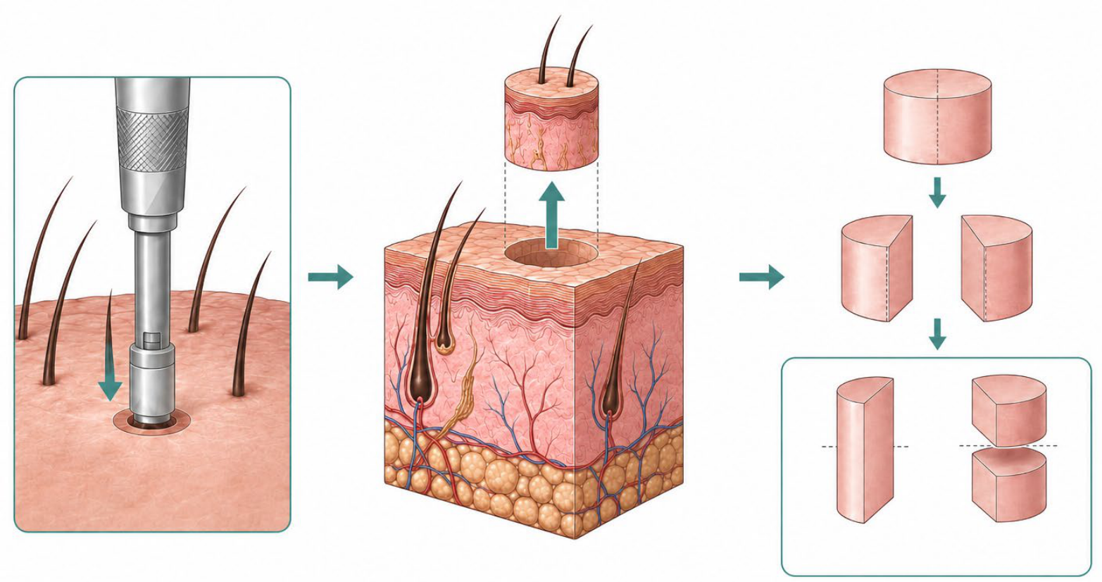
Figure 2.
(a) Horizontal (transverse) section (4x magnification). (b) Vertical (longitudinal) section of the same specimen. Hematoxylin and eosin (4x magnification).
Figure 2.
(a) Horizontal (transverse) section (4x magnification). (b) Vertical (longitudinal) section of the same specimen. Hematoxylin and eosin (4x magnification).
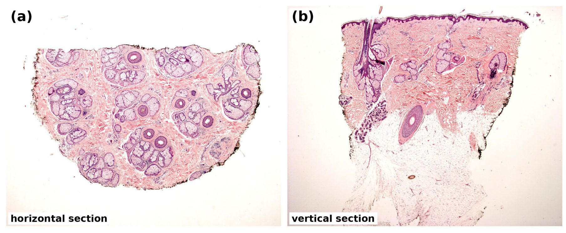
Figure 3.
Histopathology of nonscarring alopecia on horizontal sections. (a: 4x magnification; b: 10x magnification) Androgenetic alopecia: follicular miniaturization with increased vellus-like follicles and a reduced terminal-to-vellus ratio.(c: 4x magnification) Alopecia areata: preserved follicular units with increased catagen and telogen follicles; (d: 25x magnification) miniaturized anagen-vellus (“nanogen”) follicles. Hematoxylin and eosin.
Figure 3.
Histopathology of nonscarring alopecia on horizontal sections. (a: 4x magnification; b: 10x magnification) Androgenetic alopecia: follicular miniaturization with increased vellus-like follicles and a reduced terminal-to-vellus ratio.(c: 4x magnification) Alopecia areata: preserved follicular units with increased catagen and telogen follicles; (d: 25x magnification) miniaturized anagen-vellus (“nanogen”) follicles. Hematoxylin and eosin.
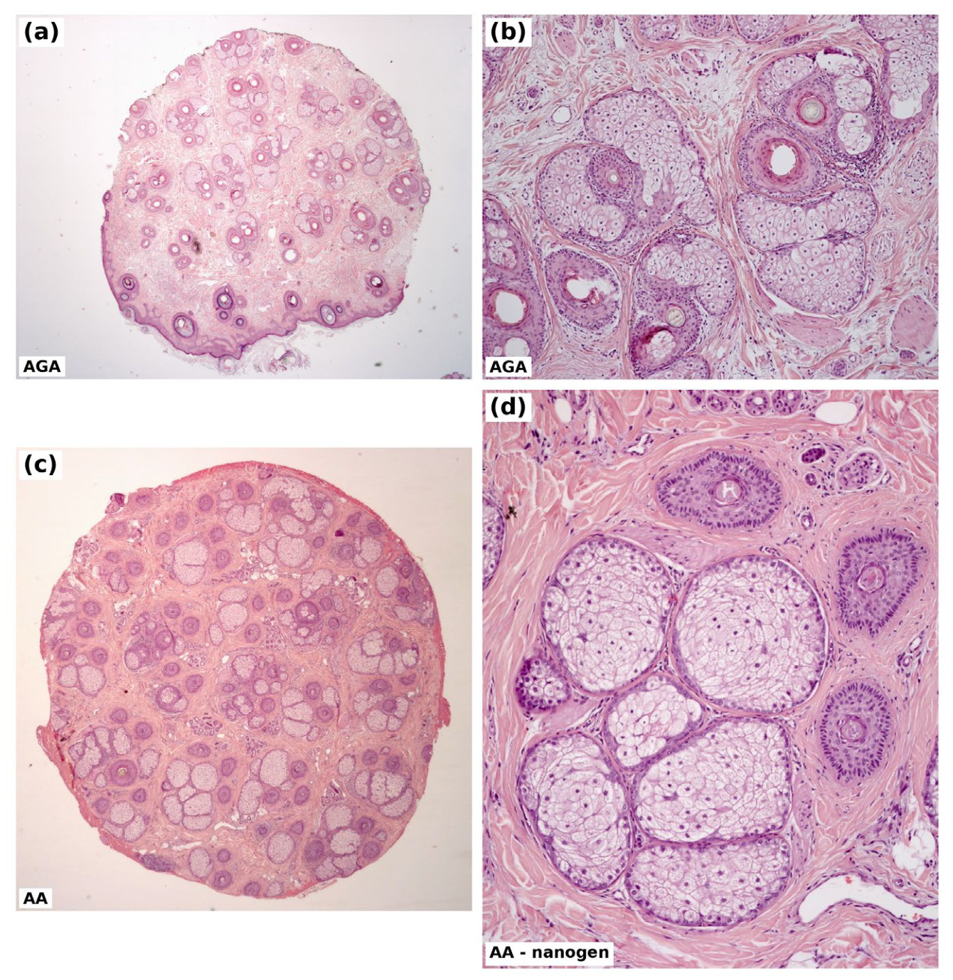
Figure 4.
Alopecia areata. Conventional histological features of alopecia areata. (a) Low-power view (H&E, 4×); the red box marks the peribulbar region enlarged in (b). (b) “Swarm of bees” pattern: mild-to-dense peribulbar lymphocytic infiltrate around the hair bulb (H&E, 30×). (c) Low-power view (H&E, 2.5×); the black box marks the region enlarged in (d). (d) Peribulbar lymphocytic infiltrate in the dermis (H&E, 25×).
Figure 4.
Alopecia areata. Conventional histological features of alopecia areata. (a) Low-power view (H&E, 4×); the red box marks the peribulbar region enlarged in (b). (b) “Swarm of bees” pattern: mild-to-dense peribulbar lymphocytic infiltrate around the hair bulb (H&E, 30×). (c) Low-power view (H&E, 2.5×); the black box marks the region enlarged in (d). (d) Peribulbar lymphocytic infiltrate in the dermis (H&E, 25×).
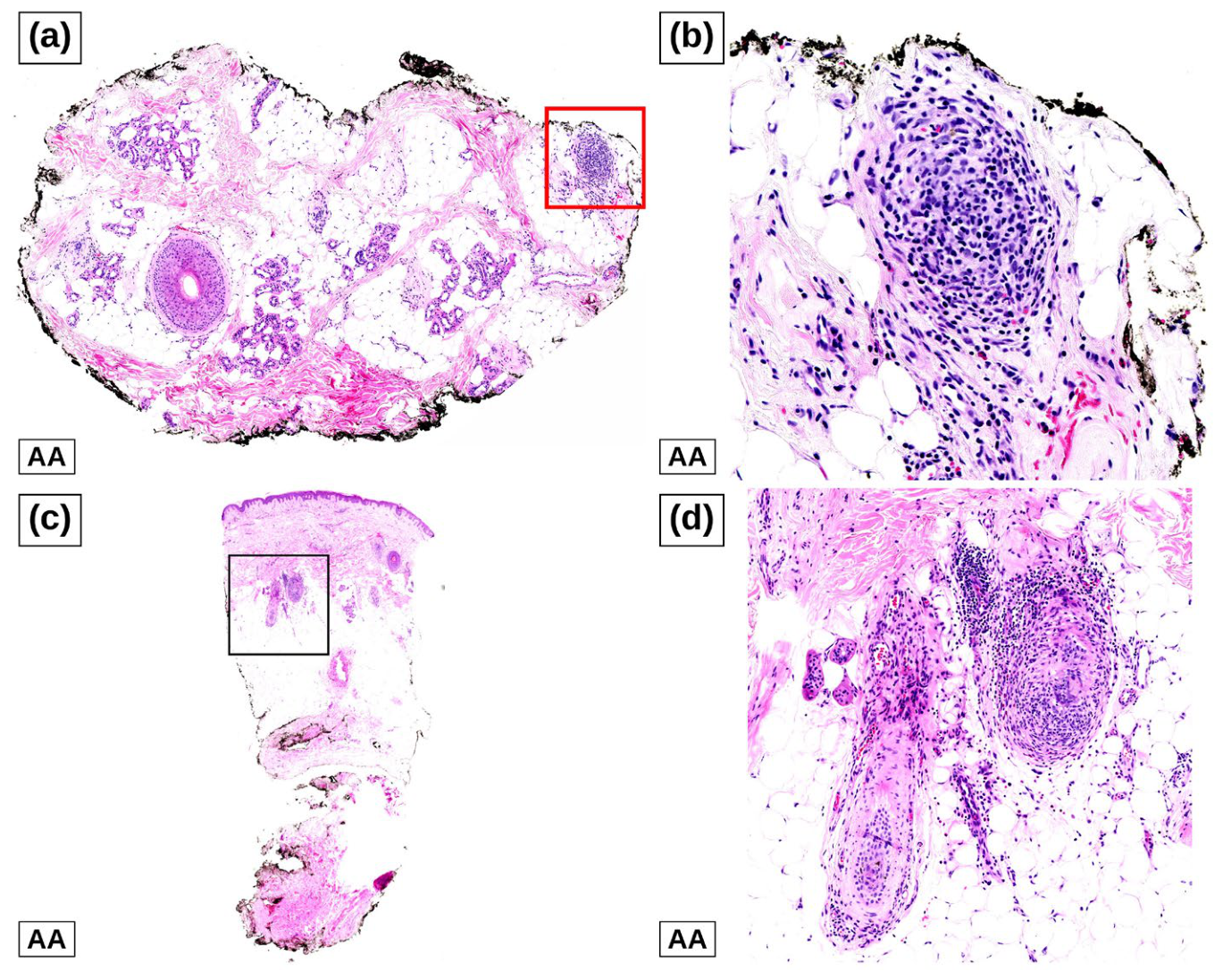
Figure 5.
Acute telogen effluvium (ATE). (a) Horizontal section, low-power view (H&E, 4×); the orange box marks the follicular unit enlarged in (b). (b) Increased number of telogen follicles within the follicular unit (H&E, 10×). (c) Vertical section, low-power view (H&E, 2.5×); the blue box marks the region enlarged in (d). (d) Two telogen follicles in the superficial and mid dermis, the peculiar histological aspect of ATE (H&E, 10×).
Figure 5.
Acute telogen effluvium (ATE). (a) Horizontal section, low-power view (H&E, 4×); the orange box marks the follicular unit enlarged in (b). (b) Increased number of telogen follicles within the follicular unit (H&E, 10×). (c) Vertical section, low-power view (H&E, 2.5×); the blue box marks the region enlarged in (d). (d) Two telogen follicles in the superficial and mid dermis, the peculiar histological aspect of ATE (H&E, 10×).
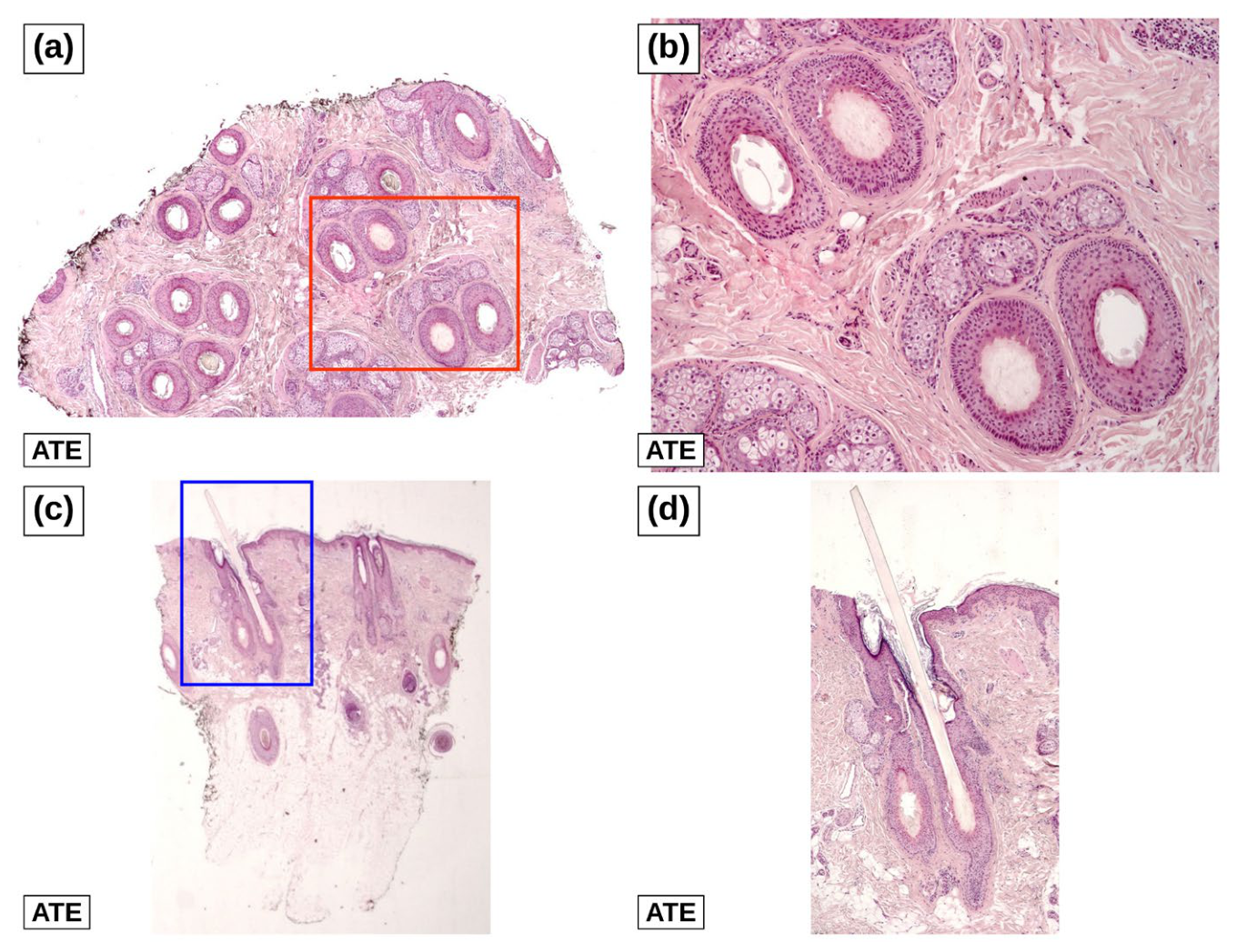
Figure 6.
Lichen planopilaris. (a; 4x magnification; b: 10x magnification) Horizontal sections showing a dense peri-isthmus lymphocytic infiltrate with perifollicular lamellar fibrosis and loss of sebaceous glands. (c: 25x magnification) Horizontal and (d: 10x magnification) vertical sections demonstrating lichenoid interface change at the infundibulum. Hematoxylin and eosin.
Figure 6.
Lichen planopilaris. (a; 4x magnification; b: 10x magnification) Horizontal sections showing a dense peri-isthmus lymphocytic infiltrate with perifollicular lamellar fibrosis and loss of sebaceous glands. (c: 25x magnification) Horizontal and (d: 10x magnification) vertical sections demonstrating lichenoid interface change at the infundibulum. Hematoxylin and eosin.
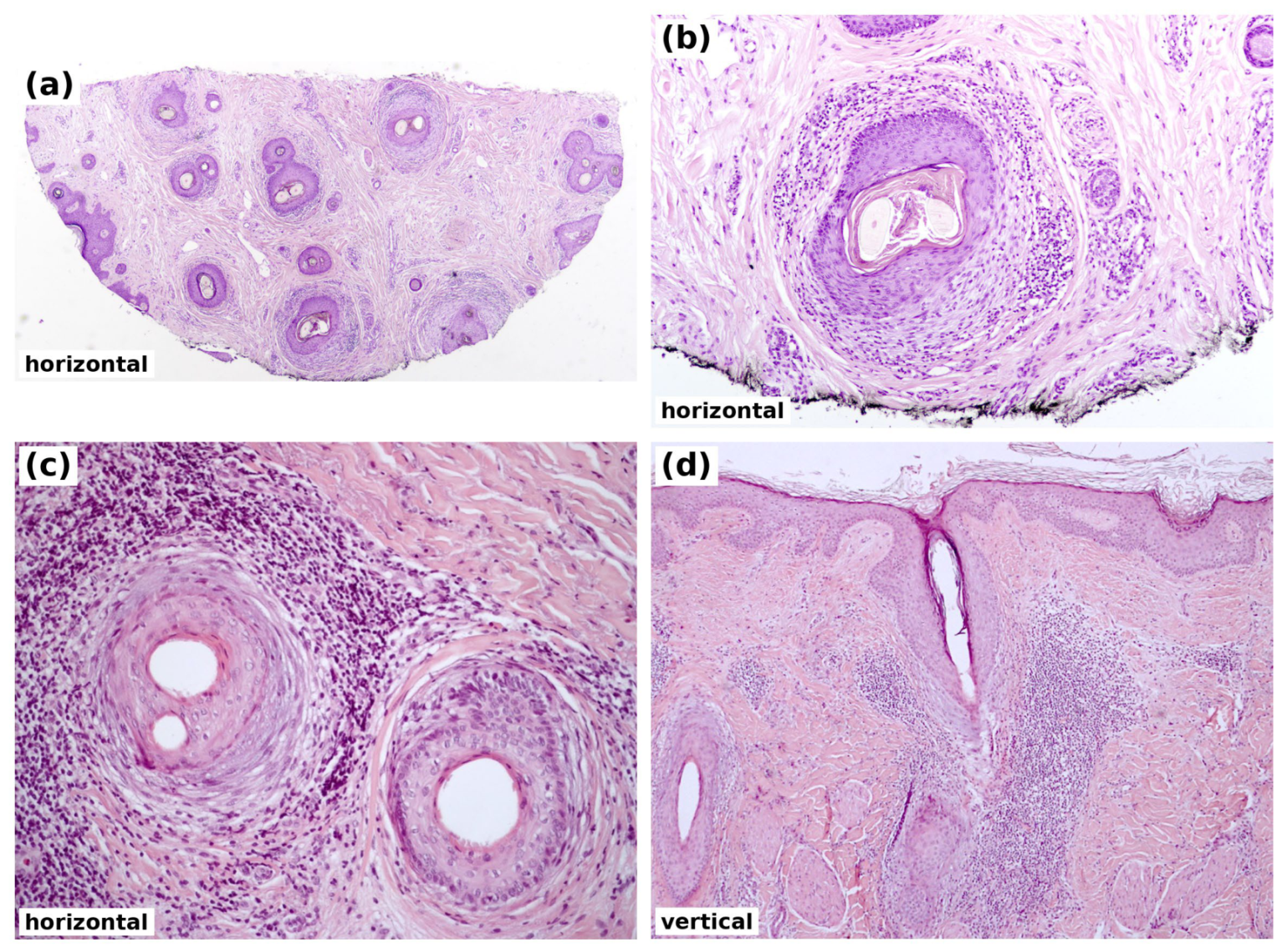
Figure 7.
Discoid lupus erythematosus. (a: 10x magnification; b: 4x magnification) Horizontal sections showing a dense peri-isthmus, peri-eccrine and perivascular lymphocytic infiltrate with vacuolar interface change at the dermoepithelial junction. (c: 40x magnification; d: 2,5x magnification) Vertical sections showing perifollicular and perivascular lymphocytic inflammation and infundibular dilation. Hematoxylin and eosin.
Figure 7.
Discoid lupus erythematosus. (a: 10x magnification; b: 4x magnification) Horizontal sections showing a dense peri-isthmus, peri-eccrine and perivascular lymphocytic infiltrate with vacuolar interface change at the dermoepithelial junction. (c: 40x magnification; d: 2,5x magnification) Vertical sections showing perifollicular and perivascular lymphocytic inflammation and infundibular dilation. Hematoxylin and eosin.
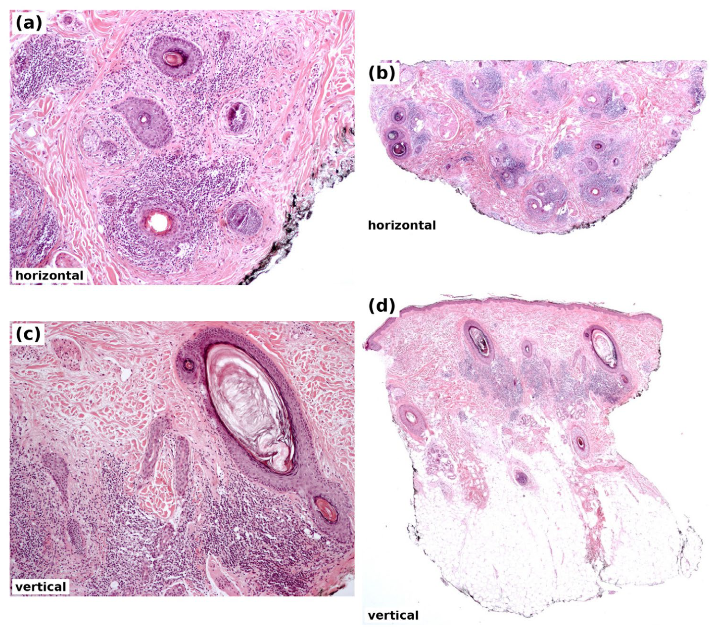
Figure 8.
Folliculitis decalvans. (a: 10 magnification; b: 4x magnification) Horizontal and (c: 40x magnification, d: 10x magnification) vertical sections showing a dense, diffuse suppurative and mixed inflammatory infiltrate with dermal fibrosis around isthmus-level follicles and follicular tufting (“tufted folliculitis”). Hematoxylin and eosin.
Figure 8.
Folliculitis decalvans. (a: 10 magnification; b: 4x magnification) Horizontal and (c: 40x magnification, d: 10x magnification) vertical sections showing a dense, diffuse suppurative and mixed inflammatory infiltrate with dermal fibrosis around isthmus-level follicles and follicular tufting (“tufted folliculitis”). Hematoxylin and eosin.
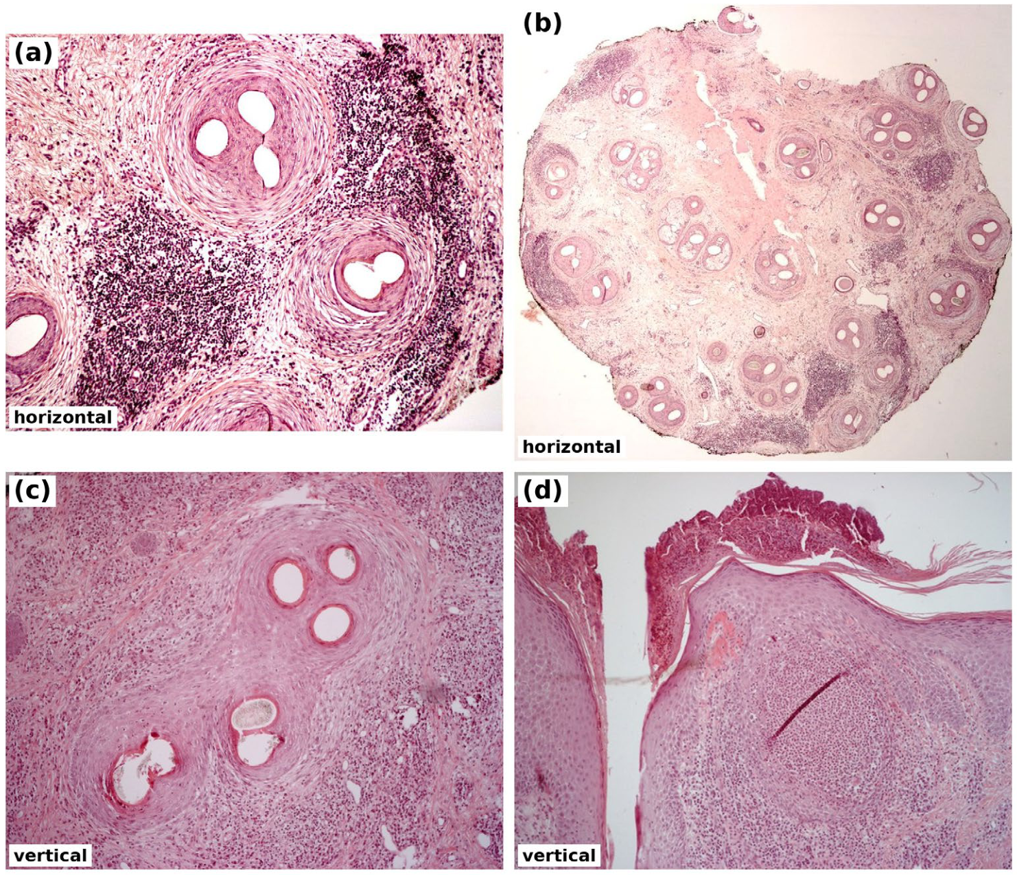
Figure 9.
Stepwise diagnostic algorithm for scalp biopsy in alopecia. After clinical history and trichoscopic assessment, biopsy is indicated when the diagnosis remains uncertain, when a scarring process cannot be excluded, or when systemic treatment requires histopathologic documentation. A 4-mm punch biopsy, oriented parallel to the direction of the hair shaft and extending into the subcutaneous fat, is processed by combined horizontal and vertical sectioning (HoVert or Tyler technique). Histopathologic findings distinguish scarring from nonscarring forms; in scarring alopecia, the predominant inflammatory cell type guides subclassification according to the NAHRS criteria. Ancillary techniques, including direct immunofluorescence, elastic fiber stains, PAS staining, and microbiologic cultures, are performed selectively when standard hematoxylin-eosin evaluation is inconclusive.
Figure 9.
Stepwise diagnostic algorithm for scalp biopsy in alopecia. After clinical history and trichoscopic assessment, biopsy is indicated when the diagnosis remains uncertain, when a scarring process cannot be excluded, or when systemic treatment requires histopathologic documentation. A 4-mm punch biopsy, oriented parallel to the direction of the hair shaft and extending into the subcutaneous fat, is processed by combined horizontal and vertical sectioning (HoVert or Tyler technique). Histopathologic findings distinguish scarring from nonscarring forms; in scarring alopecia, the predominant inflammatory cell type guides subclassification according to the NAHRS criteria. Ancillary techniques, including direct immunofluorescence, elastic fiber stains, PAS staining, and microbiologic cultures, are performed selectively when standard hematoxylin-eosin evaluation is inconclusive.
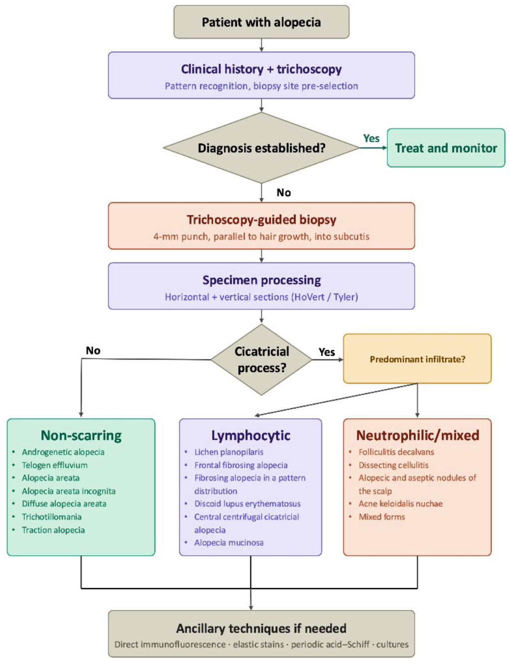

Table 1.
Recommended biopsy site according to alopecia type.
| Alopecia Type | Recommended Biopsy Site |
| Lichen planopilaris | Trichoscopy-guided area showing perifollicular concentric white scaling or casts, preferably within an active margin. |
| Frontal fibrosing alopecia | Trichoscopy-guided area of the frontal hairline showing perifollicular concentric white scaling or casts. |
| Discoid lupus erythematosus | Active trichoscopically abnormal area showing follicular red dots, keratotic plugs, or perifollicular concentric white scaling. |
| Folliculitis decalvans | Trichoscopy-guided area showing follicular tufting, preferably near an active pustular or inflammatory area. |
| Central centrifugal cicatricial alopecia | Trichoscopy-guided area showing a peripilar grey-white halo within or beyond the clinically apparent central involvement. |
| Alopecia areata | Area showing trichoscopic evidence of active disease, such as black dots, broken hairs, tapering or exclamation-mark hairs |
| Androgenetic alopecia / female pattern hair loss | Vertex or mid-scalp |
| Telogen effluvium | Vertex or mid-scalp |
| Alopecia areata incognita | Trichoscopy-guided area showing diffuse yellow dots, short regrowing hairs, or pigtail hairs. |
| Diffuse alopecia areata | Trichoscopy-guided area showing dystrophic hairs, black dots, broken hairs, or tapering hairs. |
| Fibrosing alopecia in a pattern distribution | Trichoscopy-guided area within the pattern alopecia showing perifollicular erythema, casts or scaling, loss of follicular openings, and miniaturization. |
| Traction alopecia | Area showing established hair loss at the clinically involved margin; in suspected end-stage disease, sample an area with residual follicles rather than completely smooth scarred scalp. |
| Trichotillomania | Clinically affected area showing hairs of different lengths, broken hairs, or other signs of recent traumatic activity. |
| Dissecting cellulitis of the scalp | Active inflammatory area at the margin of a nodule, abscess, or boggy plaque, avoiding a completely suppurated or end-stage scarred center. |
| Acne keloidalis nuchae | Active papular or pustular margin containing residual follicles, rather than the center of a mature keloidal plaque. |
| Multifactorial alopecia | Trichoscopy-guided watershed area in which the suspected processes overlap; for CCCA, FPHL, and traction alopecia, the frontotemporal aspect of the crown has been proposed |
Table 2.
Technical recommendations for scalp biopsy in alopecia.
| Parameter | Recommendation |
| Punch size | 4 mm |
| Depth | Into subcutaneous fat |
| Number of biopsies | One with Tyler technique |
| Orientation | Parallel to hair growth |
| Sectioning | Combined horizontal + vertical; with Tyler technique |
| Anesthesia | Lidocaine ± epinephrine |
| Closure | Simple interrupted sutures (removal at 10 days) |
Disclaimer/Publisher’s Note: The statements, opinions and data contained in all publications are solely those of the individual author(s) and contributor(s) and not of MDPI and/or the editor(s). MDPI and/or the editor(s) disclaim responsibility for any injury to people or property resulting from any ideas, methods, instructions or products referred to in the content. |
© 2026 by the authors. Licensee MDPI, Basel, Switzerland. This article is an open access article distributed under the terms and conditions of the Creative Commons Attribution (CC BY) license (http://creativecommons.org/licenses/by/4.0/).
Copyright: This open access article is published under a Creative Commons CC BY 4.0 license, which permit the free download, distribution, and reuse, provided that the author and preprint are cited in any reuse.


