

Submitted:
22 July 2026
Posted:
23 July 2026
You are already at the latest version
Abstract
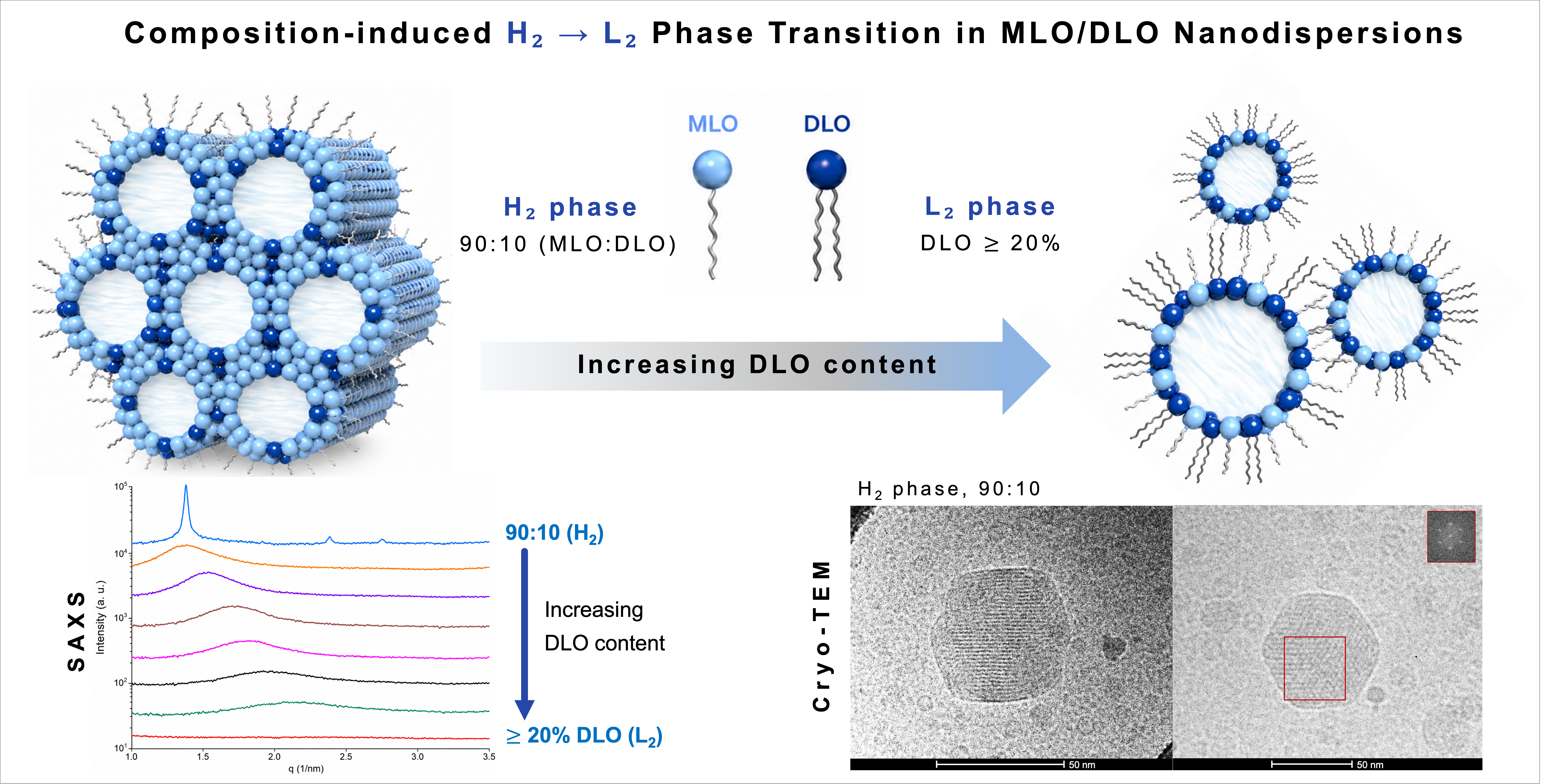
Nonlamellar liquid crystalline nanodispersions produced from single monoacylglycerols or from their combinations with fatty acids or other amphiphiles have attracted interest owing to their structural versatility and tunability. In this study, we investigated the effect of dilinolein (DLO) incorporation on the structural features of Pluronic F127-stabilized monolinolein (MLO) nanodispersions using small-angle X-ray scattering (SAXS), cryogenic transmission electron microscopy (cryo-TEM), and dynamic light scattering (DLS). We report on a lipid composition-dependent direct colloidal transformation from hexosomes, defined as nanoparticles with an ordered internal inverse hexagonal (H₂) phase, to emulsified L₂ phases (ELPs), which are nanoparticles with a more disordered internal inverse micellar (L₂) phase) upon the partial replacement of MLO by DLO. Small-angle X-ray scattering (SAXS) measurements showed that, at relatively low DLO content, the MLO-rich MLO:DLO 90:10 (w/w) nanodispersion ) retained an ordered internal H₂ phase, with three well-defined characteristic Bragg peaks; whereas the SAXS patterns recorded for all nanodispersions containing ≥ 20 wt% DLO displayed broad correlation peaks consistent with internal L₂ nanostructures. Similarly, the control nanodispersion prepared from DLO alone, displayed a single broad correlation peak, supporting its assignment as an internal L₂ phase. For the assigned internal L₂ phases, the SAXS-derived characteristic distance decreased monotonically from 4.53 to 2.87 nm as the DLO fraction increased. The experimental findings show that DLO modifies lipid packing at the MLO-water interface and promotes more negative spontaneous curvature, driving the direct H₂-to-L₂ phase transition. The ability to tune the internal nanostructure by lipid composition while maintaining nanoscale particle size and low dispersity makes these nanodispersions attractive platforms for drug nanocarrier development.
Keywords:
cryogenic transmission electron microscopy
; dilinolein
; hexosomes
; inverse hexagonal phase
; inverse micellar phase
; monolinolein
; synchrotron small-angle X-ray scattering
1. Introduction
Certain amphiphilic lipids can self-assemble in water into lyotropic liquid crystalline (LLC) phases, which have attracted long-standing interest in soft matter, colloid science, and drug delivery owing to their structural versatility and ability to encapsulate compounds with different physicochemical properties [1,2,3,4,5,6,7,8,9,10,11]. Depending on their molecular geometries, and experimental conditions, including hydration state, temperature, and pressure, these amphiphiles can self-organize into lamellar (Lα) or inverse nonlamellar phases, including bicontinuous cubic (Q2) and inverse hexagonal (H2) phases, or into inverse micellar solutions (L2 phases) [12,13,14,15,16]. These phases can be dispersed in excess water in the presence of a suitable stabilizer to produce colloidal internally self-assembled nanoobjects. Among these, cubosomes, hexosomes, and micellar cubosomes are the most extensively investigated nanodispersions [3,9,11,17]. Owing to their nanoscale dimensions and structurally versatile internal architectures with large interfacial areas, they are attractive platforms for drug and functional food delivery applications, where they may enhance the loading of bioactive compounds and modulate their release properties [9,16,18,19]. In addition to temperature, lipid type and composition play important roles in modulating the internal nanoarchitecture of cubosomes, hexosomes, micellar cubosomes, and related internally self-assembled nanodispersions by altering lipid packing, interfacial hydration, and the preferred spontaneous curvature of the lipid–water interface [20,21,22].
Monoacylglycerols (MAGs) remain among the most versatile amphiphiles of biological relevance for generating inverse nanostructures [16,21]. Monolinolein (MLO), a MAG containing a linoleoyl chain (C18:2), is structurally related to monoolein (MO), the most widely investigated monounsaturated monoacylglycerol with a strong propensity to form nonlamellar liquid-crystalline phases in excess water. Compared with fully hydrated MO, MLO undergoes similar phase transitions at lower temperatures, making it attractive for forming nonlamellar liquid crystalline phases and their fragmentation into internally structured nanodispersions under milder conditions [23,24,25]. Accordingly, MLO-based and related MAG nanodispersions have been investigated in biophysical studies, while their structurally tunable internal architectures make them attractive for drug delivery, functional food delivery, and biomedical applications [26,27]. Controlled changes in lipid composition may therefore provide a useful strategy for modulating the confined internal structural architectures of MLO-based nanoparticles.
Dilinolein (DLO), a diacylglycerol (DAG) containing two linoleoyl chains (C18:2), differs from MLO in its molecular packing and stronger tendency to promote negative spontaneous curvature at the lipid–water interface. Compared with MAGs, DAGs possess smaller effective headgroup areas and larger hydrophobic volumes, which favor inverse self-assembled structures in excess water [28,29,30,31]. In excess water, DAGs are known to form inverse micellar (L2) phases [20,32,33]. This behavior makes DLO a relevant lipid component for examining how DAG incorporation affects the confined internal phase behavior of MLO-based nanodispersions. Although temperature-induced phase transitions between internal inverse bicontinuous cubic phases and inverse discontinuous phases (including H2 and L2 nanostructures) have been reported for F127-stabilized MLO dispersions, composition-driven H2 - L2 transitions in binary MLO/DLO nanodispersions remain largely unexplored under controlled conditions. Combining MLO and DLO therefore provides a useful platform for probing how molecular-level asymmetry and dehydration influence colloidal organization. In binary systems, gradual substitution of MLO with DLO is expected to progressively shift the balance between water penetration, interfacial curvature, and lipid packing, potentially inducing transitions between ordered (H2) and disordered (L2) inverse structures. However, composition-driven H2 - L2 transitions in binary MLO/DLO nanodispersions remain largely unexplored under controlled conditions. Accordingly, the objective of this study was to investigate how systematic variation of the MLO:DLO ratio influences the internal nanostructure and colloidal behavior of these dispersions.
In this study, we report how DLO incorporation affects the internal phase behavior, morphology, and size of nanodispersions produced from binary MLO/DLO mixtures over a series of lipid compositions from MLO:DLO 90:10 to 30:70 (w/w), together with a control nanodispersion prepared from DLO alone, at fixed total lipid and Pluronic F127 concentrations. Synchrotron small-angle X-ray scattering (SAXS) and cryogenic transmission electron microscopy (cryo-TEM) were used as complementary characterization techniques. Together, the results revealed a composition-dependent colloidal transformation from hexosomes to emulsified L2-phase (ELP) nanodispersions. These findings provide insight into how DLO incorporation modulates the confined internal phase behavior of MLO-based nanodispersions. Such composition-guided control of internal nanostructure may support the rational design of structurally tunable lipid nanocarriers for drug delivery.
2. Results and Discussion
2.1. Structural Characterization of Pluronic F127-Stabilized MLO/DLO Nano-Self-Assemblies
Synchrotron SAXS was used to examine how progressive replacement of MLO by DLO affects the confined internal self-assembled architectures of the nanodispersions. The molecular structures of MLO with DLO are presented in Figure 1A. At the highest MLO content, the scattering profile of the MLO:DLO 90:10 (w/w) nanodispersion (sample MD1) displayed three characteristic Bragg peaks [(100), (110), and (200), marked with red arrows in Figure 1B] with relative peak positions following the characteristic 1:√3:√4 ratio of an inverse hexagonal (H2) phase indicating the formation of hexosomes. The corresponding lattice parameter of this internal H2 phase, calculated from the position of the first-order Bragg peak, was approximately 5.27 nm. The calculated lattice parameter is consistent with values previously reported for Pluronic F127-stabilized MLO-based hexosomes [34,35]. The sharpness and relative intensity of the Bragg reflections suggest that MLO-rich binary lipid mixture provides a sufficiently hydrated and flexible interfacial environment to stabilize this inverse phase [23].
Increasing the DLO fraction induced a composition-dependent colloidal transformation from hexosomes, characterized by an ordered internal H2 phase, to emulsified L2-phase nanodispersions (Figure 1B). In all nanodispersions containing ≥20 wt% DLO (samples MD2-MD10) , the Bragg peaks assigned to the H2 phase disappeared and were replaced by a single broad correlation peak, consistent with an inverse micellar (L2) phase lacking long-range periodicity. The SAXS profile of the control nanodispersion based on DLO alone (sample MD10, MLO:DLO 0:100, w/w) also displayed a single broad correlation peak, supporting its assignment as an internal L2 phase. The position of the correlation maximum systematically shifted toward higher q values as the fraction of DLO increased, leading to a monotonic decrease in the SAXS-derived characteristic distance, calculated as d=2π/qmax, from 4.53 nm for the MLO:DLO 80:20 (w/w) nanodispersion to 2.87 nm for the MLO:DLO 30:70 (w/w) nanodispersion. As summarized in Figure 1C, this decrease follows an approximately linear trend (R2=0.9879) with increasing DLO content, indicating a progressive reduction in the characteristic length scale of the internal L2 phase.
The monotonic decrease in the SAXS-derived characteristic distance of the internal L2 phases with increasing DLO content is most likely associated with reduced water accommodation within the internal nanostructure. Similar water expulsion has been reported during temperature-induced colloidal transformations of Pluronic F127-stabilized MLO nanodispersions from cubosomes and hexosomes toward emulsified L2 phase nanodispersions [26,34].
The pronounced impact of DLO on the structural features of MLO nanodispersions can be understood in terms of its molecular packing characteristics. DLO is a diacylglycerol containing two linoleoyl chains and a comparatively small effective polar headgroup area (Figure 1A). Relative to MLO, the additional linoleoyl chain in DLO increases the hydrophobic volume relative to the polar headgroup area, thereby shifting the self-assembled nanostructure in excess water toward stronger inverse spontaneous curvature. DLO is expected to possess a relatively large packing parameter typical of diacylglycerol-like lipids, which are known to promote negative interfacial curvature and nonlamellar organizations [28]. The pronounced structural impact of DLO can be understood in terms of its molecular packing characteristics. DLO is a diacylglycerol containing two linoleoyl chains and a comparatively small polar headgroup region (Figure 1A). Consistent with the reported phase behavior of other diglycerides, this molecular packing favors the formation of inverse micellar L2 phases under fully hydrated conditions [36,37,38].
The critical packing parameter (CPP), also referred to as the molecular shape factor, is commonly expressed as CPP = v/(a.l), where v is the hydrophobic chain volume, a is the effective headgroup area, and l is the hydrophobic chain length of the amphiphile [20]. CPP provides a useful framework for describing lipid self-assembly behavior and spontaneous curvature tendencies. Amphiphilic lipids with CPP values significantly greater than unity generally favor inverse self-assembled nanostructures; however, the specific phase depends on the magnitude of the negative spontaneous curvature of the lipid–water interface. Moderate negative curvature can support inverse bicontinuous cubic and discontinuous H2 phases, whereas stronger negative spontaneous curvature, together typically with reduced water accommodation, favors discontinuous inverse micellar nanostructures, including inverse micellar cubic Fd3m phases and inverse micellar L2 phases [22,39]. Accordingly, alterations in lipid composition can modulate the effective CPP of mixed surfactant systems and thereby influence their preferred mesophase structure. Accordingly, increasing the DLO fraction in MLO-based nanodispersions is expected to increase the effective CPP and promote more negative spontaneous curvature at the lipid–water interface, thereby favoring a direct H2-L2 phase transition [40].
The observed hexosome-to-emulsified L2-phase transformation is consistent with earlier studies on F127-stabilized MLO-based nanodispersions [26,34]. Temperature-induced transitions from cubosomes to hexosomes and finally to emulsified L2-phase nanodispersions have been reported for MLO nanodispersions, while solubilization of tetradecane in Plurnoic F127-stabilized MLO nanodispersions was also shown to promote colloidal transformations from cubosomes to hexosomes and then to emulsified L2-phase nanodispersions. Although both tetradecane and DLO can shift the internal self-assembled nanostructures of MLO-based nanodispersions toward internal L2 phases, they are expected to act through different modes of incorporation. Tetradecane, as a purely hydrophobic additive, tends to localize within the hydrophobic domains of the internal self-assembled nanostructure, whereas DLO, as an amphiphilic compound, is expected to intercalate into the MLO-water interfacial region. Thus, DLO changes the amphiphile composition of the interfacial film itself, increasing the effective CPP and promoting more negative spontaneous curvature at the lipid-water interface. This provides a composition-induced pathway toward internal L2 phases that is analogous to, but distinct from, hydrophobic-additive-induced colloidal transformations in Pluronic F127-stabilized MLO/tetradecane nanodispersions. A schematic illustration of the proposed composition-dependent H2-to-L2 transition is presented in Figure 3, highlighting the role of increasing DLO content in modulating the structural features of Pluronic F127 MLO nanodispersions.
At certain lipid compositions, the colloidal transformations from cubosomes via hexosomes to emulsified L2 phase nanodisperisons may also proceed through micellar cubosomes (nanoparticles with an internal inverse micellar cubic Fd3m phase) [35]. This discontinous (micellar) cubic phase is typically observed within narrow temperature- or composition-dependent regions in the phase diagrams of systems based on different lipids with nonlamellar phase-forming propensities [35,39,41]. In the present MLO/DLO nanodispersion, however, no intermediate cubic Fd3m phase was detected; instead, the SAXS profiles at the investigated lipid compositions indicated a composition-dependent transformation from hexosomes directly to emulsified L2-phase nanodispersions. For example, Yaghmur et al. demonstrated that incorporation of tetradecane into MLO systems induced transformations from ordered cubic and hexagonal mesophases toward disordered inverse micellar structures as interfacial curvature increased [15], showing a structural trend similar to the composition-dependent H2 - L2 transition observed in the present MLO/DLO system.
Figure 2.
Schematic illustration of the H2-L2 phase transition in MLO/DLO-based systems upon increasing DLO content. Increased DLO content is associated with reduced interfacial hydration, leading to the transformation of continuous water cylinders characteristic of the inverse hexagonal (H2) phase into discrete reverse micelles characteristic of the inverse micellar (L2) phase.
Figure 2.
Schematic illustration of the H2-L2 phase transition in MLO/DLO-based systems upon increasing DLO content. Increased DLO content is associated with reduced interfacial hydration, leading to the transformation of continuous water cylinders characteristic of the inverse hexagonal (H2) phase into discrete reverse micelles characteristic of the inverse micellar (L2) phase.
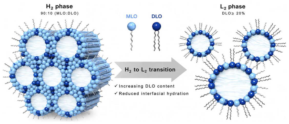
Figure 3.
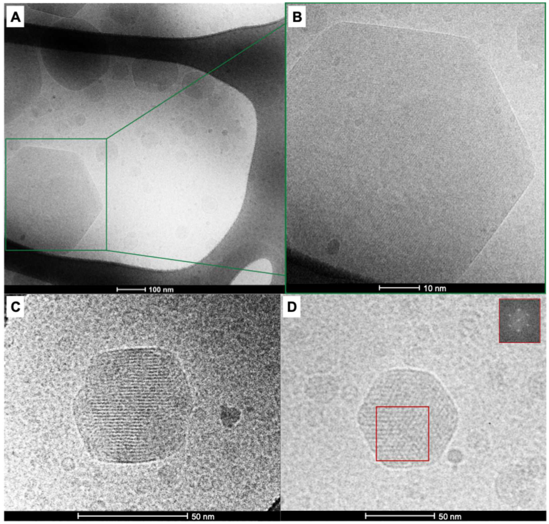
Cryo-TEM characterization and FFT analysis of the MLO-rich MLO/DLO nanodispersion (sample MD1), prepared at an MLO:DLO weight ratio of 90:10 (w/w). (A) Low-magnification overview image showing faceted, polygonal nanoparticles characteristic of hexosomes. The green boxed region is enlarged in (B), revealing the internal structural features of the particle. (C) High-magnification image showing well-defined lattice fringes appearing as curved striations, consistent with locally ordered cylindrical domains in the internal H2 phase. (D) Representative particle selected for fast Fourier transform (FFT) analysis. The red square indicates the region selected for FFT, and the corresponding inset shows discrete diffraction spots with hexagonal symmetry, from which a real-space periodicity of approximately 4.3 nm was estimated, in reasonable agreement with the first-order H2 periodicity determined by SAXS.
Figure 3.
Cryo-TEM characterization and FFT analysis of the MLO-rich MLO/DLO nanodispersion (sample MD1), prepared at an MLO:DLO weight ratio of 90:10 (w/w). (A) Low-magnification overview image showing faceted, polygonal nanoparticles characteristic of hexosomes. The green boxed region is enlarged in (B), revealing the internal structural features of the particle. (C) High-magnification image showing well-defined lattice fringes appearing as curved striations, consistent with locally ordered cylindrical domains in the internal H2 phase. (D) Representative particle selected for fast Fourier transform (FFT) analysis. The red square indicates the region selected for FFT, and the corresponding inset shows discrete diffraction spots with hexagonal symmetry, from which a real-space periodicity of approximately 4.3 nm was estimated, in reasonable agreement with the first-order H2 periodicity determined by SAXS.
Overall, these findings demonstrate that DLO incorporation strongly modulates the confined internal self-assembled architecture of Pluronic F127-stabilized MLO-based nanodispersions, enabling a lipid composition-driven tranformation from hexosomes to emulsified L2-phase nanodispersions. This lipid compositional sensitivity provides a structural basis for designing nonlamellar liquid crystalline nanocarriers with composition-controlled internal architectures, where designated colloidal transformations between different internal inverse phases may alter the connectivity and dimensions of aqueous and hydrophobic nanodomains and thereby influence drug-loading capacity and release behavior.
2.2. Morphological Characteristics of MLO/DLO Nano-Self-Assemblies
Cryo-TEM was used as a direct imaging method to gain insight into the nanoparticle morphology and the internal self-assembled architecture of a selected MLO/DLO nanodispersions, complementing the SAXS analysis. In agreement with the SAXS analysis, the micrographs of the MLO-rich nanodispersion (sample MD1), prepared at an MLO:DLO weight ratio of 90:10 (w/w), displayed faceted, polygonal nanoparticles with clearly resolved typical internal structural features of internal H2 phase (Figure 3). These colloidal objects exhibited alternating dark and bright striations arranged over locally ordered domains, consistent with projections of periodically organized cylindrical domains in the internal H2 phase and supporting their assignment as hexosomes [9].
To further support the local assignment of the internal H2 nanostructure, fast Fourier transform (FFT) analysis was performed on a selected high-resolution area within the individual nanoparticle (Figure 3). The corresponding FFT patterns showed discrete maxima arranged with approximate hexagonal symmetry, confirming the presence of locally ordered domains. The real-space periodicity estimated from the FFT analysis was approximately 4.3 nm, in reasonable agreement with the first-order H2 periodicity derived from SAXS, d100 ≈ 4.5 nm. This agreement between real-space FFT analysis and reciprocal-space SAXS measurement supports the assignment of sample MD1 as hexosomes.
In addition to the dominant hexosome population, a minor coexisting fraction of vesicle-like spherical structures of varying size were observed in different cryo-TEM images (marked with white arrows, Supplementary Figure S1). Such coexisting colloidal nanoobjects reflect the inherent morphological heterogeneity of nonlamellar lipid nanodispersions, where nanoparticle subpopulations with different morphologies and internal architectures may coexist depending on lipid type and composition, stabilizer type and concentration, and preparation conditions [9,16,42].
2.3. Particle Size Analysis of MLO/DLO
DLS was used to determine the hydrodynamic diameter (size) and size distribution, expressed as the polydispersity index (PDI), of MLO/DLO nanodispersions prepared at different lipid compositions (Table 1). The 90:10 MLO:DLO nanodispersion displayed the largest mean hydrodynamic diameter, 288.53 ± 21.39 nm, together with the highest PDI value, 0.31 ± 0.01. Progressive replacement of MLO by DLO led to smaller nanodispersions with lower PDI values, indicating that the size characteristics were strongly affected by lipid composition.
The decrease in mean hydrodynamic nanoparticle size was most pronounced upon increasing the DLO content from 10 to 50 wt%. The mean nanoparticle size decreased from 288.53 ± 21.39 nm for the 90:10 MLO:DLO nanodispersion to 201.73 ± 1.78 nm for the 80:20 MLO:DLO nanodispersion and reached values of approximately 157-164 nm for compositions between 50:50 and 30:70 MLO:DLO. These changes in size characteristics are most likely associated with the colloidal transformations described above, where progressive replacement of MLO by DLO induced colloidal transformation from hexosomes to emulsified L2-phase nanodispersions. At higher DLO contents, the mean hydrodynamic nanoparticle size moderately increased, reaching 169.07 ± 2.46 nm for the 20:80 MLO:DLO nanodispersion, 180.90 ± 1.04 nm for the 10:90 MLO:DLO nanodispersion, and 199.25 ± 0.21 nm for the nanodispersion produced from DLO alone.
The PDI values were relatively low for most DLO-containing nanodispersions, ranging from 0.06 to 0.19, compared with 0.31 ± 0.01 for the 90:10 MLO:DLO nanodispersion. Overall, the DLS results show that progressive replacement of MLO by DLO altered the mean hydrodynamic nanoparticle sizes of MLO-based nanodispersions while maintaining nanoparticle sizes below 300 nm under the investigated experimental conditions.
3. Materials and Methods
3.1. Materials
Monolinolein (MLO, with a purity of >99%) and dilinolein (DLO, with a purity of >99%) were purchased from Larodan (Stockholm, Sweden). Pluronic F127 was a gift from BASF SE (Ludwigshafen, Germany). Phosphate-buffered saline (PBS, 10 mM) at pH 7.4 was prepared using Milli-Q water (Millipore Direct-Q3 UV system, Billerica, MA, USA), sodium chloride, potassium chloride, dibasic sodium phosphate, and monobasic potassium phosphate from Sigma-Aldrich (St. Louis, MO, USA). These ingredients were of analytical grade and used as received without further purification.
3.2. Preparation of MLO/DLO-Based Aqueous Nanodispersions
MLO/DLO-based aqueous nanodispersions were prepared at a constant total lipid content of 5.0 wt% (binary mixture of MLO and DLO) and 1.0 wt% Pluronic F127. A series of formulations was prepared at MLO:DLO weight ratios ranging from 0:100 to 100:0 in 10% increments. Briefly, the required amounts of MLO and DLO were mixed thoroughly in glass vials at 25 °C to obtain homogeneous binary lipid mixtures. Each mixture was then dispersed in phosphate-buffered saline (PBS, 10 mM, pH 7.4) containing 1.0 wt% Pluronic F127. The emulsification process was performed using an ultrasonic processor (Qsonica 500, Qsonica LLC, Newtown, CT, USA) operating at 30% of its maximum power for 5 min in pulse mode (5 s pulses with 2 s breaks) as previously described [21]. No pH adjustment was applied during or after emulsification. The freshly prepared dispersions were flushed with nitrogen gas for 2 min to minimize oxidation, sealed, covered with aluminum foil, and stored at 25 °C until further analysis. No macroscopic phase separation or aggregation was visually observed during storage over at least one month at 25 °C.
3.3. Synchrotron Small-Angle X-Ray Scattering (SAXS)
The structural characterization of MLO/DLO-based nanodispersions was carried out at the Austrian SAXS beamline of the ELETTRA synchrotron light source (Trieste, Italy). Measurements were performed at 25 °C using an X-ray beam with a wavelength of 1.54 Å and an energy of 8 keV. The sample-to-detector distance was set to 1314 mm, covering a q-range of 0.07–5.0 nm−1 (q = 4π sin (θ) / λ). Silver behenate (CH3–(CH2)20–COOAg; d = 58.38 Å) was used as a calibration standard. The two-dimensional SAXS patterns were collected using a Pilatus 1 M detector (Dectris Ltd., Baden, Switzerland) and radially integrated into one-dimensional scattering profiles I(q) using Fit2D software.
Lorentzian fitting was applied to determine the q-values of the detected Bragg reflections. The d-spacing values were calculated from the SAXS reflections using the equation d = 2π / q.
3.4. Cryo-Transmission Electron Microscopy (Cryo-TEM)
Cryo-TEM analysis was performed on the MLO-rich formulation (90:10 MLO:DLO, w/w), which was selected based on the SAXS results showing the presence of a well-defined inverse hexagonal (H2) phase. The morphology of the nanodispersion was examined under frozen-hydrated conditions as previously described [38,43]. Briefly, 3-4 μL of each nanodispersion was deposited onto a lacey carbon 300-mesh copper grid (Ted Pella Inc., California, USA) that had been glow-discharged prior to use. The excess liquid was blotted using a Vitrobot IV system (FEI, Holland) with a blotting time of 5 s, blotting force 0, and 100% humidity at 4 °C. The grids were rapidly plunged into liquid ethane cooled by liquid nitrogen (−180 °C) to achieve vitrification.
Cryo-TEM imaging was performed using a Tecnai G2 20 transmission electron microscope (FEI, Holland) operated at 200 kV under low-dose conditions (~5 e-/Å2). A Gatan 626 cryo-holder (Gatan, UK) was used to maintain the vitrified state during imaging. The digital micrographs were analyzed using ImageJ software (National Institutes of Health, MD, USA), and fast Fourier transform (FFT) analyses were performed on selected regions to evaluate internal nanostructural ordering within the particles.
3.5. Dynamic Light Scattering (DLS)
The hydrodynamic diameter and polydispersity index (PDI) of the MLO/DLO-based nanodispersions were determined by dynamic light scattering (DLS) using a Zetasizer Nano ZS90 (Malvern Panalytical Ltd., Malvern, UK) equipped with a 4 mW He-Ne laser operating at 633 nm and a fixed scattering angle of 90°. Measurements were performed at 25 °C after diluting the samples 1:10 (v/v) with phosphate-buffered saline (PBS, 10 mM, pH 7.4). Each sample was measured in triplicate, and the results were expressed as mean ± standard deviation (SD).
4. Conclusions
MLO/DLO-based aqueous nanodispersions provide a simple compositionally tunable platform for internally generating self-assembled lipid nanoparticles with internal H2 or L2 internal architectures. Systematic variation of the lipid composition enabled modulation of the confined internal self-assembled architecture of the dispersed nanoparticles. This compositional modulation enabled a direct colloidal transformation from hexosomes to emulsified L2-phase nanodispersions, demonstrating that the confined self-assembled architecture and hydrodynamic size characteristics of MLO-based nanodispersions are sensitive to DLO incorporation. The structural effect of DLO is likely related to its amphiphilic DAG nature and its incorporation into the MLO-water interfacial area, where interactions with MLO alter the local packing arrangement and decrease water accommodation within the internal nanostructure.
Overall, these findings show that relatively small changes in MAG/DAG composition can strongly influence the internal nanostructure and colloidal characteristics of lipid nanodispersions. This composition-dependent control provides a practical route for tailoring internally self-assembled lipid nanocarriers through lipid molecular design rather than external triggers such as temperature variation.
From a nanocarrier design perspective, these findings highlight lipid composition as a useful design variable for directing colloidal transformations in self-assembled lipid nanodispersions. The MLO/DLO nanodispersion therefore offers a model platform for studying how changes in lipid composition influence nanoparticle self-assembled architecture. Such composition-guided tuning may support the design of lipid nanocarriers with tunable internal structural features for modulating drug loading and release behavior.
Supplementary Materials
The following supporting information can be downloaded at the website of this paper posted on Preprints.org.
Author Contributions
Conceptualization, G.D.K.; methodology, G.D.K.; SAXS analysis, G.D.K.; cryo-TEM analysis, G.D.K.; DLS analysis, G.D.K.; investigation, G.D.K.; writing-original draft preparation, G.D.K.; writing-review and editing, G.D.K.
Funding
The author acknowledges financial support from TÜBİTAK 2219-International Postdoctoral Research Fellowship Program (Project No: 1059B192203132) during a research stay.
Informed Consent Statement
Not applicable.
Data Availability Statement
The data presented in this study are available from the corresponding author upon reasonable request.
Acknowledgments
The author gratefully acknowledges Prof. Anan Yaghmur for providing access to experimental facilities, for valuable scientific discussions, and support during the work. The author also thanks the staff at the ELETTRA synchrotron facility for their support during SAXS measurements.
Conflicts of Interest
The author declare no conflicts of interest.
References
- Angelova, A.; Angelov, B.; Drechsler, M.; Bizien, T.; Gorshkova, Y.E.; Deng, Y. Plasmalogen-Based Liquid Crystalline Multiphase Structures Involving Docosapentaenoyl Derivatives Inspired by Biological Cubic Membranes. Front. Cell Dev. Biol. 2021, Volume 9 - 2021. [Google Scholar] [CrossRef]
- Angelova, A.; Garamus, V.M.; Angelov, B.; Tian, Z.; Li, Y.; Zou, A. Advances in structural design of lipid-based nanoparticle carriers for delivery of macromolecular drugs, phytochemicals and anti-tumor agents. Adv. Colloid Interface Sci. 2017, 249, 331–345. [Google Scholar] [CrossRef]
- Azmi, I.D.M.; Moghimi, S.M.; Yaghmur, A. Cubosomes and Hexosomes As Versatile Platforms for Drug Delivery. Ther. Deliv. 2015, 6, 1347–1364. [Google Scholar] [CrossRef] [PubMed]
- Azmi, I.D.M.; Østergaard, J.; Stürup, S.; Gammelgaard, B.; Urtti, A.; Moghimi, S.M.; Yaghmur, A. Cisplatin Encapsulation Generates Morphologically Different Multicompartments in the Internal Nanostructures of Nonlamellar Liquid-Crystalline Self-Assemblies. Langmuir 2018, 34, 6570–6581. [Google Scholar] [CrossRef] [PubMed]
- Azmi, I.D.M.; Wibroe, P.P.; Wu, L.-P.; Kazem, A.I.; Amenitsch, H.; Moghimi, S.M.; Yaghmur, A. A structurally diverse library of safe-by-design citrem-phospholipid lamellar and non-lamellar liquid crystalline nano-assemblies. J. Control. Release. 2016, 239, 1–9. [Google Scholar] [CrossRef] [PubMed]
- Bor, G.; Mat Azmi, I.D.; Yaghmur, A. Nanomedicines for Cancer Therapy: Current status, Challenges and Future Prospects. Ther. Deliv. 2019, 10, 113–132. [Google Scholar] [CrossRef] [PubMed]
- Fong, W.-K.; Negrini, R.; Vallooran, J.J.; Mezzenga, R.; Boyd, B.J. Responsive self-assembled nanostructured lipid systems for drug delivery and diagnostics. J. Colloid Interface Sci. 2016, 484, 320–339. [Google Scholar] [CrossRef] [PubMed]
- Murgia, S.; Biffi, S.; Mezzenga, R. Recent advances of non-lamellar lyotropic liquid crystalline nanoparticles in nanomedicine. Curr. Opin. Colloid Interface Sci. 2020, 48, 28–39. [Google Scholar] [CrossRef]
- Yaghmur, A.; Mu, H. Recent advances in drug delivery applications of cubosomes, hexosomes, and solid lipid nanoparticles. Acta Pharm. Sin. B. 2021, 11, 871–885. [Google Scholar] [CrossRef] [PubMed]
- Yu Helvig, S.; Woythe, L.; Pham, S.; Bor, G.; Andersen, H.; Moein Moghimi, S.; Yaghmur, A. A structurally diverse library of glycerol monooleate/oleic acid non-lamellar liquid crystalline nanodispersions stabilized with nonionic methoxypoly(ethylene glycol) (mPEG)-lipids showing variable complement activation properties. J. Colloid Interface Sci. 2021, 582, 906–917. [Google Scholar] [CrossRef] [PubMed]
- Zhai, J.; Fong, C.; Tran, N.; Drummond, C.J. Non-Lamellar Lyotropic Liquid Crystalline Lipid Nanoparticles for the Next Generation of Nanomedicine. ACS Nano 2019, 13, 6178–6206. [Google Scholar] [CrossRef] [PubMed]
- Caffrey, M. A comprehensive review of the lipid cubic phase or in meso method for crystallizing membrane and soluble proteins and complexes. Acta Crystallogr. F. Struct. Biol. Commun. 2015, 71, 3–18. [Google Scholar] [CrossRef] [PubMed]
- Seddon, J.M.; Templer, R.H. Chapter 3 - Polymorphism of Lipid-Water Systems. In Handbook of Biological Physics; Lipowsky, R., Sackmann, E., Eds.; North-Holland, 1995; Volume 1, pp. 97–160. [Google Scholar]
- Luzzati, V.; Husson, F. The structure of the liquid-crystalline phasis of lipid-water systems. J. Cell Biol. 1962, 12, 207–219. [Google Scholar] [CrossRef] [PubMed]
- Yaghmur, A.; de Campo, L.; Sagalowicz, L.; Leser, M.E.; Glatter, O. Control of the Internal Structure of MLO-Based Isasomes by the Addition of Diglycerol Monooleate and Soybean Phosphatidylcholine. Langmuir 2006, 22, 9919–9927. [Google Scholar] [CrossRef] [PubMed]
- Yaghmur, A.; Glatter, O. Characterization and potential applications of nanostructured aqueous dispersions. Adv. Colloid Interface Sci. 2009, 147-148, 333–342. [Google Scholar] [CrossRef] [PubMed]
- Fornasier, M.; Murgia, S. Non-lamellar lipid liquid crystalline nanoparticles: A smart platform for nanomedicine applications. Front. Soft Matter 2023, Volume 3 - 2023. [Google Scholar] [CrossRef]
- Amar-Yuli, I.; Libster, D.; Aserin, A.; Garti, N. Solubilization of food bioactives within lyotropic liquid crystalline mesophases. Curr. Opin. Colloid Interface Sci. 2009, 14, 21–32. [Google Scholar] [CrossRef]
- Eskandari, Z.; Hosseini, S.F.; Kalaycioglu, G.D.; Yaghmur, A. Layer-by-layer hybrid hexosomes with enhanced oxidative stability and bioaccessibility of omega-3 polyunsaturated fatty acids. Food Chem. 2025, 489, 145050. [Google Scholar] [CrossRef] [PubMed]
- Israelachvili, J.N.; Mitchell, D.J.; Ninham, B.W. Theory of self-assembly of lipid bilayers and vesicles. Biochim. Biophys. Acta 1977, 470, 185–201. [Google Scholar] [CrossRef] [PubMed]
- Kalaycioglu, G.D.; Bor, G.; Yaghmur, A. Simple-by-design approach for production of stabilizer-free cubosomes from phosphatidylglycerol and docosahexaenoic acid monoacylglycerol. J. Colloid Interface Sci. 2024, 675, 825–835. [Google Scholar] [CrossRef] [PubMed]
- Israelachvili, J.N. Intermolecular and surface forces; Academic press, 2011. [Google Scholar]
- Caffrey, M.; Cherezov, V. Crystallizing membrane proteins using lipidic mesophases. Nat. Protoc. 2009, 4, 706–731. [Google Scholar] [CrossRef] [PubMed]
- Kulkarni, C.V. Lipid crystallization: from self-assembly to hierarchical and biological ordering. Nanoscale 2012, 4, 5779–5791. [Google Scholar] [CrossRef] [PubMed]
- Guillot, S.; Salentinig, S.; Chemelli, A.; Sagalowicz, L.; Leser, M.E.; Glatter, O. Influence of the Stabilizer Concentration on the Internal Liquid Crystalline Order and the Size of Oil-Loaded Monolinolein-Based Dispersions. Langmuir 2010, 26, 6222–6229. [Google Scholar] [CrossRef] [PubMed]
- Yaghmur, A.; de Campo, L.; Sagalowicz, L.; Leser, M.E.; Glatter, O. Emulsified Microemulsions and Oil-Containing Liquid Crystalline Phases. Langmuir 2005, 21, 569–577. [Google Scholar] [CrossRef] [PubMed]
- Negrini, R.; Mezzenga, R. pH-Responsive Lyotropic Liquid Crystals for Controlled Drug Delivery. Langmuir 2011, 27, 5296–5303. [Google Scholar] [CrossRef] [PubMed]
- Goñi, F.M.; Alonso, A. Structure and functional properties of diacylglycerols in membranes. Prog. Lipid Res. 1999, 38, 1–48. [Google Scholar] [CrossRef] [PubMed]
- Basanez, G.; Nieva, J.L.; Rivas, E.; Alonso, A.; Goni, F.M. Diacylglycerol and the promotion of lamellar-hexagonal and lamellar-isotropic phase transitions in lipids: implications for membrane fusion. Biophys. J. 1996, 70, 2299–2306. [Google Scholar] [CrossRef] [PubMed]
- Sankhagowit, S.; Lee, E.Y.; Wong, G.C.L.; Malmstadt, N. Oxidation of Membrane Curvature-Regulating Phosphatidylethanolamine Lipid Results in Formation of Bilayer and Cubic Structures. Langmuir 2016, 32, 2450–2457. [Google Scholar] [CrossRef] [PubMed]
- Siegel, D.P.; Banschbach, J.; Alford, D.; Ellens, H.; Lis, L.J.; Quinn, P.J.; Yeagle, P.L.; Bentz, J. Physiological levels of diacylglycerols in phospholipid membranes induce membrane fusion and stabilize inverted phases. Biochemistry (Mosc.) 1989, 28, 3703–3709. [Google Scholar] [CrossRef] [PubMed]
- Das, S.; Rand, R.P. Diacylglycerol causes major structural transitions in phospholipid bilayer membranes. Biochem. Biophys. Res. Commun. 1984, 124, 491–496. [Google Scholar] [CrossRef] [PubMed]
- Gruner, S.M. Intrinsic curvature hypothesis for biomembrane lipid composition: a role for nonbilayer lipids. Proc. Natl. Acad. Sci. U. S. A. 1985, 82, 3665–3669. [Google Scholar] [CrossRef] [PubMed]
- de Campo, L.; Yaghmur, A.; Sagalowicz, L.; Leser, M.E.; Watzke, H.; Glatter, O. Reversible Phase Transitions in Emulsified Nanostructured Lipid Systems. Langmuir 2004, 20, 5254–5261. [Google Scholar] [CrossRef] [PubMed]
- Yaghmur, A.; de Campo, L.; Salentinig, S.; Sagalowicz, L.; Leser, M.E.; Glatter, O. Oil-Loaded Monolinolein-Based Particles with Confined Inverse Discontinuous Cubic Structure (Fd3m). Langmuir 2006, 22, 517–521. [Google Scholar] [CrossRef] [PubMed]
- Borné, J.; Nylander, T.; Khan, A. Phase Behavior and Aggregate Formation for the Aqueous Monoolein System Mixed with Sodium Oleate and Oleic Acid. Langmuir 2001, 17, 7742–7751. [Google Scholar] [CrossRef]
- Johnsson, M.; Lam, Y.; Barauskas, J.; Tiberg, F. Aqueous Phase Behavior and Dispersed Nanoparticles of Diglycerol Monooleate/Glycerol Dioleate Mixtures. Langmuir 2005, 21, 5159–5165. [Google Scholar] [CrossRef] [PubMed]
- Wu, C.; Kalaycioglu, G.D.; Salentinig, S.; Abboud, B.K.; Mu, H.; Yaghmur, A. The Modulatory Role of Positional Isomerism in Diolein on the Structural Features of pH-Responsive 2-Hydroxyoleic Acid Nanocarriers. J. Am. Chem. Soc. 2025, 147, 32761–32775. [Google Scholar] [CrossRef] [PubMed]
- Hyde, S.T. Identification of Lyotropic Liquid Crystalline Mesophases. 2001. [Google Scholar] [CrossRef] [PubMed]
- Das, S.; Rand, R.P. Diacylglycerol causes major structural transitions in phospholipid bilayer membranes. Biochem. Biophys. Res. Commun. 1984, 124, 491–496. [Google Scholar] [CrossRef] [PubMed]
- Yaghmur, A.; Rappolt, M. Chapter Five - The Micellar Cubic Fd3m Phase: Recent Advances in the Structural Characterization and Potential Applications. In Advances in Planar Lipid Bilayers and Liposomes; Iglič, A., Kulkarni, C.V., Eds.; Academic Press, 2013; Volume 18, pp. 111–145. [Google Scholar]
- Shao, X.; Bor, G.; Al-Hosayni, S.; Salentinig, S.; Yaghmur, A. Structural characterization of self-assemblies of new omega-3 lipids: docosahexaenoic acid and docosapentaenoic acid monoglycerides. Phys. Chem. Chem. Phys. 2018, 20, 23928–23941. [Google Scholar] [CrossRef] [PubMed]
- Bor, G.; Salentinig, S.; Şahin, E.; Nur Ödevci, B.; Roursgaard, M.; Liccardo, L.; Hamerlik, P.; Moghimi, S.M.; Yaghmur, A. Cell medium-dependent dynamic modulation of size and structural transformations of binary phospholipid/ω-3 fatty acid liquid crystalline nano-self-assemblies: Implications in interpretation of cell uptake studies. J. Colloid Interface Sci. 2022, 606, 464–479. [Google Scholar] [CrossRef] [PubMed]
Figure 1.
(A) Molecular structures of monolinolein (MLO) and dilinolein (DLO). (B) Synchrotron SAXS profiles of MLO/DLO nanodispersions produced at varying lipid compositions. At the highest MLO content (sample MD1), the SAXS profile of the nanodispersion prepared at MLO:DLO weight ratio of 90:10 (w/w) displayed three distinct Bragg peaks with relative positions consistent with an inverse hexagonal (H2) phase. Nanodispersions containing ≥20 wt% DLO display a single broad correlation peak, consistent with internal inverse micellar L2 phases lacking long-range periodicity. The systematic shift of the correlation maximum toward higher q values with increasing DLO content reflects a decrease in the SAXS-derived characteristic distance of the L2 phase. These nanodispersions (samples MD2-MD10) were prepared at the following MLO:DLO weight ratios: 80:20, 70:30, 60:40, 50:50, 40:60, 30:70, 20:80, 10:90, and 0:100, respectively. (C) Composition-dependent variation of the characteristic distance, d, of the internal L2 phases in MLO/DLO nanodispersions as a function of DLO content. The d value was calculated from the position of the SAXS correlation maximum using d = 2π/qmax.
Figure 1.
(A) Molecular structures of monolinolein (MLO) and dilinolein (DLO). (B) Synchrotron SAXS profiles of MLO/DLO nanodispersions produced at varying lipid compositions. At the highest MLO content (sample MD1), the SAXS profile of the nanodispersion prepared at MLO:DLO weight ratio of 90:10 (w/w) displayed three distinct Bragg peaks with relative positions consistent with an inverse hexagonal (H2) phase. Nanodispersions containing ≥20 wt% DLO display a single broad correlation peak, consistent with internal inverse micellar L2 phases lacking long-range periodicity. The systematic shift of the correlation maximum toward higher q values with increasing DLO content reflects a decrease in the SAXS-derived characteristic distance of the L2 phase. These nanodispersions (samples MD2-MD10) were prepared at the following MLO:DLO weight ratios: 80:20, 70:30, 60:40, 50:50, 40:60, 30:70, 20:80, 10:90, and 0:100, respectively. (C) Composition-dependent variation of the characteristic distance, d, of the internal L2 phases in MLO/DLO nanodispersions as a function of DLO content. The d value was calculated from the position of the SAXS correlation maximum using d = 2π/qmax.
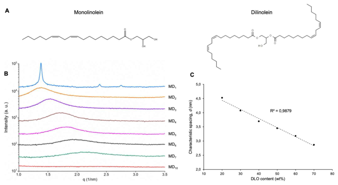

Table 1.
Composition, structural parameters, and colloidal characteristics of MLO/DLO nanodispersions. The hydrodynamic diameter and polydispersity index (PDI) were obtained from dynamic light scattering (DLS) measurements. The d-spacing values were calculated from SAXS data using d = 2π/q. The hyphen (“–“) indicates that no d-spacing could be determined due to the absence of a distinct SAXS peak. For MD1, the lattice parameter (a = 5.27 nm) was calculated from hexagonal indexing, therefore no d-spacing value is reported. Values are expressed as mean ± SD (n = 3).
Table 1.
Composition, structural parameters, and colloidal characteristics of MLO/DLO nanodispersions. The hydrodynamic diameter and polydispersity index (PDI) were obtained from dynamic light scattering (DLS) measurements. The d-spacing values were calculated from SAXS data using d = 2π/q. The hyphen (“–“) indicates that no d-spacing could be determined due to the absence of a distinct SAXS peak. For MD1, the lattice parameter (a = 5.27 nm) was calculated from hexagonal indexing, therefore no d-spacing value is reported. Values are expressed as mean ± SD (n = 3).
| Name | MLO:DLO (wt%) | d-spacing (nm) | Hydrodynamic diameter (nm) | PDI |
|---|---|---|---|---|
| MD1 | 90:10 | - | 288.53 ± 21.39 | 0.31 ± 0.01 |
| MD2 | 80:20 | 4.527 | 201.73 ± 1.78 | 0.19 ± 0.01 |
| MD3 | 70:30 | 4.075 | 165.40 ± 0.27 | 0.12 ± 0.02 |
| MD4 | 60:40 | 3.617 | 162.10 ± 0.55 | 0.08 ± 0.01 |
| MD5 | 50:50 | 3.454 | 157.07 ± 1.25 | 0.07 ± 0.02 |
| MD6 | 40:60 | 3.185 | 159.60 ± 0.63 | 0.07 ± 0.01 |
| MD7 | 30:70 | 2.873 | 163.63 ± 0.72 | 0.06 ± 0.02 |
| MD8 | 20:80 | - | 169.07 ± 2.46 | 0.09 ± 0.02 |
| MD9 | 10:90 | - | 180.90 ± 1.04 | 0.09 ± 0.01 |
| MD10 | 0:100 | - | 199.25 ± 0.21 | 0.10 ± 0.01 |
Disclaimer/Publisher’s Note: The statements, opinions and data contained in all publications are solely those of the individual author(s) and contributor(s) and not of MDPI and/or the editor(s). MDPI and/or the editor(s) disclaim responsibility for any injury to people or property resulting from any ideas, methods, instructions or products referred to in the content. |
© 2026 by the authors. Licensee MDPI, Basel, Switzerland. This article is an open access article distributed under the terms and conditions of the Creative Commons Attribution (CC BY) license (http://creativecommons.org/licenses/by/4.0/).
Copyright: This open access article is published under a Creative Commons CC BY 4.0 license, which permit the free download, distribution, and reuse, provided that the author and preprint are cited in any reuse.