

Submitted:
21 July 2026
Posted:
22 July 2026
You are already at the latest version
Abstract
Central lymphatic flow disorders remain a significant diagnostic and therapeutic challenge. Treatment typically follows a stepwise escalation approach, with reconstructive surgery being employed mainly in patients in whom conservative and interventional methods have failed or are contraindicated. Reconstructive lymphatic surgery, such as thoracic duct-vein anastomosis (TDVA), aims to restore physiological lymphatic drainage by creating a direct connection between the thoracic duct and the venous system. This review summarizes the current evidence on TDVA, including perioperative patient management, diagnostics, surgical techniques and clinical outcomes. Several case reports and case series reported successful surgical central lymphatic reconstruction, showing both safety and efficacy of this technique in both acquired and congenital lesions of the central lymphatic system. However, current evidence is limited to small case series, whereas prospective large-scale trials are lacking. Future studies should address optimal patient selection and timing of surgery in addition to conservative and interventional approaches, thus promoting the establishment of evidence-based guidelines for the treatment of central lymphatic disorders.
Keywords:
thoracic duct
; thoracic duct-vein anastomosis
; lymphovenous anastomosis
; central lymphatic system
; lymphatic reconstruction
; chylothorax
; lymphedema
; Symani
; robotic-assisted
; microsurgery
1. Introduction
The lymphatic system is essential for tissue fluid homeostasis, absorption of long-chain fatty acids and immune response [1]. Impaired drainage of the lymphatic system leads to the accumulation of lymphatic fluid in the interstitial space or body cavities, manifesting as peripheral lymphedema, chylous effusions or generalized edema [2,3]. While the microsurgical restoration of the peripheral lymphatic system has been studied extensively over the past decade [4,5,6], disorders of the central lymphatic system as well as the head and neck lymphatic system and their microsurgical reconstruction remain less explored to date. This is partly due to the relatively low prevalence of central lymphatic malformations and partly due to the limited awareness of the medical community regarding these diseases.
Overall prevalence data for central lymphatic lesions are scarce. In particular, primary central lymphatic anomalies are rare disorders, and current evidence is largely restricted to case reports and small case series, while population-based prevalence data are lacking. The incidence of iatrogenic thoracic duct (TD) injury varies by surgical site and across different studies, ranging from 0.4 to 21% after esophagectomy [7], up to 7.5% after neck dissection [8] and up to 3.9% after thoracic surgery [9].
Recent advances in imaging techniques, such as the introduction of intranodal dynamic contrast-enhanced magnetic resonance lymphangiography (DC-MRL), allowed the detailed depiction of both the individual anatomy and flow dynamics of the central lymphatic system, thus promoting the development and refinement of interventional and microsurgical treatment methods over the past decades [10].
The etiology of central lymphatic disorders can be divided into acquired (e.g., TD injury originating from trauma, surgery or malignancy) and congenital (e.g., TD malformations, central conducting lymphatic anomalies (CCLA) or other anomalies such as Gorham-Stout disease) [11]. Both acquired and congenital disorders of the central lymphatic system can lead to a variety of symptoms, which negatively impact the quality of life and mortality of affected patients [12]. Manifestations of central lymphatic anomalies and injuries include pericardial, pleural and peritoneal effusions resulting from lymphatic leakage, as well as lymphedema and anasarca [10,12]. Depending on the site of leakage, the fluid is clear lymph or chylous, whereas an accumulation of milky appearing chyle indicates a leakage from the thoracic duct or its tributaries, e.g., liver and mesenteric lymphatics within the abdomen. A loss of chyle is associated with a loss of fluid, lymphocytes, albumin and calories, thereby promoting immunosuppression and related, often life-threatening complications such as sepsis [13]. Reported overall long-term mortality of central lymphatic disorders approaches 50% [14,15].
Chyle leaks can be categorized based on both the volume of leak and the number of leak sites. Low-output chyle leaks are generally defined as a volume of <500 mL/day, or <20 mL/kg/day in children, whereas high-output leaks are characterized by a volume of >1000 mL/day, or >20mL/kg/day in children [16,17]. Typically, low-output leaks are initially managed conservatively, whereas in patients with high-output leak interventional or surgical treatment is considered earlier as conservative treatment is often insufficient in this patient group [18]. Furthermore, single lesion chyle leaks are more suitable for interventional or surgical treatment, while in multifocal leakage conservative management is more frequently considered owing to the complexity of the disease [19].
Initial therapeutic steps of low output central lymphatic leakage usually consist of conservative measures including dietary adjustments to a low-fat diet with medium-chain triglycerides (MCT) or total parenteral nutrition (TPN) to reduce the volume of chyle flowing through the thoracic duct, thus minimizing chyle leak and promoting healing [20]. This can be combined with octreotide therapy, which was proposed as effective pharmacological treatment of chyle leak in several studies [21]. Furthermore, there exist several reports on successful drug therapy with alpelisib, sirolimus, acetylsalicylic acid, propranolol, sildenafil and trametinib in patients with congenital lymphatic malformations mostly associated with congenital heart defects [22,23,24].
In cases of high output leak or absent cessation of chyle leakage, minimally invasive interventions or surgical treatment methods can be considered. Minimally invasive interventions involve therapeutic lymphangiography, which can be combined with additional percutaneous TD embolization (TDE) or selective TD branch embolization using coils and/or liquid embolic agents such as n-butyl cyanoacrylate [10,16,25,26].
Surgical methods range from TD ligation (TDL), pleurodesis, the creation of pleuro-peritoneal connections and fenestrations to microsurgical reconstructive lymphovenous anastomoses (LVA), such as TD-to-vein anastomosis (TDVA) [27,28] or peritoneovenous bypass [29]. Both TDE and TDL lead to a disruption of chyle flow through the TD, potentially risking an exacerbation of congestion and chylous effusion, which must be assessed individually for each patient [11,19]. In contrast, both TDVA and peritoneovenous bypass aim to restore the drainage of chyle into the venous system, thereby preserving physiological lymphatic circulation.
This narrative review summarizes the current literature on the surgical reconstruction of the central lymphatic system and discusses current challenges as well as future prospects regarding the management of central lymphatic disorders.
2. Diagnostic Workup
Initial suspicion of central lymphatic disorders typically arises from a patient’s medical history, most often thoracic, abdominal or neck surgery, combined with specific symptoms such as milky drainage, dyspnea, ascites or peripheral lymphedema. Confirmation of lymphatic leakage is obtained by aspiration and examination of fluid accumulated in the affected body cavity. The underlying cause is subsequently identified using conventional lymphangiography, CT lymphangiography (CTL) or DC-MRL. The choice of imaging method depends on the therapy method pursued: Conventional lymphangiography and CTL are the preferred choice in cases where an additional interventional treatment such as TDE is intended [30,31]. DC-MRL provides detailed 3D volume-rendered images of the lymphatic anatomy together with dynamic lymphatic flow data, facilitating the planning of interventional or open surgical treatment [32].
Conventional lymphangiography using an oil-based iodized contrast agent (lipiodol) is a minimally invasive imaging method with additional therapeutic effects [33,34]. Schulz et al. reported leak resolution rates of up to 80% following technically successful lymphangiography, with higher success rates particularly in patients with low-output chyle leaks [35]. While lipiodol lymphangiography has a favorable safety profile, serious complications such as lipiodol embolization to the lungs and brain as well as acute respiratory distress syndrome were reported in rare cases [36,37]. Therefore, this intervention is contraindicated in patients with right-to-left cardiac or pulmonary shunt and should be used with extreme caution in patients with severe respiratory failure, severely impaired lung function or cardiorespiratory failure [38].
On the other hand, MRL is a noninvasive, radiation-free imaging method using a gadolinium-based, water-soluble contrast material, which allows the detailed visualization of both central lymphatic structures and adjacent vascular and nonvascular anatomical structures (Figure 1) [39]. In addition to three-dimensional anatomical details, DC-MRL enables the assessment of central lymphatic flow dynamics [40].
Recently, CTL has been introduced as an alternative diagnostic tool for three-dimensional visualization of the central lymphatic system [41]. During this procedure, a water-soluble iodinated contrast medium is injected into the inguinal lymph nodes, allowing the detailed visualization of the lymphatic system and detection of lymphatic abnormalities [42]. In comparison to DC-MRL, CTL offers the advantages of superior spatial resolution, reduced technical challenge, shorter examination time as well as higher accessibility around the world [42]. Moreover, the usage of a water-soluble contrast agent during CTL avoids the risk of unintended obstruction of functioning lymph vessels, which can occur during conventional lymphangiography using an oil-based contrast agent [41].
Particularly in pediatric patients, DC-MRL is the diagnostic imaging modality of choice, providing several advantages over conventional lymphangiography and CTL including the absence of ionizing radiation and of paradoxical embolization in patients with right-to-left cardiac or pulmonary shunt [16].
3. Treatment Algorithm
Given the absence of randomized controlled trials comparing different treatment modalities, clear evidence-based guidelines for the choice and timing of therapy in patients with central lymphatic disorders are missing. Treatment decisions are currently based on case series and individual assessment of central lymphatic anatomy using MRL or conventional/CT lymphangiography.
The management of central lymphatic disorders usually follows a stepwise escalation approach, starting with conservative measures (dietary adjustments, octreotide therapy), followed by interventional therapy (lipiodol lymphangiography, embolization), and ultimately surgery (TD ligation, TDVA) if less invasive treatment fails [45]. In many cases, this escalation approach results in multiple treatment methods being employed until complete remission is achieved [19]. Surgical techniques are the method of choice in patients where conservative methods failed and interventional percutaneous treatment was either unsuccessful, technically not feasible or contraindicated [28]. Moreover, patients with pre-existing conditions, such as liver cirrhosis or cardiac insufficiency, may benefit from reconstructive lymphatic surgery instead of TDL, as a reconstructive approach allows the restoration of lymphatic flow in the TD [28,46]. For cases of congenital disorders with multiple lesions, drug therapy with propranolol, sirolimus, sildenafil, or trametinib was the preferred choice [19].
Over time, our approach has evolved into a comprehensive protocol that encompasses both conservative and surgical treatment options, tailored to the specific zone and etiology of the chyle leak. Patients are frequently referred to our center after initial conservative measures, such as dietary modifications and pharmacotherapy, have already been established. When conservative management and interventional procedures fail to achieve resolution, surgical intervention is indicated. The surgical strategy is determined by the extent and location of the lesion. Figure 2 illustrates various procedures, whereby a combination may also be indicated if required.
4. Microsurgical Reconstruction of the Central Lymphatic System
The first in-human TDVA was reported in 1953, however the technique was not adopted into widespread clinical use due to its technical difficulty and insufficient imaging methods [11,47]. Technical advances in imaging methods of the central lymphatic system led to an increased interest and a reintroduction of TDVA in the early 2000s [48,49]. In 2008, TDVA was performed in a patient presenting with chylopericardium, anastomosing the TD and the internal jugular vein in an end-to-side manner, which led to complete resolution of pericardial effusion and symptoms at seven months postoperatively [49]. Ever since, a small, but increasing number of microsurgical centers reported successful TDVA in adult and pediatric patients indicating both technical feasibility and therapeutic efficacy [28,46,50,51,52,53]. Different anatomical sites of TDVA have been described in the literature, including the neck, thorax and abdomen depending on the underlying pathology of the central lymphatic system.
Surgical planning begins with identifying the localization of lymphatic congestion or leakage, as well as visualizing the individual anatomy of the central lymphatic system using DC-MRL or conventional/CT lymphangiography. Subsequently, a surgical access to the affected section of the thoracic duct is chosen and a nearby vein, suitable for the TDVA, is identified. To clarify the venous situation, imaging, e.g., CT angiography or phlebography is performed to identify potential recipient veins. Depending on the surgical access site, an interdisciplinary approach with visceral or thoracic surgeons may be performed to access the TD by laparotomy or thoracotomy. At the beginning of the surgery, indocyanine green (ICG) is injected into the inguinal lymph nodes, allowing the intraoperative visualization of the lymphatic system using a near-infrared camera. After intraoperative identification of the TD and its lesion, TDVA is performed in an end-to-end or end-to-side fashion. Successful TDVA was reported using both end-to-end and end-to-side techniques, although there exist no comparative studies demonstrating the superiority of either method [19,46,51]. Patency of the anastomosis is confirmed intra- and postoperatively using ICG imaging. During long-term follow-ups, conventional lymphangiography or MRL can be used to assess patency of the anastomosis and continuity of lymph flow. Figure 3 shows the intraoperative visualization of the central lymphatics by ICG, vessel dissection and a patent TDVA.
The majority of studies investigating central lymphatic reconstruction describe LVA in patients with unilocular TD lesion, largely attributable to iatrogenic interventions [28,51,53,54,55].
A chylous fistula that developed after radical neck dissection was successfully treated by performing an anastomosis between the TD and the external jugular vein during wound revision. Postoperatively, no chylous fluid was observed via the intraoperatively placed drain, and the wound was healed completely after five weeks [28]. Miller et al. treated a 21-year-old patient with generalized lymphedema caused by iatrogenic TD obstruction using TDVA to the external jugular vein and an additional anastomosis between a secondary lymphatic duct and the internal jugular vein. Three months postoperatively, extremity circumference had decreased by 17% in the upper extremities and 20% in the lower extremities. Additionally, serum levels of IgG normalized from 599 mg/dL to 1230 mg/dL (physiological range 613 – 1295 mg/dL) [53]. Weissler et al. performed TDVA in two infants presenting with refractory chylothorax following cardiac surgery for congenital cardiac anomalies. The first patient had a favorable outcome, with resolution of chylothorax and ascites with extubation and chest drain removal within 10 days. The second patient required anastomosis revision because of recurrent chylothorax 18 days after surgery and absent anterograde lymph flow in lymphangiography. Following anastomosis revision, the patient was extubated nine days postoperatively, and chest tubes were removed after 21 weeks. Unfortunately, the patient passed away following an ischemic stroke during the prolonged hospitalization [51].
Apart from TDVA in patients with clinical manifestations resulting from TD leakage, several authors reported immediate central lymphatic reconstruction during oncological surgery to prevent TD leak. Yuan et al. prophylactically anastomosed the TD to the azygos vein immediately after esophagectomy. Anastomosis patency was confirmed using lymphangiography, and the patient showed no symptoms of lymphatic impairment or tumor recurrence three years postoperatively [46]. Rodi et al. reported direct repair of iatrogenic TD injury during cervical en-bloc tumor resection, which included the removal of parts of the TD resulting in a large lymph leak. TD flow was restored during the same procedure by connecting the TD stump to a branch of the subclavian vein. Six months postoperatively, the patient showed no signs of lymph leakage [55].
However, TDVA was also reported in the treatment of CCLA, addressing multilocular dysfunction of the central lymphatic system. Taghinia et al. performed TDVA in 14 patients (average age 17.5 years, range 1 to 47 years) with CCLA, achieving a complete remission in 5 patients and an improvement of the condition in 2 patients. However, 7 of 14 patients (50%) did not benefit from the surgery despite patency of the anastomosis, underlining the complexity of congenital central lymphatic disorders and indicating a poorer outcome in this patient population [52]. Othman et al. presented a case series of TDVA in four adult patients with congenital central lymphatic malformation. Complete resolution of symptoms was achieved in three patients. In the fourth patient, who had generalized lymphatic malformation, partial resolution of chylothorax was observed. However, following postoperatively developed truncal and lower-extremity lymphedema, reoperation with retroperitoneal embolization and sclerotherapy were required and peripheral LVA was planned [56].
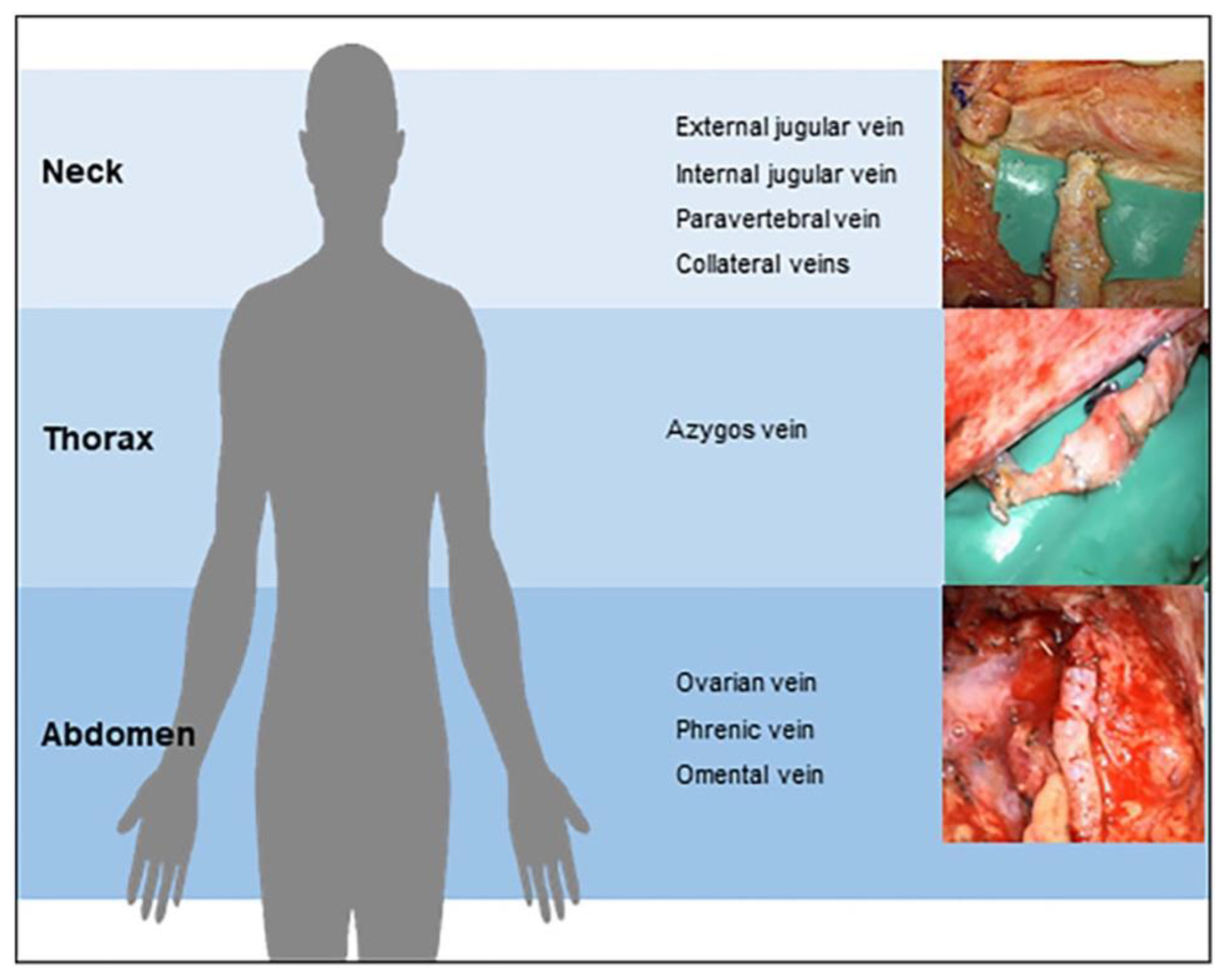
The choice of the anatomical level for thoracic duct–venous anastomosis (TDVA) is highly dependent on the underlying etiology and the individual anatomical situation. Over the past eight years, our group has established several techniques for accessing and reconstructing the central lymphatic system. Figure 4 provides an overview of the venous vessels that may be used for TDVA at different anatomical levels.
In general, the lymphatic lesion can be treated by creating a TDVA caudal to the identified site of leakage or obstruction, thereby establishing a shortcut into the venous system and bypassing the pathological segment. This approach tends to have a higher success rate, as it is less dependent on pressure gradients within the lymphatic system.
Alternatively, a TDVA may be created central to a lymphatic leak, for example, a cervical TDVA may be performed when lymphatic flow is obstructed at the left venous angle and a chylous leak is present caudal to the obstruction. In this setting, lymphatic flow must adapt to the re-established lymphovenous drainage in the neck, and chylous effusions may persist for several days to weeks until the new drainage pathway becomes established. Because lymphatic flow tends to follow the path of least resistance, careful management is required in such cases. A balance must be achieved between draining the chylous effusion, such as a pleural leak, and allowing lymphatic flow to establish a new route through the reconstructed lymphovenous pathway.
In conclusion, even though the current literature is limited to case reports and case series, previous experience indicates a good safety profile and clinical effectiveness of TDVA especially in iatrogenic TD lesions, with several reports describing remission of chylous effusions and patent anastomoses in short- and long-term follow-ups up to three years [28,51,53,54,55]. Perioperative adverse events reported in the literature include newly developed lymphedema requiring reoperation in a patient with congenital central lymphatic malformation [56], death from ischemic stroke in an infant with congenital cardiac disease (> 21 weeks postoperatively) [51] as well as status epilepticus in a patient with pre-existing epilepsy with subsequent full recovery [53]. However, the latter two adverse events were not attributed to the surgery.
5. Robotic-Assisted Reconstructive Surgery
In 2023, the first robotic-assisted central lymphatic reconstruction was reported using the Symani Surgical System® (Medical Microinstruments MMI®, Jacksonville, FL, USA) by our group. In this case report, the robotic platform was used to perform an anastomosis between a retroperitoneal aneurysmal dilatation of the left lumbar trunks and the left ovarian vein, which led to a complete regression of symptoms within three months [57]. This was followed by a case series of central lymphatic reconstruction at different anatomical levels, including robotic-assisted TDVA in the abdomen, thorax and neck in four patients (age range 8 months to 60 years) with congenital central lymphatic lesions. Three patients presented with CCLA, while the 8-months-old infant had congenital bilateral chylothorax. Symptom reduction after TDVA was observed in three patients (75%), however, one patient with CCLA showed no remission and subsequently required embolization. Abdominal and thoracic surgical access were achieved by median laparotomy and right-sided thoracotomy respectively [11].
There exist different advantages of using robotic assistance for central lymphatic reconstruction described in the literature. Motion scaling and tremor filtering provided by the robotic platform enable higher precision during vessel anastomosis, which is considered a major advantage of robotic assistance [58]. Recently, the Symani Surgical System® was used by our group to perform a total robotic-assisted peripheral LVA, performing both the vessel dissection and the anastomosis with robotic assistance [59]. Increased precision provided by the microsurgical robotic platform may be advantageous when dissecting delicate structures such as the TD and adjacent veins. However, the benefits of using robotic assistance must be weighed against associated drawbacks such as increased costs, prolonged operating times and a flat learning curve [60]. Further, current robotic microsurgical platforms use articulated arms with fixed angles, which may complicate positioning in deep anatomical regions such as the thorax or abdomen. Consequently, strategic preoperative planning and intraoperative adjustments, such as tilting of the operating table, may be required to compensate for this limitation [61].
6. Discussion
Technical feasibility and favorable clinical outcomes of TDVA have been demonstrated in several case reports and case series including both adult and pediatric patients. Underlying pathologies include both acquired and congenital lesions affecting different parts of the central lymphatic system, which result in a broad spectrum of clinical manifestations ranging from localized chylous leaks to systemic lymphatic dysfunction. This heterogeneity poses a challenge for objectively assessing and comparing outcomes and efficacy of central lymphatic reconstruction, and further hinders the direct comparison of different treatment modalities. Given the rarity of central lymphatic lesions, large-scale studies investigating the effects of surgical reconstruction are lacking to date.
To date, reconstructive lymphatic surgery is mainly considered in patients, where conservative and minimally invasive techniques failed, or if contraindications for TDE or TDL exist. Compared with embolization and ligation techniques, TDVA offers the potential benefit of restoring physiological lymphatic flow. However, the optimal indications for TDVA remain to be defined.
Advances in both imaging and surgical methods led to a growing interest in TDVA, with an increasing number of studies reporting favorable outcomes. Successful reconstructive surgery has especially been reported in patients with acquired central lymphatic lesions, which typically result from iatrogenic injury or trauma and are often characterized by isolated thoracic duct lesion.
In contrast, congenital central lymphatic disorders such as CCLA often manifest at a young age and are characterized by multiple lesions, thus complicating therapeutical management and leading to poorer surgical outcomes as highlighted by Taghinia et al. [52]. In recent years, targeted pharmacological therapies, particularly mTOR and MEK inhibitors, have emerged as important treatment option for patients with complex lymphatic anomalies [62].
Recent surgical innovations include the introduction of microsurgical robotic platforms into central lymphatic surgery, offering increased precision. However, future prototypes should address current difficulties regarding access to deep lying structures, such as the TD, within the thoracic or abdominal cavity.
Due to the high complexity and technical demands of diagnostic, interventional and surgical management, patients with central lymphatic disorders should be referred to specialized centers. Furthermore, the management requires interdisciplinary collaboration between different surgical specialties and interventional radiologists, hence early involvement of different contributing specialists is advisable for the planning of diagnostic and therapeutic steps.
This narrative review is limited by the small number and retrospective design of studies addressing central lymphatic reconstruction, which consist exclusively of case reports and case series. Further, owing to the variety of clinical manifestations, there is no standardized outcome measure used among different studies, thereby limiting the quantitative comparison of outcomes. Patient populations are highly heterogeneous, and long-term follow-ups are scarce. Lastly, publication bias may have led to preferential reporting of successful cases. These limitations underline the need for further studies with a prospective design, however the rarity of these conditions complicate the conduct of large-scale comparative studies.
7. Conclusion
Microsurgical reconstructive surgery is a safe and technically feasible approach for treating impairment of the central lymphatic system and restoring physiological lymphatic flow. To date, no clear guidelines regarding the treatment of central lymphatic disorders are available. However, with advances in imaging technologies and surgical instruments, as well as increasing clinical experience, reconstructive surgery might become a more frequently applied treatment option and may be considered earlier in the management of impaired central lymphatic system.
Author Contributions
Conceptualization, N.L., C.I., R.G.; methodology, N.L., C.I.; investigation, N.L., C.I.; writing—original draft preparation, N.L. and C.I.; writing—review and editing, C.E.Z., D.v.R., C.C.P., C.A.G., U.M., O.K., G.D.P., R.G.; visualization, N.L., C.I., C.E.Z, D.v.R.; supervision, N.L.; All authors have read and agreed to the published version of the manuscript.
Funding
This research received no external funding.
Institutional Review Board Statement
Not applicable.
Conflicts of Interest
Nicole Lindenblatt acts as a scientific consultant and clinical advisor for Medical Microinstruments (MMI). The other authors declare no conflicts of interest.
References
- Dori, Y. “Lymphatic Imaging and Intervention in Central Lymphatic Disorders,” (in eng). Lymphology 2024, vol. 57(no. 1), 4–7. [Google Scholar]
- Grada, A. A.; Phillips, T. J. Lymphedema: Pathophysiology and clinical manifestations. J. Am. Acad. Dermatol. 2017, vol. 77(no. 6), 1009–1020. [Google Scholar] [CrossRef] [PubMed]
- Rockson, S. G. “Diagnosis and management of lymphatic vascular disease,” (in eng). J. Am. Coll. Cardiol. 2008, vol. 52(no. 10), 799–806. [Google Scholar] [CrossRef] [PubMed]
- Ward, J.; et al. “A meta-analysis of the efficacy of vascularised lymph node transfer in reducing limb volume and cellulitis episodes in patients with cancer treatment-related lymphoedema,” (in eng). Eur. J. Cancer 2021, vol. 151, 233–244. [Google Scholar] [CrossRef] [PubMed]
- Basta, M. N.; Gao, L. L.; Wu, L. C. “Operative treatment of peripheral lymphedema: a systematic meta-analysis of the efficacy and safety of lymphovenous microsurgery and tissue transplantation,” (in eng). Plast. Reconstr. Surg. 2014, vol. 133(no. 4), 905–913. [Google Scholar] [CrossRef] [PubMed]
- Hahn, B. A.; et al. “Objectifying Clinical Outcomes After Lymphaticovenous Anastomosis and Vascularized Lymph Node Transfer in the Treatment of Extremity Lymphedema: A Systematic Review and Meta-Analysis,” (in eng). Microsurgery 2025, vol. 45(no. 3), e70050. [Google Scholar] [CrossRef] [PubMed]
- Koroma, P.; et al. “Chyle leak following esophagectomy: ‘a retrospective 10-year single-site experience of a tertiary center’,” (in eng). Dis. Esophagus 2025, vol. 38(no. 5). [Google Scholar] [CrossRef] [PubMed]
- Zhou, Y.; et al. Risk factors and clinical courses of chylous fistula following lateral neck dissection: a decade-long cohort study in head and neck malignancies. Eur. J. Surg. Oncol. vol. 52(no. 1), 2026. [CrossRef] [PubMed]
- Expert Panel on Vascular et al. J. Am. Coll. Radiol. 2025, vol. 22(no. 5), S177–S189. [CrossRef] [PubMed]
- Laje, P.; Dori, Y.; Smith, C.; Pinto, E.; Taha, D.; Maeda, K. “Surgical Management of Central Lymphatic Conduction Disorders: A Review,” (in eng). J. Pediatr. Surg. 2024, vol. 59(no. 2), 281–289. [Google Scholar] [CrossRef] [PubMed]
- Weinzierl, A.; et al. Microsurgical central lymphatic reconstruction-the role of thoracic duct lymphovenous anastomoses at different anatomical levels. Front. Surg. 2024, vol. 11, 1415010. [Google Scholar] [CrossRef] [PubMed]
- Trenor, A. C., 3rd; Chaudry, G. “Complex lymphatic anomalies,” (in eng). Semin Pediatr. Surg. 2014, vol. 23(no. 4), 186–90. [Google Scholar] [CrossRef] [PubMed]
- Lagarde, S. M.; Omloo, J. M.; de Jong, K.; Busch, O. R.; Obertop, H.; van Lanschot, J. J. “Incidence and management of chyle leakage after esophagectomy,” (in eng). Ann. Thorac. Surg. 2005, vol. 80(no. 2), 449–54. [Google Scholar] [CrossRef] [PubMed]
- Bolger, C.; Walsh, T. N.; Tanner, W. A.; Keeling, P.; Hennessy, T. P. “Chylothorax after oesophagectomy,” (in eng). Br. J. Surg. 1991, vol. 78(no. 5), 587–8. [Google Scholar] [CrossRef] [PubMed]
- Croteau, S. E.; et al. “Kaposiform lymphangiomatosis: a distinct aggressive lymphatic anomaly,” (in eng). J. Pediatr. 2014, vol. 164(no. 2), 383–8. [Google Scholar] [CrossRef] [PubMed]
- Nakano, T. A.; Dori, Y.; Gumer, L.; Liptzin, D. R.; Hill, L. R. S.; Kulungowski, A. M. “How we approach pediatric congenital chylous effusions and ascites,” (in eng). Pediatr. Blood Cancer 2022, vol. 69 Suppl 3, e29246. [Google Scholar] [CrossRef] [PubMed]
- Agrawal, A.; Chaddha, U.; Kaul, V.; Desai, A.; Gillaspie, E.; Maldonado, F. “Multidisciplinary Management of Chylothorax,” (in eng). Chest 2022, vol. 162(no. 6), 1402–1412. [Google Scholar] [CrossRef] [PubMed]
- Deboever, N.; et al. “Early Interventional Treatment of High Output Chyle Leak After Esophagectomy is Associated With Improved Survival,” (in eng). Ann. Surg. 2024, vol. 280(no. 1), 91–97. [Google Scholar] [CrossRef] [PubMed]
- Lindenblatt, N.; et al. Lympho-venous anastomosis for the treatment of congenital and acquired lesions of the central lymphatic system: a multidisciplinary treatment approach. Eur. J. Plast. Surg. 2022/10/01 2022, vol. 45(no. 5), 841–849. [Google Scholar] [CrossRef]
- Smith, R.; Higginson, J.; Breik, O.; Praveen, P.; Parmar, S. “Nutritional management of chyle leak after head and neck surgery: a systematic review and proposed protocol for management,” (in eng). Oral Maxillofac. Surg. 2024, vol. 28(no. 1), 51–62. [Google Scholar] [CrossRef] [PubMed]
- Magoo, S.; Bhate, K.; Santhosh Kumar, S. N.; Kakodkar, P.; Gajul, M.; Mastud, S. “Effect of Octreotide in stopping post surgical chyle leak in neck Dissection-A systematic review,” (in eng). J. Oral Biol. Craniofac Res. 2022, vol. 12(no. 5), 737–741. [Google Scholar] [CrossRef] [PubMed]
- Hollman, D.; Cheema, H.; Yu, A. C.; Mahinpey, N.; AlZahrani, F. “Medical Therapies for Pediatric Lymphatic Malformations: A Systematic Review,” (in eng). J. Cutan. Med. Surg. 2026, vol. 30(no. 3), 259–264. [Google Scholar] [CrossRef] [PubMed]
- Xu, Y.; Zhou, J.; Lan, Y.; Ji, Y. “Efficacy and Safety of Trametinib Therapy for Complex Lymphatic Malformations,” (in eng). Pediatr. Blood Cancer 2026, vol. 73(no. 1), e32119. [Google Scholar] [CrossRef] [PubMed]
- Li, D.; et al. “ARAF recurrent mutation causes central conducting lymphatic anomaly treatable with a MEK inhibitor,” (in eng). Nat. Med. 2019, vol. 25(no. 7), 1116–1122. [Google Scholar] [CrossRef] [PubMed]
- Toliyat, M.; Singh, K.; Sibley, R. C.; Chamarthy, M.; Kalva, S. P.; Pillai, A. K. Interventional radiology in the management of thoracic duct injuries: Anatomy, techniques and results. Clin. Imaging 2017, vol. 42, 183–192. [Google Scholar] [CrossRef] [PubMed]
- Itkin, M.; Kucharczuk, J. C.; Kwak, A.; Trerotola, S. O.; Kaiser, L. R. “Nonoperative thoracic duct embolization for traumatic thoracic duct leak: experience in 109 patients,” (in eng). J. Thorac. Cardiovasc Surg. 2010, vol. 139(no. 3), 584-89; discussion 589-90. [Google Scholar] [CrossRef] [PubMed]
- Meguid, R. A. Chylothorax: Surgical Ligation of the Thoracic Duct Through Thoracotomy. Oper. Tech. Thorac. Cardiovasc. Surg. 2016, vol. 21(no. 2), 139–151. [Google Scholar] [CrossRef]
- Lindenblatt, N.; Puippe, G.; Broglie, M. A.; Giovanoli, P.; Grünherz, L. “Lymphovenous Anastomosis for the Treatment of Thoracic Duct Lesion: A Case Report and Systematic Review of Literature,” (in eng). Ann. Plast. Surg. 2020, vol. 84(no. 4), 402–408. [Google Scholar] [CrossRef] [PubMed]
- Klifto, M.; Card, E. B.; Itkin, M.; Kovach, S. J. “Microsurgical Peritoneovenous Bypass for the Treatment of Recalcitrant Chylous Ascites,” (in eng). Plast. Reconstr. Surg. 2023, vol. 152(no. 2), 433–439. [Google Scholar] [CrossRef] [PubMed]
- Pieper, C. C.; et al. Back to the Future: Lipiodol in Lymphography—From Diagnostics to Theranostics. Investig. Radiol. 2019, vol. 54(no. 9), 600–615. [Google Scholar] [CrossRef] [PubMed]
- Schoellnast, H.; Maybody, M.; Getrajdman, G. I.; Bains, M. S.; Finley, D. J.; Solomon, S. B. “Computed tomography-guided access to the cisterna chyli: introduction of a technique for direct lymphangiography to evaluate and treat chylothorax,” (in eng). Cardiovasc Interv. Radiol. 2011, vol. 34 Suppl 2, S240–4. [Google Scholar] [CrossRef] [PubMed]
- Schwartz, F. R.; James, O.; Kuo, P. H.; Witte, M. H.; Koweek, L. M.; Pabon-Ramos, W. M. “Lymphatic Imaging: Current Noninvasive and Invasive Techniques,” (in eng). Semin Interv. Radiol. 2020, vol. 37(no. 3), 237–249. [Google Scholar] [CrossRef] [PubMed]
- Sommer, C. M.; et al. “Conventional Lymphangiography (CL) in the Management of Postoperative Lymphatic Leakage (PLL): A Systematic Review,” (in eng). Rofo;Konv. Lymphangiografie (KL) beim Management postoperativer Lymphleckagen (PLL) Eine Syst. Übersicht 2020, vol. 192(no. 11), 1025–1035. [Google Scholar] [CrossRef] [PubMed]
- Alejandre-Lafont, E.; Krompiec, C.; Rau, W. S.; Krombach, G. A. “Effectiveness of therapeutic lymphography on lymphatic leakage,” (in eng). Acta Radiol. 2011, vol. 52(no. 3), 305–11. [Google Scholar] [CrossRef] [PubMed]
- Schulz, S. N.; Miftaroski, A.; Rouiller, B.; Egger, B.; Lutz, J. A.; Widmer, L. “Ultrasound-Guided Intranodal Lipiodol Lymphangiography for the Assessment and Treatment of Chylous Leaks: A Retrospective Case Series from a Single Center in Switzerland and a Systematic Review of the Literature,” (in eng). J. Clin. Med. 2024, vol. 13(no. 21). [Google Scholar] [CrossRef] [PubMed]
- Taki, Y.; et al. “A case of acute respiratory distress syndrome due to lymphography with Lipiodol for chylothorax after esophagectomy,” (in eng). Surg. Case Rep. 2019, vol. 5(no. 1), 1. [Google Scholar] [CrossRef] [PubMed]
- Sheybani, A.; Gaba, R. C.; Minocha, J. “Cerebral Embolization of Ethiodized Oil following Intranodal Lymphangiography,” (in eng). Semin Interv. Radiol. 2015, vol. 32(no. 1), 10–3. [Google Scholar] [CrossRef] [PubMed]
- Kariya, S.; et al. “Nodal Lymphangiography and Embolization for Postoperative Lymphatic Leakage,” (in eng). Interv. Radiol. (Higashimatsuyama) 2025, vol. 10, e20240012. [Google Scholar] [CrossRef] [PubMed]
- Pabon-Ramos, W. M.; Raman, V.; Schwartz, F. R.; Tong, B. C.; Koweek, L. M. “Magnetic Resonance Lymphangiography of the Central Lymphatic System: Technique and Clinical Applications,” (in eng). J. Magn. Reson Imaging 2021, vol. 53(no. 2), 374–380. [Google Scholar] [CrossRef] [PubMed]
- Ahn, Y.; et al. “Single-Center Experience With Dynamic Contrast-Enhanced Magnetic Resonance Lymphangiography for Diagnosing Lymphatic Disorders and Guiding Percutaneous Embolization,” (in eng). J. Korean Med. Sci. 2024, vol. 39(no. 39), e260. [Google Scholar] [CrossRef] [PubMed]
- Ahn, Y.; et al. “Contrast-enhanced CT Lymphangiography for Various Central Lymphatic Disorders,” (in eng). Radiographics 2024, vol. 44(no. 12), e240058. [Google Scholar] [CrossRef] [PubMed]
- Patel, S.; Hur, S.; Khaddash, T.; Simpson, S.; Itkin, M. “Intranodal CT Lymphangiography with Water-soluble Iodinated Contrast Medium for Imaging of the Central Lymphatic System,” (in eng). Radiology 2022, vol. 302(no. 1), 228–233. [Google Scholar] [CrossRef] [PubMed]
- Plutecki, D.; et al. “The Anatomy of the Thoracic Duct and Cisterna Chyli: A Meta-Analysis with Surgical Implications,” (in eng). J. Clin. Med. 2024, vol. 13(no. 15). [Google Scholar] [CrossRef] [PubMed]
- Rabattu, P. Y.; et al. Anatomical study of the thoracic duct and its clinical implications in thoracic and pediatric surgery, a 70 cases cadaveric study. Surg. Radiol. Anat. 2021/09/01 2021, vol. 43(no. 9), 1481–1489. [Google Scholar] [CrossRef] [PubMed]
- R. Expert Panel on Interventional et al., Journal of the American College of Radiology, vol. 23, no. 6, pp. 1078- 1087, 2026. [CrossRef] [PubMed]
- Yuan, Y.; Chen, L. Q.; Zhao, Y. “Anastomosis Between Thoracic Duct and Azygos Vein During Esophagectomy: A Novel Technique with 3-year Follow-up,” (in eng). World J. Surg. 2016, vol. 40(no. 12), 2984–2987. [Google Scholar] [CrossRef] [PubMed]
- R. F., “Side-to-side anastomosis of the thoracic duct and the internal jugular vein.,” vol. 24, ed. Chirurg., 1953, pp. 476-777.
- Shen, W. B.; et al. “[The diagnosis and therapy of chylous ascites],” (in chi). Zhonghua Wai Ke Za Zhi 2005, vol. 43(no. 1), 25–8. [Google Scholar]
- Melduni, R. M.; Oh, J. K.; Bunch, T. J.; Sinak, L. J.; Gloviczki, P. “Reconstruction of occluded thoracic duct for treatment of chylopericardium: a novel surgical therapy,” (in eng). J. Vasc. Surg. 2008, vol. 48(no. 6), 1600–2. [Google Scholar] [CrossRef] [PubMed]
- Veziant; Sakka, L.; Galvaing, G.; Tardy, M. M.; Cassagnes, L.; Filaire, M. “Lymphovenous anastomosis for recurrent swelling syndrome and chylous effusion due to cervical thoracic duct cyst,” (in eng). J. Vasc. Surg. 2015, vol. 62(no. 4), 1068–70. [Google Scholar] [CrossRef] [PubMed]
- Weissler, J. M.; et al. “Lymphovenous Anastomosis for the Treatment of Chylothorax in Infants: A Novel Microsurgical Approach to a Devastating Problem,” (in eng). Plast. Reconstr. Surg. 2018, vol. 141(no. 6), 1502–1507. [Google Scholar] [CrossRef] [PubMed]
- Taghinia, A. H.; et al. Lymphaticovenous bypass of the thoracic duct for the treatment of chylous leak in central conducting lymphatic anomalies. J. Pediatr. Surg. 2019, vol. 54(no. 3), 562–568. [Google Scholar] [CrossRef] [PubMed]
- Miller, T. J.; Gilstrap, J. N.; Maeda, K.; Rockson, S.; Nguyen, D. H. “Correction of complete thoracic duct obstruction with lymphovenous bypass: A case report,” (in eng). Microsurgery 2019, vol. 39(no. 3), 255–258. [Google Scholar] [CrossRef] [PubMed]
- Tang, H.; Bai, Y.; Shen, W.; Zhao, J. Anastomosis of the thoracic duct and the azygos vein for the treatment of recurrent chylothoraxes. Eur. J. Cardio-Thorac. Surg. 2018, vol. 53(no. 5), 1093–1094. [Google Scholar] [CrossRef] [PubMed]
- Rodi, T.; Tung Nguyen, B.; Fritsche, E.; Rajan, G.; Scaglioni, M. F. “Direct repair of iatrogenic thoracic duct injury through lymphovenous anastomosis (LVA): A case report,” (in eng). J. Surg. Oncol. 2020, vol. 121(no. 2), 224–227. [Google Scholar] [CrossRef] [PubMed]
- Othman, S.; Azoury, S. C.; Klifto, K.; Toyoda, Y.; Itkin, M.; Kovach, S. J. “Microsurgical Thoracic Duct Lymphovenous Bypass in the Adult Population,” (in eng). Plast. Reconstr. Surg. Glob. Open 2021, vol. 9(no. 10), e3875. [Google Scholar] [CrossRef] [PubMed]
- Grunherz, et al. “First-in-human Use of a Microsurgical Robotic System for Central Lymphatic Reconstruction,” Plastic and reconstructive surgery. Glob. Open 2023, vol. 11(no. 12), e5484. [Google Scholar] [CrossRef] [PubMed]
- Kueckelhaus, et al. “Clinical experience in open robotic-assisted microsurgery: user consensus of the European Federation of Societies for Microsurgery,” (in eng). J. Robot Surg. 2025, vol. 19(no. 1), 171. [Google Scholar] [CrossRef] [PubMed]
- Imholz, C.; Zurfluh, C. E.; Watson, J. A.; Lindenblatt, N. The first total robotic-assisted lymphovenous anastomosis: prospects for innovation and future development. Int. J. Comput Assist Radiol. Surg. (in eng). 2026. [Google Scholar] [CrossRef] [PubMed]
- Imholz, C.; Grünherz, L.; Lindenblatt, N. “Systematic Review of Robotic-Assisted Peripheral and Central Lymphatic Surgery,” (in eng). J. Surg. Oncol. 2025, vol. 131(no. 1), 62–71. [Google Scholar] [CrossRef] [PubMed]
- Weinzierl, A.; Lindenblatt, N. “Advancing Robotic-assisted Microsurgery for Access to Deep Anatomical Structures,” (in eng). Plast. Reconstr. Surg. Glob. Open 2026, vol. 14(no. 4), e7668. [Google Scholar] [CrossRef] [PubMed]
- van den Brink, V. C.; et al. “Molecular targeted treatment in infants with central conducting lymphatic anomalies,” (in eng). Eur. J. Pediatr. 2025, vol. 184(no. 9), 567. [Google Scholar] [CrossRef] [PubMed]
Figure 1.
(A) MR-lymphangiography of a thrombotic occlusion at the left venous angle in a 6-month-old female patient as an example of an acquired central lymphatic flow disorder. (B) MR-lymphangiography of a 19-year-old patient with significant reflux in the context of a lymphatic drainage disorder, the congenital lesion is caused by a 13q deletion. (C) Exemplary CT-image of a recurrent chylopericardium. (D) Exemplary CT-image of a recurrent bilateral chylothorax. Reproduced from Weinzierl et al. [11] and created under a Creative Commons Attribution 4.0 International License (CC BY) https://creativecommons.org/licenses/by/4.0/.
Figure 1.
(A) MR-lymphangiography of a thrombotic occlusion at the left venous angle in a 6-month-old female patient as an example of an acquired central lymphatic flow disorder. (B) MR-lymphangiography of a 19-year-old patient with significant reflux in the context of a lymphatic drainage disorder, the congenital lesion is caused by a 13q deletion. (C) Exemplary CT-image of a recurrent chylopericardium. (D) Exemplary CT-image of a recurrent bilateral chylothorax. Reproduced from Weinzierl et al. [11] and created under a Creative Commons Attribution 4.0 International License (CC BY) https://creativecommons.org/licenses/by/4.0/.
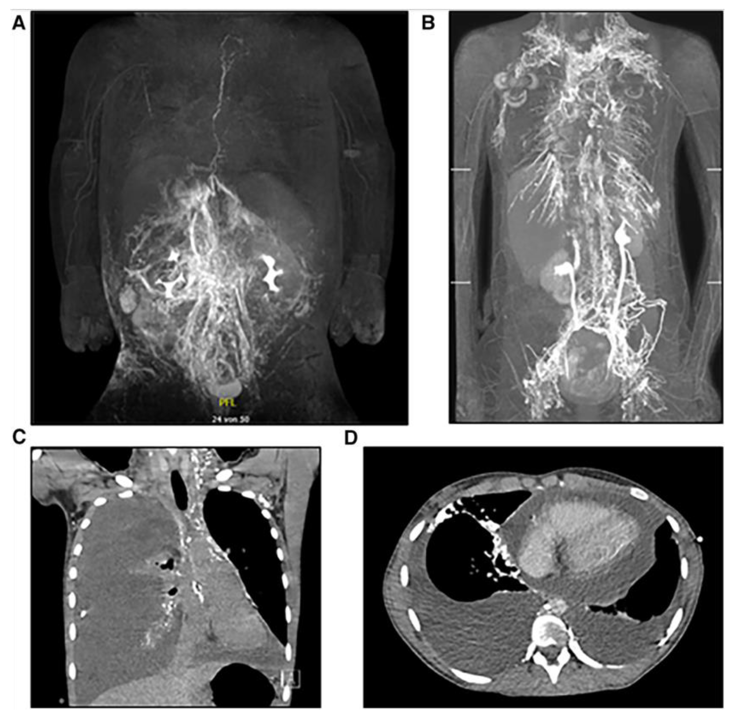
Figure 2.
Different diagnostic and therapeutic options for central lymphatic flow disorders.
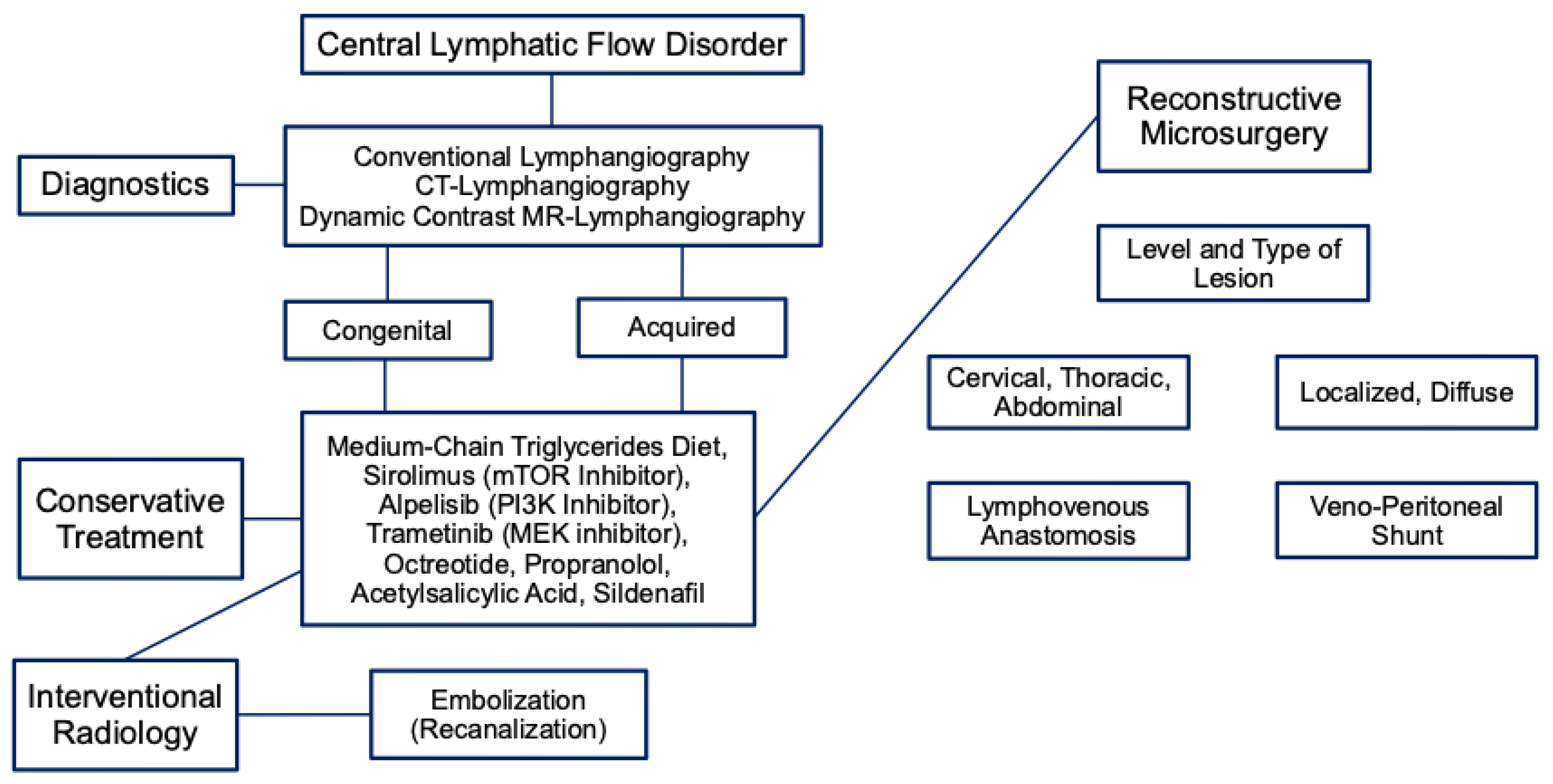
Figure 3.
Anastomosis of the thoracic duct to azygos vein (A) thoracoscopy after ICG injection into groin lymph nodes. The image shows the thoracic duct (central) as well as multiple pleural collaterals (left). (B) Surgical preparation of the thoracic duct (*) and the azygos vein (►). (C) Patent lymphovenous anastomosis as verified by the flow of ICG into the azygos vein. Reproduced from Weinzierl et al. [11] and created under a Creative Commons Attribution 4.0 International License (CC BY) https://creativecommons.org/licenses/by/4.0/.
Figure 3.
Anastomosis of the thoracic duct to azygos vein (A) thoracoscopy after ICG injection into groin lymph nodes. The image shows the thoracic duct (central) as well as multiple pleural collaterals (left). (B) Surgical preparation of the thoracic duct (*) and the azygos vein (►). (C) Patent lymphovenous anastomosis as verified by the flow of ICG into the azygos vein. Reproduced from Weinzierl et al. [11] and created under a Creative Commons Attribution 4.0 International License (CC BY) https://creativecommons.org/licenses/by/4.0/.
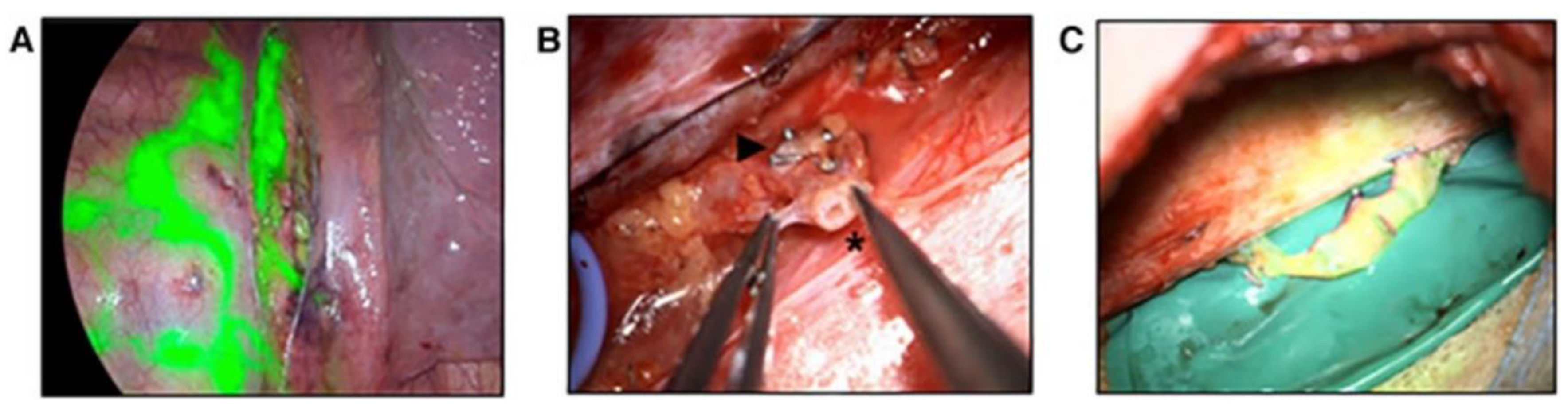
Figure 4.
Different venous vessels used for TDVA at different anatomical levels. Reproduced from Weinzierl et al. [11] and created under a Creative Commons Attribution 4.0 International License (CC BY) https://creativecommons.org/licenses/by/4.0/.
Figure 4.
Different venous vessels used for TDVA at different anatomical levels. Reproduced from Weinzierl et al. [11] and created under a Creative Commons Attribution 4.0 International License (CC BY) https://creativecommons.org/licenses/by/4.0/.
Disclaimer/Publisher’s Note: The statements, opinions and data contained in all publications are solely those of the individual author(s) and contributor(s) and not of MDPI and/or the editor(s). MDPI and/or the editor(s) disclaim responsibility for any injury to people or property resulting from any ideas, methods, instructions or products referred to in the content. |
© 2026 by the authors. Licensee MDPI, Basel, Switzerland. This article is an open access article distributed under the terms and conditions of the Creative Commons Attribution (CC BY) license (http://creativecommons.org/licenses/by/4.0/).
Copyright: This open access article is published under a Creative Commons CC BY 4.0 license, which permit the free download, distribution, and reuse, provided that the author and preprint are cited in any reuse.


