

Submitted:
22 July 2026
Posted:
22 July 2026
You are already at the latest version
Abstract
Congenital colonic stenosis (CCS) is an exceptionally rare gastrointestinal anomaly. It presents with nonspecific clinical symptoms and can mimic other pediatric bowel disorders. We report a 1-year-old girl with a history of prematurity who presented to the emergency department with progressive abdominal distension and vomiting. She had maintained steady growth and age-appropriate weight gain during early infancy because a liquid-based milk diet allowed soft stool to pass through a pinhole-sized narrowing. The transition to solid foods at a corrected age of 9 months produced formed stools, precipitating an acute mechanical large bowel obstruction and precipitous weight loss. A repeat fluoroscopic contrast enema successfully identified a classic, concentric "ring sign" characteristic of structural stenosis, correcting an earlier misinterpreted study. A short-segment resection and primary anastomosis were curative.
Keywords:
colon stenosis
; contrast enema
; infant
; intestinal obstruction
1. Introduction
Congenital colonic stenosis (CCS) is a rare cause of pediatric lower gastrointestinal obstruction, with an estimated incidence of 1 in 40,000 live births [1]. While the exact etiology remains debated, it is frequently attributed to fetal mesenteric vascular accidents, intrauterine ischemic insults, incomplete intestinal recanalization, or maternal exposure to vasoconstrictive agents or tobacco [2,3,4].
Due to incomplete luminal narrowing, clinical presentation is highly variable, ranging from acute neonatal ileus or failure to thrive in early childhood to insidious chronic constipation that may masquerade as Hirschsprung disease [2,3,5]. Preoperative diagnosis of CCS presents a significant challenge for both pediatric surgeons and radiologists. Although a contrast enema is considered the diagnostic gold standard to delineate the transition from a small-caliber distal colon to a dilated proximal colon, it is frequently inconclusive or misleading in practice [3,5,6]. This report examines a diagnostic pitfall in which an infant’s liquid-exclusive diet masked a severe, pinpoint descending colon stenosis, delaying definitive diagnosis until the introduction of solid foods precipitated acute mechanical obstruction.
2. Case Report
A 1-year-old girl, born prematurely at 28 weeks’ gestation with a very low birth weight of 1095 g, presented to the emergency department with a 3-day history of progressive abdominal distension, bilious vomiting, and failure to pass flatus or stool.
Her neonatal and early infantile history was notable for intermittent bowel dilatation investigated at 66 days of age. An initial fluoroscopic contrast enema performed at that time had demonstrated an abrupt retrograde obstruction of the contrast medium within the distal descending colon (Figure 1). However, because a subsequent rectal suction biopsy confirmed the presence of ganglion cells (ruling out Hirschsprung disease) and the infant remained able to tolerate a liquid milk diet and spontaneously pass loose stools, the abnormal contrast enema was clinically dismissed and misinterpreted as a technical artifact or procedural study failure.
Throughout her first year of life, the patient remained exclusively on a liquid-based milk diet and demonstrated steady, age-appropriate weight gain (Figure 2), which effectively concealed the underlying structural anomaly. The clinical turning point occurred at a corrected age of 9 months, coinciding precisely with weaning and the introduction of solid foods. The transition to bulkier, formed stools led to severe, obstinate obstipation, progressive abdominal distension, and a precipitous decline in body weight from 7.82 kg to 7.04 kg over a 2-month period.
Upon presentation to the emergency department, physical examination revealed an acutely distressed, malnourished infant with a severely distended, tympanitic abdomen and visible bowel loops. Laboratory evaluation was unremarkable except for mild dehydration and metabolic alkalosis.
An urgent repeat contrast colon enema was performed, which clearly revealed a localized, severe annular constriction with a prominent concentric “ring sign” within the descending colon (Figure 3). This diagnostic breakthrough stood in sharp contrast to the inconclusive neonatal enema, where lower hydrostatic pressure and liquid stool patency had obscured the lesion.
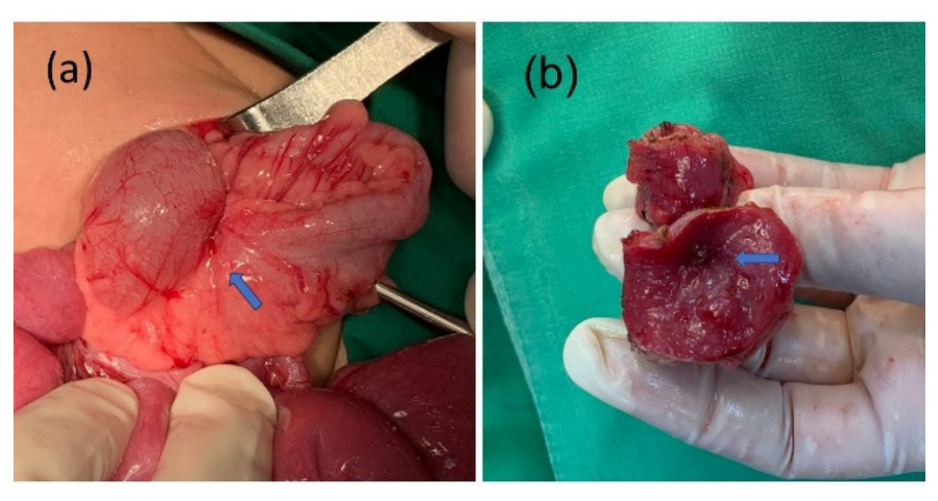
The patient underwent an emergent exploratory laparotomy. Intraoperative findings revealed a segment of descending colon stenosis surrounded and partially obscured by dense mesenteric adhesions and reactive fibrofatty tissue wrapping (Figure 4a). Gross examination of the short cross-sectionally resected specimen revealed an extreme luminal narrowing with a pinpoint stricture orifice measuring less than 2 mm in diameter (Figure 4b). A short-segment colonic resection and primary end-to-end anastomosis were performed. Histopathological evaluation confirmed the presence of normal ganglion cells and absence of malignancy, confirming a diagnosis of congenital membranous colonic stenosis. Postoperatively, the patient tolerated dietary advancement smoothly and exhibited robust catch-up growth, reaching 9.50 kg by 17 months of age.
4. Discussion
Congenital colonic stenosis is rare, accounting for only 1.8% to 15% of all cases of congenital intestinal atresia [3]. Unlike colonic atresia, which manifests as complete obstruction necessitating immediate neonatal diagnosis, CCS is characterized by persistent, incomplete narrowing that maintains a minimal patent lumen. This patency allows for variable transit of luminal contents, often resulting in subacute or intermittent symptoms, such as chronic constipation, that may be initially misdiagnosed [3,6,7]. Consequently, CCS remains a diagnostic dilemma, often mimicking functional constipation or Hirschsprung disease [7,8,9].
While neonatal presentations typically include progressive abdominal distension, failure to pass meconium, and bilious vomiting [3], delayed presentations during late infancy or childhood—manifesting as failure to thrive, colicky abdominal pain, recurrent vomiting, and chronic constipation—are well-documented [3,4,5,6,8]. The mechanisms underlying these delayed presentations may involve a progressive loss of patency within the stenotic segment, potentially secondary to inflammation or scarring, or the anatomical location of the stenosis, with distal lesions often associated with later clinical onset [5,7,10].
This case highlights a unique ‘masking effect’ related to diet. During early infancy, the low viscosity of breast milk or formula allows liquid stool to pass through a sub-millimeter pinpoint stricture. Consequently, because the patient remains asymptomatic and continues to gain weight, high-grade mechanical obstruction is easily overlooked. Only when the introduction of solid fibers produces bulkier stools does the stenosis become clinically apparent, converting a subclinical anomaly into an acute emergency. Intraoperative findings in our case confirmed a colonic stenosis surrounded by dense mesenteric fibrofatty tissue, without acute inflammation. Histological evaluation of the resected specimen revealed severe luminal narrowing with a pinpoint orifice and an absence of neutrophilic infiltration.
Contrast enema remains essential for distinguishing colonic stenosis from atresia. While atresia presents with a microcolon and a distended proximal bowel separated by an abrupt cutoff, stenosis appears as a concentric or annular “ring-like filling defect” [4,6,7,10]. This radiological feature also differentiates CCS from the non-relaxed, cone-shaped “transition zone” characteristic of Hirschsprung disease [11]. Because CCS can present with multiple synchronous stenoses, a comprehensive pan-colonic evaluation is imperative to avoid overlooking proximal or distal lesions [6,8].
Diagnostic workup may be inconclusive, with the diagnosis sometimes established only intraoperatively [3,5,6]. False-negative findings often stem from inadequate bowel preparation, insufficient hydrostatic pressure during contrast administration, or cognitive bias focusing solely on the rectosigmoid region. When persistent gastrointestinal symptoms remain unexplained, a repeat contrast enema is warranted. In our patient, a high index of clinical suspicion prompted repeat fluoroscopy, which successfully demonstrated the diagnostic ring-like filling defect and facilitated definitive surgical intervention.
Surgical management must be tailored to the patient’s clinical status, the anatomical location of the lesion, and the luminal diameter discrepancy. Primary end-to-end anastomosis is generally safe when the proximal-to-distal lumen diameter ratio is 3:1 or less [3,10]. In cases involving poor nutritional status or significant comorbidities, a staged approach—utilizing an initial divided stoma followed by delayed secondary anastomosis—is recommended to preserve bowel length and minimize morbidity [3,10].
5. Conclusions
Persistent abdominal distension, recurrent vomiting, and a failure to thrive following dietary transitions from liquid to solid food warrant a low threshold for repeating diagnostic contrast studies in infants. Fluoroscopic contrast colon enema remains a highly effective tool for detecting underlying congenital colonic stenosis, provided it is repeated when the clinical index of suspicion remains high despite historical “normal” or inconclusive reports.
Conflicts of Interest
The authors declare no conflicts of interest.
Abbreviations
The following abbreviations are used in this manuscript:
| CCS | Congenital colonic stenosis |
References
- Galvan-Montano, A.; Suarez-Roa, M.; de, L.; Carmona-Moreno, E. Congenital stenosis of the colon with foreign bodies. Case report. Cir. Cir. 2010, 78(3), 259–261. [Google Scholar] [PubMed]
- El-Asmar, K.M.; Abdel-Latif, M.; El-Kassaby, A.A.; Soliman, M.H.; El-Behery, M.M. Colonic Atresia: Association with Other Anomalies. J. Neonatal Surg. 2016, 5(4), 47. [Google Scholar] [CrossRef] [PubMed]
- Gupta, A.; Singh, A.K.; Sunil, K.; Pandey, A.; Rawat, J.D.; Kureel, S.N. Congenital Colonic Stenosis: A Rare Gastrointestinal Malformation in Children. J. Indian Assoc. Pediatr. Surg. 2021, 26(5), 317–323. [Google Scholar] [CrossRef] [PubMed]
- Natarajan, S.; Vijayshankar, R.; Periasamy, M.; Rangasamy, S.; Ramasamy, R. Delayed Presentation of Congenital Colonic Stenosis. J. Indian Assoc. Pediatr. Surg. 2017, 22(3), 191–2. [Google Scholar] [CrossRef] [PubMed]
- Garcia, D.J.; Hamade, M.; Lin, L.; Matias, M.; Sobhan, A.; Zaritsky, M.; Thorson, C. Failure to thrive in infant secondary to congenital colonic stenosis: a case report. J. Surg. Case Rep. 2024, 2024(12), rjae766. [Google Scholar] [CrossRef] [PubMed]
- Ekenze, S.O.; Ezomike, U.O.; Nwachukwu, I.E.; Ariom, A.I.; Chukwubuike, K.E.; Nwangwu, E.I.; Onoh, U.S.; Uwah, E.A. Chronic bowel obstruction from colonic stenosis in early infancy-A report of two cases. Malawi Med. J. 2019, 31(1), 82–85. [Google Scholar] [CrossRef] [PubMed]
- Dahal, P.; Dawadi, K.; Parajuli, S. Delayed presentation of congenital sigmoid colon stenosis: a rare entity. BJR Case Rep. 2024, 10(1), uaae003. [Google Scholar] [PubMed]
- Zambaiti, E.; Chiaramonte, C.; Salerno, S.; Li Voti, G.; Siracusa, F. Multiple Congenital Colonic Stenosis: A Rare Gastrointestinal Malformation. Case Reports in Pediatrics 2016, 2016, 6329793. [Google Scholar] [CrossRef]
- Naik, P.; Mandelia, A.; Agarwal, N.; Sen Sarma, M. Congenital colonic stenosis: an unusual cause of colonic obstruction masquerading as Hirschsprung’s disease in infancy. BMJ Case Rep. 2023, 16(9). [Google Scholar] [CrossRef]
- El-Mefleh, N.; Ba’Ath, M.E. Congenital colonic stenosis after the neonatal period: A case report and review of the literature with criteria for safe primary anastomosis. J. Neonatal Surg. 2025, 14, 541–546. [Google Scholar] [CrossRef]
- Hamid, R.; Ali, I.; Bhat, N.; Baba, A.A.; Mufti, G.; et al. Management Congenital Colonic Stenosis. J. Neonatal Biol. 2015, 4, 166. [Google Scholar] [CrossRef]
Figure 1.
Initial fluoroscopic contrast enema performed at 66 days of age. (a) Anterior-posterior and (b) lateral radiographic views demonstrating a complete, abrupt retrograde obstruction (block) of the contrast medium within the distal descending colon. Clinically, this imaging finding clashed with the infant’s ongoing ability to pass loose stool. This clinicoradiological discrepancy originally led to the study being falsely interpreted and discarded as an executive or technical artifact (procedural failure) rather than a true structural or functional pathology.
Figure 1.
Initial fluoroscopic contrast enema performed at 66 days of age. (a) Anterior-posterior and (b) lateral radiographic views demonstrating a complete, abrupt retrograde obstruction (block) of the contrast medium within the distal descending colon. Clinically, this imaging finding clashed with the infant’s ongoing ability to pass loose stool. This clinicoradiological discrepancy originally led to the study being falsely interpreted and discarded as an executive or technical artifact (procedural failure) rather than a true structural or functional pathology.
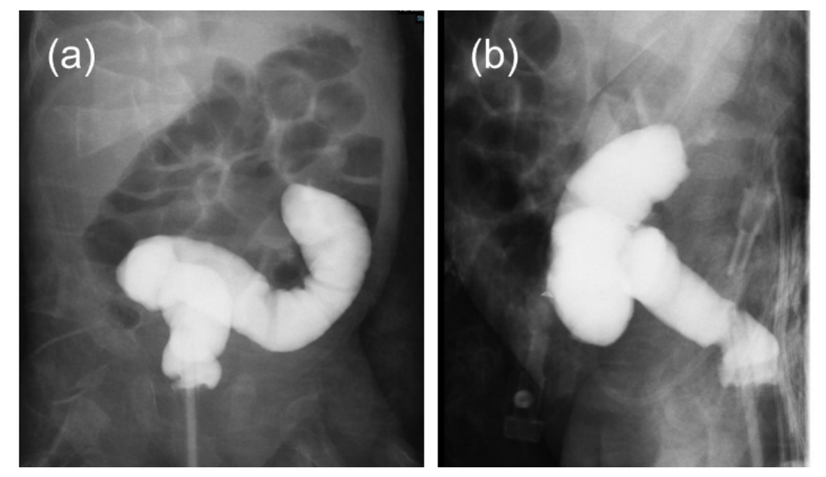
Figure 2.
Infant growth chart illustrating chronological body weight variations. The patient’s body weight trend demonstrates steady, age-appropriate weight gain during early infancy, which initially masked the underlying distal colonic obstruction. The introduction of solid foods at approximately 7 months of age triggered a progressive decline in weight (blue arrow), secondary to obstructive symptoms. Following surgical intervention at 9 months of age (red arrow; body weight 7.04 kg), the weight loss trend reversed, leading to sustained catch-up growth and weight restoration.
Figure 2.
Infant growth chart illustrating chronological body weight variations. The patient’s body weight trend demonstrates steady, age-appropriate weight gain during early infancy, which initially masked the underlying distal colonic obstruction. The introduction of solid foods at approximately 7 months of age triggered a progressive decline in weight (blue arrow), secondary to obstructive symptoms. Following surgical intervention at 9 months of age (red arrow; body weight 7.04 kg), the weight loss trend reversed, leading to sustained catch-up growth and weight restoration.
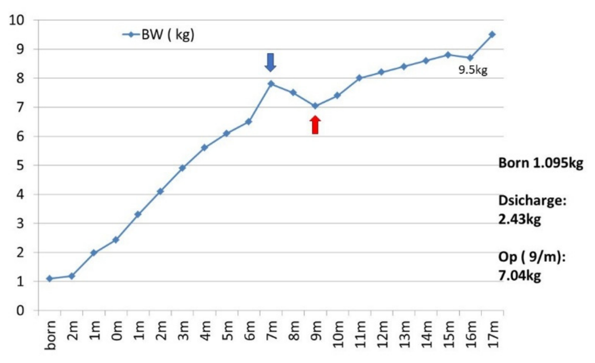
Figure 3.
Repeat fluoroscopic contrast enema performed at 9 months of age. (a) Anterior-posterior and (b) lateral radiographic views clearly demonstrating a localized, severe annular constriction with a prominent concentric “ring sign” (red arrows) within the descending colon. In contrast to the initial study, these images establish a definitive, high-grade focal mechanical obstruction, corresponding structurally with the clinical onset of severe obstructive symptoms following the introduction of solid foods.
Figure 3.
Repeat fluoroscopic contrast enema performed at 9 months of age. (a) Anterior-posterior and (b) lateral radiographic views clearly demonstrating a localized, severe annular constriction with a prominent concentric “ring sign” (red arrows) within the descending colon. In contrast to the initial study, these images establish a definitive, high-grade focal mechanical obstruction, corresponding structurally with the clinical onset of severe obstructive symptoms following the introduction of solid foods.
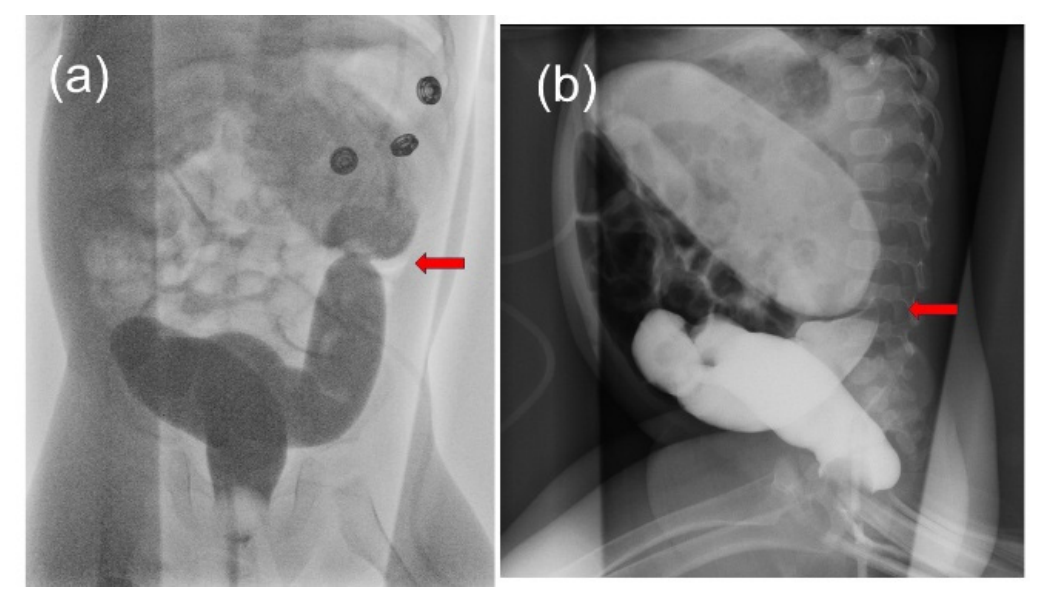
Figure 4.
Intraoperative findings and gross pathology of the colonic lesion. (a) Intraoperative photograph demonstrating a segment of colonic stenosis surrounded and obscured by dense mesenteric adhesions and fibrofatty tissue wrapping (blue arrow). (b) Macroscopic evaluation of the short cross-sectionally resected colonic specimen revealing marked luminal narrowing with a pinpoint stricture orifice (blue arrow).
Figure 4.
Intraoperative findings and gross pathology of the colonic lesion. (a) Intraoperative photograph demonstrating a segment of colonic stenosis surrounded and obscured by dense mesenteric adhesions and fibrofatty tissue wrapping (blue arrow). (b) Macroscopic evaluation of the short cross-sectionally resected colonic specimen revealing marked luminal narrowing with a pinpoint stricture orifice (blue arrow).
Disclaimer/Publisher’s Note: The statements, opinions and data contained in all publications are solely those of the individual author(s) and contributor(s) and not of MDPI and/or the editor(s). MDPI and/or the editor(s) disclaim responsibility for any injury to people or property resulting from any ideas, methods, instructions or products referred to in the content. |
© 2026 by the authors. Licensee MDPI, Basel, Switzerland. This article is an open access article distributed under the terms and conditions of the Creative Commons Attribution (CC BY) license (http://creativecommons.org/licenses/by/4.0/).
Copyright: This open access article is published under a Creative Commons CC BY 4.0 license, which permit the free download, distribution, and reuse, provided that the author and preprint are cited in any reuse.


