

Submitted:
21 July 2026
Posted:
22 July 2026
You are already at the latest version
Abstract
Ischemic heart disease is currently diagnosed mainly through cardiac computed tomography (CT), angiography and functional testing, both of which detect only advanced arterial macrocalcification, at a stage when treatment can merely slow disease progression rather than reverse it. Yet macrocalcification represents the end product of a much earlier molecular process, which is microcalcification. This process is driven by smooth muscle cell and macrophage apoptosis, matrix vesicle release, and osteogenic phenotypic transitions within the arterial intima, occurring years to decades before mineral deposits become visible on CT. [18F]Sodium fluoride (NaF) positron emission tomography (PET) exploits the tracer’s affinity for hydroxyapatite to visualize this early, biologically active microcalcification directly, offering a diagnostic window during which the disease may still be responsive to targeted therapy. Studies demonstrate that anti-atherosclerotic interventions, including statins, and tissue-nonspecific alkaline phosphatase inhibition, can suppress NaF uptake even when CT-based calcium scores remain unchanged or continue to rise, a dissociation now also observed in human trials of statins and PCSK9 inhibitors. This review traces the cellular and histological basis of arterial calcification, outlines the principles and limitations of NaF-PET imaging, and evaluates its emerging role—supported by artificial intelligence-based quantification—as a tool for monitoring targeted anti-atherosclerotic treatment.
Keywords:
ischemic heart disease
; microcalcification
; sodium fluoride
; positron emission tomography
1. Introduction
Cardiovascular disease remains the leading cause of morbidity and mortality worldwide, accounting for an estimated 19.8 million deaths in 2022 alone, equating to 396 million years of life lost and 44.9 million years lived with disability [1]. Atherosclerosis, the pathological substrate underlying most ischemic heart disease and stroke, progresses through a prolonged, largely silent process in which lipid-laden plaques accumulate within the arterial wall over decades before producing clinical symptoms [2]. Rudolf Virchow first proposed inflammation as the initiating event in atherogenesis, and this concept remains foundational: circulating monocytes are recruited to sites of endothelial injury, differentiate into macrophages, and internalize oxidized low-density lipoprotein (LDL) to become lipid-laden foam cells that form the core of the nascent atheroma [3].
Embedded within this inflammatory cascade is a less appreciated but clinically consequential process which is calcification. Coronary artery calcification (CAC) has long served as a surrogate marker of atherosclerotic burden and is quantified clinically using the Agatston score derived from cardiac computed tomography (CT) [4]. However, this metric captures only macroscopic, late-stage mineralization—by the time calcification is visible on CT, the underlying disease process has often been active for years. The molecular precursor to this visible calcification, microcalcification, forms via distinct cellular mechanisms and is invisible to conventional structural imaging [5].
This diagnostic gap has motivated interest in molecular imaging techniques capable of detecting biologically active processes that precede anatomical change. Positron emission tomography (PET) imaging using 18F-sodium fluoride (NaF) exploits the tracer’s affinity for hydroxyapatite, the mineral phase of arterial calcification, enabling visualization of microcalcification at a stage when disease-modifying therapy may still alter its trajectory [6,7]. This review traces the biological narrative from the earliest cellular events of calcification through to the clinical application of NaF-PET, culminating in an assessment of its emerging role as a tool for monitoring pharmacological and lifestyle interventions.
2. The Molecular and Histological Basis of Arterial Calcification
2.1. Two Distinct Calcification Phenotypes
Arterial calcification is not a single, uniform process. Depending on its anatomical location within the vessel wall, calcification is classified into two categories with distinct etiologies and clinical implications: Mönckeberg medial calcification and intimal atherosclerotic calcification [8]. Mönckeberg calcification predominantly affects the tunica media of peripheral arteries in patients with long-standing diabetes, chronic kidney disease, hypercalcemia, elevated phosphate, or hyperparathyroidism. It arises largely from osteoblast-like differentiation of vascular cells driven by altered intracellular signaling and calcium-sensing receptor activity, and notably does not typically involve lipid deposition or substantial inflammatory infiltration in its advanced stages [9].
Intimal atherosclerotic calcification, by contrast, is intrinsically linked to atherosclerosis progression and is the process most relevant to ischemic heart disease [10]. It is driven by chondrocyte-like rather than osteoblast-like cellular transformation and by an active inflammatory cascade involving macrophage infiltration and cytokine release [11]. Because CAC scoring reflects primarily this intimal process [10,12], the remainder of this review focuses on intimal calcification as the substrate for NaF-PET imaging.
2.2. Two Distinct Calcification Phenotypes
Several non-mutually exclusive mechanisms contribute to the initiation of intimal microcalcification. Death of inflammatory cells within the atheroma releases apoptotic bodies that nucleate hydroxyapatite crystal formation [13]; death of smooth muscle cells (SMCs) and macrophages liberates matrix vesicles that serve a similar nucleating function [14]; and phenotypic modulation of SMCs into chondrocyte-like cells promotes bone-like matrix deposition [15,16]. These processes are further exacerbated by reduced expression of endogenous mineralization inhibitors such as osteopontin, fetuin, and pyrophosphate [17,18,19].
Matrix vesicles, extracellular vesicles ranging from exosomes (10–100 nm) to microparticles (100–500 nm) and apoptotic bodies (500–1000 nm), are now understood to play a central nucleating role [20]. Localized collagen degradation allows larger vesicles (100–300 nm) to accumulate within the extracellular matrix, followed by annexin A1-mediated vesicle aggregation [21]. Within these vesicles, phosphatase orphan 1 cleaves phosphate ions from two membrane phospholipids—phosphoethanolamine and phosphocholine, while tissue-nonspecific alkaline phosphatase (TNAP) and nucleotide pyrophosphatases (NPP1, NPP3) liberate additional phosphate from ATP, ADP, and pyrophosphate in the extracellular space [22,23,24]. Pit1 and Pit2 transporters import phosphate into the vesicles while annexins A2, A5, and A6 mediate calcium transport, culminating in nucleation of calcium phosphate, vesicle membrane fusion, and eventual mineral maturation into microcalcification [25,26].
Experimental work has further implicated macrophage-derived matrix vesicles specifically. Under calcium and phosphate concentrations resembling those seen in chronic kidney disease patients on dialysis, macrophage-derived vesicles exhibit heightened calcification potential, mediated by a phosphatidylserine-annexin A5-S100A9 membrane complex that functions as a nucleation site for hydroxyapatite [27]. Given the relative abundance of macrophages over SMCs in rupture-prone plaques, this mechanism may be particularly relevant to the initiation of microcalcification in unstable lesions [28]. A second, independent mechanism involves phenotypic transition of SMCs into osteoblast-like cells that subsequently synthesize osteogenic factors capable of promoting vascular calcium deposition, while a third implicates direct hydroxyapatite crystal formation during apoptosis of SMCs and macrophages themselves [29]. These mechanisms are not mutually exclusive, and current evidence has not established which predominates at any given stage of plaque development.
2.3. Histological Progression from Microcalcification to Macrocalcification
The earliest histologically identifiable form of CAC is microcalcification, ranging in size from 0.5 to 15.0 μm, best visualized using von Kossa or Alizarin red staining within regions of pathological intimal thickening [30,31]. These deposits originate predominantly from SMC apoptosis, producing fine microcalcifications, while apoptotic macrophages generate relatively larger punctate deposits, typically located near the internal elastic lamina at the outer margins of the necrotic core [20]. Initial calcification occurring within matrix vesicles measuring only 100 to 700 nm can be resolved only by electron microscopy, underscoring how far this process precedes any form of clinical detectability [31].
Over time, microcalcifications coalesce into larger speckles and fragments, progressing outward from the necrotic core into the surrounding collagenous matrix, eventually forming calcified sheets or plates spanning more than one quadrant of the vessel wall [32]. These sheets may fracture into nodular calcifications that protrude into the lumen, disrupt the endothelial lining, and precipitate acute luminal thrombosis, a mechanism implicated in 2–7% of coronary and 4–14% of carotid thrombotic events identified in pathological studies [33,34].
This progression mirrors, in several respects, the process of skeletal bone formation, with bone-related proteins such as bone morphogenetic proteins (BMP-1, BMP-4), bone sialoprotein, osteocalcin, osteonectin, osteopontin, and osteoprotegerin identified within calcified arterial tissue [35,36]. Osteoprotegerin, osteopontin, and matrix Gla protein appear at sites of microcalcification even in early plaques—uncarboxylated matrix Gla protein specifically emerges with the transition from adaptive to pathologic intimal thickening—while BMP-2, BMP-4, osteopontin, and osteonectin become more prominent in advanced fibrocalcific lesions [36,37]. Despite this parallel with osteogenesis, true bone formation within the human coronary vessel wall remains rare, and the precise triggers governing the transition from microcalcification to macroscopic sheet calcification remain incompletely understood. Calcification extent also tracks with plaque type and degree of luminal narrowing: it is minimal in early adaptive or pathological intimal thickening, increases progressively in fibroatheroma and thin-cap fibroatheroma, and becomes disproportionately extensive relative to necrotic core size in healed ruptures and fibrocalcific plaques [38], where diffuse calcification on radiography corresponds histologically to sheet calcification [38].
2.4. Biomechanical Consequences: Microcalcification and Plaque Instability
The clinical significance of microcalcification extends beyond its role as a histological marker—it directly influences plaque biomechanics and rupture risk. Macrocalcification tends to stabilize plaques by acting as a mechanical barrier, whereas microcalcification introduces biomechanical heterogeneity that increases vulnerability [39]. This effect is strongly size-dependent: microcalcifications smaller than 5 μm are predicted to be biomechanically inert, since their associated voids do not undergo explosive growth under tensile stress owing to high surface energy. However, deposits between 5 and 65 μm can amplify plaque tensile stress by up to fivefold, and the interfacial debonding theory proposed by Maldonado and colleagues identifies microcalcifications exceeding 65 μm as a distinct failure mode in which fibrous tissue separates from the mineral surface under threshold stress, precipitating rupture [39]. Consistent with this, nearly all microcalcifications identified within ruptured fibrous caps measured under 65 μm, with a mean diameter of 28 ± 13 μm [30].
Morphology and spatial clustering further modulate risk. Elongated, prolate spheroidal microcalcifications generate substantially higher stress at their poles than spherical deposits, with elliptical microcalcifications of aspect ratio greater than 2 producing up to a fourfold elevation in localized stress concentration compared with a twofold increase around spherical deposits [40,41,42]. Collagen appears to act as a structural scaffold for microcalcification formation, with collagen fiber alignment directly shaping the resulting mineral geometry by guiding matrix vesicle aggregation into elongated forms, while collagen degradation itself accompanies microcalcification development [40,43].
Proximity between adjacent microcalcifications compounds this danger. When the gap-to-diameter ratio (h/D) falls below 0.1, peak tissue stress can increase up to sixfold, and stress concentration rises exponentially as h/D drops below 0.4. Analysis of nearly 35,000 microcalcifications across 22 unruptured plaques found that closely juxtaposed pairs (h/D <0.4) were rare, occurring in only 3 instances, suggesting either a genuinely low incidence or that such configurations are disproportionately associated with rupture and therefore rarely captured in surviving, unruptured tissue [39]. Laboratory models using silicone-embedded microbeads confirm that closely spaced microcalcifications reduce the ultimate tensile strength (UTS) of fibrous cap tissue by 10.9%, 22.3%, and 39.4% as h/D ratios decreased from 0.86 to 0.60 to 0.54, respectively, with thinner caps showing more pronounced weakening. High concentrations of small hydroxyapatite particles (≤5 μm) similarly reduce UTS and tissue stiffness, an effect attributed partly to a reduction in collagen fiber matrix content [44,45]. Collectively, this body of biomechanical evidence establishes microcalcification—not macrocalcification—as the histological feature most directly implicated in plaque rupture, providing the rationale for imaging techniques capable of detecting it before it accumulates into stabilizing macrocalcific sheets.
3. Principles of NaF-PET Imaging
3.1. Molecular Basis and Dual-Tracer Complementarity
PET imaging enables visualization of biologically active processes that remain invisible to structural modalities such as CT, ultrasound, or MRI. Two radiotracers dominate atherosclerosis imaging: 18F-fluorodeoxyglucose (FDG), which reflects macrophage glucose metabolism and thus arterial inflammation, and NaF, which binds hydroxyapatite and thus reflects microcalcification [46]. Because inflammation and microcalcification are distinct, only partially interconnected biological processes, the two tracers are not interchangeable and often diverge within the same lesion or vascular bed [46].
This divergence has been directly demonstrated in preclinical models. In ApoE-/- mice fed an atherogenic diet, FDG uptake appeared in the aortic arch while NaF uptake instead localized to pulmonary arteries; FDG uptake in the aortic arch was linked to hypoxia rather than inflammation, and FDG did not appear earlier than NaF, challenging the assumption that inflammation strictly precedes calcification [46]. In patients with pseudoxanthoma elasticum, aortic NaF uptake correlated with pulse wave velocity in a pattern opposite to arterial FDG uptake, and the two tracers did not correlate with one another in any arterial segment examined [47]. A dual-tracer PET/CT study of atherosclerotic plaque similarly found that FDG-based inflammation and NaF-based mineral deposition were not directly correlated within the same plaque segments, with mineralization patterns suggesting that active calcification represents a distinct, later biological stage relative to inflammatory activity [48].
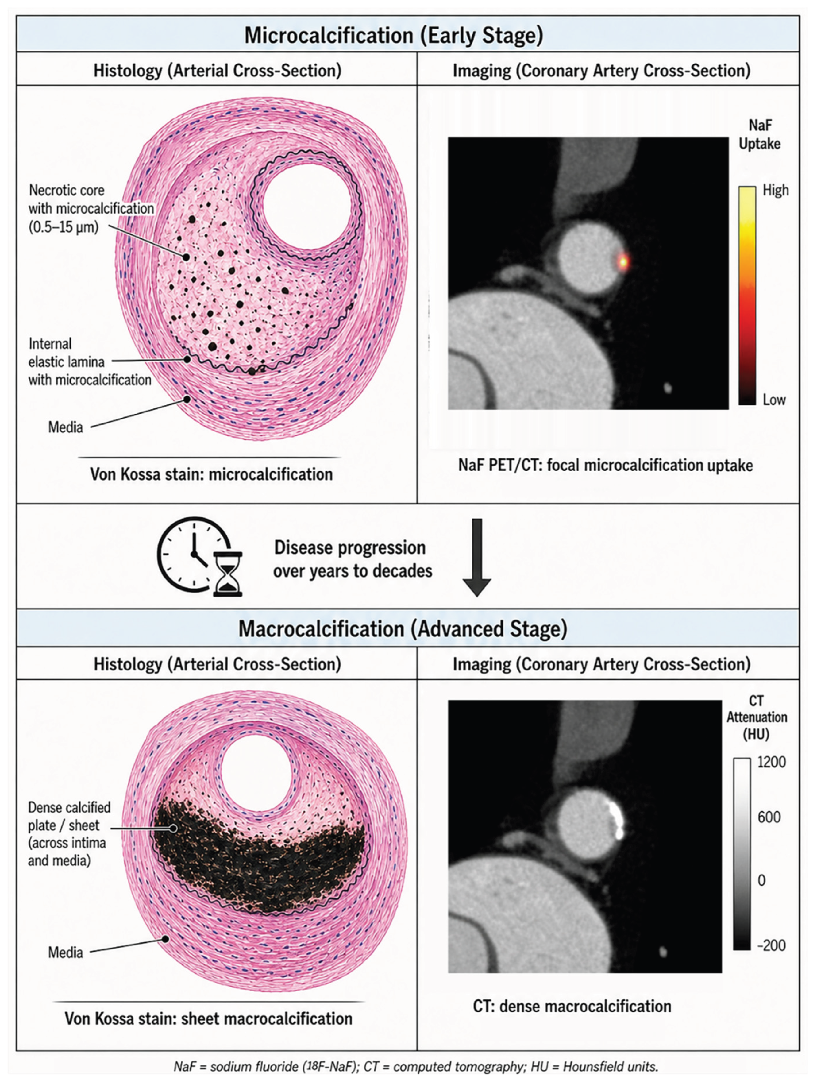
Beyond their distinct biological targets, NaF offers several practical imaging advantages over FDG for vascular quantification: reduced spillover from adjacent avid tissues—a particular problem for FDG given high myocardial glucose uptake—and faster blood clearance, which lowers background blood-pool activity and improves the accuracy of arterial wall uptake measurement [49]. NaF uptake also appears more temporally stable than FDG uptake, which can fluctuate over short intervals in ways that complicate serial monitoring for treatment response [50,51]. Together, these properties position NaF as the more tractable tracer for longitudinal, quantitative studies of calcification biology, while FDG remains complementary for characterizing the inflammatory component of plaque activity. Figure 1 provides a schematic, illustrative comparison of early microcalcification versus advanced macrocalcification, juxtaposing representative histological staining patterns with corresponding imaging appearances on NaF PET/CT and CT to conceptually demonstrate why molecular imaging can detect disease activity at a stage structurally invisible to CT.
3.2. Clinical Evidence in Humans
Patients with angina pectoris show significantly elevated pulmonary artery NaF uptake compared with healthy controls [52], and patients with rheumatoid arthritis—a condition associated with accelerated atherosclerosis—demonstrate elevated abdominal aortic NaF uptake alongside higher CT calcium scores, while FDG uptake shows no such difference [53]. In patients with peripheral arterial disease, NaF-PET revealed 30% higher total atherosclerotic burden in non-lower-extremity arteries compared with disease-free controls, driven by elevated carotid and thoracic aortic uptake [54]. In patients imaged for prostate cancer or chest pain, coronary NaF uptake was significantly higher in those with ischemic versus normal myocardial perfusion, and both obstructive and non-obstructive coronary lesions showed higher NaF uptake than disease-free segments, although the correlation with CT calcification itself was often poor—reinforcing that NaF uptake and structural calcium burden are related but non-identical measures [55].
NaF uptake also correlates with plaque vulnerability. Culprit plaques in patients with ST-elevation myocardial infarction show 23–27% higher coronary NaF uptake than plaques in stable angina patients [56]. Intracoronary optical coherence tomography studies have shown that NaF-positive coronary segments have higher lipid arc, greater macrophage prevalence, thinner plaque free wall, and greater total and calcified plaque burden than NaF-negative segments [57]. Similarly, carotid culprit lesions show higher NaF uptake than non-culprit plaques, and thin-cap fibroatheromas—the histological hallmark of vulnerable plaque—cluster within coronary segments exhibiting elevated NaF target-to-background ratios above 1.28 [58]. In carotid stenosis patients, NaF uptake was significantly associated with MRI-defined culprit lesion features including necrosis, intraplaque hemorrhage, ulceration, and calcification [59]. These findings collectively position NaF PET not merely as a marker of calcification burden, but as a potential indicator of plaque instability itself.
4. NaF-PET for Monitoring Targeted Anti-Atherosclerotic Therapy
The most clinically compelling application of NaF-PET lies in its apparent ability to detect treatment response at a molecular level, even when structural imaging shows no change—or paradoxically, an increase—in calcification.
4.1. Pharmacological Interventions in Preclinical Models
Pharmacological interventions show similarly dissociated effects between molecular and structural imaging. In a rabbit model combining cholesterol-enriched diet with balloon injury of the abdominal aorta, animals treated with atorvastatin (5 mg/kg/day) showed only a 2% increase in NaF uptake from baseline over 18 weeks, compared with a 20% increase in untreated controls—despite total calcium density, measured by von Kossa staining, increasing by 235% in the atorvastatin group relative to untreated animals. Notably, although overall calcium density rose sharply with statin treatment, the areas of microcalcification within plaque were significantly lower, and microcalcification deposits co-localized more closely with regions of elevated NaF uptake, suggesting a shift toward more mature, stable macrocalcification alongside suppression of the biologically active microcalcific component [60].
Vitamin K status has also been examined mechanistically. In ApoE-/- mice maintained on a Western-type diet for twelve weeks followed by a further twelve weeks on control, advanced Western, vitamin K, or Warfarin diets, the Warfarin group—which impairs vitamin K-dependent calcification inhibitors—developed spotty aortic calcifications on CT corresponding to dense mineralization on von Kossa staining, and showed the highest NaF uptake in the aortic arch and left ventricle alongside the advanced-diet group. The vitamin K supplemented group had the lowest NaF uptake after controls, while the advanced Western-diet group showed elevated NaF uptake without developing CT-visible spotty calcification at all, directly demonstrating that NaF-PET can detect active microcalcification in the complete absence of structural CT findings [61].
Direct inhibition of TNAP, the enzyme central to matrix vesicle-mediated mineralization, was evaluated using NaF-PET/CT, ex vivo osteosense imaging, and in vitro Alizarin red staining in ApoE-deficient mice. TNAP inhibition prevented aortic calcification both in vivo and in human vascular smooth muscle cells, and additionally reduced blood cholesterol and triglyceride levels, without adversely affecting skeletal bone structure—raising the possibility of a targeted anti-calcific therapy with a favorable off-target metabolic profile [62]. By contrast, a study of semaglutide, a glucagon-like peptide-1 receptor agonist, in a non-diabetic rat model of atherosclerosis found reduced uptake of the macrophage-tracking tracers 64Cu-DOTATATE and FDG, but no change in NaF uptake, supporting the interpretation that semaglutide’s vascular benefit operates through suppression of inflammatory macrophage activity rather than direct inhibition of the calcification pathway [63].
4.2. Human Intervention Studies
In a notable case report, a 64-year-old man with mild hypercholesterolemia and a history of limited teenage smoking underwent baseline NaF-PET/CT, which showed a mixed plaque near the right coronary artery ostium, a smaller plaque in the left main coronary artery, and minor left anterior descending artery calcification. After six months of rosuvastatin 10 mg/day, aspirin 75 mg/day, and lifestyle modification, his LDL cholesterol fell by 65%. A repeat scan showed an unchanged CT appearance and unchanged Agatston score, yet revealed a 37% decrease in focal NaF uptake in the right coronary artery plaque and a 40% decrease in the left main plaque, with no new hotspots detected anywhere, including the left anterior descending artery [64]. This case offered an early demonstration that molecular imaging can capture a therapeutic response invisible to structural CT.
This pattern was formally replicated in a larger cohort of 38 patients, most of whom had diabetes and were classified at high or very high cardiovascular risk [65]. In this group, the highest baseline NaF uptake was most commonly observed in the abdominal aorta, followed by the descending aorta, aortic arch, and carotid arteries. Patients with subclinical atherosclerosis and significant plaque NaF uptake were treated with rosuvastatin 20 mg/day for six months, resulting in a statistically significant 19.2% reduction in maximum plaque NaF uptake, with median target-to-background ratio falling from 1.96 to 1.53. Together, the case report and cohort data indicate that statin therapy can measurably suppress microcalcification activity well before any corresponding change becomes apparent on CT.
The PCSK9 inhibitor evolocumab, a monoclonal antibody that blocks hepatic LDL receptor degradation and thereby enhances LDL clearance, has shown comparable effects in a cohort of 47 patients (mean age 61.8 years, 87% male) with extensive non-calcified coronary plaque and 196 evaluable lesions [66]. Over 18 months of serial coronary CT angiography and NaF PET, evolocumab significantly reduced both coronary microcalcification activity, from a mean of 1.35 to 1.08, and lesion-level NaF target-to-background ratio, from 1.73 to 1.62. The proportion of lesions classified as high microcalcification activity fell from 34.1% to 21.3%, and high NaF uptake lesions fell from 63.8% to 55.6%. Reductions in microcalcification activity occurred in most participants, while changes in target-to-background ratio were more variable, suggesting these two quantitative metrics may capture partially distinct facets of the underlying biological response to intensive lipid-lowering therapy.
Not all interventions have shown benefit. A double-blind, placebo-controlled 2×2 factorial trial of vitamin K1 (10 mg/day) and colchicine (0.5 mg/day) in 149 type 2 diabetic patients with existing CT coronary calcification found no significant effect of either agent, alone or combined, on coronary , aortic NaF target-to-background ratio over three months or Agatston score [67]. A subsequent post hoc analysis applying a revised upper limit of normal—defined from a separate zero-calcium diabetic cohort—suggested that vitamin K1 independently decreased the odds of developing new NaF-positive lesions in the coronary arteries (odds ratio 0.35), aorta (odds ratio 0.27), and both territories combined (odds ratio 0.28) [68]. This illustrates how threshold definitions can materially alter the interpretation of otherwise negative trial data.
Dietary intervention data add further nuance to this body of evidence. In eleven healthy older adults without clinical cardiovascular disease, six months of supplementation with high-polyphenol extra virgin olive oil significantly reduced both FDG and NaF uptake, indicating attenuation of both arterial inflammation and microcalcification activity [69]. Standard extra virgin olive oil produced no significant effect, while refined olive oil was associated with increased NaF uptake, suggesting active progression of microcalcification. This dose-dependent divergence across the three olive oil preparations indicates that polyphenol content specifically, rather than olive oil consumption in general, drives the anti-atherosclerotic molecular imaging signal observed in this study.
4.3. Natural History and Prognostic Value
Beyond intervention monitoring, longitudinal studies underscore the slow, variable natural history of arterial NaF uptake, which contrasts with the more consistently progressive nature of CT calcification. In healthy controls followed for two years, global cardiac NaF uptake changed minimally despite a measurable 6% decrease in CT-calcification within the same regions of interest. This finding is reinforced by a dedicated two-year longitudinal study comparing carotid and aortic NaF uptake in 29 healthy controls and 20 angina pectoris patients: aortic partial-volume-corrected mean uptake changed only marginally over two years in both groups (1.14 to 1.29 in angina patients versus 0.99 to 0.95 in healthy controls), with a similar pattern observed in the carotids, and baseline NaF uptake did not predict subsequent change in CT-calcification [69]. Notably, angina patients showed a persistent, though statistically insignificant, trend toward higher NaF uptake than healthy controls at both time points, consistent with a slow but continuously active microcalcification process even under statin therapy, which half of the angina cohort was receiving; NaF uptake across all aortic segments also correlated positively with age [70]. Among patients with pseudoxanthoma elasticum, only two of fourteen showed a two-year increase in CT-calcification of major extracardial arteries, while NaF uptake metrics remained unchanged or even decreased. In a small cohort with 51 CT-defined coronary lesions, already-low NaF uptake showed no change over a follow-up period of three to five years [47].
This slow natural history appears to be modulated by sex and cardiovascular history, according to a separate two-year NaF-PET/CT study in patients with type 2 diabetes. Piri et al. found that microcalcification progression differed significantly by sex, with menopausal women—particularly those without a prior history of cardiovascular events—showing disproportionately higher NaF uptake progression than men or women with established cardiovascular disease [71]. This finding suggests that hormonal status may independently influence microcalcification biology in diabetic patients, adding a further layer of heterogeneity to the already variable natural history of arterial NaF uptake, and raising the possibility that future NaF-PET-based trials may need to stratify or adjust for sex and menopausal status when interpreting longitudinal uptake changes.
Prognostically, baseline NaF positivity predicts future events across several independent cohorts. Among patients with known coronary artery disease, myocardial infarction occurred only in those with abnormal baseline coronary NaF uptake [72]. In diabetic patients, calcification progression was more common in NaF-positive than NaF-negative coronary arteries, and in patients with multivessel disease, more rapid CT-calcification progression—and increase in calcium score—occurred specifically in coronary segments with elevated baseline NaF uptake [73]. In a large analysis of 461 patients with cardiovascular disease followed for approximately one year, progression of thoracic aortic calcium volume correlated with baseline thoracic aortic NaF activity; over six years of follow-up, elevated baseline thoracic aortic NaF uptake was specifically associated with subsequent ischemic stroke, while elevated coronary NaF activity was instead associated with myocardial infarction, suggesting that NaF PET may carry vascular bed-specific prognostic information not captured by a single global calcification score [74]. In one distinct clinical setting, examination of coronary artery bypass grafts found that arterial and venous grafts were entirely devoid of NaF activity nearly three years after implantation, while native, non-bypassed coronary arteries proximal to the graft anastomosis showed three times higher NaF uptake and greater calcium score progression than bypassed segments—an intriguing observation suggesting that surgical revascularization or altered hemodynamics may locally suppress microcalcification activity .
4.4. The Emerging Role of Artificial Intelligence
A recurring limitation of NaF-PET, beyond partial volume effects, is the time-intensive nature of manual image analysis, particularly for small, curved structures like the coronary arteries. Artificial intelligence, and specifically convolutional neural networks (CNNs), offer a potential solution. CNNs are computational models composed of sequential, connected processing layers that extract hierarchical image features and have demonstrated efficiency and accuracy in automated volumetric CT segmentation, detection, and classification tasks across other disease domains, including oncology [75]. Applied to NaF-PET/CT, such models could standardize quantification of tracer uptake, reduce inter-observer variability inherent to manual region-of-interest analysis, and accelerate the transition of NaF-PET from a research tool into a scalable clinical and trial-monitoring instrument [76,77,78]. Dedicated validation studies applying CNN-based segmentation specifically to vascular NaF-PET quantification remain an important area for future development, particularly given the granularity required to resolve microcalcification signal from partial-volume-degraded coronary artery images.
5. Conclusions
Arterial calcification begins as a molecular process—governed by matrix vesicle biology, SMC and macrophage apoptosis, and osteogenic phenotypic transitions—long before it becomes visible as macrocalcification on conventional imaging. NaF-PET imaging bridges this gap, offering a window onto microcalcification activity at a stage when the underlying biology remains dynamic and potentially reversible. Accumulating preclinical and clinical evidence demonstrates that anti-atherosclerotic interventions, can measurably reduce NaF uptake independent of, or even in the setting of rising, CT-based calcium scores. At the same time, negative and mixed findings—from vitamin K trials to semaglutide illustrate that not every therapeutic mechanism engages the calcification pathway detectable by NaF, underscoring the tracer’s specificity rather than representing a limitation of the imaging technique itself. As quantification methods mature particularly through AI-assisted segmentation, NaF-PET is well positioned to become a central tool not only for early diagnosis of subclinical atherosclerosis but for the direct, biologically grounded monitoring of novel targeted therapies.
Author Contributions
Conceptualization, R.P., P.F.H.C.; investigation, R.P., S.T.; writing—original draft preparation, R.P., P.F.H.C..; writing—review and editing, P.F.H.C.; visualization, R.P., S.T; supervision, P.F.H.C.
Funding
This research received no external funding.
Institutional Review Board Statement
Not applicable.
Informed Consent Statement
Not applicable.
Data Availability Statement
No new data were created or analyzed in this study. Data sharing is not applicable to this article.
Acknowledgments
In this section, you can acknowledge any support given which is not covered.
Conflicts of Interest
The authors declare no conflicts of interest.
Abbreviations
The following abbreviations are used in this manuscript:
| ADP | adenosine diphosphate; |
| AI | artificial intelligence; |
| ApoE−/− | apolipoprotein E knockout; |
| ATP | adenosine triphosphate; |
| BMP | bone morphogenetic protein; |
| CAC | coronary artery calcification; |
| CNN | convolutional neural network; |
| CT | computed tomography; |
| [18F]FDGFDG | fluorine-18 fluorodeoxyglucose; |
| [18F]NaF | fluorine-18 sodium fluoride; |
| [64Cu]DOTATATE | copper-64 DOTA-(Tyr3)-octreotate; |
| h/D | gap-to-diameter ratio; |
| LDL | low-density lipoprotein; |
| MRI | magnetic resonance imaging; |
| NPP1/NPP3 | nucleotide pyrophosphatase/phosphodiesterases 1 and 3; |
| PCSK9 | proprotein convertase subtilisin/kexin type 9; |
| PET | positron emission tomography; |
| PET/CT | positron emission tomography/computed tomography; |
| PiT1/PiT2 | type III sodium-dependent phosphate cotransporters 1 and 2; |
| SMC | smooth muscle cell; |
| ST | ST segment; |
| TNAP | tissue-nonspecific alkaline phosphatase; |
| UTS | ultimate tensile strength. |
References
- Timmis, A.; Aboyans, V.; Vardas, P.; Townsend, N.; Torbica, A.; Kavousi, M.; Boriani, G.; Huculeci, R.; Kazakiewicz, D.; Scherr, D. European society of cardiology: the 2023 atlas of cardiovascular disease statistics. Eur. Heart J. 2024, 45, 4019–4062. [Google Scholar] [CrossRef] [PubMed]
- Gambardella, J.; Wang, X.; Mone, P.; Khondkar, W.; Santulli, G. Genetics of adrenergic signaling drives coronary artery calcification. Atherosclerosis 2020, 310, 88–90. [Google Scholar] [CrossRef] [PubMed]
- Ross, R. Atherosclerosis—an inflammatory disease. N. Engl. J. Med. 1999, 340, 115–126. [Google Scholar] [CrossRef] [PubMed]
- Gidding, S.S.; Rana, J.S.; Prendergast, C.; McGill, H.; Carr, J.J.; Liu, K.; Colangelo, L.A.; Loria, C.M.; Lima, J.; Terry, J.G.; et al. Pathobiological Determinants of Atherosclerosis in Youth (PDAY) Risk Score in Young Adults Predicts Coronary Artery and Abdominal Aorta Calcium in Middle Age. Circulation 2016, 133, 139–146. [Google Scholar] [CrossRef] [PubMed]
- Parikh, P.; Shah, N.; Ahmed, H.; Schoenhagen, P.; Fares, M. Coronary artery calcium scoring: Its practicality and clinical utility in primary care. Clevel. Clin. J. Med. 2018, 85, 707–716. [Google Scholar] [CrossRef] [PubMed]
- Ortega, M.A.; De Leon-Oliva, D.; Gimeno-Longas, M.J.; Boaru, D.L.; Fraile-Martinez, O.; García-Montero, C.; de Castro, A.V.; Barrena-Blázquez, S.; López-González, L.; Amor, S.; et al. Vascular Calcification: Molecular Networking, Pathological Implications and Translational Opportunities. Biomolecules 2024, 14. [Google Scholar] [CrossRef] [PubMed]
- Høilund-Carlsen, P.F.; Piri, R.; Gerke, O.; Sturek, M.; Werner, T.J.; Revheim, M.-E.; Alavi, A. NaF-PET imaging of atherosclerosis burden. J. Imaging 2023, 9, 31. [Google Scholar] [CrossRef] [PubMed]
- Lanzer, P.; Hannan, F.M.; Lanzer, J.D.; Janzen, J.; Raggi, P.; Furniss, D.; Schuchardt, M.; Thakker, R.; Fok, P.W.; Saez-Rodriguez, J.; et al. Medial Arterial Calcification: JACC State-of-the-Art Review. J. Am. Coll. Cardiol. 2021, 78, 1145–1165. [Google Scholar] [CrossRef] [PubMed]
- Doherty, T.M.; Fitzpatrick, L.A.; Inoue, D.; Qiao, J.H.; Fishbein, M.C.; Detrano, R.C.; Shah, P.K.; Rajavashisth, T.B. Molecular, endocrine, and genetic mechanisms of arterial calcification. Endocr. Rev. 2004, 25, 629–672. [Google Scholar] [CrossRef] [PubMed]
- Nakahara, T.; Dweck, M.R.; Narula, N.; Pisapia, D.; Narula, J.; Strauss, H.W. Coronary Artery Calcification: From Mechanism to Molecular Imaging. JACC Cardiovasc Imaging 2017, 10, 582–593. [Google Scholar] [CrossRef] [PubMed]
- Otsuka, F.; Sakakura, K.; Yahagi, K.; Joner, M.; Virmani, R. Has our understanding of calcification in human coronary atherosclerosis progressed? Arterioscler. Thromb. Vasc. Biol. 2014, 34, 724–736. [Google Scholar] [CrossRef] [PubMed]
- Mori, H.; Torii, S.; Kutyna, M.; Sakamoto, A.; Finn, A.V.; Virmani, R. Coronary Artery Calcification and its Progression: What Does it Really Mean? JACC Cardiovasc Imaging 2018, 11, 127–142. [Google Scholar] [CrossRef] [PubMed]
- Kirsch, T.; Wang, W.; Pfander, D. Functional differences between growth plate apoptotic bodies and matrix vesicles. J. Bone Min. Res. 2003, 18, 1872–1881. [Google Scholar] [CrossRef] [PubMed]
- Ghadially, F.N. As You Like It, Part 3: A Critique and Historical Review of Calcification as Seen with the Electron Microscope. Ultrastruct. Pathol. 2001, 25, 243–267. [Google Scholar] [CrossRef] [PubMed]
- Bobryshev, Y.V. Transdifferentiation of smooth muscle cells into chondrocytes in atherosclerotic arteries in situ: implications for diffuse intimal calcification. J. Pathol. 2005, 205, 641–650. [Google Scholar] [CrossRef] [PubMed]
- Aikawa, E.; Nahrendorf, M.; Figueiredo, J.L.; Swirski, F.K.; Shtatland, T.; Kohler, R.H.; Jaffer, F.A.; Aikawa, M.; Weissleder, R. Osteogenesis associates with inflammation in early-stage atherosclerosis evaluated by molecular imaging in vivo. Circulation 2007, 116, 2841–2850. [Google Scholar] [CrossRef] [PubMed]
- Rocha-Singh, K.J.; Zeller, T.; Jaff, M.R. Peripheral arterial calcification: prevalence, mechanism, detection, and clinical implications. Catheter Cardiovasc Interv. 2014, 83, E212-220. [Google Scholar] [CrossRef] [PubMed]
- Zheng, J.; Huang, M.; Huang, Q.; Chen, Q.; Chen, Z. The relationship between fetuin-A and coronary atherosclerotic heart disease (CHD) and CHD-related risk factors: A retrospective study. Medicine 2021, 100, e27481. [Google Scholar] [CrossRef] [PubMed]
- Villa-Bellosta, R. Vascular Calcification: Key Roles of Phosphate and Pyrophosphate. Int. J. Mol. Sci. 2021, 22. [Google Scholar] [CrossRef] [PubMed]
- New, S.E.; Goettsch, C.; Aikawa, M.; Marchini, J.F.; Shibasaki, M.; Yabusaki, K.; Libby, P.; Shanahan, C.M.; Croce, K.; Aikawa, E. Macrophage-derived matrix vesicles: an alternative novel mechanism for microcalcification in atherosclerotic plaques. Circ. Res. 2013, 113, 72–77. [Google Scholar] [CrossRef] [PubMed]
- Anderson, H.C. Matrix vesicles and calcification. Curr. Rheumatol. Rep. 2003, 5, 222–226. [Google Scholar] [CrossRef] [PubMed]
- Ma, W.; Jia, K.; Cheng, H.; Xu, H.; Li, Z.; Zhang, H.; Xie, H.; Sun, H.; Yi, L.; Chen, Z.; et al. Orphan Nuclear Receptor NR4A3 Promotes Vascular Calcification via Histone Lactylation. Circ. Res. 2024, 134, 1427–1447. [Google Scholar] [CrossRef] [PubMed]
- Nitschke, Y.; Weissen-Plenz, G.; Terkeltaub, R.; Rutsch, F. Npp1 promotes atherosclerosis in ApoE knockout mice. J. Cell Mol. Med. 2011, 15, 2273–2283. [Google Scholar] [CrossRef] [PubMed]
- Kapustin, A.N.; Chatrou, M.L.; Drozdov, I.; Zheng, Y.; Davidson, S.M.; Soong, D.; Furmanik, M.; Sanchis, P.; De Rosales, R.T.; Alvarez-Hernandez, D.; et al. Vascular smooth muscle cell calcification is mediated by regulated exosome secretion. Circ. Res. 2015, 116, 1312–1323. [Google Scholar] [CrossRef] [PubMed]
- Méndez-Barbero, N.; San Sebastian-Jaraba, I.; Blázquez-Serra, R.; Martín-Ventura, J.L.; Blanco-Colio, L.M. Annexins and cardiovascular diseases: Beyond membrane trafficking and repair. Front. Cell Dev. Biol. 2022, Volume 10 - 2022. [Google Scholar] [CrossRef] [PubMed]
- Verzili, D.; Zamparelli, C.; Mattei, B.; Noegel, A.A.; Chiancone, E. The sorcin-annexin VII calcium-dependent interaction requires the sorcin N-terminal domain. FEBS Lett. 2000, 471, 197–200. [Google Scholar] [CrossRef] [PubMed]
- Rattazzi, M.; Bertacco, E.; Del Vecchio, A.; Puato, M.; Faggin, E.; Pauletto, P. Aortic valve calcification in chronic kidney disease. Nephrol. Dial. Transpl. 2013, 28, 2968–2976. [Google Scholar] [CrossRef] [PubMed]
- Speer, M.Y.; Yang, H.Y.; Brabb, T.; Leaf, E.; Look, A.; Lin, W.L.; Frutkin, A.; Dichek, D.; Giachelli, C.M. Smooth muscle cells give rise to osteochondrogenic precursors and chondrocytes in calcifying arteries. Circ. Res. 2009, 104, 733–741. [Google Scholar] [CrossRef] [PubMed]
- Shanahan, C.M.; Crouthamel, M.H.; Kapustin, A.; Giachelli, C.M. Arterial calcification in chronic kidney disease: key roles for calcium and phosphate. Circ. Res. 2011, 109, 697–711. [Google Scholar] [CrossRef] [PubMed]
- Kelly-Arnold, A.; Maldonado, N.; Laudier, D.; Aikawa, E.; Cardoso, L.; Weinbaum, S. Revised microcalcification hypothesis for fibrous cap rupture in human coronary arteries. Proc. Natl. Acad. Sci. U S A 2013, 110, 10741–10746. [Google Scholar] [CrossRef] [PubMed]
- Vengrenyuk, Y.; Carlier, S.; Xanthos, S.; Cardoso, L.; Ganatos, P.; Virmani, R.; Einav, S.; Gilchrist, L.; Weinbaum, S. A hypothesis for vulnerable plaque rupture due to stress-induced debonding around cellular microcalcifications in thin fibrous caps. Proc. Natl. Acad. Sci. U S A 2006, 103, 14678–14683. [Google Scholar] [CrossRef] [PubMed]
- Yahagi, K.; Kolodgie, F.D.; Lutter, C.; Mori, H.; Romero, M.E.; Finn, A.V.; Virmani, R. Pathology of Human Coronary and Carotid Artery Atherosclerosis and Vascular Calcification in Diabetes Mellitus. Arterioscler. Thromb. Vasc. Biol. 2017, 37, 191–204. [Google Scholar] [CrossRef] [PubMed]
- Virmani, R.; Kolodgie, F.D.; Burke, A.P.; Farb, A.; Schwartz, S.M. Lessons From Sudden Coronary Death. Arterioscler. Thromb. Vasc. Biol. 2000, 20, 1262–1275. [Google Scholar] [CrossRef] [PubMed]
- Mauriello, A.; Sangiorgi, G.M.; Virmani, R.; Trimarchi, S.; Holmes, D.R., Jr.; Kolodgie, F.D.; Piepgras, D.G.; Piperno, G.; Liotti, D.; Narula, J.; et al. A pathobiologic link between risk factors profile and morphological markers of carotid instability. Atherosclerosis 2010, 208, 572–580. [Google Scholar] [CrossRef] [PubMed]
- Dhore, C.R.; Cleutjens, J.P.; Lutgens, E.; Cleutjens, K.B.; Geusens, P.P.; Kitslaar, P.J.; Tordoir, J.H.; Spronk, H.M.; Vermeer, C.; Daemen, M.J. Differential expression of bone matrix regulatory proteins in human atherosclerotic plaques. Arterioscler. Thromb. Vasc. Biol. 2001, 21, 1998–2003. [Google Scholar] [CrossRef] [PubMed]
- Roijers, R.B.; Debernardi, N.; Cleutjens, J.P.; Schurgers, L.J.; Mutsaers, P.H.; van der Vusse, G.J. Microcalcifications in early intimal lesions of atherosclerotic human coronary arteries. Am. J. Pathol. 2011, 178, 2879–2887. [Google Scholar] [CrossRef] [PubMed]
- Luo, G.; Ducy, P.; McKee, M.D.; Pinero, G.J.; Loyer, E.; Behringer, R.R.; Karsenty, G. Spontaneous calcification of arteries and cartilage in mice lacking matrix GLA protein. Nature 1997, 386, 78–81. [Google Scholar] [CrossRef] [PubMed]
- Ijichi, T.; Nakazawa, G.; Torii, S.; Nakano, M.; Yoshikawa, A.; Morino, Y.; Ikari, Y. Evaluation of coronary arterial calcification - Ex-vivo assessment by optical frequency domain imaging. Atherosclerosis 2015, 243, 242–247. [Google Scholar] [CrossRef] [PubMed]
- Maldonado, N.; Kelly-Arnold, A.; Vengrenyuk, Y.; Laudier, D.; Fallon, J.T.; Virmani, R.; Cardoso, L.; Weinbaum, S. A mechanistic analysis of the role of microcalcifications in atherosclerotic plaque stability: potential implications for plaque rupture. Am. J. Physiol. Heart Circ. Physiol. 2012, 303, H619–628. [Google Scholar] [CrossRef] [PubMed]
- Vengrenyuk, Y.; Cardoso, L.; Weinbaum, S. Micro-CT based analysis of a new paradigm for vulnerable plaque rupture: cellular microcalcifications in fibrous caps. Mol. Cell Biomech. 2008, 5, 37–47. [Google Scholar] [PubMed]
- Bluestein, D.; Alemu, Y.; Avrahami, I.; Gharib, M.; Dumont, K.; Ricotta, J.J.; Einav, S. Influence of microcalcifications on vulnerable plaque mechanics using FSI modeling. J. Biomech. 2008, 41, 1111–1118. [Google Scholar] [CrossRef] [PubMed]
- Cardoso, L.; Kelly-Arnold, A.; Maldonado, N.; Laudier, D.; Weinbaum, S. Effect of tissue properties, shape and orientation of microcalcifications on vulnerable cap stability using different hyperelastic constitutive models. J. Biomech. 2014, 47, 870–877. [Google Scholar] [CrossRef] [PubMed]
- Bertazzo, S.; Gentleman, E.; Cloyd, K.L.; Chester, A.H.; Yacoub, M.H.; Stevens, M.M. Nano-analytical electron microscopy reveals fundamental insights into human cardiovascular tissue calcification. Nat. Mater. 2013, 12, 576–583. [Google Scholar] [CrossRef] [PubMed]
- Jansen, I.; Cahalane, R.; Hengst, R.; Akyildiz, A.; Farrell, E.; Gijsen, F.; Aikawa, E.; van der Heiden, K.; Wissing, T. The interplay of collagen, macrophages, and microcalcification in atherosclerotic plaque cap rupture mechanics. Basic Res. Cardiol. 2024, 119, 193–213. [Google Scholar] [CrossRef] [PubMed]
- Corti, A.; Khalil, D.; De Paolis, A.; Cardoso, L. Size and proximity of micro-scale hard-inclusions increase the risk of rupture in fibroatheroma-like laboratory models. J. Mech. Behav. BioMed Mater. 2023, 141, 105749. [Google Scholar] [CrossRef] [PubMed]
- McKenney-Drake, M.L.; Moghbel, M.C.; Paydary, K.; Alloosh, M.; Houshmand, S.; Moe, S.; Salavati, A.; Sturek, J.M.; Territo, P.R.; Weaver, C.; et al. 18F-NaF and 18F-FDG as molecular probes in the evaluation of atherosclerosis. Eur. J. Nucl. Med. Mol. Imaging 2018, 45, 2190–2200. [Google Scholar] [CrossRef] [PubMed]
- Omarjee, L.; Mention, P.-J.; Janin, A.; Kauffenstein, G.; Le Pabic, E.; Meilhac, O.; Blanchard, S.; Navasiolava, N.; Leftheriotis, G.; Couturier, O.; et al. Assessment of Inflammation and Calcification in Pseudoxanthoma Elasticum Arteries and Skin with 18F-FluroDeoxyGlucose and 18F-Sodium Fluoride Positron Emission Tomography/Computed Tomography Imaging: The GOCAPXE Trial. J. Clin. Med. 2020, 9, 3448. [Google Scholar] [CrossRef] [PubMed]
- Derlin, T.; Tóth, Z.; Papp, L.; Wisotzki, C.; Apostolova, I.; Habermann, C.R.; Mester, J.; Klutmann, S. Correlation of Inflammation Assessed by <sup>18</sup>F-FDG PET, Active Mineral Deposition Assessed by <sup>18</sup>F-Fluoride PET, and Vascular Calcification in Atherosclerotic Plaque: A Dual-Tracer PET/CT Study. J. Nucl. Med. 2011, 52, 1020. [Google Scholar] [CrossRef] [PubMed]
- Blomberg, B.A.; de Jong, P.A.; Thomassen, A.; Lam, M.G.E.; Vach, W.; Olsen, M.H.; Mali, W.P.T.M.; Narula, J.; Alavi, A.; Høilund-Carlsen, P.F. Thoracic Aorta Calcification but Not Inflammation Is Associated with Increased Cardiovascular Disease Risk: Results of the CAMONA Study. Eur. J. Nucl. Med. Mol. Imaging 2017. [Google Scholar] [CrossRef] [PubMed]
- Blomberg, B.A.; Thomassen, A.; Takx, R.A.P.; Hildebrandt, M.G.; Simonsen, J.A.; Buch-Olsen, K.M.; Diederichsen, A.C.P.; Mickley, H.; Alavi, A.; Høilund-Carlsen, P.F. Delayed 18F-fluorodeoxyglucose PET/CT imaging improves quantitation of atherosclerotic plaque inflammation: Results from the CAMONA study. J. Nucl. Cardiol. 2014, 21, 588–597. [Google Scholar] [CrossRef] [PubMed]
- Blomberg, B.A.; Thomassen, A.; de Jong, P.A.; Simonsen, J.A.; Lam, M.G.; Nielsen, A.L.; Mickley, H.; Mali, W.P.; Alavi, A.; Høilund-Carlsen, P.F. Impact of Personal Characteristics and Technical Factors on Quantification of Sodium 18F-Fluoride Uptake in Human Arteries: Prospective Evaluation of Healthy Subjects. J. Nucl. Med. 2015, 56, 1534–1540. [Google Scholar] [CrossRef] [PubMed]
- Zhang, V.; Borja, A.J.; Rojulpote, C.; Padmanabhan, S.; Patil, S.; Gonuguntla, K.; Revheim, M.E.; Werner, T.J.; Høilund-Carlsen, P.F.; Alavi, A. Global quantification of pulmonary artery atherosclerosis using (18)F-sodium fluoride PET/CT in at-risk subjects. Am. J. Nucl. Med. Mol. Imaging 2020, 10, 119–126. [Google Scholar] [CrossRef] [PubMed]
- Mehdizadeh Seraj, S.; Raynor, W.; Rojulpote, C.; Zirakchian Zadeh, M.; Arani, L.; Werner, T.; Hoilund-Carlsen, P.F.; Baker, J.; Alavi, A.; Hunt, S. Assessing the feasibility of NaF or FDG as PET probes to evaluate atherosclerosis in rheumatoid arthritis patients. J. Nucl. Med. 2019, 60, 1439. [Google Scholar]
- Asadollahi, S.; Rojulpote, C.; Bhattaru, A.; Patil, S.; Gonuguntla, K.; Karambelkar, P.; Borja, A.J.; Vuthaluru, K.; Seraj, S.M.; Zhang, V.; et al. Comparison of atherosclerotic burden in non-lower extremity arteries in patients with and without peripheral artery disease using (18)F-NaF-PET/CT imaging. Am. J. Nucl. Med. Mol. Imaging 2020, 10, 272–278. [Google Scholar] [PubMed]
- Hayrapetian, A.; Berenji, G.R.; Nguyen, K.L.; Li, Y. (18)F-Sodium fluoride uptake is associated with severity of atherosclerotic stenosis in stable ischemic heart disease. J. Nucl. Cardiol. 2021, 28, 3058–3066. [Google Scholar] [CrossRef] [PubMed]
- Ashwathanarayana, A.G.; Singhal, M.; Satapathy, S.; Sood, A.; Mittal, B.R.; Kumar, R.M.; Parmar, M.; Krishnappa, D.; Rana, N. 18F-NaF PET uptake characteristics of coronary artery culprit lesions in a cohort of patients of acute coronary syndrome with ST-elevation myocardial infarction and chronic stable angina: A hybrid fluoride PET/CTCA study. J. Nucl. Cardiol. 2022, 29, 558–568. [Google Scholar] [CrossRef]
- Majeed, K.; Bellinge, J.W.; Butcher, S.C.; Alcock, R.; Spiro, J.; Playford, D.; Hillis, G.S.; Newby, D.E.; Mori, T.A.; Francis, R.; et al. Coronary 18F-sodium fluoride PET detects high-risk plaque features on optical coherence tomography and CT-angiography in patients with acute coronary syndrome. Atherosclerosis 2021, 319, 142–148. [Google Scholar] [CrossRef] [PubMed]
- Wurster, T.H.; Landmesser, U.; Abdelwahed, Y.S.; Skurk, C.; Morguet, A.; Leistner, D.M.; Fröhlich, G.; Haghikia, A.; Engel, L.C.; Schuster, A.; et al. Simultaneous [18F]fluoride and gadobutrol enhanced coronary positron emission tomography/magnetic resonance imaging for in vivo plaque characterization. Eur. Heart J.-Cardiovasc. Imaging 2022, 23, 1391–1398. [Google Scholar] [CrossRef] [PubMed]
- Mechtouff, L.; Sigovan, M.; Douek, P.; Costes, N.; Le Bars, D.; Mansuy, A.; Haesebaert, J.; Bani-Sadr, A.; Tordo, J.; Feugier, P.; et al. Simultaneous assessment of microcalcifications and morphological criteria of vulnerability in carotid artery plaque using hybrid 18F-NaF PET/MRI. J. Nucl. Cardiol. 2022, 29, 1064–1074. [Google Scholar] [CrossRef] [PubMed]
- Zhang, X.; Chen, X.; Liang, Z.; Nie, M.; Yan, Y.; Zhao, Q. Atorvastatin Promotes Macrocalcification, But Not Microcalcification in Atherosclerotic Rabbits: An 18F-NaF PET/CT Study. J. Cardiovasc. Pharmacol. 2021, 78. [Google Scholar] [CrossRef] [PubMed]
- Bellinge, J.W.; Francis, R.J.; Lee, S.C.; Vickery, A.; Macdonald, W.; Gan, S.K.; Chew, G.T.; Phillips, M.; Lewis, J.R.; Watts, G.F.; et al. The effect of Vitamin-K1 and Colchicine on Vascular Calcification Activity in subjects with Diabetes Mellitus (ViKCoVaC): A double-blind 2x2 factorial randomized controlled trial. J. Nucl. Cardiol. 2022, 29, 1855–1866. [Google Scholar] [CrossRef] [PubMed]
- Bessueille, L.; Kawtharany, L.; Quillard, T.; Goettsch, C.; Briolay, A.; Taraconat, N.; Balayssac, S.; Gilard, V.; Mebarek, S.; Peyruchaud, O.; et al. Inhibition of alkaline phosphatase impairs dyslipidemia and protects mice from atherosclerosis. Transl. Res. 2023, 251, 2–13. [Google Scholar] [CrossRef] [PubMed]
- Jensen, J.K.; Binderup, T.; Grandjean, C.E.; Bentsen, S.; Ripa, R.S.; Kjaer, A. Semaglutide reduces vascular inflammation investigated by PET in a rabbit model of advanced atherosclerosis. Atherosclerosis 2022, 352, 88–95. [Google Scholar] [CrossRef]
- Dietz, M.; Chironi, G.; Faraggi, M. Reduced 18F-sodium fluoride activity in coronary plaques after statin therapy. Eur. Heart J.-Cardiovasc. Imaging 2021, 22, e133–e133. [Google Scholar] [CrossRef] [PubMed]
- Oliveira-Santos, M.; Borges-Rosa, J.; Silva, R.; Paixão, L.; Santo, C.E.; Abrunhosa, A.; Castelo-Branco, M.; Slomka, P.J.; Gonçalves, L.; Ferreira, M.J. Rosuvastatin effect on atherosclerotic plaque metabolism: A subclinical atherosclerosis imaging study with (18)F-NaF PET-CT. Atherosclerosis 2024, 395, 117481. [Google Scholar] [CrossRef] [PubMed]
- Han, D.; Tzolos, E.; Park, R.; Gransar, H.; Hyun, M.; Friedman John, D.; Hayes Sean, W.; Thomson Louise, E.J.; Kwan Alan, C.; Budoff, M.; et al. Effects of Evolocumab on Coronary Plaque Composition and Microcalcification Activity by Coronary PET and CT Angiography. JACC Cardiovasc. Imaging 2025, 18, 589–599. [Google Scholar] [CrossRef] [PubMed]
- Meer, R.; Romero Prats, M.L.; Vervloet, M.G.; van der Schouw, Y.T.; de Jong, P.A.; Beulens, J.W.J. The effect of six-month oral vitamin K supplementation on calcification propensity time in individuals with type 2 diabetes mellitus: A post hoc analysis of a randomized, double-blind, placebo-controlled trial. Atherosclerosis 2024, 394, 117307. [Google Scholar] [CrossRef] [PubMed]
- Bellinge, J.W.; Francis, R.J.; Lee, S.C.; Bondonno, N.P.; Sim, M.; Lewis, J.R.; Watts, G.F.; Schultz, C.J. The effect of vitamin K1 on arterial calcification activity in subjects with diabetes mellitus: a post hoc analysis of a double-blind, randomized, placebo-controlled trial. Am. J. Clin. Nutr. 2022, 115, 45–52. [Google Scholar] [CrossRef] [PubMed]
- Zoubdane, N.; Abdo, R.A.; Nguyen, M.; Bentourkia, M.; Turcotte, E.E.; Berrougui, H.; Fulop, T.; Khalil, A. High Tyrosol and Hydroxytyrosol Intake Reduces Arterial Inflammation and Atherosclerotic Lesion Microcalcification in Healthy Older Populations. Antioxidants 2024, 13. [Google Scholar] [CrossRef] [PubMed]
- Piri, R.; Lici, G.; Riyahimanesh, P.; Gerke, O.; Alavi, A.; Høilund-Carlsen, P.F. Two-year change in 18F-sodium fluoride uptake in major arteries of healthy subjects and angina pectoris patients. Int. J. Cardiovasc. Imaging 2021, 37, 3115–3126. [Google Scholar] [CrossRef] [PubMed]
- Piri, R.; Stidsen, J.V.; Nielsen, J.S.; Henriksen, J.E.; Thomsen, R.W.; Olesen, T.B.; Pareek, M.; Diederichsen, A.C.P.; Olsen, M.H.; Høilund-Carlsen, P.F. NaF-PET/CT imaging of atherosclerosis in type 2 diabetes: Associations with sex and history of cardiovascular events in a 2-year follow-up study. Eur. J. Nucl. Med. Mol. Imaging 2025, 52, 5006–5016. [Google Scholar] [CrossRef] [PubMed]
- Doris, M.K.; Meah, M.N.; Moss, A.J.; Andrews, J.P.M.; Bing, R.; Gillen, R.; Weir, N.; Syed, M.; Daghem, M.; Shah, A.; et al. Coronary 18F-Fluoride Uptake and Progression of Coronary Artery Calcification. Circ. Cardiovasc. Imaging 2020, 13, e011438. [Google Scholar] [CrossRef] [PubMed]
- Bellinge, J.W.; Francis, R.J.; Lee, S.C.; Phillips, M.; Rajwani, A.; Lewis, J.R.; Watts, G.F.; Schultz, C.J. 18F-Sodium Fluoride Positron Emission Tomography Activity Predicts the Development of New Coronary Artery Calcifications. Arterioscler. Thromb. Vasc. Biol. 2021, 41, 534–541. [Google Scholar] [CrossRef] [PubMed]
- Fletcher Alexander, J.; Tew Yong, Y.; Tzolos, E.; Joshi Shruti, S.; Kaczynski, J.; Nash, J.; Debono, S.; Lembo, M.; Kwiecinski, J.; Bing, R.; et al. Thoracic Aortic 18F-Sodium Fluoride Activity and Ischemic Stroke in Patients With Established Cardiovascular Disease. JACC Cardiovasc. Imaging 2022, 15, 1274–1288. [Google Scholar] [CrossRef] [PubMed]
- Song, D.; Yang, S.; Han, J.Y.; Kim, K.G.; Kim, S.T.; Yi, W.-J. Comparison of segmentation performance of cnns, vision transformers, and hybrid networks for paranasal sinuses with sinusitis on CT images. Sci. Rep. 2025, 15, 32087. [Google Scholar] [CrossRef] [PubMed]
- Piri, R.; Hamakan, Y.; Vang, A.; Edenbrandt, L.; Larsson, M.; Enqvist, O.; Gerke, O.; Høilund-Carlsen, P.F. Common carotid segmentation in (18) F-sodium fluoride PET/CT scans: Head-to-head comparison of artificial intelligence-based and manual method. Clin. Physiol. Funct. Imaging 2023, 43, 71–77. [Google Scholar] [CrossRef]
- Li, S.; Kendrick, J.; Ebert, M.A.; Hassan, G.M.; Barry, N.; Wright, K.; Lee, S.C.; Bellinge, J.W.; Schultz, C. Auto-segmentation, radiomic reproducibility, and comparison of radiomics between manual and AI-derived segmentations for coronary arteries in cardiac [18F]NaF PET/CT images. EJNMMI Phys. 2025, 12, 42. [Google Scholar] [CrossRef]
- Piri, R.; Edenbrandt, L.; Larsson, M.; Enqvist, O.; Nøddeskou-Fink, A.H.; Gerke, O.; Høilund-Carlsen, P.F. Aortic wall segmentation in 18F-sodium fluoride PET/CT scans: Head-to-head comparison of artificial intelligence-based versus manual segmentation. J. Nucl. Cardiol. 2022, 29, 2001–2010. [Google Scholar] [CrossRef] [PubMed]
Figure 1.
Histology-to-imaging correlation of arterial calcification across disease stages. Top row: Early-stage microcalcification. Left, von Kossa staining of an arterial cross-section with pathological intimal thickening demonstrates small, scattered, dark punctate deposits (0.5–15.0 μm) localized near the internal elastic lamina and at the outer margins of the necrotic core, representing the earliest histologically identifiable calcific deposits arising from smooth muscle cell and macrophage apoptosis. Right, corresponding NaF-PET/CT fusion image shows a focal, elevated tracer uptake in a region of the coronary vessel wall without a corresponding dense signal on the unenhanced CT component, illustrating NaF-PET’s ability to detect molecularly active microcalcification before it becomes visible as macroscopic mineral deposition. Bottom row: Advanced-stage macrocalcification. Left, von Kossa staining demonstrates a large, confluent, dense calcified sheet spanning the arterial wall, reflecting coalescence of microcalcific deposits over years to decades. Right, corresponding CT image shows a well-defined, dense, hyperattenuating (bright white) calcified plaque within the coronary vessel wall, consistent with a positive Agatston calcium score. Central arrow indicates disease progression from molecular microcalcification to structural macrocalcification over a timescale of years to decades, during which NaF-PET, but not CT, can detect ongoing disease activity.
Figure 1.
Histology-to-imaging correlation of arterial calcification across disease stages. Top row: Early-stage microcalcification. Left, von Kossa staining of an arterial cross-section with pathological intimal thickening demonstrates small, scattered, dark punctate deposits (0.5–15.0 μm) localized near the internal elastic lamina and at the outer margins of the necrotic core, representing the earliest histologically identifiable calcific deposits arising from smooth muscle cell and macrophage apoptosis. Right, corresponding NaF-PET/CT fusion image shows a focal, elevated tracer uptake in a region of the coronary vessel wall without a corresponding dense signal on the unenhanced CT component, illustrating NaF-PET’s ability to detect molecularly active microcalcification before it becomes visible as macroscopic mineral deposition. Bottom row: Advanced-stage macrocalcification. Left, von Kossa staining demonstrates a large, confluent, dense calcified sheet spanning the arterial wall, reflecting coalescence of microcalcific deposits over years to decades. Right, corresponding CT image shows a well-defined, dense, hyperattenuating (bright white) calcified plaque within the coronary vessel wall, consistent with a positive Agatston calcium score. Central arrow indicates disease progression from molecular microcalcification to structural macrocalcification over a timescale of years to decades, during which NaF-PET, but not CT, can detect ongoing disease activity.
Disclaimer/Publisher’s Note: The statements, opinions and data contained in all publications are solely those of the individual author(s) and contributor(s) and not of MDPI and/or the editor(s). MDPI and/or the editor(s) disclaim responsibility for any injury to people or property resulting from any ideas, methods, instructions or products referred to in the content. |
© 2026 by the authors. Licensee MDPI, Basel, Switzerland. This article is an open access article distributed under the terms and conditions of the Creative Commons Attribution (CC BY) license.
Copyright: This open access article is published under a Creative Commons CC BY 4.0 license, which permit the free download, distribution, and reuse, provided that the author and preprint are cited in any reuse.


