

Submitted:
19 July 2026
Posted:
22 July 2026
You are already at the latest version
Abstract
Theasaponin E2 (TE2) is a triterpenoid saponin isolated from Camellia sinensis seeds with reported anti-inflammatory, antibacterial, and antitumor activities; however, its effects on osteoclasts remain unclear. In this study, TE2 markedly inhibited RANKL-induced osteoclast differentiation in RAW264.7 cells without affecting precursor-cell viability. TE2 also significantly reduced the number and viability of mature, differentiated osteoclasts. Mechanistically, TE2 suppressed RANKL-induced phosphorylation of AKT at Ser473 and GSK-3β at Ser9, as determined by ELISA. Molecular docking predicted that TE2 could interact with the interdomain allosteric pocket of AKT1 at the pleckstrin homology–kinase domain interface (PDB ID: 7NH5; docking score, −9.8 kcal/mol) and the ATP-binding pocket of GSK-3β (PDB ID: 5K5N; docking score, −7.7 kcal/mol). The selected docking poses contained multiple hydrogen-bonding interactions with donor–acceptor distances of 2.79–3.19 Å. Collectively, these findings indicate that TE2 suppresses osteoclast differentiation and reduces mature osteoclast viability, potentially through modulation of the AKT/GSK-3β signaling axis. TE2 may therefore represent a promising natural-product candidate for further investigation in conditions characterized by excessive osteoclast-mediated bone resorption.
Keywords:
theasaponin E2
; osteoclastogenesis
; RANKL
; AKT/GSK-3β signaling
; molecular docking
; bone resorption
Introduction
Osteoclasts are multinucleated cells primarily responsible for bone resorption [1]. Their differentiation from monocyte/macrophage-lineage precursors is regulated principally by receptor activator of nuclear factor-κB ligand (RANKL) and macrophage colony-stimulating factor (M-CSF) [2]. Binding of RANKL to its receptor, RANK, activates multiple downstream signaling pathways, including the phosphoinositide 3-kinase/AKT pathway [3]. Activated AKT phosphorylates glycogen synthase kinase-3β (GSK-3β) at Ser9, thereby suppressing its activity and promoting the nuclear accumulation and transcriptional activity of nuclear factor of activated T cells c1 (NFATc1), the master regulator of osteoclast differentiation [3,4]. Excessive osteoclast formation and activity contribute to pathological bone resorption in disorders such as osteoporosis, periodontitis, and inflammatory bone loss [5]. Therefore, identifying compounds that inhibit osteoclastogenesis by modulating the AKT/GSK-3β signaling axis may support the development of new therapeutic strategies for osteolytic diseases.
Natural products remain an important source of lead compounds for regulating kinase-dependent signaling pathways because of their considerable structural and chemical diversity [6]. Theasaponin E2 (TE2) is a triterpenoid saponin isolated from the seeds of Camellia sinensis [7]. Structurally, TE2 belongs to the barringtogenol C-type oleanane triterpenoid saponins and contains an acylated aglycone core linked to an oligosaccharide chain. Tea saponins have also been reported to modulate kinase-associated signaling pathways, including MAPK/ERK signaling, in other cellular systems [8,9]. However, the effects of TE2 on osteoclast differentiation and survival have not been characterized, and its potential interactions with kinases involved in osteoclast signaling remain unknown.
In this study, we investigated the effects of TE2 on RANKL-induced osteoclast differentiation and mature osteoclast viability in vitro. We further examined its effects on AKT and GSK-3β phosphorylation and performed molecular docking analyses to explore potential interactions of TE2 with both kinases.
Materials and Methods
Cell Culture and Reagents. Murine macrophage RAW264.7 cells were cultured in Dulbecco’s Modified Eagle Medium (DMEM; Gibco) supplemented with 10% fetal bovine serum and 1% penicillin–streptomycin at 37 °C in a humidified incubator with 5% CO2 as described earlier [10]. Theasaponin E2 (TE2) (purity ≥ 98.76%; CAS No.: 220114-30-7; HY-N12153) was purchased from MedChemExpress (NJ, USA). Recombinant mouse RANKL (462-TEC-010) was obtained from R&D Systems (MN, USA).
Cell Viability Assay. RAW264.7 cells were seeded in 96-well plates (5 × 10³ cells/well) and treated with vehicle or TE2 (1 μM) for 48 h. Cell viability was assessed using a Cell Counting Kit-8 (CCK-8; Service Bio, China) according to the manufacturer’s protocol. Cell viability was expressed as a percentage relative to vehicle-treated controls.
TRAP Staining and osteoclast differentiation. RAW264.7 cells were seeded in 96-well plates (5 × 10³ cells/well) and stimulated with RANKL (50 ng/mL) in the presence or absence of TE2 (1 μM) for 5 days. The culture medium containing RANKL and TE2 was refreshed every 2 days. Cell morphology and attachment were monitored microscopically throughout the differentiation period. After differentiation, cells were fixed with 4% paraformaldehyde for 15 min and stained for tartrate-resistant acid phosphatase (TRAP) activity using a commercial TRAP staining kit (Servicebio, Hubei, China) according to the manufacturer’s protocol. TRAP-positive multinucleated cells (≥3 nuclei) were counted as mature osteoclasts under light microscopy.
Mature osteoclast treatment. RAW264.7 cells were differentiated into mature osteoclasts with RANKL for 5 days and subsequently treated with TE2 (1 μM) for 24 h. TRAP staining and cell viability (CCK-8 assay) were assessed to evaluate osteoclast survival.
Signaling analysis. RAW264.7 cells were pre-treated with TE2 (1 μM) for 1 h prior to RANKL stimulation (50 ng/mL, 10 min). Phosphorylation of AKT (Ser473) and GSK-3β (Ser9) was measured by ELISA as per manufacturer’s protocol (Abcam, USA) and expressed as fold change relative to vehicle control.
Molecular docking. The three-dimensional structure of TE2 was docked against AKT1 (PDB ID: 7NH5) and GSK-3β (PDB ID: 5K5N) using CB-Dock3 [11]. The resulting protein–ligand complexes were analyzed to identify key intermolecular interactions, and two-dimensional interaction diagrams were generated using PoseView. Hydrogen bonds were defined using a donor–acceptor distance cutoff of <3.5 Å between ligand donor or acceptor atoms and the corresponding protein residue atoms.
Statistical analysis. Data are presented as the mean ± standard deviation (SD) from three independent experiments. Comparisons between two groups were performed using an unpaired, two-tailed Student’s t-test. Comparisons among three or more groups were performed using one-way analysis of variance (ANOVA), followed by Tukey’s multiple-comparisons post hoc test. Statistical significance was defined as P < 0.05.
Results
TE2 Suppresses RANKL-Induced Osteoclast Differentiation Without Affecting Precursor Viability
We first examined the effect of TE2 on RANKL-induced osteoclastogenesis in RAW264.7 cells. TRAP staining showed that TE2 treatment (1 μM) markedly reduced the formation of TRAP-positive multinucleated osteoclasts compared with RANKL treatment alone (Figure 1A). In contrast, TE2 did not significantly affect the viability of RAW264.7 osteoclast precursor cells (Figure 1B), indicating that its inhibitory effect on osteoclast differentiation was not attributable to nonspecific cytotoxicity.
TE2 Suppresses AKT/GSK-3β Signaling and Impairs Mature Osteoclast Survival
To investigate the signaling mechanism underlying the inhibitory effect of TE2, we examined RANKL-induced phosphorylation of AKT and GSK-3β. ELISA analysis showed that TE2 significantly reduced the phosphorylation of AKT at Ser473 and GSK-3β at Ser9 (Figure 2A). These findings indicate that TE2 interferes with RANKL-induced activation of the AKT/GSK-3β signaling axis.
We next evaluated the effect of TE2 on differentiated osteoclasts. RAW264.7 cells were differentiated with RANKL for 5 days and subsequently treated with TE2 for 24 h. TRAP staining revealed a marked reduction in the number of mature osteoclasts following TE2 treatment (Figure 2B). Consistently, the CCK-8 assay showed a significant reduction in the viability of mature osteoclast cultures (Figure 2C). These results suggest that mature osteoclasts are more susceptible to TE2 treatment than undifferentiated RAW264.7 precursor cells under the conditions tested.
Molecular Docking Predicts Potential Interactions of TE2 with AKT1 and GSK-3β
To examine whether TE2 could potentially interact with AKT1 and GSK-3β, molecular docking analyses were performed. TE2 was predicted to occupy the allosteric pocket at the interface between the pleckstrin homology and kinase domains of AKT1 (PDB ID: 7NH5), with a predicted docking score of −9.8 kcal/mol. The selected pose formed hydrogen bonds with Tyr18A, Asp274A, Glu278A, and Leu156A, with donor–acceptor distances ranging from 2.79 to 3.19 Å, together with hydrophobic contacts involving Lys297A, Gly311A, and Cys310A.
TE2 was also predicted to bind within the ATP-binding pocket of GSK-3β (PDB ID: 5K5N), with a predicted docking score of −7.7 kcal/mol. The selected pose formed hydrogen bonds with Thr138A, Ser203A, Arg220A, Cys218A, Ser66A, and Ser219A, with distances ranging from 2.82 to 3.06 Å, and hydrophobic contacts with Gln185A and Leu188A (Figure 3 and Table 1). These computational findings provide a possible structural basis for the observed suppression of AKT and GSK-3β phosphorylation observed by ELISA.
Discussion
In this study, we demonstrate that theasaponin E2 (TE2) inhibits RANKL-induced osteoclast differentiation in vitro and reduces the viability of mature osteoclasts. The AKT signaling pathway plays an important role in osteoclast differentiation and survival. Inhibition of AKT phosphorylation decreases the inhibitory phosphorylation of GSK-3β at Ser9, thereby maintaining GSK-3β activity and suppressing NFATc1-dependent osteoclastogenesis [12,13]. Accordingly, the reductions in both p-AKT and p-GSK-3β observed in the present study indicate that TE2 interferes with the AKT/GSK-3β signaling axis.
Molecular docking provided a possible structural basis for these functional effects. TE2 was predicted to occupy the interdomain allosteric pocket of AKT1 and the ATP-binding pocket of GSK-3β, with favorable docking scores and multiple hydrogen-bonding interactions at donor–acceptor distances of 2.79–3.19 Å. The predicted AKT1-binding pose was located within the allosteric pocket represented in structure 7NH5 [14], whereas the GSK-3β pose was located within the ATP-binding region represented in structure 5K5N [15]. These findings raise the possibility that TE2 interacts with both kinases and modulates the AKT/GSK-3β signaling axis through a dual-target mechanism. However, because docking scores and predicted interaction patterns do not establish direct binding or biochemical inhibition, this interpretation requires experimental confirmation.
TE2 also produced different effects in precursor and mature osteoclast cultures. At the concentration tested, TE2 suppressed RANKL-induced osteoclast differentiation without reducing the viability of RAW264.7 precursor cells, whereas treatment of differentiated osteoclasts markedly decreased both osteoclast number and culture viability. These findings suggest that the differential response may reflect the dependence of mature osteoclast survival on sustained prosurvival signaling, including the PI3K/AKT pathway, or other stage-specific vulnerabilities [16]. This response is potentially relevant to pathological bone resorption because a compound that inhibits the formation of new osteoclasts while reducing the persistence of existing mature osteoclasts could exert complementary antiresorptive effects. Nevertheless, additional experiments in primary osteoclast precursors, mature osteoclasts, and other myeloid-lineage cells are required before conclusions regarding cell-type selectivity can be drawn.
Several limitations should be acknowledged. First, the molecular docking analysis is computational and hypothesis-generating and does not demonstrate direct physical binding of TE2 to AKT1 or GSK-3β. Direct target engagement should therefore be examined using orthogonal approaches such as surface plasmon resonance, isothermal titration calorimetry, cellular thermal shift assays, or related binding methods. Cell-free kinase assays are also required to determine whether TE2 directly inhibits AKT1, GSK-3β, or both enzymes. Second, the mechanism responsible for the reduced viability of mature osteoclasts remains unclear. Further studies should determine whether TE2 induces apoptosis, alters cytoskeletal organization, promotes osteoclast detachment, or affects other survival pathways. Finally, the present findings were obtained in an in vitro RAW264.7-cell model. Validation in primary bone marrow macrophages and relevant animal models of osteoporosis or inflammatory bone loss, together with pharmacokinetic and toxicity studies, will be necessary to establish the therapeutic potential of TE2.
Conclusion
TE2 suppresses RANKL-induced osteoclastogenesis by inhibiting the AKT/GSK-3β signaling axis and reduces the viability of mature osteoclast cultures. Molecular docking predicts that TE2 may interact with both AKT1 and GSK-3β, providing a testable hypothesis for its mechanism of action. These findings identify TE2 as a promising natural-product candidate for further investigation as a modulator of pathological osteoclast formation and bone resorption.
Funding
This work was supported by the WKU-ISRG grant (ISRG2023032) and the International Frontier Interdisciplinary Research Institute of Wenzhou-Kean University (IFIRI-WKU) grant (KY20250604000450), both awarded to Vishwa Deepak. Additional support was provided by the Wenzhou-Kean University Talent Introduction Grant (WB20220901000092), awarded to Yili Ding.
Acknowledgments
We thank the core facilities of Wenzhou-Kean University for providing the necessary resources.
Data Availability
The authors confirm that all data generated during this study are included in this published article.
Ethics Approval and Consent to Participate
Not applicable.
Consent for Publication
Not applicable.
Competing Interests
The authors declare no competing interests.
References
- Veis, D.J.; O’Brien, C.A. Osteoclasts, Master Sculptors of Bone. Annu Rev. Pathol. 2023, 18, 257–281. [Google Scholar] [CrossRef] [PubMed]
- Feng, X.; Teitelbaum, S.L. Osteoclasts: New Insights. Bone Res. 2013, 1, 11–26. [Google Scholar] [CrossRef] [PubMed]
- Yin, L.; Sun, C.; Zhang, J.; Li, Y.; Wang, Y.; Bai, L.; Lei, Z. Critical signaling pathways in osteoclast differentiation and bone resorption: mechanisms and therapeutic implications for periprosthetic osteolysis. Front Cell Dev. Biol. 2025, 13, 1639430. [Google Scholar] [CrossRef] [PubMed]
- Deepak, V.; Yang, S.T.; Li, Z.; Li, X.; Ng, A.; Xu, D.; Li, Y.P.; Oursler, M.J.; Yang, S. IFT80 negatively regulates osteoclast differentiation via association with Cbl-b to disrupt TRAF6 stabilization and activation. Proc. Natl. Acad. Sci. U S A 2022, 119, e2201490119. [Google Scholar] [CrossRef] [PubMed]
- Charles, J.F.; Aliprantis, A.O. Osteoclasts: more than ‘bone eaters’. Trends Mol. Med. 2014, 20, 449–459. [Google Scholar] [CrossRef] [PubMed]
- Pye, C.R.; Bertin, M.J.; Lokey, R.S.; Gerwick, W.H.; Linington, R.G. Retrospective analysis of natural products provides insights for future discovery trends. Proc. Natl. Acad. Sci. U S A 2017, 114, 5601–5606. [Google Scholar] [CrossRef] [PubMed]
- Kitagawa, I.; Hori, K.; Motozawa, T.; Murakami, T.; Yoshikawa, M. Structures of new acylated oleanene-type triterpene oligoglycosides, theasaponins E1 and E2, from the seeds of tea plant, Camellia sinensis (L.) O. Kuntze. Chem. Pharm. Bull. 1998, 46, 1901–1906. [Google Scholar] [CrossRef] [PubMed]
- Bhardwaj, J.; Chaudhary, N.; Seo, H.J.; Kim, M.Y.; Shin, T.S.; Kim, J.D. Immunomodulatory effect of tea saponin in immune T-cells and T-lymphoma cells via regulation of Th1, Th2 immune response and MAPK/ERK2 signaling pathway. Immunopharmacol. Immunotoxicol. 2014, 36, 202–210. [Google Scholar] [CrossRef] [PubMed]
- Yu, X.L.; He, Y. Tea saponins: effective natural surfactants beneficial for soil remediation, from preparation to application. RSC Adv. 2018, 8, 24312–24321. [Google Scholar] [CrossRef] [PubMed]
- Zhang, L.; Deepak, V. 8-Epixanthatin Suppresses RANKL-Induced Osteoclast Differentiation via Inhibition of NF-kappaB and MAPK Signaling. Int. J. Mol. Sci. 2026, 27. [Google Scholar] [CrossRef] [PubMed]
- Liu, Y.; Ding, J.; Gan, J.; Xiong, X.; Zong, F.; Xiao, Z.X.; Cao, Y. CB-Dock3: an enhanced web server for protein-ligand blind docking. Nucleic Acids Res. 2026, 54, W238–W245. [Google Scholar] [CrossRef] [PubMed]
- Moon, J.B.; Kim, J.H.; Kim, K.; Youn, B.U.; Ko, A.; Lee, S.Y.; Kim, N. Akt induces osteoclast differentiation through regulating the GSK3beta/NFATc1 signaling cascade. J. Immunol. 2012, 188, 163–169. [Google Scholar] [CrossRef] [PubMed]
- Takayanagi, H.; Kim, S.; Koga, T.; Nishina, H.; Isshiki, M.; Yoshida, H.; Saiura, A.; Isobe, M.; Yokochi, T.; Inoue, J.; et al. Induction and activation of the transcription factor NFATc1 (NFAT2) integrate RANKL signaling in terminal differentiation of osteoclasts. Dev. Cell 2002, 3, 889–901. [Google Scholar] [CrossRef] [PubMed]
- Quambusch, L.; Depta, L.; Landel, I.; Lubeck, M.; Kirschner, T.; Nabert, J.; Uhlenbrock, N.; Weisner, J.; Kostka, M.; Levy, L.M.; et al. Cellular model system to dissect the isoform-selectivity of Akt inhibitors. Nat. Commun. 2021, 12, 5297. [Google Scholar] [CrossRef] [PubMed]
- Liang, S.H.; Chen, J.M.; Normandin, M.D.; Chang, J.S.; Chang, G.C.; Taylor, C.K.; Trapa, P.; Plummer, M.S.; Para, K.S.; Conn, E.L.; et al. Discovery of a Highly Selective Glycogen Synthase Kinase-3 Inhibitor (PF-04802367) That Modulates Tau Phosphorylation in the Brain: Translation for PET Neuroimaging. Angew. Chem. Int. Ed. Engl. 2016, 55, 9601–9605. [Google Scholar] [CrossRef] [PubMed]
- Lee, S.E.; Chung, W.J.; Kwak, H.B.; Chung, C.H.; Kwack, K.B.; Lee, Z.H.; Kim, H.H. Tumor necrosis factor-alpha supports the survival of osteoclasts through the activation of Akt and ERK. J. Biol. Chem. 2001, 276, 49343–49349. [Google Scholar] [CrossRef] [PubMed]
Figure 1.
Theasaponin E2 (TE2) inhibits RANKL-induced osteoclast differentiation in RAW264.7 cells. (A) RAW264.7 cells were cultured with RANKL (50 ng/mL) in the presence or absence of TE2 (1 μM) for 5 days. Cells were fixed and stained for tartrate-resistant acid phosphatase (TRAP) to visualize mature osteoclasts. Representative images of the vehicle, RANKL, and RANKL + TE2 groups are shown. TRAP-positive multinucleated cells containing three or more nuclei were counted as osteoclasts. The bar graph shows the number of osteoclasts per well. Scale bar, 100 μm. (B) RAW264.7 cells were treated with vehicle or TE2 (1 μM) for 48 h, and cell viability was assessed using the CCK-8 assay. Data are presented as the mean ± SD from three independent experiments (n = 3). Statistical significance was determined using an unpaired, two-tailed Student’s t-test. ****P < 0.0001 versus the RANKL-treated group; ns, not significant versus the vehicle group.
Figure 1.
Theasaponin E2 (TE2) inhibits RANKL-induced osteoclast differentiation in RAW264.7 cells. (A) RAW264.7 cells were cultured with RANKL (50 ng/mL) in the presence or absence of TE2 (1 μM) for 5 days. Cells were fixed and stained for tartrate-resistant acid phosphatase (TRAP) to visualize mature osteoclasts. Representative images of the vehicle, RANKL, and RANKL + TE2 groups are shown. TRAP-positive multinucleated cells containing three or more nuclei were counted as osteoclasts. The bar graph shows the number of osteoclasts per well. Scale bar, 100 μm. (B) RAW264.7 cells were treated with vehicle or TE2 (1 μM) for 48 h, and cell viability was assessed using the CCK-8 assay. Data are presented as the mean ± SD from three independent experiments (n = 3). Statistical significance was determined using an unpaired, two-tailed Student’s t-test. ****P < 0.0001 versus the RANKL-treated group; ns, not significant versus the vehicle group.
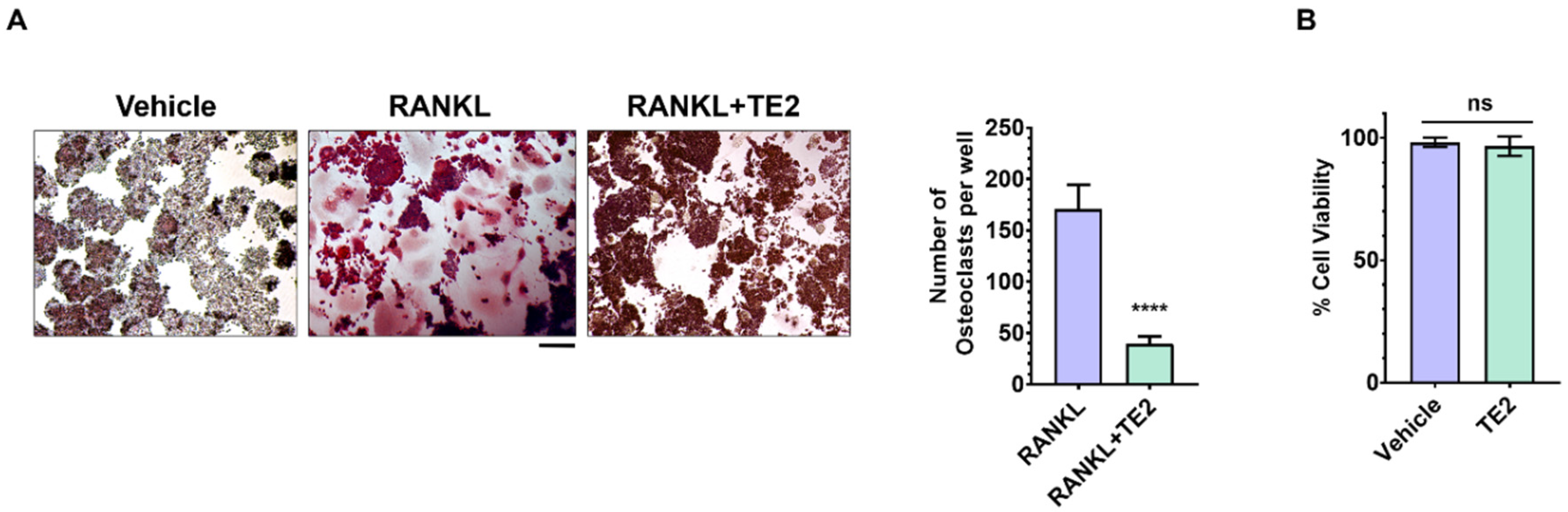
Figure 2.
Theasaponin E2 (TE2) suppresses RANKL-induced AKT/GSK-3β phosphorylation and reduces the number and viability of mature osteoclasts. (A) RAW264.7 cells were pretreated with TE2 (1 μM) for 1 h and subsequently stimulated with RANKL (50 ng/mL) for 10 min. The phosphorylation levels of AKT at Ser473 and GSK-3β at Ser9 were measured by ELISA and expressed as fold changes relative to the vehicle-treated group. (B) RAW264.7 cells were differentiated into mature osteoclasts by treatment with RANKL for 5 days and subsequently treated with vehicle or TE2 (1 μM) for 24 h. Representative TRAP-stained images of the RANKL and RANKL + TE2 groups are shown. TRAP-positive multinucleated cells containing three or more nuclei were counted as osteoclasts, and the graph shows the number of osteoclasts per well. Scale bar, 100 μm. (C) The viability of mature osteoclast cultures following treatment with TE2 (1 μM) for 24 h was assessed using the CCK-8 assay. Data are presented as the mean ± SD from three independent experiments (n = 3). Panel A was analyzed using one-way ANOVA followed by Tukey’s multiple-comparisons test, whereas panels B and C were analyzed using an unpaired, two-tailed Student’s t-test. ****P < 0.0001 versus the RANKL-treated group.
Figure 2.
Theasaponin E2 (TE2) suppresses RANKL-induced AKT/GSK-3β phosphorylation and reduces the number and viability of mature osteoclasts. (A) RAW264.7 cells were pretreated with TE2 (1 μM) for 1 h and subsequently stimulated with RANKL (50 ng/mL) for 10 min. The phosphorylation levels of AKT at Ser473 and GSK-3β at Ser9 were measured by ELISA and expressed as fold changes relative to the vehicle-treated group. (B) RAW264.7 cells were differentiated into mature osteoclasts by treatment with RANKL for 5 days and subsequently treated with vehicle or TE2 (1 μM) for 24 h. Representative TRAP-stained images of the RANKL and RANKL + TE2 groups are shown. TRAP-positive multinucleated cells containing three or more nuclei were counted as osteoclasts, and the graph shows the number of osteoclasts per well. Scale bar, 100 μm. (C) The viability of mature osteoclast cultures following treatment with TE2 (1 μM) for 24 h was assessed using the CCK-8 assay. Data are presented as the mean ± SD from three independent experiments (n = 3). Panel A was analyzed using one-way ANOVA followed by Tukey’s multiple-comparisons test, whereas panels B and C were analyzed using an unpaired, two-tailed Student’s t-test. ****P < 0.0001 versus the RANKL-treated group.
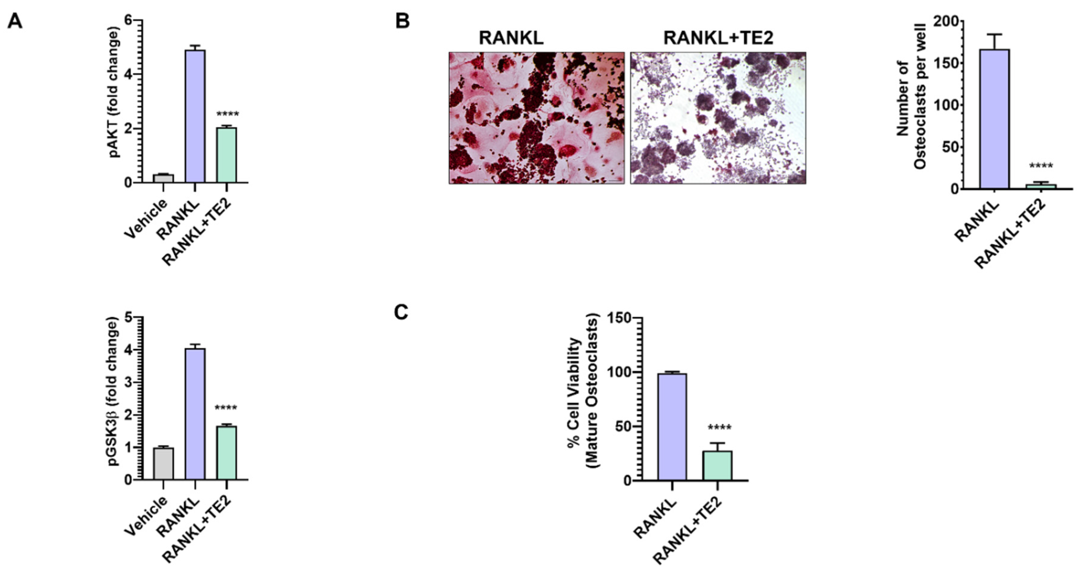
Figure 3.
Predicted molecular interactions of theasaponin E2 (TE2) with AKT1 and GSK-3β. Two-dimensional interaction diagrams of the selected docking poses of TE2 with (A) AKT1 (PDB ID: 7NH5; predicted binding energy, −9.8 kcal/mol) and (B) GSK-3β (PDB ID: 5K5N; predicted binding energy, −7.7 kcal/mol). Molecular docking was performed using CB-Dock3, and protein–ligand interaction diagrams were generated using PoseView. In AKT1, TE2 was predicted to bind within the interdomain allosteric pocket between the pleckstrin homology and kinase domains, forming hydrogen bonds with Tyr18A, Asp274A, Glu278A, and Leu156A, and hydrophobic contacts with Lys297A, Gly311A, and Cys310A. The corresponding hydrogen-bond distances were 2.86, 2.79, 2.97, and 3.19 Å, respectively. In GSK-3β, TE2 was predicted to bind within the ATP-binding pocket, forming hydrogen bonds with Thr138A, Ser203A, Arg220A, Ser66A, Cys218A, and Ser219A, and hydrophobic contacts with Gln185A and Leu188A. The corresponding hydrogen-bond distances were 2.82, 2.90, 2.95, 3.00, 2.99, and 3.06 Å, respectively. Hydrogen bonds were defined using a donor–acceptor distance cutoff of <3.5 Å. Dashed lines indicate hydrogen bonds, solid green lines indicate hydrophobic contact regions, and R groups represent continuation of the protein backbone beyond the depicted residues.
Figure 3.
Predicted molecular interactions of theasaponin E2 (TE2) with AKT1 and GSK-3β. Two-dimensional interaction diagrams of the selected docking poses of TE2 with (A) AKT1 (PDB ID: 7NH5; predicted binding energy, −9.8 kcal/mol) and (B) GSK-3β (PDB ID: 5K5N; predicted binding energy, −7.7 kcal/mol). Molecular docking was performed using CB-Dock3, and protein–ligand interaction diagrams were generated using PoseView. In AKT1, TE2 was predicted to bind within the interdomain allosteric pocket between the pleckstrin homology and kinase domains, forming hydrogen bonds with Tyr18A, Asp274A, Glu278A, and Leu156A, and hydrophobic contacts with Lys297A, Gly311A, and Cys310A. The corresponding hydrogen-bond distances were 2.86, 2.79, 2.97, and 3.19 Å, respectively. In GSK-3β, TE2 was predicted to bind within the ATP-binding pocket, forming hydrogen bonds with Thr138A, Ser203A, Arg220A, Ser66A, Cys218A, and Ser219A, and hydrophobic contacts with Gln185A and Leu188A. The corresponding hydrogen-bond distances were 2.82, 2.90, 2.95, 3.00, 2.99, and 3.06 Å, respectively. Hydrogen bonds were defined using a donor–acceptor distance cutoff of <3.5 Å. Dashed lines indicate hydrogen bonds, solid green lines indicate hydrophobic contact regions, and R groups represent continuation of the protein backbone beyond the depicted residues.
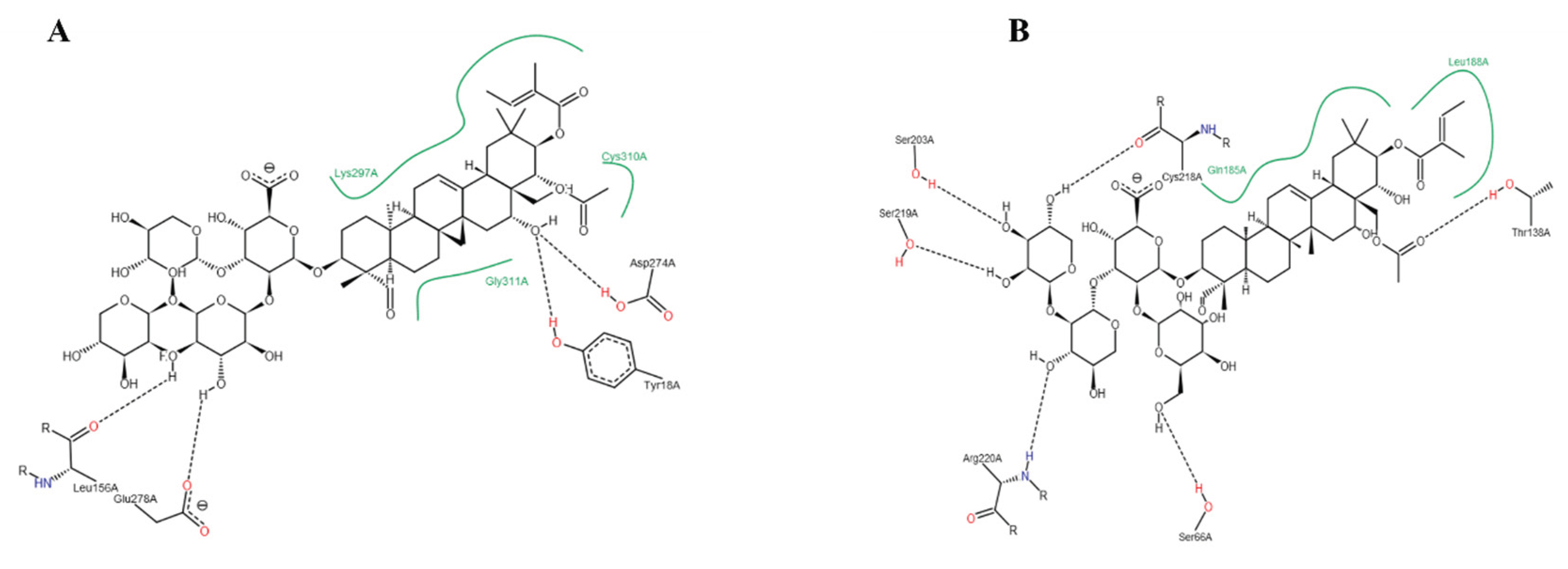

Table 1.
Predicted docking scores and key residue interactions of theasaponin E2 (TE2) with AKT1 and GSK-3β.
Table 1.
Predicted docking scores and key residue interactions of theasaponin E2 (TE2) with AKT1 and GSK-3β.
| Target | PDB ID | Binding Energy (kcal/mol) | Hydrogen-bonding residue | Distance (Å) | Hydrophobic Contacts |
| AKT1 | 7NH5 | −9.8 | Tyr18A | 2.86 | Lys297A, Gly311A, Cys310A |
| Asp274A | 2.79 | ||||
| Glu278A | 2.97 | ||||
| Leu156A | 3.19 | ||||
| GSK-3β | 5K5N | −7.7 | Thr138A | 2.82 | Gln185A, Leu188A |
| Ser203A | 2.90 | ||||
| Arg220A | 2.95 | ||||
| Ser66A | 3.00 | ||||
| Cys218A | 2.99 | ||||
| Ser219A | 3.06 | ||||
Note: Binding energies are predicted docking scores obtained using CB-Dock3. Hydrogen-bond distances represent donor–acceptor distances. Protein–ligand interactions were identified using PoseView. The suffix “A” in the residue labels denotes chain A. PDB, Protein Data Bank; Å, angstrom; TE2, theasaponin E2.
Disclaimer/Publisher’s Note: The statements, opinions and data contained in all publications are solely those of the individual author(s) and contributor(s) and not of MDPI and/or the editor(s). MDPI and/or the editor(s) disclaim responsibility for any injury to people or property resulting from any ideas, methods, instructions or products referred to in the content. |
© 2026 by the authors. Licensee MDPI, Basel, Switzerland. This article is an open access article distributed under the terms and conditions of the Creative Commons Attribution (CC BY) license (http://creativecommons.org/licenses/by/4.0/).
Copyright: This open access article is published under a Creative Commons CC BY 4.0 license, which permit the free download, distribution, and reuse, provided that the author and preprint are cited in any reuse.


