
Submitted:
17 July 2026
Posted:
22 July 2026
You are already at the latest version
Abstract
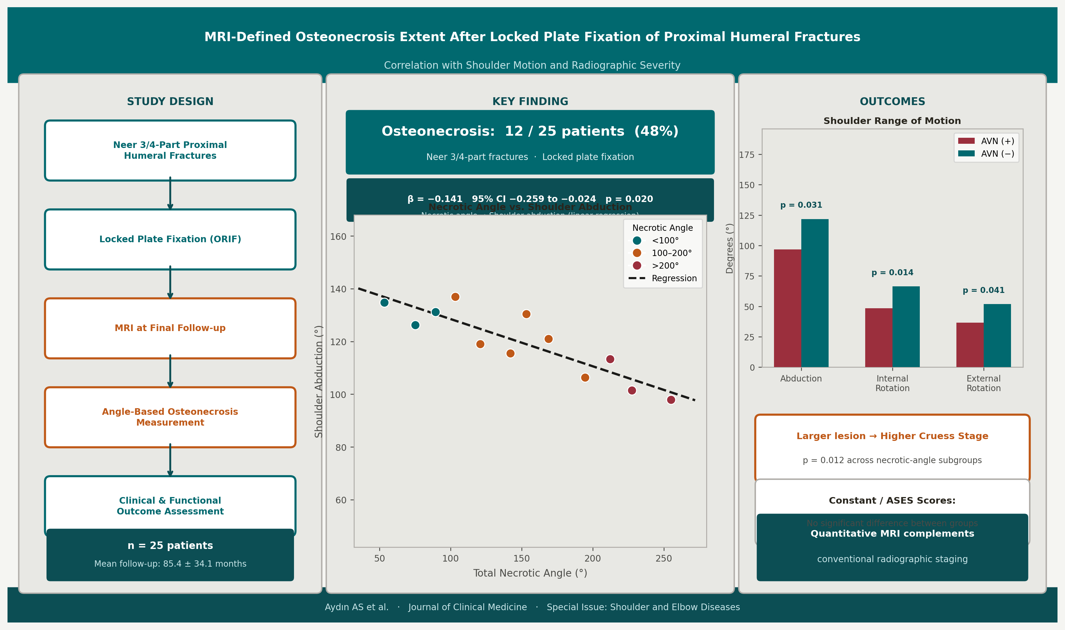
Background/Objectives: Humeral head osteonecrosis is a recognised complication following locked plate fixation of complex proximal humeral fractures, yet the clinical relevance of necrotic lesion extent remains poorly characterised. This study aimed to quantify MRI-defined osteonecrosis extent and examine its associations with radiographic severity, shoulder motion, and functional outcomes. Methods: Twenty-five patients with Neer three- or four-part proximal humeral fractures treated by locked plate fixation between 2010 and 2022 were retrospectively reviewed. Active shoulder range of motion, Constant score, American Shoulder and Elbow Surgeons (ASES) score, and visual analogue scale (VAS) pain were assessed at final follow-up. Osteonecrosis severity was graded using the Cruess classification. MRI necrotic extent was quantified with a modified angle-based method on coronal and axial images; associations with outcomes were assessed by linear regression. Results: At a mean follow-up of 85.4 ± 34.1 months, osteonecrosis was identified in 12 of 25 patients (48%). Patients with osteonecrosis demonstrated significantly lower shoulder abduction (96.7 ± 27.1° vs. 121.8 ± 29.2°; p = 0.031), internal rotation (48.3 ± 15.9° vs. 66.4 ± 15.5°; p = 0.014), and external rotation (36.7 ± 16.7° vs. 51.8 ± 26.4°; p = 0.041) than those without. Functional outcome scores did not differ significantly between groups. Greater total necrotic angle was associated with higher radiographic Cruess stage and reduced shoulder abduction (β = −0.141; 95% CI −0.259 to −0.024; p = 0.020). Conclusions: MRI-defined osteonecrosis extent correlates with radiographic severity and reduced shoulder motion after locked plate fixation of proximal humeral fractures. Larger necrotic lesions are associated with decreased shoulder abduction, whereas no clear relationship was identified between necrotic extent and global functional outcome scores. Quantitative MRI assessment may provide additional clinically relevant information on the structural severity of post-traumatic humeral head osteonecrosis.
Keywords:
proximal humeral fracture
; osteonecrosis
; avascular necrosis
; magnetic resonance imaging
; shoulder motion
; locked plate fixation
; Cruess classification
; functional outcome
1. Introduction
Among adults, proximal humeral fractures represent approximately 4–5% of all skeletal injuries and rank among the most frequently encountered upper-extremity fractures [1,2]. Within this spectrum, Neer three- and four-part configurations present a particularly demanding surgical problem owing to significant displacement, metaphyseal fragmentation, and vulnerability of the humeral head blood supply [3]. Operative stabilisation is often necessary, and locked plate fixation has established itself as a widely adopted technique for these complex injuries [4].
Despite advances in implant design and surgical technique, post-operative complications remain a meaningful clinical concern. Avascular necrosis (AVN) of the humeral head is among the most consequential, given its potential to cause articular collapse, secondary osteoarthritis, pain, and progressive loss of shoulder function [5,6]. Published rates after proximal humeral fracture fixation vary substantially across series, owing to heterogeneity in fracture severity, vascular disruption, reduction adequacy, construct stability, and duration of follow-up [5,6,7].
Research into post-traumatic humeral head osteonecrosis has identified several contributing factors. Fracture pattern and integrity of the medial calcar hinge are critical determinants of humeral head viability after intracapsular injuries [5]. Fracture classification systems have also been evaluated for their utility in estimating osteonecrosis risk [8]. Nevertheless, prior investigations have largely focused on whether AVN develops rather than on the extent of necrotic humeral head involvement and its functional consequences.
The relationship between structural AVN severity and shoulder function is not well defined. Inferior outcomes following post-traumatic humeral head osteonecrosis have been documented in some cohorts [9], whereas other investigators have shown that satisfactory function can coexist with radiographic necrosis [10]. This dissociation implies that tissue destruction and functional impairment do not necessarily progress in parallel.
MRI affords detailed evaluation of bone marrow viability and osteonecrotic boundaries after fracture fixation [11]. Angle-based measurements—originally developed for femoral head osteonecrosis to quantify lesion size and collapse risk [12]—have since been applied to humeral head osteonecrosis [13]. Optimised postoperative MRI protocols have improved image quality by mitigating metal artefacts from fixation hardware [14]. Despite these advances, MRI-based quantification of AVN extent after locked plate fixation of proximal humeral fractures remains limited.
The purpose of this study was to quantify the extent of humeral head osteonecrosis using MRI after locked plate fixation of three- and four-part proximal humeral fractures and to investigate its relationship with radiographic severity, shoulder motion, and functional outcomes.
2. Materials and Methods
2.1. Study Design and Patient Selection
This retrospective cohort study included consecutive patients who underwent locked plate fixation for proximal humeral fractures at a tertiary referral centre between January 2010 and December 2022. Institutional review board approval was obtained prior to study initiation (Istanbul University Faculty of Medicine IRB, Approval No: E-29624016-050.99-917586). All procedures conformed to the ethical standards of the 1964 Declaration of Helsinki and its later amendments.
Eligible patients were adults (age >18 years) with a Neer three- or four-part proximal humeral fracture [15] treated by open reduction and internal fixation with a locking plate. Exclusion criteria were: follow-up shorter than 6 months; pathological fracture; nonunion; revision osteosynthesis; primary hemiarthroplasty or reverse shoulder arthroplasty; postoperative loss of medial calcar continuity; incomplete radiological or clinical data; and refusal to participate.
Of 110 patients screened for eligibility, 25 met all inclusion criteria and formed the final study cohort (Figure 1).
2.2. Clinical Assessment
At the latest follow-up visit, all patients underwent standardised clinical examination by a senior orthopaedic surgeon blinded to MRI findings. Active shoulder range of motion—including abduction, internal rotation at 90° abduction, and external rotation—was measured with a goniometer. Functional outcomes were assessed using the Constant score [16], the American Shoulder and Elbow Surgeons (ASES) score [17], and a visual analogue scale (VAS) for pain [18].
2.3. Radiological Evaluation
Standardised anteroposterior and oblique shoulder radiographs were obtained at final follow-up. Osteonecrosis severity was graded according to the Cruess classification [19]. Radiographic and MRI evaluations were independently performed by two experienced musculoskeletal radiologists (G.D. and M.D.) blinded to all clinical findings and functional outcome data. Disagreements were resolved by consensus.
2.4. MRI Protocol and Quantification of Osteonecrosis Extent
MRI examinations were performed at final follow-up using a dedicated shoulder coil. The imaging protocol included coronal oblique T1-weighted, coronal oblique fat-suppressed T2-weighted or proton density-weighted, axial fat-suppressed proton density-weighted, and sagittal oblique sequences for assessment of bone marrow signal, subchondral integrity, and humeral head morphology. To minimise implant-related susceptibility artefacts, fast spin-echo–based sequences with thin-slice acquisition and high-bandwidth parameters were preferentially employed. Additional metal artefact reduction strategies were applied when available, in accordance with previously described postoperative MRI protocols for proximal humeral fractures [14].
Osteonecrosis extent was quantified using a modified angle-based technique adapted from established assessment methods [12,13]. Mid-coronal and mid-axial images through the centre of the humeral head were selected. The humeral head centre was determined geometrically using a best-fit circle, and lines were drawn from this centre to the opposing margins of the necrotic region. The angle subtended by these lines defined the necrotic angle on each imaging plane. The total necrotic angle was calculated as the sum of the axial and coronal measurements (Figure 2). Measurements were obtained from slices affording the clearest visualisation of necrotic margins and humeral head contour. Examinations with insufficient visualisation of necrotic boundaries owing to artefact were excluded from quantitative analysis. Patients with MRI evidence of osteonecrosis were subsequently categorised by total necrotic angle: <100°, 100–200°, and >200°.
2.5. Statistical Analysis
Analyses were performed using IBM SPSS Statistics (version 25.0; IBM Corp., Armonk, NY, USA). Continuous variables are reported as mean ± standard deviation; categorical variables as frequencies and percentages. Distributional normality was assessed with the Shapiro–Wilk test. Between-group comparisons (osteonecrosis vs. no osteonecrosis) used the independent-samples t-test. Differences across necrotic-angle subgroups were examined with one-way analysis of variance; when significant, pairwise comparisons used Bonferroni correction. Linear regression was used to examine associations between total necrotic angle and clinical outcomes; an age-adjusted multivariable model was additionally fitted for Constant score. Statistical significance was set at p < 0.05.
3. Results
Of 110 patients screened, 25 fulfilled all eligibility criteria and were included in the final analysis (Figure 1). The cohort comprised 11 women (44%) and 14 men (56%), with a mean age of 50.9 ± 13.2 years (range 23–72 years). Mean follow-up was 85.4 ± 34.1 months (range 36–156 months). The dominant extremity was involved in 13 patients (52%).
Osteonecrosis of the humeral head was identified by radiography and MRI in 12 of 25 patients (48%). Comparisons between patients with and without osteonecrosis are summarised in Table 1. Shoulder abduction, internal rotation, and external rotation were each significantly lower in the osteonecrosis group. Mean abduction was 96.7 ± 27.1° in osteonecrosis patients versus 121.8 ± 29.2° in those without (p = 0.031). Mean internal rotation at 90° abduction was 48.3 ± 15.9° versus 66.4 ± 15.5° (p = 0.014). Mean external rotation was 36.7 ± 16.7° versus 51.8 ± 26.4° (p = 0.041). Constant score, ASES score, and VAS pain score did not differ significantly between groups (Table 1).
Among the 12 patients with osteonecrosis, five had total necrotic angles <100°, four had angles between 100° and 200°, and three had angles >200°. Comparisons across necrotic-angle subgroups are presented in Table 2. Patients with larger lesions demonstrated significantly higher Cruess stages (p = 0.012). No statistically significant differences in shoulder range of motion, Constant score, ASES score, or VAS pain score were observed across subgroups (Table 2).
Linear regression results are presented in Table 3. Greater total necrotic angle was associated with reduced shoulder abduction (β = −0.141; 95% CI −0.259 to −0.024; p = 0.020) (Figure 3). A negative trend was also apparent for Constant score, but this did not reach statistical significance (β = −0.037; p = 0.217). In the age-adjusted multivariable model, total necrotic angle was not independently associated with Constant score (Table 3).
4. Discussion
This study examined MRI-defined humeral head osteonecrosis extent after locked plate fixation of complex proximal humeral fractures and its relationship with radiographic severity and clinical outcomes. Three principal observations emerged. First, osteonecrosis was present in nearly half the patients at long-term follow-up. Second, osteonecrosis was associated with reduced shoulder motion—particularly abduction and rotation—whereas composite functional outcome scores were less markedly affected. Third, greater MRI-defined necrotic lesion size correlated with higher radiographic Cruess stages and lower shoulder abduction.
The 48% osteonecrosis prevalence observed here lies at the upper boundary of rates published for locked plate fixation of proximal humeral fractures [5,6,7,8,11]. This likely reflects the deliberate restriction of the cohort to Neer three- and four-part fractures—injury patterns recognised for their elevated risk of vascular compromise [5]. The extended mean follow-up exceeding seven years may also have facilitated detection of osteonecrotic changes that remain radiographically occult during earlier postoperative evaluations.
Shoulder motion was measurably reduced in patients with osteonecrosis compared with those without, a pattern consistent with outcomes reported in prior series on post-traumatic humeral head osteonecrosis [9,10]. Yet Constant and ASES composite scores did not differ significantly between groups despite these motion deficits. The dissociation between motion loss and global functional scores suggests that patients may compensate for restricted range of motion through preserved strength, adaptation to chronic discomfort, or modification of daily activities—mechanisms that can partially sustain overall function scores even as structural integrity deteriorates.
A distinguishing feature of the present investigation is the use of quantitative MRI-based lesion measurement. The combined necrotic angle approach, originally refined for femoral head osteonecrosis to estimate lesion volume and collapse probability [12], has been translated to the humeral head setting [13]. In the present series, larger necrotic angles were associated with more advanced radiographic osteonecrosis stages and reduced shoulder abduction. The inverse relationship between total necrotic angle and shoulder abduction is illustrated in Figure 3. In contrast, no significant association was identified between necrotic angle and Constant score. These findings suggest that MRI-defined lesion extent may reflect specific impairments in shoulder motion rather than overall functional status.
When osteonecrosis patients were divided into necrotic-angle subgroups, those with larger lesions had higher Cruess stages, but inter-subgroup differences in motion and functional scores did not reach significance. Given that only 12 patients had osteonecrosis—with subgroups of five, four, and three patients—this analysis was substantially underpowered, and negative findings should not be interpreted as evidence of equivalence.
From a clinical standpoint, quantitative MRI assessment of osteonecrosis extent may usefully supplement conventional radiographic staging after proximal humeral fracture fixation. The association between necrotic angle and shoulder abduction suggests that lesion size contributes to motion-specific impairment, even when patient-reported global function appears relatively preserved. However, imaging severity alone should not direct clinical decision-making without integration of the patient's symptoms, functional demands, and trajectory over time.
The present study also underscores the challenges specific to MRI evaluation after metallic fixation. Susceptibility artefacts from the locking plate can obscure humeral head margins and necrotic boundaries. Measurements were therefore restricted to slices with adequate visualisation, and metal artefact reduction sequences were employed in accordance with published postoperative MRI protocols [14].
4.1. Limitations
Several limitations warrant consideration. The cohort was small, particularly after stratification of osteonecrosis cases by necrotic angle; however, previously published studies focusing specifically on MRI-defined osteonecrosis after proximal humeral fractures have similarly included limited patient numbers (Table 4). The retrospective design precluded longitudinal tracking of osteonecrosis progression and associated functional trajectories. Variables including fracture morphology, reduction quality, bone density, and implant positioning may have independently influenced both osteonecrotic progression and clinical results but could not be fully controlled. Finally, MRI measurements in the presence of metallic implants retain susceptibility to residual image distortion despite artefact-reduction measures.
5. Conclusions
Post-traumatic humeral head osteonecrosis after locked plate fixation of Neer three- and four-part proximal humeral fractures was identified in nearly half of patients at long-term follow-up and was associated with significantly reduced shoulder motion—most prominently in abduction and rotational arcs—while global composite functional scores were less clearly affected. Larger MRI-defined necrotic lesions correlated with more advanced radiographic Cruess staging and lower shoulder abduction values. These findings suggest that quantitative MRI assessment of osteonecrosis extent provides clinically relevant structural information that complements conventional radiographic classification. Prospective studies in larger cohorts employing standardised MRI protocols are warranted to confirm these associations and to clarify the role of quantitative MRI characterisation in post-operative monitoring and treatment planning for this patient population.
Author Contributions
Conceptualization, A.S.A. and D.K.; Methodology, A.S.A. and A.Ş.K.; Formal Analysis, A.S.A.; Investigation, A.S.A. and M.B.; Data Curation, M.B.; Writing – Original Draft Preparation, A.S.A.; Writing – Review and Editing, A.Ş.K. and A.E.; Visualization, A.S.A.; Radiology and MRI Evaluation, G.D. and M.D.; Supervision, A.E. All authors have read and agreed to the published version of the manuscript.
Funding
This research received no external funding.
Institutional Review Board Statement
This study was conducted in accordance with the Declaration of Helsinki and was approved by the Institutional Review Board of Istanbul University Faculty of Medicine (Approval No. E-29624016-050.99-917586) on 27 May 2022.
Informed Consent Statement
Written informed consent was obtained from all participants prior to the final clinical and radiological evaluation.
Data Availability Statement
The datasets generated and/or analysed during the current study are available from the corresponding author on reasonable request.
Acknowledgments
The authors thank all patients who participated in this study and the staff of the Departments of Orthopaedics and Traumatology and Radiology for their assistance during patient follow-up and data collection.
Conflicts Of Interest
The authors declare no conflicts of interest.
Abbreviations
AVN, avascular necrosis; ASES, American Shoulder and Elbow Surgeons score; VAS, visual analogue scale; MRI, magnetic resonance imaging; ORIF, open reduction and internal fixation; CI, confidence interval.
References
- Court-Brown, C.M.; Caesar, B. Epidemiology of adult fractures: A review. Injury 2006, 37, 691–697. [Google Scholar] [CrossRef] [PubMed]
- Launonen, A.P.; Lepola, V.; Saranko, A.; Flinkkilä, T.; Laitinen, M.; Mattila, V.M. Epidemiology of proximal humerus fractures. Arch. Osteoporos. 2015, 10, 209. [Google Scholar] [CrossRef] [PubMed]
- Neer, C.S. Displaced proximal humeral fractures. Part I. Classification and evaluation. J. Bone Jt. Surg. Am. 1970, 52, 1077–1089. [Google Scholar] [CrossRef]
- Handoll, H.H.; Brorson, S. Interventions for treating proximal humeral fractures in adults. Cochrane Database Syst. Rev. 2015, 11, CD000434. [Google Scholar] [CrossRef]
- Hertel, R.; Hempfing, A.; Stiehler, M.; Leunig, M. Predictors of humeral head ischemia after intracapsular fracture. J. Shoulder Elb. Surg. 2004, 13, 427–433. [Google Scholar] [CrossRef]
- Clavert, P.; Adam, P.; Bevort, A.; Bonnomet, F.; Kempf, J.F. Pitfalls and complications with locking plate for proximal humerus fracture. J. Shoulder Elb. Surg. 2010, 19, 489–494. [Google Scholar] [CrossRef]
- Neviaser, A.S.; Hettrich, C.M.; Dines, J.S.; Lorich, D.G. Rate of avascular necrosis following proximal humerus fractures treated with a lateral locking plate. Arch. Orthop. Trauma Surg. 2011, 131, 1617–1622. [Google Scholar] [CrossRef] [PubMed]
- Fazzari, F.; Canton, G.; Giraldi, G.; Falcioni, N.; Clocchiatti, S.; Rasio, N.; et al. Avascular necrosis of humeral head after proximal humerus fracture: Comparison between classification systems. Acta Biomed. 2023, 94, e2023089. [Google Scholar] [CrossRef] [PubMed]
- Gerber, C.; Hersche, O.; Berberat, C. The clinical relevance of posttraumatic avascular necrosis of the humeral head. J. Shoulder Elb. Surg. 1998, 7, 586–590. [Google Scholar] [CrossRef]
- Roddy, E.; Kandemir, U. High rate of avascular necrosis but excellent patient-reported outcomes after ORIF of proximal humerus fracture dislocations. J. Shoulder Elb. Surg. 2023, 32, 2097–2104. [Google Scholar] [CrossRef]
- Bastian, J.D.; Hertel, R. Initial post-fracture humeral head ischemia does not predict development of necrosis. J. Shoulder Elb. Surg. 2008, 17, 2–8. [Google Scholar] [CrossRef]
- Kerboul, M.; Thomine, J.; Postel, M.; Merle d'Aubigné, R. The conservative surgical treatment of idiopathic aseptic necrosis of the femoral head. J. Bone Jt. Surg. Br. 1974, 56, 291–296. [Google Scholar] [CrossRef]
- Sakai, T.; Sugano, N.; Nishii, T.; Hananouchi, T.; Yoshikawa, H. Extent of osteonecrosis on MRI predicts humeral head collapse. Clin. Orthop. Relat. Res. 2008, 466, 1074–1080. [Google Scholar] [CrossRef] [PubMed]
- Garkisch, A.; Fischer, D.C.; Mittlmeier, T.; Märdian, S.; Klaan, B.; Weber, M.A.; et al. Assessment of humeral head perfusion after proximal humeral fracture: MRI-based protocol. J. Orthop. Surg. Res. 2025, 20, 560. [Google Scholar] [CrossRef] [PubMed]
- Neer, C.S. Displaced proximal humeral fractures. Part II. Treatment of three-part and four-part displacement. J. Bone Jt. Surg. Am. 1970, 52, 1090–1103. [Google Scholar] [CrossRef]
- Constant, C.R.; Murley, A.H. A clinical method of functional assessment of the shoulder. Clin. Orthop. Relat. Res. 1987, 214, 160–164. [Google Scholar] [CrossRef]
- Richards, R.R.; An, K.N.; Bigliani, L.U.; Friedman, R.J.; Gartsman, G.M.; Iannotti, J.P.; et al. A standardized method for the assessment of shoulder function. J. Shoulder Elb. Surg. 1994, 3, 347–352. [Google Scholar] [CrossRef]
- Huskisson, E.C. Measurement of pain. Lancet 1974, 304, 1127–1131. [Google Scholar] [CrossRef]
- Cruess, R.L. Osteonecrosis of bone: Current concepts as to etiology and pathogenesis. Clin. Orthop. Relat. Res. 1978, 130, 3–9. [Google Scholar] [CrossRef]
Figure 1.
Flow diagram illustrating patient screening, application of inclusion and exclusion criteria, and final cohort selection.
Figure 1.
Flow diagram illustrating patient screening, application of inclusion and exclusion criteria, and final cohort selection.
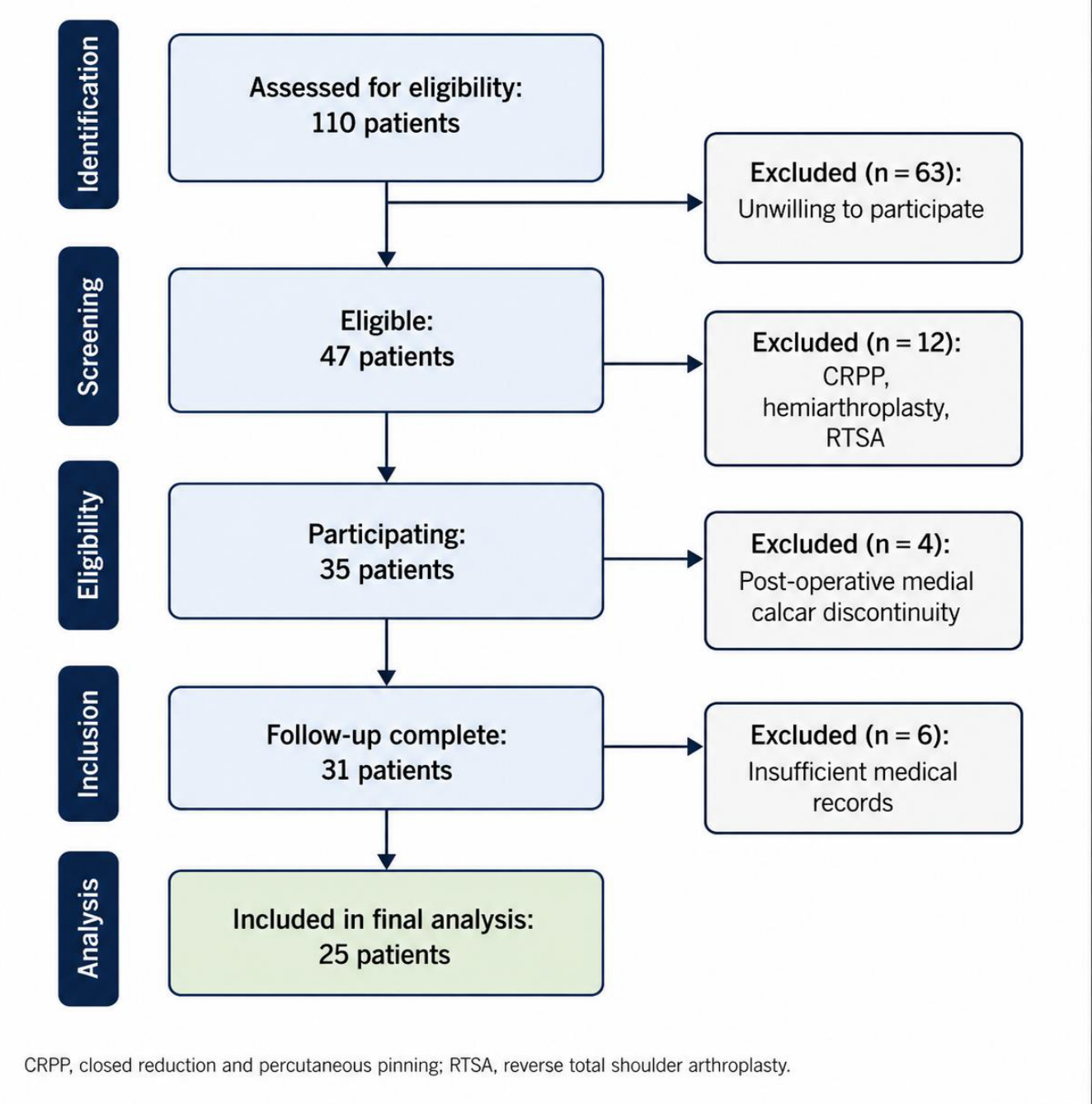
Figure 2.
MRI-based quantification of humeral head osteonecrosis extent after locked plate fixation of proximal humeral fractures. (a) 58-year-old man with Cruess stage II osteonecrosis (total necrotic angle 75°). Mid-coronal T1-weighted image demonstrating the angle-based necrotic extent measurement; lines are drawn from the geometrically determined humeral head centre to the opposing margins of the necrotic region. (b) 47-year-old woman with Cruess stage III osteonecrosis (total necrotic angle 145°). Mid-axial fat-suppressed proton density-weighted image showing the necrotic angle measurement. (c) 63-year-old man with Cruess stage IV osteonecrosis (total necrotic angle 230°). Representative coronal and axial images demonstrating a large necrotic lesion with advanced structural involvement. Total necrotic angle was calculated as the sum of coronal and axial measurements.
Figure 2.
MRI-based quantification of humeral head osteonecrosis extent after locked plate fixation of proximal humeral fractures. (a) 58-year-old man with Cruess stage II osteonecrosis (total necrotic angle 75°). Mid-coronal T1-weighted image demonstrating the angle-based necrotic extent measurement; lines are drawn from the geometrically determined humeral head centre to the opposing margins of the necrotic region. (b) 47-year-old woman with Cruess stage III osteonecrosis (total necrotic angle 145°). Mid-axial fat-suppressed proton density-weighted image showing the necrotic angle measurement. (c) 63-year-old man with Cruess stage IV osteonecrosis (total necrotic angle 230°). Representative coronal and axial images demonstrating a large necrotic lesion with advanced structural involvement. Total necrotic angle was calculated as the sum of coronal and axial measurements.
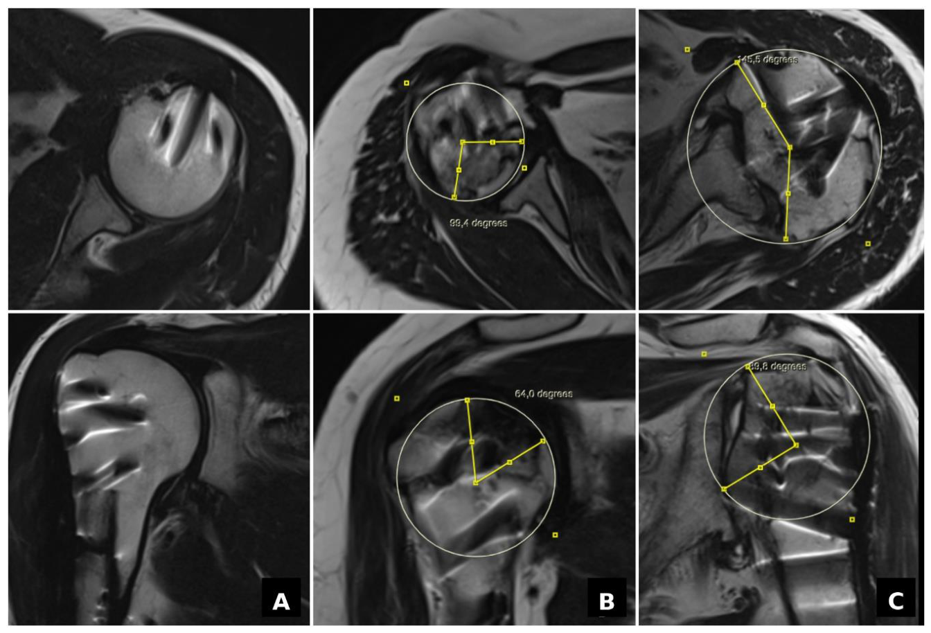
Figure 3.
Scatter diagrams illustrating the relationship between total necrotic angle (degrees) and clinical outcomes in patients with osteonecrosis (n = 12). (a) Total necrotic angle versus Constant score (β = −0.037; p = 0.217). (b) Total necrotic angle versus shoulder abduction in degrees (β = −0.141; 95% CI −0.259 to −0.024; p = 0.020). Each data point represents one patient; the linear regression line with 95% confidence interval is shown.
Figure 3.
Scatter diagrams illustrating the relationship between total necrotic angle (degrees) and clinical outcomes in patients with osteonecrosis (n = 12). (a) Total necrotic angle versus Constant score (β = −0.037; p = 0.217). (b) Total necrotic angle versus shoulder abduction in degrees (β = −0.141; 95% CI −0.259 to −0.024; p = 0.020). Each data point represents one patient; the linear regression line with 95% confidence interval is shown.
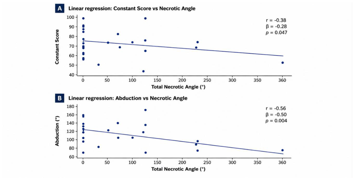

Table 1.
Clinical outcomes and shoulder range of motion in patients with and without osteonecrosis.
| Variable | Osteonecrosis (+) (n = 12) | Osteonecrosis (−) (n = 13) | p Value |
| Age (years) | 48.9 ± 12.8 | 52.7 ± 13.6 | 0.462 |
| Follow-up (months) | 88.3 ± 36.2 | 82.7 ± 32.4 | 0.684 |
| Constant score | 68.4 ± 13.9 | 75.6 ± 10.8 | 0.167 |
| ASES score | 72.1 ± 14.7 | 80.5 ± 12.1 | 0.138 |
| VAS pain score | 2.8 ± 1.7 | 1.9 ± 1.4 | 0.176 |
| Abduction (°) | 96.7 ± 27.1 | 121.8 ± 29.2 | 0.031 * |
| Internal rotation (°) | 48.3 ± 15.9 | 66.4 ± 15.5 | 0.014 * |
| External rotation (°) | 36.7 ± 16.7 | 51.8 ± 26.4 | 0.041 * |
Values are mean ± SD. * p < 0.05. ASES, American Shoulder and Elbow Surgeons score; VAS, visual analogue scale; SD, standard deviation.
Table 2.
Clinical and radiographic characteristics according to MRI-defined osteonecrosis extent.
| Variable | <100° (n = 5) | 100–200° (n = 4) | >200° (n = 3) | p Value |
| Constant score | 74.8 ± 10.4 | 67.5 ± 13.2 | 58.7 ± 14.5 | 0.236 |
| ASES score | 78.2 ± 11.5 | 70.1 ± 13.6 | 62.3 ± 16.8 | 0.281 |
| VAS pain score | 2.0 ± 1.2 | 2.9 ± 1.5 | 3.7 ± 2.1 | 0.314 |
| Abduction (°) | 112.0 ± 24.1 | 96.3 ± 18.4 | 73.3 ± 11.5 | 0.071 |
| Internal rotation (°) | 58.0 ± 13.5 | 46.3 ± 11.1 | 36.7 ± 15.3 | 0.108 |
| External rotation (°) | 45.0 ± 17.1 | 35.0 ± 12.9 | 23.3 ± 10.4 | 0.174 |
| Cruess stage | 2.0 ± 0.7 | 3.0 ± 0.8 | 4.3 ± 0.6 | 0.012 * |
Values are mean ± SD. * p < 0.05. ASES, American Shoulder and Elbow Surgeons score; VAS, visual analogue scale; SD, standard deviation.
Table 3.
Linear regression analyses: associations between total necrotic angle and clinical outcomes.
Table 3.
Linear regression analyses: associations between total necrotic angle and clinical outcomes.
| Dependent Variable | β | 95% CI | p Value | R² |
| Constant score | −0.037 | −0.096 to 0.023 | 0.217 | 0.063 |
| Abduction (°) | −0.141 | −0.259 to −0.024 | 0.020 * | 0.204 |
| Constant score (age-adjusted) | −0.035 | −0.095 to 0.025 | 0.236 | 0.106 |
β, unstandardised regression coefficient; CI, confidence interval; R², coefficient of determination. * p < 0.05.
Table 4.
Sample sizes in selected studies evaluating osteonecrosis after proximal humeral fractures.
Table 4.
Sample sizes in selected studies evaluating osteonecrosis after proximal humeral fractures.
| Study | Focus | Imaging | n |
| Gerber et al. [9] | Clinical relevance of post-traumatic osteonecrosis | Radiography | 25 |
| Hertel et al. [5] | Predictors of humeral head ischaemia | Radiography | 40 |
| Roddy et al. [10] | Functional outcomes after fracture-dislocation ORIF | Radiography | 26 |
| Sakai et al. [13] | MRI quantification of osteonecrotic extent | MRI | 46 |
| Present study | MRI-defined osteonecrosis extent and clinical outcomes | MRI | 25 |
MRI, magnetic resonance imaging; ORIF, open reduction and internal fixation.
Disclaimer/Publisher’s Note: The statements, opinions and data contained in all publications are solely those of the individual author(s) and contributor(s) and not of MDPI and/or the editor(s). MDPI and/or the editor(s) disclaim responsibility for any injury to people or property resulting from any ideas, methods, instructions or products referred to in the content. |
© 2026 by the authors. Licensee MDPI, Basel, Switzerland. This article is an open access article distributed under the terms and conditions of the Creative Commons Attribution (CC BY) license.
Copyright: This open access article is published under a Creative Commons CC BY 4.0 license, which permit the free download, distribution, and reuse, provided that the author and preprint are cited in any reuse.


