

Submitted:
21 July 2026
Posted:
22 July 2026
You are already at the latest version
Abstract
Former smokers in lung cancer screening remain at elevated risk for lung cancer despite smoking cessation. We and others have shown that curcumin exhibits anti-inflammatory and antiproliferative effects but is limited by poor bioavailability. However, since curcumin is lipophilic, co-administration with omega-3 fatty acids (ω-3 ω-3 ω-3 FA) represents a mechanistically rational strategy to enhance delivery and target complementary pathways, including STAT3 and NF-κB signaling for lung cancer chemoprevention.
Methods:
We conducted a randomized, single-blind, placebo-controlled pilot study evaluating curcumin combined with ω-3 FA in high-risk former and current smokers with CT-detected pulmonary nodules. Participants received intervention agents or placebo for 6 months. Primary endpoints included radiologic changes in nodule size, number, and density. Secondary endpoints included safety, adherence, bioavailability, and exploratory biomarker analyses. Correlation analyses of imaging-derived metrics were performed to assess relationships among LDCT parameters.
Results:
Nineteen participants were enrolled (intervention, n=12; placebo, n=7). No statistically significant between-group differences were observed in primary imaging endpoints. The intervention was well tolerated, with predominantly grade 1 adverse events. Exploratory analyses demonstrated consistent positive correlations among LDCT-derived measures, with clustering of size-based metrics (mean diameter, volume, sum of longest diameters) and density-based parameters. Multidimensional scaling supported this structure, indicating internal coherence among imaging-derived endpoints.
Conclusions:
Although no statistically significant treatment effect on the image biomarkers was observed, this pilot study demonstrates feasibility challenges and identifies coherent imaging biomarkers that may serve as intermediate endpoints in early-phase chemoprevention trials. These results support further investigation of strategies utilizing agent combinations with enhanced bioavailability and safety and refinement of trial design in high-risk lung cancer patient populations.
Keywords:
lung cancer chemoprevention
; low-dose ct
; lung nodules
; curcumin
; ω-3 fatty acids
; smokers
1. Introduction
1.1. Current Chemoprevention Options for Former Smokers
Lung cancer remains the leading cause of cancer-related mortality among men and women in Florida, the United States, and worldwide [1,2]. Approximately 80-90% of lung cancers occur in individuals with a history of smoking, and over 50% arise in former smokers. Tobacco smoking is the most important and prevalent lung cancer risk factor. Current strategies to reduce mortality in high-risk populations include lung cancer screening via low-dose computed tomography (LDCT) screening and tobacco control interventions. However, former and current smokers, particularly those undergoing active surveillance for pulmonary nodules, remain at persistently elevated risk and are highly motivated to pursue additional risk-reduction strategies [3].
Chemoprevention during this window represents a rational and potentially effective approach to promote regression of precursor lesions, including pulmonary nodules, thereby complementing LDCT screening and smoking cessation. The rationale for chemoprevention in this population is supported by several interrelated concepts: (a) lung carcinogenesis arises within airway epithelial cells exposed to shared genetic and environmental insults; (b) the “field cancerization” paradigm suggests that the entire airway epithelium is at risk and amenable to systemic intervention; (c) histopathologic precursor lesions such as metaplasia and dysplasia can be reproducibly identified and graded;[1,4,5] (d) resolution of smoking-induced inflammation is an active, regulated process mediated by specialized pro-resolving lipid mediators (SPMs);[6,7](e) progression to malignancy requires sustained proliferation and evasion of apoptosis driven by inflammatory and growth signaling pathways; and (f) interventions in disease-free, high-risk individuals must promote repair without immunosuppression and with minimal toxicity [2,7,8].
Based on these principles, phytochemicals and biologically active agents capable of modulating inflammatory and proliferative pathways have emerged as promising candidates for lung cancer chemoprevention.
1.2. Curcuminoids in Lung Cancer Chemoprevention
Curcumin (diferuloylmethane), the active component of Curcuma longa, modulates multiple signaling pathways implicated in carcinogenesis, including cell cycle regulation, apoptosis, proliferation, survival, angiogenesis, invasion, and inflammation.[10,11,12,13,14,15,16,17,18,25]. In particular, signal transducer and activator of transcription 3 (Stat3), activated in approximately 50% of lung cancers, plays a central role in inflammation-driven tumorigenesis [19,20,21,22]. Our preclinical studies demonstrate that CUR suppresses Stat3 phosphorylation and reduces proliferative markers (Cyclin D1, MCM2) in lung cancer cell lines and murine models in a dose-dependent manner [16]. CUR also inhibits NF-κB signaling through suppression of IκB-α kinase activity and downregulation of pro-inflammatory cytokines (TNF-α, IL-1, IL-6, IL-8) and enzymes (COX-2, 5-LOX) [10,12,25,26,27,28,29,54,55]. In lung cancer cell lines with constitutive Stat3 and NF-κB activation, CUR effectively reduces inflammatory signaling and proliferation [23].
Preclinical models further demonstrate that CUR suppresses K-ras–driven lung tumor progression and inflammation, supporting its role in delaying malignant transformation [30]. CUR has also shown antioxidant and antifibrotic effects in lung tissue without impairing tumor cell killing [3]. Clinical studies confirm favorable safety and bioavailability profiles [32,33]. Phase II data demonstrate biological activity, including a 40% reduction in aberrant crypt foci with CUR administration [34]. Novel formulations combining CUR with ω-3 FA improve systemic and pulmonary bioavailability and reduce tumor growth in murine lung cancer models [35].
1.3. Omega-3 Fatty Acids in Lung Cancer Chemoprevention
Omega-3 fatty acids (ω-3 FA) exert anti-inflammatory and pro-resolving effects central to lung carcinogenesis. [6,7,36]. A key mechanism involves inhibition of NF-κB signaling, a regulator of inflammatory cytokines, adhesion molecules, and COX-2 expression [37,38,39,40,41]. ω-3 FA reduce NF-κB activation by preventing IκB phosphorylation and nuclear translocation, thereby decreasing TNF-α and other pro-inflammatory mediators [38,39]. In clinical studies, ω-3 FA supplementation attenuates NF-κB activation, reduces proteasome activity, and lowers inflammatory cytokine levels in cancer patients [38]. Additional studies demonstrate reduced cytokine release and inflammatory signaling in human alveolar cells [40,41,42]. Importantly, ω-3 FA serve as precursors to specialized pro-resolving mediators (SPMs), including resolvins, protectins, and maresins, which actively promote resolution of inflammation and tissue repair without immunosuppression [6,7,43,44,45]. Clinical supplementation increases circulating SPMs (RvE1, RvD1, RvD2), supporting their biological relevance [46,47,48]. These mechanisms provided a strong rationale for targeting persistent inflammation in former smokers.
1.4. Synergistic Effects of Curcumin and Omega-3 Fatty Acids
The central limitation of curcumin is its poor systemic and tissue bioavailability, which has hindered its translation from promising preclinical findings to consistent clinical efficacy. One of the most compelling strategies to overcome this limitation is co-administration with lipids, particularly omega-3 fatty acids (ω-3 FA) [49,54]. Curcumin is inherently lipophilic, and its absorption is markedly enhanced in lipid-rich environments, providing a strong pharmacokinetic rationale for combination with ω-3 FA. Co-administration facilitates improved solubilization and intestinal absorption, leading to enhanced systemic distribution and more effective delivery to target tissues, including the lung. Consistent with this, lipid-based formulations combining curcumin with ω-3 FA have demonstrated measurable lung bioavailability and significant reductions in tumor growth in preclinical models [35]. Beyond pharmacokinetic advantages, the combination is supported by complementary and potentially synergistic biological mechanisms. Both agents modulate key inflammatory pathways, including inhibition of NF-κB signaling, while curcumin additionally targets STAT3, and ω-3 FA contribute to the generation of specialized pro-resolving mediators that actively promote resolution of inflammation [35,49,50,51,52,53]. Preclinical studies demonstrate that co-administration results in greater suppression of cell proliferation, enhanced induction of apoptosis, including increased caspase-3 activity, and more pronounced attenuation of inflammatory signaling compared with either agent alone [35,49,50,51,52,53]. In lung cancer cell models, combined treatment with curcumin and ω-3 FA significantly reduces cell viability relative to single-agent exposure [35]. Importantly, this strategy addresses a key translational barrier while simultaneously amplifying biological efficacy, and both agents have demonstrated favorable safety profiles in clinical settings, supporting the feasibility of this combination as a chemopreventive approach in high-risk populations. To further explore the potential role of combination of standardized agents- CUR + ω-3 FA for lung cancer chemoprevention, we conducted a pilot, randomized, single-blind, placebo-controlled trial in former and current smokers.
1.5. Hypothesis and Clinical Translation
Based on this evidence, we hypothesize that a combination of standardized agents- CUR + ω-3 FA will target both Stat3 and NF-κB signaling pathways relevant to lung carcinogenesis and provide for a novel, robust, synergistic, effective and safe means of lung cancer chemoprevention as indicated by regression of number and size of nodules, in former and current smokers, mediated via anti-proliferative, anti-inflammatory and pro-resolving effects in the bronchoepithelium in and around field areas of precancerous lesions. If successful, this Phase II trial will establish a safe, non-toxic, and biologically rational strategy for lung cancer interception, inform dietary-based risk reduction approaches, and provide the foundation for well powered Phase II trials. We will determine the best dose of this combination of agents by including a promising combination of safety and efficacy criteria that yields the largest probability of success (PoS) as follows: (a) minimum qualitative and quantitative toxicities assessed by Common Toxicity Criteria version 5.0 (safety); (b) minimum dose that delivers a pre-defined percentage of modulation of the primary endpoint biomarker (efficacy); (c) maximum dose beyond which no further beneficial effect is seen.
2. Materials and Methods
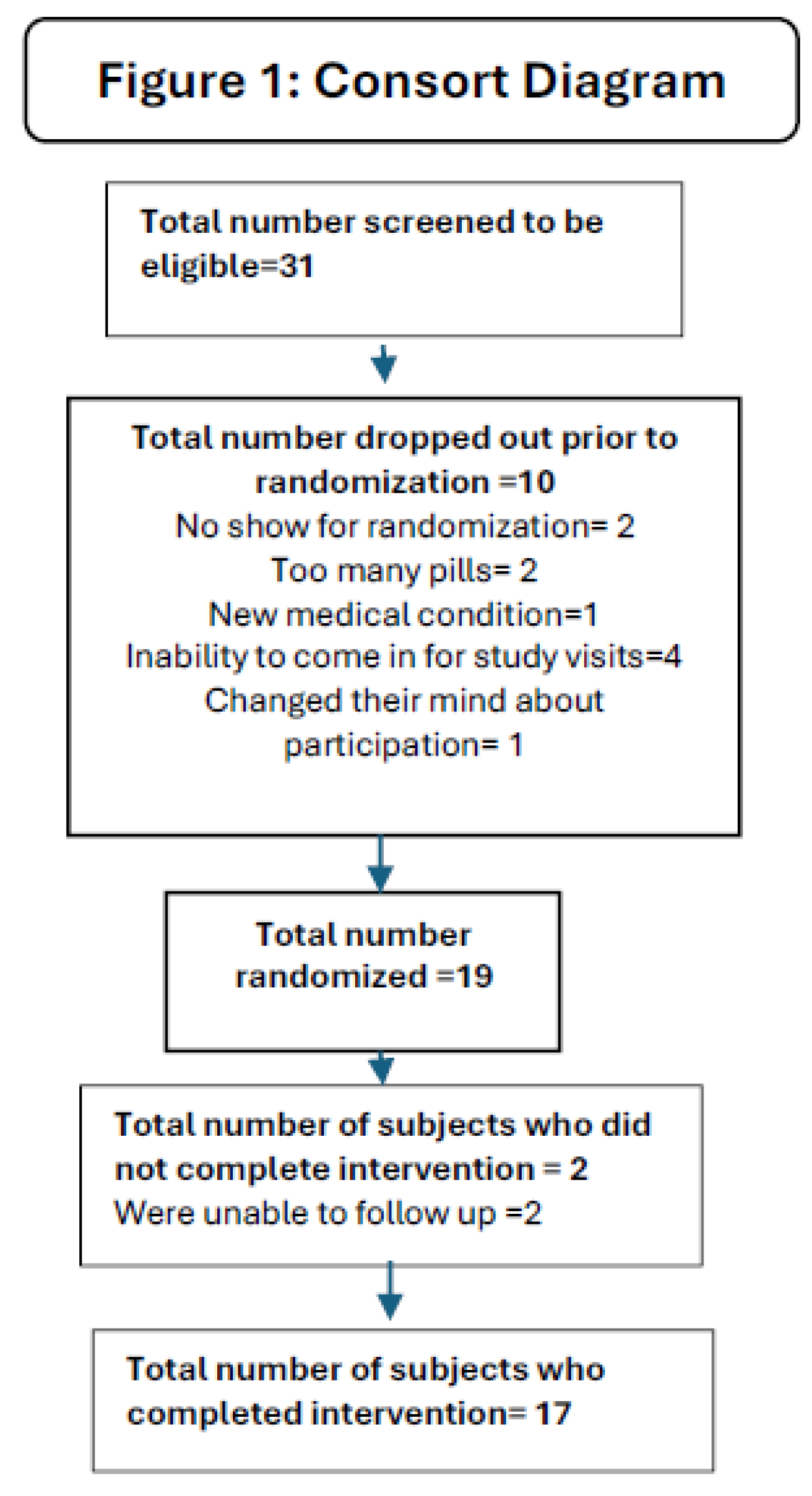
Prior to developing the methods for this trial, we consulted with the Patient Advisory Committee for the Thoracic Oncology Program. Based on their recommendation, we conducted a feasibility study to evaluate the interest of this target subject population to participate in an intervention trial [59]. The clinical trial was registered in https://clinicaltrials.gov (Trial registration number: NCT03598309 and Date of registration: 07-18-2018). The study and the consent procedures were approved by the institutional review board, Advara (Approval Code: Pro00028018) on 03-08-2018. A consort diagram depicting the number of subjects screened, enrolled, randomized and completed intervention is shown in Figure 1. Due to delay because of the COVID epidemic, participants were enrolled at the Moffitt Cancer, a National Cancer Institute designated Comprehensive Cancer Center from 6/2019 to 11/2024. Potential participants were identified by the Thoracic Oncology Program who were scheduled for an LDCT in the lung cancer screening program or being evaluated for a suspicious pulmonary nodule. Criteria for inclusion included male or female, 55 years of age or older who were former or current smokers and enrolled in the lung cancer screening program or those who are detected using a diagnostic CT, and have Lung-RADS 3 category lesion(s), that would get a 6 month follow-up LDCT or diagnostic CT based on Lung-RADS recommendations or have Lung-RADS 2 category lesions with lung nodule ≥4mm mean diameter detected during screening LDCT or regular CT scan; history of cigarette smoking with ≥ 20 pack years; ECOG performance score of ≤ 1; able to swallow study agents; willing and able to undergo CT scans; not allergic to components of the study agents; willing to comply with requirement to not consume additional dietary ω-3 fatty acids or curcumin supplements during the intervention period.
All current smokers were offered smoking cessation service, whether or not they agreed to participate in the trial. Participants were required to have normal organ and marrow function based on screening CBC and CMP. Exclusion Criteria included history of lung cancer or other invasive malignancy (with the exclusion of basal cell carcinoma or skin squamous cell carcinoma) and recent use of doxycycline or tetracycline(within ≤ 2 weeks).
The PI obtained an US FDA IND (139704) for the combination study agents and conducted the trial in compliance with the FDA Regulatory requirements.
Throughout the trial, we ensured that the criteria for inclusion/exclusion, safety and symptom monitoring procedures ensured safety of the agent administered to this target population for the duration of intervention. Thus benefit versus harm was monitored throughout the trial.
2.1. Randomization and Blinding
After eligibility was confirmed and consent obtained, participants were assigned to the intervention or placebo arm (1:1) using the SRAR system, a web-delivered subject registration application. All study staff and participants, with the exception of the clinical pharmacist and randomization team, were blinded to the assignments until the completion of the trial. After a potential subject is identified as having at least one lung nodule (see criteria), the LDCT or diagnostic CT were reviewed by the study radiologist (DK) to confirm eligibility. Each lung nodule was measured at baseline screening and 6 month LDCT or standard CT 1 mm thick axial images using standard lung window settings (C -500 HU, W 1500 HU), with the nodule diameter representing the average of the longest nodule diameter and the perpendicular diameter. New lung nodules at the 6 month LDCT or diagnostic CT were measured on the screening CT if they are present in retrospect. Both the overall nodule size and the size of the solid component of the nodule were measured and recorded for part-solid lung nodules. For both part-solid and non-solid lung nodules, average density of the solid and nonsolid portions of the nodule were determined at screening and 6 month LDCT or standard CT. In addition, all lung nodules ≥ 4mm at screening and 6 month LDCT or standard CT were assessed for change in size of > 1mm average diameter.
After the informed consent process, the participant had a baseline visit. Participants who meet inclusion/exclusion criteria were then randomized to one of 2 doses of CUR and ω-3 FA vs. placebo administered twice daily. Curcumin C3 complex® and matching placebo were obtained from Sabinsa Corporation and Lovaza® was purchased from GSK Pharma. Group A received Lovaza at a dose of 4 grams per day divided into 2 doses 2 grams q AM and 2 grams q PM and Curcumin C3 Complex, 8 grams per day divided into 2 doses of 4 grams po q AM and q PM. Group B received Lovaza at a dose of 2 grams per day divided into 2 doses of 1 gram po q AM and q PM and Curcumin C3 Complex, 8 grams per day divided into 2 doses of 4 grams po q AM and q PM. A single placebo arm received matching placebo for Lovaza consisting of 2 gram po q AM and q PM and 4 grams placebo for Curcumin C-3 complex, po q AM and q PM. At the end of intervention visit, a follow-up LDCT or standard CT evaluation of nodule characteristics occurred at 6 months +/- 15 days. All screening of LDCT or diagnostic CTs were categorized by the study radiologist in a blinded fashion as to intervention group and were read after the completion of each LDCT or standard CT scan. Intervention continued until the 6 month LDCT or diagnostic CT, intolerable toxicity or withdrawal from study. The data were summarized as average change in size of nodules (primary endpoint) from the LDCT or standard CT. In addition, we observed trends in the number and size of nodules ≥4 mm, and lung nodule density of partially solid and non-solid between the intervention and placebo arms. Toxicities were evaluated according to CTCAE version 5.0 guidelines.
2.2. Intervention
An investigator-initiated IND (139704, Kumar NB PI) was obtained for this combination of agents (Curcumin + ω-3 FA) at the dose proposed for this trial and for this indicationAnnual testing of study agents was conducted to ensure drug stability with full potency of agent documented until 2025 when the last subject completed the trial. To minimize the use of other supplements, a standard vitamin and mineral formulation containing 100% U.S. recommended daily allowance was provided to all participants for the duration of the study. The intervention was terminated if a participant developed lung or other cancers or a serious adverse event. All subjects were contacted 7±3 days following the 6-month intervention to assess toxicity and concomitant medications.
2.3. Study End Points
The primary endpoint was to evaluate the effectiveness of Curcumin C3 complex® combined with Lovaza® (ω-3 -Acid Ethyl Esters) vs. placebo for 6 months to asymptomatic former and current smokers with lung nodules (detected during LDCT or detected using diagnostic CT) and followed in the Thoracic Oncology nodule surveillance program, as indicated by a) Change of size of CT-detected lung nodules; b) Number of nodules ≥ 4mm in the longest diameter, and c) lung density of solid, partially solid and non-solid nodules between the treatment and placebo arms. Secondary Endpoints included safety of the combination agents (Curcumin C3 complex® + Lovaza® (ω-3 -Acid Ethyl Esters) (vs. placebo) as indicated by incidence of adverse events and toxicities, monitored using Common Toxicity Criteria version 5.0 throughout the trial as well as results of CBC, and CMP at baseline, midpoint, and at end of trial. Bioavailability of the combination agents (vs. placebo), we measured change in plasma as well as plasma ω-3 Index from baseline, and at end of study. Adherence and acceptability of the combination agents (vs. placebo), we measured pill counts and diet and pill logs at midpoint and at end of study. All subjects who met inclusion/exclusion criteria, who were compliant , took 85% dose of study medication, and underwent a post-treatment CT were included in the efficacy analysis. All participants were evaluated for toxicity from the time that the informed consent is signed up to the time of the post-intervention telephone contact (day 7± 3 days after stopping study medication) according to the NCI CTCAE version 5.0. All participants included in the study were assessed for response to intervention, even if there are major protocol deviations. All conclusions regarding efficacy were based on all eligible participants.
2.4. Data Management and Study Monitoring
All collected data were entered from source documents or case report forms (CRF’s) directly into the web-based ONCORE system by authorized, trained staff. Toxicities were monitored continuously through the trial by the PI and study physicians. The study was monitored in accordance with the Protocol Review and Monitoring System at the Moffitt Cancer Center.
Intervention was administered on an outpatient basis. Intervention agents were self-administered in two divided doses, on an outpatient basis with clear instructions provided to the subjects by the study team. All subjects were also provided with a daily multivitamin/mineral supplement to ensure equivalent intake of essential nutrients. Study drug administration continued from the time of randomization until the time of LDCT or diagnostic CT at 6 months or earlier if medically indicated; the planned/maximum duration of intervention is 6 months. With the challenges that we have faced with recruitment of subjects in this clinical trial, and the safety of this agent combination observed, we eliminated the low-dose arm of the study.
As required by NIH rules, we will make the data collected as part of this protocol available to outside investigators. In order to maintain compliance with HIPAA regulations, our preferred method will be to execute a data sharing agreement with the requestor for a limited use dataset as defined by the US Department of Health and Human Services (DHHS).
2.5. Statistical methods
The analytic dataset included 19 participants (Group A, n=3; Group B, n=9; Group C, n=7). For analysis, Groups A and B (curcumin + ω-3 fatty acids) were combined and compared with Group C (placebo). Given the pilot nature of the study and limited sample size, all analyses were considered exploratory. Baseline demographic and clinical characteristics were summarized using descriptive statistics. Continuous variables are presented as mean (standard deviation) or median (range), as appropriate, and categorical variables as frequencies and percentages.
For imaging endpoints, nodule-level measurements were aggregated at the patient level to account for within-subject correlation. Specifically, baseline-to-end-of-treatment ratios (end-of-treatment divided by baseline) were calculated for each nodule, and summary measures (e.g., sum of longest diameters) were derived at the patient level for analysis.
Between-group comparisons of continuous outcomes were performed using the Wilcoxon rank-sum test (Mann–Whitney U test) due to small sample size and non-normal distributions. Parametric (Welch t-test) and non-parametric (Kruskal–Wallis) tests were additionally performed as sensitivity analyses.
Correlations among LDCT-derived imaging variables were assessed using Spearman rank correlation coefficients, given the non-parametric nature of the data. Correlation matrices were constructed to evaluate pairwise relationships among size- and density-based metrics.
To further explore the structure of relationships among imaging variables, multidimensional scaling (MDS) was applied to the correlation matrix to visualize clustering patterns among related metrics.
Given the hierarchical structure of nodules within patients and the limited sample size, formal mixed-effects modeling was not performed, and analyses were conducted at the patient level. Accordingly, findings should be interpreted as exploratory and hypothesis-generating. All statistical analyses were conducted using R software (version 4.5.0). No adjustments were made for multiple comparisons.
Because of the small sample size, no interim analysis was planned.
3. Results
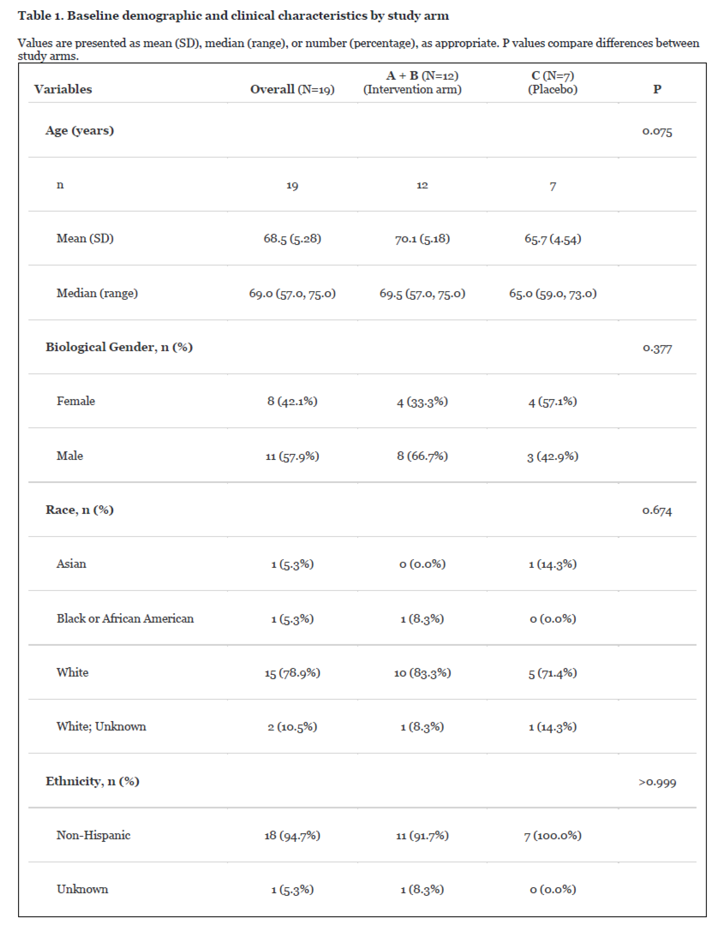
Participant Characteristics and Study Completion: (Table 1) A total of 19 participants were enrolled and included in the analysis, with 12 randomized to the intervention group (curcumin + ω-3 fatty acids) and 7 to placebo. Baseline demographic and clinical characteristics were generally balanced between groups, with a median age of 69 years (range 57–75) and a predominance of male participants (57.9%). Study completion was impacted by challenges in recruitment, adherence, and retention, with the majority of participants discontinuing prior to study completion. These factors resulted in a reduced evaluable sample size for efficacy analyses.
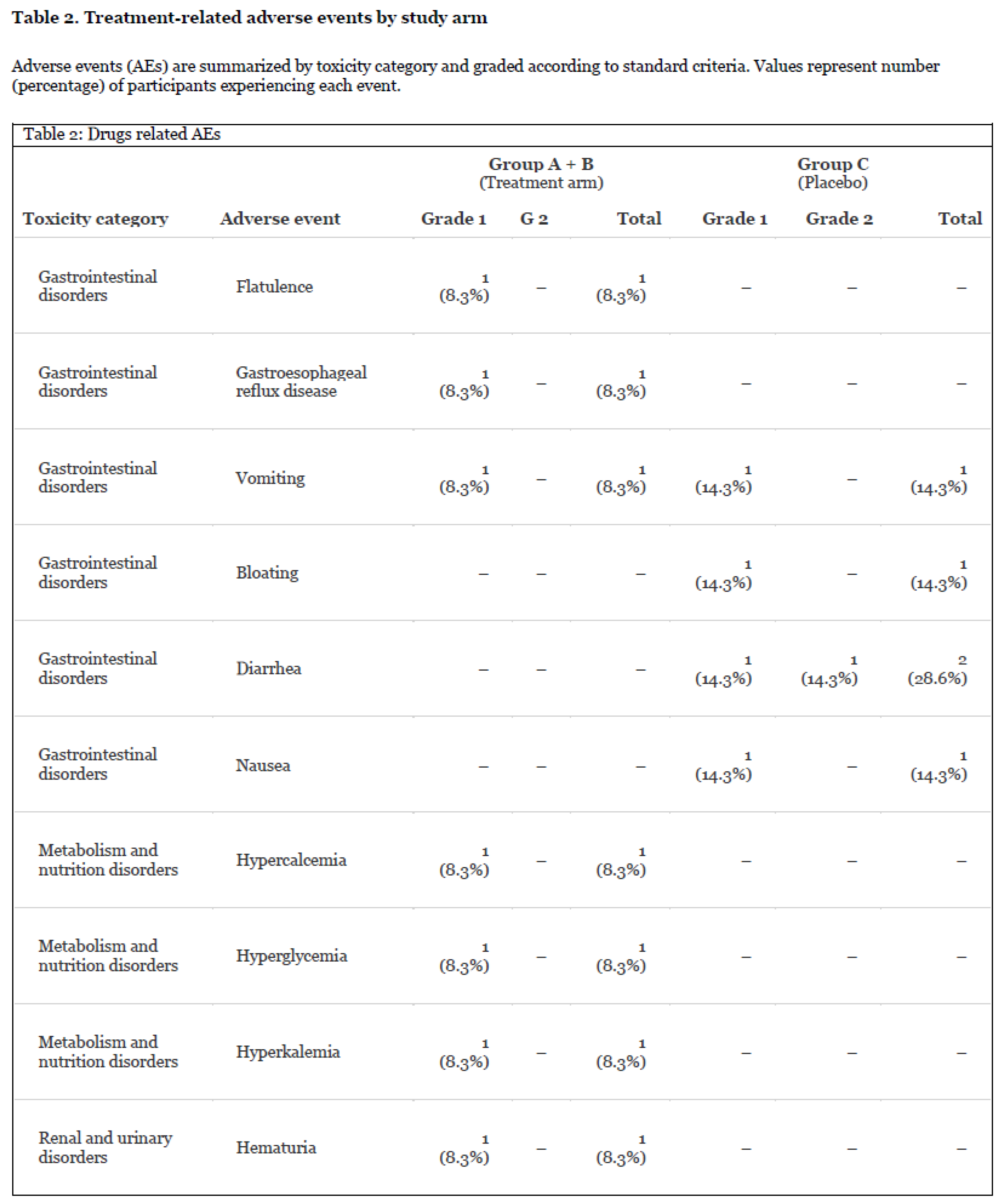
Safety and Tolerability: (Table 2) The combination of curcumin and ω-3 fatty acids was generally well tolerated. A total of 7 participants experienced study drug–related adverse events, most of which were grade 1 in severity. Reported events included gastrointestinal symptoms (e.g., flatulence, reflux, vomiting), fatigue, and mild metabolic abnormalities. No unexpected safety signals or high-grade toxicities attributable to the intervention were observed.
Adherence and Feasibility: Adherence to study intervention was variable, as assessed by pill counts, self-reported logs, and plasma ω-3 index measurements. The study was conducted during the COVID-19 pandemic, which, along with the high comorbidity burden of the study population, contributed to challenges in participant retention and compliance. These factors limited statistical power but provided important feasibility insights relevant to the conduct of chemoprevention trials in high-risk populations.
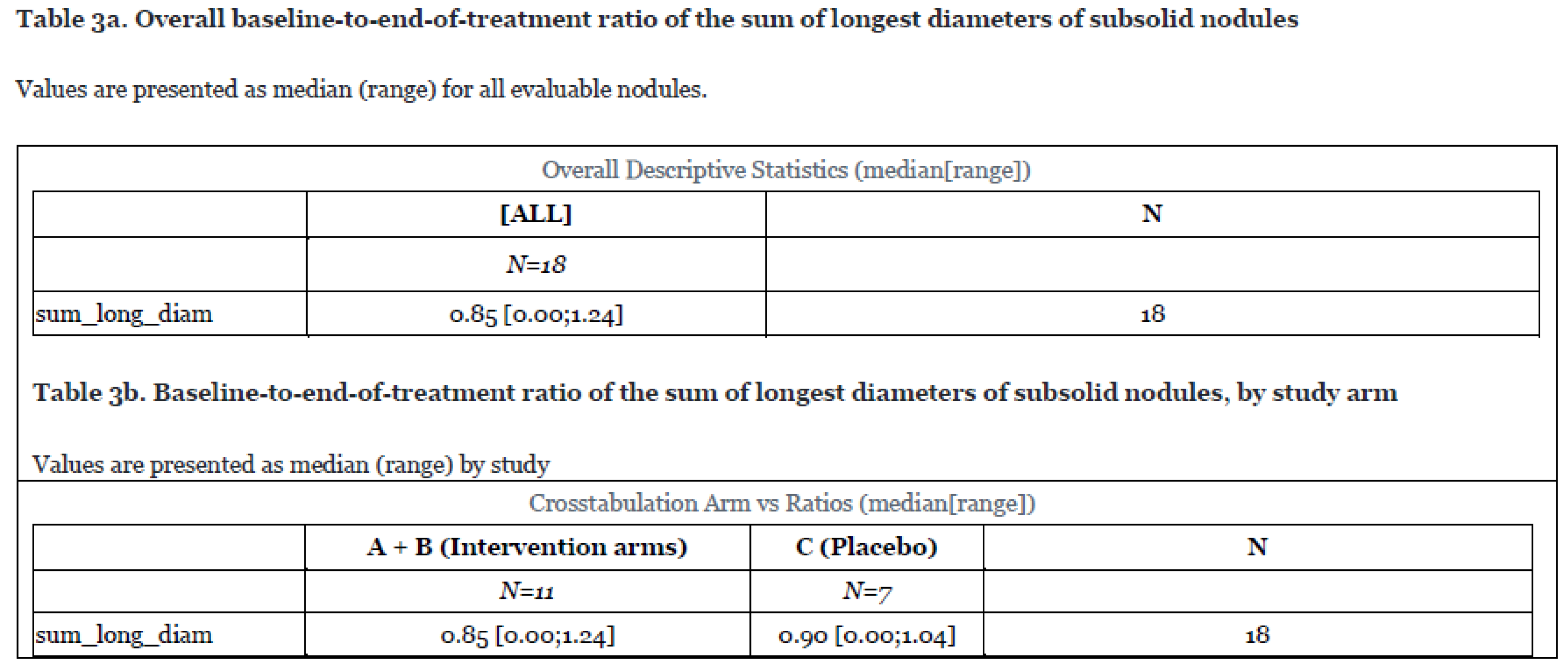
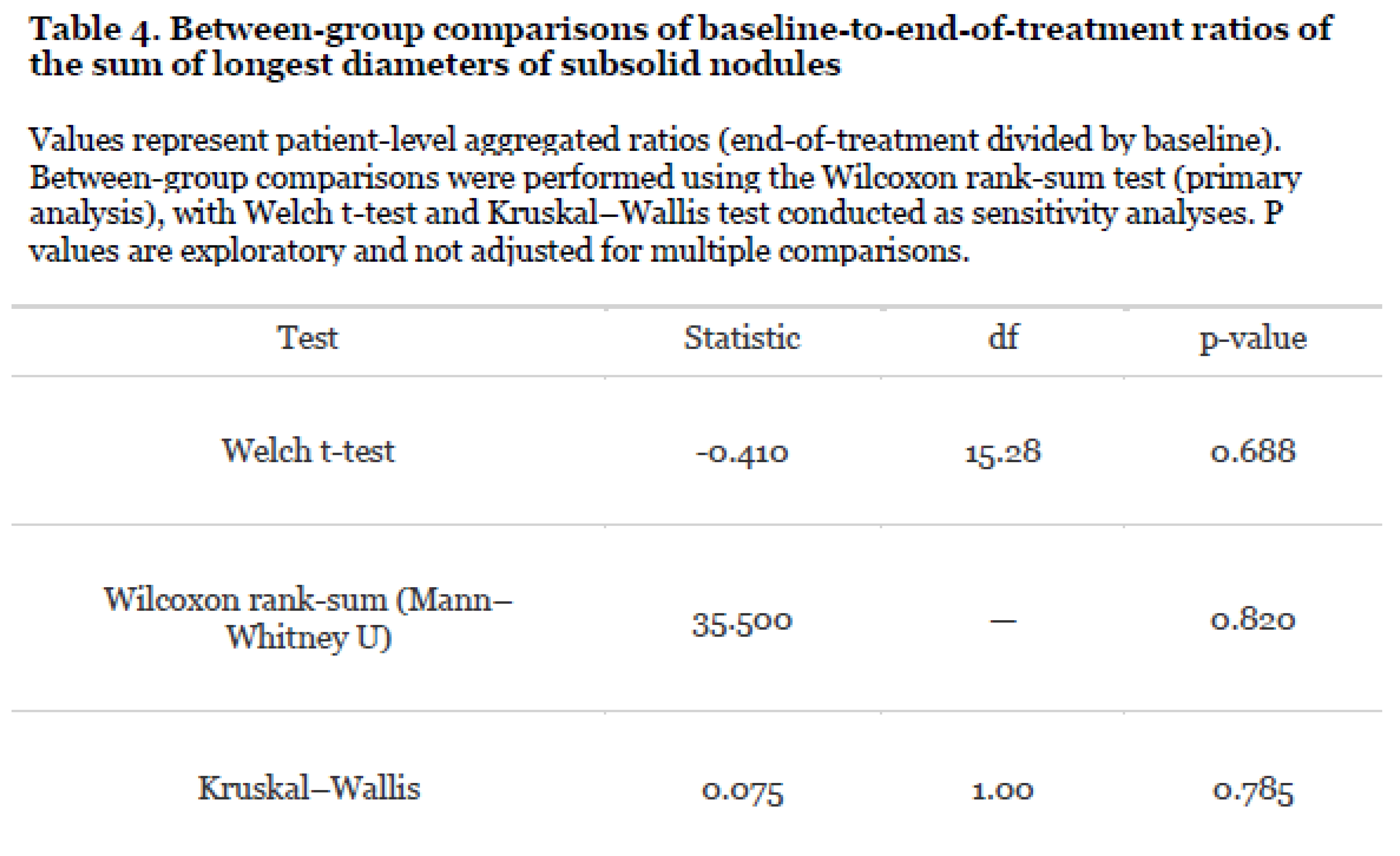
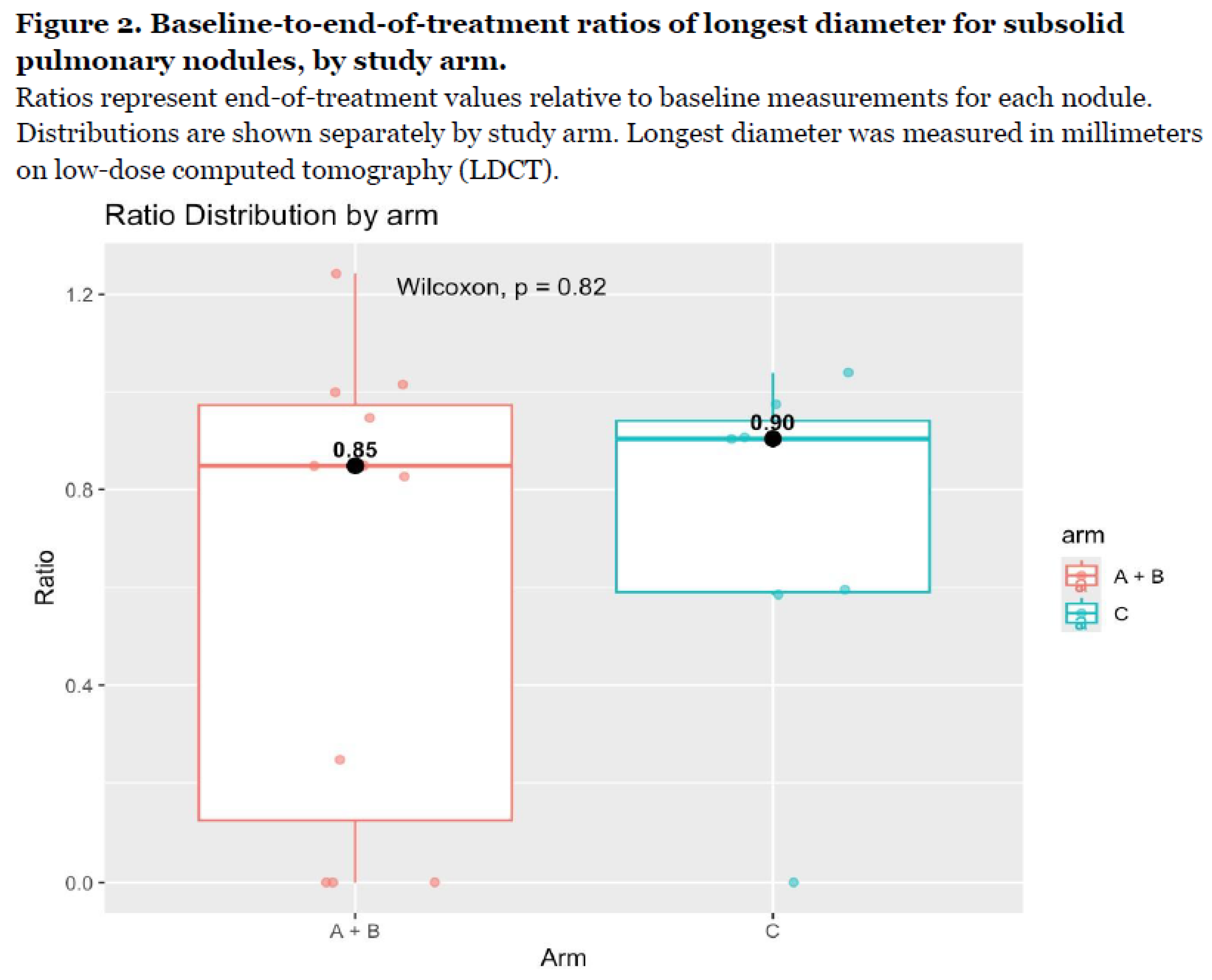
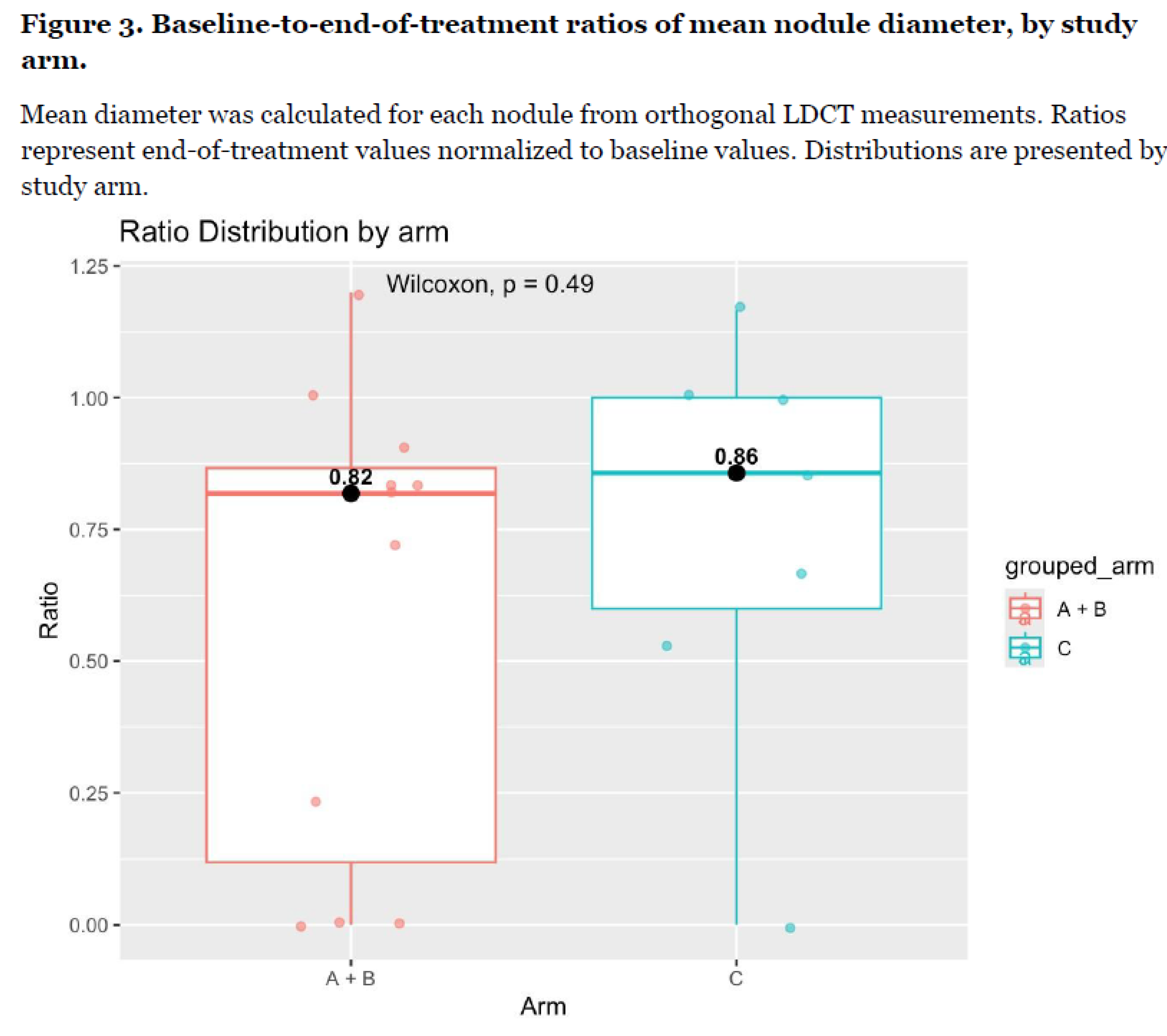
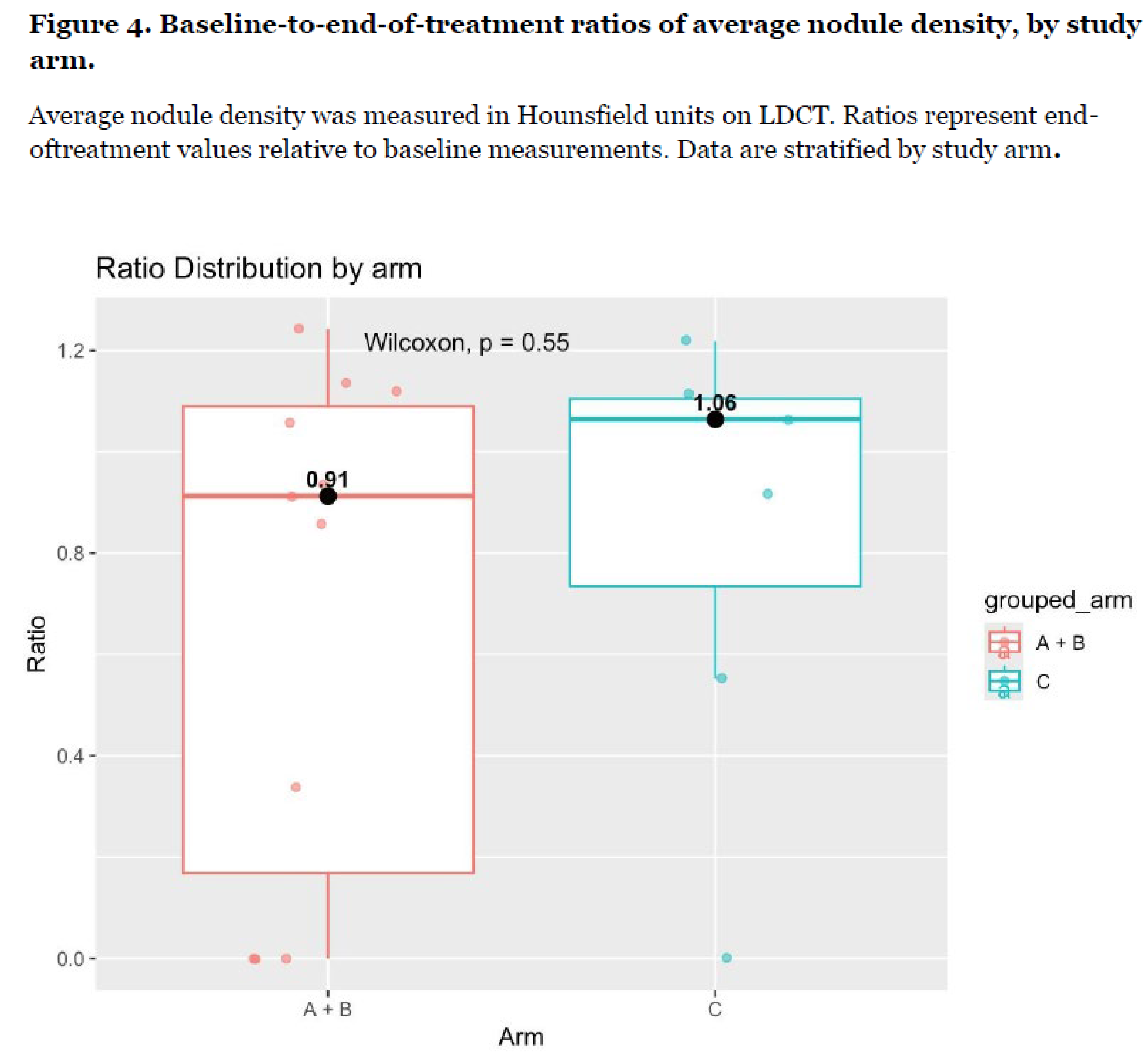
Primary Imaging Outcomes: Table 3a provides the overall baseline-to-end-of-treatment ratio of the sum of longest diameters of subsolid nodules. Values are presented as median (range) for all evaluable nodules. Table 3b provides baseline-to-end-of-treatment ratio of the sum of longest diameters of subsolid nodules, by study arm. Values are presented as median (range) by study. Figure 2 presents the baseline-to-end-of-treatment ratios of longest diameter for subsolid pulmonary nodules, by study arm. Ratios represent end-of-treatment values relative to baseline measurements for each nodule. Distributions are shown separately by study arm. Longest diameter was measured in millimeters on low-dose computed tomography (LDCT). Figure 3 presents the baseline-to-end-of-treatment ratios of mean nodule diameter, by study arm. Mean diameter was calculated for each nodule from orthogonal LDCT measurements. Ratios represent end-of-treatment values normalized to baseline values. Distributions are presented by study arm. Figure 4 presents the baseline-to-end-of-treatment ratios of average nodule density, by study arm. Average nodule density was measured in Hounsfield units on LDCT. Ratios represent end-of-treatment values relative to baseline measurements. Data are stratified by study arm. Table 4 provides the between-group comparisons of baseline-to-end-of-treatment ratios of the sum of longest diameters of subsolid nodules. Values represent patient-level aggregated ratios (end-of-treatment divided by baseline). Between-group comparisons were performed using the Wilcoxon rank-sum test (primary analysis), with Welch t-test and Kruskal–Wallis test conducted as sensitivity analyses. P values are exploratory and not adjusted for multiple comparisons. Although relatively lower increase in these nodule parameters were observed in the intervention arm compared to the placebo arm, no statistically significant differences were observed between the intervention and placebo groups across primary LDCT-derived imaging endpoints, including changes in nodule size, volume, number of nodules ≥4 mm, and density-related measures. Across all evaluated parameters, distributions of aggregated baseline-to-follow-up ratios were comparable between groups, with no consistent directional trends favoring the intervention.
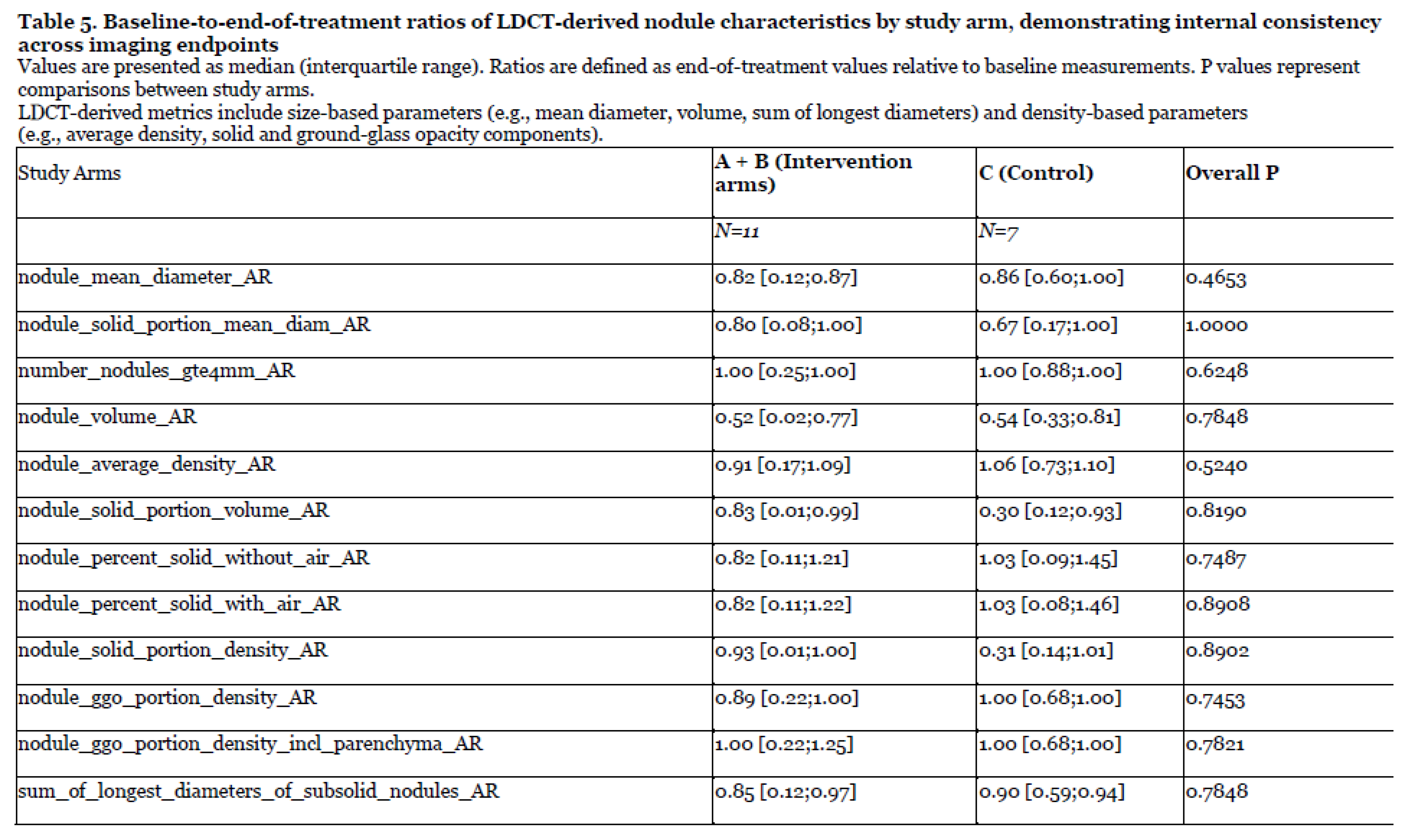
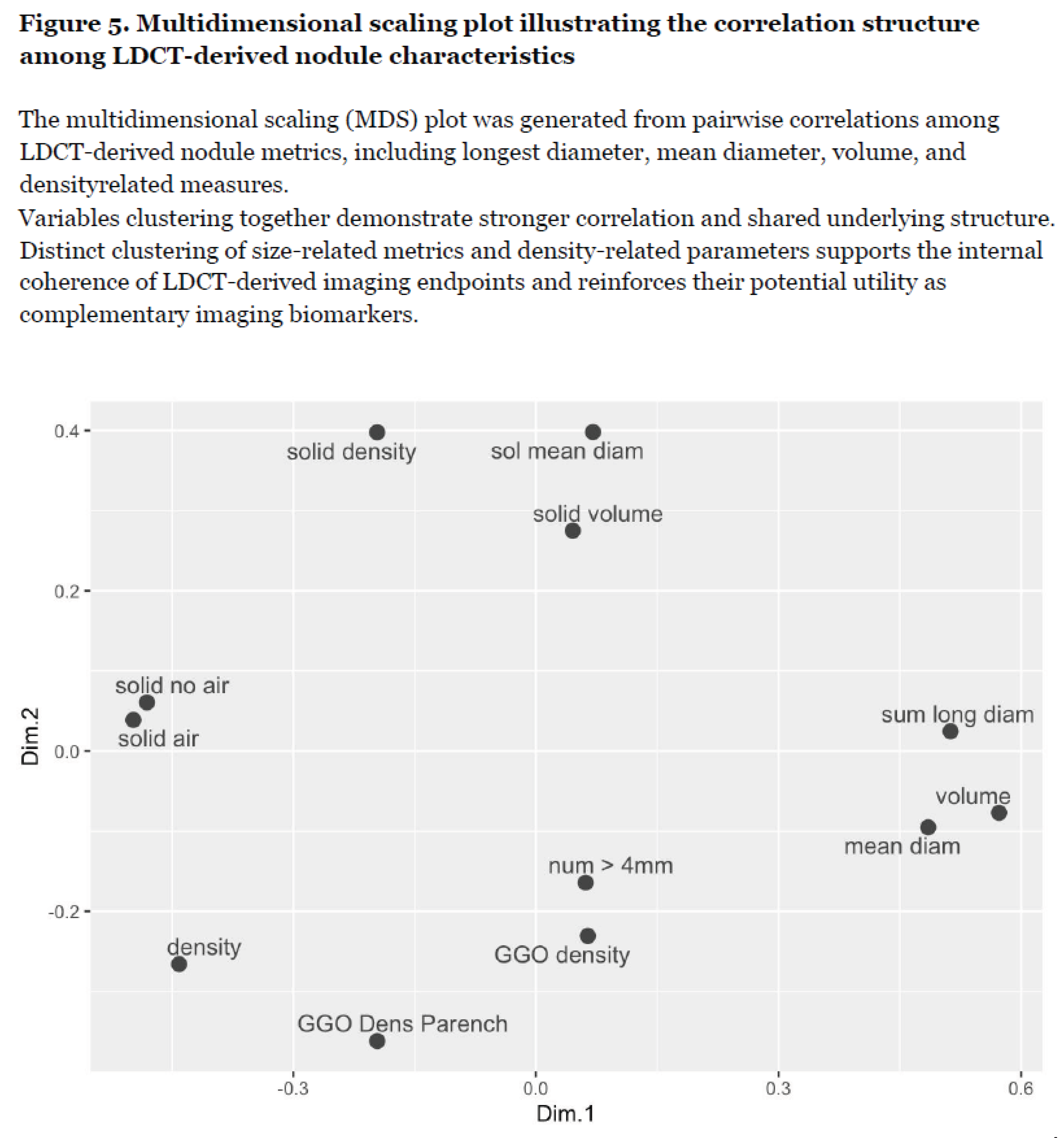
Exploratory Imaging Biomarker Analyses: Table 5 presents the baseline-to-end-of-treatment ratios of LDCT-derived nodule characteristics by study arm, demonstrating internal consistency across imaging endpoints. Values are presented as median (interquartile range). Ratios are defined as end-of-treatment values relative to baseline measurements. P values represent comparisons between study arms. LDCT-derived metrics include size-based parameters (e.g., mean diameter, volume, sum of longest diameters) and density-based parameters (e.g., average density, solid and ground-glass opacity components). Figure 5 displays a multidimensional scaling plot illustrating the correlation structure among LDCT-derived nodule characteristics. The multidimensional scaling (MDS) plot was generated from pairwise correlations among LDCT-derived nodule metrics, including longest diameter, mean diameter, volume, and density-related measures. These exploratory analyses demonstrated consistent positive correlations among LDCT-derived measures of pulmonary nodule characteristics. Strong clustering was observed among size-based metrics, including mean diameter, nodule volume, and the sum of longest diameters, while density-related variables formed a separate but internally consistent cluster. These relationships were visualized through correlation matrix analyses and further supported by multidimensional scaling, which demonstrated spatial grouping of related variables. Collectively, these findings indicate internal coherence among LDCT-derived parameters, suggesting that these measures capture related but distinct dimensions of nodule biology. While not indicative of treatment effect, this consistency supports the potential utility of LDCT-based metrics as candidate intermediate endpoints for signal detection in early-phase chemoprevention studies. These structured interrelationships among LDCT-derived metrics are consistent with prior radiomics studies [56,57,58] demonstrating clustering of imaging features into biologically relevant domains, supporting the validity of these measures as candidate intermediate endpoints.
4. 4. Discussion
In this first, pilot randomized study evaluating the combination of curcumin and omega-3 fatty acids (ω-3 FA) in high-risk current and former smokers with imaging-detected pulmonary nodules, we did not observe statistically significant differences between intervention and placebo groups across primary imaging endpoints. However, this study was designed as an exploratory, signal-generating trial, and several findings provide important insights relevant to the design and conduct of future chemoprevention studies.
First, exploratory analyses demonstrated consistent positive correlations among low-dose computed tomography (LDCT)-derived measures of nodule size, volume, and density, suggesting internal coherence among imaging-based endpoints. These relationships, supported by correlation matrix and multidimensional scaling analyses, indicate that LDCT-derived metrics may capture related but distinct aspects of nodule biology and could serve as integrated biomarkers in early-phase chemoprevention trials. Prior radiomics studies have demonstrated that CT-derived imaging features cluster into correlated domains reflecting tumor size, morphology, and density [56,57,58]. Our findings extend this concept to clinically accessible LDCT-derived metrics, demonstrating that routinely measured parameters exhibit similar internal structure, supporting their use as coherent intermediate endpoints. In settings where clinical endpoints require prolonged follow-up, such imaging-based measures may provide a practical and biologically relevant approach to signal detection and intermediate endpoint evaluation.
Second, despite the absence of a measurable efficacy signal, the biological rationale for the combined use of curcumin and ω-3 FA remains compelling. Curcumin targets key oncogenic and inflammatory pathways, including STAT3 and NF-κB signaling, while ω-3 FA modulate inflammatory responses and promote resolution through specialized pro-resolving mediators. Importantly, the lipophilic nature of curcumin presents a known limitation to its clinical translation, and co-administration with ω-3 FA offers a mechanistically grounded strategy to enhance bioavailability and tissue delivery. Preclinical evidence supports both improved pharmacokinetics and synergistic biological activity of this combination, including enhanced suppression of proliferation and inflammatory signaling. Thus, the absence of observed clinical effects in this pilot study likely reflects limitations in study design and execution rather than a lack of underlying biological activity.
Third, this study highlights critical feasibility challenges inherent to chemoprevention trials in high-risk populations. Conducted during the COVID-19 pandemic, the study experienced substantial barriers to recruitment, adherence, and retention. The target population—older individuals with a history of smoking and a high burden of comorbidities—represents a clinically relevant but complex group in whom competing health priorities and treatment fatigue may limit sustained participation. These factors contributed to reduced evaluable sample size and limited statistical power, constraining the ability to detect modest intervention effects. Importantly, these challenges reflect real-world conditions and underscore the need for pragmatic trial designs that incorporate adherence support, simplified intervention strategies, and optimized patient selection criteria.
This study has several limitations. First, the sample size was small and the study was not powered to detect modest between-group differences, consistent with its design as a pilot, exploratory trial. Second, pulmonary nodules are clustered within patients; to address this, nodule-level measurements were aggregated at the patient level prior to analysis. Given the limited sample size and number of observations per subject, more complex hierarchical modeling approaches, such as mixed-effects models, were not feasible without risk of overfitting. Third, multiple comparisons were performed without formal adjustment, and findings should therefore be interpreted descriptively. Finally, challenges in recruitment, adherence, and retention—exacerbated by the COVID-19 pandemic and the comorbidity burden of the study population—limited the evaluable sample size and statistical power. Accordingly, the results should be considered hypothesis-generating and intended to inform the design of future adequately powered studies.
Collectively, these findings emphasize that early-phase chemoprevention trials must balance biological rigor with feasibility. The present study provides preliminary evidence supporting the use of LDCT-derived imaging metrics as potential intermediate endpoints and reinforces the importance of addressing pharmacologic delivery challenges for agents such as curcumin. Future studies should build on these insights by employing adequately powered designs, refining intervention delivery approaches to enhance bioavailability, and incorporating strategies to improve adherence in high-risk populations. In addition, further validation of imaging biomarkers in relation to biological and clinical outcomes will be essential to advance their role in chemoprevention research.
5. Conclusions
In conclusion, although this pilot study did not demonstrate statistically significant changes in primary endpoints, it provides important feasibility data and highlights key methodological and biological considerations for the development of effective lung cancer chemoprevention strategies. The integration of mechanism-based interventions with robust and scalable biomarker platforms remains a critical next step in advancing prevention efforts in high-risk populations.
These results support continued investigation of combination strategies that address both biological efficacy and pharmacologic delivery, which remain central challenges in cancer chemoprevention. Future studies should evaluate composite imaging approaches that integrate size and density metrics to refine risk stratification and endpoint selection in early-phase lung cancer prevention trials.
6. Future Directions
The future of cancer chemoprevention depends not only on the identification of effective preventive agents but also on the development and validation of rigorous intermediate endpoints capable of efficiently detecting biological activity in early-phase trials. This pilot study contributes to that objective by demonstrating the feasibility and internal coherence of multidimensional LDCT-derived imaging endpoints while highlighting the practical challenges inherent in conducting prevention studies in high-risk populations. These findings provide a methodological framework for future lung cancer interception trials and support continued refinement of biomarker-driven trial design.
Author Contributions
Dr. Nagi Kumar is the principal investigator and was responsible for conceptualization, methodology, sponsor of IND, interpretation, writing, funding acquisition and submission of this manuscript. Dr. Klippenstein was responsible for the imaging studies conducted in this trial. All co-authors of this manuscript were responsible for interpretation, writing, editing prior to submission of this manuscript. Drs. Schell and Laborde were responsible for performing the statistical analysis and interpretation of the data.
Funding
Funding for this manuscript was provided by the State of Florida James Esther King Award to Dr. Nagi Kumar.
Institutional Review Board Statement
Prior to initiating the study and receiving agent, the Moffitt Cancer Center received written approval to conduct the study from the appropriate IRB (Advara). All amendments to the protocol were also approved by the IRB prior to implementation, unless the change involves risk to the subjects, in which case it was implemented immediately. The full protocol and informed consent were approved by the Institutional Review Board (Ethic Committee) Name: Advara, Approval Code: Pro00028018 and Approval Date: 3 Aug 2018. The trial was registered in https://clinicaltrials.gov,—Trial registration number: NCT03598309, Date of registration: 2018-07-13.:.
Informed Consent Statement
All potential study participants were given a copy of the IRB-approved Informed Consent to review. The investigator explained all aspects of the study in lay language and answer all questions regarding the study. If the participant decided to participate in the study, he/she will be asked to sign the IRB approved Informed Consent Form. The subject is considered enrolled in the study from the time the informed consent document is signed. Any and all AEs and concomitant medication taken by the study participant were captured up to end of intervention or at the time of termination from the study. The study agent(s) were not released to a participant who had not signed the Informed Consent document. Subjects who refuse to participate or who withdraw from the study will be treated without prejudice.
Data Availability Statement
As required by NIH rules, we will make the data collected as part of this protocol available to outside investigators. In order to maintain compliance with HIPAA regulations, our preferred method will be to execute a data sharing agreement with the requestor for a limited use dataset as defined by the US Department of Health and Human Services (DHHS).
Acknowledgments
We acknowledge the clinical trials office research team for their work on this trial.
Conflicts of Interest
The authors declare no conflicts of interest. .:.
Abbreviations
The following abbreviations are used in this manuscript:
| Abbreviation | Description |
| 5-LOX | 5-Lipoxygenase |
| 14R/S-HDHA | 14(S)-Hydroxy Docosahexaenoic Acid |
| 17R/S-HDHA | 17(S)-Hydroxy Docosahexaenoic Acid |
| ω-3 FA | Omega-3 Fatty Acids |
| AE(s) | Adverse Event(s) |
| AP-1 | Activator Protein 1 |
| ARDS | Acute Respiratory Distress Syndrome |
| BALF | Bronchoalveolar Lavage Fluid |
| Bcl-2 | B-Cell Lymphoma 2 |
| Bcl-xl | B-Cell Lymphoma-Extra Large |
| CBC | Complete Blood Count |
| cFLIP | Cellular FLICE (FADD-like IL-1β-converting enzyme)-Inhibitory Protein |
| CMP | Complete Metabolic Profile |
| COX-2 | Cyclooxygenase-2 |
| CRFs | Case Report Forms |
| CT | Computed Tomography Scan |
| CTC | Common Terminology Criteria |
| CTCAE | Common Terminology Criteria Adverse Events |
| CTDIvol | CT Dose Index |
| CUR | Curcuminoids |
| CXCR-4 | Chemokine Receptor Type 4 |
| CXR | Chest X-Ray |
| CycD1 | Cyclin-D1 |
| DHA | Docosahexaenoic Acid |
| DSMB | Date Safety Monitoring Board |
| ECOG | Eastern Cooperative Oncology Group |
| EDSMB | External Data Safety Monitoring Board |
| EGFR | Epidermal Growth Factor Receptor |
| EIA | Enzyme Immunoassay |
| ELISA | Enzyme-Linked Immunosorbent Assay |
| EPA | Eicosapentaenoic Acid |
| FDA | Food and Drug Administration |
| GI | Gastrointestinal |
| H157 | Human Oral Squamous Cell Carcinoma |
| H322 | Human Adenocarcinoma Cell Lines |
| HER-2 | Human Epidermal Growth Factor Receptor 2 |
| HPLC | Liquid Chromatography-Tandem |
| HU | Hounsfield Scale |
| IAP | Inhibitor of Apoptosis |
| IEBs | Intermediate Endpoint Biomarker(s) |
| IĸB | IkappaB Kinase |
| IĸB-α | IkappaB Kinase –Alpha |
| IL-1 | Interleukin-1 |
| IL-6 | Interleukin-6 |
| IL-8 | Interleukin-8 |
| IL-12 | Interleukin-12 |
| IND | Investigational New Drug Application |
| IRB | Institutional Review Board |
| LC-MS-MS | Liquid Chromatography-Tandem Mass Spectrometry |
| LDCT | Low Dose Computer Tomography |
| LM | Lipid Mediator |
| LM-SPM | Lipid Mediator |
| LX | Lipoxines |
| MaR | Maresin |
| MaR1 | Maresin 1 |
| MCC | Moffitt Cancer Center |
| MCM2 | Mini-Chromosome Maintenance Proteins |
| MD(s) | Medical Doctor(s) |
| MMP-9 | Matrix Metallopeptidase 9 |
| NCCN | National Comprehensive Cancer Network |
| NCI | National Cancer Institute |
| NDS-R | Minnesota Nutritional Data Systems Research |
| NF-ĸB | Transcription Factor |
| NLST | The National Lung Screening Trial |
| NPD1/PD1 | Neuroprotectin D1/protectin D1 |
| PA | Physical Activity |
| PD | Protectins |
| PGE2 | Prostaglandin E2 |
| PI | Principal Investigator |
| PoS | Probability of Success |
| RvD1 | Resolvin D1 |
| RvD2 | Resolvin D2 |
| RvD3 | Resolvin D3 |
| RvE1 | Resolvin E1 |
| Rv | Resolvins |
| SCC | Squamous Cell Carcinoma |
| SF-36 | Rand Short-Form 36 Medical Outcomes Study SF36 |
| SPM | Specialized Lipid Mediators |
| SRC | Scientific Research Committee |
| SSN(s) | Subsolid Nodule(s) |
| Stat3 | Signal Transducer and Activator of Transcription 3 |
| TNF | Tumor Necrosis Factor |
| TNF-α | Tumor Necrosis Factor Alpha |
| TRIP | Tobacco Research and Intervention Program |
| QOL | Quality of Life |
| VEGF | Vascular Endothelial Growth Factor |
References
- Keith, R.L. Chemoprevention of lung cancer. Proc. Am. Thorac. Soc. 2009, 6, 187–193. [Google Scholar] [CrossRef] [PubMed] [PubMed Central]
- Keith, R.L.; Blatchford, P.J.; Kittelson, J.; Minna, J.D.; Kelly, K.; Massion, P.P.; Franklin, W.A.; Mao, J.; Wilson, D.O.; Merrick, D.T.; Hirsch, F.R.; Kennedy, T.C.; Bunn, P.A.; Jr Geraci, M.W.; Miller, Y.E. Oral iloprost improves endobronchial dysplasia in former smokers. Cancer Prev. Res. 2011, 4, 793–802. [Google Scholar] [CrossRef] [PubMed] [PubMed Central]
- Kumar, N.B.; Quinn, G.P.; Alexandrow, M.G.; Gray, J.; Schell, M.; Sutton, S.; Haura, E.B. Chemoprevention Trial Feasibility Using Botanicals in Exceptionally High Risk Populations for Lung Cancer. J. Clin. Trials 2014, 4. [Google Scholar] [CrossRef] [PubMed] [PubMed Central]
- Lynch, T.J.; Adjei, A.A.; Bunn, P.A.; Jr Eisen, T.G.; Engelman, J.; Goss, G.D.; Haber, D.A.; Heymach, J.V.; Janne, P.A.; Johnson, B.E.; Johnson, D.H.; Lilenbaum, R.C.; Meyerson, M.; Sandler, A.B.; Sequist, L.V.; Settleman, J.; Wong, K.K.; Hart, C.S. Summary statement: novel agents in the treatment of lung cancer: advances in epidermal growth factor receptor-targeted agents. Clin. Cancer Res. An. Off. J. Am. Assoc. Cancer Res. 2006, 12, 4365s–71s. [Google Scholar] [CrossRef] [PubMed]
- O’Shaughnessy, J.A.; Kelloff, G.J.; Gordon, G.B.; Dannenberg, A.J.; Hong, W.K.; Fabian, C.J.; Sigman, C.C.; Bertagnolli, M.M.; Stratton, S.P.; Lam, S.; Nelson, W.G.; Meyskens, F.L.; Alberts, D.S.; Follen, M.; Rustgi, A.K.; Papadimitrakopoulou, V.; Scardino, P.T.; Gazdar, A.F.; Wattenberg, L.W.; Sporn, M.B.; Sakr, W.A.; Lippman, S.M.; Von Hoff, D.D. Treatment and prevention of intraepithelial neoplasia: an important target for accelerated new agent development. Clin. Cancer Res. An. Off. J. Am. Assoc. Cancer Res. 2002, 8, 314–346. [Google Scholar] [PubMed]
- Duvall, M.G.; Levy, B.D. DHA- and EPA-derived resolvins, protectins, and maresins in airway inflammation. Eur. J. Pharmacol. 2015. [Google Scholar] [CrossRef] [PubMed]
- Hsiao, H.M.; Thatcher, T.H.; Colas, R.A.; Serhan, C.N.; Phipps, R.P.; Sime, P.J. Resolvin D1 Reduces Emphysema and Chronic Inflammation. Am. J. Pathol. 2015, 185, 3189–3201. [Google Scholar] [CrossRef] [PubMed]
- Kelly, R.J.; Lopez-Chavez, A.; Szabo, E. Criteria of evidence to move potential chemopreventive agents into late phase clinical trials. Curr. Drug Targets 2011, 12, 1983–1988. [Google Scholar] [CrossRef] [PubMed]
- Block KI, Gyllenhaal C, Lowe L, Amedei A, Amin AR, Amin A, Aquilano K, Arbiser J, Arreola A, Arzumanyan A, Ashraf SS, Azmi AS, Benencia F, Bhakta D, Bilsland A, Bishayee A, Blain SW, Block PB, Boosani CS, Carey TE, Carnero A, Carotenuto M, Casey SC, Chakrabarti M, Chaturvedi R, Chen GZ, Chen H, Chen S, Chen YC, Choi BK, Ciriolo MR, Coley HM, Collins AR, Connell M, Crawford S, Curran CS, Dabrosin C, Damia G, Dasgupta S, DeBerardinis RJ, Decker WK, Dhawan P, Diehl AM, Dong JT, Dou QP, Drew JE, Elkord E, El-Rayes B, Feitelson MA, Felsher DW, Ferguson LR, Fimognari C, Firestone GL, Frezza C, Fujii H, Fuster MM, Generali D, Georgakilas AG, Gieseler F, Gilbertson M, Green MF, Grue B, Guha G, Halicka D, Helferich WG, Heneberg P, Hentosh P, Hirschey MD, Hofseth LJ, Holcombe RF, Honoki K, Hsu HY, Huang GS, Jensen LD, Jiang WG, Jones LW, Karpowicz PA, Keith WN, Kerkar SP, Khan GN, Khatami M, Ko YH, Kucuk O, Kulathinal RJ, Kumar NB, Kwon BS, Le A, Lea MA, Lee HY, Lichtor T, Lin LT, Locasale JW, Lokeshwar BL, Longo VD, Lyssiotis CA, MacKenzie KL, Malhotra M, Marino M, Martinez-Chantar ML, Matheu A, Maxwell C, McDonnell E, Meeker AK, Mehrmohamadi M, Mehta K, Michelotti GA, Mohammad RM, Mohammed SI, Morre DJ, Muralidhar V,Muqbil I, Murphy MP, Nagaraju GP, Nahta R, Niccolai E, Nowsheen S, Panis C, Pantano F, Parslow VR, Pawelec G, Pedersen PL, Poore B, Poudyal D, Prakash S, Prince M, Raffaghello L, Rathmell JC, Rathmell WK, Ray SK, Reichrath J, Rezazadeh S, Ribatti D, Ricciardiello L, Robey RB, Rodier F, Rupasinghe HP, Russo GL, Ryan EP, Samadi AK, Sanchez-Garcia I, Sanders AJ, Santini D, Sarkar M, Sasada T, Saxena NK, Shackelford RE, Shantha Kumara HM, Sharma D, Shin DM, Sidransky D, Siegelin MD, Signori E, Singh N, Sivanand S, Sliva D, Smythe C, Spagnuolo C, Stafforini DM, Stagg J, Subbarayan PR, Sundin T, Talib WH, Thompson SK, Tran PT, Ungefroren H, Vander Heiden MG, Venkateswaran V, Vinay DS, Vlachostergios PJ, Wang Z, Wellen KE, Whelan RL, Yang ES, Yang H, Yang X, Yaswen P, Yedjou C, Yin X, Zhu J, Zollo M. Designing a broad-spectrum integrative approach for cancer prevention and treatment. Seminars in cancer biology 2015, 35, S276–S304. [CrossRef] [PubMed]
- Aggarwal, B.B.; Harikumar, K.B. Potential therapeutic effects of curcumin, the anti-inflammatory agent, against neurodegenerative, cardiovascular, pulmonary, metabolic, autoimmune and neoplastic diseases. Int. J. Biochem. Cell Biol. 2009, 41, 40–59. [Google Scholar] [CrossRef] [PubMed] [PubMed Central]
- Bill, M.A.; Fuchs, J.R.; Li, C.; Yui, J.; Bakan, C.; Benson, D.M.; Jr Schwartz, E.B.; Abdelhamid, D.; Lin, J.; Hoyt, D.G.; Fossey, S.L.; Young, G.S.; Carson, W.E.; Li, P.K., 3rd; Lesinski, G.B. The small molecule curcumin analog FLLL32 induces apoptosis in melanoma cells via STAT3 inhibition and retains the cellular response to cytokines with anti-tumor activity. Mol. Cancer 2010, 9, 165. [Google Scholar] [CrossRef] [PubMed] [PubMed Central]
- Choudhuri, T.; Pal, S.; Das, T.; Sa, G. Curcumin selectively induces apoptosis in deregulated cyclin D1- expressed cells at G2 phase of cell cycle in a p53-dependent manner. J. Biol. Chem. 2005, 280, 20059–20068. [Google Scholar] [CrossRef] [PubMed]
- Goel, A.; Kunnumakkara, A.B.; Aggarwal, B.B. Curcumin as “Curecumin”: from kitchen to clinic. Biochem. Pharmacol. 2008, 75, 787–809. [Google Scholar] [CrossRef] [PubMed]
- Kunnumakkara, A.B.; Anand, P.; Aggarwal, B.B. Curcumin inhibits proliferation, invasion, angiogenesis and metastasis of different cancers through interaction with multiple cell signaling proteins. Cancer Lett. 2008, 269, 199–225. [Google Scholar] [CrossRef] [PubMed]
- Lin, S.S.; Lai, K.C.; Hsu, S.C.; Yang, J.S.; Kuo, C.L.; Lin, J.P.; Ma, Y.S.; Wu, C.C.; Chung, J.G. Curcumin inhibits the migration and invasion of human A549 lung cancer cells through the inhibition of matrix metalloproteinase-2 and -9 and Vascular Endothelial Growth Factor (VEGF). Cancer Lett. 2009, 285, 127–133. [Google Scholar] [CrossRef] [PubMed]
- Sameermahmood, Z.; Balasubramanyam, M.; Saravanan, T.; Rema, M. Curcumin modulates SDF- 1alpha/CXCR4-induced migration of human retinal endothelial cells (HRECs). Investig. Ophthalmol. Vis. Sci. 2008, 49, 3305–3311. [Google Scholar] [CrossRef] [PubMed]
- Sung, B.; Prasad, S.; Yadav, V.R.; Aggarwal, B.B. Cancer cell signaling pathways targeted by spice-derived nutraceuticals. Nutr. Cancer 2012, 64, 173–197. [Google Scholar] [CrossRef] [PubMed] [PubMed Central]
- Wu, S.H.; Hang, L.W.; Yang, J.S.; Chen, H.Y.; Lin, H.Y.; Chiang, J.H.; Lu, C.C.; Yang, J.L.; Lai, T.Y.; Ko, Y.C.; Chung, J.G. Curcumin induces apoptosis in human non-small cell lung cancer NCI-H460 cells through ER stress and caspase cascade- and mitochondria-dependent pathways. Anticancer Res. 2010, 30, 2125–33. [Google Scholar] [PubMed]
- Halappanavar, S.; Russell, M.; Stampfli, M.R.; Williams, A.; Yauk, C.L. Induction of the interleukin 6/ signal transducer and activator of transcription pathway in the lungs of mice sub-chronically exposed to mainstream tobacco smoke. BMC Med. Genom. 2009, 2, 56. [Google Scholar] [CrossRef] [PubMed] [PubMed Central]
- Yu, H.; Jove, R. The STATs of cancer--new molecular targets come of age. Nat. Rev. Cancer 2004, 4, 97–105. [Google Scholar] [CrossRef] [PubMed]
- Haura, E.B.; Turkson, J.; Jove, R. Mechanisms of disease: Insights into the emerging role of signal transducers and activators of transcription in cancer. Nat. Clin. Pract. Oncol. 2005, 2, 315–24. [Google Scholar] [CrossRef] [PubMed]
- Li, Y.; Du, H.; Qin, Y.; Roberts, J.; Cummings, O.W.; Yan, C. Activation of the signal transducers and activators of the transcription 3 pathway in alveolar epithelial cells induces inflammation and adenocarcinomas in mouse lung. Cancer Res. 2007, 67, 8494–8503. [Google Scholar] [CrossRef] [PubMed]
- Alexandrow, M.G.; Song, L.J.; Altiok, S.; Gray, J.; Haura, E.B.; Kumar, N.B. Curcumin: a novel Stat3 pathway inhibitor for chemoprevention of lung cancer. European journal of cancer prevention: the official journal of the European Cancer Prevention Organisation 2012, 21, 407–412. [Google Scholar] [CrossRef] [PubMed] [PubMed Central]
- Gao, S.P.; Mark, K.G.; Leslie, K.; Pao, W.; Motoi, N.; Gerald, W.L.; Travis, W.D.; Bornmann, W.; Veach, D.; Clarkson, B.; Bromberg, J.F. Mutations in the EGFR kinase domain mediate STAT3 activation via IL-6 production in human lung adenocarcinomas. J. Clin. Investig. 2007, 117, 3846–3856. [Google Scholar] [CrossRef] [PubMed] [PubMed Central]
- Panahi, Y.; Darvishi, B.; Ghanei, M.; Jowz, N.; Beiraghdar, F.; Varnamkhasti, B.S. Molecular mechanisms of curcumins suppressing effects on tumorigenesis, angiogenesis and metastasis, focusing on NF-kB pathway. Cytokine Growth Factor Rev. 2016, Pss: A1359-6101((15)30012-5). [Google Scholar] [CrossRef] [PubMed]
- Cho, J.W.; Lee, K.S.; Kim, C.W. Curcumin attenuates the expression of IL-1beta, IL-6, and TNF-alpha as well as cyclin E in TNF-alpha-treated HaCaT cells; NF-kappaB and MAPKs as potential upstream targets. Int. J. Mol. Med. 2007, 19, 469–474. [Google Scholar] [PubMed]
- Pal, S.; Bhattacharyya, S.; Choudhuri, T.; Datta, G.K.; Das, T.; Sa, G. Amelioration of immune cell number depletion and potentiation of depressed detoxification system of tumor-bearing mice by curcumin. Cancer Detect Prev. 2005, 29, 470–478. [Google Scholar] [CrossRef] [PubMed]
- Sehgal, A.; Kumar, M.; Jain, M.; Dhawan, D.K. Synergistic effects of piperine and curcumin in modulating benzo(a)pyrene induced redox imbalance in mice lungs. Toxicol. Mech. Methods 2012, 22, 74–80. [Google Scholar] [CrossRef] [PubMed]
- Dance-Barnes, S.T.; Kock, N.D.; Moore, J.E.; Lin, E.Y.; Mosley, L.J.; D’Agostino, R.B.; Jr McCoy, T.P.; Townsend, A.J.; Miller, M.S. Lung tumor promotion by curcumin. Carcinogenesis 2009, 30, 1016–1023. [Google Scholar] [CrossRef] [PubMed] [PubMed Central]
- Moghaddam, S.J.; Barta, P.; Mirabolfathinejad, S.G.; Ammar-Aouchiche, Z.; Garza, N.T.; Vo, T.T.; Newman, R.A.; Aggarwal, B.B.; Evans, C.M.; Tuvim, M.J.; Lotan, R.; Dickey, B.F. Curcumin inhibits COPD-like airway inflammation and lung cancer progression in mice. Carcinogenesis 2009, 30, 1949–1956. [Google Scholar] [CrossRef] [PubMed] [PubMed Central]
- Lee, J.C.; Kinniry, P.A.; Arguiri, E.; Serota, M.; Kanterakis, S.; Chatterjee, S.; Solomides, C.C.; Javvadi, P.; Koumenis, C.; Cengel, K.A.; Christofidou-Solomidou, M. Dietary curcumin increases antioxidant defenses in lung, ameliorates radiation-induced pulmonary fibrosis, and improves survival in mice. Radiat. Res. 2010, 173, 590–601. [Google Scholar] [CrossRef] [PubMed] [PubMed Central]
- Sharma, R.A.; Euden, S.A.; Platton, S.L.; Cooke, D.N.; Shafayat, A.; Hewitt, H.R.; Marczylo, T.H.; Morgan, B.; Hemingway, D.; Plummer, S.M.; Pirmohamed, M.; Gescher, A.J.; Steward, W.P. Phase I clinical trial of oral curcumin: biomarkers of systemic activity and compliance. Clin. Cancer Res. An. Off. J. Am. Assoc. Cancer Res. 2004, 10, 6847–6854. [Google Scholar] [CrossRef] [PubMed]
- Cheng, A.L.; Hsu, C.H.; Lin, J.K.; Hsu, M.M.; Ho, Y.F.; Shen, T.S.; Ko, J.Y.; Lin, J.T.; Lin, B.R.; Ming-Shiang, W.; Yu, H.S.; Jee, S.H.; Chen, G.S.; Chen, T.M.; Chen, C.A.; Lai, M.K.; Pu, Y.S.; Pan, M.H.; Wang, Y.J.; Tsai, C.C.; Hsieh, C.Y. Phase I clinical trial of curcumin, a chemopreventive agent, in patients with high-risk or pre-malignant lesions. Anticancer Res. 2001, 21, 2895–2900. [Google Scholar] [PubMed]
- Carroll, R.E.; Benya, R.V.; Turgeon, D.K.; Vareed, S.; Neuman, M.; Rodriguez, L.; Kakarala, M.; Carpenter, P.M.; McLaren, C.; Meyskens, F.L.; Jr Brenner, D.E. Phase IIa clinical trial of curcumin for the prevention of colorectal neoplasia. Cancer Prev. Res. 2011, 4, 354–364. [Google Scholar] [CrossRef] [PubMed] [PubMed Central]
- Busquets Ruis, S. Effects of Nutritfriend(Smartfish+CUR) on tumor growth and cachexia in mice bearning Lewis Lung Carcinoma. In Cancer Research Group, Department of Biochemistry and Molecular Biology of Cancer; (Unplublished); University of Barcelona.
- Fasano, E.; Serini, S.; Piccioni, E.; Innocenti, I.; Calviello, G. Chemoprevention of lung pathologies by dietary ω-3 polyunsaturated fatty acids. Curr. Med. Chem. 2010, 17, 3358–3376. [Google Scholar] [CrossRef] [PubMed]
- Aggarwal, B.B.; Kumar, A.; Bharti, A.C. Anticancer potential of curcumin: preclinical and clinical studies. Anticancer Res. 2003, 23, 363–398. [Google Scholar] [PubMed]
- Kumar, N.B.; Kazi, A.; Smith, T.; Crocker, T.; Yu, D.; Reich, R.R.; Reddy, K.; Hastings, S.; Exterman, M.; Balducci, L.; Dalton, K.; Bepler, G. Cancer cachexia: traditional therapies and novel molecular mechanism-based approaches to treatment. Curr. Treat. Options Oncol. 2010, 11, 107–117. [Google Scholar] [CrossRef] [PubMed] [PubMed Central]
- Calder, P.C. Polyunsaturated fatty acids, inflammatory processes and inflammatory bowel diseases. Mol. Nutr. Food Res. 2008, 52, 885–897. [Google Scholar] [CrossRef] [PubMed]
- Nys, M.; Deby-Dupont, G.; Habraken, Y.; Legrand-Poels, S.; Kohnen, S.; Ledoux, D.; Canivet, J.L.; Damas, P.; Lamy, M. Bronchoalveolar lavage fluids of ventilated patients with acute lung injury activate NF-kappaB in alveolar epithelial cell line: role of reactive oxygen/nitrogen species and cytokines. Nitric Oxide Biol. Chem. Off. J. Nitric Oxide Soc. 2003, 9, 33–43. [Google Scholar] [CrossRef] [PubMed]
- Singer, P.; Shapiro, H.; Theilla, M.; Anbar, R.; Singer, J.; Cohen, J. Anti-inflammatory properties of omega-3 fatty acids in critical illness: novel mechanisms and an integrative perspective. Intensive Care Med. 2008, 34, 1580–1592. [Google Scholar] [CrossRef] [PubMed]
- Cotogni, P.; Trombetta, A.; Muzio, G.; Maggiora, M.; Canuto, R.A. The Omega-3 Fatty Acid Docosahexaenoic Acid Modulates Inflammatory Mediator Release in Human Alveolar Cells Exposed to Bronchoalveolar Lavage Fluid of ARDS Patients. BioMed Res. Int. 2015, 2015, 642520. [Google Scholar] [CrossRef] [PubMed] [PubMed Central]
- Weylandt, K.H.; Chiu, C.Y.; Gomolka, B.; Waechter, S.F.; Wiedenmann, B. Omega-3 fatty acids and their lipid mediators: towards an understanding of resolvin and protectin formation. Prostaglandins Other Lipid Mediat. 2012, 97, 73–82. [Google Scholar] [CrossRef] [PubMed]
- Weylandt, K.H. Docosapentaenoic acid derived metabolites and mediators—The new world of lipid mediator medicine in a nutshell. Eur. J. Pharmacol. 2015. [Google Scholar] [CrossRef] [PubMed]
- Souza, P.R.; Norling, L.V. Implications for eicosapentaenoic acid- and docosahexaenoic acid-derived resolvins as therapeutics for arthritis. Eur. J. Pharmacol. 2015. [Google Scholar] [CrossRef] [PubMed]
- Barden, A.; Mas, E.; Croft, K.D.; Phillips, M.; Mori, T.A. Short-term ω-3 fatty acid supplementation but not aspirin increases plasma proresolving mediators of inflammation. J. Lipid Res. 2014, 55, 2401–2407. [Google Scholar] [CrossRef] [PubMed] [PubMed Central]
- Mas, E.; Barden, A.; Burke, V.; Beilin, L.J.; Watts, G.F.; Huang, R.C.; Puddey, I.B.; Irish, A.B.; Mori, T.A. A randomized controlled trial of the effects of ω-3 fatty acids on resolvins in chronic kidney disease. Clin. Nutr. 2015. [Google Scholar] [CrossRef] [PubMed]
- Mas, E.; Croft, K.D.; Zahra, P.; Barden, A.; Mori, T.A. Resolvins D1, D2, and other mediators of self-limited resolution of inflammation in human blood following ω-3 fatty acid supplementation. Clin. Chem. 2012, 58, 1476–1484. [Google Scholar] [CrossRef] [PubMed]
- Zou, L.; Liu, W.; Liu, C.; Xiao, H.; McClements, D.J. Utilizing food matrix effects to enhance nutraceutical bioavailability: increase of curcumin bioaccessibility using excipient emulsions. J. Agric. Food Chem. 2015, 63, 2052–2062. [Google Scholar] [CrossRef] [PubMed]
- Siddiqui, R.A.; Harvey, K.A.; Walker, C.; Altenburg, J.; Xu, Z.; Terry, C.; Camarillo, I.; Jones-Hall, Y.; Mariash, C. Characterization of synergistic anti-cancer effects of docosahexaenoic acid and curcumin on DMBA- induced mammary tumorigenesis in mice. BMC Cancer 2013, 13, 418. [Google Scholar] [CrossRef] [PubMed] [PubMed Central]
- Altenburg, J.D.; Bieberich, A.A.; Terry, C.; Harvey, K.A.; Vanhorn, J.F.; Xu, Z.; Jo Davisson, V.; Siddiqui, R.A. A synergistic antiproliferation effect of curcumin and docosahexaenoic acid in SK-BR-3 breast cancer cells: unique signaling not explained by the effects of either compound alone. BMC Cancer 2011, 11, 149. [Google Scholar] [CrossRef] [PubMed] [PubMed Central]
- Halder, R.C.; Almasi, A.; Sagong, B.; Leung, J.; Jewett, A.; Fiala, M. Curcuminoids and omega-3 fatty acids with anti-oxidants potentiate cytotoxicity of natural killer cells against pancreatic ductal adenocarcinoma cells and inhibit interferon gamma production. Front. Physiol. 2015, 6, 129. [Google Scholar] [CrossRef] [PubMed] [PubMed Central]
- Fiala, M. Curcumin and omega-3 fatty acids enhance NK cell-induced apoptosis of pancreatic cancer cells but curcumin inhibits interferon-gamma production: benefits of omega-3 with curcumin against cancer. Molecules 2015, 20, 3020–3026. [Google Scholar] [CrossRef] [PubMed]
- Wang, J.; Zhu, R.; Sun, D.; Sun, X.; Geng, Z.; Kiu, H.; Wang, S.L. Inracellular Uptake of Curcumin-Loaded Solid Lipid Nanoparticles Exhibit Anti-Inflammatory Activities Superior to Those of Curcumin Through the NF-kB Signaling Pathways. J. BioMed Nanotechnl 2015, 11, 403–415. [Google Scholar] [CrossRef] [PubMed]
- Fan, Z.; Yaoi, J.; Li, Y.; Hu, X.; Shao, H.; Tian, X. Anti-inflammatory and antioxidant effects of curcumin on acute lung injury in a rodent model of intestinal ischemia reperfusion by inhibiting the pathway of NF- kB. Int. J. Clin. Exp. Pathol. 2015, 8, 3451–3459. [Google Scholar] [PubMed] [PubMed Central]
- Aerts, H.J.; Velazquez, E.R.; Leijenaar, R.T.; Parmar, C.; Grossmann, P.; Carvalho, S.; Bussink, J.; Monshouwer, R.; Haibe-Kains, B.; Rietveld, D.; Hoebers, F.; Rietbergen, M.M.; Leemans, C.R.; Dekker, A.; Quackenbush, J.; Gillies, R.J.; Lambin, P. Decoding tumour phenotype by noninvasive imaging using a quantitative radiomics approach. Nat. Commun. 2014, 5, 4006, Erratum in: Nat Commun. 2014, 5:4644. Cavalho, Sara [corrected to Carvalho, Sara]. PMID: 24892406; PMCID: PMC4059926. [Google Scholar] [CrossRef]
- Hawkins, S.; Wang, H.; Liu, Y.; Garcia, A.; Stringfield, O.; Krewer, H.; Li, Q.; Cherezov, D.; Gatenby, R.A.; Balagurunathan, Y.; Goldgof, D.; Schabath, M.B.; Hall, L.; Gillies, R.J. Predicting Malignant Nodules from Screening CT Scans. J. Thorac. Oncol. 2016, 11, 2120–2128, Erratum in: J Thorac Oncol. 2018, 13, 280–281. doi: 10.1016/j.jtho.2017.09.1959. PMID: 27422797; PMCID: PMC5545995. [Google Scholar] [CrossRef]
- Lambin, P.; Leijenaar, R.T.H.; Deist, T.M.; Peerlings, J.; de Jong, E.E.C.; van Timmeren, J.; Sanduleanu, S.; Larue, R.T.H.M.; Even, A.J.G.; Jochems, A.; van Wijk, Y.; Woodruff, H.; van Soest, J.; Lustberg, T.; Roelofs, E.; van Elmpt, W.; Dekker, A.; Mottaghy, F.M.; Wildberger, J.E.; Walsh, S. Radiomics: the bridge between medical imaging and personalized medicine. Nat. Rev. Clin. Oncol. 2017, 14, 749–762. [Google Scholar] [CrossRef] [PubMed]
- Kumar, N.B.; Quinn, G.P.; Alexandrow, M.G.; Gray, J.; Schell, M.; Sutton, S.; Haura, E.B. Chemoprevention Trial Feasibility Using Botanicals in Exceptionally High Risk Populations for Lung Cancer. J. Clin. Trials 2014, 4, 180. [Google Scholar] [CrossRef] [PubMed] [PubMed Central]
Disclaimer/Publisher’s Note: The statements, opinions and data contained in all publications are solely those of the individual author(s) and contributor(s) and not of MDPI and/or the editor(s). MDPI and/or the editor(s) disclaim responsibility for any injury to people or property resulting from any ideas, methods, instructions or products referred to in the content. |
© 2026 by the authors. Licensee MDPI, Basel, Switzerland. This article is an open access article distributed under the terms and conditions of the Creative Commons Attribution (CC BY) license (http://creativecommons.org/licenses/by/4.0/).
Copyright: This open access article is published under a Creative Commons CC BY 4.0 license, which permit the free download, distribution, and reuse, provided that the author and preprint are cited in any reuse.


