
Submitted:
05 July 2026
Posted:
22 July 2026
You are already at the latest version
Abstract
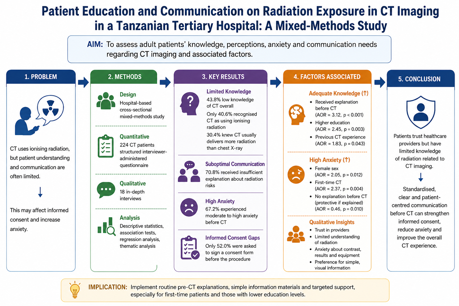
Background: Computed tomography (CT) is essential for diagnosis, but its use of ionising radiation requires clear patient communication and informed consent. This study assessed knowledge, perception and communication needs regarding CT imaging among adult patients at Benjamin Mkapa Hospital, Tanzania. Methods: A hospital-based cross-sectional mixed-methods study was conducted among inpatients and outpatients attending CT services. Quantitative data were collected from 224 patients using a structured interviewer-administered questionnaire, and qualitative data were obtained through 18 semi-structured interviews. Descriptive statistics, association tests, regression analysis and thematic analysis were used. Findings were integrated through triangulation. Results: Overall, 43.8% of participants had low CT knowledge, 37.9% had moderate knowledge and 18.3% had high knowledge. Although 65.6% knew that CT produces cross-sectional images, only 40.6% identified CT as ionising radiation and 30.4% knew that CT usually delivers more radiation than a plain chest X-ray. Trust in healthcare providers was high, while pre-scan anxiety was moderate. Adequate knowledge was associated with receiving an explanation before CT, higher education and previous CT experience. High anxiety was more common among female and first-time CT patients, but was lower among those who received explanations. Conclusion: Patients accepted CT mainly through professional trust, but radiation knowledge and interactive communication were limited. Standard pre-CT explanations, simple information materials and targeted support for first-time patients may strengthen informed consent and reduce avoidable anxiety. The findings highlight a practical patient-education gap within routine Tanzanian tertiary imaging services.
Keywords:
computed tomography
; patient education
; radiation awareness
; informed consent
; patient perception
Introduction
Computed tomography (CT) is an essential diagnostic imaging modality in modern clinical practice. It provides rapid cross-sectional images that support diagnosis, disease staging, treatment planning and follow-up. In tertiary hospitals, CT is particularly valuable because it can provide detailed anatomical information that may not be adequately obtained from clinical assessment or plain radiography alone. However, CT uses ionising radiation and therefore requires appropriate justification, optimisation and patient-centred communication. International radiation-protection guidance emphasises that medical exposure should be justified by clinical benefit and optimised to achieve the required diagnostic information at the lowest reasonable dose [1,2]. Evidence from international CT practice has also shown substantial variation in radiation dose between institutions and countries, indicating that radiation safety depends not only on technology but also on protocols, systems and communication practices [3].
Patient understanding is an important part of safe and ethical CT imaging. Before undergoing CT, patients should receive clear information about why the scan is needed, what the procedure involves, whether contrast media will be used, what safety checks are required and whether the examination involves ionising radiation. If this information is not communicated clearly, informed consent may become a routine administrative act rather than a meaningful patient-centred process. Sweetman and Bernard [4] argue that informed consent for imaging procedures using ionising radiation requires patients to appreciate both the benefits and potential risks of the examination. Similarly, radiation-risk communication studies show that patients and families often ask about imaging risks, but health professionals may struggle to communicate dose and risk in ways that are both accurate and understandable [5].
Previous studies have consistently reported limited patient knowledge of radiation exposure from diagnostic imaging. Ribeiro et al. [6], in a review of patient awareness, concluded that patients are generally poorly informed about radiation exposure during diagnostic scans. Bastiani et al. [7], in a multicentre survey of 2,866 patients undergoing radiological examinations in Italy, found important misconceptions about medical radiation and showed that better knowledge was associated with receiving information from medical staff and having a higher educational level. CT-specific evidence has also shown persistent knowledge gaps. In Saudi Arabia, Alashban and Alghamdi [8] reported that only 56.8% of CT patients correctly classified CT as an ionising-radiation procedure. These findings suggest that patient attendance for CT should not be interpreted as evidence of adequate understanding.
The African and Tanzanian context makes this issue more important. Mkoloma et al. [9] reported low knowledge and awareness of medical radiation among patients, household members and clinicians in Tanzania, despite generally positive attitudes towards the medical use of radiation. This pattern is clinically important because patients may accept imaging services mainly through trust in healthcare workers while still lacking the knowledge needed for meaningful informed consent. In such circumstances, communication must do more than provide technical information; it should explain the purpose, benefits, radiation issue, contrast safety checks and opportunity to ask questions in simple and reassuring language.
Patient perception of CT is also shaped by anxiety, trust and previous imaging experience. Patients may worry about unfamiliar equipment, contrast injection, possible results or the meaning of radiation exposure. Lambertova et al. [10] showed that patient information on contrast-enhanced CT can improve awareness, but communication must be handled carefully because information may also increase fear if it is not accompanied by reassurance. Salerno et al. [11] further demonstrated that patients are interested in radiation-dose information, but communication is more effective when it uses simple and patient-friendly formats rather than technical dose quantities alone. Therefore, improving CT communication requires a balance between accuracy, clarity and emotional reassurance.
At Benjamin Mkapa Hospital in Dodoma, Tanzania, CT forms part of a specialised diagnostic imaging service used by both inpatients and outpatients. However, local evidence on how patients understand and perceive CT imaging remains limited. In particular, little is known about whether patients understand CT as an ionising-radiation procedure, whether pre-scan explanations are sufficient, what factors contribute to anxiety and which patient groups may need additional communication support. This study therefore assessed knowledge and perception regarding CT imaging among adult patients attending CT services at Benjamin Mkapa Hospital. Specifically, it examined patients’ knowledge of CT procedures and ionising radiation, their perceptions of anxiety, trust and overall experience, and the sociodemographic and previous-experience factors associated with knowledge and perception outcomes.
Materials and Methods
Study design and setting
A hospital-based cross-sectional mixed-methods study was conducted from December 2025 to June 2026 at Benjamin Mkapa Hospital in Dodoma, Tanzania. The quantitative component assessed patients’ knowledge and perceptions of computed tomography (CT) imaging and associated factors, whereas the qualitative component explored patients’ understanding, anxiety, trust and communication experiences.
Benjamin Mkapa Hospital is a tertiary hospital providing specialised diagnostic imaging services, including CT, magnetic resonance imaging and conventional radiography. The CT unit serves both inpatients and outpatients referred from different clinical departments.
Study participants and sampling
The study population comprised adult patients attending the CT unit during the study period. Patients were eligible if they were aged 18 years or above, attended the hospital for a CT examination, were clinically stable, could respond to the study questions and provided informed consent. Critically ill patients, patients with severe cognitive impairment, those clinically unable to participate and those who declined consent were excluded. Questionnaires with incomplete responses were excluded from the quantitative analysis.
Participants in the quantitative component were recruited consecutively as they attended the CT unit. Consecutive sampling was appropriate because the study was conducted in a routine clinical-service setting where patients arrived according to referral and appointment flow. A total of 224 participants with complete questionnaires were included in the quantitative analysis.
For the qualitative component, 18 participants were purposively selected to achieve variation in sex, age, educational level, inpatient or outpatient status, previous CT experience and quantitative knowledge and perception outcomes. Recruitment continued until the interviews provided sufficient depth and no substantially new themes were emerging from the data.
Study variables
The primary outcome variables were knowledge of CT imaging and perceptions of CT services. Knowledge was assessed using items covering the nature of CT imaging, the use of ionising radiation, comparative radiation dose, pregnancy-related safety checks, contrast-media screening and patients’ right to ask questions before examination. Correct responses were assigned a score of 1, while incorrect and “do not know” responses were assigned a score of 0. The total knowledge score ranged from 0 to 10 and was categorised as low, moderate or high. Knowledge scores of 0–3 were classified as low, 4–6 as moderate and 7–10 as high. For regression analysis, knowledge was dichotomised into adequate knowledge, defined as moderate or high knowledge, and low knowledge.
Perception measures included anxiety before CT, fear of radiation, concern about contrast administration or injection, trust in healthcare providers, clarity of pre-examination explanations and overall CT experience. The items were measured using a five-point Likert scale, where higher scores indicated stronger agreement with each statement. High anxiety was defined as a score of 4 or 5 on the anxiety item, indicating agreement or strong agreement that the participant felt anxious before CT.
The independent variables included age, sex, educational level, place of residence, inpatient or outpatient status, previous CT experience, receipt and source of pre-scan information, opportunity to ask questions and use of contrast media.
Data collection tool and technique
Quantitative data were collected using a structured interviewer-administered questionnaire developed from previous literature on CT imaging, radiation awareness, patient perception and informed consent, and adapted to the local clinical context. The questionnaire consisted of sections covering sociodemographic characteristics, previous CT experience, information received before CT, knowledge of CT procedures and ionising radiation, anxiety, trust, perceived safety, clarity of explanations and overall experience. The questionnaire was administered in Kiswahili or English according to participant preference by trained data collectors.
Qualitative data were collected using a semi-structured interview guide. The interviews explored participants’ understanding of CT, sources of pre-scan information, causes of anxiety, trust in healthcare providers and preferred information before CT. Interviews were conducted in a private or semi-private area without interfering with routine clinical care. With participants’ consent, interviews were audio-recorded and later transcribed verbatim for thematic analysis. Participation was voluntary, and refusal did not affect treatment or access to imaging services.
Validity and reliability
The questionnaire was reviewed by experts in radiology and research methodology to assess its content validity and relevance to CT knowledge, radiation awareness, patient perceptions and informed consent. It was pretested among patients with characteristics similar to the main study population, and participants involved in the pretest were not included in the final study. Ambiguous, repetitive or technically difficult questions were revised before data collection.
The internal consistency of the perception items was evaluated using Cronbach’s alpha. Qualitative credibility was strengthened through careful probing, use of participants’ own accounts, verbatim transcription and comparison of themes across participants with different characteristics and CT experiences.
Data analysis
Quantitative data were cleaned, coded and analysed using IBM SPSS Statistics version 27.0. Categorical variables were summarised using frequencies and percentages. Continuous variables were summarised using means and standard deviations or medians and interquartile ranges, depending on distribution.
Associations between categorical variables were assessed using the Chi-square test or Fisher’s exact test, as appropriate. Variables with a p-value below 0.20 in bivariable analysis and variables considered clinically relevant were entered into multivariable binary logistic regression models. Separate models were fitted to identify factors independently associated with adequate CT knowledge and high anxiety. Results were reported as adjusted odds ratios with 95% confidence intervals. Statistical significance was established at p < 0.05.
Qualitative data were analysed thematically. The transcripts were read repeatedly, and meaningful units were coded and grouped into categories. Related categories were compared and refined into themes describing patients’ understanding, anxiety, trust and communication needs. Quantitative and qualitative findings were integrated through triangulation during interpretation.
Ethical considerations
Ethical approval was obtained from the Directorate of Research and Training, Benjamin Mkapa Hospital, Dodoma, Tanzania, with approval number 17/2026/BMH. Written informed consent was obtained from all participants before data collection. Participation was voluntary, and participants were informed of their right to withdraw at any time without affecting their care. Confidentiality was maintained by excluding personal identifiers from questionnaires, anonymising interview data and storing all study records securely with access limited to the research team.
Results
Participant recruitment
Of the 242 patients approached, 230 consented to participate, giving a consent rate of 95.0%. Six questionnaires were incomplete and were excluded from the quantitative analysis. The final quantitative analysis therefore included 224 participants, representing 97.4% of consenting participants and 92.6% of all patients approached. Eighteen participants were purposively selected for qualitative interviews.
Participant characteristics
Participants aged 31–45 years constituted the largest age group, 71 (31.7%), followed by those aged 46–60 years, 63 (28.1%). Of the 224 participants, 118 (52.7%) were male, 148 (66.1%) had secondary education or below, and 155 (69.2%) were outpatients. Most participants, 141 (62.9%), had not previously undergone CT. Although 150 participants (67.0%) received an explanation before CT, only 71 (31.7%) asked questions before the examination [Table 1].
Knowledge of CT imaging and ionising radiation
The mean knowledge score was 4.8 ± 2.1 out of 10. Based on the total knowledge score, 98 participants (43.8%) had low knowledge, 85 (37.9%) had moderate knowledge and 41 (18.3%) had high knowledge [Table 3].
Correct responses to selected knowledge items are presented in Table 2. Knowledge was highest for the importance of pregnancy status before selected CT examinations, 165 (73.7%), followed by the ability of CT to produce cross-sectional images, 147 (65.6%). Radiation-related knowledge was weaker. Only 91 participants (40.6%) recognised that CT uses ionising radiation, while 68 (30.4%) knew that CT generally delivers a higher radiation dose than plain chest radiography.
CT: Computed tomography. Values represent participants who gave correct responses.
Patient perceptions of CT
Trust in healthcare providers had the highest domain score, 4.18 ± 0.74, followed by overall CT experience, 3.91 ± 0.81, and clarity of explanation, 3.64 ± 0.89. The mean anxiety score was 3.42 ± 0.96. Concern about contrast administration or injection, 3.35 ± 1.01, was higher than fear of radiation, 2.88 ± 1.03 [Table 4]. These findings indicate that high trust in healthcare providers coexisted with moderate pre-scan anxiety.
Factors associated with adequate knowledge
Receiving an explanation before CT was associated with higher odds of adequate knowledge (adjusted odds ratio [AOR] = 3.12; 95% confidence interval [CI]: 1.74–5.60; P < 0.001). College or university education was also independently associated with adequate knowledge (AOR = 2.45; 95% CI: 1.36–4.41; P = 0.003), as was previous CT experience (AOR = 1.83; 95% CI: 1.02–3.29; P = 0.043) [Table 5].
Factors associated with high anxiety
Female participants had higher odds of high anxiety than male participants (AOR = 2.05; 95% CI: 1.17–3.59; P = 0.012). Participants undergoing CT for the first time also had higher odds of high anxiety than those with previous CT experience (AOR = 2.37; 95% CI: 1.31–4.28; P = 0.004). Receiving an explanation before CT was associated with lower odds of high anxiety (AOR = 0.46; 95% CI: 0.25–0.83; P = 0.010) [Table 5].
Qualitative findings
Four themes were identified from the qualitative interviews: professional trust as the basis for accepting CT, practical but limited technical knowledge, anxiety surrounding CT and preference for simple and visual information.
Participants generally accepted CT because it had been recommended by clinicians or radiology personnel. One participant stated,
“I agreed because the doctor said the scan would help them know the problem clearly.”
Another participant explained,
“I trusted the staff because they know what they are doing, even though I did not understand everything about the machine.”
These accounts supported the quantitative finding that trust in healthcare providers had the highest perception-domain score.
Although many participants understood CT as a machine that produces images of internal body structures, fewer understood ionising radiation or comparative radiation dose. One participant said,
“I knew it takes pictures inside the body, but I did not know about the radiation part.”
This was consistent with the quantitative finding that only 40.6% of participants recognised CT as using ionising radiation and only 30.4% understood that CT generally delivers a higher radiation dose than plain chest radiography.
Anxiety was mainly linked to unfamiliar equipment, contrast injection, the need to remain still and fear of examination results. One participant reported,
“I was worried about the injection and what the results might show.”
Another stated,
“The machine looked serious, so I became afraid before they explained what would happen.”
This explained why first-time CT patients had higher odds of high anxiety.
Participants preferred short verbal explanations, visual materials and nontechnical language before CT. One participant suggested,
“A simple explanation before the scan would help patients relax and know what to expect.”
This supported the quantitative finding that receiving an explanation before CT was associated with adequate knowledge and lower anxiety.

Table 6.
Integration of qualitative and quantitative findings.
| Qualitative theme | Illustrative quotation | Related quantitative finding |
| Professional trust | “I trusted the staff because they know what they are doing, even though I did not understand everything about the machine.” | Trust had the highest perception score: 4.18 ± 0.74. |
| Practical but limited technical knowledge | “I knew it takes pictures inside the body, but I did not know about the radiation part.” | Only 40.6% identified CT as using ionising radiation and 30.4% understood comparative dose. |
| Anxiety surrounding CT | “I was worried about the injection and what the results might show.” | First-time patients had higher odds of high anxiety: AOR = 2.37 (95% CI: 1.31–4.28). |
| Preference for simple information | “A simple explanation before the scan would help patients relax and know what to expect.” | Explanation before CT was associated with adequate knowledge and lower anxiety. |
Discussion
This study found that patients attending CT services at Benjamin Mkapa Hospital generally trusted healthcare providers and reported positive experiences, despite limited knowledge of ionising radiation and comparative radiation dose. Overall, 43.8% had low CT knowledge, only 40.6% recognised that CT uses ionising radiation and 30.4% knew that CT generally delivers a higher radiation dose than plain chest radiography. These findings suggest that acceptance of CT may be based partly on professional trust rather than complete understanding of the examination. Such knowledge gaps may restrict patients’ meaningful participation in informed-consent discussions.
The limited radiation-related knowledge observed in this study is consistent with previous research. Bastiani et al. reported widespread misconceptions about ionising radiation among patients undergoing radiological examinations [7]. Similarly, Ribeiro et al. concluded that patients are frequently inadequately informed about radiation exposure from diagnostic imaging [6]. The findings also correspond with those of Alashban and Alghamdi, who found that a substantial proportion of patients did not correctly identify CT as an ionising-radiation procedure [8]. Evidence from Tanzania has likewise demonstrated limited public and patient awareness of medical radiation despite generally positive attitudes towards its clinical use [9].
Knowledge was higher for practical safety issues than for radiation concepts. Most participants understood the importance of pregnancy screening before selected CT examinations, whereas fewer understood the use of ionising radiation or comparative radiation dose. Patients may therefore understand the immediate purpose and practical requirements of CT without fully appreciating its risk–benefit context. This does not imply that technical dose information should be provided in excessive detail. Rather, patients should receive simple and balanced explanations of why CT is required, the safety precautions involved and how its anticipated benefits are weighed against potential risks.
Trust in healthcare providers had the highest perception-domain score. Trust can facilitate cooperation and strengthen the patient–provider relationship. However, the qualitative findings indicated that some patients accepted CT principally because it was recommended by healthcare professionals, despite having limited understanding of radiation and contrast-related considerations. Bastiani et al. similarly found that patients preferred to obtain radiation information from healthcare professionals [7]. International evidence also shows that healthcare workers may experience difficulty communicating radiation risks accurately and understandably [5]. Professional trust should therefore facilitate communication rather than replace meaningful explanation.
Anxiety was related to several aspects of the CT experience, including unfamiliar equipment, contrast injection, remaining still and fear of examination results. Concern about contrast administration or injection was greater than concern about radiation. This finding indicates that pre-examination communication should address both radiation-related information and patients’ immediate procedural concerns. Previous research has shown that information about contrast-enhanced CT may improve awareness, although poorly framed information can increase fear without appropriate reassurance [10]. Information should therefore be accurate, brief and accompanied by an explanation of the measures used to protect patients.
Receiving an explanation before CT was associated with greater odds of adequate knowledge and lower odds of high anxiety. Because of the cross-sectional design, these relationships cannot establish causality. Nevertheless, they identify pre-examination communication as a potentially modifiable aspect of CT services. The association between college or university education and adequate knowledge also suggests that information should be adapted to different health-literacy levels. Patients with less formal education may benefit from simpler language, visual materials and opportunities to confirm their understanding.
Previous CT experience was associated with adequate knowledge, whereas undergoing CT for the first time was associated with high anxiety. Previous exposure may reduce uncertainty by increasing familiarity with the equipment and procedure. However, previous experience should not be considered a substitute for structured explanation because patients may have undergone earlier examinations without receiving sufficient information. Every CT encounter should include a brief explanation, with additional orientation offered to first-time patients.
The qualitative findings complemented the quantitative results. Participants commonly understood CT as a procedure for producing images of internal body structures but had difficulty explaining ionising radiation and comparative dose. This helps explain the coexistence of high trust, positive experiences and limited radiation knowledge. Salerno et al. recommended communicating radiation information through simple, patient-friendly formats instead of presenting technical dose quantities alone [11]. Participants in the present study similarly preferred brief explanations covering the purpose of CT, the procedure, contrast administration, safety checks and opportunities to ask questions.
Implications for practice
A standard pre-CT communication checklist could improve the consistency of information provided at Benjamin Mkapa Hospital. It should cover the purpose of the examination, use of ionising radiation, possible contrast administration, pregnancy screening, allergy and kidney-function checks, expected sensations and opportunities to ask questions. Brief patient-information materials in Kiswahili and English could reinforce verbal explanations without substantially disrupting clinical workflow. First-time patients may also benefit from a short orientation before entering the examination room.
Strengths and limitations
The mixed-methods design enabled quantitative patterns to be explored through patients’ accounts. Including both inpatients and outpatients also provided a broader representation of experiences within the CT unit.
However, the cross-sectional design prevented causal interpretation of the reported associations. The single-centre setting limits the generalisability of the findings to other hospitals and patient populations. Knowledge and perceptions were self-reported and may have been affected by recall and social-desirability biases. The study did not assess the appropriateness of CT referrals, image quality or actual radiation doses. In addition, the operational definitions of adequate knowledge and high anxiety should be reported clearly to support interpretation and reproducibility.
Conclusions
Patients attending CT services at Benjamin Mkapa Hospital reported high trust in healthcare providers and generally positive experiences, but their understanding of ionising radiation and comparative radiation dose was limited. Pre-examination explanation was associated with adequate knowledge and lower anxiety. Standardised, accessible and patient-centred communication particularly for first-time patients and those requiring additional health-literacy support may strengthen informed consent and improve the CT experience.
Competing interests
The authors declare that no competing interests exist.
Authors’ contributions
B.K.A. was the main author and was responsible for conceptualisation, literature review, study protocol development, data collection, data analysis, interpretation of findings, manuscript drafting and discussion. A.W. acted as the academic supervisor from India and was responsible for methodological guidance, supervision, protocol review, interpretation of findings, critical revision and overall academic review of the manuscript. E.N. provided field-level guidance during data collection at Benjamin Mkapa Hospital, supported coordination of data-collection procedures within the CT unit and reviewed the manuscript for practical radiology-service relevance. All authors reviewed and approved the final manuscript. B.K.A. is the guarantor of the work.
Funding information
This research received no specific grant from any funding agency in the public, commercial or not-for-profit sectors.
Data availability statement
The datasets collected and analysed during the current study are available from the corresponding author upon reasonable request, subject to ethical approval, institutional permission and protection of participant confidentiality.
Acknowledgments
This manuscript was developed from research work conducted by Brenda Kadari Athumani as part of her academic programme at the School of Allied & Health Care Sciences, GNA University, India. The authors acknowledge the support of Benjamin Mkapa Hospital, Dodoma, Tanzania, for permitting the study to be conducted within its CT imaging service. The authors also thank the radiology staff and all patients who voluntarily participated in the study.
References
- International Commission on Radiological Protection. Diagnostic reference levels in medical imaging. Ann. ICRP 2017, 46(1), 1–144. [Google Scholar] [CrossRef] [PubMed]
- United Nations Scientific Committee on the Effects of Atomic Radiation. Sources, effects and risks of ionizing radiation: UNSCEAR 2020/2021 report. Volume I, Scientific Annex A: Evaluation of medical exposure to ionizing radiation; United Nations: New York, 2022. [Google Scholar]
- Smith-Bindman, R.; Wang, Y.; Chu, P.; Chung, R.; Einstein, A.J.; Balcombe, J.; et al. International variation in radiation dose for computed tomography examinations: prospective cohort study. BMJ 2019, 364, k4931. [Google Scholar] [CrossRef] [PubMed]
- Sweetman, S.J.; Bernard, J. Patient knowledge and perception of radiation risk in diagnostic imaging: a cross-sectional study. J. Patient Exp. 2020, 7(1), 110–115. [Google Scholar] [CrossRef] [PubMed]
- Vassileva, J.; Zewde, N.T.; Reim, M.; Holmberg, O.; Rehani, M.M. Communication of radiation risk from imaging studies: an IAEA-coordinated international survey. J. Radiol. Prot. 2022, 42(2), 021524. [Google Scholar] [CrossRef] [PubMed]
- Ribeiro, A.; Husson, O.; Drey, N.; Murray, I.; May, K.; Thurston, J.; et al. Ionising radiation exposure from medical imaging: a review of patient’s (un)awareness. Radiography (Lond) 2020, 26(2), e25–e30. [Google Scholar] [CrossRef] [PubMed]
- Bastiani, L.; Paolicchi, F.; Faggioni, L.; Martinelli, M.; Gerasia, R.; Martini, C.; et al. Patient perceptions and knowledge of ionizing radiation from medical imaging. JAMA Netw. Open. 2021, 4(10), e2128561. [Google Scholar] [CrossRef] [PubMed]
- Alashban, Y.; Alghamdi, S.A. Patient perspectives on ionising radiation exposure from computed tomography in Saudi Arabia: a knowledge and perception study. Radiat. Prot. Dosim. 2024, 200(7), 687–692. [Google Scholar] [CrossRef] [PubMed]
- Mkoloma, S.S.; Motto, J.A.; Muhogora, W.E. Knowledge, attitude and awareness of the community about medical radiation in Tanzania. Health Phys. 2026, 130(2), 188–201. [Google Scholar] [CrossRef] [PubMed]
- Lambertova, A.; Harsa, P.; Lambert, L.; Kuchynka, P.; Briza, J.; Burgetova, A. Patient awareness, perception and attitude to contrast-enhanced CT examination: implications for communication and compliance with patients’ preferences. Adv. Clin. Exp. Med. 2019, 28(7), 923–929. [Google Scholar] [CrossRef] [PubMed]
- Salerno, S.; Nardi, C.; Pace, M.; Rabiolo, L.; Flammia, F.; Loverre, F.; et al. Communicating radiation dose in medical imaging: how to best inform our patients? Acta Radiol. Open. 2023, 12(4), 20584601231168967. [Google Scholar] [CrossRef] [PubMed]
Table 1.
Sociodemographic and CT-related characteristics of participants (n = 224).
| Characteristic | Category | n | Percentage |
| Age group | 18–30 years | 42 | 18.8 |
| 31–45 years | 71 | 31.7 | |
| 46–60 years | 63 | 28.1 | |
| >60 years | 48 | 21.4 | |
| Sex | Male | 118 | 52.7 |
| Female | 106 | 47.3 | |
| Education | No formal/primary | 52 | 23.2 |
| Secondary | 96 | 42.9 | |
| College/university | 76 | 33.9 | |
| Patient type | Outpatient | 155 | 69.2 |
| Inpatient | 69 | 30.8 | |
| Residence | Urban | 143 | 63.8 |
| Rural | 81 | 36.2 | |
| Previous CT experience | Yes | 83 | 37.1 |
| No | 141 | 62.9 | |
| Explanation before CT | Yes | 150 | 67.0 |
| No | 74 | 33.0 | |
| Main source of explanation | Radiographer | 96 | 42.9 |
| Physician/referring clinician | 34 | 15.2 | |
| Nurse | 20 | 8.9 | |
| No explanation | 74 | 33.0 | |
| Asked questions before CT | Yes | 71 | 31.7 |
| No | 153 | 68.3 | |
| Contrast used | Yes | 132 | 58.9 |
| No | 92 | 41.1 |
Table 2.
Correct responses to selected CT knowledge items among participants (n = 224).
| Knowledge item | n | Percentage |
| CT produces cross-sectional images | 147 | 65.6 |
| CT uses ionising radiation | 91 | 40.6 |
| CT generally delivers a higher radiation dose than plain chest radiography | 68 | 30.4 |
| Pregnancy status is important before selected CT examinations | 165 | 73.7 |
| Contrast administration may require allergy or kidney-function screening | 122 | 54.5 |
| Patients can ask questions before examination | 136 | 60.7 |
Table 3.
Overall CT knowledge score and knowledge level among participants (n = 224).
| Knowledge measure | Category/statistic | n | percentage | Mean | SD |
| Overall knowledge score | Mean score | — | — | 4.8 | 2.1 |
| Knowledge level | Low | 98 | 43.8 | — | — |
| Moderate | 85 | 37.9 | — | — | |
| High | 41 | 18.3 | — | — |
CT: Computed tomography; SD: standard deviation. Knowledge scores ranged from 0 to 10.
Table 4.
Patient perception-domain scores (n = 224).
| Perception domain | Mean ± SD |
| Anxiety before CT | 3.42 ± 0.96 |
| Fear of radiation | 2.88 ± 1.03 |
| Concern about contrast administration or injection | 3.35 ± 1.01 |
| Trust in healthcare providers | 4.18 ± 0.74 |
| Clarity of explanation | 3.64 ± 0.89 |
| Overall CT experience | 3.91 ± 0.81 |
CT: Computed tomography; SD: Standard deviation. Items were measured on a five-point scale.
Table 5.
Multivariable analysis of factors associated with adequate knowledge and high anxiety.
| Outcome | Predictor | Reference category | AOR | 95% CI | P value |
| Adequate knowledge | Explanation before CT | No explanation | 3.12 | 1.74–5.60 | <0.001 |
| College/university education | Secondary or below | 2.45 | 1.36–4.41 | 0.003 | |
| Previous CT experience | No previous CT | 1.83 | 1.02–3.29 | 0.043 | |
| High anxiety | Female sex | Male | 2.05 | 1.17–3.59 | 0.012 |
| First CT examination | Previous CT experience | 2.37 | 1.31–4.28 | 0.004 | |
| Explanation before CT | No explanation | 0.46 | 0.25–0.83 | 0.010 |
AOR: adjusted odds ratio; CI: confidence interval; CT: computed tomography. Adequate knowledge was defined as moderate or high knowledge. High anxiety was defined as a score of 4 or 5 on the anxiety item. Statistical significance was set at P < 0.05.
Disclaimer/Publisher’s Note: The statements, opinions and data contained in all publications are solely those of the individual author(s) and contributor(s) and not of MDPI and/or the editor(s). MDPI and/or the editor(s) disclaim responsibility for any injury to people or property resulting from any ideas, methods, instructions or products referred to in the content. |
© 2026 by the authors. Licensee MDPI, Basel, Switzerland. This article is an open access article distributed under the terms and conditions of the Creative Commons Attribution (CC BY) license.
Copyright: This open access article is published under a Creative Commons CC BY 4.0 license, which permit the free download, distribution, and reuse, provided that the author and preprint are cited in any reuse.


