
Submitted:
21 July 2026
Posted:
22 July 2026
You are already at the latest version
Abstract
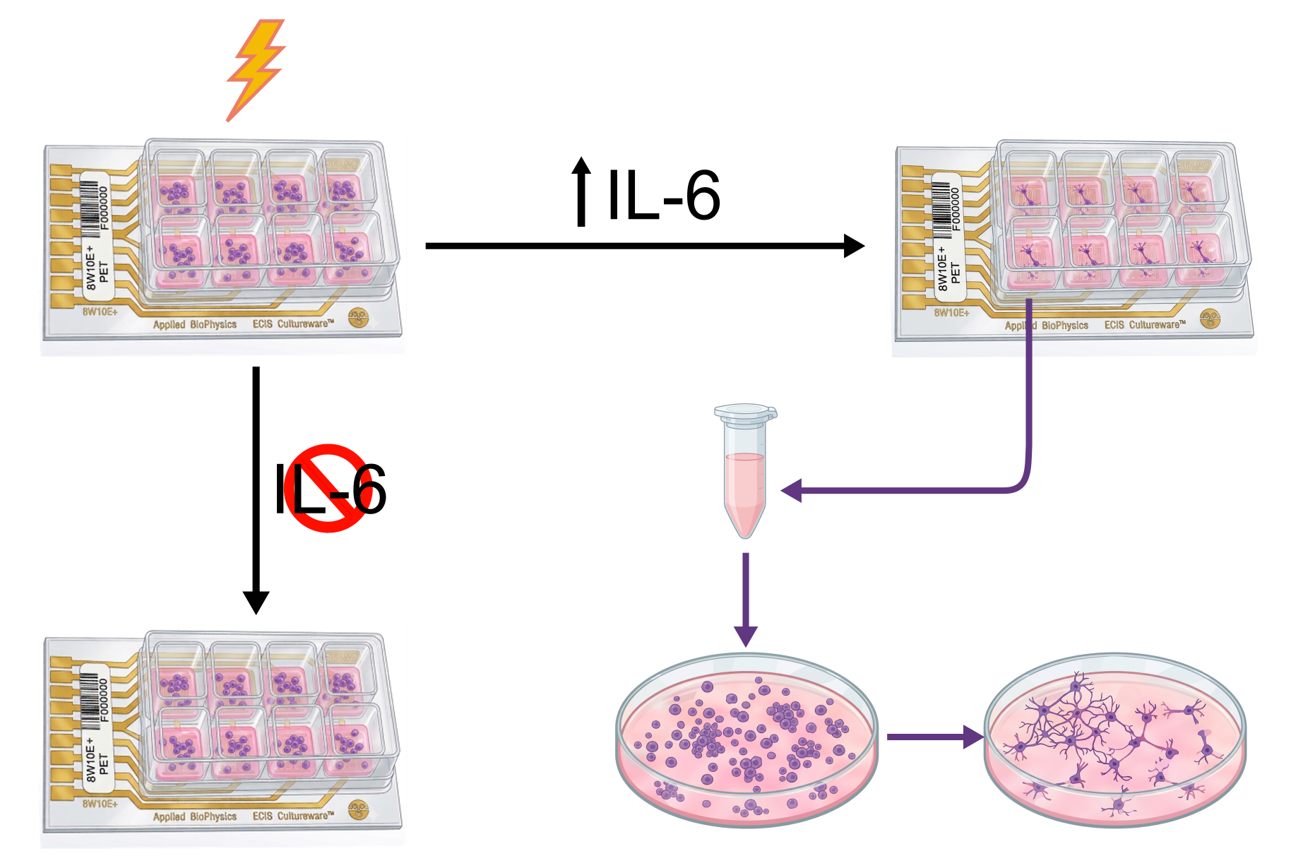
The bystander effect describes the induction of responses in non-targeted cells through cell signaling by directly stimulated cells. While this phenomenon has been extensively studied in the context of ionizing radiation, its occurrence following electrical stimulation (ES) remains poorly understood. Conditioned medium from N2a neuroblastoma cells exposed to voltage-controlled biphasic pulses at 500 mV/mm and 100 Hz induced neuronal differentiation in non-stimulated cells through an ES bystander effect. Bystander medium promoted morphological changes associated with neuronal differentiation, including increased neurite outgrowth and a reduction in the proliferation marker KI-67, indicating that the effects of ES extend to neighboring non-targeted cells. Molecular analysis revealed increased expression and secretion of interleukin-6 (IL-6) following ES, while neutralization of IL-6 receptor inhibited the effects of ES, highlighting the role of IL-6 as a key mediator of this effect. We provide the first evidence that ES promotes a differentiation-associated bystander effect mediated by IL-6.
Keywords:
bystander effect
; electrical stimulation
; neuroblastoma
; differentiation therapy
1. Introduction
The bystander effect is defined as the induction of biological changes in cells that have not been directly targeted by a specific stimulus but have received molecular signals from cells that were exposed. This phenomenon challenges the traditional “targeted” dogma of biology, which assumes that heritable effects and cellular damage require direct interaction with the initial stressor [1,2]. While the bystander effect is extensively documented in the context of ionizing radiation (IR) and ultraviolet (UV) light, its manifestation following electrical stimulation (ES) is a more recent and promising field of study [2].
In the well-established framework of radiation-induced bystander effects, irradiated cells release signaling molecules which can be transmitted: 1) via gap junctions, for molecules of up to 1,000–1,500 Da which include ions such as Ca++, peptides, nucleotides and other secondary messengers; and 2) through the release of soluble factors into the extracellular environment diffused in the medium, molecules like cytokines, such as IL-1, IL-6, IL-8, IL-10, brain derived neurotrophic factor (BDNF), tumor necrosis factor alpha, and transforming growth factor, nitric oxide, and reactive oxygen species (ROS) [1,2,3]. All these signals trigger responses in unirradiated cells ranging from micronuclei, aberrations and sister chromatid exchanges to apoptosis and cell cycle arrest [2,3]. Similar responses have been observed following UV irradiation, where oxidative stress and physical damage to the cell membrane prompt the release of mediators that affect in the same way unexposed cells [4]. Moreover, these consequences have also been observed in photodynamic therapy, heat and chemotherapy agents, therefore, the bystander effect must be considered in a wide range or therapies to avoid killing of non-treated cells [3].
Electrical stimulation (ES) is a technique that consists of applying an electric field (voltage, current or charge) to living tissue or cell cultures. ES has been shown to promote specific cell responses, including adhesion, proliferation, migration, and differentiation [5,6]. On the other hand, ES is increasingly recognized not only as a tool for direct cellular manipulation but also as a potent inducer of a “bystander-like” effect. Research indicates that ES can significantly enhance the secretion of extracellular vesicles and different molecules that modulate the physiology of distant cells [4,7]. For example, conditioned medium derived from ES-treated mesenchymal stem cells has demonstrated the ability to inhibit tumor cell migration and 3D spheroids growth and promote bone healing in unexposed tissues [8]. Furthermore, in cardiac models, electrical stimulation increases the release of cardioprotective EVs that shield non-stimulated cardiomyocytes from hypoxia-induced apoptosis [9].
ES-induced signaling is frequently associated with regenerative and therapeutic potential, offering a non-invasive pathway to enhance tissue repair [10] but the applications of ES are not limited to tissue engineering, and its capability to affect cell destiny can be very interesting in other fields like differentiation cancer therapy [11,12]. The advantage of this therapy lies in its ability to stop cancer progression, minimizing the harmful effects caused by traditional therapies (chemical or radiation), as it promotes cell differentiation without damage [13,14]. On the other hand, and in the context of differentiation therapy, hormones or cytokines have been found to promote differentiation in vitro, modifying in this way the phenotype of cancer cells [15].
We have previously reported that neuroblastoma cells can be differentiated into neurons through ES; by using this methodology, we were able to induce the differentiation of N2a cells in both 2D and 3D, as well as halting proliferation without DNA damage [16]. Unlike the well-characterized radiation-induced bystander effect, which is generally associated with detrimental biological outcomes, the ES bystander effect under specific conditions of cellular differentiation, as previously described, may offer a therapeutic opportunity by suppressing cellular proliferation and thereby limiting cancer progression. This study focuses on the use of conditioned medium of electrically stimulated N2a cells in order to promote neuronal differentiation of non-stimulated cells through a kind of bystander effect. We demonstrated that this conditioned medium is able to induce cell differentiation in N2a cells and that this bystander effect is mediated by cytokine interleukin 6.
2. Materials and Methods
2.1. Cell Culture
N2a (Neuro-2a) mouse neuroblastoma cells (ATCC CCL-131) were generously provided by Dr. Diego Ruano Caballero. Cells were routinely cultured in a 1:1 mixture of high-glucose Dulbecco’s Modified Eagle Medium (DMEM, Gibco 11965092) and Opti-MEM (Gibco 31985062), supplemented with 10% (v/v) fetal bovine serum (FBS, Gibco A5256701), 2mM L-glutamine (Gibco 25030081), 50μg/mL streptomycin (Gibco 15140122), and 50U/mL penicillin (Gibco 15140122). Cultures were maintained at 37 °C in a humidified incubator with 5% CO₂. Cells were routinely passaged to ensure cells were in exponential growth phase when used for experiments.
10000 N2a cells were seeded on each well of an 8W10E+ plate. The next day serum-free medium was added. On the third day ES was delivered and 75000 cells were seeded in 35mm petri dishes. Right when the 6h ES ends the medium of the 8W10E+ plate wells (medium A) was transferred to one of the dishes seeded, this was done for control and ES. The medium of the 8W10E+ plate was refreshed. The following day, approximately 24 hours after ES, the medium of the electrode plate was transferred to another two petri dishes (medium B). Images of the cells in the petri dishes were taken on day five. The images were analyzed using ImageJ software. A diagram of the experimental protocol can be seen in Figure 1.
Recombinant mouse IL-6 (R&D, 406-ML/CF) was added at a concentration of 50ng/mL 2h prior to ES to the conditions that required it. For the inhibition of IL-6 receptor, 1 hour prior to ES, cells were treated with 1µg/mL of IL-6 receptor blocking antibody (R&D, AF1830). This blocking antibody will be referred to as αIL-6R.
2.2. Electrical Stimulation
Electrical stimulation was performed as described in [16]. Briefly, the applied ES signals were biphasic square voltage signals with a duty cycle of 50% (25% positive and 25% negative) at 100Hz, with amplitude ±500mV. These signals were previously studied by the authors in [6], where many amplitudes and frequencies were tested and characterized, 500mV-100Hz was found to be the best conditions for neuronal differentiation of N2a cells.
2.3. Morphological Analysis Method
For morphological analysis, images were taken with a 20X objective in an inverted microscope (Leica, Wetzlar, Germany). In order to obtain cell differentiation percentage, differentiated and nondifferentiated cells on top of the electrode were counted using ImageJ software as described in [5]. Differentiation percentage was calculated as the number of cells displaying a differentiated morphology divided by the number of total counted cells. Neurite length was measured the ruler tool on ImageJ and expressed in µm.
2.4. ELISA Assay
Cytokine levels were quantified using the BD™ Cytometric Bead Array (CBA) Mouse Inflammation Kit (BD Biosciences, Cat. No. 552364) according to the manufacturer’s instructions. Briefly, 10µl of bystander medium samples and cytokine standards were incubated with a mixture of capture beads coated with cytokine-specific antibodies, followed by incubation with phycoerythrin (PE) conjugated detection antibodies. After washing, samples were analyzed by flow cytometry (Miltenyi MACSQuant VYB). Cytokine concentrations were determined by comparison to the standard curves generated.
2.5. RT-qPCR
Cells were lysed using Trizol reagent (gTPzol01 CViral). RNA was isolated 24 hours after ES started, using the E.Z.N.A. Total RNA Kit I (Omega Bio-Tek. GA, USA). Total RNA was reverse transcribed with the iScript cDNA Synthesis Kit (Bio-Rad. CA, USA). The concentration of the extracted RNA was determined using a NanoDrop spectrophotometer. The real-time PCR amplification was done in a CFX Connect Real-Time System (Bio-Rad) using SYBRGreen (Bio-Rad), and the cDNA obtained as template. Primer sequences can be found in the supplementary information.
2.6. Immunofluorescence
Cells were washed with PBS and fixed for 30 minutes with 4% paraformaldehyde in PBS, then permeabilized for 10 minutes with 0.5% Triton-X. After this, cells were treated with blocking solution (1% bovine serum albumin + 0.1% Tween 20 in PBS) for 15 minutes. Primary antibody for KI-67 (BD Pharmingen 550609) was diluted 1:500 in blocking solution. Cells were incubated in this antibody at 4ºC overnight after which they were washed 3 times with blocking solution. The same process was followed for the secondary antibodies (Invitrogen A32731), diluted 1:500 in blocking solution, and incubated for 45 minutes. DAPI staining was carried out after the secondary antibody. Immunofluorescence (IF) images were taken with Leica DMi8 microscope with a 63X objective. Images were analyzed with Cell Profiler, where the nuclei were isolated using the DAPI channel and green fluorescence was measured inside the nucleus.
2.7. Statistical Analysis
Each experiment was independently performed at least in duplicate. All statistical analyses were performed using one-way analysis of variance (ANOVA) test, where p values < 0.05 were considered statistically significant. Unless otherwise noted, all tests have been performed with reference to the control condition.
3. Results
3.1. ES can Provoke a Bystander Effect
Firstly, the ES induced bystander effect was characterized. For that matter, the medium from electrically stimulated cells was transferred to non-stimulated cells, right after ES (bystander medium A) and 24h after the start of ES (bystander medium B). The separation of medium A and B allows us to distinguish between the effects produced by electrochemical byproducts, if present (medium A), and pure cell secretion (medium B). 24 hours after the last medium transfer, images of bystander cells were taken and analyzed. Representative images are displayed in Figure 2A. Control cells were tightly packed and exhibited occasional neurites, while largely retaining a mostly undifferentiated morphology. On the contrary, bystander cells displayed a more polygonal shape with more frequent and longer neurites. Quantification of cells displaying differentiated morphology and neurite length can be seen in Figure 2B and C respectively. Both bystander mediums induced the differentiation and neurite outgrowth of N2a cells, although medium B was more effective. Cells exposed to medium B exhibited an increment in differentiation percentage of around 50% compared to the control, as well as a significant increase in neurite length. Next, alterations of proliferative capabilities produced by the bystander effect were characterized by KI-67 immunofluorescence. Bystander medium was transferred 24h after ES, as medium B was the most effective. KI-67 IF representative images can be seen in Figure 2D. The intensity of KI-67 was significantly lower on bystander cells (Figure 2E). Bystander cells displayed a decrease in KI-67 IF intensity of around 60% compared to the control. This is consistent with the morphological analysis, as neuronal differentiation entails the arrest of the cell cycle and thus a decrease in KI-67.
3.2. ES Bystander Effect may be Mediated by Interleukins
After analyzing the effect of the bystander effect, the content of this medium was assessed. RT-qPCR analysis of BDNF, IL-6 and IL-10 was performed in ES and control cells (Figure 3A). No change was observed in BDNF expression. IL-6 was upregulated in ES cells with the consequent reduction in IL-10 expression. Furthermore, IL-6 concentration in the bystander medium was assessed by ELISA analysis (Figure 3B) and found to be higher than in the control. Although the absolute value of the concentration was different in each replicate it was always higher in the bystander medium. These results point to IL-6 as a possible candidate mediating the bystander effect, as ES cells produced and secreted it.
3.3. Interleukin-6 is the Main Cytokine Implicated in the ES Bystander Effect
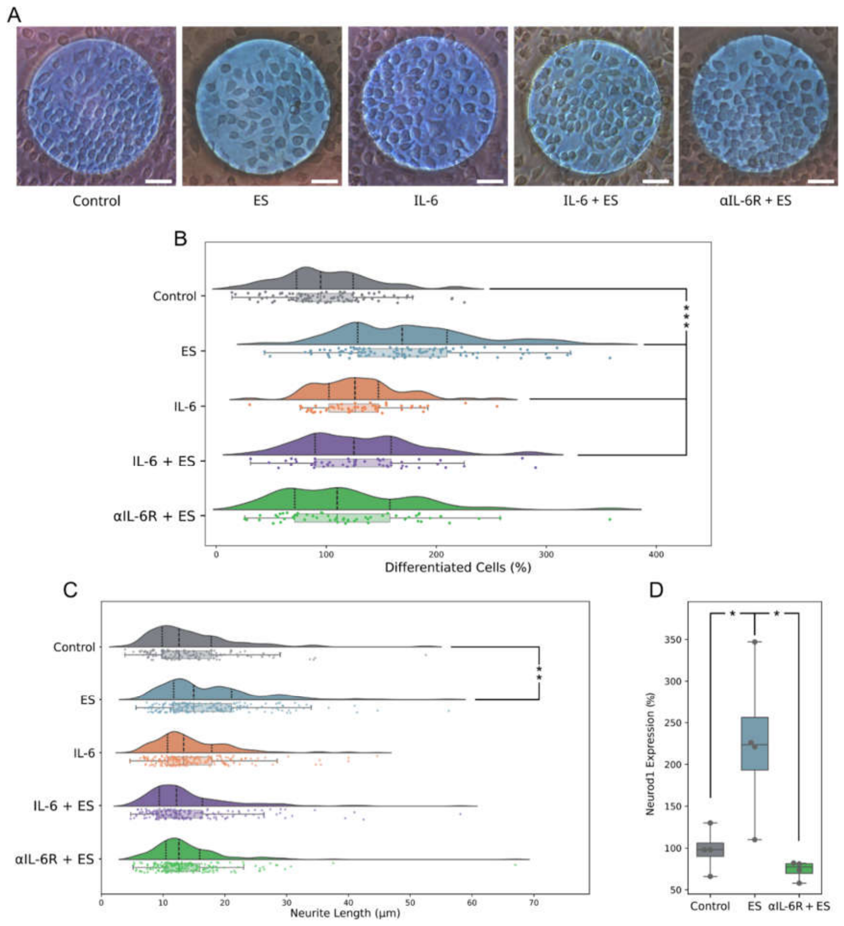
To confirm the role of IL-6 in the bystander effect produced by ES, combinatory treatments of ES with IL-6 and IL-6 receptor blocking antibodies were tested. Representative images are displayed in Figure 4A. Again, control cells were tightly packed and exhibited occasional neurites with mainly undifferentiated morphology. On the contrary, ES cells displayed a more polygonal shape with more and longer neurites. Cells treated with 50 ng/mL of recombinant IL-6 exhibited an intermediate morphology between control and ES-treated cells, but a significant difference respect to control cells morphology was observed. While a variety of cells displayed a differentiated phenotype, some retained a morphology similar to control cells and, overall, neurites were not as long, regardless of whether IL-6 was administered alone or in combination with ES. Lastly, stimulated cells in the presence of the αIL-6R remained very similar to the control, with few neurite processes. This is confirmed by the morphological analysis in Figure 4B. ES induced the most significant difference in morphologically differentiated cells compared to the control. Recombinant IL-6 produced an increase in differentiation regardless of the combination with ES that was slightly lower than ES cells but also statistically significant. αIL-6R suppressed the effects of ES on N2a morphology. A very similar outcome can be observed when analyzing neurite length (Figure 4C). In this case, the only significant change regarding the control was observed for the ES condition. Recombinant IL-6 alone also produced a very slight increase in neurite length. Lastly, αIL-6R suppressed the effects of ES on neurite length.
The effect of the αIL-6R was further confirmed by Neurod1 RT-qPCR. Neurod1 is a neuronal differentiation onset marker that was overexpressed in ES cells and suppressed by the αIL-6R (Figure 4D). These results indicate clearly the involvement of IL-6 in the induction of neuronal differentiation produced by the ES bystander effect.
4. Discussion
The ES induced bystander effect was first evaluated through morphological analysis and was found to provoke an increase in differentiated cells. KI-67 immunofluorescence confirmed that ES bystander medium produced a decrease of proliferating cells previously associated with neuronal differentiation [16]. We have previously shown that ES reduced the proliferative capabilities of N2a cells both in monolayers and in neurospheres. This effect could be linked to the neuronal differentiation of these neuroblastoma cells, since as progenitor cells progress through the differentiation process their proliferation capabilities are reduced [16]. These new results relating to differentiation therapy expand the traditional understanding of bystander signaling into the field of electrical stimulation, which, as we have previously described, has been extensively documented in response to ionizing radiation and UV light. They indicate that ES acts not only on the directly stimulated cells but also on the surrounding population via cellular communication. The distinction between the bystander mediums extracted right after ES and the next day provided insight into the nature of the signaling mechanism. While both media induced some level of differentiation, the medium containing only cell secretion appeared more effective, suggesting that the primary driver of differentiation is likely a biological response involving cell-secreted factors rather than transient electrochemical byproducts generated during ES delivery. This is consistent with other research indicating that while ES can generate ROS in the culture medium, these factors alone may be insufficient to fully replicate the effects observed with direct stimulation [17].
The use of ES-conditioned medium as a catalyst for the bystander effect is a novel approach, but precedents exist. For instance, in [18], ES-conditioned medium from Schwann cells promoted sustained neurite outgrowth in a neuronal culture. Similarly, conditioned medium obtained from electrically stimulated mesenchymal stem cells and Jurkat T cells showed tumor-suppressive activity, reducing tumor cell migration, 3D spheroid growth, and cancer tissue fragment viability [8]. In both studies, medium from ES cells is transferred to a different cell line. In this study, the same cell line is used to generate the bystander medium and to assess its effect on it, in an attempt to standardize a possible protocol to halt cell progression in neuroblastoma cells.
Molecular analysis pointed toward IL-6 as a potential mediator of this ES-induced bystander effect. While IL-6 is frequently characterized as a proinflammatory cytokine in various bystander models, it is known to function as a neurotrophin molecule within the central nervous system [19]. Higher concentrations of IL-6 were found in the ES bystander medium as per ELISA. This correlates with [20], where a higher concentration of IL-6 was found on ES mesenchymal stem cells via cytokine arrays. Recombinant IL-6 was also responsible for a slight increase in N2a differentiation, accordant with [21] and [22], where IL-6 was shown to induce the neuronal differentiation of PC12 and SH-SY5Y cells respectively. The observed upregulation of IL-6 in ES-treated cells and its increased concentration in the bystander medium suggest a similar role in N2a cells, potentially triggering the transition toward a mature phenotype. This was further confirmed by the inhibition of the IL-6 receptor using a blocking antibody. This inhibitor cancelled the induction of differentiation produced by ES as detected by morphological analysis and Neurod1 RT-qPCR.
No earlier evidence pointing to Il-6 as the effector of the ES induced bystander effects has been found. Other authors have explained the effect of ES bystander medium through the Piezo1 pathway [8]. This pathway has been associated with ES in several studies, as Piezo1 is a protein that reacts to physical stimuli [8,17]. Furthermore, the Piezo1 pathway and IL-6 have been related. Specifically, recent research in skeletal muscle models indicates that ES mimics the effects of mechanical exercise and induces the secretion of IL-6 in a Piezo1 dependent manner [23].The connection between Piezo1 and IL-6 pathways has been attributed to calcium ion [24] which is also involved in the neuronal differentiation induction produced by ES [25]. This evidence points to IL-6 as a possible responsible for the ES bystander effect produced in N2a, probably through a Piezo1 dependent mechanism.
More studies must be performed to determine the exact role of IL-6 and possibly Piezo1 as differentiating agents. On the other hand, as we have previously described, extracellular vesicles generated by ES could play a relevant role in the modulation of physiology of distant cells [7] and it would be of great interest to assess the possible presence of IL6 in exosomes secreted by the cells after ES. Furthermore, in our opinion, it would be very useful in order to try to establish a role for this described bystander effect as a possible therapy for cancer progression.
5. Conclusions
This study focused on characterizing the ES induced bystander effect in N2a cells. It was shown that the ES bystander effect could promote neuronal differentiation in neuroblastoma cells through soluble factors released by directly stimulated cells. Bystander medium from ES-treated N2a cells induced morphological changes consistent with neuronal differentiation, including increased neurite outgrowth and a reduction in the proliferation marker KI-67, indicating that ES-mediated signaling extends beyond the directly stimulated cell population. Molecular analysis identified IL-6 as a key mediator of this response, as ES increased both its expression and secretion, while neutralization of the IL-6 receptor inhibited the effects of ES on neuronal differentiation. These findings provide the first evidence that IL-6 mediates an ES-induced bystander effect associated with neuronal differentiation. From a therapeutic perspective, the ability of ES to trigger differentiation indirectly through paracrine signaling may broaden its potential application as a non-genotoxic differentiation agent for differentiation therapy. Nevertheless, this study was performed in a single in vitro cell line and focused primarily on soluble cytokine signaling. Further studies are required to validate these findings in additional models, determine the contribution of extracellular vesicles and other signaling pathways, and assess the relevance of this mechanism in vivo.
Supplementary Materials
The following supporting information can be downloaded at the website of this paper posted on Preprints.org.
Author Contributions
Conceptualization, L.O. and P.D.; methodology, P.D, A.D and D.M.; investigation, D.M and A.D.; data curation, D.M and A.A.; writing—original draft preparation, D.M and P.D.; writing—review and editing, all authors.; visualization, D.M and A.A.; supervision, L.O, A.Y and P.D.; project administration, A.Y and P.D.; funding acquisition, A.Y and P.D. All authors have read and agreed to the published version of the manuscript.
Acknowledgments and Funding
This publication has been funded by grant USECHIP (TSI-069100-2023-001), project funded by the Secretary of State for Telecommunications and Digital Infrastructure, Ministry for Digital Transformation and Civil Service and by the European Union–NextGenerationEU/PRTR. Additionally, we acknowledge project PID2021-1225290B-100, funded by MICIU/ AEI/ 10.13039/501100011033. This publication is also part of the applied research and innovation project SOL2024-31706, ‘Bioengineered mesenchymal stromal cell-derived extracellular vesicles for the treatment of type 1 diabetes,’ co-financed by the EU—Ministry of Finance and Public Function—European Funds—Regional Government of Andalusia—Regional Ministry of University, Research and Innovation.
Data Availability Statement
The datasets used and/or analyzed during the current study are available from the corresponding author on reasonable request.
Conflicts of Interest
The authors declare no conflicts of interest.
References
- Hall, E. J. THE BYSTANDER EFFECT. Health Phys. 2003, vol. 85(no. 1), 31–35. [Google Scholar] [CrossRef]
- Xu, S.; et al. Exosome-mediated microRNA transfer plays a role in radiation-induced bystander effect. RNA Biol. 2015, vol. 12(no. 12), 1355–1363. [Google Scholar] [CrossRef] [PubMed]
- Prise, K. M.; O’Sullivan, J. M. Radiation-induced bystander signalling in cancer therapy. Nat. Rev. Cancer 2009, vol. 9(no. 5), 351–360. [Google Scholar] [CrossRef] [PubMed]
- Ng, C. Y.; et al. Scalable Production of Extracellular Vesicles and Its Therapeutic Values: A Review. Int. J. Mol. Sci. 2022, vol. 23(no. 14), 7986. [Google Scholar] [CrossRef] [PubMed]
- Martín, D.; et al. DC electrical stimulation enhances proliferation and differentiation on N2a and MC3T3 cell lines. J. Biol. Eng. 2022, vol. 16(no. 1). [Google Scholar] [CrossRef] [PubMed]
- Martín, D.; Ruano, D.; Yúfera, A.; Daza, P. Electrical pulse stimulation parameters modulate N2a neuronal differentiation. Cell Death Discov. 2024, vol. 10(no. 1). [Google Scholar] [CrossRef] [PubMed]
- Fukuta, T.; Nishikawa, A.; Kogure, K. Low level electricity increases the secretion of extracellular vesicles from cultured cells. Biochem. Biophys. Rep. 2020, vol. 21, 100713. [Google Scholar] [CrossRef] [PubMed]
- Cui, C.; et al. Electrical Stimulation Generates Induced Tumor-Suppressing Cells, Offering a Potential Option for Combatting Breast Cancer and Bone Metastasis. Int. J. Mol. Sci. 2025, vol. 26(no. 3), 1030. [Google Scholar] [CrossRef] [PubMed]
- Zhang, H.; et al. Electrical Stimulation Increases the Secretion of Cardioprotective Extracellular Vesicles from Cardiac Mesenchymal Stem Cells. Cells 2023, vol. 12(no. 6), 875. [Google Scholar] [CrossRef] [PubMed]
- Meng, S.; Rouabhia, M.; Zhang, Z. “Electrical Stimulation in Tissue Regeneration,” in Applied Biomedical Engineering; Gargiulo, G. D., McEwan, A., Eds.; IntechOpen: Rijeka, 2011; p. ch. 3. [Google Scholar] [CrossRef] [PubMed]
- Leszczyniecka, M.; Roberts, T.; Dent, P.; Grant, S.; Fisher, P. B. Differentiation therapy of human cancer: basic science and clinical applications. Pharmacol. Ther. 2001, vol. 90(no. 2–3), 105–156. [Google Scholar] [CrossRef] [PubMed]
- Diego-Santiago, M. del P.; et al. Bioelectric stimulation outperforms brain derived neurotrophic factor in promoting neuronal maturation. Sci. Rep. 2025, vol. 15(no. 1), 4772. [Google Scholar] [CrossRef] [PubMed]
- Jin, Z.; Lu, Y.; Wu, Y.; Che, J.; Dong, X. Development of differentiation modulators and targeted agents for treating neuroblastoma. Eur. J. Med. Chem. 2020, vol. 207, 112818. [Google Scholar] [CrossRef] [PubMed]
- Zhu, K.; et al. Characterization and therapeutic perspectives of differentiation-inducing therapy in malignant tumors. Front. Genet. 2023, vol. 14. [Google Scholar] [CrossRef] [PubMed]
- de Thé, H. Differentiation therapy revisited. Nat. Rev. Cancer 2018, vol. 18(no. 2), 117–127. [Google Scholar] [CrossRef] [PubMed]
- Martín, D.; et al. Electrical stimulation induces differentiation onset consistent with a therapeutic approach in neuroblastoma cells. Sci. Rep. 2026. [Google Scholar] [CrossRef] [PubMed]
- Bielfeldt, M.; et al. Discrimination between the effects of pulsed electrical stimulation and electrochemically conditioned medium on human osteoblasts. J. Biol. Eng. 2023, vol. 17(no. 1). [Google Scholar] [CrossRef] [PubMed]
- Koppes, N.; et al. Electrical Stimulation of Schwann Cells Promotes Sustained Increases in Neurite Outgrowth. Tissue Eng. Part A 2013, 130924230853000. [Google Scholar] [CrossRef] [PubMed]
- Erta, M.; Quintana, A.; Hidalgo, J. Interleukin-6, a Major Cytokine in the Central Nervous System. Int. J. Biol. Sci. 2012, vol. 8(no. 9), 1254–1266. [Google Scholar] [CrossRef] [PubMed]
- Hwang, S. J.; Cho, T. H.; Lee, B.; Kim, I. S. Bone-healing capacity of conditioned medium derived from three-dimensionally cultivated human mesenchymal stem cells and electrical stimulation on collagen sponge. J. Biomed. Mater. Res. A 2018, vol. 106(no. 2), 311–320. [Google Scholar] [CrossRef] [PubMed]
- Satoh, T.; et al. Induction of Neuronal Differentiation in PC12 Cells by B-Cell Stimulatory Factor 2/Interleukin 6. Mol. Cell. Biol. 1988, vol. 8(no. 8), 3546–3549. [Google Scholar] [CrossRef] [PubMed]
- Barron, et al. Maternal pre-eclampsia serum increases neurite growth and mitochondrial function through a potential IL-6-dependent mechanism in differentiated SH-SY5Y cells. Front. Physiol. 2023, vol. 13. [Google Scholar] [CrossRef] [PubMed]
- Sciancalepore, M.; Massaria, G.; Tramer, F.; Zacchi, P.; Lorenzon, P.; Bernareggi, A. A preliminary study on the role of Piezo1 channels in myokine release from cultured mouse myotubes. Biochem. Biophys. Res. Commun. 2022, vol. 623, 148–153. [Google Scholar] [CrossRef] [PubMed]
- Hasan, R.; et al. The Alteration of Microglial Calcium Homeostasis in Central Nervous System Disorders: A Comprehensive Review. Neuroglia 2024, vol. 5(no. 4), 410–444. [Google Scholar] [CrossRef]
- Wang, S.; Guan, S.; Sun, C.; Liu, H.; Liu, T.; Ma, X. Electrical stimulation enhances the neuronal differentiation of neural stem cells in three-dimensional conductive scaffolds through the voltage-gated calcium ion channel. Brain Res. 2023, vol. 1798, 148163. [Google Scholar] [CrossRef] [PubMed]
Figure 1.
Bystander experimental protocol.
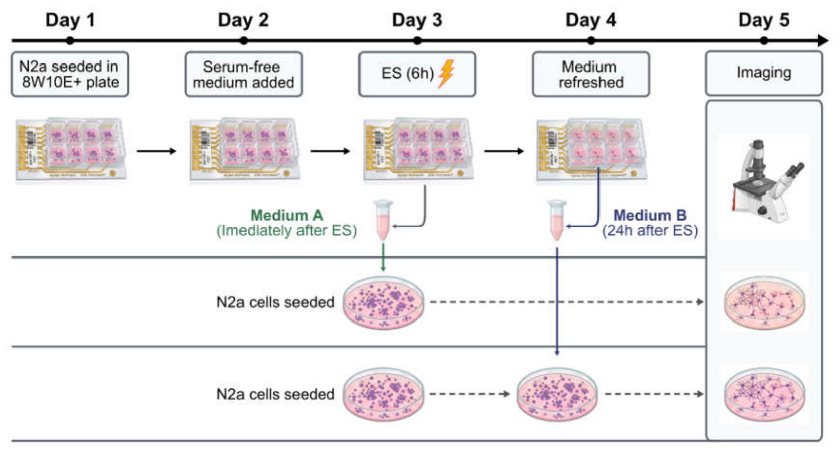
Figure 2.
Bystander effect morphological assessment and KI-67 analysis. (A) Phase contrast microscopy images taken at 20X with a 2X digital zoom. Scale bar represents 50μm. Quantitative analysis of differentiated cells (B) (** p<0.01, ***p<0.001; n=21) and neurite length (C) (***p<0.001; n=200). (D) Representative immunofluorescence images of KI-67 (green) and DAPI (blue), images taken at 63X, scale bar represents 20µm. (E) Quantification of KI-67 intensity represented as percentage to the control (*** p<0.001; n=63).
Figure 2.
Bystander effect morphological assessment and KI-67 analysis. (A) Phase contrast microscopy images taken at 20X with a 2X digital zoom. Scale bar represents 50μm. Quantitative analysis of differentiated cells (B) (** p<0.01, ***p<0.001; n=21) and neurite length (C) (***p<0.001; n=200). (D) Representative immunofluorescence images of KI-67 (green) and DAPI (blue), images taken at 63X, scale bar represents 20µm. (E) Quantification of KI-67 intensity represented as percentage to the control (*** p<0.001; n=63).
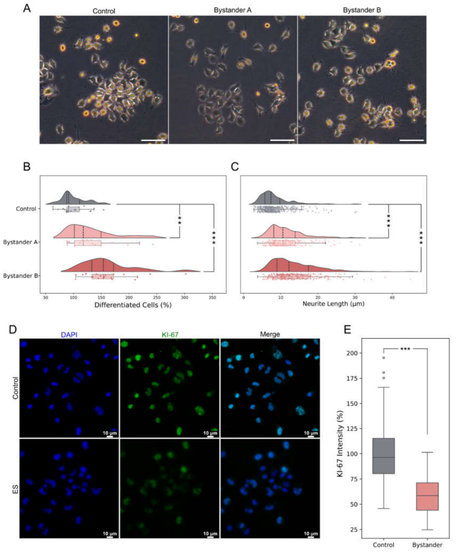
Figure 3.
Molecular analysis of cell excretion and bystander medium. (A) RT-qPCR of interleukins and BDNF on directly stimulated cells. (**p<0.01, ***p<0.001; n=4) (B) ELISA analysis of IL-6 concentration on bystander medium, paired experiments are represented with lines.
Figure 3.
Molecular analysis of cell excretion and bystander medium. (A) RT-qPCR of interleukins and BDNF on directly stimulated cells. (**p<0.01, ***p<0.001; n=4) (B) ELISA analysis of IL-6 concentration on bystander medium, paired experiments are represented with lines.
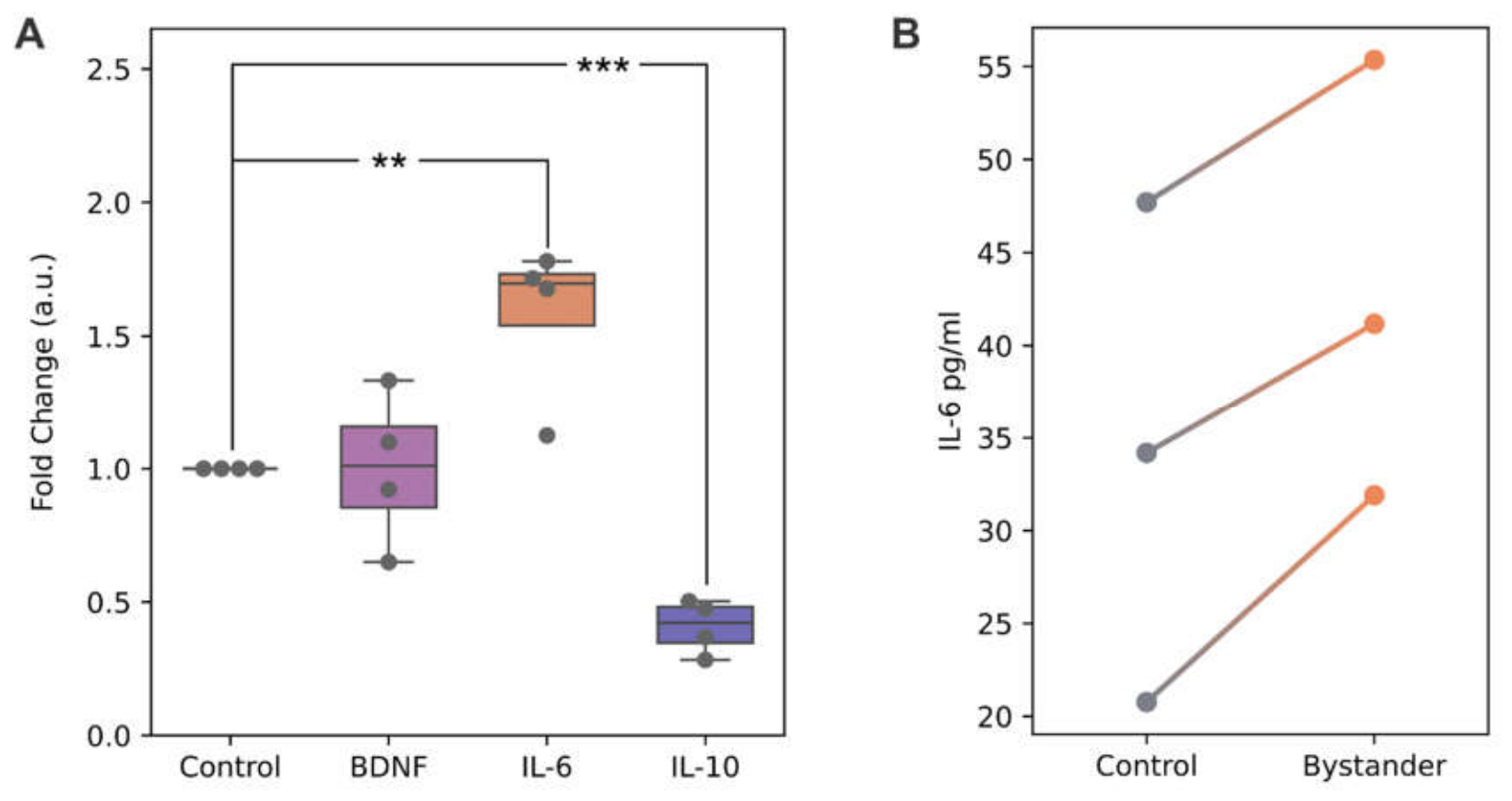
Figure 4.
Effect of recombinant IL-6 and neutralization of IL-6R in combination with ES. (A) Phase contrast microscopy representative images. Scale bar represents 50µm. Quantitative analysis of differentiated cells (B) (***p<0.001; n=60) and neurite length (C) (**p<0.01; n=151). (D) RT-qPCR analysis of Neurod1 expression (*p<0.05; n=4).
Figure 4.
Effect of recombinant IL-6 and neutralization of IL-6R in combination with ES. (A) Phase contrast microscopy representative images. Scale bar represents 50µm. Quantitative analysis of differentiated cells (B) (***p<0.001; n=60) and neurite length (C) (**p<0.01; n=151). (D) RT-qPCR analysis of Neurod1 expression (*p<0.05; n=4).
Disclaimer/Publisher’s Note: The statements, opinions and data contained in all publications are solely those of the individual author(s) and contributor(s) and not of MDPI and/or the editor(s). MDPI and/or the editor(s) disclaim responsibility for any injury to people or property resulting from any ideas, methods, instructions or products referred to in the content. |
© 2026 by the authors. Licensee MDPI, Basel, Switzerland. This article is an open access article distributed under the terms and conditions of the Creative Commons Attribution (CC BY) license (http://creativecommons.org/licenses/by/4.0/).
Copyright: This open access article is published under a Creative Commons CC BY 4.0 license, which permit the free download, distribution, and reuse, provided that the author and preprint are cited in any reuse.


