

Submitted:
18 July 2026
Posted:
21 July 2026
You are already at the latest version
Abstract
The intensification of human–companion animal interactions has amplified the risk of zoonotic infections and antimicrobial resistance at the human–animal–environment interface. Dogs and cats serve as important reservoirs for a range of fungal pathogens, including dermatophytes such as Microsporum canis, yeast-associated infections involving Malassezia species, and environmentally acquired systemic mycoses. Transmission to humans occurs through direct contact with infected animals, indirect exposure via contaminated fomites, and shared environmental sources. Urbanisation, increasing pet density, climate change, and environmental disturbance have further reshaped the epidemiology of fungal zoonoses, facilitating outbreaks and enhancing fungal adaptation to warm-blooded hosts. In parallel, inappropriate antimicrobial and antifungal use in companion animals has contributed to the emergence of resistant pathogens, including mobile colistin resistance (mcr) genes and antifungal-resistant dermatophytes, raising serious public health concerns. Viral infections, such as SARS-CoV-2, may exacerbate this risk by impairing host immunity and predisposing animals and humans to secondary fungal infections. This review critically examines pet-associated fungal infections, their transmission dynamics, and the role of close contact, urbanisation, and antimicrobial misuse in driving zoonotic risk, highlighting the urgent need for integrated One Health surveillance, responsible pet ownership, and evidence-based policy interventions.
Keywords:
zoonotic fungi
; zoonotic transmission
; human - animal interface
; antifungal resistance
; zoonotic disease control
; pet mycose
; one health approach
1. Introduction
Companion animals are increasingly integrated into human households, resulting in frequent and intimate contact between pets and their owners. Behaviours such as petting, licking, grooming, and shared living spaces facilitate the bidirectional exchange of microorganisms and antimicrobial resistance genes, positioning dogs and cats as key contributors to zoonotic risk within domestic environments (Hamame et al., 2022). As global pet adoption rises, the health consequences of this close coexistence have become an important concern within the One Health framework.
Fungal infections represent a substantial yet often underestimated component of pet-associated zoonoses. In companion animals, fungal diseases range from superficial infections, such as dermatophytosis, to more severe systemic mycoses acquired from environmental reservoirs (Winterfeld et al., 2024). Dermatophytosis remains the most prevalent zoonotic fungal infection worldwide and is caused by keratinophilic fungi belonging to the genera Microsporum, Trichophyton, and Nannizzia (Pasquetti et al., 2017; Zineldar et al., 2025). Among these, Microsporum canis predominates in cats and dogs, with cats acting as major reservoir hosts and posing a disproportionate risk of transmission to humans, particularly paediatric populations (Pasquetti et al., 2017).
Zoonotic transmission of fungal pathogens occurs through multiple pathways. Direct contact with infected skin, hair, or scales is the primary route for dermatophyte spread, while indirect transmission via contaminated fomites including grooming tools, bedding, furniture, and household surfaces is facilitated by the prolonged environmental persistence of infective arthroconidia (Pasquetti et al., 2017; Winterfeld et al., 2024). In contrast, systemic mycoses (Figure 1) are typically acquired by both humans and pets from shared environmental sources, such as soil contaminated with bird or bat droppings, underscoring the importance of environmental exposure in disease emergence (Winterfeld et al., 2024; Carpouron et al., 2022). Close contact practices, grooming behaviours, and veterinary care further shape transmission dynamics and contribute to the development of antifungal resistance. Inadequate hygiene, contaminated grooming equipment, and inappropriate antifungal use particularly sub therapeutic dosing and premature treatment discontinuation have been associated with reduced treatment efficacy and the emergence of resistant fungal strains (Winterfeld et al., 2024). In parallel, the widespread use of antimicrobials in companion animals has facilitated the dissemination of resistant bacteria, including strains carrying mobile colistin resistance (mcr) genes, which have been shown to overlap genetically with isolates from human infections (Hamame et al., 2022).
Urbanisation and environmental change have intensified these risks by altering fungal ecology and host exposure patterns. Urban soil disturbance, increased pet density in confined settings, and climate-driven temperature increases have enhanced fungal survival, transmission efficiency, and thermotolerance, enabling certain species to adapt more readily to warm-blooded hosts (Carpouron et al., 2022). The emergence of antifungal resistant dermatophytes and shifting patterns of zoonotic mycoses reflect the growing public health relevance of these infections (Zineldar et al., 2025).
Viral infections may further compound fungal disease burden by compromising host immune defences. Evidence indicates that infections such as SARS-CoV-2 can predispose animals and humans to secondary fungal colonisation, increasing disease severity and complicating clinical management (Silva et al., 2024). These findings highlight the need for integrated One Health strategies (Figure 1) that prioritise surveillance, accurate diagnostics, responsible antimicrobial use, and public awareness to mitigate the rising threat of pet associated fungal zoonoses and antimicrobial resistance.
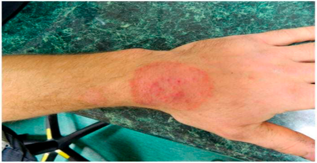
Figure 2.
Clinical picture of glabrous skin mycosis caused by T. verrucosum infection (Piorunek et al.,2024).
Figure 2.
Clinical picture of glabrous skin mycosis caused by T. verrucosum infection (Piorunek et al.,2024).
2. Common Fungal Infections in Pets
Fungal infections in pets are a varied group of diseases that differ in severity, type of tissue affected, and risk of transmission to humans (Seyedmousavi et al., 2018; Dr. Aakriti Guleria, 2025). These infections are generally categorized by how deeply they invade tissues: superficial, subcutaneous, and systemic mycoses (Dr. Aakriti Guleria, 2025). Dermatological issues are the most common cases seen in veterinary practice (Beraldo et al., 2011). Pets, especially dogs and cats, often live close to humans, which raises the risk of passing fungal pathogens back and forth (Bruno B. Chomel, 2014; Naz et al., 2023). Factors like temperature, humidity, climate change, ventilation, and the immune status of the host significantly influence fungal ecology, how infections spread, and the results of the disease (Kundu et al., 2024; Seyedmousavi et al., 2018).
2.1. Aspergillosis
Aspergillosis is one of the key systemic fungal infections (Seyedmousavi et al., 2018). It affects many animal species and is caused by filamentous fungi from the genus Aspergillus, which are commonly found in soil, decaying plants, seeds, and grains (Seyedmousavi et al., 2018). While most Aspergillus species are harmless, some, especially Aspergillus fumigatus, can lead to localized infections, allergic reactions, or severe widespread disease in animals with weak immune systems (Seyedmousav et al., 2018). In dogs, aspergillosis typically manifests as a nasal or sinonasal infection (Figure 3). Birds and mammals may also develop severe forms depending on their exposure and immune strength (Seyedmousavi et al., 2018).
2.2. Mucormycosis
Another serious opportunistic infection is mucormycosis, caused by fungi in the order Mucorales, which includes genera like Rhizopus, Mucor, and Lichtheimia (Seyedmousavi et al., 2018). These fungi are found in soil, decaying matter, dust, food, and outdoor and indoor air (Seyedmousavi et al., 2018). Mucormycosis (Figure 4) in animals closely mirrors the disease in humans in how it spreads, enters the body, forms lesions, and progresses (Seyedmousavi et al., 2018). Both domestic and wild animals are vulnerable, especially if they have weakened immune systems or existing injuries (Seyedmousavi et al., 2018).
2.3. Candidiasis
Candidiasis is another frequent fungal issue, mainly affecting the skin and mucosal surfaces (Seyedmousavi et al., 2018). Cutaneous candidiasis (Figure 5) is common in dogs and often occurs alongside conditions like allergies, hormone disorders, immunosuppressive treatments, or systemic immune problems. It resembles skin issues caused by Malassezia (Seyedmousavi et al., 2018). In birds, especially poultry, the main form of mucosal candidiasis, known as thrush or sour crop, affects the oral and gastrointestinal tract (Seyedmousavi et al., 2018). It is marked by white-greyish plaques and thickened skin. Similar infections have been noted in horses, cattle, dogs, cats, and pigs, mainly in younger animals or those on long-term antibiotic treatments (Seyedmousavi et al., 2018). While systemic candidiasis is rare in dogs and cats, it can happen after surgery or trauma, leading to granuloma formation or peritonitis (Seyedmousavi et al., 2018).
2.4. Dermatophytosis
Dermatophytosis, commonly referred to as ringworm, is the most common superficial fungal infection in pets and poses a major concern for zoonotic transmission (Beraldo et al., 2011; Pasquetti et al.,2017). Dermatophytes are fungi that thrive on keratin and belong to the genera Microsporum, Trichophyton, Nannizzia, and Epidermophyton (Smagulova et al., 2023; Kundu et al., 2024). The majority of infections in cats and dogs are caused by zoophilic species like Microsporum canis and Trichophyton mentagrophytes (Figure 5) (Pasquetti et al., 2017; Kundu et al., 2024). Among these, M. canis is the most frequent pathogen, with cats acting as the main carriers (Pasquetti et al., 2017; Ibrahim et al., 2021). The fungus invades keratinized tissues such as skin, hair, and nails, creating circular, hairless patches with scaling and central clearing (Pasquetti et al., 2017; Ibrahim et al., 2021). Arthroconidia produced during hair invasion are the main infective agents, allowing for effective transmission between animals and humans (Piorunek et al., 2024; Pasquetti et al., 2017).
Studies across various regions reveal the global reach and changing patterns of dermatophytosis (Piorunek et al., 2024). Research from Italy, Portugal, and the United States shows M. canis as the most common isolate in cats, while T. mentagrophytes and T. rubrum appear more often in people (Piorunek et al., 2024). Urban growth, pet domestication, population density, travel, and migration have led to new zoonotic species emerging and increasing infection rates (Kundu et al., 2024; Naz et al., 2023). Each time zoophilic dermatophytes appear in animals, there are often simultaneous infections in humans who come into contact with them.

Table 1.
Main etiological agents of dermatophytosis in different animal species. (Francisco Javier Cabanes.,2000).
Table 1.
Main etiological agents of dermatophytosis in different animal species. (Francisco Javier Cabanes.,2000).
| Domestic animals | Dermatophytes |
|---|---|
| Cats and dogs |
M. canis Others: T. mentagrophytes, M. gypseum, M. persicolor |
| Horses |
T. equinum Others: M. canis, M. equinum, M. gypseum, T. mentagrophytes, T. verrucosum |
| Cattle, goats and sheep |
T. verrucosum Others: M. canis, M. gypseum, T. mentagrophytes, T. equinum |
| Rabbits |
T. mentagrophytes Other: M.canis |
| Pigs |
M. nanum Others: M. canis, M. gypseum, T. mentagrophytes, T. verrucosum |
| Poultry |
M. gallinae Other: T. simii |
2.5. Sporotrichosis
Sporotrichosis is a subcutaneous fungal infection caused by thermally dimorphic fungi from the genus Sporothrix, primarily S. schenckii (Figure 6), S. globosa, and the highly contagious S. brasiliensis (Ronald D. Welsh, 2003; Kundu et al., 2024). This disease affects many animals, including cats, dogs, rodents, livestock, and wildlife (Barros et al., 2007; Etchecopaz et al., 2021). Cats are particularly significant as they can transmit the fungus to humans due to a high fungal load in their skin lesions, their tendency to scratch, and their close interactions with people(Ronald D. Welsh, 2003; Bruno B. Chomel, 2014). Human infections with S. brasiliensis usually stem from bites, scratches, or direct contact with infected lesions of cats (Ronald D. Welsh, 2003; Bruno B. Chomel, 2014). The increased heat tolerance of S. brasiliensis at cat body temperatures may link to climate change, aiding its spread (Ronald D. Welsh, 2003; Corrêa-Junior et al., 2023). Successful infection requires the fungus to change from conidia to yeast forms, with immune recognition involving fungal components such as β-glucans and mannans (Ronald D. Welsh, 2003; Corrêa-Junior et al., 2023).
2.6. Malassezia
Malassezia species, especially Malassezia pachydermatis, are common yeasts that often cause ear infections and skin issues in dogs (Figure 7) (Aruna Maramulla, 2023). About 10% of dogs receiving primary veterinary care show signs of otitis externa, with around two-thirds attributed to Malassezia overgrowth (Figure 7) (Aruna Maramulla, 2023). Some dog breeds, such as West Highland White Terriers, Basset Hounds, Cocker Spaniels, and Shih Tzus, are more susceptible (Aruna Maramulla, 2023). Conditions like excessive ear wax, antibiotic or corticosteroid use, moisture, and other concurrent diseases like allergies or skin infections promote yeast growth (Aruna Maramulla, 2023). While Malassezia-related skin issues are similar in humans and animals, systemic infections are mostly seen in humans (Aruna Maramulla, 2023).
2.7. Cryptococcosis and Histoplasmosis
Other notable fungal diseases include cryptococcosis, caused by Cryptococcus neoformans and C. gattii, with birds like pigeons and parrots being key carriers (Seyedeh A. Hosseinian, 2022). While these birds often show no symptoms, they shed the fungus in their droppings, contaminating soil and cages and exposing humans to the fungus through inhalation (Seyedeh A. Hosseinian, 2022). Histoplasmosis, caused by Histoplasma capsulatum, occurs in dirt contaminated with bird or bat droppings and can remain in soil for years, posing inhalation risks to people and animals (Seyedeh A. Hosseinian, 2022). Most human cases are mild, but severe respiratory or systemic diseases can develop if left untreated (Seyedeh A. Hosseinian, 2022).
Emerging fungal infections like phaeohyphomycosis involve melanized fungi, such as Exophiala dermatitidis, and have been reported in both people and animals, mainly as skin or subcutaneous lesions, sometimes leading to systemic spread (Seyedmousavi et al., 2018). Fungal infections in reptiles, birds, and exotic pets are gaining recognition, emphasizing their importance as indicators of zoonotic threats (Seyedmousavi et al., 2018). Researchers have identified fungi within the WHO's critical and high-priority pathogen groups among pets and wildlife, highlighting the increasing public health significance of veterinary mycology (Seyedmousavi et al., 2018).
3. Transmission of Fungal Infections from Pets to Humans
Diseases transmitted from animals to humans are collectively referred to as zoonoses and may be caused by bacteria, viruses, parasites, or fungi (Currie et al, 2023). Although the number of recognized zoonotic fungal agents is relatively limited compared to other pathogen groups, several species associated with companion animals exert a substantial public health impact (Seyedmousavi et al., 2018). Among these, Microsporum canis and Sporothrix brasiliensis, particularly transmitted from cats, are considered some of the most significant zoonotic fungal pathogens affecting humans worldwide (Seyedmousavi et al., 2018; Kundu et al., 2024). In certain rural settings, it has been estimated that up to 80% of human fungal skin infections originate from animals, highlighting the importance of pets and domestic animals as reservoirs of infection (Zineldar et al., 2025).
Traditionally, fungal zoonoses were considered environmentally acquired infections, with transmission occurring following traumatic contact with soil, plants, or decaying organic matter contaminated with fungal spores (Seyedmousavi et al., 2018; Ronald D. Welsh, 2003). However, emerging evidence demonstrates that alternative transmission routes, especially animal to animal and animal to human transmission play a major role in modern outbreaks (Figure 8) (Naz et al., 2023; Carpouron et al., 2022). This shift is most clearly observed in sporotrichosis epidemics driven by Sporothrix brasiliensis, where direct transmission via scratches, bites, and skin trauma during cat fights or close contact has replaced the classical environmental route (Kundu et al., 2024; Etchecopaz et al., 2021). Cats act as the primary reservoir host for S. brasiliensis, transmitting the fungus not only to humans but also to other animals such as dogs and rats (Naz et al., 2023; Bruno B. Chomel, 2014). The rapid geographic spread of sporotrichosis across adjacent regions, particularly in Brazil, underscores the highly contagious nature of this species, for which no universally effective treatment is currently available (Barros et al., 2007; Kundu et al., 2024).
Zoonotic fungal transmission occurs through multiple pathways, broadly classified into direct and indirect modes (Carpouron et al., 2022). Direct transmission involves close physical contact between an infected animal and a human host, including skin contact, bites, scratches, and exposure to secretions or excretions (Carpouron et al., 2022; Naz et al., 2023). This route is particularly efficient in the spread of sporotrichosis, where inoculation of the fungus occurs through minor skin abrasions, puncture wounds, or scratches inflicted by infected cats (Ronald D. Welsh, 2003; Barros et al., 2007). Indirect transmission occurs when fungal pathogens shed into the environment by infected animals are acquired by humans through contaminated air, soil, water, food, or inanimate objects (fomites) (Carpouron et al., 2022; Piorunek et al., 2024). Shared household environments, including carpets, bedding, furniture, grooming tools, cages, brushes, clippers, and even washed textiles such as socks, can serve as long term reservoirs for infectious fungal elements (Beraldo et al., 2011).
Dermatophytes represent the most common group of zoonotic fungi transmitted from pets to humans (Beraldo et al., 2011; Piorunek et al., 2024). The infectious form of dermatophytes is the arthroconidium (arthrospore), produced by fragmentation of fungal hyphae during hair or skin invasion (Piorunek et al., 2024; Pasquetti et al.,2017). These arthroconidia are highly resistant, capable of surviving in the environment for months to years, especially when embedded in hair, fur, or skin scales. Floors, walls, grooming equipment, transport carriers, sofas, beds, and other household surfaces can remain contaminated long after the infected animal is removed (Beraldo et al., 2011; Neves et al., 2018). Although direct contact with infected animals remains the primary transmission route, indirect environmental exposure also plays a significant role, particularly in urban settings (Pasquetti et al.,2017; Neves et al., 2018).
Among dermatophytes, Microsporum canis is the most frequently encountered zoonotic species worldwide and is the predominant dermatophyte infecting cats and dogs, with cats considered the most important reservoir hosts (Pasquetti et al.,2017; Ibrahim et al., 2021). Transmission occurs via infective arthrospores present on animal hair coats or in contaminated environments (Pasquetti et al.,2017; Smagulova et al., 2023). Humans commonly develop Tinea corporis and Tinea capitis following exposure (Katiraee et al., 2017; Beraldo et al., 2011). During infection, M. canis reproduces asexually, producing arthroconidia during hair invasion, while macroconidia and microconidia are formed under laboratory culture conditions (Currie et al., 2023). Molecular studies using internal transcribed spacer (ITS) primers have confirmed that both symptomatic and asymptomatic indoor and outdoor cats and dogs serve as major sources of human infection (Pasquetti et al.,2017). Epidemiological investigations from multiple regions, including Iran, have shown M. canis to be the dominant etiological agent of dermatophytosis, followed by M. gypseum and Trichophyton mentagrophytes(Smagulova et al., 2023; Zineldar et al., 2025).
Sporotrichosis, a subcutaneous mycosis caused by dimorphic fungi of the genus Sporothrix, is widely distributed globally, particularly in tropical and subtropical regions (Ronald D. Welsh., 2003; Corrêa-Junior et al., 2023). Infection classically occurs following traumatic inoculation of contaminated soil or plant material, leading to the historical designation of sporotrichosis as “rose growers’ disease” (Ronald D. Welsh., 2003; Kundu et al., 2024). In humans, the disease typically manifests as a subacute to chronic cutaneous (Figure 9) or lymphocutaneous infection (Figure 10) (Ronald D. Welsh., 2003). Following inoculation, a papule develops at the site of entry, enlarges into a nodule, and often ulcerates within one to two weeks (Ronald D. Welsh., 2003; Corrêa-Junior et al., 2023). If untreated, the infection may spread along lymphatic channels, producing a chain of nodular lesions. Differential diagnosis includes cat-scratch disease, plague, tularemia, nocardiosis, no tubercular mycobacteriosis, and leishmaniasis (Seyedeh Alemeh Hosseinian., 2022; Ugochukwu et al., 2024; Z. Sun et al., 2025).
The pathogenicity of Sporothrix species is closely linked to their dimorphic nature (Naz et al., 2023; Kundu et al., 2024). Environmental isolates exist in a mycelial form at temperatures of 25–30 °C but convert to yeast like forms at mammalian body temperatures of 35–37 °C. This dimorphic transition is essential for disease establishment. Some strains grow optimally below 35 °C and are associated with localized cutaneous lesions, while thermotolerant strains, particularly S. brasiliensis, exhibit enhanced virulence and transmissibility (Naz et al., 2023; Kundu et al., 2024). Host immune recognition involves fungal cell wall components such as mannose, rhamnomannans, and β-glucans; failure of effective phagocytosis facilitates disease progression (Naz et al., 2023; Kundu et al., 2024).
Several documented outbreaks highlight the public health importance of sporotrichosis (Bruno B. Chomel, 2014). In Rio de Janeiro during the early 2000s, a major epidemic affected over 750 people, with more than 80% reporting contact with infected cats and over half sustaining scratches or bites (Barros et al., 2007). Case investigations have demonstrated localized transmission clusters, such as the outbreak identified following fungal culture confirmation of sporotrichosis in a domestic cat, which led to subsequent detection of infection in nearby cats and humans within a defined geographic radius (Bruno B. Chomel, 2014; Barros et al., 2007; Martins-Filho et al., 2023).
Other fungal zoonoses include coccidioidomycosis, with rare documented cases resulting from cat bites or inhalation of spores during necropsy of infected animals, and trichosporonosis, caused by emerging opportunistic Trichosporon species (Kundu et al., 2024). These fungi inhabit soil, decaying wood, aquatic environments, and wildlife, with human infection occurring through ingestion of contaminated food or occupational exposure among zookeepers and fishers (Kundu et al., 2024). Similarly, birds play a major role in the transmission of systemic mycoses such as histoplasmosis and cryptococcosis by contaminating soil with infected droppings, enabling aerosolized spores to be inhaled by humans (Seyedeh Alemeh Hosseinian, 2022). Bats further amplify this risk by dispersing Histoplasma capsulatum across long distances (Seyedeh Alemeh Hosseinian, 2022).
Exotic pets and wildlife species also contribute to zoonotic fungal transmission (Ugochukwu et al., 2024). Reptiles, snakes, rabbits, hedgehogs, parrots, and meerkats have been identified as carriers of pathogenic or opportunistic fungi, often without over clinical signs (Ugochukwu et al., 2024; V. Chupia et al., 2025; Z. Sun et al., 2025). Reptiles may harbor pathogenic yeasts and contaminate environments through feces, while hedgehogs covered with spines facilitate fungal transmission through micro injuries during handling (Ugochukwu et al., 2024). Rabbits and hedgehogs have been implicated in transmission of dermatophytes and Candida albicans to humans, particularly when handled without protective equipment (V. Chupia et al., 2025). Molecular and clinical evidence has also confirmed pet parrots as sources of zoonotic microsporidia keratoconjunctivitis, with infection occurring through hand to eye contact even in the absence of direct ocular exposure (Z. Sun et al., 2025).
The emergence and spread of zoonotic fungal infections depend on several interrelated factors, including prevalence of infection in animal populations, frequency of human animal contact, environmental persistence of fungal spores, and host immune status (Naz et al., 2023; Kundu et al., 2024). Immunocompromised individuals are particularly susceptible to opportunistic fungal infections, which may progress to severe or invasive disease (Naz et al., 2023; Kundu et al., 2024). Climate change, urbanisation, globalisation of pet ownership, and rising global temperatures further enhance fungal survival, virulence, and geographic expansion (Kundu et al., 2024; Seyedmousavi et al., 2018).
4. Human—Pet interactions and Antifungal Resistance
Humans and animals have lived together for centuries, sharing the same ecological space. However, in recent years, this relationship has changed with the rise in pet ownership and the growing presence of animals in homes (Naz et al., 2023; Dr. Aakriti Guleria,2025). Pets are not just outside anymore; they share beds, furniture, living areas, and daily routines with their owners. This close contact has changed how zoonotic infections, including fungal diseases, spread. It allows for direct transmission, environmental contamination, and the rise of antifungal resistance (Naz et al., 2023).
Certain behaviours of animals, especially cats, are key in transmitting fungi. Cats often engage in toileting behaviours that bring them into contact with soil. They also scratch surfaces and engage in mating and territorial disputes that can lead to scratches and bites spreading the fungus to other hosts (Seyedmousavi et al., 2018). These activities create chances for fungal pathogens to attach to their claws, fur, and skin, which can then spread to other animals or humans. When cats, particularly stray or roaming ones, fight, they can easily spread fungal pathogens like Sporothrix species through wounds (Ronald D. Welsh, 2003).
The emotional connection between pets and their owners raises the risk of transmission. Pet owners often engage in risky behaviours like kissing their animals, letting them sleep in beds, and neglecting proper hand hygiene after handling them (Bruno B. Chomel,2014). Children are especially at risk because they have close contact with pets and may not practice good hygiene (Piorunek et al., 2024).
Grooming is another significant pathway for spreading fungi. Dermatophytes create infectious arthroconidia that stick to hair and skin. These spores can survive for months or even years on grooming tools and household items, including towels, combs, brushes, clippers, cages, bowls, litter boxes, toys, blankets, bedding, furniture, carpets, and cleaning tools (Pasquetti et al., 2017; Piorunek et al, 2024). Even after washing, fungal spores might remain on items like socks or gloves. While exposure to the environment is usually considered a less common source of infection compared to direct contact with infected animals, contaminated objects greatly increase transmission within homes, veterinary clinics, shelters, and pet shops (Pasquetti et al., 2017).
Veterinary professionals face a high risk due to consistent exposure to infected animals during exams, grooming, and treatments. There have been cases where veterinarians developed sporotrichosis after handling infected cats without any visible injuries, confirming that a high fungal load can lead to transmission (Ronald D. Welsh, 2003). Data shows that sporotrichosis is much more common among those who care for sick animals, with caregiving being a major risk factor during outbreaks (Barros et al., 2007). Family outbreaks also support zoonotic transmission, as similarities have been seen between fungal strains from infected pets and their owners living in the same home (Correa-Junior et al.,2023).
The risk of fungal transmission is heavily influenced by how animals are managed and the hygiene practices followed. Poor grooming, overcrowding, lack of sanitation, and delayed veterinary care can create higher fungal loads and environmental contamination (Zineldar et al.,2025). Cats infected with Microsporum canis may show few or unusual lesions, making visual diagnosis unreliable and highlighting the need for regular laboratory screening before adoption (Winterfeld et al.,2024).
Veterinary care practices are vital in shaping antifungal resistance. Canine and feline dermatophytosis is often treated with systemic antifungals like griseofulvin, ketoconazole, itraconazole, and terbinafine. However, inconsistent treatment durations, too-low doses, and stopping treatment too soon can lead to treatment failures and drug-resistant fungi (Winterfeld.,2024). Studies have found higher minimum inhibitory concentrations (MICs) of ketoconazole and itraconazole in animal isolates compared to human isolates (Itoi et al., 2012). Resistance mechanisms, like efflux pumps that remove antifungal drugs from fungal cells, complicate treatment outcomes (Winterfeld et al.,2024).
The rise of antifungal-resistant dermatophytes is a significant concern, especially after the large outbreak in India caused by Trichophyton indotineae (Figure 11) (Carpouron et al., 2022; Silvestre et al., 2021). This new species is more virulent and resistant to common antifungals, likely due to widespread unsupervised antifungal use by the public (Carpouron et al, 2022). The resulting infections are severe, persistent, and hard to control, illustrating the consequences of misusing antimicrobials.
Beyond fungi, pets can also exchange antimicrobial resistance genes (ARGs) with humans. Research has shown that dogs and cats carry resistant bacteria with genes like mcr and blaCTX-M, which have been found in human patients (Hamame et al.,2022). In some instances, ARGs detected in hospital patients have also been found in their pets (Hamame et al.,2022). The use of antimicrobials in veterinary medicine, including colistin for treatment and prevention, speeds up resistance development (Hamame et al.,2022). One Health initiatives stress the importance of careful antimicrobial use in both human and veterinary medicine to slow the global spread of resistance (Dr. Aakriti Guleria,2025).
The role of animal shelters, pet shops, and exotic pet ownership adds another layer of complexity. Healthy shelter dogs can carry various zoonotic and antibiotic-resistant pathogens, shedding them into the environment and posing risks to workers, adopters, and other animals (Shringi et al.,2025). Similarly, rabbits, hedgehogs, parrots, meerkats, reptiles, and marine mammals can carry zoonotic fungi or microsporidia (Ruszkowski et al.,2021; Chupia et al.,2025; Sun et al.,2025; Garcia-Bustos et al.,2024). Handling rabbits or hedgehogs without protective gear raises the risk of dermatophytosis due to skin contact and small injuries (Chang et al.,2022; Ruszkowski et al.,2021). Parrots have been linked to cases of microsporidia keratoconjunctivitis, where infection can happen through hand-to-eye contact, even without direct eye exposure (Sun et al.,2025).
Environmental hygiene is crucial in controlling fungal spread. Before using disinfectants, it’s essential to remove organic debris because disinfectants don't work well with organic material (Winterfeld.,2024). High cleaning standards, proper animal density, good ventilation, and regular cleaning can significantly lower fungal contamination in animal enclosures (Chupia et al.,2025). While antifungal vaccines don’t prevent infections, they can be helpful in disease control programs (Winterfeld.,2024).
The broader public health significance is highlighted by global data showing that over 60% of known human infectious diseases are zoonotic, according to the World Health Organization (Figure 12) (Dr. Aakriti Guleria,2025). Domestic pets can act as reservoirs for a wide range of pathogens, including fungi, which pose greater risks to immunocompromised individuals, children, the elderly, veterinarians, shelter workers, and pet owners (Dr. Aakriti Guleria,2025). As human-animal interactions become more intense, combined with environmental changes, misuse of antimicrobials, and diagnostic challenges, conditions for the spread of fungi and the emergence of antifungal resistance improve.
5. Urbanisation and the Increase in Fungal Infections
Urbanization has significantly changed the patterns of fungal infections by affecting how humans, animals, and the environment interact. As cities grow, natural ecosystems are disrupted and wildlife habitats are broken up. Animals that once lived in rural areas are increasingly moving into urban and suburban regions (Figure 13). This shift has created new opportunities for fungal spread, especially involving pets, wildlife that thrives alongside humans, and people in crowded urban areas (Silvestre et al., 2021; Neves et al., 2018).
Household rodents illustrate this trend. Rats and mice, often seen as pests, have greatly expanded their range due to urban growth and globalization. These rodents can carry and spread a variety of fungal pathogens, acting as natural reservoirs, alternate hosts, or carriers depending on their interaction with the pathogens (Seyedmousavi et al, 2018). While they pose an ongoing infection risk to humans and pets, they can also serve as indicators of pathogens like Cryptococcus (Seyedmousavi et al., 2018). Their presence signals environmental contamination linked to overcrowding, waste buildup, and poor sanitation.
Rapid population growth has raised food demand and changed land use, resulting in encroachments into wildlife habitats. This has increased contact between humans, pets, and wild animals, promoting disease spread, particularly for fungal pathogens that linger in soil and organic matter (Naz et al., 2023). Urban poverty, poor waste disposal, lack of clean water, and overcrowding make fungal infections more common in vulnerable communities near urban centers (Neves et al., 2018).
One notable case of urban-driven fungal emergence is sporotrichosis caused by Sporothrix brasiliensis. Once thought to be a rural occupational disease tied to farming and contact with soil or plants, it has begun to emerge as a significant urban zoonosis since the late 1990s (Orofino-Costa et al., 2017). The largest outbreak of feline sporotrichosis started in Rio de Janeiro State, Brazil, in 1998. From 1998 to 2003, nearly 500 humans and over 1,000 feline cases were confirmed, with many affected individuals reporting scratches or bites from cats (Seyedmousavi et al., 2018). Women, children, and the elderly were impacted more often due to caregiving roles and close contact with pets (Orofino-Costa et al., 2017).
In the following decades, sporotrichosis became widely prevalent in urban areas of Brazil, particularly in Rio de Janeiro and the adjacent Baixada Fluminense (Orofino-Costa et al., 2017). These regions have high population density, poor socioeconomic conditions, and weak healthcare systems (Etchecopaz et al., 2021). Between 2012 and 2017, more than 100 human cases were reported in southern Brazil, with over 90% connected to zoonotic transmission, and family clusters linked to domestic cats were observed (Kundu et al., 2024). A major outbreak in the early 2000s affected over 750 people, with over 80% having contact with infected cats and more than half sustaining scratches or bites, and more than 1,500 feline cases were diagnosed (Bruno B. Chomel,2014).
The spread of sporotrichosis into non-endemic areas further highlights its mobility. The discovery of feline-associated sporotrichosis in Sergipe, a coastal state in northeastern Brazil that previously had no cases, shows how urban connectivity can drive geographic spread. In Aracaju, a city with around 600,000 residents facing considerable socioeconomic challenges, proactive monitoring after a cat was diagnosed revealed more feline and human cases nearby (Martins-Filho et al., 2023). This pattern underscores how urban living conditions affect transmission.
Several factors contribute to the success of S. brasiliensis in cities. The fungus seems to have shifted from being a soil dwelling organism to a highly adapted zoonotic pathogen (Silvestre et al., 2021). Its survival and growth at cats' higher body temperatures may relate to global warming and urban heat island effects, which favour heat-tolerant fungal strains (Silvestre et al, 2021; Winterfeld et al., 2024). Data show a strong link between the number of infected cats in urban areas and the rate of human disease (Silvestre et al., 2021). While cats are the main carriers, S. brasiliensis is also found in dogs and rodents, further integrating the pathogen into urban life (Silvestre et al., 2021).
Urban development disrupts the delicate balance between environmental, animal, and human health. Building activities and soil digging release fungal spores into the air, increasing exposure risks for both pets and people (Winterfeld et al., 2024). High pet density in cities, especially in shared places like shelters, parks, dog day-care centers, and apartment buildings encourages quick transmission of contagious fungal pathogens (Winterfeld et al., 2024). Urban microclimates, which have higher temperatures and humidity, help fungi adapt to warm-blooded hosts more effectively, increasing their potential to cause disease (Winterfeld et al., 2024).
Dermatophytosis is another example of the link between urbanisation and fungal disease. While dermatophytes occur worldwide, their incidence is higher in low-income urban neighbourhoods lacking essential services like clean drinking water, wastewater management, and regular garbage removal (Neves et al, 2018). Overcrowding, poor hygiene, and limited access to veterinary care help sustain transmission among pets and people. Fungal skin infections are among the most common skin conditions globally and particularly impact urban populations living in difficult situations.
Urbanisation also affects fungal ecology beyond common pets. Environmental factors, animal care practices, and geographic location are closely tied to the types of fungi found in reptiles and other exotic pets. Snakes kept in crowded, poorly ventilated cages in urban settings show higher rates of fungal colonisation (Ugochukwu et al.,2024). Similarly, hedgehogs are increasingly found in suburban yards and parks, bringing them closer to humans (Ruszkowski et al.,2021). As hedgehog populations grow in urban areas, there are concerns about their potential to carry fungal pathogens and parasites, which need more study (Ruszkowski et al.,2021).
Brazil’s large pet population amplifies these trends. With millions of dogs and cats living close to humans, urban pet ownership is a defining aspect of modern life. Dogs and cats have been human companions for over 10,000 years, now playing important roles in households by sharing living spaces and daily routines (Bruno B. Chomel,2014). According to national statistics, Brazil has one of the largest pet populations in the world, further increasing the potential for zoonotic transmission (Correa-Junior et al.,2023). Outdoor cats especially face increased exposure to fungi and are crucial in linking environmental and domestic transmission (Bruno B. Chomel,2014).
Despite the rising rate of urban fungal infections, our understanding of the eco-epidemiology of many fungal pathogens is limited (Correa-Junior et al.,2023). It is crucial to learn how urbanization, climate change, animal behaviour, and socio-economic factors interact to influence fungal transmission for effective disease prevention and control. The growing challenges posed by urban fungal diseases highlight the need for integrated monitoring, public awareness, improved sanitation, responsible pet ownership, and collaborative research based on the One Health approach.
6. The Emerging Importance of Fungal Pathogens
From a public health viewpoint, fungal pathogens represent a serious but often overlooked threat to both human and animal health. Unlike viruses, which typically cause sudden outbreaks with clear symptoms, fungal infections often develop gradually, leading to their nickname as “silent killers” (Naz et al., 2023). Fungal diseases affect nearly one billion people worldwide each year and result in over 1.5 million deaths annually (Carpouron et al, 2022; Naz et al, 2023). Superficial mycoses alone impact about one quarter of the global population (Potkonjak et al., 2013; Piorunek et al., 2024). Although invasive fungal infections are largely treatable, they are responsible for around 1.5 million deaths annually, which is similar to the death toll from tuberculosis or malaria (Carpouron et al., 2022). Since the mid-20th century, both the number of fungal infections and related deaths have been steadily increasing worldwide (Carpouron et al., 2022).
One of the most harmful ways fungi affect human health is through mycotoxins. These toxins are considered internal contaminants because toxigenic fungi produce them directly in food (Seyedmousavi et al., 2018). From a food safety perspective, the most worrisome mycotoxins include aflatoxins (B1, B2, G1, G2, and M1), ochratoxin A, patulin, and various toxins from Fusarium species, such as fumonisins and trichothecenes (including nivalenol, deoxynivalenol, and T-2 and HT-2 toxins), along with zearalenone (Seyedmousavi et al., 2018). When livestock eat mouldy feed, these mycotoxins can build up in their tissues or be metabolised and passed onto products like milk and eggs (Seyedmousavi et al., 2018). Given that infants and children consume a lot of dairy, this poses a serious public health issue (Seyedmousavi et al., 2018).
Among mycotoxins, aflatoxins and ochratoxins are the most toxic and well researched (Seyedmousavi et al., 2018). Aflatoxins are difuranocoumarin lactones with over 20 known derivatives, with aflatoxins B1, B2, G1, and G2 being the most common (Seyedmousavi et al., 2018). Their toxicity is ranked as AFB1 > AFG1 > AFB2 > AFG2 (Seyedmousavi et al., 2018). Aflatoxin B1 is regarded as one of the most potent naturally occurring human carcinogens and is classified as a Group I carcinogen by the International Agency for Research on Cancer (Seyedmousavi et al., 2018). Its metabolite, aflatoxin M1, shows similar harmful effects on the liver and can cause DNA damage (Seyedmousavi et al., 2018). These long term effects set fungal toxins apart from viral infections, which tend to damage hosts through quick replication and immune responses.
Fungal diseases disproportionately affect vulnerable groups, especially people with respiratory issues like asthma, patients who have undergone organ transplants, and those with AIDS or cancer (Naz et al., 2023). In these individuals, fungal infections often worsen the severity of illnesses and increase death rates when combined with viral or bacterial infections (Naz et al., 2023; Winterfeld et al., 2024). Despite this, fungal diseases receive much less research attention, funding, and monitoring compared to viral diseases, leading to delays in diagnosis and poor health outcomes (Naz et al., 2023).
Climate change is also favouring fungi. Rising global temperatures are promoting the ability of fungi to withstand heat. Species that once thrived only in decaying matter are now adapting to survive at body temperatures of mammals (Kundu et al., 2024). This change is allowing fungi to colonise humans and animals more frequently. Such shifts in hosts can lead to genetic mutations, clonal expansion, and the emergence of drug-resistant strains (Kundu et al., 2024). These evolutionary changes occur more subtly and persistently compared to viral adaptations.
Unlike viruses, fungi have complex cellular systems that help them survive in different environments, evade immune responses, and persist in tissues. Zoonotic fungi, in particular, demonstrate significant ecological adaptability. For example, reptiles are increasingly seen as carriers of various fungal, bacterial, and viral pathogens. Research has identified several fungal species classified by the World Health Organisation as critical and high-priority pathogens, including Cryptococcus neoformans, Candida tropicalis, and Candida parapsilosis, from snakes (Ugochukwu et al.,2024). These discoveries are concerning, indicating a broader range of fungal hosts and potential new pathways for zoonotic transmission.
Dermatophytosis highlights the widespread impact of fungal infections. Caused by fungi that feed on keratin, such as Microsporum, Trichophyton, and Epidermophyton, dermatophytosis is one of the most common communicable fungal diseases globally (Ibrahim et al., 2021). Skin, hair, and nail infections are the most prevalent form of mycoses in both humans and animals, leading to significant economic and public health challenges (Ibrahim et al., 2021). The global burden of dermatophytosis continues to grow, especially in areas with overcrowding, poor hygiene, and close interactions between humans and animals (Piorunek et al., 2024).
Wildlife reservoirs complicate the picture of fungal epidemiology. Hedgehogs, for example, have been found to carry bacterial, viral, and fungal pathogens that can be transmitted to humans (Ruszkowski et al.,2021) Their increasing presence in human habitats raises concerns about spillover events, especially among people with weakened immune systems. These reservoirs, along with the ability of fungi to stay in the environment and resist elimination, give them an edge over many viral pathogens, which usually require direct host-to-host transmission.
7. Current Screening Methods
Current screening methods for fungal infections in pets mainly rely on a mix of clinical observation and laboratory-based diagnostic techniques (Demirbilek et al., 2022). Many infected animals may show mild, unusual, or even no visible signs on their skin (Pasquetti et al., 2017). This makes visual examination alone unreliable, especially in cases involving Microsporum canis, where asymptomatic carriers are common (Pasquetti et al., 2017; Katiraee et al.,2016). Therefore, laboratory confirmation is crucial before diagnosing, treating, or adopting animals (Pasquetti et al., 2017; Demirbilek et al.,2022).
One of the most common initial screening tools is Wood’s lamp examination (Pasquetti et al., 2017). It is a quick method for screening dermatophytosis in dogs and cats (Pasquetti et al., 2017). When affected hair shafts are exposed to ultraviolet light, those infected with most strains of M. canis emit a yellow-green glow (Pasquetti et al., 2017). This glow comes from tryptophan metabolites produced by certain dermatophyte species (Pasquetti et al., 2017). Despite its usefulness, Wood’s lamp examination is not definitive (Pasquetti et al., 2017). False positive and false negative results can happen due to poor equipment, lack of magnification, ineffective technique, insufficient training, or noncompliance from the patient (Pasquetti et al., 2017; Demirbilek et al.,2022).
Because of these limitations, results from Wood’s lamp screening must always be confirmed through direct microscopic examination and fungal culture (Demirbilek et al.,2022; Beraldo et al.,2011). These remain essential for definitive diagnosis (Pasquetti et al., 2017; Katiraee et al., 2016). Direct examination helps find fungal elements in hair or skin samples, while culture techniques allow for identifying the specific species causing the infection (Pasquetti et al., 2017; Katiraee et al., 2016). These methods are particularly important since infected cats can silently carry the fungus (Pasquetti et al., 2017; Katiraee et al., 2016). The source of infection is not always limited to animals living with the infected individual (Katiraee et al., 2016). In such cases, animal involvement needs to be confirmed through laboratory testing rather than assumed (Katiraee et al., 2016; Demirbilek et al., 2022).
It highlights the importance of environmental screening. The environment can hold significant amounts of fungal elements (Neves et al., 2018). Arthroconidia can survive for long periods on surfaces like grooming tools, furniture, bedding, and floors, which contributes to reinfection and ongoing spread (Neves et al., 2018; Beraldo et al.,2011). Veterinary intervention is vital to identify the true source of infection through proper diagnostic tests, reducing the risk of recurrence (Katiraee et al., 2016; Demirbilek et al.,2022).
Recent studies said that early and accurate diagnosis is key for effective disease management and preventing severity (Kundu et al., 2024). While conventional diagnostic methods are still widely used, molecular diagnostic techniques are increasingly recognized for their higher sensitivity, specificity, and quicker results (Kundu et al., 2024; Zineldar et al., 2025). These methods are becoming more accessible and will likely complement traditional screening approaches soon (Kundu et al., 2024).
8. Policy, Awareness, and Responsible Pet Ownership
In addressing zoonotic fungal infections, it is not only a matter of providing medical treatment but also a holistic approach that takes into account the interlinkages between human health, animal health, and the environment (Naz et al., 2023). In this regard, the One Health approach is the most appropriate framework that recognizes the fact that fungal zoonoses are not just isolated biological events but are also the result of complex interactions between ecological disruption, human behaviour, animal management, and public health infrastructure (Naz et al., 2023; Kundu et al., 2024). In developing countries, where access to basic health education and veterinary care may be limited, education and awareness are essential tools in addressing the effects of fungal zoonoses (Naz et al., 2023).
Zoonotic diseases caused by fungi are inherently global in nature because they are a result of shared ecosystems and interactions between humans, animals, and the environment (Naz et al., 2023; Kundu et al., 2024). Prevention of such diseases, therefore, needs coordination between scientists, veterinarians, and medical professionals (Naz et al., 2023). The One Health approach is a systematic way of doing so, and it allows the health sector and the government to design strategies for surveillance and control (Naz et al., 2023). Otherwise, fungal diseases could go undetected, be misdiagnosed, or be inappropriately treated, leading to continued transmission (Kundu et al., 2024).
Health education and awareness are important components of preventive strategies (Kundu et al., 2024). Many pet owners are not aware that skin lesions in animals or humans could be indicative of contagious fungal infections, and not just allergies or irritation (Demirbilek et al., 2022). It has been found that laboratory confirmed dermatophytosis is not uncommon in pets that have presented with suspicious skin lesions, and this highlights the need for laboratory testing rather than just relying on clinical appearance (Pasquetti et al., 2017). Animals infected with Microsporum canis, for instance, may have very few, atypical, or even absent skin lesions, and this makes it more likely for the infection to be transmitted to humans, particularly children and the elderly (Katiraee et al., 2016). Therefore, laboratory testing of animals, especially before they are adopted, is important regardless of the presence or absence of typical skin lesions (Pasquetti et al., 2017).
Animal health professionals are critical in combating zoonoses, but professional behaviour in the workplace is sometimes a matter of concern (Etchecopaz et al., 2021). In most cases, veterinarians fail to use personal protective equipment like gloves when handling cats or taking clinical samples (Etchecopaz et al., 2021). This should be corrected. Veterinarians and healthcare providers should be trained to think about fungal diseases like sporotrichosis in endemic areas when dealing with patients who have complicated diseases like cancer, AIDS, respiratory infections, or atypical skin lesions (Etchecopaz et al., 2021; Orofino-Costa et al., 2017). Gloves should be used when examining, treating, and taking samples from suspected animals with fungal infections, and care should be taken to prevent scratches and bites (Etchecopaz et al., 2021).
Responsible pet care also involves community efforts (Orofino-Costa et al., 2017). Methods of controlling fungal zoonoses include spaying street animals, early diagnosis and treatment of infected pets, and health education on proper pet care (Orofino-Costa et al., 2017). In hyperendemic regions, carcasses of infected animals with Sporothrix brasiliensis should be burned instead of being buried to avoid contaminating the environment and the soil with the fungal organism (Orofino-Costa et al., 2017). In regions where the disease has newly emerged, early detection is crucial in preventing the development of endemic cycles (Martins-Filho et al., 2023).
Personal and environmental hygiene are also important (Neves et al., 2018). Dermatophyte spores are resistant to many disinfectants (Neves et al., 2018). However, thorough cleaning procedures can lower fungal counts (Neves et al., 2018). Organic matter must be cleared before disinfection because disinfectants are not effective in the presence of organic matter (Neves et al., 2018). Although alcohols and phenols are not very effective, other compounds like sodium hypochlorite, benzalkonium chloride, glutaraldehyde, and lime sulfur are more effective against dermatophytes (Neves et al., 2018; Beraldo et al., 2011). The control of mycoses also demands the treatment of both symptomatic and asymptomatic cases, as well as the environment (Beraldo et al., 2011).
Birds, farm animals, and exotic animals also pose challenges (Seyedeh Alemeh Hosseinian, 2022). Avoiding contact with infected birds, practicing hand hygiene, disinfecting bird cages and feeding utensils, and quarantining infected animals are major control measures to prevent zoonoses (Seyedeh Alemeh Hosseinian, 2022). High-risk persons, including infants, the elderly, and immunocompromised patients, should avoid direct contact with birds and other potential sources (Seyedeh Alemeh Hosseinian, 2022). Cattle, too, remain the main reservoir of Trichophyton verrucosum, and human infections are common in children and farm workers due to direct contact or poor personal hygiene (Kundu et al., 2024). Immunization of livestock, improved hygiene conditions, and strict prophylactic measures are thus crucial in controlling the disease (Kundu et al., 2024).
The appearance of animal-related fungal infections in humans can either be due to the incidence or switching of hosts of known fungal species (Katiraee et al., 2016). The use of veterinary services is important in establishing the role of animals in the infection and in preventing re-infection. The environment is also a major reservoir of fungi (Katiraee et al., 2016).
The policy environment should encourage awareness, ownership practices, and surveillance (Naz et al., 2023). Public health campaigns should focus on the contagious nature of fungal infections, recognizing the early symptoms, and practicing good hygiene habits like frequent hand washing and cleaning pet environments (Naz et al., 2023). Good pet ownership practices include vaccination when possible, parasite management, proper grooming, and seeking veterinary attention when necessary (Naz et al., 2023). While pet ownership provides psychological and social benefits, high-risk behaviours like kissing pets, sharing food or eating utensils, or allowing pets to sleep in beds can raise the risk of transmission, especially for high risk groups (Bruno B. Chomel, 2014).
Antimicrobial resistance is another factor that adds to the complexity of controlling fungal infections (Naz et al., 2023). It has been observed that the mcr gene for antimicrobial resistance is zoonotically transmitted from pets to humans (Naz et al., 2023). This has led to concerns about the development of resistance to colistin (Naz et al., 2023). It is important to screen and monitor the resistance patterns in pets (Naz et al., 2023). To control resistance, it is important to use antimicrobials properly and follow good hygiene practices (Naz et al., 2023). Research on resistance mechanisms, as per the One Health approach, is also important. Animals in shelters can carry bacterial, viral, fungal, and protozoal infections, which are often asymptomatic The close animal proximity, stress, and mingling contribute to susceptibility and transmission (Naz et al., 2023). Quarantine, pre-transfer medical evaluation, shelter cleanliness, and funding policies are essential in mitigating disease. Upgrading shelter facilities, enhancing diagnostic capabilities, and increasing access to spaying/neutering are realistic policy goals (Naz et al., 2023).
Improved diagnostic tools are important for early detection and prevention (Kundu et al., 2024). Molecular methods, such as high-throughput and metagenomics, enable rapid, sensitive, and specific detection of pathogens, thus minimising errors in diagnosis (Kundu et al., 2024). Early detection of dermatophytosis, in particular, prevents the progression of the disease and limits its spread (Pasquetti et al., 2017).
Finally, effective control of zoonotic fungal infections requires a concerted effort. As stated by the World Health Organisation, most emerging infectious diseases in humans have animal origins, thus underscoring the importance of a multi-sectoral approach. Successful implementation of the One Health approach requires education of veterinarians, medical practitioners, policymakers, and the general public, facilitated by policies that promote collaboration between the human, animal, and environmental health sectors (Naz et al., 2023).
9. Discussion
Fungal infections in domestic animals have received increasing attention in recent years owing to their growing clinical significance, zoonotic potential, and intricate epidemiology (Dr. Aakriti Guleria, 2025). In contrast to bacterial and viral infections, fungal infections are frequently chronic, hard to diagnose, slow to clear, and have the potential to persist in the environment for a long time (Dr. Aakriti Guleria, 2025; Seyedmousavi et al., 2018). It emphasizes the fact that domestic animals, especially cats and dogs, are important reservoirs for a broad range of fungal pathogens that impact animal as well as human health (Dr. Aakriti Guleria, 2025; Bruno B. Chomel, 2014).
Amongst the superficial mycoses, dermatophytosis or ringworm has been recognized as the most common fungal infection in domestic animals (Beraldo et al., 2011). Microsporum canis and Trichophyton mentagrophytes are the two species that predominantly infect cats and dogs, with cats being the major reservoir host (Pasquetti et al., 2017; Smagulova et al., 2023). The alarming aspect is that a large number of infected animals remain asymptomatic or develop atypical lesions, which enables them to continuously shed infective arthroconidia into the environment without being noticed (Pasquetti et al., 2017; Katiraee et al., 2016). These spores show extraordinary resistance and can persist on hair, skin scales, furniture, carpets, grooming instruments, and environmental surfaces for months to years, thus enabling indirect transmission to humans (Neves et al., 2018; Beraldo et al., 2011).
Systemic and subcutaneous fungal infections, such as sporotrichosis, histoplasmosis, cryptococcosis, aspergillosis, and mucormycosis, although less frequent, are more dangerous to human health (Seyedmousavi et al., 2018; Seyedeh Alemeh Hosseinian, 2022). Sporotrichosis, especially due to Sporothrix brasiliensis, is one of the most impressive examples of emerging zoonoses associated with pets (Etchecopaz et al., 2021; Bruno B. Chomel, 2014). The zoonotic potential of cats is exceptionally high because of their high fungal load in lesions, the tendency to scratch and bite, and their close contact with humans (Etchecopaz et al., 2021; Barros et al., 2007). Large-scale outbreaks in urban Brazil show how animal-to-human transmission can become a public health crisis, especially in areas of high human density and poor sanitation and veterinary care (Barros et al., 2007; Orofino-Costa et al., 2017).
The transmission of fungal infections is a complex phenomenon (Carpouron et al., 2022). Direct transmission occurs through bites, scratches, and direct contact with infected animals, while indirect transmission occurs through fomites such as infected bedding, clothing, grooming tools, cages, and veterinary equipment (Carpouron et al., 2022; Piorunek et al., 2024). In systemic mycoses such as cryptococcosis and histoplasmosis, humans and animals are frequently infected from a common source, namely soil contaminated with bird or bat guano (Seyedeh Alemeh Hosseinian, 2022).
A close relationship between humans and animals increases the risk of transmission. Activities such as sleeping with pets, kissing, poor hand hygiene, and grooming practices facilitate the frequent transfer of microorganisms (Bruno B. Chomel, 2014; Naz et al., 2023). Veterinary workers, pet store employees, shelter workers, and animal handlers are found to be at a high risk of infection as a result of frequent exposure (Etchecopaz et al., 2021; Piorunek et al., 2024). Children are also at a high risk because they have close contact with pets and do not follow proper hygiene practices. Immunocompromised patients are at a high risk of developing severe or disseminated fungal infections, thus emphasizing the importance of pet-related mycoses in public health (Katiraee et al., 2016; Kundu et al., 2024).
Urbanization is a critical factor in determining the epidemiology of fungal infections. The rapid increase in population, increased pet ownership, decreased habitats for wildlife, and the expansion of slums have increased human, domestic animal, and wildlife contact (Seyedmousavi et al., 2018; Naz et al., 2023). Poor sanitation, crowding, and the lack of veterinary care provide optimal conditions for the growth and transmission of fungi (Etchecopaz et al., 2021; Neves et al., 2018). Moreover, climate change and the increase in global temperatures may increase the adaptability and pathogenicity of fungi, thus allowing some species to survive and thrive in higher body temperatures and expanding their range (Kundu et al., 2024).
One of the emerging issues that has been identified in the studies is the development of antifungal resistance (Naz et al., 2023). The overuse and misuse of antifungal drugs in veterinary and human medicine have resulted in the development of reduced susceptibility in various fungal species (Naz et al., 2023; Zineldar et al., 2025). Sub-therapeutic concentrations, treatment cessation before completion, and the indiscriminate use of broad-spectrum antifungals have all contributed to the failure of treatment and the persistence of infectivity (Naz et al., 2023; Kundu et al., 2024). Resistant strains make it difficult to control the disease and also contribute to the potential for persistent household and community transmission (Zineldar et al., 2025; Naz et al., 2023).
In light of the above findings, it is clear that fungal diseases in domestic pets are not merely veterinary issues but are, in fact, complex zoonotic diseases that are affected by behaviour, environment, medical practices, and socio-economic issues (Naz et al., 2023; Kundu et al., 2024). The evidence clearly indicates that there is a need for a One Health approach that recognizes the inextricable connections between animal health, human health, and the environment (Naz et al., 2023; Kundu et al., 2024).
10. Conclusion
Fungal diseases in domestic animals are an emerging and underappreciated public health issue because of their capacity to survive in the environment, escape early diagnosis, and be transmitted between animals and humans. Domestic animals, particularly cats and dogs, are essential reservoirs for a broad spectrum of superficial, subcutaneous, and systemic fungal pathogens, such as dermatophytes, Sporothrix, Cryptococcus, Histoplasma, Malassezia, and opportunistic yeasts. These are transmitted by direct contact, biting and scratching, fomites, and shared environmental exposure, often in a domestic setting.
The growing prevalence of pet ownership, strong human-animal bonding, urbanization, and environmental changes have increased the risk of zoonotic fungal infections, especially among children, immunocompromised persons, and veterinary and animal care workers. The presence of viable infective spores in the domestic environment and the existence of asymptomatic carriers in animals add to the complexity of prevention and control. In addition, the rising prevalence of antifungal resistance poses a serious challenge to successful treatment and underscores the dangers of improper antifungal therapy.
The increasing trends of pet ownership, pet humanization, urbanization, and environmental change have heightened the risk of zoonotic fungal infections, particularly among children. Addressing fungal zoonoses associated with companion animals requires a comprehensive One Health approach that integrates veterinary screening, accurate laboratory diagnosis, judicious antifungal therapy, improved hygiene practices, environmental decontamination, and public health awareness campaigns. Educating pet owners about the early recognition of fungal infections, routine grooming, and proper hygiene is essential to reducing the risk of transmission. Furthermore, strengthening surveillance systems and advancing research on fungal ecology, transmission dynamics, and antifungal resistance are critical for the effective prevention and control of emerging fungal zoonoses.
In conclusion, fungal infections at the human-animal interface are an emerging global issue that requires a concerted effort from the veterinary and human health sectors, as well as environmental management. Active prevention and cooperation are essential to reduce the impact of plant-associated fungal infections and protect animal and human health. Immunocompromised human individuals, veterinary practitioners, and animal handlers. The presence of viable fungal spores in the domestic environment and the role of asymptomatic animal carriers add to the challenges of prevention and control. Additionally, the emergence of antifungal resistance poses a threat to treatment.
References
- Barbosa JD, Barbosa CC, Ferreira Filho CES, Moran JFG, Oliveira CMC, Bomjardim HA, Costa PSC, Brito MF, Paz MC, Lamego EC, Spanamberg A, Driemeier D: Cutaneous mucormycosis in buffalos in the Brazilian Amazon biome. Animals 2024, 14:1327. [CrossRef]
- Barros MBL, Schubach AO, Wanke B: Cat-transmitted sporotrichosis epidemic in Brazil: Description of a series of cases. Clinical Infectious Diseases 2007, 45(10):1335–1342.
- Barros MBL, Schubach TP, Galhardo MCG, Schubach AO, Monteiro PCF, Reis RS, Wanke B: Sporotrichosis: An emergent zoonosis in Rio de Janeiro. Memórias do Instituto Oswaldo Cruz 2004, 99(8):777–783. [CrossRef]
- Beraldo RM, Gasparoto AK, Siqueira AM, Dias ALT: Dermatophytes in household cats and dogs. Revista Brasileira de Ciência Veterinária 2011, 18(2–3):85–91. [CrossRef]
- Buck JD: Occurrence of human-associated yeasts in the feces and pool waters of captive bottlenosed dolphins (Tursiops truncatus). Journal of Wildlife Diseases 1980, 16(1):141–149. [CrossRef]
- Cabañes FJ: Dermatophytes in domestic animals. Revista Iberoamericana de Micología 2000, 17(3):104–108.
- Carpouron JE, de Hoog S, Gentekaki E, Hyde KD: Emerging animal-associated fungal diseases. Journal of Fungi 2022, 8(6):611. [CrossRef]
- Cates MB, Kaufman L, Grabau JH, Pletcher JM, Schroeder JP: Blastomycosis in an Atlantic bottlenose dolphin. Journal of the American Veterinary Medical Association 1986, 189(9):1148–1150. [CrossRef]
- Chang C-C, Wechtaisong W, Chen S-Y, Cheng M-C, Chung C-S, Lin L-S, Lien Y-Y, Tsai Y-L: Prevalence and risk factors of zoonotic dermatophyte infection in pet rabbits in Northern Taiwan. Journal of Fungi 2022, 8(6):627. [CrossRef]
- Chang HJ, Miller HL, Watkins N, Arduino MJ, Ashford DA, Midgley G, Aguero SM, Pinto-Powell R, von Reyn CF, Edwards W, McNeil MM, Jarvis WR: An epidemic of Malassezia pachydermatis in an intensive care nursery associated with colonization of health care workers’ pet dogs. New England Journal of Medicine 1998, 338(11):706–711. [CrossRef]
- Chingizova EA, Menchinskaya ES, Chingizov AR, Pislyagin EA, Girich EV, Yurchenko AN, Guzhova IV, Mikhailov VV, Aminin DL, Yurchenko EA: Marine fungal cerebroside flavuside B protects HaCaT keratinocytes against Staphylococcus aureus–induced damage. Marine Drugs 2021, 19(10):553. [CrossRef]
- Chomel BB: Emerging and re-emerging zoonoses of dogs and cats. Animals 2014, 4(3):434–445. [CrossRef]
- Chupia V, Suriyasathaporn W, Nomsiri R, Pirintr P: Environmental fungal contamination on the fur of healthy captive meerkats: Implications for human health. Open Veterinary Journal 2025, 15(11):6066–6071. [CrossRef]
- Copetti MV, Santurio JM, Cavalheiro AS, Boeck AA, Argenta JS, Aguiar LC, Alves SH: Dermatophytes isolated from dogs and cats suspected of dermatophytosis in Southern Brazil. Acta Scientiae Veterinariae 2006, 34(2):119–124. [CrossRef]
- Currie A, Altrichter S, Hötzenecker W: Dermatological differential diagnoses in pet owners. Allergo Journal International 2023, 32:117–122. [CrossRef]
- Dalis JS, Kazeem HM, Chah KF: Occurrence of dermatophytosis among sheep and goats in Zaria, Nigeria. Sokoto Journal of Veterinary Sciences 2023, 21(2):89–95. [CrossRef]
- Demirbilek SK, Ardıçlı Ö, Kurnaz H: Evaluation of fungi isolated in the veterinary microbiology laboratory in terms of human health. Indian Journal of Animal Research 2022, 56(10):1264–1268. [CrossRef]
- Duncan C, Stephen C, Campbell J: Clinical characteristics and predictors of mortality for Cryptococcus gattii infection in dogs and cats of southwestern British Columbia. Canadian Veterinary Journal 2006, 47(10):993–998.
- Dunn JL, Buck JD, Spotte S: Candidiasis in captive cetaceans. Journal of the American Veterinary Medical Association 1982, 181(11):1316–1321. [CrossRef]
- Dworecka-Kaszak B, Biegańska MJ, Dąbrowska I: Occurrence of various pathogenic and opportunistic fungi in skin diseases of domestic animals: A retrospective study. BMC Veterinary Research 2020, 16:248. [CrossRef]
- Etchecopaz A, Toscanini MA, Gisbert A, Mas J, Scarpa M, Iovannitti CA, Bendezú K, Nusblat AD, Iachini R, Cuestas ML: Sporothrix brasiliensis: A review of an emerging South American fungal pathogen, its related disease, presentation and spread in Argentina. Journal of Fungi 2021, 7(3):170.21. [CrossRef]
- Fedele M, Gualillo O, Vecchione A: Animal models of human pathology 2012. Journal of Biomedicine and Biotechnology 2012, 2012:404130.
- Garcia-Bustos V, Rosario Medina I, Cabanero-Navalon MD, Williams RS, Macgregor SK, John SK, Aznar FJ, Gozalbes P, Acosta-Hernández B: Aspergillus infections in cetaceans: A systematic review of clinical, ecological, and conservation perspectives. Biology 2025, 14(6):664. [CrossRef]
- Garcia-Bustos V, Rosario Medina I, Cabañero Navalón MD, Ruiz Gaitán AC, Pemán J, Acosta-Hernández B: Candida spp. in cetaceans: Neglected emerging challenges in marine ecosystems. Microorganisms 2024, 12(6):1128. [CrossRef]
- Gómez-De-Anda FR, Flores-Jiménez NG, de-la-Rosa-Arana JL, Zepeda-Velázquez AP: Isolation and identification of filamentous fungi and yeasts with zoonotic potential obtained from cattle egret (Bubulcus ibis) droppings. Veterinary Research Forum 2023, 14(10):525–530. [CrossRef]
- Groch KR, Díaz-Delgado J, Sacristán C, Oliveira DE, Souza G, Sánchez-Sarmiento AM, Catão-Dias JL: Pulmonary and systemic fungal infections in an Atlantic spotted dolphin and a Bryde’s whale, Brazil. Diseases of Aquatic Organisms 2018, 128:73–79. [CrossRef]
- Guillot J, Bond R: Malassezia yeasts in veterinary dermatology: An updated overview. Frontiers in Cellular and Infection Microbiology 2020, 10:79. [CrossRef]
- Guleria A: Pathogenic microorganisms in cats and dogs: A review of zoonotic potential, diagnosis and management. International Journal of Research Publication and Reviews 2025, 6(5):15272–15281.
- Gupta AK, Wang T, Susmita, Talukder M, Bakotic WL: Global dermatophyte infections linked to human and animal health: A scoping review. Microorganisms 2025, 13(3):575. [CrossRef]
- Halsby KD, Walsh AL, Campbell C, Hewitt K, Morgan D: Healthy animals, healthy people: Zoonosis risk from animal contact in pet shops—A systematic review of the literature. PLoS ONE 2014, 9(2):e89309. [CrossRef]
- Hamame A, Davoust B, Cherak Z, Rolain J-M, Diene SM: Mobile colistin resistance (mcr) genes in cats and dogs and their zoonotic transmission risks. Pathogens 2022, 11(6):698. [CrossRef]
- Hong N, Chen M, Xu J: Molecular markers reveal epidemiological patterns and evolutionary histories of the human pathogenic Cryptococcus. Frontiers in Cellular and Infection Microbiology 2021, 11:683670. [CrossRef]
- Hosseinian SA: Zoonotic diseases associated with pet birds. Journal of Zoonotic Diseases 2022, 6(3):91–112.
- Ibrahim MA, Abdel-Latef GK, Abdel Rahim MM, Abdel Aziz SAA: Epidemiologic and molecular characterization of zoonotic dermatophytes from pet dogs and cats in Egypt. Advances in Animal and Veterinary Sciences 2021, 9(12):2225–2233. [CrossRef]
- Ipek DNS: Molecular characterization and zoonotic significance of Cryptosporidium spp. and Giardia duodenalis in asymptomatic adult stray cats and dogs in Turkey. Indian Journal of Animal Research 2024, BF-1707. [CrossRef]
- Itoi S, Kano R, Hasegawa A, Kamata H: In vitro activities of antifungal agents against clinical isolates of dermatophytes from animals. Journal of Veterinary Medical Science 2012, 74(8):1067–1069. [CrossRef]
- Jarjees KI, Issa NA: First study on molecular epidemiology of dermatophytosis in cats, dogs, and their companions in the Kurdistan region of Iraq. Veterinary World 2022, 15(12):2971–2978. [CrossRef]
- Jazdarehee A, Malekafzali L, Lee J, Lewis R, Mukovozov I: Transmission of onychomycosis and dermatophytosis between household members: A scoping review. Journal of Fungi 2022, 8(1):60. [CrossRef]
- Kamaljyoti, Sharma AK, Filia G, Sharma S: Studies on Malassezia infection in otitis external of dogs. Journal of Animal Research 2017, 7(1):197–200. [CrossRef]
- Katiraee F, Asharafi Helan J, Teifoori F: Multiple cases of feline dermatophytosis due to Microsporum canis transmitted to their owners. Journal of Zoonotic Diseases 2016, 1(1):24–30.
- Keskin Yılmaz N, Baş B: Epidemiological analysis of dermatophytes isolated from cats and dogs in Ankara. Veteriner Hekimler Derneği Dergisi 2024, 95(2):132–139.
- Kottferová L, Molnár L, Conková E, Major P, Sesztáková E, Szarková A, Slivková M, Kottferová J: Fungal flora in asymptomatic pet guinea pigs and rabbits. Animals 2022, 12(18):2387. [CrossRef]
- Kundu R, Bansal Y, Singla N: The zoonotic potential of fungal pathogens: Another dimension of the One Health approach. Diagnostics 2024, 14:2050. [CrossRef]
- Legendre AM, Walker M, Buyukmihci N, Stevens R: Canine blastomycosis: A review of 47 clinical cases. Journal of the American Veterinary Medical Association 1981, 178(11):1163–1168. [CrossRef]
- Lysková P, Dobiáš R, Cmoková A, Kolařík M, Hamal P, Šmatláková K, Hušek J, Mencl K, Mallátová N, Poláčková Z, et al.: An outbreak of Trichophyton quinckeanum zoonotic infections in the Czech Republic transmitted from cats and dogs. Journal of Fungi 2021, 7(9):684. [CrossRef]
- Mancianti F, Pedonese F, Zullino C: Efficacy of oral administration of itraconazole to cats with dermatophytosis caused by Microsporum canis. Journal of the American Veterinary Medical Association 1998, 213(7):993–995. [CrossRef]
- Maramulla A: Malassezia pachydermatis infections in dogs: Epidemiology, clinical features, and management. Veterinary World 2023, 16(4):823–830.
- Marques GN, Barny R, Guerra JS, Leal M, Urbani N, Flanagan C: A suggested in-house respiratory fungal culture protocol for bottlenose dolphins (Tursiops truncatus). Aquatic Mammals 2025, 51(1):37–45. [CrossRef]
- Martins-Filho PR, Araújo BC, Santos VS: Spatial and temporal dynamics of sporotrichosis transmission in Brazil. Travel Medicine and Infectious Disease 2020, 34:101541.
- Martins-Filho PR, Silva JA, Pereira AG, Araújo BC: Emergence of feline-associated sporotrichosis in Northeastern Brazil. PLOS Neglected Tropical Diseases 2023.
- Mattei AS, Beber MA, Madrid IM: Dermatophytosis in small animals. SOJ Microbiology & Infectious Diseases 2014, 2(3):1–6.
- Mazzotta E, Lucchese L, Corrò M, Ceglie L, Danesi P, Capello K, Natale A: Zoonoses in dog and cat shelters in North-East Italy: Update on emerging, neglected and known zoonotic agents. Frontiers in Veterinary Science 2024, 11:1490649. [CrossRef]
- McEwen JG, Taylor JW, Carter D, Xu J, Felipe MSS, Vilgalys R, Mitchell TG, Kasuga T, White T, Bui T, Soares CMA: Molecular typing of pathogenic fungi. Medical Mycology 2000, 38(Suppl 1):189–197. [CrossRef]
- Mengistu TS, van Wyk I, Oosthuizen M, Cohen L, Wentzel J: Bats and zoonoses: A pilot study to investigate bats as a potential source of zoonotic mycoses. Journal of Tropical Diseases 2024, 12:442. [CrossRef]
- Morris DO, O’Shea K, Shofer FS, Rankin SC: Malassezia pachydermatis carriage in dog owners. Emerging Infectious Diseases 2005, 11(1):83–88. [CrossRef]
- Mueller RS, Bettenay SV, Shipstone M: Cutaneous candidiasis in a dog caused by Candida guilliermondii . Veterinary Record 2002, 150(23):728–730. [CrossRef]
- Murmu S, Debnath C, Pramanik AK, Mitra T, Jana S, Dey S, Banerjee S, Batabyal K: Detection and characterization of zoonotic dermatophytes from dogs and cats in and around Kolkata. Veterinary World 2015, 8(9):1078–1082. [CrossRef]
- Naz G, Rasheed M, Sarwar A, Mehmood S, Farooq W, Imran U, Uroos A, Javed U: A one health approach to combat common pet associated fungal zoonosis. Zoonosis 2023, 4:587–598.
- Nehru BS, Mohanraj N, Reddy KH, Kumar PKS, Kumar MCS, Tanmaie K, Vasu J, Srinivas VMV: Molecular detection of dermatophytes associated with dermatitis in dogs of Puducherry, India. Microbiology Research Journal International 2024, 34(7):72–79. [CrossRef]
- Neves JJA, Paulino AO, Vieira RG, Nishida EK, Coutinho SDA: The presence of dermatophytes in infected pets and their household environment. Arquivo Brasileiro de Medicina Veterinária e Zootecnia 2018, 70(6):1747–1753. [CrossRef]
- Orofino-Costa R, Macedo PM, Rodrigues AM, Bernardes-Engemann AR: Sporotrichosis: An update on epidemiology, etiopathogenesis, laboratory and clinical therapeutics. Anais Brasileiros de Dermatologia 2017, 92(5):606–620. [CrossRef]
- Orsini M, Ianni A, Zinzula L: Brucella ceti and Brucella pinnipedialis genome characterization unveil genetic features that highlight their zoonotic potential. MicrobiologyOpen 2022, 11:e1329. [CrossRef]
- Osada H, Nagashima-Fukui M, Okazawa T, Omura M, Makimura K, Ohmori K: Case report: First isolation of Exophiala dermatitidis from subcutaneous phaeohyphomycosis in a cat. Frontiers in Veterinary Science 2023, 10:1259115. [CrossRef]
- Pasquetti M, Molinar Min AR, Scacchetti S, Dogliero A, Peano A: Infection by Microsporum canis in paediatric patients: A veterinary perspective. Veterinary Sciences 2017, 4(3):46. [CrossRef]
- Peralta DP, Najjar H, Garcia-Chan J: Cryptococcal meningitis in an immunocompetent man exposed to a pet cockatoo: An overlooked zoonosis. Cureus 2022, 14(8):e28122. [CrossRef]
- Piorunek M, Kubisiak-Rzepczyk H, Trafas T, Piorunek T: Superficial zoonotic mycoses in humans associated with cattle. Pathogens 2024, 13:848. [CrossRef]
- Potkonjak AS, Lako BL, Vračar VS, Jovanović MA, Čanak GJ, Suvajdžić LD, Milanov DS: The occurrence of trichophytosis in humans and cattle on a farm in Vojvodina, Serbia. Journal for Natural Sciences, Matica Srpska Novi Sad 2013, 124:281–286. [CrossRef]
- Prado MR, Brilhante RSN, Cordeiro RA, Monteiro AJ, Sidrim JJC, Rocha MFG: Frequency of yeasts and dermatophytes from healthy and diseased dogs. Journal of Veterinary Diagnostic Investigation 2008, 20(2):197–202. [CrossRef]
- Proverbio D, Perego R, Spada E, Bagnagatti de Giorgi G, Della Pepa A, Ferro E: Survey of dermatophytes in stray cats with and without skin lesions in Northern Italy. Veterinary Medicine International 2014, 2014:841470. [CrossRef]
- Pulhin JM, Sajise AJU, Predo CD, Anders CS, Tatlonghari RV, Mendoza MD, Palmiery FQ, Tasico SLC, Evaristo MBS, Yu JBOL, Sevilla FY: Catalysts and barriers in the development and implementation of payment for water ecosystem services in the Philippines: Retrospects and prospects. Science and Engineering Journal 2024, 17(Suppl):281–303. [CrossRef]
- Reeb D, Best PB, Botha A, Cloete KJ, Thornton M, Mouton M: Fungi associated with the skin of a southern right whale (Eubalaena australis) from South Africa. Journal of Medical Mycology 2018, 28(1):85–94. [CrossRef]
- Reidarson TH, Griner LA, Pappagianis D, McBain J: Coccidioidomycosis in a bottlenose dolphin. Journal of Wildlife Diseases 1998, 34(3):629–631. [CrossRef]
- Reis EG, Gremião IDF, Kitada AAB, Rocha RFDB, Castro VSP, Barros MBL, Menezes RC, Pereira SA, Schubach TMP: Potassium iodide capsule treatment of feline sporotrichosis. Journal of Feline Medicine and Surgery 2012, 14:1–6. [CrossRef]
- Reis RS, Bonna ICF, Antonio IMS, Pereira SA, Nascimento CRS, Ferraris FK, Brito-Santos F, Ferreira Gremião ID, Trilles L: Cryptococcus neoformans VNII as the main cause of cryptococcosis in domestic cats from Rio de Janeiro, Brazil. Journal of Fungi 2021, 7(11):980. [CrossRef]
- Riley PY, Chomel BB: Hedgehog zoonoses. Emerging Infectious Diseases 2005, 11(1):1–5. [CrossRef]
- Rocha JLT, Oliveira MGX: Esporotricose felina: Sinais clínicos e prevenção em animais e humanos. PUBVET 2024, 18(5): e1591.
- Rodrigues AM, Teixeira MM, de Hoog GS, Schubach TMP, Pereira SA, Fernandes GF, Camargo ZP: Phylogenetic analysis reveals a high prevalence of Sporothrix brasiliensis in feline sporotrichosis outbreaks. PLoS Neglected Tropical Diseases 2013, 7(6):e2281. [CrossRef]
- Rodríguez PA, Guzmán G, García P, Asz-Sigall D, Arenas R: Human dermatophytosis acquired from pets: Report of three cases. Journal of Dermatology & Cosmetology 2018, 2(4):38–41.
- Rosa S, Silvestre-Ferreira AC, Queiroga FP: A review of the sentinel role of Erinaceus europaeus in zoonotic diseases across urban and rural environments: A One Health perspective. Veterinary Sciences 2026, 13(1):29. [CrossRef]
- Ruszkowski JJ, Hetman M, Turlewicz-Podbielska H, Pomorska-Mól M: Hedgehogs as a potential source of zoonotic pathogens—A review and an update of knowledge. Animals 2021, 11:1754. [CrossRef]
- Seyedmousavi S, Bosco SMG, de Hoog S, Ebel F, Elad D, Gomes RR, Jacobsen ID, Jensen HE, Martel A, Mignon B, Pasmans F, Piecková E, Rodrigues AM, Singh K, Vicente VA, Wibbelt G, Wiederhold NP, Guillot J: Fungal infections in animals: A patchwork of different situations. Medical Mycology 2018, 56(Suppl 1):S165–S187. [CrossRef]
- Shah N, Manikkam M, Parakhoodi H: A rare case of simultaneous histoplasmosis and coccidioidomycosis. Cureus 2024, 16:e67145.
- Shringi S, Shah DH, Carney K, Verma A: Pathogen detection and resistome analysis in healthy shelter dogs using whole metagenome sequencing. Pathogens 2025, 14(1):33. [CrossRef]
- Sierra-Maeda KY, Martínez-Hernández F, Arenas R, Boeta-Ángeles L, Martínez-Chavarría LC, Rodríguez-Colín SF, Xicohtencatl-Cortes J, Hernández-Castro R: Tinea corporis intrafamilial infection in pets due to Microsporum canis. Revista do Instituto de Medicina Tropical de São Paulo 2024, 66:e30. [CrossRef]
- Silva MJA, Santana DS, Lima MBM, Silva CS, de Oliveira LG, Monteiro EOL, Costa Lima LNG: Assessment of the risk impact of SARS-CoV-2 infection prevalence between cats and dogs in America and Europe: A systematic review and meta-analysis. Pathogens 2024, 13:314. [CrossRef]
- Silvestre GS, Gütschow MERP, Meira J, Custódio CS, Motheo TF, Pereira ML: Prevalence of dermatopathies in dogs and cats in the highland of Santa Catarina State, Brazil. Acta Veterinaria Brasilica 2021, 15(3):220–224. [CrossRef]
- Smagulova AM, Kukhar EV, Glotova TI, Glotov AG: Phylogenetic analysis of dermatophytes isolated from small domestic animals. Veterinary Science Today 2023, 12(3):259–264. [CrossRef]
- Song Z, Yue R, Sun Y, Liu C, Khan SH, Li C, Zhao D: Fatal bacterial septicemia in a bottlenose dolphin (Tursiops truncatus) caused by Streptococcus iniae. Diseases of Aquatic Organisms 2017, 122:195–203. [CrossRef]
- Stockmann A, Lützen L, Astvad KMT, Bergmann K, Svendsen SV, Svendsen MT: Family-wide infection with Microsporum canis following exposure to a domestic cat. Journal of Dermatology & Skin Science 2025, 7(3):1–3. [CrossRef]
- Sun Z, Zhang P, Li Y, Zhang C, Liu Y, Ma B, Lan Q, Qi H: Microsporidia keratoconjunctivitis identified as an emerging zoonotic threat from pet parrots: Clinical and metagenomic next-generation sequencing evidence. Virulence 2026, 17(1):2605385. [CrossRef]
- Tarra M, Davis KJ, Vinodkumar K, Vijayakumar K, Menon VK: Occurrence of dermatophytosis in dogs from Thrissur, Kerala. Journal of Veterinary and Animal Sciences 2022, 53(2):322–327. [CrossRef]
- Tonu NS, Hossain A, Islam S, Debnath BK: Brucellosis in marine mammals: Meta-analysis of prevalence, infection patterns, host specificity and zoonotic potential. Veterinary Medicine and Science 2025, 11(1):e70557. [CrossRef]
- Thakur SD, Vaid RK, Panda AK, Saini Y: Marine mammal brucellosis: A new dimension to an old zoonosis. Current Science 2012, 103(4):369–382.
- Ulbert ÁB, Juhász H, Karácsony Z, Bencze K, Deim Z, Burián K, Terhes G: The occurrence of Chlamydia felis in cats and dogs in Hungary. Pathogens 2024, 13(9):771. [CrossRef]
- Vazquez JM, Khudyakov JI, Madelaire CB, Godard-Codding CA, Routti H, Lam EK, Vázquez-Medina JP: Ex vivo and in vitro methods as a platform for studying anthropogenic effects on marine mammals. Frontiers in Marine Science 2024, 11:1466968. [CrossRef]
- Waltzek TB, Cortés-Hinojosa G, Wellehan JFX Jr, Gray GC: Marine mammal zoonoses: A review of disease manifestations. Zoonoses and Public Health 2012, 59(8):521–535. [CrossRef]
- Welsh RD: Sporotrichosis. Journal of the American Veterinary Medical Association 2003, 223(8):1123–1126.
- Winterfeld DT, Schauer B, Globokar M, Pantchev N, Mouchantat S, Conraths FJ, Kampen H, Dups-Bergmann J, Schares G, Maksimov P: Comparison of different diagnostic protocols for the detection of Toxocara spp. in faecal samples of cats and dogs. Parasites & Vectors 2024, 17:436. [CrossRef]
- Woods J, Richmond H: Nasal fungal (Aspergillus) infections in dogs. Savannah Veterinary Internal Medicine 2026.
- Zhou X, Ahmed SA, Tang C, Grisolia ME, Warth JFG, Webster K, Peano A, Uhrlass S, Cafarchia C, Hayette M-P, Sacheli R, Matos T, Kang Y, de Hoog GS, Feng P: Human adaptation and diversification in the Microsporum canis complex. IMA Fungus 2023, 14:14. [CrossRef]
- Zineldar HA, El-Neshwy WM, Cristina RT, Abouzeid NZ, Eisa MI, Muselin F, Dumitrescu E, Abdelkhalek A, Tartor YH: Molecular diagnostics and control of zoonotic dermatophytosis: First detection of Trichophyton indotineae in a dog in Africa. Animals 2025, 15(17):2622. [CrossRef]
Figure 1.
Conceptual representation of the One Health interface illustrating the interaction between human health.
Figure 1.
Conceptual representation of the One Health interface illustrating the interaction between human health.
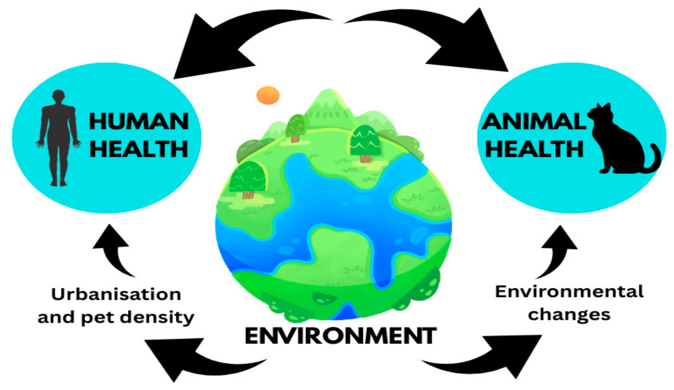
Figure 3.
Nasal fungal (Aspergillus) infections in dogs (Woods et al., 2024).
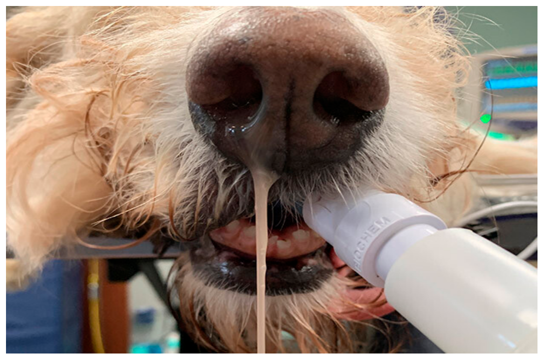
Figure 4.
Cutaneous mucormycosis in buffalo in the Brazilian Amazon biome (Barbosa et al.,2024).
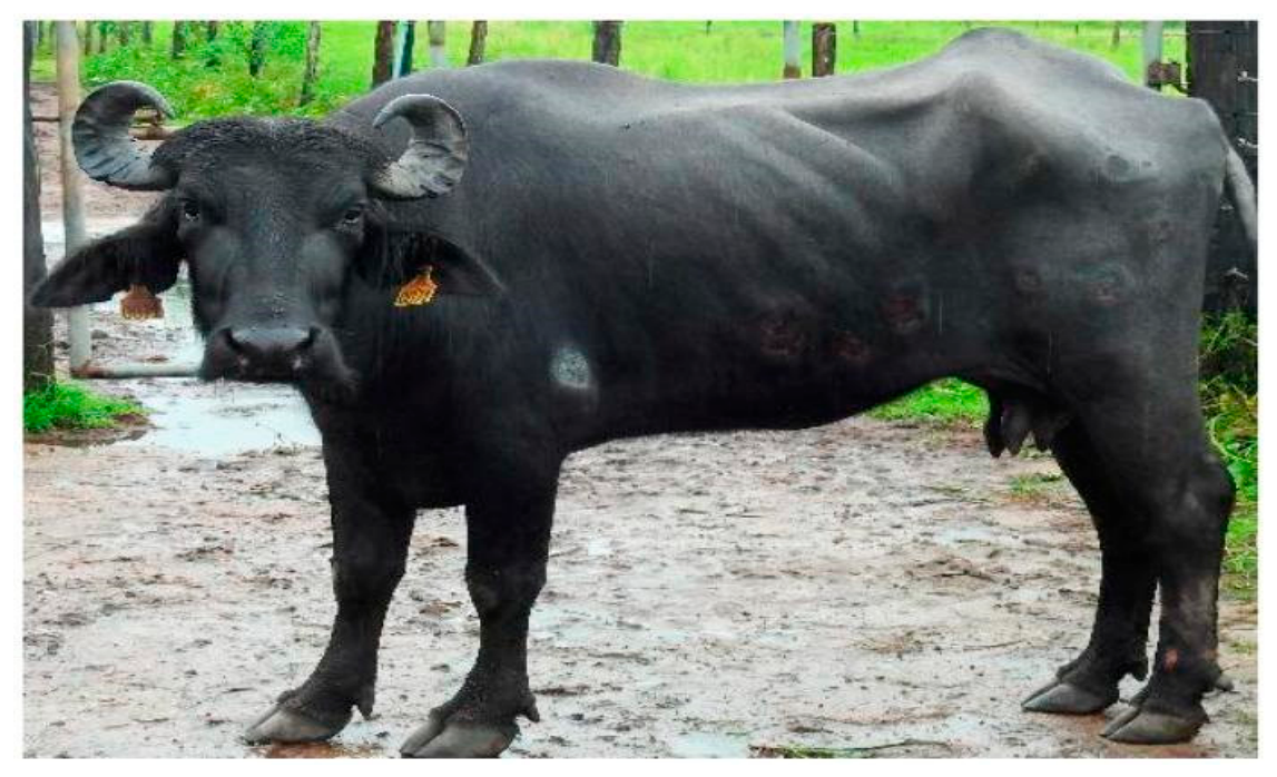
Figure 5.
Severe erythema, scaling and crusting in the inguinal area of an eight-month-old male, neutered Jack Russell terrier, between the prepuce and the scrotum bilaterally, due to Candida guilliermondii (Mueller et al.,2011).
Figure 5.
Severe erythema, scaling and crusting in the inguinal area of an eight-month-old male, neutered Jack Russell terrier, between the prepuce and the scrotum bilaterally, due to Candida guilliermondii (Mueller et al.,2011).
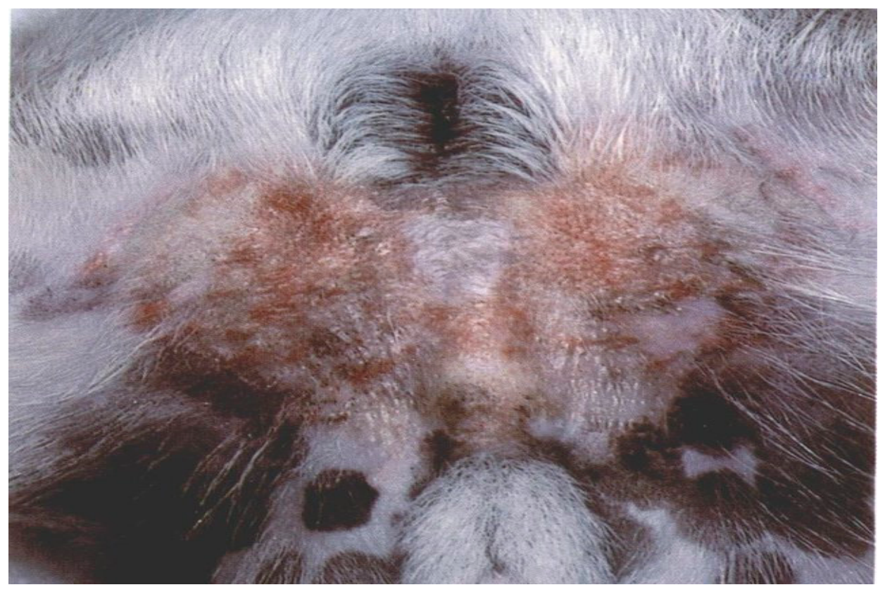
Figure 5.
A kitten infected by M. canis with no evidence of clinical lesions, sampled by the tooth-brush technique (Osada et al.,2023).
Figure 5.
A kitten infected by M. canis with no evidence of clinical lesions, sampled by the tooth-brush technique (Osada et al.,2023).
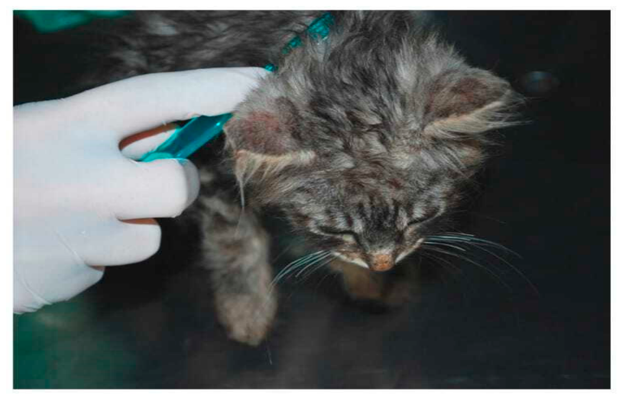
Figure 6.
(A, C) Cats with ulcerated cutaneous lesions caused by Sporotrix schenckii before treatment. (B, D) The cats after KI treatment — the lesions have resolved (Reis et al.,2012).
Figure 6.
(A, C) Cats with ulcerated cutaneous lesions caused by Sporotrix schenckii before treatment. (B, D) The cats after KI treatment — the lesions have resolved (Reis et al.,2012).
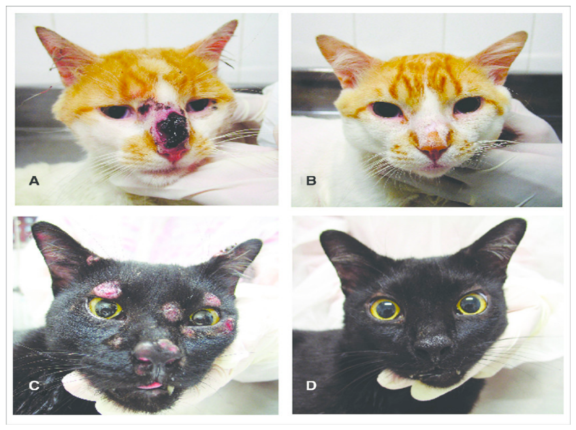
Figure 7.
Erythematous lesions and lichenification on pinna due to Malassezia (Kamaljyoti et al.,2017).
Figure 7.
Erythematous lesions and lichenification on pinna due to Malassezia (Kamaljyoti et al.,2017).
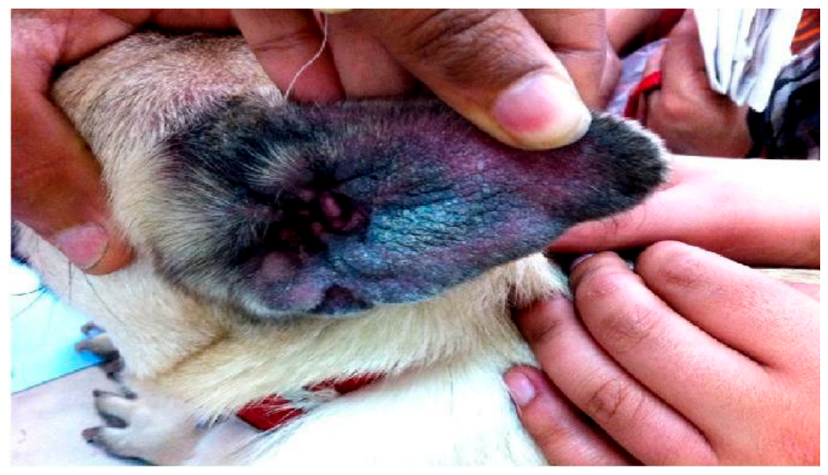
Figure 8.
Conceptual representation of Transmission of fungal infections from animals to humans.
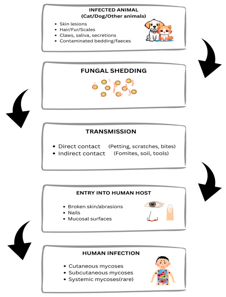
Figure 9.
Cutaneous disseminated sporotrichosis in a 66-year-old man. This patient presents nodular-ulcerated lesions on the dorsa of their hands and a lymphocutaneous/zosteriform distribution on their abdomen (Seyedmousavi et al., 2018).
Figure 9.
Cutaneous disseminated sporotrichosis in a 66-year-old man. This patient presents nodular-ulcerated lesions on the dorsa of their hands and a lymphocutaneous/zosteriform distribution on their abdomen (Seyedmousavi et al., 2018).
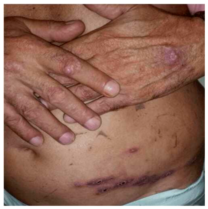
Figure 10.
Lymphocutaneous sporotrichosis with tenosynovitis involvement in a 75-year-old woman. Two nodules/masses on the dorsum of the left wrist (Seyedmousavi et al., 2018).
Figure 10.
Lymphocutaneous sporotrichosis with tenosynovitis involvement in a 75-year-old woman. Two nodules/masses on the dorsum of the left wrist (Seyedmousavi et al., 2018).
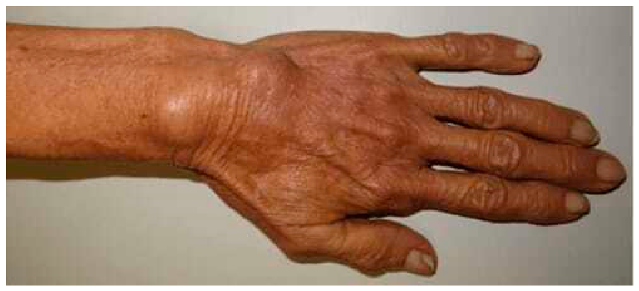
Figure 11.
An occipital erythematous swollen area about 4 × 4 cm in size with embedded pustules and honey-yellow crusts associated with infection with Trichophyton indotinea (Currie et al., 2023).
Figure 11.
An occipital erythematous swollen area about 4 × 4 cm in size with embedded pustules and honey-yellow crusts associated with infection with Trichophyton indotinea (Currie et al., 2023).
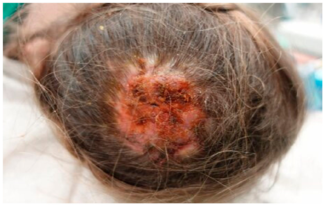
Figure 12.
Mycological investigations of samples collected from animals with dermatitis. Colour bars represent indicated fungi and the number of isolates (Dworecka-Kaszak et al.,2020).
Figure 12.
Mycological investigations of samples collected from animals with dermatitis. Colour bars represent indicated fungi and the number of isolates (Dworecka-Kaszak et al.,2020).
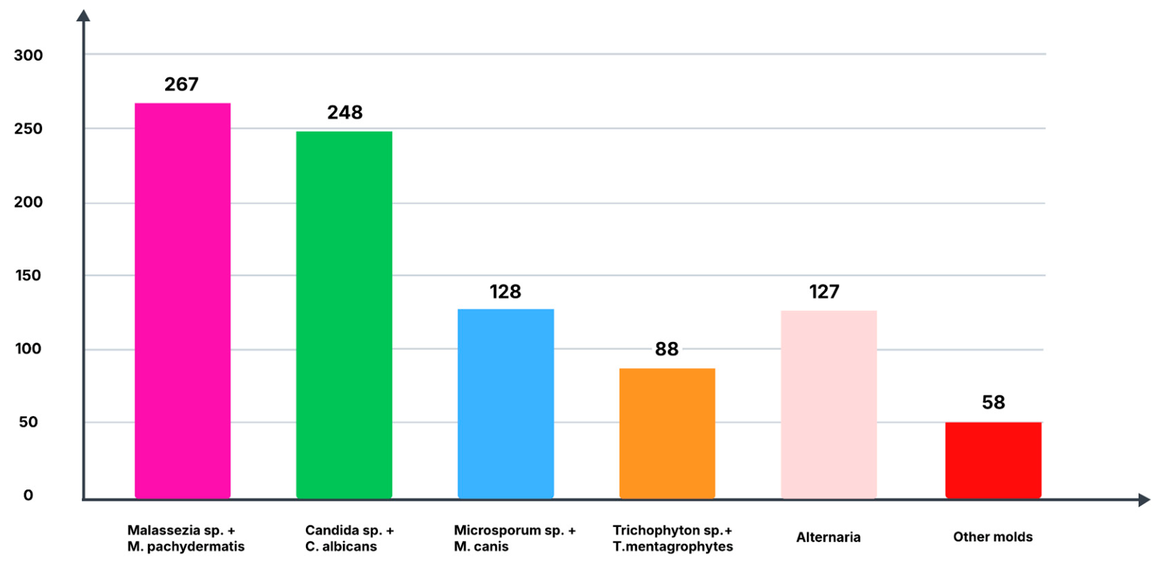
Figure 13.
Fungal diversity in the soil Mycobiome (Yiallouris et al.,2024).
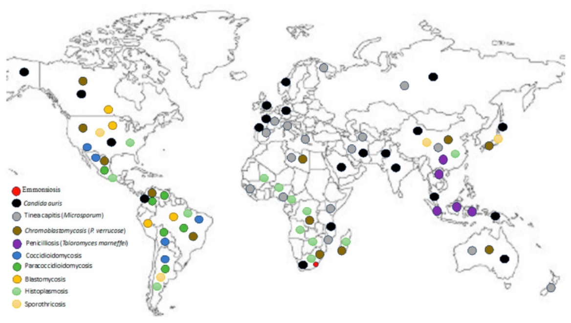
Disclaimer/Publisher’s Note: The statements, opinions and data contained in all publications are solely those of the individual author(s) and contributor(s) and not of MDPI and/or the editor(s). MDPI and/or the editor(s) disclaim responsibility for any injury to people or property resulting from any ideas, methods, instructions or products referred to in the content. |
© 2026 by the authors. Licensee MDPI, Basel, Switzerland. This article is an open access article distributed under the terms and conditions of the Creative Commons Attribution (CC BY) license (http://creativecommons.org/licenses/by/4.0/).
Copyright: This open access article is published under a Creative Commons CC BY 4.0 license, which permit the free download, distribution, and reuse, provided that the author and preprint are cited in any reuse.


