

Submitted:
20 July 2026
Posted:
21 July 2026
You are already at the latest version
Abstract
Stroke is the second leading cause of death after ischemic heart disease, accounting for 10% of all deaths worldwide. Stroke is also a major cause of long-term neurological disability, yet olfactory dysfunction remains an underrecognized consequence of cerebrovascular injury. Smell is rarely evaluated in stroke patients despite its central role in appetite, hedonia, hazard detection, memory, emotional processing, and quality of life. The olfactory system extends from the olfactory epithelium and olfactory bulb to primary olfactory cortex, limbic regions, orbitofrontal cortex, insula, thalamus, hippocampus, broader fronto-limbic networks as well as cerebellum. The broad anatomical organization of the olfactory system subjects it to frequent perturbation due to stroke or other lesions. Post-stroke olfactory deficits may result from direct injury to olfactory structures, disruption of higher-order olfactory networks, diaschisis, altered sniffing and sensorimotor control, or secondary degeneration. Clinically, these deficits are manifested as hyposmia, anosmia, impaired odor identification, discrimination and memory, altered odor pleasantness, reduced flavor perception, loss of appetite, or reduced odor-guided behavior. Current evidence suggests that olfactory dysfunction may persist into the chronic phase of stroke although the prevalence estimates vary due to the differences in population, testing methods, and reporting. This review provides an overview of stroke-related olfactory dysfunction, emphasizing olfactory pathway anatomy, lesion-relevant mechanisms, clinical consequences, measurement limitations, rehabilitation approaches, and future research priorities. We argue that post-stroke olfactory dysfunction should be understood as a multidimensional network-based disorder rather than a simple binary loss of smell.
Keywords:
anosmia
; dysosmia
; flavor
; hyposmia
; insula
; olfaction
; olfactory bulb
; piriform cortex
; olfactory training
; orbitofrontal cortex
; rehabilitation
; smell dysfunction
; thalamus
1. Introduction
Stroke is an acute neurological injury caused by interruption of cerebral blood flow or bleeding into brain tissue. It remains one of the leading causes of death and long-term disability worldwide and produces a broad spectrum of motor, sensory, cognitive, language, visual, affective, swallowing, and functional impairments [1,2]. Ranking among the leading causes of death and long-term disability worldwide, there are an estimated 12.2 million new cases and 6.55 million deaths recorded annually as of 2019 [1]. In the US alone, over 795,000 people suffer from a stroke every year, accounting for 1 in 6 deaths due to a cardiovascular disease [3]. Between 2019 and 2020, the stroke-related costs equal $56.2 billion in the US, which include health care services, treatment, and missed work [3]. A non-Hispanic black adult possesses twice the risk of having a first stroke compared to a white adult, suggesting a strong population bias in manifestation of stroke pathology [3]. Shared modifiable risk factors span both subtypes and include hypertension, the single largest contributor, accounting for over 55% of stroke-related disability-adjusted life years globally, as well as elevated body mass index, high fasting plasma glucose, ambient particulate matter exposure, and smoking [1].
This review synthesizes current evidence on the relationship between stroke and olfactory function, examines the neuroanatomical substrates underlying post-stroke smell loss, and identifies critical gaps in screening, rehabilitation, and post-stroke care. The discussion leads us to conclude that stroke-related olfactory dysfunction is a prevalent, underreported, and underrecognized consequence of cerebrovascular events that significantly impacts patient safety, nutrition, and mood. It requires specialized assessment and targeted, multidisciplinary interventions.
2. Stroke Pathology: Cellular and Anatomical
Broadly, stroke is classified into two pathological categories. Ischemic stroke is the most prevalent type, accounting for ~87% of all stroke cases [3]. The principal vascular etiologies include large artery atherosclerosis, cardioembolism, most commonly associated with atrial fibrillation, and small vessel disease, also referenced as lacunar stroke [2]. The current window for the treatment of ischemic stroke is about 4.5 hours and primarily involves restoration of blood flow through the administration of tissue plasminogen activator (tPA) or use of clot retrieval devices [4]. The second category of stroke, hemorrhagic stroke, results from the rupture of a blood vessel within or around the brain parenchyma; small vessel disease is the most frequent cause of intracerebral hemorrhage, while subarachnoid hemorrhage most commonly follows rupture of an intracranial aneurysm [2].
At the cellular level, disruption of blood supply produces severe hypoxic stress in neurons which are very sensitive to this stress [5]. Within minutes of blood flow cessation, ATP depletion triggers membrane depolarization, excitotoxic glutamate release, and calcium influx. This process activates proteases and lipases that contribute to irreversible cell death through several pathways including apoptosis, necrosis, autophagy, and pyroptosis [6]. Subsequently, white matter axon fibers and astrocytes are degenerated, and the blood-brain-barrier is disrupted [5]. The disruption of the blood-brain-barrier leads to invasion of peripheral immune cells into the brain parenchyma, contributing to neuroinflammation and neuropathology [7]. The ischemic core contains severely injured tissue, while the surrounding tissue, known as “ischemic penumbra,” may remain functionally silent but potentially salvageable for a limited window of hours. This forms the biological rationale for time-sensitive reperfusion therapies, including intravenous thrombolysis, as well as mechanical thrombectomy [2]. Glial cells further play protective roles by limiting the expansion of the ischemic core and by active tissue remodeling through phagocytosis of the dead cells and cellular debris, angiogenesis and neurogenesis [7].
Beyond the acute lesion, stroke can also produce diaschisis, remote hypoperfusion, degeneration of connected pathways, and long-term structural remodeling. These mechanisms are especially relevant for olfaction because smell depends on distributed and recurrent networks rather than a single linear pathway. The clinical manifestations of stroke are determined largely by the location and extent of the affected vascular territory. The middle cerebral artery is the most commonly occluded vessel and may produce contralateral hemiparesis, hemisensory loss, and, with dominant hemisphere involvement, aphasia. Anterior cerebral artery infarcts affect the medial frontal and parietal lobes, whereas posterior circulation strokes, involving the vertebrobasilar system, disrupt the brainstem, cerebellum, thalamus, and occipital cortex [2]. Notably, several of these territories overlap with structures integral to olfactory processing, including the insula, orbitofrontal cortex (OFC), and thalamus, an anatomical relationship that may contribute to the vulnerability of the olfactory system following stroke.
3. Olfactory Dysfunction: Causes and Consequences
Despite its clinical relevance, olfactory dysfunction remains one of the most underrecognized sequelae of stroke. The sense of smell is integral to functions that directly affect patient health and quality of life, including appetite regulation and nutritional intake, detection of environmental hazards such as smoke or spoiled food, and the mediation of memory and emotional experience [8,9,10]. Anosmia and hyposmia, the complete or partial loss of smell, are estimated to affect between 3% and 20% of the general population, with neurological injury representing a significant contributing factor [11]. Olfactory dysfunction is broadly categorized as congenital and acquired; congenital dysfunction affecting 0.01%-0.02% of the general population [12]. Congenital anosmia can be a result of rare genetic abnormalities such as in Kallmann syndrome, characterized by deficits in embryonic migration of gonadotropin-releasing hormone (GnRH) neurons and olfactory nerves originated in the nasal placode (e.g., mutations in KAL1, FGFR1, PROK2 genes), or due to infection- or stress-induced events during pregnancy [13,14]. The majority of Kallmann syndrome cases are due to sporadic mutations, and familial inheritance cases are rare with 64% displaying an autosomal dominant pattern [15]. Acquired olfactory dysfunction is largely attributed to sinonasal disease, upper respiratory tract infection, traumatic brain injury or idiopathic [16]. Viral infections (e.g., Covid-19), neurodegenerative diseases and toxins are well identified mechanisms underlying acquired olfactory dysfunction [17,18].
In the context of stroke, olfactory deficits are frequently underreported: patients are rarely screened for smell loss during acute or rehabilitation care, and many remain unaware of their own impairment [19]. However, objective assessment demonstrates that olfactory dysfunction is a measurable consequence of cerebrovascular injury, ranging from sudden anosmia following ischemic infarction of olfactory structures to subtler deficits in odor identification and hedonic appraisal [19,20]. The olfactory system’s anatomically distributed organization, spanning peripheral receptors, the olfactory bulb and tract, piriform cortex, amygdala, entorhinal cortex, insula, and OFC, renders it vulnerable to disruption across multiple stroke territories, likely contributing to the heterogeneity of deficits observed clinically.
4. The Olfactory System and Stroke-Relevant Stages of the Olfactory Pathway
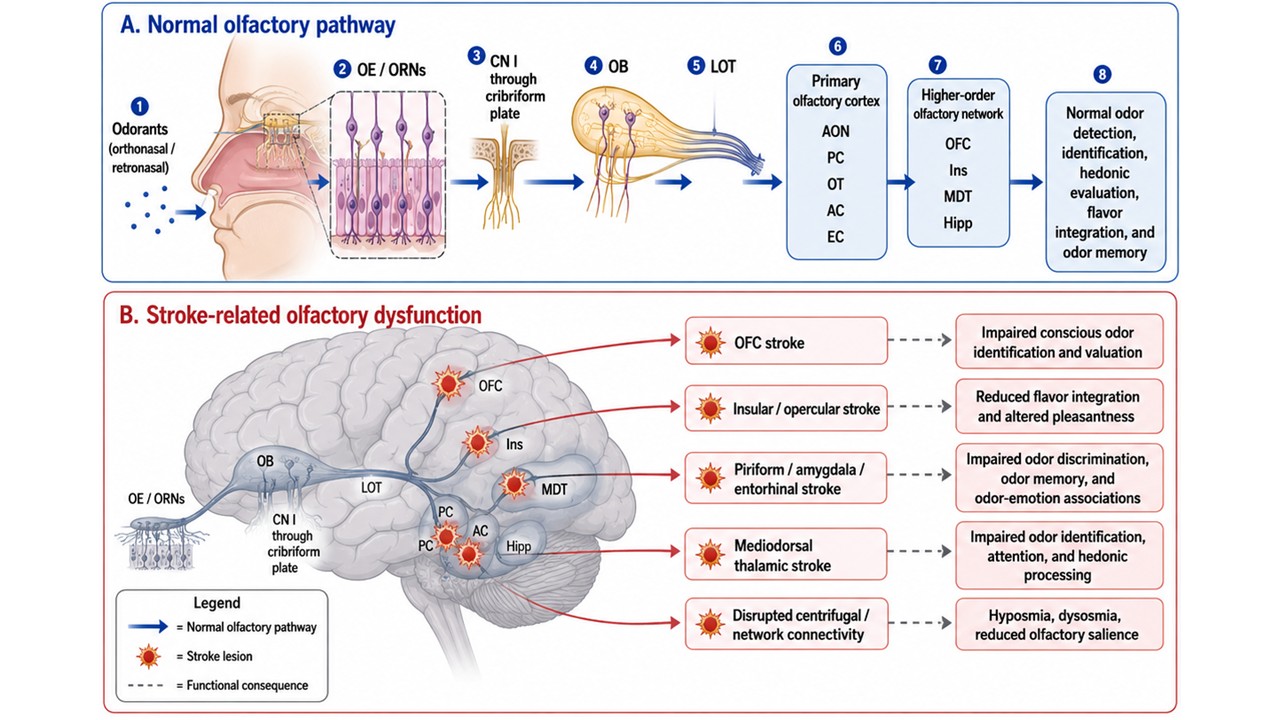
Olfaction begins when volatile odorant molecules reach the olfactory mucosa either orthonasally, during sniffing or inhalation through the nares, or retronasally, when odorants released from food and drink pass from the oral cavity through the nasopharynx during chewing, swallowing, and exhalation (Figure 1). Retronasal olfaction is especially important for flavor perception because it allows food-related volatile compounds to be integrated with taste, oral somatosensation, and trigeminal input in higher cortical regions such as the insula and OFC [21].
Both orthonasal and retronasal signals activate olfactory receptor neurons in the olfactory epithelium, a pseudostratified neuroepithelium located in the superior nasal cavity. This epithelium contains bipolar olfactory receptor neurons, sustentacular supporting cells, and basal stem cells. Odorant receptors are G protein-coupled receptors expressed on the cilia of olfactory receptor neurons, where odorant binding initiates transduction and generates activity in cranial nerve I [22,23,24]. The olfactory pathway begins in the olfactory epithelium, with olfactory receptor neuron axons crossing the cribriform plate and projecting ipsilaterally to the olfactory bulb, where they synapse in glomeruli [23,24].
The olfactory bulb is the first central relay of the main olfactory system (Figure 1). Axons from olfactory receptor neurons converge onto olfactory bulb glomeruli, where they contact mitral and tufted projection neurons as well as juxtaglomerular interneurons, including periglomerular cells, short-axon cells, and external tufted cells [25,26]. This glomerular organization allows odor information to be transformed from peripheral receptor activation into distributed spatial and temporal patterns of bulbar activity. Periglomerular and short-axon cells provide local and interglomerular inhibition, often using GABA and dopamine, while external tufted cells are glutamatergic and can exhibit rhythmic bursting that helps coordinate glomerular output [26,27]. Olfactory sensory neuron terminals contact mitral cells, external tufted cells, periglomerular cells, and short-axon cells, with dopaminergic/GABAergic short-axon cells forming oligoglomerular or polyglomerular networks that support interglomerular modulation [28].
These inhibitory and excitatory microcircuits are relevant to stroke because the olfactory bulb does not function only as a passive relay. It receives extensive centrifugal input from olfactory cortex, frontal cortex, hippocampal structures, cholinergic basal forebrain, locus coeruleus, raphe nuclei, and GABAergic systems, which shape odor responses, attention, salience, and experience-dependent modulation [24,29]. Therefore, vascular injury to frontal, limbic, or neuromodulatory systems may alter bulbar gain, odor sensitivity, attention to odors, or odor learning even when the olfactory epithelium and bulb are structurally intact.
Mitral and tufted cell axons leave the olfactory bulb through the lateral olfactory tract and project directly to primary olfactory cortical and limbic structures. Primary olfactory cortex includes the anterior olfactory nucleus, olfactory tubercle, piriform cortex, cortical and periamygdaloid amygdala-related regions, nucleus of the lateral olfactory tract, and parts of the entorhinal cortex [24,30]. The piriform cortex is the largest primary olfactory cortical area and plays a central role in odor identity, odor object formation, olfactory learning, and associative memory [24,31].
A distinctive feature of olfaction is that early cortical processing does not require a mandatory thalamic relay before reaching primary cortex, unlike vision, audition, and somatosensation [32]. This direct bulb-to-cortex organization may help explain why focal cortical and limbic lesions can produce selective deficits in odor identification, discrimination, memory, or hedonic appraisal rather than a simple loss of detection threshold. Human connectivity work also shows that primary olfactory subregions form distinct large-scale functional networks, with the anterior olfactory nucleus, olfactory tubercle, and frontal and temporal piriform cortices showing separable whole-brain connectivity patterns [33].
After primary olfactory cortex, odor information is distributed to secondary and multimodal regions, especially the OFC, insula, amygdala, hippocampal formation, hypothalamus, cingulate cortex, and thalamus [34,35]. The OFC is a major higher-order target for conscious odor perception, odor identification, reward value, pleasantness, and integration of smell with taste and oral texture [34,36]. The insula is also strongly implicated in olfaction and flavor because it participates in multisensory, affective, interoceptive, and gustatory processing. Tractography, primate tracing, functional imaging, and lesion evidence support connections between primary olfactory cortex and the insula, and insular lesions can produce olfactory disturbances, particularly altered pleasantness, subjective smell changes, or difficulty distinguishing odors [37]. This is important for stroke because the insula is commonly involved in middle cerebral artery territory infarcts. Thus, insular stroke may contribute to altered flavor, odor unpleasantness, distorted odor quality, reduced food enjoyment, or reduced appetite, even when basic odor detection is preserved [37,38,39].
Although olfaction bypasses the thalamus on its way to primary olfactory cortex, the mediodorsal thalamus remains important for higher-order olfactory processing. The mediodorsal thalamic nucleus receives input from primary olfactory regions, including piriform cortex, olfactory tubercle, amygdala, lateral entorhinal cortex, and anterior olfactory nucleus, and has reciprocal connections with OFC and agranular insular regions [32]. Thalamic involvement is particularly relevant to stroke phenotypes. Patients with focal thalamic lesions may show relatively spared odor detection but impaired odor identification and altered hedonic ratings, especially reduced pleasantness for pleasant odors [40]. Case reports of mediodorsal or bilateral thalamic infarction similarly describe disturbances of smell, taste, food intake, and odor pleasantness [41,42]. Okamoto et al. also reported that acute ischemic stroke patients without MRI-visible thalamic lesions but with thalamic hypoperfusion showed reduced odor identification and recognition, suggesting that diaschisis or remote perfusion changes may affect olfactory behavior even when the visible infarct is outside the classic olfactory pathway [43]. These observations support a model in which thalamic injury preferentially affects attention-dependent, semantic, affective, and evaluative dimensions of olfaction rather than peripheral odor detection.
5. Direct and Indirect Effects of Stroke on Olfactory Processing
Stroke may affect the olfactory system directly, indirectly, or through a combination of mechanisms. Direct effects occur when the lesion involves olfactory pathway structures themselves, such as the anterior olfactory region, olfactory tract, piriform cortex, amygdala, entorhinal cortex, OFC, insula, or thalamus [37,40,44]. Indirect effects occur when stroke affects regions that modulate olfactory processing, disrupts distributed networks, alters perfusion in connected areas, impairs cognition or language required for odor identification, or changes respiratory and sniffing behavior [32,33,43,45].
This distinction is clinically important because a patient does not need to have an infarct in the olfactory bulb or primary olfactory cortex to develop olfactory symptoms. A frontal stroke may impair conscious odor recognition and valuation. A thalamic stroke may impair odor identification or pleasantness. An insular stroke may alter flavor integration or odor affect (hedonic value) as the insular cortex (insula) acts as a critical multisensory hub where taste, smell, and internal bodily sensations converge to create our perception of flavor. A cerebellar stroke may change sniff dynamics and olfactomotor control. A large hemispheric stroke may disrupt attention, naming, memory, or awareness, thereby affecting performance on odor identification tasks [36,37,40,45]. In addition, diaschisis or remote hypoperfusion may impair olfactory behavior even when the visible infarct is outside classical olfactory structures [43].
From a stroke perspective, olfactory dysfunction can therefore arise at several levels. Damage to inferior frontal or orbitofrontal regions may impair conscious odor recognition, valuation, and flavor-related reward [34,36]. Medial temporal or limbic infarcts involving piriform cortex, amygdala, hippocampus, or entorhinal cortex may affect odor identity, odor memory, odor-emotion associations, and learned odor meaning [9,24,31]. Insular and opercular strokes may disturb multisensory flavor integration and the affective or interoceptive experience of odors [37,38,39]. Thalamic strokes, especially involving mediodorsal regions, may impair odor identification and pleasantness while leaving basic detection relatively intact [40,41,42]. Finally, disruption of fronto-limbic, thalamo-cortical, or centrifugal feedback circuits may alter olfactory attention, salience, and odor-guided behavior [29,32,43]. Clinically, this means that post-stroke olfactory dysfunction may present not only as anosmia or hyposmia, but also as impaired odor naming, reduced food enjoyment, altered pleasantness, dysosmia, impaired flavor perception, impaired odor memory, or reduced awareness of the deficit [19,42,46].
6. How Common is Post-Stroke Olfactory Dysfunction?
The true prevalence of post-stroke olfactory dysfunction remains uncertain. Estimates vary because studies use different methods, including self-report surveys, bedside questionnaires, psychophysical smell tests, acute cohorts, chronic cohorts, rehabilitation samples, and case-control designs [19,46,47]. Despite this variability, available evidence suggests that olfactory impairment after stroke is not rare and may persist well beyond the acute phase.
Objective testing appears to detect more dysfunction than self-report alone. In a prospective cohort of 78 chronic stroke patients tested one year after stroke admission, Wehling and colleagues found 28.2% hyposmia and 15.4% olfactory loss, including functional and complete anosmia [19]. Age and the National Institutes of Health Stroke Scale score were predictors of olfactory dysfunction, and patients with olfactory dysfunction rated odor items as less pleasant [19]. Importantly, self-reported olfactory function did not reliably distinguish patients with normal olfaction from those with reduced olfactory performance, suggesting that routine clinical questioning may miss many patients with meaningful olfactory impairment [19].
Bedside questionnaire studies similarly suggest that smell and taste dysfunction occur in both acute and chronic stroke populations. Schön and colleagues reported subjective smell dysfunction in approximately 12% of stroke patients and taste dysfunction in approximately 38%, emphasizing that routine bedside assessment is feasible but should be validated against objective testing [47]. Case-control psychophysical work has reported worse smell identification in patients with stroke than in healthy controls, along with concurrent taste impairment and reduced food liking [46]. Such findings are valuable because they connect olfactory dysfunction to patient-centered outcomes such as appetite and food enjoyment; however, they should be interpreted in light of sampling, since rehabilitation or clinic populations may overrepresent more symptomatic patients.
7. What does Olfactory Dysfunction Look Like after Stroke?
Post-stroke olfactory deficits are heterogeneous and may involve sensitivity, identification, discrimination, hedonic evaluation, flavor integration, memory, or qualitative odor perception. For this reason, “smell loss” is an imprecise umbrella term.
One common pattern is reduced sensitivity, or threshold elevation, which may manifest as hyposmia or anosmia. In some patients, this may be lateralized, particularly if the lesion affects unilateral olfactory structures or olfactomotor pathways [19,45]. A second pattern is impaired odor identification or discrimination. These patients may detect that an odor is present but cannot name it, recognize it, or distinguish it from similar odors. This phenotype may reflect central olfactory network injury, but it may also be influenced by aphasia, semantic impairment, memory dysfunction, attention deficits, or executive dysfunction [19,43,48].
A third pattern is hedonic or affective distortion. Patients may report that odors have lost their pleasant character, smell unpleasant, or no longer contribute normally to food enjoyment. This phenotype is particularly important because it may drive appetite changes, reduced oral intake, weight loss, or maladaptive dietary compensation. Classic thalamic lesion reports show that odor identification may be preserved while pleasantness is selectively impaired, demonstrating that hedonic olfaction can dissociate from basic detection [40,42]. Wehling and colleagues similarly found altered pleasantness ratings in chronic stroke patients with olfactory dysfunction [19].
A fourth pattern includes qualitative phenomena such as dysosmia, parosmia-like distortions, or phantosmia-like complaints. These symptoms are less commonly discussed in stroke than in post-viral or traumatic etiologies, but insular and temporal network involvement may plausibly contribute to altered odor quality, unpleasant odor bias, or olfactory hallucinations. Beume and colleagues described olfactory hallucinations as a primary symptom of ischemia in the right posterior insula, supporting the possibility that focal ischemia can produce qualitative olfactory phenomena [37,49].
Finally, patients may experience reduced flavor perception even when they describe the issue as a “taste” problem rather than a smell problem. Because flavor depends heavily on retronasal olfaction, stroke-related disruption of olfactory, gustatory, insular, orbitofrontal, and somatosensory integration may cause food to seem bland, unpleasant, or less rewarding [21,38,46].
8. Lesion Locations that Matter: What Do We Learn from Animal and Case Studies?
Because olfaction is networked, smell loss is not attributed to one localization. Still, several stroke-relevant nodes show repeated associations with specific olfactory phenotypes (Figure 2).
8.1. Anterior Olfactory Skull-Base Region and Olfactory Tract
Sudden isolated anosmia can, in rare cases, be a cerebrovascular presentation. Ischemic infarcts involving the anterior olfactory skull-base region adjacent to the olfactory sulci and straight gyri, including the olfactory tract distribution, may produce acute smell loss and mimic more common causes such as viral anosmia. Theodorou and colleagues described CT- and MRI-visible bilateral ischemic lesions adjacent to the olfactory sulci in a patient with anosmia, supporting ischemic olfactory infarction as a vascular cause of sudden smell loss [44]. This is clinically relevant since the patient had a history of type-2 diabetes and gastritis and presented with symptoms mimicking potential coronavirus infection. Thus, sudden anosmia may be attributed to infection, sinonasal disease, or post-viral dysfunction, potentially delaying consideration of vascular causes in appropriate contexts.
8.2. Insula and Opercular/Frontoparietal Cortex
The insula is a multisensory integration hub tightly tied to flavor, taste, oral somatosensation, interoception, and affective response. Although the OFC can be the primary center for integrating and processing of taste and smell inputs, the insula and opercular or frontoparietal cortex receive taste afferents from the nucleus solitarius and thalamus. Thus, lesions involving insula and opercular/frontoparietal cortex can disrupt integration of taste and smell signals in the brain [37,38,39]. In addition, left angular gyrus and inferior parietal gyrus in the parietal lobe is involved in olfactory working memory [50]. Functional MRI studies reveal that these areas are highly activated during processing of olfactory working memory [50]. Therefore, these areas are critical for olfactory perception, integration and memory.
Moo and Wityk described olfactory and taste dysfunction after bilateral middle cerebral artery strokes involving posterior insula and opercular/frontoparietal cortex relevant to multisensory integration [39]. The fact that the patient was presented with the symptoms only after the second stroke affecting the other side of the brain, suggests that bilateral lesions in these areas may be necessary to produce a measurable deficit. Because the insula is frequently affected in middle cerebral artery strokes, it may be an important but underrecognized contributor to post-stroke changes in food enjoyment, appetite, and flavor perception. Insular involvement may also explain why standard threshold or odor identification tests can miss clinically meaningful deficits. A patient may perform adequately on odor detection using standard testing such as Sniffin’ Sticks test but still experience odors as unpleasant, distorted, weak, or poorly integrated with taste [37]. The patient with left insular cavernoma developed an altered perception of odor detection; the unpleasant odors appeared stronger, and the pleasant odors either became indistinguishable or unpleasant [37]. Since distinct olfactory regions are shown to be responsible for the presentation of valence and intensity [51], it is plausible that a left insular lesion could only produce subjective deficits in the patient’s olfactory perception. This further supports the need for hedonic and flavor-specific assessment in patients with insular or opercular lesions and perhaps the necessity for an improved methodological advancement toward understanding of olfactory perception governed by the insular function.
A heightened sensitivity to unpleasant odors, as well as olfactory hallucinations for unpleasant odors following insular lesions have been reported [38,49] . Mak and colleagues reported altered suprathreshold odor (and taste) intensity perception after a left posterior insular stroke, with prominent contralateral intensity changes, suggesting cortical disinhibition mechanisms [38]. It has been postulated that the right hemisphere may be more attuned with processing of pleasant stimuli, the left hemisphere toward more unpleasant stimuli for various modalities [40]. Overall, left insular lesions are more frequently associated with olfactory dysfunction [37,38,39]. It remains unknown whether olfactory dysfunction associated with left and right insular lesions are measurably distinct. Nonetheless, an alteration in hedonic perception of odor may be indicative of damage to the insula requiring neurological intervention.
8.3. Thalamus
Although olfaction is often construed as bypassing the thalamus, multiple lines of evidence in rodent models using electrophysiology, behavior, and immunohistochemistry approaches show that thalamic nucleus, especially mediodorsal thalamus, known for its role in higher order cognitive function, contributes to experience-dependent, modality-specific olfactory functions including olfactory attention, discrimination, and hedonic processing, as well as odor-guided decision making [32,52]. As such, mediodorsal thalamus receives projection from piriform cortex and gustatory cortex and is connected reciprocally with OFC. A novel odor compared with a familiar odor, leads to greater neuronal activation determined by c-Fos reactivity, whereas a familiar taste or odor-taste mixture results in greater neuronal activation in the mediodorsal thalamus [52]. In addition, the preference for a familiar odor over a novel odor (such as preference for water over isoamyl acetate-odorized water) is eliminated when the novel odor is coupled with a novel taste (such as isoamyl acetate-odorized water with sucrose) over several days, suggesting an experience-driven integration of behaviorally relevant chemosensory stimuli including odor neophobia, potentially involving mediodorsal thalamus and the higher-order structures in food-related behavior [52]. The mediodorsal thalamus enhances neural coupling with piriform cortex during odor sampling and with OFC during decision making during two-alternative odor discrimination tasks [32]. Specifically, the mediodorsal thalamus is likely to be spared for sensory thalamic relay relevant to odor encoding, and state-dependent modulation, but remains an important node for sensorimotor representation of odor-guided, goal-directed behavior [32,53].
Thalamic lesions have further enhanced our understanding of the role of mediodorsal thalamus in olfaction. Although the thalamus is prone to ischemia, the reporting of thalamic infarctions associated with olfactory deficits is rare. Patients with thalamic infarctions suffer from co-occurring mental disturbances and consciousness issues, or mild olfactory deficits are often overlooked during standard examination. Reports of mediodorsal thalamic infarction describe persistent loss of pleasantness of odors and tastes with major appetite and weight consequences, despite preserved identification [42]. While bilateral mediodorsal infarction leads to olfactory deficits, infarctions involving the left mediodorsal thalamus and right sided adjacent nuclei such as right ventral posterior nucleus and ventral lateral nucleus have been associated with transient odor abnormality, supporting the role of thalamic involvement in olfactory disturbance [41]. The best evidence for thalamic involvement in olfaction comes from unilateral lesion studies in 17 patients and 18 age-matched healthy controls [40]. Sela and colleagues reported that thalamic lesions significantly affect odor identification, sparing odor detection. In addition, only right sided lesions diminish the pleasantness of pleasant odors, suggesting olfactory hedonic perceptions may be attributed to the structural and functional asymmetry associated with thalamus [40]. Both right and left thalamic lesion patients display significantly less sniff vigor compared with healthy controls, without significant changes in odor detection threshold, suggesting that thalamus may act as a component of olfactomotor network [40]. In addition, 19 acute ischemic stroke patients without MRI-visible thalamic lesions but with thalamic hypoperfusion as detected by Technetium-99m ethyl cysteinate dimer (99mTc-ECD) SPECT, display lower scores in odor detection and recognition threshold olfactory disturbances, compared with 9 stroke controls with normal thalamic hypoperfusion, further reinforcing the role for thalamus or its associated remote network function in olfactory behavior [43].
8.4. OFC and Frontal Regions
The OFC has long been visualized as a secondary olfactory cortex receiving smell information from the primary olfactory cortex, such as the piriform cortex [54,55]. Functional neuroimaging studies in human reveal that the rostral OFC displays highest responsivity [34]. Lesion studies indicate that OFC plays a critical role in human olfactory consciousness [36]. Li and colleagues combine fMRI and peripheral autonomic recording in a traumatic brain injury patient with right orbitofrontal lesion to show that despite complete absence of conscious olfaction, the odor-evoked neural activity remains intact in left OFC [36]. The autonomic responses to odor hedonics through presentation in left nostril also remain normal. Together these results suggest that right OFC drives olfactory consciousness [36]. Broader reviews of chemical-sense processing emphasize orbitofrontal involvement in odor and flavor valuation [35]. This may be especially relevant when patients report that food no longer has emotional or rewarding value, rather than simply reporting reduced smell sensitivity. In addition, superior, middle and inferior frontal gyrus, as well as motor cortex including precentral gyrus and supplemental motor cortex are activated during olfactory working memory tasks in fMRI studies [50]. Strokes involving orbitofrontal or frontal regions may therefore impair odor identification, odor valuation, flavor reward, conscious awareness of odors and olfactory working memory.
8.5. Medial Temporal and Limbic Structures
The piriform cortex, amygdala, entorhinal cortex, and hippocampus support odor identity, associative learning, odor memory, and odor-emotion links [9,24,31]. Because odors are strongly tied to autobiographical memory and affect, which involve the amygdala-hippocampus as well as medial OFC and posterior cingulate cortex, damage to limbic olfactory structures may have psychological and quality-of-life consequences that are not captured by threshold testing alone [9,56]. Matsunaga and colleagues report that odor-evoked specific memories generate 6.5 times more nostalgia than odors that do not elicit a specific memory or an event [57]. Reid and colleagues further observe that nostalgic odors bring three times more positive than negative emotion [58]. In fact, odor-evoked nostalgia is more powerful than music-evoked nostalgia [59]. Matsunaga and colleagues further note that plasma levels of proinflammatory cytokines such as tumor necrosis factor α (TNF-α) and interferon-γ (IFN-γ) are significantly reduced following odor experience evoking positive emotions and autobiographical memory, implicating odor-evoked emotions in modulating neuroimmune responses [57]. Both medial OFC and posterior cingulate cortex are activated by odor-evoked autobiographical memory, as observed by PET imaging – further emphasizing the role for limbic system in olfaction. Thus, stroke involving the limbic system may impair odor recognition, odor memory, emotional associations with odors, and learned odor meaning, which in turn may critically impact both psychological and physiological well-being.
8.6. Cerebellum and Olfactomotor Control
Although the cerebellum is not traditionally considered as a part of the olfactory system, fMRI studies suggest that the cerebellum can be activated during human olfaction [60]. Sobel and colleagues observe that odorants such as vanillin and propionic acid induce significant activation in posterior lateral hemisphere in concentration-dependent manner, while sniffing non-odorized air activates anterior cerebellum [60]. These results suggest that specific cerebellar areas are involved in distinct control of olfactory functions such as odor detection and sniff dynamics. Olfaction depends not only on receptors and cortical processing but also on sniff dynamics. Sniffing controls odorant delivery to the olfactory mucosa and influences odor perception. The olfactomotor system produces sniffs that are strong enough to sample the odor signal but inversely proportional to their concentration. Lesion studies suggest that unilateral cerebellar damage can produce selective olfactory impairments such as odor identification and detection, supporting a role for the olfacto-cerebellar circuits connecting each nostril to the contralateral cerebellum [45]. In addition, patients with bilateral cerebellar degeneration display impairment in olfactory identification, further highlighting the role for cerebellum in olfaction [61]. In stroke, altered breathing patterns, reduced sniff vigor, facial weakness, impaired motor coordination, or reduced sensorimotor control could degrade olfactory input even if receptor neurons remain intact.
9. Secondary Degeneration and Structural Remodeling After Stroke
Beyond the location of the infarct, stroke can drive longer-term remodeling in olfactory structures. In an MRI case-control study of 41 patients with stroke and 41 controls, Kültür and colleagues measured peripheral olfactory structures, including olfactory bulb volume and olfactory sulcus depth, as well as central regions such as the insula and amygdala [62]. They reported smaller peripheral and central smell-related regions in patients with stroke, and left olfactory bulb volume decreased with longer stroke duration [62]. These findings suggest that post-stroke olfactory dysfunction may involve ongoing degeneration or inflammation-associated change rather than simple acute injury followed by recovery.
This aligns with a broader model in which post-stroke olfactory dysfunction reflects a mixture of direct circuit injury, remote network dysfunction, diaschisis, hypoperfusion, and chronic structural change. Such mechanisms may explain why some patients continue to experience olfactory impairment long after the acute lesion and why olfactory deficits may not map precisely onto the visible infarct alone [43,62].
Although long-term recovery of olfactory function remains considerably unknown, available documentation indicates that olfactory deficiency may extend well beyond the acute phase, with impairment persisting and remaining measurable within a significant number of patients following up to one year after stroke [19]. Restoration may differ due to the underlying mechanics of impairment; dysfunction sourced from transient network-level disturbances likely alter from those triggered by direct trauma to olfactory structures or structural modification [43,44,62]. Regardless, the determinants that anticipate recovery continue to be unexplored, and longitudinal studies monitoring olfactory effects after stroke are understudied. Additional research is essential for understanding whether post-stroke olfactory dysfunction adheres to predictable recovery patterns.
10. Why Olfaction Matters Clinically After Stroke
Smell loss is not merely a sensory curiosity. It can affect patient outcomes. First, olfactory dysfunction can affect nutrition and appetite. Loss or distortion of smell reduces food enjoyment and may contribute to reduced oral intake, weight loss, or poor nutritional status [8,20,42,46]. Some patients may compensate by adding excessive salt or sugar to food, which may complicate management of hypertension, diabetes, or vascular risk factors. Others may lose interest in eating altogether, reducing energy intake during a period when nutrition is important for rehabilitation and recovery.
Second, olfactory dysfunction creates safety risks. Patients with impaired smell may fail to detect smoke, gas leaks, spoiled food, chemicals, or other environmental hazards [10,11]. Because self-report is unreliable, patients may not spontaneously disclose smell loss and may not receive appropriate counseling unless clinicians ask directly and test objectively [19].
Third, olfactory dysfunction may affect mood, quality of life, memory, and social behavior. Odor hedonics are tightly linked to emotion and memory circuits [9,35]. Reduced pleasantness, distorted odor perception, or loss of odor-evoked memory may contribute to diminished pleasure, social withdrawal, anxiety, or depressive symptoms. Wehling and colleagues found altered pleasantness ratings in chronic stroke patients with olfactory impairment, supporting the relevance of affective olfactory changes after stroke [19].
Fourth, olfactory dysfunction may exacerbate an inflammatory response. Inflammation is often identified as an underlying condition in several systemic, peripheral as well as neurological, neuropsychiatric and neurodegenerative diseases. Odor-evoked memories may mitigate inflammation through psychoneuroimmune interactions [57]. Plasma level of IFN-γ is negatively correlated with the activation of medial OFC and posterior cingulate cortex, areas associated with odor-evoked autobiographical memory.
Fifth, olfactory dysfunction may indirectly affect rehabilitation engagement. Poor appetite, reduced enjoyment, fatigue related to inadequate nutrition, and mood changes may reduce motivation and participation in therapy. Therefore, olfactory dysfunction should be considered part of the broader post-stroke recovery environment rather than an isolated sensory deficit (Figure 3).
While current findings are inadequate to determine olfactory dysfunction as a prognostic biomarker, these findings suggest that olfactory assessment may provide insight outside of the presence or absence of sensory impairment alone. Considering the widespread nature of olfactory processing and its perceived relationship with stroke severity and network dysfunction, olfactory deficits may provide information into broader neurological consequences of cerebrovascular injury. Future studies should consider investigating whether post-stroke olfactory dysfunction is aligned with cognitive outcomes, rehabilitation engagement, nutritional status, or long-term functional recovery. If such relationships exist, olfactory assessment could present as an underutilized tool for identifying patients who may benefit beyond conventional measures of stroke severity.
11. Measurement: Why the Problem is Underestimated
The single biggest reason post-stroke olfactory dysfunction is underestimated is measurement failure. Self-report is unreliable. Patients may not notice their deficit, may attribute it to aging or congestion, or may describe it as a taste problem, appetite problem, or food preference change. Cognitive impairment, aphasia, neglect, reduced awareness, or competing neurological symptoms may further reduce reporting. Patients may report reduced flavor or appetite rather than smell loss. Therefore, objective testing is needed (Figure 4).
In Wehling’s chronic stroke cohort, self-reported olfactory function did not separate patients with normal olfaction from those with objective impairment [19]. Objective psychophysical tests are therefore essential. Practical tools include the University of Pennsylvania Smell Identification Test and Sniffin’ Sticks [63,64,65]. Sniffin’ Sticks is especially useful because it can separately assess threshold, discrimination, and identification, often summarized as a TDI score [64,65]. This separation matters because stroke may impair one domain while sparing another. For example, thalamic or frontal lesions may affect identification or pleasantness more than detection threshold [40].
Hedonic measures should be added, when possible, particularly if patients complain that food is unpleasant, smells are distorted, or “everything smells wrong” while standard smell scores appear normal. This is particularly relevant for insular and thalamic lesions, where hedonic or affective olfactory processing may be disrupted despite preserved basic detection [37,40,42]. Retronasal olfaction and flavor-related testing may also be important because many patients experience the functional problem during eating rather than during isolated odor sniffing [21,46].
Lateralized testing may be helpful in selected patients because stroke effects may be asymmetric. Nostril-specific testing can reveal deficits that bilateral testing may obscure, especially when olfactomotor or unilateral pathway involvement is suspected [45]. Research settings may also use olfactory event-related potentials or neuroimaging-based approaches to detect objective abnormalities even when subjective perception appears preserved. Cecchini and colleagues highlighted the potential value of olfactory testing and neurophysiological approaches in patients with ischemic stroke [48].
A multidimensional post-stroke olfactory assessment framework should therefore include odor threshold, discrimination, identification, pleasantness, retronasal olfaction, taste, flavor perception, appetite, food liking, nutrition, mood, cognition, language, and quality of life (Figure 4). The steps include screening: initial symptom questions used to identify possible olfactory dysfunction; psychophysical testing: behavioral smell testing using standardized odor stimuli; threshold: lowest concentration at which an odor can be detected; discrimination: ability to tell whether odors are the same or different; identification: ability to name or select the correct label for an odor; pleasantness rating: judgment of how pleasant or unpleasant an odor is; retronasal testing: testing odor perception through the mouth/nasopharynx, relevant to flavor; peripheral-like pattern: pattern dominated by reduced odor sensitivity; central pattern: pattern dominated by impaired identification, memory, pleasantness, attention, or flavor integration.
12. Management and Rehabilitation
There is currently no stroke-specific, universally accepted treatment pathway for olfactory loss comparable to dysphagia, aphasia, or motor rehabilitation pathways. However, several management steps are clinically defensible.
First, clinicians should evaluate non-stroke contributors that are common in stroke survivors, including sinonasal disease, allergies, smoking, xerostomia, medication effects, neurodegenerative disease, and recent viral infections [11,66]. Identifying these factors is important because some may be treatable or may confound interpretation of olfactory testing.
Second, safety counseling should be provided when dysfunction is present. Patients should be advised about smoke and gas detectors, food spoilage strategies, labeling food dates, using visual or caregiver checks, and avoiding reliance on smell alone for safety decisions. This recommendation is supported by the broader olfactory-disorder literature showing that smell loss affects personal safety and well-being [10].
Third, nutrition and appetite should be assessed. Patients with reduced food enjoyment, weight loss, or poor intake may benefit from dietary counseling, flavor enhancement strategies, texture modification, or involvement of nutrition specialists. Because some patients compensate with excess salt or sugar, counseling should also consider vascular risk management [8,46].
Fourth, olfactory training may be reasonable. Across etiologies, olfactory training has the strongest evidence base among noninvasive interventions for olfactory dysfunction. A recent meta-analysis reported significant improvements across several olfactory domains, especially identification, discrimination, and composite olfactory scores, supporting olfactory training as a reasonable rehabilitative approach even though stroke-specific randomized trial data remain limited [67].
Finally, stroke-specific trials remain limited but emerging. A registered effort focused on re-education of olfactory disorders after cerebrovascular accident, RE-OLF/NCT04703218, reflects increasing recognition of post-stroke olfactory rehabilitation as a research priority [68,69] . Until stronger stroke-specific evidence is available, post-stroke olfactory care should remain cautious and individualized, with emphasis on screening, education, safety counseling, nutrition support, and referral to neurology or otolaryngology when appropriate.
13. Olfaction as a Marker of Broader Cerebrovascular and Aging Risk
Emerging evidence also suggests that olfactory dysfunction may not only follow stroke but may also relate to broader cerebrovascular vulnerability. Chamberlin and colleagues examined whether poor olfaction was associated with adverse cerebrovascular events and future stroke risk in the Atherosclerosis Risk in Communities Study. They found that poor olfaction assessed by a single olfaction test was associated with a higher risk of stroke over the following decade [70]. This does not mean that smell loss directly causes stroke, but it suggests that olfactory dysfunction may serve as a marker of vascular, neurodegenerative, inflammatory, or aging-related vulnerability.
This association also overlaps with broader work on frailty. Pleasants and colleagues reported that olfactory dysfunction was associated with frailty risk in older adults, with olfactory function assessed using the 12-item Sniffin’ Sticks Test and frailty measured using both the Fried Frailty Phenotype and the Cumulative Frailty Index [71]. Older adults with better olfactory function showed lower frailty risk than those with poor olfaction [71]. These findings suggest that olfactory dysfunction may represent more than an isolated sensory problem. It may also reflect systemic vulnerability relevant to aging, vascular health, and recovery potential.
14. Research Gaps and Future Directions
Although olfactory dysfunction is increasingly recognized as a clinically meaningful consequence of stroke, the literature remains comparatively underdeveloped relative to other post-stroke sensory, motor, cognitive, and swallowing outcomes. Existing studies suggest that smell and taste deficits may be common after stroke, may persist into the chronic phase, and may affect appetite, food liking, safety, and quality of life [19,46,47]. However, mechanisms remain insufficiently defined, and the field still lacks a clear lesion-to-phenotype model that explains why some patients primarily show reduced odor sensitivity while others show impaired odor identification, altered pleasantness, reduced flavor perception, or impaired odor memory.
A major limitation of the current literature is the heterogeneity of stroke populations. Studies often include patients with diverse lesion locations, stroke types, stroke severities, chronicity, and comorbidities, making it difficult to determine whether olfactory impairment reflects direct damage to olfactory-related regions, disruption of distributed olfactory networks, diaschisis, thalamic hypoperfusion, cognitive-language impairment, mood-related changes, or non-stroke causes such as sinonasal disease, viral infection, medication effects, smoking, or age-related decline [19,43]. This is especially important because olfactory processing depends on a distributed network that includes the olfactory bulb, primary olfactory cortex, OFC, insula, amygdala, entorhinal cortex, hippocampus, mediodorsal thalamus, and fronto-limbic connections [24,32,33]. Evidence from thalamic lesion studies shows that odor detection can be relatively spared while odor identification and hedonic judgments are impaired, supporting the need to distinguish basic sensory loss from higher-order olfactory deficits [32,40].
Future studies should move beyond simple presence-or-absence descriptions of smell loss and adopt a multidimensional testing framework (Figure 5). Objective psychophysical assessment should include, where feasible, odor threshold, discrimination, identification, pleasantness ratings, retronasal olfaction, flavor-related outcomes, and odor memory. This approach is preferable to relying on self-report alone because stroke patients may not recognize or report olfactory impairment, or may describe the problem as reduced taste, reduced appetite, or food tasting “flat” [19,46]. Standardized tools such as the University of Pennsylvania Smell Identification Test and Sniffin’ Sticks can provide validated measures of olfactory function, with Sniffin’ Sticks allowing separate assessment of threshold, discrimination, and identification [63,64,65].
An ideal future study design would enroll patients in the acute or subacute period after ischemic or hemorrhagic stroke and follow them longitudinally into the chronic phase. Participants should undergo structural MRI with lesion segmentation, vascular imaging, perfusion imaging when available, and standardized olfactory and gustatory testing. The olfactory battery should separate peripheral-like sensitivity deficits from central deficits in identification, discrimination, hedonic evaluation, retronasal flavor perception, and odor memory. Taste testing, appetite measures, food liking, nutritional status, mood, cognition, language, and quality-of-life instruments should be included because these domains may mediate or modify the clinical impact of olfactory dysfunction. The 2025 case-control study by Graham and colleagues is a useful model because it assessed smell, taste, hedonic groupings, and food liking in patients with stroke, but future work should add more detailed imaging and longitudinal follow-up to clarify lesion-specific mechanisms [37,46].
Lesion-symptom mapping and network-based analyses are especially important next steps. Voxel-based lesion-symptom mapping could test whether damage to specific regions, such as OFC, insula, piriform cortex, amygdala, entorhinal cortex, hippocampus, or mediodorsal thalamus, predicts specific olfactory phenotypes. Lesion network mapping could then examine whether different lesion locations produce similar olfactory symptoms by disrupting a shared olfactory network rather than a single anatomical site. These approaches are already used in stroke and lesion neuroscience to connect focal lesions with behavioral deficits and distributed brain circuits, and they could help explain cases where olfactory dysfunction occurs despite no visible infarct in the olfactory bulb or primary olfactory cortex [72,73,74].
Another major gap is the lack of interventional and rehabilitation research. At present, post-stroke olfactory dysfunction is rarely screened systematically, and there is little stroke-specific evidence on whether olfactory training, nutritional counseling, flavor enhancement, safety education, or targeted cognitive-sensory rehabilitation improves outcomes [67,68]. Future trials should test whether structured olfactory training or combined smell-taste-flavor rehabilitation can improve odor identification, food enjoyment, appetite, nutritional status, safety awareness, and quality of life.
Overall, the field needs a shift from descriptive studies toward lesion-informed, longitudinal, multimodal research. The most valuable future studies would combine standardized smell and taste testing, detailed lesion mapping, perfusion imaging, cognitive and affective assessment, appetite and nutrition measures, quality-of-life outcomes, and follow-up across recovery stages. Such work could produce a clinically useful lesion-to-phenotype map, identify predictors of recovery or persistence, clarify whether olfactory impairment contributes to post-stroke nutritional risk, and establish evidence-based screening and rehabilitation recommendations.
15. Conclusions
Olfactory dysfunction after stroke is likely underrecognized, undermeasured, and undertreated. The olfactory system is a distributed network rather than a simple linear pathway, and stroke can disrupt smell through direct injury to olfactory structures, impairment of higher-order cortical and limbic regions, thalamic involvement, diaschisis, hypoperfusion, secondary degeneration, altered sniffing, and cognitive or affective mechanisms. As a result, post-stroke olfactory dysfunction may present not only as anosmia or hyposmia, but also as impaired odor identification, altered pleasantness, reduced flavor perception, appetite disturbance, impaired odor memory, and reduced odor-guided behavior.
Because self-report is unreliable, objective and multidimensional assessment is necessary. Clinically, olfactory dysfunction matters because it affects nutrition, safety, quality of life, mood, social behavior, and rehabilitation engagement. Although stroke-specific treatment evidence remains limited, screening, safety counseling, evaluation of reversible contributors, nutrition support, and consideration of olfactory training are reasonable current steps.
Future research should prioritize standardized olfactory phenotyping, acute-to-chronic longitudinal cohorts, lesion-symptom mapping, network-based biomarkers, perfusion imaging, and rehabilitation trials with meaningful patient-centered outcomes. Recognizing olfactory dysfunction as part of the post-stroke syndrome may improve patient safety, quality of life, and recovery while opening an important and currently underdeveloped area of stroke neuroscience.
Author Contributions
Conceptualization, T.H. and S.N.; resources, T.H. and S.N.; writing—original draft preparation, T.H., G.V., L.S. and S.N.; writing—review and editing, T.H., G.V., L.S. and S.N.; project administration, T.H. and S.N.; funding acquisition, T.H. and S.N. All authors have read and agreed to the final version of the manuscript.
Funding
This publication resulted in part from support to T.H. from the National Science Foundation [NSF IOS-1355034], Howard University College of Medicine, and the District of Columbia Center for AIDS Research, an NIH funded program [P30AI117970], which is supported by the following NIH Co-Funding and Participating Institutes and Centers: NIAID, NCI, NICHD, NHLBI, NIDA, NIMH, NIA, NIDDK, NIMHD, NIDCR, NINR, FIC, and OAR. In addition, this work was partially supported by Howard University College of Medicine and Office of Research funds to S.N., NIH grants, R21 MH124294 and R21 NS116480 to S.N., the NARSAD Young Investigator grant to S.N., and a Thurgood Marshall College Fund (TMCF)/Novartis faculty research grant to S.N. The content of this publication is solely the responsibility of the authors and does not necessarily represent the official views of the NIH.
Data Availability Statement
No new data were created or analyzed in this study.
Conflicts of Interest
The authors declare no conflicts of interest. The funders had no role in the design of the study; in the collection, analyses, or interpretation of data; in the writing of the manuscript; or in the decision to publish the results.
References
- Collaborators, G. B. D. S., Global, regional, and national burden of stroke and its risk factors, 1990-2019: a systematic analysis for the Global Burden of Disease Study 2019. Lancet Neurol 2021, 20, (10), 795-820. [CrossRef]
- Hilkens, N. A.; Casolla, B.; Leung, T. W.; de Leeuw, F. E., Stroke. Lancet 2024, 403, (10446), 2820-2836.
- Tsao, C. W.; Aday, A. W.; Almarzooq, Z. I.; Anderson, C. A. M.; Arora, P.; Avery, C. L.; Baker-Smith, C. M.; Beaton, A. Z.; Boehme, A. K.; Buxton, A. E.; Commodore-Mensah, Y.; Elkind, M. S. V.; Evenson, K. R.; Eze-Nliam, C.; Fugar, S.; Generoso, G.; Heard, D. G.; Hiremath, S.; Ho, J. E.; Kalani, R.; Kazi, D. S.; Ko, D.; Levine, D. A.; Liu, J.; Ma, J.; Magnani, J. W.; Michos, E. D.; Mussolino, M. E.; Navaneethan, S. D.; Parikh, N. I.; Poudel, R.; Rezk-Hanna, M.; Roth, G. A.; Shah, N. S.; St-Onge, M. P.; Thacker, E. L.; Virani, S. S.; Voeks, J. H.; Wang, N. Y.; Wong, N. D.; Wong, S. S.; Yaffe, K.; Martin, S. S.; American Heart Association Council on, E.; Prevention Statistics, C.; Stroke Statistics, S., Heart Disease and Stroke Statistics-2023 Update: A Report From the American Heart Association. Circulation 2023, 147, (8), e93-e621. [CrossRef]
- National Institute of Neurological, D.; Stroke rt, P. A. S. S. G., Tissue plasminogen activator for acute ischemic stroke. N Engl J Med 1995, 333, (24), 1581-7. [CrossRef]
- Sekerdag, E.; Solaroglu, I.; Gursoy-Ozdemir, Y., Cell Death Mechanisms in Stroke and Novel Molecular and Cellular Treatment Options. Curr Neuropharmacol 2018, 16, (9), 1396-1415. [CrossRef]
- Neves, D.; Salazar, I. L.; Almeida, R. D.; Silva, R. M., Molecular mechanisms of ischemia and glutamate excitotoxicity. Life Sci 2023, 328, 121814. [CrossRef]
- Qin, C.; Yang, S.; Chu, Y. H.; Zhang, H.; Pang, X. W.; Chen, L.; Zhou, L. Q.; Chen, M.; Tian, D. S.; Wang, W., Signaling pathways involved in ischemic stroke: molecular mechanisms and therapeutic interventions. Signal Transduct Target Ther 2022, 7, (1), 215. [CrossRef]
- Boesveldt, S.; Parma, V., The importance of the olfactory system in human well-being, through nutrition and social behavior. Cell Tissue Res 2021, 383, (1), 559-567. [CrossRef]
- Herz, R. S., The Role of Odor-Evoked Memory in Psychological and Physiological Health. Brain Sci 2016, 6, (3). [CrossRef]
- Lee, L.; Luke, L.; Boak, D.; Philpott, C., Impact of olfactory disorders on personal safety and well-being: a cross-sectional observational study. Eur Arch Otorhinolaryngol 2024, 281, (7), 3639-3647. [CrossRef]
- Boesveldt, S.; Postma, E. M.; Boak, D.; Welge-Luessen, A.; Schopf, V.; Mainland, J. D.; Martens, J.; Ngai, J.; Duffy, V. B., Anosmia-A Clinical Review. Chem Senses 2017, 42, (7), 513-523. [CrossRef]
- Croy, I.; Negoias, S.; Novakova, L.; Landis, B. N.; Hummel, T., Learning about the functions of the olfactory system from people without a sense of smell. PLoS One 2012, 7, (3), e33365. [CrossRef]
- Abolmaali, N. D.; Hietschold, V.; Vogl, T. J.; Huttenbrink, K. B.; Hummel, T., MR evaluation in patients with isolated anosmia since birth or early childhood. AJNR Am J Neuroradiol 2002, 23, (1), 157-64.
- Chu, G.; Li, P.; Zhao, Q.; He, R.; Zhao, Y., Mutation spectrum of Kallmann syndrome: identification of five novel mutations across ANOS1 and FGFR1. Reprod Biol Endocrinol 2023, 21, (1), 23. [CrossRef]
- Kumar Yadav, R.; Qi, B.; Wen, J.; Gang, X.; Banerjee, S., Kallmann syndrome: Diagnostics and management. Clin Chim Acta 2025, 565, 119994. [CrossRef]
- Keller, A.; Malaspina, D., Hidden consequences of olfactory dysfunction: a patient report series. BMC Ear Nose Throat Disord 2013, 13, (1), 8. [CrossRef]
- Hummel, T.; Whitcroft, K. L.; Andrews, P.; Altundag, A.; Cinghi, C.; Costanzo, R. M.; Damm, M.; Frasnelli, J.; Gudziol, H.; Gupta, N.; Haehne, A.; Holbrook, E.; Hong, S. C.; Hornung, D.; Huttenbrink, K. B.; Kamel, R.; Kobayashi, M.; Konstantinidis, I.; Landis, B. N.; Leopold, D. A.; Macchi, A.; Miwa, T.; Moesges, R.; Mullol, J.; Mueller, C. A.; Ottaviano, G.; Passali, G. C.; Philpott, C.; Pinto, J. M.; Ramakrishnan, V. J.; Rombaux, P.; Roth, Y.; Schlosser, R. A.; Shu, B.; Soler, G.; Stjarne, P.; Stuck, B. A.; Vodicka, J.; Welge-Luessen, A., Position paper on olfactory dysfunction. Rhinol Suppl 2017, 54, (26), 1-30. [CrossRef]
- Parma, V.; Ohla, K.; Veldhuizen, M. G.; Niv, M. Y.; Kelly, C. E.; Bakke, A. J.; Cooper, K. W.; Bouysset, C.; Pirastu, N.; Dibattista, M.; Kaur, R.; Liuzza, M. T.; Pepino, M. Y.; Schopf, V.; Pereda-Loth, V.; Olsson, S. B.; Gerkin, R. C.; Rohlfs Dominguez, P.; Albayay, J.; Farruggia, M. C.; Bhutani, S.; Fjaeldstad, A. W.; Kumar, R.; Menini, A.; Bensafi, M.; Sandell, M.; Konstantinidis, I.; Di Pizio, A.; Genovese, F.; Ozturk, L.; Thomas-Danguin, T.; Frasnelli, J.; Boesveldt, S.; Saatci, O.; Saraiva, L. R.; Lin, C.; Golebiowski, J.; Hwang, L. D.; Ozdener, M. H.; Guardia, M. D.; Laudamiel, C.; Ritchie, M.; Havlicek, J.; Pierron, D.; Roura, E.; Navarro, M.; Nolden, A. A.; Lim, J.; Whitcroft, K. L.; Colquitt, L. R.; Ferdenzi, C.; Brindha, E. V.; Altundag, A.; Macchi, A.; Nunez-Parra, A.; Patel, Z. M.; Fiorucci, S.; Philpott, C. M.; Smith, B. C.; Lundstrom, J. N.; Mucignat, C.; Parker, J. K.; van den Brink, M.; Schmuker, M.; Fischmeister, F. P. S.; Heinbockel, T.; Shields, V. D. C.; Faraji, F.; Santamaria, E.; Fredborg, W. E. A.; Morini, G.; Olofsson, J. K.; Jalessi, M.; Karni, N.; D’Errico, A.; Alizadeh, R.; Pellegrino, R.; Meyer, P.; Huart, C.; Chen, B.; Soler, G. M.; Alwashahi, M. K.; Welge-Lussen, A.; Freiherr, J.; de Groot, J. H. B.; Klein, H.; Okamoto, M.; Singh, P. B.; Hsieh, J. W.; Author, G. G.; Reed, D. R.; Hummel, T.; Munger, S. D.; Hayes, J. E., More Than Smell-COVID-19 Is Associated With Severe Impairment of Smell, Taste, and Chemesthesis. Chem Senses 2020, 45, (7), 609-622. [CrossRef]
- Wehling, E.; Naess, H.; Wollschlaeger, D.; Hofstad, H.; Bramerson, A.; Bende, M.; Nordin, S., Olfactory dysfunction in chronic stroke patients. BMC Neurol 2015, 15, 199. [CrossRef]
- Green, T. L.; McGregor, L. D.; King, K. M., Smell and taste dysfunction following minor stroke: a case report. Can J Neurosci Nurs 2008, 30, (2), 10-3.
- Small, D. M.; Green, B. G., A Proposed Model of a Flavor Modality. In The Neural Bases of Multisensory Processes, Murray, M. M.; Wallace, M. T., Eds. Boca Raton (FL), 2012.
- Buck, L.; Axel, R., A novel multigene family may encode odorant receptors: a molecular basis for odor recognition. Cell 1991, 65, (1), 175-87. [CrossRef]
- Han, S. A.; Kim, J. K.; Cho, D. Y.; Patel, Z. M.; Rhee, C. S., The Olfactory System: Basic Anatomy and Physiology for General Otorhinolaryngologists. Clin Exp Otorhinolaryngol 2023, 16, (4), 308-316. [CrossRef]
- Heinbockel, T.; Straiker, A., Cannabinoids Regulate Sensory Processing in Early Olfactory and Visual Neural Circuits. Front Neural Circuits 2021, 15, 662349. [CrossRef]
- Shipley, M. T.; Ennis, M., Functional organization of olfactory system. J Neurobiol 1996, 30, (1), 123-76. [CrossRef]
- Ennis, M.; Zhu, M.; Heinbockel, T.; Hayar, A., Olfactory nerve-evoked, metabotropic glutamate receptor-mediated synaptic responses in rat olfactory bulb mitral cells. J Neurophysiol 2006, 95, (4), 2233-41. [CrossRef]
- Kiyokage, E.; Pan, Y. Z.; Shao, Z.; Kobayashi, K.; Szabo, G.; Yanagawa, Y.; Obata, K.; Okano, H.; Toida, K.; Puche, A. C.; Shipley, M. T., Molecular identity of periglomerular and short axon cells. J Neurosci 2010, 30, (3), 1185-96. [CrossRef]
- Heinbockel, T.; Brown, E. A., Multiple roles of cannabinoids in the olfactory system. Brain Sci 2026, 16, (2). [CrossRef]
- Cleland, T. A.; Linster, C., Central olfactory structures. Handb Clin Neurol 2019, 164, 79-96.
- Haberly, L. B., Parallel-distributed processing in olfactory cortex: new insights from morphological and physiological analysis of neuronal circuitry. Chem Senses 2001, 26, (5), 551-76. [CrossRef]
- Wilson, D. A.; Sullivan, R. M., Cortical processing of odor objects. Neuron 2011, 72, (4), 506-19. [CrossRef]
- Courtiol, E.; Wilson, D. A., The olfactory thalamus: unanswered questions about the role of the mediodorsal thalamic nucleus in olfaction. Front Neural Circuits 2015, 9, 49. [CrossRef]
- Zhou, G.; Lane, G.; Cooper, S. L.; Kahnt, T.; Zelano, C., Characterizing functional pathways of the human olfactory system. Elife 2019, 8. [CrossRef]
- Gottfried, J. A.; Zald, D. H., On the scent of human olfactory orbitofrontal cortex: meta-analysis and comparison to non-human primates. Brain Res Brain Res Rev 2005, 50, (2), 287-304. [CrossRef]
- Lundstrom, J. N.; Boesveldt, S.; Albrecht, J., Central Processing of the Chemical Senses: an Overview. ACS Chem Neurosci 2011, 2, (1), 5-16. [CrossRef]
- Li, W.; Lopez, L.; Osher, J.; Howard, J. D.; Parrish, T. B.; Gottfried, J. A., Right orbitofrontal cortex mediates conscious olfactory perception. Psychol Sci 2010, 21, (10), 1454-63. [CrossRef]
- Roy-Cote, F.; Zahal, R.; Frasnelli, J.; Nguyen, D. K.; Boucher, O., Insula and Olfaction: A Literature Review and Case Report. Brain Sci 2021, 11, (2). [CrossRef]
- Mak, Y. E.; Simmons, K. B.; Gitelman, D. R.; Small, D. M., Taste and olfactory intensity perception changes following left insular stroke. Behav Neurosci 2005, 119, (6), 1693-700. [CrossRef]
- Moo, L.; Wityk, R. J., Olfactory and taste dysfunction after bilateral middle cerebral artery stroke. J Stroke Cerebrovasc Dis 1999, 8, (5), 353-4. [CrossRef]
- Sela, L.; Sacher, Y.; Serfaty, C.; Yeshurun, Y.; Soroker, N.; Sobel, N., Spared and impaired olfactory abilities after thalamic lesions. J Neurosci 2009, 29, (39), 12059-69. [CrossRef]
- Asai, H.; Udaka, F.; Hirano, M.; Ueno, S., Odor abnormalities caused by bilateral thalamic infarction. Clin Neurol Neurosurg 2008, 110, (5), 500-1. [CrossRef]
- Rousseaux, M.; Muller, P.; Gahide, I.; Mottin, Y.; Romon, M., Disorders of smell, taste, and food intake in a patient with a dorsomedial thalamic infarct. Stroke 1996, 27, (12), 2328-30. [CrossRef]
- Okamoto, K.; Shiga, H.; Nakamura, H.; Matsui, M.; Miwa, T., Relationship Between Olfactory Disturbance After Acute Ischemic Stroke and Latent Thalamic Hypoperfusion. Chem Senses 2020, 45, (2), 111-118. [CrossRef]
- Theodorou, D. J.; Theodorou, S. J.; Tsaggou, V.; Tsifetaki, N., Anosmia caused by ischaemic olfactory infarction: false alert for COVID-19 infection. QJM 2021, 114, (1), 50-51. [CrossRef]
- Mainland, J. D.; Johnson, B. N.; Khan, R.; Ivry, R. B.; Sobel, N., Olfactory impairments in patients with unilateral cerebellar lesions are selective to inputs from the contralesional nostril. J Neurosci 2005, 25, (27), 6362-71. [CrossRef]
- Graham, C. A.; Stevens, H.; Piluso, F.; Camarda, S.; Mavrommatis, Y.; Gasparini, P.; Concas, M. P., Differences in smell and taste performance and food liking between patients with stroke and healthy controls. Sci Rep 2025, 15, (1), 43687. [CrossRef]
- Schon, M.; Cruz, D. M.; Tomas, R.; Sotero, F.; Alves, P. N.; Canhao, P., Assessing Taste and Smell Dysfunction in Acute and Chronic Stroke Patients: Insights From an Adapted Bedside Questionnaire. Eur J Neurol 2025, 32, (5), e70089. [CrossRef]
- Cecchini, M. P.; Bojanowski, V.; Bodechtel, U.; Hummel, T.; Hahner, A., Olfactory function in patients with ischemic stroke: a pilot study. Eur Arch Otorhinolaryngol 2012, 269, (4), 1149-53. [CrossRef]
- Beume, L. A.; Klingler, A.; Reinhard, M.; Niesen, W. D., Olfactory hallucinations as primary symptom for ischemia in the right posterior insula. J Neurol Sci 2015, 354, (1-2), 138-9. [CrossRef]
- Li, Z.; Li, S. B.; Tan, S.; Liu, L. L.; Yan, C.; Zou, L. Q., Neural correlates of olfactory working memory in the human brain. Neuroimage 2025, 306, 121005. [CrossRef]
- Anderson, A. K.; Christoff, K.; Stappen, I.; Panitz, D.; Ghahremani, D. G.; Glover, G.; Gabrieli, J. D.; Sobel, N., Dissociated neural representations of intensity and valence in human olfaction. Nat Neurosci 2003, 6, (2), 196-202. [CrossRef]
- Fredericksen, K. E.; McQueen, K. A.; Samuelsen, C. L., Experience-Dependent c-Fos Expression in the Mediodorsal Thalamus Varies With Chemosensory Modality. Chem Senses 2019, 44, (1), 41-49. [CrossRef]
- Murakami, M.; Kashiwadani, H.; Kirino, Y.; Mori, K., State-dependent sensory gating in olfactory cortex. Neuron 2005, 46, (2), 285-96. [CrossRef]
- Savic, I.; Gulyas, B.; Larsson, M.; Roland, P., Olfactory functions are mediated by parallel and hierarchical processing. Neuron 2000, 26, (3), 735-45. [CrossRef]
- Zald, D. H.; Pardo, J. V., Functional neuroimaging of the olfactory system in humans. Int J Psychophysiol 2000, 36, (2), 165-81. [CrossRef]
- Herz, R. S.; Eliassen, J.; Beland, S.; Souza, T., Neuroimaging evidence for the emotional potency of odor-evoked memory. Neuropsychologia 2004, 42, (3), 371-8. [CrossRef]
- Matsunaga, M.; Bai, Y.; Yamakawa, K.; Toyama, A.; Kashiwagi, M.; Fukuda, K.; Oshida, A.; Sanada, K.; Fukuyama, S.; Shinoda, J.; Yamada, J.; Sadato, N.; Ohira, H., Brain-immune interaction accompanying odor-evoked autobiographic memory. PLoS One 2013, 8, (8), e72523. [CrossRef]
- Reid, C. A.; Green, J. D.; Wildschut, T.; Sedikides, C., Scent-evoked nostalgia. Memory 2015, 23, (2), 157-66. [CrossRef]
- Barrett, F. S.; Grimm, K. J.; Robins, R. W.; Wildschut, T.; Sedikides, C.; Janata, P., Music-evoked nostalgia: affect, memory, and personality. Emotion 2010, 10, (3), 390-403. [CrossRef]
- Sobel, N.; Prabhakaran, V.; Hartley, C. A.; Desmond, J. E.; Zhao, Z.; Glover, G. H.; Gabrieli, J. D.; Sullivan, E. V., Odorant-induced and sniff-induced activation in the cerebellum of the human. J Neurosci 1998, 18, (21), 8990-9001. [CrossRef]
- Abele, M.; Riet, A.; Hummel, T.; Klockgether, T.; Wullner, U., Olfactory dysfunction in cerebellar ataxia and multiple system atrophy. J Neurol 2003, 250, (12), 1453-5. [CrossRef]
- Kultur, T.; Bayar Muluk, N.; Inal, M.; Komurcu Erkmen, S. P.; Rasulova, G., Peripheral and central smell regions in patients with stroke: an MRI evaluation. Neurol Sci 2022, 43, (7), 4287-4296. [CrossRef]
- Doty, R. L.; Shaman, P.; Kimmelman, C. P.; Dann, M. S., University of Pennsylvania Smell Identification Test: a rapid quantitative olfactory function test for the clinic. Laryngoscope 1984, 94, (2 Pt 1), 176-8. [CrossRef]
- Hummel, T.; Sekinger, B.; Wolf, S. R.; Pauli, E.; Kobal, G., ‘Sniffin’ sticks’: olfactory performance assessed by the combined testing of odor identification, odor discrimination and olfactory threshold. Chem Senses 1997, 22, (1), 39-52. [CrossRef]
- Su, B.; Bleier, B.; Wei, Y.; Wu, D., Clinical Implications of Psychophysical Olfactory Testing: Assessment, Diagnosis, and Treatment Outcome. Front Neurosci 2021, 15, 646956. [CrossRef]
- Li, X.; Lui, F., Anosmia. In StatPearls, Treasure Island (FL) companies. Disclosure: Forshing Lui declares no relevant financial relationships with ineligible companies., 2026.
- Delgado-Lima, A. H.; Bouhaben, J.; Delgado-Losada, M. L., The efficacy of olfactory training in improving olfactory function: a meta-analysis. Eur Arch Otorhinolaryngol 2024, 281, (10), 5267-5284. [CrossRef]
- Hazelton, C.; Todhunter-Brown, A.; Campbell, P.; Thomson, K.; Nicolson, D. J.; McGill, K.; Chung, C. S.; Dorris, L.; Gillespie, D. C.; Hunter, S. M.; Williams, L. J.; Brady, M. C., Interventions for people with perceptual disorders after stroke: the PIONEER scoping review, Cochrane systematic review and priority setting project. Health Technol Assess 2024, 28, (69), 1-141. [CrossRef]
- Watch, W. C. Reeducation of Olfactory Disorders After a Cerebral Vascular Accident in Adults. https://www.centerwatch.com/clinical-trials/listings/NCT04703218/reeducation-of-olfactory-disorders-after-a-cerebral-vascular-accident-in-adults (3/7/2026),.
- Chamberlin, K. W.; Li, C.; Kucharska-Newton, A.; Luo, Z.; Reeves, M.; Shrestha, S.; Pinto, J. M.; Deal, J. A.; Kamath, V.; Palta, P.; Couper, D.; Mosley, T. H., Jr.; Chen, H., Poor Olfaction and Risk of Stroke in Older Adults: The Atherosclerosis Risk in Communities Study. Stroke 2025, 56, (2), 465-474. [CrossRef]
- Pleasants, H.; Yuan, Y.; Chamberlin, K.; Li, C.; Couper, D.; Shrestha, S.; Kamath, V.; Deal, J. A.; Mosley, T. H., Jr.; Palta, P.; Pinto, J. M.; Chen, H.; Kucharska-Newton, A., Longitudinal Association of Olfactory Function with Frailty in Older Adults: The Atherosclerosis Risk in Communities Study. J Gerontol A Biol Sci Med Sci 2025, 80, (4). [CrossRef]
- Joutsa, J.; Corp, D. T.; Fox, M. D., Lesion network mapping for symptom localization: recent developments and future directions. Curr Opin Neurol 2022, 35, (4), 453-459. [CrossRef]
- Nabizadeh, F.; Aarabi, M. H., Functional and structural lesion network mapping in neurological and psychiatric disorders: a systematic review. Front Neurol 2023, 14, 1100067. [CrossRef]
- Rajashekar, D.; Wilms, M.; Hecker, K. G.; Hill, M. D.; Dukelow, S.; Fiehler, J.; Forkert, N. D., The Impact of Covariates in Voxel-Wise Lesion-Symptom Mapping. Front Neurol 2020, 11, 854. [CrossRef]
Figure 1.
Schematic overview of the olfactory pathway and major sites where stroke can disrupt olfactory processing. In the normal pathway, odorants reach the olfactory epithelium orthonasally or retronasally, activate olfactory receptor neurons, and project via cranial nerve I to the olfactory bulb. Mitral and tufted cell output then travels through the lateral olfactory tract to primary olfactory cortical areas, including the anterior olfactory nucleus, piriform cortex, olfactory tubercle, amygdaloid complex, and entorhinal cortex, with further processing in orbitofrontal, insular, thalamic, and hippocampal networks. Stroke affecting these regions can impair odor detection, odor identification, hedonic evaluation, flavor integration, odor memory, and odor-guided behavior. Post-stroke disruption of olfactory processing may present as hyposmia, functional anosmia, impaired odor identification, altered odor pleasantness, dysosmia, reduced appetite, diminished flavor perception, impaired odor memory, or reduced odor-guided behavior. In some patients, basic odor detection may be relatively preserved while higher-order functions such as odor naming, recognition, pleasantness judgment, and flavor integration are impaired. OE - Olfactory epithelium, ORNs - Olfactory receptor neurons, CN I - Olfactory nerve, OB - Olfactory bulb, LOT - Lateral olfactory tract, AON - Anterior olfactory nucleus, PC - Piriform cortex, OT - Olfactory tubercle, AC - Amygdaloid complex, EC - Entorhinal cortex, OFC - Orbitofrontal cortex, Ins - Insula, MDT - Mediodorsal thalamus, Hipp - Hippocampus.
Figure 1.
Schematic overview of the olfactory pathway and major sites where stroke can disrupt olfactory processing. In the normal pathway, odorants reach the olfactory epithelium orthonasally or retronasally, activate olfactory receptor neurons, and project via cranial nerve I to the olfactory bulb. Mitral and tufted cell output then travels through the lateral olfactory tract to primary olfactory cortical areas, including the anterior olfactory nucleus, piriform cortex, olfactory tubercle, amygdaloid complex, and entorhinal cortex, with further processing in orbitofrontal, insular, thalamic, and hippocampal networks. Stroke affecting these regions can impair odor detection, odor identification, hedonic evaluation, flavor integration, odor memory, and odor-guided behavior. Post-stroke disruption of olfactory processing may present as hyposmia, functional anosmia, impaired odor identification, altered odor pleasantness, dysosmia, reduced appetite, diminished flavor perception, impaired odor memory, or reduced odor-guided behavior. In some patients, basic odor detection may be relatively preserved while higher-order functions such as odor naming, recognition, pleasantness judgment, and flavor integration are impaired. OE - Olfactory epithelium, ORNs - Olfactory receptor neurons, CN I - Olfactory nerve, OB - Olfactory bulb, LOT - Lateral olfactory tract, AON - Anterior olfactory nucleus, PC - Piriform cortex, OT - Olfactory tubercle, AC - Amygdaloid complex, EC - Entorhinal cortex, OFC - Orbitofrontal cortex, Ins - Insula, MDT - Mediodorsal thalamus, Hipp - Hippocampus.
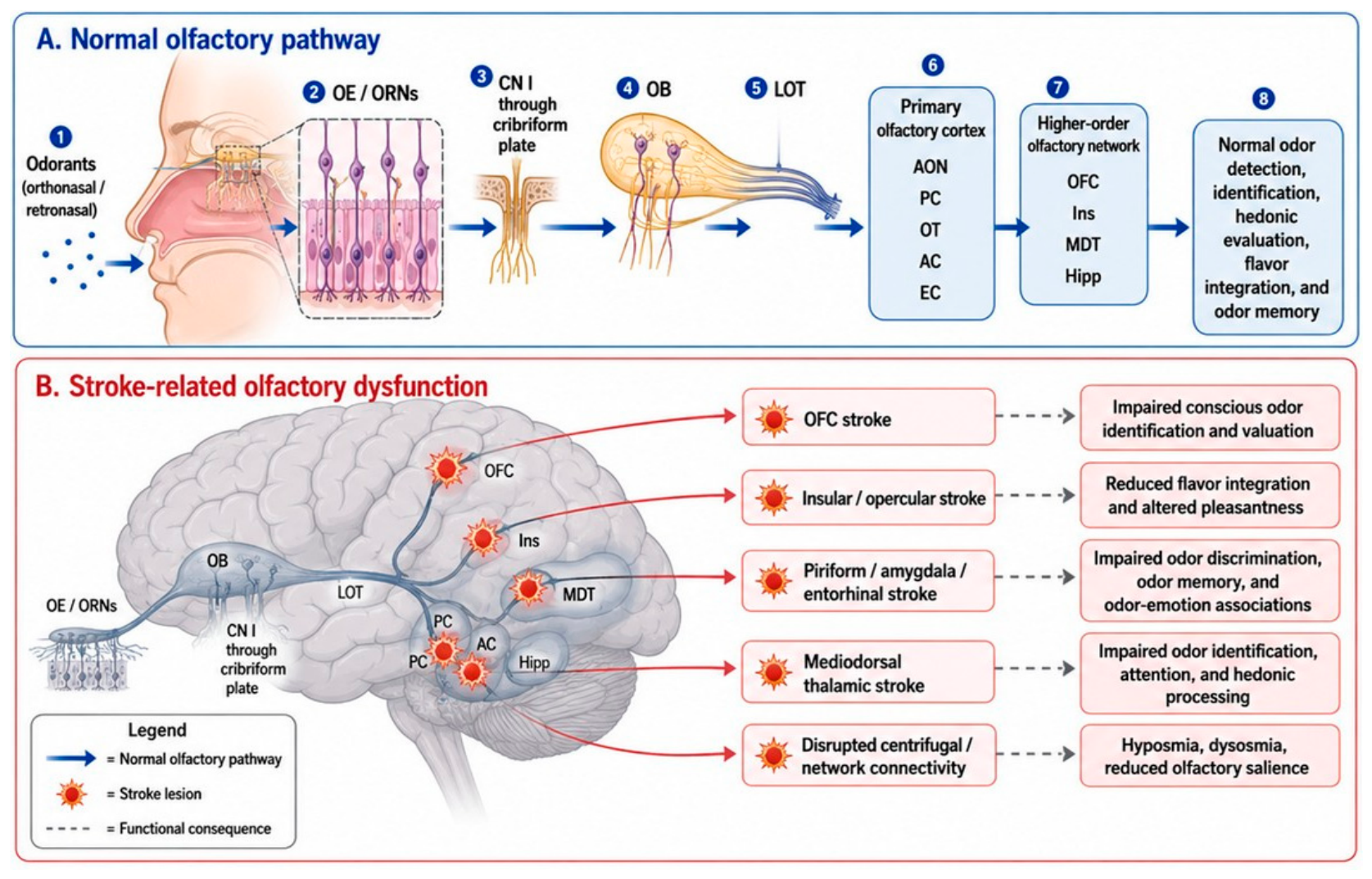
Figure 2.
Stroke lesion location predicts distinct olfactory phenotypes. Olfactory impairment after stroke is not a single clinical entity. Lesions affecting different parts of the olfactory network may produce distinct patterns of dysfunction, including impaired odor detection, odor discrimination, odor identification, hedonic evaluation, flavor integration, odor memory, or odor-guided behavior. Orbitofrontal lesions are expected to affect conscious odor identification and valuation; insular and opercular lesions may affect flavor integration and odor pleasantness; piriform, amygdaloid, entorhinal, and hippocampal lesions may affect odor discrimination, odor-emotion associations, and odor memory; and mediodorsal thalamic lesions may impair odor identification, attention, and hedonic processing despite relatively preserved detection. This lesion-to-phenotype framework may help distinguish peripheral smell loss from central olfactory network dysfunction after stroke. EC - entorhinal cortex, Hipp – hippocampus, Ins – insula, MDT - mediodorsal thalamus, OFC – orbitofrontal cortex.
Figure 2.
Stroke lesion location predicts distinct olfactory phenotypes. Olfactory impairment after stroke is not a single clinical entity. Lesions affecting different parts of the olfactory network may produce distinct patterns of dysfunction, including impaired odor detection, odor discrimination, odor identification, hedonic evaluation, flavor integration, odor memory, or odor-guided behavior. Orbitofrontal lesions are expected to affect conscious odor identification and valuation; insular and opercular lesions may affect flavor integration and odor pleasantness; piriform, amygdaloid, entorhinal, and hippocampal lesions may affect odor discrimination, odor-emotion associations, and odor memory; and mediodorsal thalamic lesions may impair odor identification, attention, and hedonic processing despite relatively preserved detection. This lesion-to-phenotype framework may help distinguish peripheral smell loss from central olfactory network dysfunction after stroke. EC - entorhinal cortex, Hipp – hippocampus, Ins – insula, MDT - mediodorsal thalamus, OFC – orbitofrontal cortex.
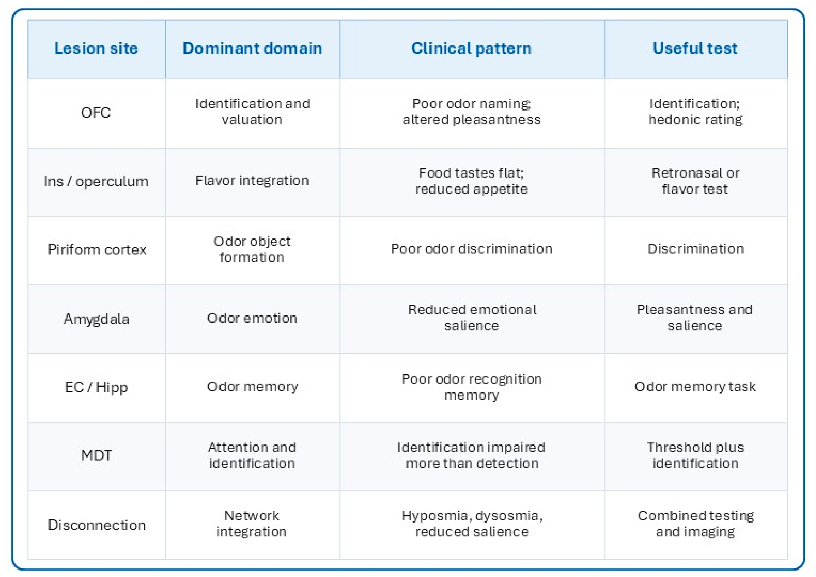
Figure 3.
Orthonasal and retronasal olfaction, taste pathways and flavor dysfunction after stroke. Odorants reach the olfactory system through two principal routes. In orthonasal olfaction, odorants enter the nasal cavity during sniffing. In retronasal olfaction, food volatiles released during eating pass from the oral cavity to the nasal cavity and stimulate the same olfactory apparatus. Thus, both orthonasal and retronasal odorants reach the olfactory epithelium (OE) and then follow the shared olfactory pathway through the olfactory bulb (OB) and primary olfactory cortex. In parallel, taste information travels from taste buds to the nucleus of the solitary tract (NTS), then to the ventral posteromedial thalamic nucleus (VPM), and onward to the insula/operculum, where gustatory processing contributes to flavor perception. Olfactory and gustatory information converge in higher-order flavor networks involving the insula/operculum, orbitofrontal cortex (OFC), amygdala, hypothalamus, and hippocampus. Stroke affecting these pathways may cause reduced environmental odor detection, impaired retronasal flavor perception such that food “tastes bland,” altered pleasantness, dysosmia or parosmia, reduced appetite, and impaired food-related quality of life. NTS - nucleus of the solitary tract, OB - olfactory bulb, OE - olfactory epithelium, OFC - orbitofrontal cortex, Primary OC – primary olfactory cortex, QoL - quality of life, VPM - ventral posteromedial thalamic nucleus.
Figure 3.
Orthonasal and retronasal olfaction, taste pathways and flavor dysfunction after stroke. Odorants reach the olfactory system through two principal routes. In orthonasal olfaction, odorants enter the nasal cavity during sniffing. In retronasal olfaction, food volatiles released during eating pass from the oral cavity to the nasal cavity and stimulate the same olfactory apparatus. Thus, both orthonasal and retronasal odorants reach the olfactory epithelium (OE) and then follow the shared olfactory pathway through the olfactory bulb (OB) and primary olfactory cortex. In parallel, taste information travels from taste buds to the nucleus of the solitary tract (NTS), then to the ventral posteromedial thalamic nucleus (VPM), and onward to the insula/operculum, where gustatory processing contributes to flavor perception. Olfactory and gustatory information converge in higher-order flavor networks involving the insula/operculum, orbitofrontal cortex (OFC), amygdala, hypothalamus, and hippocampus. Stroke affecting these pathways may cause reduced environmental odor detection, impaired retronasal flavor perception such that food “tastes bland,” altered pleasantness, dysosmia or parosmia, reduced appetite, and impaired food-related quality of life. NTS - nucleus of the solitary tract, OB - olfactory bulb, OE - olfactory epithelium, OFC - orbitofrontal cortex, Primary OC – primary olfactory cortex, QoL - quality of life, VPM - ventral posteromedial thalamic nucleus.
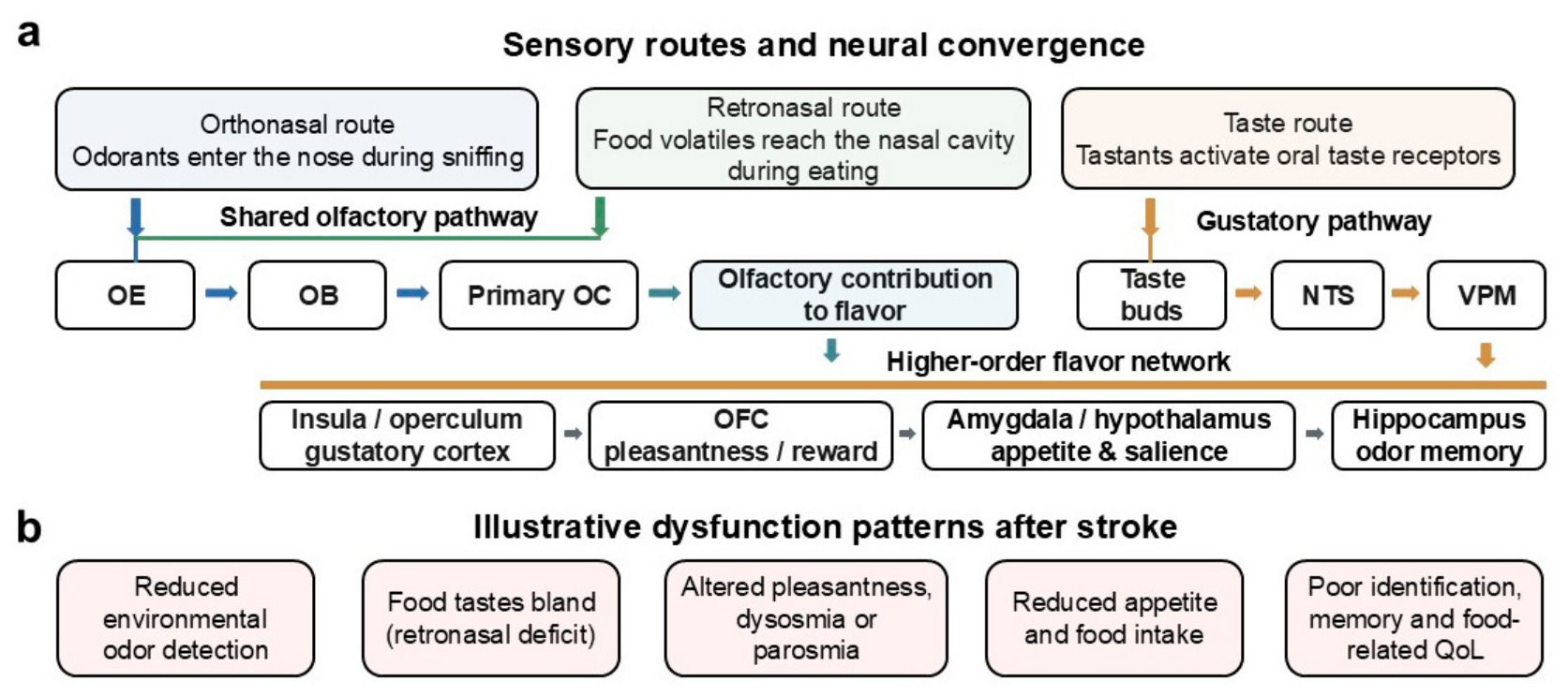
Figure 4.
Proposed clinical screening and testing algorithm for olfactory dysfunction after stroke. Because olfactory dysfunction after stroke may be underrecognized, clinical evaluation should include both symptom screening and objective testing. Patients should be asked about smell loss, distorted odors, reduced food enjoyment, reduced appetite, impaired detection of smoke or spoiled food, and changes in flavor perception. Non-stroke causes of olfactory dysfunction, including sinonasal disease, viral illness, head trauma, medication effects, and neurodegenerative disease, should be considered. Objective testing can then separate odor threshold (odor sensitivity), discrimination (same vs. different odor judgement), identification (odor naming or recognition), pleasantness, retronasal olfaction (odor perception through the mouth/nasopharynx), and odor memory. The resulting profile may help distinguish peripheral-like smell loss from central deficits involving orbitofrontal, insular, limbic, thalamic, or network-level dysfunction. Management may include patient education, safety counseling, nutrition support, olfactory training, follow-up testing, and referral when appropriate.
Figure 4.
Proposed clinical screening and testing algorithm for olfactory dysfunction after stroke. Because olfactory dysfunction after stroke may be underrecognized, clinical evaluation should include both symptom screening and objective testing. Patients should be asked about smell loss, distorted odors, reduced food enjoyment, reduced appetite, impaired detection of smoke or spoiled food, and changes in flavor perception. Non-stroke causes of olfactory dysfunction, including sinonasal disease, viral illness, head trauma, medication effects, and neurodegenerative disease, should be considered. Objective testing can then separate odor threshold (odor sensitivity), discrimination (same vs. different odor judgement), identification (odor naming or recognition), pleasantness, retronasal olfaction (odor perception through the mouth/nasopharynx), and odor memory. The resulting profile may help distinguish peripheral-like smell loss from central deficits involving orbitofrontal, insular, limbic, thalamic, or network-level dysfunction. Management may include patient education, safety counseling, nutrition support, olfactory training, follow-up testing, and referral when appropriate.
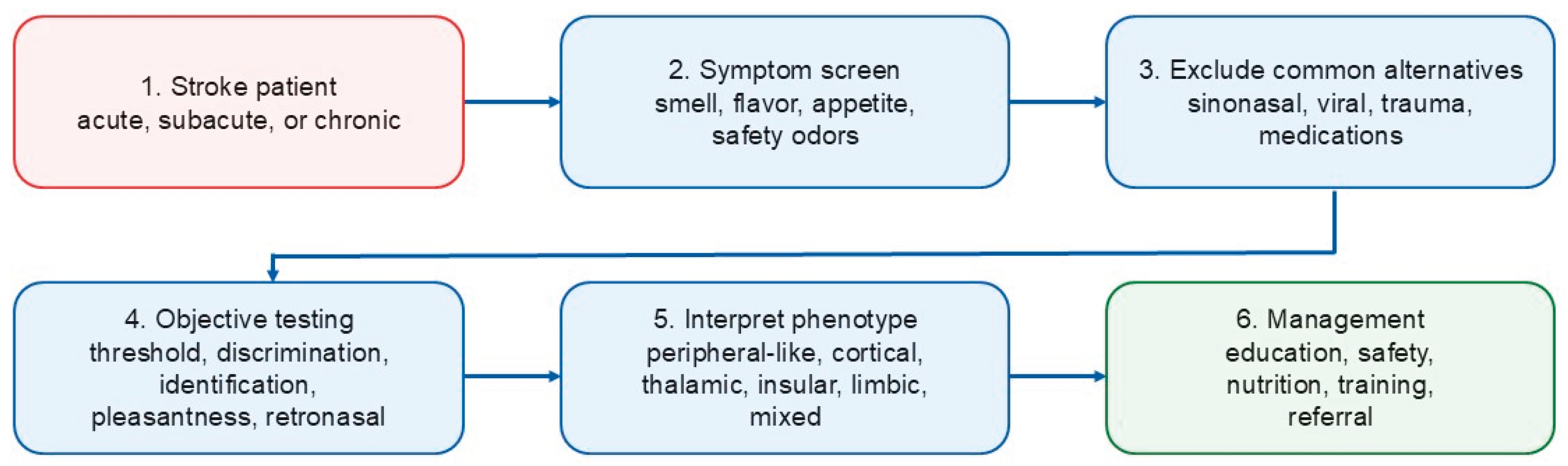
Figure 5.
Research gaps and proposed framework for future studies of stroke and olfaction. Current studies of post-stroke olfactory dysfunction are limited by heterogeneous stroke populations, small sample sizes, underuse of objective olfactory testing, limited assessment of retronasal olfaction and flavor, incomplete lesion-symptom mapping, and sparse longitudinal or interventional data. Future studies should combine acute and chronic stroke cohorts with structural MRI, perfusion imaging, lesion segmentation, olfactory threshold, discrimination, identification, pleasantness, retronasal flavor testing, cognitive and language assessment, mood and appetite measures, quality-of-life instruments, and longitudinal follow-up. This approach could identify lesion-specific olfactory phenotypes, clarify mechanisms of recovery or persistence, and guide screening, counseling, and rehabilitation strategies. MRI - magnetic resonance imaging, QoL - quality of life, lesion-symptom mapping - linking lesion sites to behavioral deficits, connectivity analysis - structural or functional network analysis.
Figure 5.
Research gaps and proposed framework for future studies of stroke and olfaction. Current studies of post-stroke olfactory dysfunction are limited by heterogeneous stroke populations, small sample sizes, underuse of objective olfactory testing, limited assessment of retronasal olfaction and flavor, incomplete lesion-symptom mapping, and sparse longitudinal or interventional data. Future studies should combine acute and chronic stroke cohorts with structural MRI, perfusion imaging, lesion segmentation, olfactory threshold, discrimination, identification, pleasantness, retronasal flavor testing, cognitive and language assessment, mood and appetite measures, quality-of-life instruments, and longitudinal follow-up. This approach could identify lesion-specific olfactory phenotypes, clarify mechanisms of recovery or persistence, and guide screening, counseling, and rehabilitation strategies. MRI - magnetic resonance imaging, QoL - quality of life, lesion-symptom mapping - linking lesion sites to behavioral deficits, connectivity analysis - structural or functional network analysis.
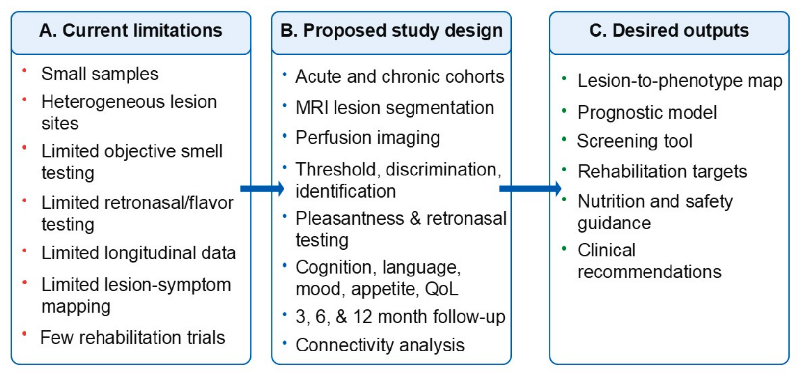
Disclaimer/Publisher’s Note: The statements, opinions and data contained in all publications are solely those of the individual author(s) and contributor(s) and not of MDPI and/or the editor(s). MDPI and/or the editor(s) disclaim responsibility for any injury to people or property resulting from any ideas, methods, instructions or products referred to in the content. |
© 2026 by the authors. Licensee MDPI, Basel, Switzerland. This article is an open access article distributed under the terms and conditions of the Creative Commons Attribution (CC BY) license (http://creativecommons.org/licenses/by/4.0/).
Copyright: This open access article is published under a Creative Commons CC BY 4.0 license, which permit the free download, distribution, and reuse, provided that the author and preprint are cited in any reuse.


