

Submitted:
17 July 2026
Posted:
21 July 2026
You are already at the latest version
Abstract
Neonatal hypoxic-ischemic encephalopathy (HIE) remains one of the leading causes of neonatal mortality and long-term neurodevelopmental disability despite therapeutic hypothermia, which provides only partial neuroprotection. Among adjunctive therapies, magnesium sulphate (MgSO4) has emerged as one of the most biologically plausible neuroprotective agents because of its ability to modulate glutamate-mediated excitotox-icity, intracellular calcium influx, oxidative stress, neuroinflammation and apoptotic pathways. Nevertheless, encouraging molecular and preclinical findings have not translated into consistent clinical benefit.
This narrative review critically examines the translational gap between the molecular mechanisms of magnesium sulphate and its clinical performance in neonatal HIE. Evi-dence from experimental models, clinical studies and recent meta-analyses was integrated to identify the biological and methodological factors potentially responsible for this dis-crepancy. We discuss the evolving pathophysiology of HIE across the primary, latent, secondary and tertiary phases of brain injury and analyse how the timing of intervention, lesion heterogeneity and inadequate biological stratification may influence therapeutic responsiveness.
Current clinical research has largely evaluated broad neurological outcomes, particularly cerebral palsy, despite the heterogeneous neuropathological substrates underlying ne-onatal brain injury. We argue that this strategy may dilute genuine treatment effects by grouping together distinct lesion phenotypes with different biological mechanisms. Accordingly, we propose the **Neuroprotection per Effective Therapy (NET)** framework, a conceptual translational model integrating molecular targets, experimental evidence, MRI-defined lesion phenotypes, methodological quality and advanced statistical ap-proaches to improve patient stratification and outcome selection.
Rather than questioning the biological efficacy of magnesium sulphate itself, this review suggests that future progress will depend on aligning molecular mechanisms with clinically meaningful phenotypes. Precision-based translational strategies may ultimately allow magnesium sulphate and other neuroprotective therapies to demonstrate their true therapeutic potential in neonatal HIE.
Keywords:
hypoxic-ischemic encephalopathy
; magnesium sulphate
; neuroprotection
; excitotoxicity
; NMDA receptor
; neuroinflammation
; therapeutic hypothermia
; translational medicine
1. Introduction
1.1. Neonatal HIE as an Unmet Clinical Challenge
Neonatal hypoxic-ischemic encephalopathy (HIE) is currently a major global challenge. The Global Burden of Disease study highlighted that in 2021 it has been among the ten neurological conditions contributing to the highest age-standardized disability adjusted life years (DALYs) [1]. Furthermore, its diagnosis could be difficult in the absence of sentinel events and radiological signs (MRI) [2,3,4]. Besides various attempts to explore the distinctive and predictive HIE pathophysiology (placental markers, changes in bilirubin, continuous glucose monitoring, protein S100, urine markers of abnormal MRI features) [5,6,7,8,9,10], neuroprotective drugs are increasingly being investigated to reduce and prevent brain injury incidence in relation with mortality and negative neurodevelopmental outcome [11,12,13,14].
1.2. Limitations of Therapeutic Hypothermia
Therapeutic hypothermia remains the standard of care. Researchers focused mainly on how to extend the benefits of neonatal populations and neuroprotective effects [15]. However, it does not necessarily protect neonates from brain injury after HIE as described by Ghi et al. [16] despite the promising results emerged in the TOBY trial [17,18]. The optimal timing of MRI imaging for long-term prognostic purposes remains unclear, as reviewed by Flyger et al. [19]. Moreover, hypothermia could influence interpretation of post-therapeutic signs in MRI. For instance, Zhuang et al. [20] included routine treatment of severe neonatal HIE as a limitation of their radiomic study aiming to detect venous signature of this pathology because of hypothermia-related changes in images and metabolites.
1.2. Why Magnesium Sulphate?
Over the past three decades, magnesium sulphate has progressively emerged as one of the most biologically plausible neuroprotective agents. However, biological plausibility alone may be insufficient to guarantee clinical efficacy. Since HIE in term infants mirrors a progressive cascade of excito-oxidative events unfolding in the brain after an asphyxial insult, Johnston et al. recommended in 2011 to identify additional medications adjuvating hypothermia [21]. Above neuroprotective drugs, magnesium sulphate (MgSO4) is considered as a promising neuroprotective strategy [22], especially before maternal labour [23]. Chemically, magnesium is an ionized mineral essential to hundreds of enzymatic processes, including hormone receptor binding, energy metabolism, muscle contractility as well as neuronal and neurotransmitter function [24] while sulphate is a crucial anion implicated in modulating exogen, polysaccharide chains of proteoglycans, cholesterol and its derivates, and tyrosine residues of a myriad of proteins [25]. Preclinical in vivo studies in neonatal rodent models have consistently demonstrated that magnesium sulphate attenuates excitotoxic and inflammatory pathways after hypoxic-ischemic injury, thereby providing the biological rationale for its subsequent clinical evaluation [26]. However, decades of research showed that preclinical studies should be more rigorous [27]. Experimental evidence accumulated progressively during the late 1980s [28], and throughout the 1990s, establishing magnesium sulphate as one of the earliest candidate neuroprotective agents for neonatal brain injury, despite some scientific accidents described by the group of Levene et al. [29] in which neonates received 150 mg of MgSO4 that revealed itself as a bolus of 250 mg when diluting it in water. Importantly, the publication by Levene et al. certified the foundation of the international network for neuroprotective evaluation after severe birth asphyxia [30] in 1999. Maternal antenatal administration of magnesium sulphate prior to preterm delivery (before 32 or 34 weeks) demonstrated a reduction of cerebral palsy (CP) incidence (a motor condition) in trials and meta-analyses [31,32,3334]. Notwithstanding these encouraging results [35,36], current data are suffering from selective reporting bias and pathophysiology misalignments: magnesium sulphate administration is not sufficient to provide a complete neuroprotective care according to survey studies [37], and its effect on the reduction of intraventricular haemorrhage (IVH) is uncertain [38]. Furthermore, pathophysiological evidence demonstrates the CP could result from diverse brain injury per phenotype and epidemiological time-dependent incidence (haemorrhagic lesions like intraventricular and cerebellar haemorrhage; ischemic lesions like periventricular leukomalacia and white matter injury) [39,40,41,42,43,44]. For instance, Calandrino et al. [42] reported cerebellar disruptions in preterm neonates with germinal matrix and intraventricular haemorrhages. Conversely, antenatal magnesium sulphate does not accelerate white matter maturation in preterm neonates at term-equivalent age (TEA) [45] and in a further Canadian study, functional connectivity is enhanced at TEA [46]. Hence, these groups of lesions lead to the white matter necrosis [47,48]. The inconsistencies in antenatal magnesium sulphate effects may reflect inadequate biological stratification [49]. Accordingly, a target-unified magnesium therapy could not be appropriate without considering molecular pathways and pathophysiological patterns. Hence, the neuroprotective effect of antenatal magnesium sulphate in early preterm newborns cannot automatically be extrapolated to term newborns with HIE. To the best of our knowledge, no review is available on substantially exploring the gap between molecular biology of magnesium sulphate and HIE pathophysiology.
1.3. Aim of the Review
To critically examine why strong molecular neuroprotective effects have not translated into consistent clinical benefit.
A narrative review was considered the most appropriate approach because the topic encompasses complex translational questions that require integration of molecular, preclinical and clinical evidence rather than quantitative synthesis alone [50].
2. Pathophysiology of Hypoxic-Ischemic Brain Injury
2.1. Primary Energy Failure
The pathogenesis of hypoxic-ischemic injury is complex and multifactorial, focusing primarily on cellular and molecular mechanisms [51]. To better understand the neuroprotective role of magnesium sulphate, it is important to describe the molecular pathophysiology of HIE and its links with MgSO4 pharmacodynamics [3,13,52,53]. The hypoxic-ischemic injury is characterized by three stages: latent, secondary and tertiary [54] as illustrated in Figure 1. Specifically, during the primary phase of hypoxic-ischemic injury, depletion of intracellular ATP disrupts ionic homeostasis, causing membrane depolarization and excessive extracellular accumulation of glutamate. Sustained activation of NMDA receptors results in pathological calcium influx, which initiates multiple downstream cascades including mitochondrial dysfunction, oxidative stress, activation of calcium-dependent enzymes and programmed cell death. These processes constitute the core of glutamate-mediated excitotoxicity and represent one of the earliest therapeutic targets in neonatal HIE. Since Mg2+ physiologically blocks the NMDA receptor channel in a voltage-dependent manner, this pathway represents one of the earliest and most biologically plausible therapeutic targets in neonatal HIE. If excitotoxicity predominates, NMDA blockade may theoretically exert greater benefit than in infants whose injury is primarily driven by inflammation or delayed repair failure.
2.2. Latent Phase
During profound hypoxia-ischemia, brain cells might die, although some could recover oxidative metabolism. This period is defined as “latent” in which neural metabolism and activity are suppressed [55]. Thereafter, after moderate to severe hypoxia-ischemia, this transient recovery is followed by a 6 h phase of secondary deterioration, with delayed seizures, failure of mitochondrial function, cytotoxic edema, and cell death over 72 h.
2.3. Secondary Energy Failure
Progression of acute cerebral hypoxia-ischemia to an inflammatory response is a major contributor to the pathophysiology of neonatal brain injury [56]. As portraited by Li et al. [56], hypoxia-ischemia elicits an intravascular inflammatory cascade. Subsequently, the activation of resident immune cells and the cerebral infiltration of peripheral immune cells response to cellular damages in the brain parenchyma amplyfing the effects of the on-course inflammation. Thus, this protracted neuroinflammation brings about a secondary brain tissue injury. Indeed, the secondary energy failure could be generated by multiple neurotoxic phenomena like oxidative stress, mitochondrial dysfunction, neuroinflammation and apoptosis [12,13,57]. Specifically, oxidative stress is a crucial dimension for detecting potential brain susceptibility to neural damage [58,59]. To demonstrate how it is important to consider a multisystem approach in neuroprotective strategies—including MgSO4 administration—the crosstalk between neonatal lung, heart and brain is linked to apoptosis, glutamate signaling, oxidative stress, breakdown of the blood brain barrier and pro-inflammatory cytokines which could be interconnected with inflammatory pulmonary state and low cardiac output [60].
2.4. Tertiary Injury
Tertiary injuries could occur in presence of persistent inflammation and impaired repair mechanisms leading to neonatal seizures, [55,12,61]. Some outcomes include sensitization to inflammation or injury, incremented seizure susceptibility, persistent inflammation and gliosis, impaired oligodendrocyte maturation and myelination, altered proliferation and synaptogenesis, and epigenetic alteration [62]. This phase could last between weeks or years after perinatal injury [55]. Although rehabilitation is one step of the main therapeutic path fostering neuroplasticity [63], current evidence is still limited because of small sample size in the majority of studies. In this sense, intervention timing during tertiary injury is crucial for preventing further lesions. Davidson et al. [55] described in a masterclass way the underlying cellular phenomena. Largely thanks to neurodegeneration models, endogenous brain plasticity could be impaired by protracted pro-inflammatory activation (M1) of microglia [64,65], and sustained activation of astrocytes (A1) causing gliosis. These phenomena are opposed to neuroprotective and boost repair activation (M2 and A2, respectively for microglia and astrocytes) [66,67]. Beyond the increasingly diffusion of pro-inflammatory factors damaging neighboring cells and endogenous plasticity mechanisms, M1 could not manage physiological neurodevelopmental functions (i.e., synaptic pruning and control of synaptic functioning) [68]. Noteworthy, some drugs may enhance M2 over M1 activition like pioglitazone, metformin, and resveratrol [69,70,71,72]. Interestingly, evidence is increasingly developing on the conditioning power of inflammation on the integrated stress response in male fetal mice. there is emerging evidence that inflammation can affect the integrated stress response only in male fetal mice. Deterring this response improved neurobehavioral outcomes in male but not female offspring [73]. Therefore, possible sex-specific effects might modulate the inflammation process in the tertiary phase. Accordingly, drug neuroprotection might be tailored according to the timing and phase of hypoxic-ischemic injury with the available and evidence-grounded therapies [54].
3. Molecular Basis of Magnesium Sulphate Neuroprotection
Analogously to adult cohorts, N-methyl-D-aspartate (NMDA) receptors are involved in the excitotoxicity induced by hypoxia-ischemia in HIE. GluN2A- and GluN2B-containing NMDA receptor subtypes have distinct roles in HIE pathophysiology [52]. MgSo4 modulates immunological activity [74]. Indeed, it inhibits cellular calcium influx and excitatory amino acid release in neurons via blockade of the NMDA-receptor channel, which is provided by a magnesium-dependent calcium gate [75,76]. As described by Nowak et al. [76], the properties of Mg2+-free solutions, L-glutamate, L-aspartate and NMDA open cation channels are voltage independent. When Mg2+ is present, the probability of opening of channels decreases as the single-channel currents at resting potential disintegrates themselves. Hyperpolarization increments both effects, sloping negatively the I-V relationship of the glutamate response. The voltage dependence of the NMDA receptor-linked conductance may be derived from the voltage dependence of the Mg2+ block. Accordingly, its interpretation does not necessarily imply an intramembrane voltage-dependent gate.
Reviewing the most recent literature, Okazaki et al. concluded that combining hypothermia and further medications such erythropoietin and melatonin is becoming an increasingly popular approach for neonates with perinatal asphyxia [77]. However, magnesium sulphate in combination with hypothermia did not reduce mortality in a randomised controlled trial in India [78].
4. Clinical Evidence in Neonatal HIE
4.1. Early Pilot Studies
Pilot studies were aimed to assess safety and feasibility of administering neuroprotective drugs in HIE neonates in multiple combinations (erythropoietin, magnesium sulphate, hypothermia) [79]. Concerning magnesium sulphate, in an Indian prospective, longitudinal, placebo-controlled trial, postnatal magnesium sulphate treatment improved neurologic outcomes at discharge for term neonates with severe perinatal asphyxia [80]. Although no adverse effects are associated with magnesium sulphate according to a systematic review of a randomized trials settled in low and middle-income countries [81], few studies reported an increase in neonatal apnoea requiring ventilation such as in the trial by Bhat et al. [80] who identified two infants with this condition. Nonetheless, more robust, multicentric randomized controlled trials are needed to confirm the potential of the wide spectrum of neuroprotective drugs including magnesium sulphate. Indeed, multicentre randomized double-blind controlled trials such as the PENUTS study [82] in extremely preterm neonates, showed no significant differences between erythropoietin and placebo in the rates of retinopathy of prematurity, intracranial haemorrhage, sepsis, necrotizing enterocolitis, bronchopulmonary dysplasia, death or in the frequency of serious adverse events; moreover, high-dose erythropoietin treatment administered to extremely preterm infants from 24 h after birth through 32 weeks of postmenstrual age did not result in a lower risk of severe neurodevelopmental impairment or death at 2 years of age.
4.2. Recent Meta-Analyses and Systematic Reviews
Meta-analysis represents one of the highest tier of the evidence pyramid [83] together with randomized trials and systematic reviews. In this review, the evidence on magnesium sulphate is fragmented into multiple categories of methodological strength connected with statistical significancy. Analysing 20 randomized trials (total sample size: 1485), Gowda et al. [84] showed that magnesium sulphate could improve neurological outcomes in settings where therapeutic hypothermia is not available, but it does not reduce mortality. This meta-analysis is an illustrative example of what we define the “Ping-Pong effect”, i.e., the recurrent shift of statistical significance from one clinical endpoint to another across successive randomized trials and meta-analyses, resulting in inconsistent perceptions of therapeutic efficacy despite a stable biological rationale [49].
5. The Translational Paradox
5.1. Why Does Molecular Efficacy Not Translate into Clinical Benefit?
Magnesium sulphate literature is a text-book example of disappointing efficacy in clinical research despite preclinical evidence. Regarding term neonates with hypoxic lesions, Gressens et al. [85] argued a misalignment between in-vitro models and in-vivo phenotype. In international research on HIE, mice studies underscored the evidence of molecular and excito-oxidic pathways, but they failed in clinical trials on human neonates [86] despite the neonatal brain being susceptible to excitotoxicity because of its immaturity [23]. Besides, neonatologists do not know when an hypoxic-ischemic event could onset in the human neonates rather than in mice [87]. Recent observational studies have progressively incorporated biologically grounded hypotheses and clinically defined high-risk populations, partially addressing the translational gap. However, patient stratification still relies predominantly on clinical characteristics rather than molecular endotypes or biomarker-guided precision approaches [88].
5.2. The Neuroprotection per Effective Therapy (NET) Framework: A Conceptual Proposal for Translational Neonatal Neuroprotection
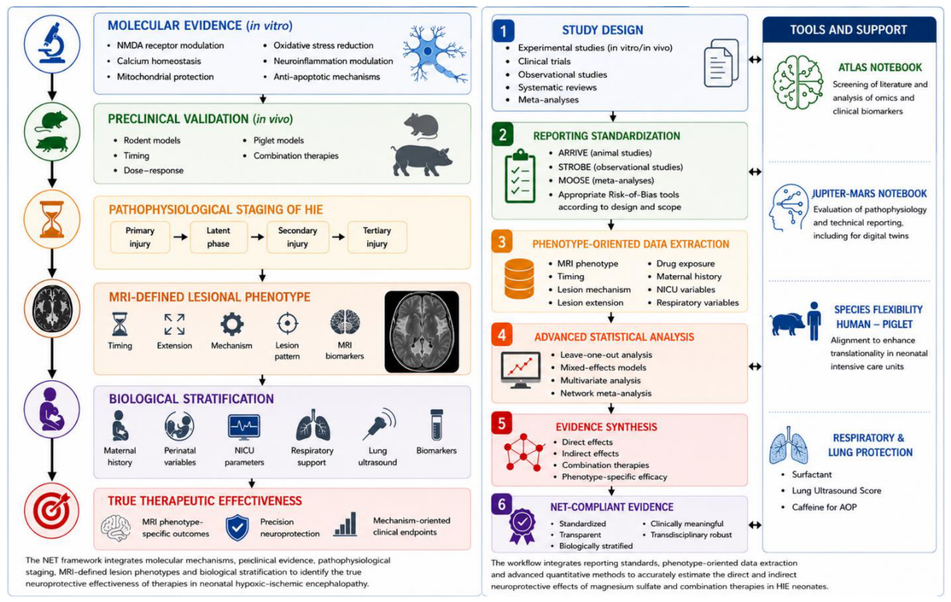
Considering the neuroprotective drug pitfalls underscored by Gressens and Galinsky [49,85], we propose the NET framework (Neuroprotection per Effective Therapy) in Figure 2, consisting in a vectorial structure from in-vitro evidence to in-vivo MRI phenotype as clinical endpoints, mediated by defining MRI lesional phenotype per timing, extension and mechanism using appropriate statistical methods to quantify authentic drug effectiveness. If feasible, omics analysis could be included as molecular biomarkers, as we will be describing in the section number 7. NET is a proposal for enhancing precision neuroprotection integrating imaging, clinical biomarkers and biological phenotype helping neonatologists and healthcare providers to guide treatment decision. Future evidence synthesis should incorporate biologically informed lesion phenotypes and sensitivity analyses (e.g., leave-one-out procedures) to better identify studies contributing disproportionately to heterogeneity (effect size and quantitative evidence synthesis). Further studies should integrate clinical, imaging and biological variables within multivariable analytical frameworks [89]. These approaches may clarify whether magnesium sulphate exerts differential effects according to lesion phenotype, treatment timing, respiratory status and concomitant neuroprotective interventions. Network meta-analysis may further help compare combination strategies involving hypothermia and adjunctive therapies [90,91,92,93]. In this way, researchers could also approach magnesium sulphate experimentations in alignment with species flexibility between human and animal models, shaping the translationality of neonatological research [90]. Studies implementing the NET framework should adhere to internationally accepted reporting standards according to study design (e.g., STROBE, MOOSE and ARRIVE) [91,92,93], ensuring methodological transparency and reproducibility. The clinical dimensions (Risk of Bias and reporting assessment) should consider the appropriate checklists according to the design and scope. In such manner, methodology and reporting may be more standardized and transparent to the benefit of pharmacological research on MgSO4, overcoming the limitation that some neural anti-inflammatory therapies studies did not clearly state whether they controlled for a number of potential biases [94].
5.3. Tools for Applying NET Principles
To perform a preliminarily screening of literature in artificial intelligence (AI) applied to imaging, radiomics and clinical biomarkers (if necessary), we have developed the ATLAS Notebook. Being benchmarked on multiple papers on adult oncology, pediatric oncology and neonatology, it extracts directly from Portable Document Format files relevant keywords associated with standard checklists [95]. Additionally, to evaluate pathophysiology reporting even for digital twins (virtual replicas of organs, patients and biological systems to inform clinical decisions) [96], the JUPITER-MARS Notebook [97] could assist healthcare researchers to evaluate both pathophysiology and technical reporting analogously to the ATLAS Notebook.
5.4. Failure of “One Drug-One Mechanism” Approaches
HIE is a multifactorial disease as much as magnesium sulphate is a multieffect drug. We mean that magnesium sulphate effect is diluted into a major outcome-container named “Cerebral Palsy” (CP), leading to a more prominent “pathology of evidence” contaminating both primary study results and systematic reviews. With NET, we suggest considering lesional pattern according to onset timing and imaging signs. Analogously, Brenner et al. [98] analysed the trial CRASH-3 on the neuroprotective effect of tranexamic acid (TXA) in adult intensive care patients with head traumas. Crucially, TXA effects were diluted into a further outcome-container, that is, all-cause mortality reduction. Similarly to our approach, Brenner et al. underscored how effective neuroprotection was dependent on TXA administration timing considering the underlying mechanism of haemorrhage.
6. Towards MRI Biomarkers of Neuroprotective Drug Response
This review has depicted how molecular pathways are helpful for understanding HIE pathophysiology and magnesium sulphate effects. However, as previously remarked, the current contribution is not sufficient to provide a complete knowledge of its neuroprotective role. Furthermore, omics and molecular biology labs are not available in every hospital. Therefore, a more feasible approach is needed to balance current evidence and medical research instruments. For instance, imaging could be a potential biomarker source rather than a monitoring tool alone. Radiomics is one potential strategy to study pathology and pathophysiology of HIE [99,100,101,102]. Additionally, the informatic ComBat MRI harmonization [103,104] represents a further strategy to both overcome scanner bias across research centres in MRI diffusion tensor imaging (DTI), and enlarge neonatal cohorts to analyse neuroprotective drugs effects [105]; this strategy is being employed in the ACUMEN study [106] on melatonin administration for moderate and severe HIE neonates treated with hypothermia.
7. Towards Molecular Biomarkers of Neuroprotective Response: The Omics Contribution
As radiomics approach could be more generalizable when combining with a molecular analysis (genomics, etc.) as indicated by the specific Radiomic Quality Score item [107], molecular biomarkers should not simply predict prognosis, but identify biological endotypes potentially responsive to specific neuroprotective interventions.in integration with the preexisting radiological and clinical features [108] as previously seen in neonatal mice [109].
Whilst proteomics, lipidomics, transcriptomics and epigenomics are promising and less validated in the clinical use, metabolomics remains the most mature approach despite reduced sample size numerosity, standardization and external validation per study [110]. Based on the previous research [111,112], these studies identify alterations that are coherent with mitochondrial energetic deficit, oxidative stress, amino acids metabolism, purines, acylcarnitine and lipids. Some models combined blood cord or in integration with plasma and urine metabolites, mass spectrometry and MRI and other clinical parameters to improve HIE or further brain injuries stratification on MRI as showed by O’Boyle et al. Locci et al., respectively [113,114]. Particularly, Locci et al. revealed lactates, succinates, hypoxanthine, amino acids and acylcarnitine as biomarkers; nonetheless, this methodology is not replicable across centres. In order to capture the metabolomic signature varying per the HIE-induced brain injury severity [115], underlying neurological process that could be not necessiraly reflected on MRI [116], and the plastic nature of metabolomic signature of being modified across the first two hours of life by treatment and disease [112], one replicable, feasible and non-invasive analysis is on the urine samples. Studies by Solevåg et al. on oxidative stress and Pineiro et al. concerning HIE metabolome underscored, respectively, that this approach could a serial monitoring strategy before and after hypothermia treatment without serial blood samples [117,118]. As such, lipidomics could be a promising integrative framework with metabolomics. Indeed, Nixon et al. [119] founded lipidomic signatures on dried blood spots in hypothermia-treated, HIE neonates as they may provide biomarkers related to membrane and myelinization damages.
8. Conclusions
Magnesium sulphate remains one of the most biologically plausible neuroprotective agents in neonatal HIE. However, the discrepancy between molecular efficacy and clinical outcomes highlights the complexity of translational neuroprotection and underscores the need for precision-based therapeutic strategies.
Author Contributions
MEC: Conceptualization; Writing—Draft; Writing—Review and Editing; Reference retrieval. FP: Writing—Review and Editing; Validation; PS: Writing—Review and Editing; Validation. LAR: Conceptualization; Writing—Review and Editing; Validation; Supervision.
Acknowledgments
We thank international communities for providing evidence with their precious works.
Conflicts of Interest
The authors declare no conflict of interest.
References
- Global, regional, and national burden of disorders affecting the nervous system, 1990–2021: A systematic analysis for the Global Burden of Disease Study 2021. Lancet Neurol. 2024, 23, 344–381. [CrossRef]
- Cimino, C.; Sortino, V.; Sapuppo, A.; Saporito, M.A.N.; Scarlata, F.; Falsaperla, R. Beyond hypoxic–ischemic encephalopathy: Genetic insights and precision diagnosis in neonatal encephalopathies. Front Med. 2026, 13, 1868036. [Google Scholar] [CrossRef] [PubMed]
- Chakkarapani, E.; De Vries, L.S.; Ferriero, D.M.; Gunn, A.J. Neonatal encephalopathy and hypoxic-ischemic encephalopathy: The state of the art. Pediatr. Res. 2025, 98, 2444–2458. [Google Scholar] [CrossRef] [PubMed]
- Christoffel, K.; Mulkey, S.B. Shifting outlooks after neonatal encephalopathy in the era of therapeutic hypothermia. Pediatr. Res. 2025, 98, 2518–2529. [Google Scholar] [CrossRef] [PubMed]
- Dani, C.; Poggi, C.; Fancelli, C.; Pratesi, S. Changes in bilirubin in infants with hypoxic–ischemic encephalopathy. Eur. J. Pediatr. 2018, 177, 1795–1801. [Google Scholar] [CrossRef] [PubMed]
- Espinoza, M.L.; Brundler, M.A.; Hasan, S.U.; Mohammad, K.; Momin, S.; Al Shaikh, B.; et al. Placental pathology as a marker of brain injury in infants with hypoxic ischemic encephalopathy. Early Hum. Dev. 2022, 174, 105683. [Google Scholar] [CrossRef] [PubMed]
- Kalogeropoulou, M.S.; Thomson, L.; Beardsall, K. Continuous glucose monitoring during therapeutic hypothermia for hypoxic ischaemic encephalopathy: A feasibility study. Arch. Dis. Child Fetal Neonatal Ed. 2023, 108, 309–315. [Google Scholar] [CrossRef] [PubMed]
- Basu, S.K.; Salemi, J.L.; Gunn, A.J.; Kaiser, J.R. Hyperglycaemia in infants with hypoxic–ischaemic encephalopathy is associated with improved outcomes after therapeutic hypothermia: A post hoc analysis of the CoolCap Study. Arch. Dis. Child Fetal Neonatal Ed. 2017, 102, F299–F306. [Google Scholar] [CrossRef] [PubMed]
- Nadeem, M.; Murray, D.M.; Boylan, G.B.; Dempsey, E.M.; Ryan, C.A. Early blood glucose profile and neurodevelopmental outcome at two years in neonatal hypoxic-ischaemic encephalopathy. BMC Pediatr. 2011, 11, 10. [Google Scholar] [CrossRef] [PubMed]
- Bersani, I.; Gasparroni, G.; Bashir, M.; Aboulgar, H.; Mufeed, H.; Iskander, I.; et al. Early predictors of abnormal MRI patterns in asphyxiated infants: S100B protein urine levels. Clin. Chem. Lab. Med. (CCLM) 2022, 60, 1745–1752. [Google Scholar] [CrossRef] [PubMed]
- Chan, N.H.; Hawkins, C.C.; Rodrigues, B.V.; Cornet, M.; Gonzalez, F.F.; Wu, Y.W. Neuroprotection for neonatal hypoxic–ischemic encephalopathy: A review of novel therapies evaluated in clinical studies. Dev. Med. Child Neuro 2025, 67, 591–599. [Google Scholar] [CrossRef] [PubMed]
- Zhou, K.Q.; Dhillon, S.K.; Bennet, L.; Davidson, J.O.; Gunn, A.J. How do we reach the goal of personalized medicine for neuroprotection in neonatal hypoxic-ischemic encephalopathy? Semin. Perinatol. 2024, 48, 151930. [Google Scholar] [CrossRef] [PubMed]
- Dixon, B.; Reis, C.; Ho, W.; Tang, J.; Zhang, J. Neuroprotective Strategies after Neonatal Hypoxic Ischemic Encephalopathy. Int. J. Mol. Sci. 2015, 16, 22368–22401. [Google Scholar] [CrossRef] [PubMed]
- Dhillon, S.K.; Lear, C.A.; Galinsky, R.; Wassink, G.; Davidson, J.O.; Juul, S.; et al. The fetus at the tipping point: Modifying the outcome of fetal asphyxia. J. Physiol. 2018, 596, 5571–5592. [Google Scholar] [CrossRef] [PubMed]
- Zhou, T.; Liang, P.; Dai, N.; Ma, M.; Lin, J.; Zhu, J.; et al. Therapeutic hypothermia for neonates: A bibliometric analysis and visualization research. Front Neurol. 2025, 16, 1565749. [Google Scholar] [CrossRef] [PubMed]
- Ghi, T.; Mercuri, E.; Ramenghi, L.A.; De Luca, D. Uncertainty in brain imaging after therapeutic hypothermia. Lancet Child Adolesc. Health 2025, 9, 759–760. [Google Scholar] [CrossRef] [PubMed]
- Azzopardi, D.V.; Strohm, B.; Edwards, A.D.; Dyet, L.; Halliday, H.L.; Juszczak, E.; et al. Moderate Hypothermia to Treat Perinatal Asphyxial Encephalopathy. N. Engl. J. Med. 2009, 361, 1349–1358. [Google Scholar] [CrossRef] [PubMed]
- Rutherford, M.; Ramenghi, L.A.; Edwards, A.D.; Brocklehurst, P.; Halliday, H.; Levene, M.; et al. Assessment of brain tissue injury after moderate hypothermia in neonates with hypoxic–ischaemic encephalopathy: A nested substudy of a randomised controlled trial. Lancet Neurol. 2010, 9, 39–45. [Google Scholar] [CrossRef] [PubMed]
- Flyger, H.; Holdsworth, S.J.; Gunn, A.J.; Bennet, L.; Abbasi, H. Temporal dynamics of neonatal hypoxic–ischemic encephalopathy injuries on magnetic resonance imaging. Neural Regen. Res. 2025, 20, 3144–3150. [Google Scholar] [CrossRef] [PubMed]
- Zhuang, X.; Jin, K.; Li, J.; Yin, Y.; Dong, X.; Lin, H. A radiomics-based study of deep medullary veins in infants: Evaluation of neonatal brain injury with hypoxic-ischemic encephalopathy via susceptibility-weighted imaging. Front Neurosci. 2022, 16, 1093499. [Google Scholar] [CrossRef] [PubMed] [PubMed Central]
- Johnston, M.V.; Fatemi, A.; Wilson, M.A.; Northington, F. Treatment advances in neonatal neuroprotection and neurointensive care. Lancet Neurol. 2011, 10, 372–382. [Google Scholar] [CrossRef] [PubMed]
- Robertson, N.J.; Tan, S.; Groenendaal, F.; Van Bel, F.; Juul, S.E.; Bennet, L.; et al. Which Neuroprotective Agents are Ready for Bench to Bedside Translation in the Newborn Infant? J. Pediatr. 2012, 160, 544–552.e4. [Google Scholar] [CrossRef] [PubMed]
- Lingam, I.; Robertson, N.J. Magnesium as a Neuroprotective Agent: A Review of Its Use in the Fetus, Term Infant with Neonatal Encephalopathy, and the Adult Stroke Patient. Dev. Neurosci. 2018, 40, 1–12. [Google Scholar] [CrossRef] [PubMed]
- Fawcett, W.J.; Haxby, E.J.; Male, D.A. Magnesium: Physiology and pharmacology. Br. J. Anaesth. 1999, 83, 302–320. [Google Scholar] [CrossRef] [PubMed]
- Den Bakker, E.; Smith, D.E.C.; Finken, M.J.J.; Wamelink, M.M.C.; Salomons, G.S.; Van De Kamp, J.M.; et al. Sulfate: A neglected (but potentially highly relevant) anion. Essays Biochem. 2024, 68, 391–399. [Google Scholar] [CrossRef] [PubMed]
- Le Dieu-Lugon, B.; Dupré, N.; Derambure, C.; Janin, F.; Gonzalez, B.J.; Marret, S.; et al. Effect of Neuroprotective Magnesium Sulfate Treatment on Brain Transcription Response to Hypoxia Ischemia in Neonate Mice. Int. J. Mol. Sci. 2021, 22, 4253. [Google Scholar] [CrossRef] [PubMed]
- Galinsky, R.; Dean, J.M.; Lingam, I.; Robertson, N.J.; Mallard, C.; Bennet, L.; et al. A Systematic Review of Magnesium Sulfate for Perinatal Neuroprotection: What Have We Learnt From the Past Decade? Front Neurol. 2020, 11, 449. [Google Scholar] [CrossRef] [PubMed]
- Robertson, C.S.; Foltz, R.; Grossman, R.G.; Goodman, J.C. Protection against experimental ischemic spinal cord injury. J. Neurosurg. 1986, 64, 633–642. [Google Scholar] [CrossRef] [PubMed]
- Levene, M.; Blennow, M.; Whitelaw, A.; Hanko, E.; Fellman, V.; Hartley, R. Acute effects of two different doses of magnesium sulphate in infants with birth asphyxia. Arch. Dis. Childhood—Fetal Neonatal Ed. 1995, 73, F174–F177. [Google Scholar] [CrossRef] [PubMed]
- Levene, M.I.; Evans, D.J.; Mason, S.; Brown, J. An international network for evaluating neuroprotective therapy after severe birth asphyxia. Semin. Perinatol. 1999, 23, 226–233. [Google Scholar] [CrossRef] [PubMed]
- Doyle, L.W.; Crowther, C.A.; Middleton, P.; Marret, S.; Rouse, D. Magnesium sulphate for women at risk of preterm birth for neuroprotection of the fetus. In Cochrane Database of Systematic Reviews; Cochrane Pregnancy and Childbirth Group, Ed.; John Wiley & Sons, Inc.: Hoboken, NJ, USA, 2009. [Google Scholar] [CrossRef] [PubMed]
- Crowther, C.A. Effect of Magnesium Sulfate Given for Neuroprotection Before Preterm BirthA Randomized Controlled Trial. JAMA 2003, 290, 2669. [Google Scholar] [CrossRef] [PubMed]
- Wolf, H.; Huusom, L.; Henriksen, T.; Hegaard, H.; Brok, J.; Pinborg, A. Magnesium sulphate for fetal neuroprotection at imminent risk for preterm delivery: A systematic review with meta-analysis and trial sequential analysis. BJOG 2020, 127, 1180–1188. [Google Scholar] [CrossRef] [PubMed]
- Crowther, C.A.; Middleton, P.F.; Voysey, M.; Askie, L.; Duley, L.; Pryde, P.G.; et al. Assessing the neuroprotective benefits for babies of antenatal magnesium sulphate: An individual participant data meta-analysis. PLoS Med. 2017, 14, e1002398. [Google Scholar] [CrossRef] [PubMed]
- Maier, J.A.M.; Locatelli, L.; Fedele, G.; Cazzaniga, A.; Mazur, A. Magnesium and the Brain: A Focus on Neuroinflammation and Neurodegeneration. Int. J. Mol. Sci. 2022, 24, 223. [Google Scholar] [CrossRef] [PubMed]
- Koning, G.; Leverin, A.L.; Nair, S.; Schwendimann, L.; Ek, J.; Carlsson, Y.; et al. Magnesium induces preconditioning of the neonatal brain via profound mitochondrial protection. J. Cereb. Blood Flow Metab. 2019, 39, 1038–1055. [Google Scholar] [CrossRef] [PubMed]
- Chollat, C.; Marret, S. Magnesium sulfate and fetal neuroprotection: Overview of clinical evidence. Neural Regen. Res. 2018, 13, 2044–2049. [Google Scholar] [CrossRef] [PubMed] [PubMed Central]
- Moradi, Y.; Khateri, R.; Haghighi, L.; Dehghani, S.; Hanis, S.M.; Valipour, M.; et al. The effect of antenatal magnesium sulfate on intraventricular hemorrhage in premature infants: A systematic review and meta-analysis. Obstet. Gynecol. Sci. 2020, 63, 395–406. [Google Scholar] [CrossRef] [PubMed]
- Al Rifai, M.T.; Al Tawil, K.I. The Neurological Outcome of Isolated PVL and Severe IVH in Preterm Infants: Is It Fair to Compare? Pediatr. Neurol. 2015, 53, 427–433. [Google Scholar] [CrossRef] [PubMed]
- Jary, S.; De Carli, A.; Ramenghi, L.A.; Whitelaw, A. Impaired brain growth and neurodevelopment in preterm infants with posthaemorrhagic ventricular dilatation. Acta Paediatr. 2012, 101, 743–748. [Google Scholar] [CrossRef] [PubMed]
- El-Dib, M.; Abend, N.S.; Austin, T.; Boylan, G.; Chock, V.; Cilio, M.R.; et al. Neuromonitoring in neonatal critical care part I: Neonatal encephalopathy and neonates with possible seizures. Pediatr. Res. 2023, 94, 64–73. [Google Scholar] [CrossRef] [PubMed]
- Calandrino, A.; Minghetti, D.; Brigati, G.; Parodi, A.; Nozza, P.; Ramenghi, L.A. Disruption of Cerebellar Granular Layer as a Consequence of Germinal Matrix Intraventricular Hemorrhage in Extreme Prematurity: An Acute Direct Mechanism Too? Pediatr. Neurol. 2024, 152, 20–22. [Google Scholar] [CrossRef] [PubMed]
- Boswinkel, V.; Steggerda, S.J.; Fumagalli, M.; Parodi, A.; Ramenghi, L.A.; Groenendaal, F.; et al. The CHOPIn Study: A Multicenter Study on Cerebellar Hemorrhage and Outcome in Preterm Infants. Cerebellum 2019, 18, 989–998. [Google Scholar] [CrossRef] [PubMed]
- Benders, M.J.N.L.; Kersbergen, K.J.; De Vries, L.S. Neuroimaging of White Matter Injury, Intraventricular and Cerebellar Hemorrhage. Clin. Perinatol. 2014, 41, 69–82. [Google Scholar] [CrossRef] [PubMed]
- Poppe, T.; Thompson, B.; Boardman, J.P.; Bastin, M.E.; Alsweiler, J.; Deib, G.; et al. Effect of antenatal magnesium sulphate on MRI biomarkers of white matter development at term equivalent age: The MagNUM Study. eBioMedicine 2022, 78, 103923. [Google Scholar] [CrossRef] [PubMed]
- Ufkes, S.; Kennedy, E.; Poppe, T.; Miller, S.P.; Thompson, B.; Guo, J.; et al. Prenatal Magnesium Sulfate and Functional Connectivity in Offspring at Term-Equivalent Age. JAMA Netw. Open 2024, 7, e2413508. [Google Scholar] [CrossRef] [PubMed]
- Lee, Y.A. White Matter Injury of Prematurity: Its Mechanisms and Clinical Features. J. Pathol. Transl. Med. 2017, 51, 449–455. [Google Scholar] [CrossRef] [PubMed] [PubMed Central]
- Ballabh, P.; de Vries, L.S. White matter injury in infants with intraventricular haemorrhage: Mechanisms and therapies. Nat. Rev. Neurol. 2021, 17, 199–214. [Google Scholar] [CrossRef] [PubMed] [PubMed Central]
- Galinsky, R.; Bennet, L.; Groenendaal, F.; Lear, C.A.; Tan, S.; Van Bel, F.; et al. Magnesium Is Not Consistently Neuroprotective for Perinatal Hypoxia-Ischemia in Term-Equivalent Models in Preclinical Studies: A Systematic Review. Dev. Neurosci. 2014, 36, 73–82. [Google Scholar] [CrossRef] [PubMed]
- Sukhera, J. Narrative Reviews: Flexible, Rigorous, and Practical. J. Grad. Med. Educ. 2022, 14, 414–417. [Google Scholar] [CrossRef] [PubMed]
- McLean, C.; Ferriero, D. Mechanisms of hypoxic—Ischemic injury in the term infant. Semin. Perinatol. 2004, 28, 425–432. [Google Scholar] [CrossRef] [PubMed]
- Zhang, X.; Peng, K.; Zhang, X. The Function of the NMDA Receptor in Hypoxic-Ischemic Encephalopathy. Front Neurosci. 2020, 14, 567665. [Google Scholar] [CrossRef] [PubMed]
- Nair, J.; Kumar, V.H.S. Current and Emerging Therapies in the Management of Hypoxic Ischemic Encephalopathy in Neonates. Children 2018, 5, 99. [Google Scholar] [CrossRef] [PubMed]
- Chakkarapani, A.A.; Aly, H.; Benders, M.; Cotten, C.M.; El-Dib, M.; Gressens, P.; et al. Therapies for neonatal encephalopathy: Targeting the latent, secondary and tertiary phases of evolving brain injury. Semin. Fetal Neonatal Med. 2021, 26, 101256. [Google Scholar] [CrossRef] [PubMed]
- Davidson, J.O.; Gonzalez, F.; Gressens, P.; Gunn, A.J. Update on mechanisms of the pathophysiology of neonatal encephalopathy. Semin. Fetal Neonatal Med. 2021, 26, 101267. [Google Scholar] [CrossRef] [PubMed]
- Li, B.; Concepcion, K.; Meng, X.; Zhang, L. Brain-immune interactions in perinatal hypoxic-ischemic brain injury. Prog. Neurobiol. 2017, 159, 50–68. [Google Scholar] [CrossRef] [PubMed]
- Samaiya, P.K.; Krishnamurthy, S.; Kumar, A. Mitochondrial dysfunction in perinatal asphyxia: Role in pathogenesis and potential therapeutic interventions. Mol. Cell Biochem. 2021, 476, 4421–4434. [Google Scholar] [CrossRef] [PubMed]
- Perrone, S.; Tataranno, L.M.; Stazzoni, G.; Ramenghi, L.; Buonocore, G. Brain susceptibility to oxidative stress in the perinatal period. J. Matern.-Fetal Neonatal Med. 2015, 28, 2291–2295. [Google Scholar] [CrossRef] [PubMed]
- Panfoli, I.; Candiano, G.; Malova, M.; De Angelis, L.; Cardiello, V.; Buonocore, G.; et al. Oxidative Stress as a Primary Risk Factor for Brain Damage in Preterm Newborns. Front Pediatr. 2018, 6, 369. [Google Scholar] [CrossRef] [PubMed]
- Polglase, G.R.; Miller, S.L.; Barton, S.K.; Kluckow, M.; Gill, A.W.; Hooper, S.B.; et al. Respiratory support for premature neonates in the delivery room: Effects on cardiovascular function and the development of brain injury. Pediatr. Res. 2014, 75, 682–688. [Google Scholar] [CrossRef] [PubMed]
- Chakkarapani, E.; De Vries, L.S.; Ferriero, D.M.; Gunn, A.J. Neonatal encephalopathy and hypoxic-ischemic encephalopathy: The state of the art. Pediatr. Res. 2025, 98, 2444–2458. [Google Scholar] [CrossRef] [PubMed]
- Fleiss, B.; Gressens, P. Tertiary mechanisms of brain damage: A new hope for treatment of cerebral palsy? Lancet Neurol. 2012, 11, 556–566. [Google Scholar] [CrossRef] [PubMed]
- Novak, I.; Morgan, C. High-risk follow-up: Early intervention and rehabilitation. Handb. Clin. Neurol. 2019, 162, 483–510. [Google Scholar] [CrossRef] [PubMed]
- Jaworska, J.; Ziemka-Nalecz, M.; Sypecka, J.; Zalewska, T. The potential neuroprotective role of a histone deacetylase inhibitor, sodium butyrate, after neonatal hypoxia-ischemia. J. Neuroinflammation 2017, 14, 34. [Google Scholar] [CrossRef] [PubMed]
- Zhou, D.; Ji, L.; Chen, Y. TSPO Modulates IL-4-Induced Microglia/Macrophage M2 Polarization via PPAR-γ Pathway. J. Mol. Neurosci. 2020, 70, 542–549. [Google Scholar] [CrossRef] [PubMed]
- Miyamoto, N.; Magami, S.; Inaba, T.; Ueno, Y.; Hira, K.; Kijima, C.; et al. The effects of A1/A2 astrocytes on oligodendrocyte linage cells against white matter injury under prolonged cerebral hypoperfusion. Glia 2020, 68, 1910–1924. [Google Scholar] [CrossRef] [PubMed]
- Yun, S.P.; Kam, T.I.; Panicker, N.; Kim, S.; Oh, Y.; Park, J.S.; et al. Block of A1 astrocyte conversion by microglia is neuroprotective in models of Parkinson’s disease. Nat. Med. 2018, 24, 931–938. [Google Scholar] [CrossRef] [PubMed]
- Fleiss, B.; Van Steenwinckel, J.; Bokobza, C.; K. Shearer, I.; Ross-Munro, E.; Gressens, P. Microglia-Mediated Neurodegeneration in Perinatal Brain Injuries. Biomolecules 2021, 11, 99. [Google Scholar] [CrossRef] [PubMed]
- Karalis, F.; Soubasi, V.; Georgiou, T.; Nakas, C.T.; Simeonidou, C.; Guiba-Tziampiri, O.; et al. Resveratrol ameliorates hypoxia/ischemia-induced behavioral deficits and brain injury in the neonatal rat brain. Brain Res. 2011, 1425, 98–110. [Google Scholar] [CrossRef] [PubMed]
- Mandrekar-Colucci, S.; Karlo, J.C.; Landreth, G.E. Mechanisms Underlying the Rapid Peroxisome Proliferator-Activated Receptor-γ-Mediated Amyloid Clearance and Reversal of Cognitive Deficits in a Murine Model of Alzheimer’s Disease. J. Neurosci. 2012, 32, 10117–10128. [Google Scholar] [CrossRef] [PubMed]
- Jin, Q.; Cheng, J.; Liu, Y.; Wu, J.; Wang, X.; Wei, S.; et al. Improvement of functional recovery by chronic metformin treatment is associated with enhanced alternative activation of microglia/macrophages and increased angiogenesis and neurogenesis following experimental stroke. Brain Behav. Immun. 2014, 40, 131–142. [Google Scholar] [CrossRef] [PubMed]
- Zhao, Q.; Wang, Q.; Wang, J.; Tang, M.; Huang, S.; Peng, K.; et al. Maternal immune activation-induced PPARγ-dependent dysfunction of microglia associated with neurogenic impairment and aberrant postnatal behaviors in offspring. Neurobiol. Dis. 2019, 125, 1–13. [Google Scholar] [CrossRef] [PubMed]
- Kalish, B.T.; Kim, E.; Finander, B.; Duffy, E.E.; Kim, H.; Gilman, C.K.; et al. Maternal immune activation in mice disrupts proteostasis in the fetal brain. Nat. Neurosci. 2021, 24, 204–213. [Google Scholar] [CrossRef] [PubMed]
- Sugimoto, J.; Romani, A.M.; Valentin-Torres, A.M.; Luciano, A.A.; Ramirez Kitchen, C.M.; Funderburg, N.; et al. Magnesium Decreases Inflammatory Cytokine Production: A Novel Innate Immunomodulatory Mechanism. J. Immunol. 2012, 188, 6338–6346. [Google Scholar] [CrossRef] [PubMed]
- Peruche, B.; Krieglstein, J. Mechanisms of drug actions against neuronal damage caused by ischemia—An overview. Prog. Neuro-Psychopharmacol. Biol. Psychiatry 1993, 17, 21–70. [Google Scholar] [CrossRef] [PubMed]
- Nowak, L.; Bregestovski, P.; Ascher, P.; Herbet, A.; Prochiantz, A. Magnesium gates glutamate-activated channels in mouse central neurones. Nature 1984, 307, 462–465. [Google Scholar] [CrossRef] [PubMed]
- Okazaki, K.; Nakamura, S.; Koyano, K.; Konishi, Y.; Kondo, M.; Kusaka, T. Neonatal asphyxia as an inflammatory disease: Reactive oxygen species and cytokines. Front Pediatr. 2023, 11, 1070743. [Google Scholar] [CrossRef] [PubMed]
- Kumar, C.; Adhisivam, B.; Bobby, Z.; Bhat, B.V. Magnesium Sulfate as an Adjunct to Therapeutic Hypothermia in the Management of Term Infants with Hypoxic–Ischemic Encephalopathy: A Randomized, Parallel-Group, Controlled Trial. Indian J. Pediatr. 2023, 90, 886–892. [Google Scholar] [CrossRef] [PubMed]
- Nonomura, M.; Harada, S.; Asada, Y.; Matsumura, H.; Iwami, H.; Tanaka, Y.; et al. Combination therapy with erythropoietin, magnesium sulfate and hypothermia for hypoxic-ischemic encephalopathy: An open-label pilot study to assess the safety and feasibility. BMC Pediatr. 2019, 19, 13. [Google Scholar] [CrossRef] [PubMed]
- Bhat, M.A.; Charoo, B.A.; Bhat, J.I.; Ahmad, S.M.; Ali, S.W.; Mufti M ul, H. Magnesium Sulfate in Severe Perinatal Asphyxia: A Randomized, Placebo-Controlled Trial. Pediatrics 2009, 123, e764–e769. [Google Scholar] [CrossRef] [PubMed]
- Ayeni, V.A.; Abdullahi, A.A.; Isah, A.; Ayamolowo, L.B.; Adeyemi, T.; Oladimeji, M.A.; et al. Pharmacologic neuroprotective agents for the treatment of perinatal asphyxia in low-income and lower-middle-income countries: A systematic review and meta-analysis of randomised controlled trials. PLoS ONE 2025, 20, e0337798. [Google Scholar] [CrossRef] [PubMed] [PubMed Central]
- Juul, S.E.; Comstock, B.A.; Wadhawan, R.; Mayock, D.E.; Courtney, S.E.; Robinson, T.; et al. A Randomized Trial of Erythropoietin for Neuroprotection in Preterm Infants. N. Engl. J. Med. 2020, 382, 233–243. [Google Scholar] [CrossRef] [PubMed]
- Bellini, V.; Ori, E.; Coccolini, F.; Bignami, E. Evidence pyramid and artificial intelligence: A metamorphosis of clinical research. Discov. Health Syst. 2023, 2, 40. [Google Scholar] [CrossRef]
- Gowda, B.B.; Rath, C.; Muthusamy, S.; Nagarajan, L.; Rao, S. Outcomes of Neonates with Hypoxic-Ischemic Encephalopathy Treated with Magnesium Sulfate: A Systematic Review with Meta-analysis. J. Pediatr. 2023, 262, 113610. [Google Scholar] [CrossRef] [PubMed]
- Gressens, P.; Le Verche, V.; Fraser, M.; Rousset, C.I.; Schwendimann, L.; Bennet, L.; et al. Pitfalls in the Quest of Neuroprotectants for the Perinatal Brain. Dev. Neurosci. 2011, 33, 189–198. [Google Scholar] [CrossRef] [PubMed]
- Leonardo, C.C.; Pennypacker, K.R. Neuroinflammation and MMPs: Potential therapeutic targets in neonatal hypoxic-ischemic injury. J. Neuroinflammation 2009, 6, 13. [Google Scholar] [CrossRef] [PubMed]
- Dammann, O.; Ferriero, D.; Gressens, P. Neonatal Encephalopathy or Hypoxic-Ischemic Encephalopathy? Appropriate Terminology Matters. Pediatr. Res. 2011, 70, 1–2. [Google Scholar] [CrossRef] [PubMed]
- Minor, K.C.; Liu, J.; Druzin, M.L.; El-Sayed, Y.Y.; Hintz, S.R.; Bonifacio, S.L.; et al. Magnesium sulfate and risk of hypoxic-ischemic encephalopathy in a high-risk cohort. Am. J. Obstet. Gynecol. 2024, 231, 647.e1–647.e12. [Google Scholar] [CrossRef] [PubMed] [PubMed Central]
- Sera, F.; Armstrong, B.; Blangiardo, M.; Gasparrini, A. An extended mixed-effects framework for meta-analysis. Stat. Med. 2019, 38, 5429–5444. [Google Scholar] [CrossRef] [PubMed]
- Dam, M.S.; Juhl, S.M.; Sangild, P.T.; Svendsen, M.N. Feeding premature neonates: Kinship and species in translational neonatology. Soc. Sci. Med. 2017, 179, 129–136. [Google Scholar] [CrossRef] [PubMed]
- Von Elm, E.; Altman, D.G.; Egger, M.; Pocock, S.J.; Gøtzsche, P.C.; Vandenbroucke, J.P. The Strengthening the Reporting of Observational Studies in Epidemiology (STROBE) statement: Guidelines for reporting observational studies. J. Clin. Epidemiol. 2008, 61, 344–349. [Google Scholar] [CrossRef] [PubMed]
- Brooke, B.S.; Schwartz, T.A.; Pawlik, T.M. MOOSE Reporting Guidelines for Meta-analyses of Observational Studies. JAMA Surg. 2021, 156, 787. [Google Scholar] [CrossRef] [PubMed]
- Percie du Sert, N.; Hurst, V.; Ahluwalia, A.; Alam, S.; Avey, M.T.; Baker, M.; et al. The ARRIVE guidelines 2.0: Updated guidelines for reporting animal research. PLoS Biol. 2020, 18, e3000410. [Google Scholar] [CrossRef] [PubMed] [PubMed Central]
- Kelly, S.B.; Tran, N.T.; Polglase, G.R.; Hunt, R.W.; Nold, M.F.; Nold-Petry, C.A.; et al. A systematic review of immune-based interventions for perinatal neuroprotection: Closing the gap between animal studies and human trials. J. Neuroinflammation 2023, 20, 241. [Google Scholar] [CrossRef] [PubMed]
- Canepa, M.E.; Striano, P.; Cangelosi, D.; Rossi, A.; Tortora, D.; Calandrino, A.; Ramenghi, L.A. The ATLAS Method: A Framework for Systematic Reviews in Quantitative Radiology Integrating AI, Radiomics, Clinical Biomarkers and Imaging Data Resources. Eur. Radiol. Exp. 2026, Under review. [Google Scholar]
- Drummond, D.; Gonsard, A. Definitions and Characteristics of Patient Digital Twins Being Developed for Clinical Use: Scoping Review. J. Med. Internet Res. 2024, 26, e58504. [Google Scholar] [CrossRef] [PubMed]
- Canepa, M.E.; Striano, P.; Vinci, F.; et al. JUPITER-MARS: A reporting framework and preliminary assessment score for pathophysiology-driven clinical digital twin studies. JMIR Form. Res. 2026, preprint. [Google Scholar]
- On behalf of the CRASH-3 trial collaborators; Brenner, A.; Belli, A.; Chaudhri, R.; Coats, T.; Frimley, L.; et al. Understanding the neuroprotective effect of tranexamic acid: An exploratory analysis of the CRASH-3 randomised trial. Crit. Care 2020, 24, 560. [Google Scholar] [CrossRef] [PubMed]
- Zhuang, X.; Jin, K.; Li, J.; Yin, Y.; Dong, X.; Lin, H. A radiomics-based study of deep medullary veins in infants: Evaluation of neonatal brain injury with hypoxic-ischemic encephalopathy via susceptibility-weighted imaging. Front Neurosci. 2023, 16, 1093499. [Google Scholar] [CrossRef] [PubMed]
- Zhuang, X.; Jin, K.; Lin, H.; Li, J.; Yin, Y.; Dong, X. Can radiomics be used to detect hypoxic–ischemic encephalopathy in neonates without magnetic resonance imaging abnormalities? Pediatr. Radiol. 2023, 53, 1927–1940. [Google Scholar] [CrossRef] [PubMed]
- Zhuang, X.; Lin, H.; Li, J.; Yin, Y.; Dong, X.; Jin, K. Radiomics based of deep medullary veins on susceptibility-weighted imaging in infants: Predicting the severity of brain injury of neonates with perinatal asphyxia. Eur. J. Med. Res. 2023, 28, 9. [Google Scholar] [CrossRef] [PubMed]
- Pacchiano, F.; Tortora, M.; Bordin, V.; Gentile, F.; Cirillo, M.; Tortora, F.; et al. Radiomics in fetal brain MRI: A narrative review. Eur. Radiol. Exp. 2026, 10, 30. [Google Scholar] [CrossRef] [PubMed]
- Orlhac, F.; Lecler, A.; Savatovski, J.; Goya-Outi, J.; Nioche, C.; Charbonneau, F.; et al. How can we combat multicenter variability in MR radiomics? Validation of a correction procedure. Eur. Radiol. 2021, 31, 2272–2280. [Google Scholar] [CrossRef] [PubMed]
- Tassi, E.; Bianchi, A.M.; Calesella, F.; Vai, B.; Bellani, M.; Nenadić, I.; et al. Assessment of ComBat Harmonization Performance on Structural Magnetic Resonance Imaging Measurements. Hum. Brain Mapp. 2024, 45, e70085. [Google Scholar] [CrossRef] [PubMed]
- Bonthrone, A.F.; Blesa Cábez, M.; Edwards, A.D.; Hajnal, J.V.; Counsell, S.J.; Boardman, J.P. Harmonizing multisite neonatal diffusion-weighted brain MRI data for developmental neuroscience. Dev. Cogn. Neurosci. 2025, 71, 101488. [Google Scholar] [CrossRef] [PubMed]
- Pang, R.; Macneil, A.; Wadke, A.; Jaami, Y.; Marlow, N.; Standing, J.F.; et al. Acute High Dose Melatonin for Encephalopathy of the Newborn (ACUMEN) Study: A protocol for a multicentre phase 1 safety trial of melatonin to augment therapeutic hypothermia for moderate/severe hypoxic ischaemic encephalopathy. BMJ Open 2025, 15, e107083. [Google Scholar] [CrossRef] [PubMed]
- Lambin, P.; Leijenaar, R.T.H.; Deist, T.M.; Peerlings, J.; De Jong, E.E.C.; Van Timmeren, J.; et al. Radiomics: The bridge between medical imaging and personalized medicine. Nat. Rev. Clin. Oncol. 2017, 14, 749–762. [Google Scholar] [CrossRef] [PubMed]
- Cascant-Vilaplana, M.M.; Piñeiro-Ramos, J.D.; Soláz-García, Á.; Lara-Cantón, I.; Izquierdo, I.; Llorens, R.; et al. Searching molecular biomarkers correlating with BSID-III at 24 months in infants with neonatal hypoxic-ischemic encephalopathy. Eur. J. Pediatr. 2024, 183, 3933–3942. [Google Scholar] [CrossRef] [PubMed]
- Dupré, N.; Arabo, A.; Orset, C.; Maucotel, J.; Detroussel, Y.; Hauchecorne, M.; et al. Neonatal cerebral hypoxia-ischemia in mice triggers age-dependent vascular effects and disabilities in adults; implication of tissue plasminogen activator (tPA). Exp. Neurol. 2020, 323, 113087. [Google Scholar] [CrossRef] [PubMed]
- Rasineni, G.K.; Panigrahy, N.; Rath, S.N.; Chinnaboina, M.; Konanki, R.; Chirla, D.K.; et al. Diagnostic and Therapeutic Roles of the “Omics” in Hypoxic–Ischemic Encephalopathy in Neonates. Bioengineering 2022, 9, 498. [Google Scholar] [CrossRef] [PubMed]
- O’Sullivan, M.P.; Looney, A.M.; Moloney, G.M.; Finder, M.; Hallberg, B.; Clarke, G.; et al. Validation of Altered Umbilical Cord Blood MicroRNA Expression in Neonatal Hypoxic-Ischemic Encephalopathy. JAMA Neurol. 2019, 76, 333. [Google Scholar] [CrossRef] [PubMed]
- Toorell, H.; Carlsson, Y.; Hallberg, B.; O’Riordian, M.N.; Walsh, B.H.; O’Sullivan, M.P.; et al. Neuro-Specific and Immuno-Inflammatory Biomarkers in Umbilical Cord Blood in Neonatal Hypoxic-Ischemic Encephalopathy. Neonatology 2024, 121, 25–33. [Google Scholar] [CrossRef] [PubMed]
- Locci, E.; Bazzano, G.; Demontis, R.; Chighine, A.; Fanos, V.; d’Aloja, E. Exploring Perinatal Asphyxia by Metabolomics. Metabolites 2020, 10, 141. [Google Scholar] [CrossRef] [PubMed]
- O’Boyle, D.S.; Dunn, W.B.; O’Neill, D.; Kirwan, J.A.; Broadhurst, D.I.; Hallberg, B.; et al. Improvement in the Prediction of Neonatal Hypoxic-Ischemic Encephalopathy with the Integration of Umbilical Cord Metabolites and Current Clinical Makers. J. Pediatr. 2021, 229, 175–181.e1. [Google Scholar] [CrossRef] [PubMed]
- Denihan, N.M.; Kirwan, J.A.; Walsh, B.H.; Dunn, W.B.; Broadhurst, D.I.; Boylan, G.B.; et al. Untargeted metabolomic analysis and pathway discovery in perinatal asphyxia and hypoxic-ischaemic encephalopathy. J. Cereb. Blood Flow Metab. 2019, 39, 147–162. [Google Scholar] [CrossRef] [PubMed]
- Piñeiro-Ramos, J.D.; Núñez-Ramiro, A.; Llorens-Salvador, R.; Parra-Llorca, A.; Sánchez-Illana, Á.; Quintás, G.; et al. Metabolic Phenotypes of Hypoxic-Ischemic Encephalopathy with Normal vs. Pathologic Magnetic Resonance Imaging Outcomes. Metabolites 2020, 10, 109. [Google Scholar] [CrossRef] [PubMed]
- Solevåg, A.L.; Zykova, S.N.; Thorsby, P.M.; Schmölzer, G.M. Metabolomics to Diagnose Oxidative Stress in Perinatal Asphyxia: Towards a Non-Invasive Approach. Antioxidants 2021, 10, 1753. [Google Scholar] [CrossRef] [PubMed]
- Piñeiro-Ramos, J.D.; Cascant, M.M.; Núñez-Ramiro, A.; López-Gonzálvez, Á.; Solaz-García, Á.; Albiach-Delgado, A.; et al. Noninvasive monitoring of evolving urinary metabolic patterns in neonatal encephalopathy. Pediatr. Res. 2022, 91, 598–605. [Google Scholar] [CrossRef] [PubMed]
- Nixon, R.; Ip, T.H.R.; Jenkins, B.; Yip, P.K.; Clarke, P.; Ponnusamy, V.; et al. Lipid Profiles from Dried Blood Spots Reveal Lipidomic Signatures of Newborns Undergoing Mild Therapeutic Hypothermia after Hypoxic-Ischemic Encephalopathy. Nutrients 2021, 13, 4301. [Google Scholar] [CrossRef] [PubMed]
Figure 1.
Molecular cascade of neonatal hypoxic-ischemic brain injury.
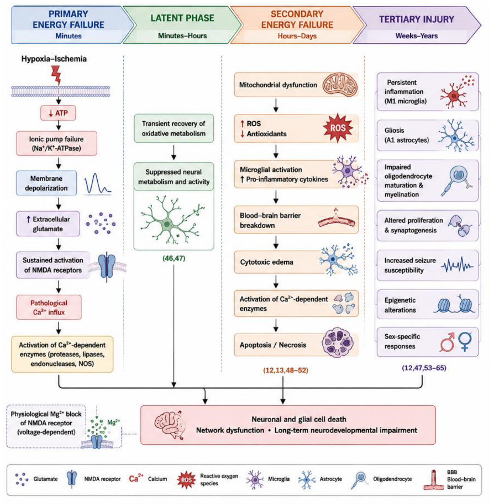
Figure 2.
NET framework from molecular mechanisms to clinically meaningful neuroprotection (on the left); NET-compliant methodology for translational neonatal neuroprotection research (on the right).
Figure 2.
NET framework from molecular mechanisms to clinically meaningful neuroprotection (on the left); NET-compliant methodology for translational neonatal neuroprotection research (on the right).
Disclaimer/Publisher’s Note: The statements, opinions and data contained in all publications are solely those of the individual author(s) and contributor(s) and not of MDPI and/or the editor(s). MDPI and/or the editor(s) disclaim responsibility for any injury to people or property resulting from any ideas, methods, instructions or products referred to in the content. |
© 2026 by the authors. Licensee MDPI, Basel, Switzerland. This article is an open access article distributed under the terms and conditions of the Creative Commons Attribution (CC BY) license (http://creativecommons.org/licenses/by/4.0/).
Copyright: This open access article is published under a Creative Commons CC BY 4.0 license, which permit the free download, distribution, and reuse, provided that the author and preprint are cited in any reuse.


