
Submitted:
19 July 2026
Posted:
21 July 2026
You are already at the latest version
Abstract
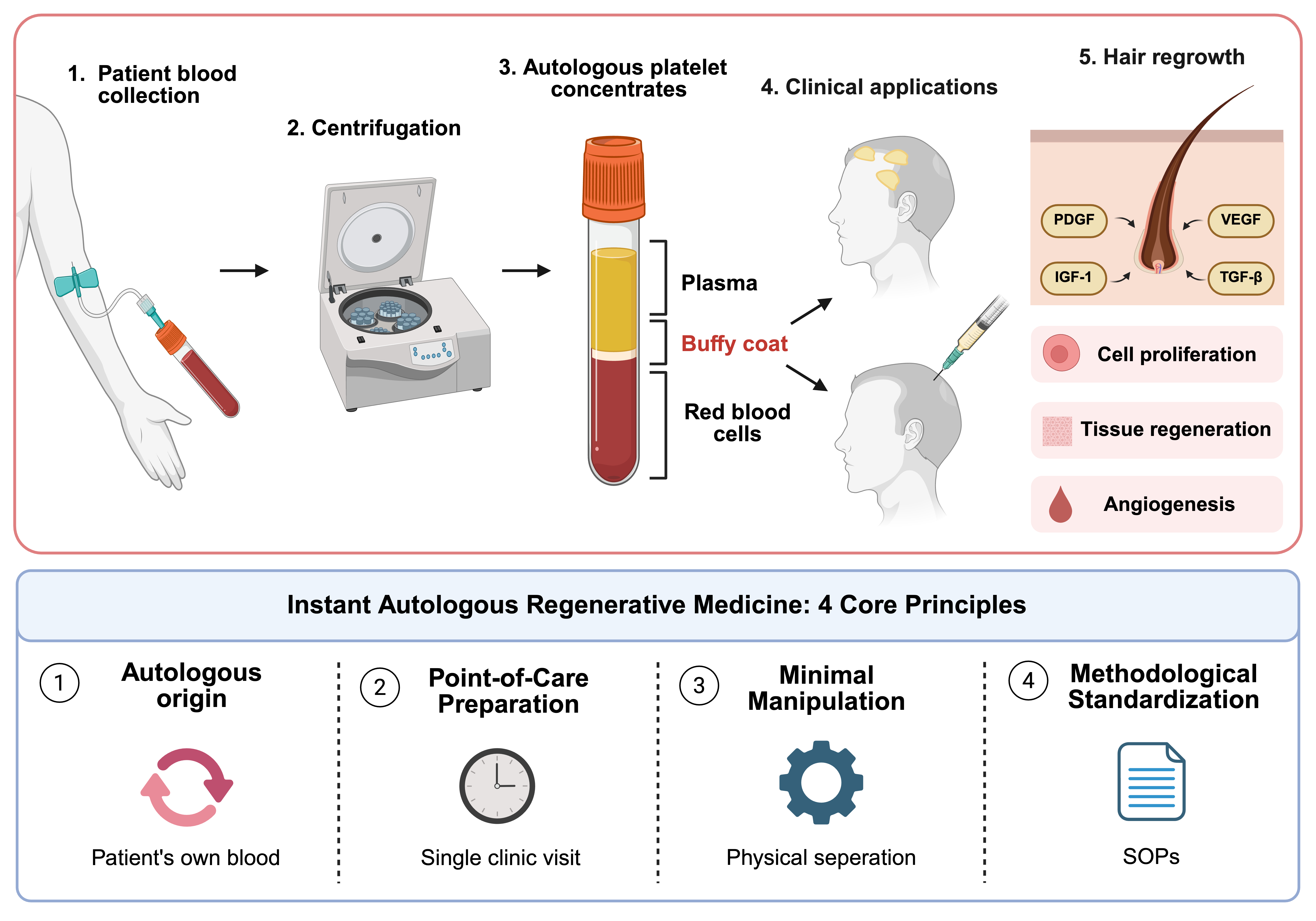
Androgenetic alopecia is a progressive hair loss disorder characterized by reduced hair density and hair follicle miniaturization. Autologous blood concentrates, including platelet-rich plasma, platelet-rich fibrin, and concentrated growth factors, are increasingly utilized for wound healing and hair restoration. However, despite extensive clinical evidence, these therapies still lack a unified conceptual framework, and their underlying molecular mechanisms remain to be fully elucidated. This review summarizes the gradual optimization of platelet concentrates, while proposing extracellular vesicles as a potential key mediator in next-generation platelet concentrates. Moreover, we define the four core principles of “Instant Autologous Regenerative Medicine” and discuss the current limits and research priorities. This unifying framework will facilitate the development, standardization, and broader clinical application of point-of-care regenerative therapies.
Keywords:
instant autologous regenerative medicine
; autologous platelet concentrates
; platelet-rich plasma
; platelet-rich fibrin
; concentrated growth factors
; extracellular vesicles
1. Introduction
Androgenetic alopecia (AGA) is the most common hair loss disorder globally, affecting up to 85% of men and 40% of women, causing diminished self-esteem and quality of life [1]. Alterations in androgen metabolism are widely recognized as the primary cause of AGA, leading to a shortened anagen phase and a subsequent reduction in hair density and hair shaft diameter. Men typically experience frontal bitemporal thinning that gradually extends to the vertex, whereas women exhibit widespread hair thinning across the mid-scalp [2]. Currently available FDA-approved pharmacological treatments for AGA are topical minoxidil and oral finasteride. However, variable treatment response, the need for lifelong use, and adverse effects such as sexual dysfunction and scalp irritation limit their clinical utility [3]. Consequently, there is a growing demand for safe and effective regenerative strategies.
In recent years, autologous platelet concentrates (APCs) have become a promising therapeutic intervention. Prepared at the point of care by centrifuging the patients’ own blood, APCs offer a safe and straightforward administration profile, eliminating the risks of immune rejection or transmission of infectious diseases [4,5]. Moreover, APCs have garnered significant attention in dermatological diseases for their capacity to promote tissue healing and regeneration through the release of morphogens, growth factors (GFs), and proteins [6].
In the field of hair restoration, extensive studies have been performed to elucidate the clinical efficacy of APCs. For example, platelet-rich plasma (PRP) was the first platelet-rich formulation to be developed and commercialized, followed by platelet-rich fibrin (PRF) and concentrated growth factors (CGF) [4]. The rationale behind these therapies lies in the intricate biology of the hair follicle, where growth and cycling are closely regulated by various GFs. Platelet concentrates serve as reservoirs of platelet-derived growth factor (PDGF), vascular endothelial growth factor (VEGF), insulin-like growth factor-1 (IGF-1), fibroblast growth factor (FGF), and epidermal growth factor (EGF) [7].
2. Evolution of autologous platelet concentrates for regenerative medicine: from PRP to CGF
The field of regenerative medicine has focused mainly on the body’s inherent healing capabilities, and APCs have exemplified their efficacy in hair regeneration research. APCs have undergone continuous advancements, and their progression across generations is depicted in Figure 1. The evolution of APCs reflects continuous effort toward optimizing preparation protocols, enhancing therapeutic efficacy, and expanding clinical applications.
2.1. First-generation platelet concentrates: PRP
PRP, developed in the 1970s, is the first generation of APC [4]. It is the most extensively studied therapy among the three generations and has been shown to improve hair density and thickness in multiple clinical studies [7]. PRP is prepared by centrifuging freshly collected blood, yielding a GF concentration approximately 3 to 5 times higher than that of whole blood [8]. The obtained plasma comprises highly concentrated GFs, including PDGF, TGF-β, and VEGF, that actively promote cell proliferation, tissue regeneration, and angiogenesis, all of which are essential for hair regrowth [2]. PRP also contains leukocytes, plasma proteins, and extracellular vesicles (EVs), which act synergistically to modulate immunomodulation, scaffolding, and intercellular signaling [8].
The underlying mechanism of hair follicle stimulation involves the secretion of a variety of GFs from platelets upon activation, which subsequently activate signaling pathways such as the extracellular signal-regulated kinase (ERK) and protein kinase B (Akt) pathways. Specifically, the Akt pathway promotes follicular cell persistence through anti-apoptotic effects via increased BCL-2 expression, while the ERK pathway primarily regulates cell growth [8]. A meta-analysis of nine randomized controlled trials (RCTs) comparing PRP with topical minoxidil for AGA found that both minoxidil and PRP improved hair density, with no significant difference in clinical efficacy. However, PRP yielded greater patient satisfaction than minoxidil [2]. Recent reports have also described the efficacy of intraoperative PRP integration. The use of PRP for the pretreatment of follicular units before grafting improved hair density and accelerated hair regrowth [9]. Furthermore, combining PRP injection with suture-embedded scalp lifting has shown therapeutic synergy in improving scalp thickness, enhancing local blood supply, and increasing hair shaft diameter, which further reflects its clinical value as a promising adjunctive method for combination therapy [10].
However, a major limitation of PRP is its burst release kinetics. Specifically, GFs are secreted abruptly within 5 to 10 minutes [4], with approximately 95% of the total payload released within an hour of activation [11]. The short-lived bioavailability (typically less than 7 days) and the lack of a fibrin scaffold impair treatment efficacy and negatively impact patient compliance, as frequent injections are required [4]. In addition, the use of anticoagulants may preclude therapeutic intervention in patient groups allergic to these additives [11].
Currently, a PRP preparation lacks a standardized consensus. Differences in procedural parameters (centrifugation parameters, injection volume, thermal conditions, and anticoagulants) lead to profound variations in the final amount of GF payload and bioactivity. Consequently, clinical outcomes of PRP remain inconsistent, making a direct comparison of results between studies difficult [5,7,9]. The two main methods of centrifugation are single-spin and double-spin centrifugation. The single-spin approach involves a single centrifugation step with calcium gluconate activation, resulting in a platelet concentration three to six-fold higher than that of whole blood [2]. However, most PRP preparation protocols utilize a double-spin centrifugation consisting of low-speed and high-speed centrifugation, designed to isolate a small volume of platelet-rich supernatant in the lower one-third part of the plasma column [9]. Based on the necessity for a standardized protocol, Sharma et al. proposed a detailed recommendation for the preparation and clinical application of PRP. They recommended a nonactivated, manual double-spin method targeting a platelet concentration of 1.0 to 1.5 million/µL. For patients with Norwood-Hamilton grades II to V, they recommended using 5 to 7 mL (0.05 to 0.1 mL/cm²) across a minimum of three to five sessions over one month [9].
2.2. Second generation platelet concentrates: platelet-rich fibrin (PRF)
To overcome the limitations of PRP, PRF was introduced in 2001. PRF stands out for its rapid preparation, anticoagulant-free composition, and a sustained GF release profile [11]. Low-temperature storage of PRP helps preserve the natural fibrin network, protecting GFs against proteolytic degradation [12] and facilitating a more prolonged delivery of GFs over several days to weeks [4]. In addition, PRF strongly supports cell growth and differentiation by activating key signaling pathways, while also mitigating inflammatory responses, upregulating the expression of GFs in mesenchymal cells, and exhibiting antimicrobial properties [13].
Similar to PRP, the regenerative performance of PRF is highly related to the technical variables of its preparation process. For instance, centrifugal force, rotor angulation, and spin duration directly influence the fibrin architecture, GF concentration, and cellular distribution, inducing significant variations in scaffold quality, potentially compromising its therapeutic efficacy [14]. Extensive literature has employed fixed-angle centrifugation operated at low relative centrifugal force(RCF) levels, typically ranging from 600 to 800, and reduced spin durations of 3 to 6 minutes [11]. However, these conventional fixed-angle methods often suffer from an uneven distribution of centrifugal force, leading to inconsistent cell separation. Consequently, horizontal centrifugation has emerged as an alternative method. This approach ensures a homogeneous distribution of cellular components and achieves up to a fourfold enrichment of platelets and leukocytes compared to fixed-angle methods [14].
PRF can be categorized into solid PRF and liquid injectable PRF. Early clinical interventions relied mostly on solid PRF, including leukocyte-rich PRF (L-PRF), titanium PRF (T-PRF), advanced PRF (A-PRF), and advanced PRF plus (A-PRF+) [11,13]. The solid autologous scaffolds mimic the natural wound healing cascade through the sustained release of platelets, leukocytes, and GFs, which drive effective tissue repair [14]. However, their high cell density and gel-like viscosity made them unsuitable for direct injection, restricting their broader clinical utility [11]. In 2014, injectable PRF (i-PRF), the first anticoagulant-free liquid APC, was developed [14]. It was obtained through further refinements in centrifugation protocols and tube technology [13]. Compared to solid PRF, i-PRF maintains a higher concentration of platelets and leukocytes, thereby ensuring robust tissue regeneration [15]. More recently, concentrated platelet-rich fibrin (C-PRF) has been investigated. C-PRF, obtained from the buffy coat layer through horizontal high-speed centrifugation, further enhanced fibroblast migration, proliferation, and collagen I synthesis [16].
Clinical studies evaluating PRF for AGA have been increasingly reported. For instance, a systematic review of the efficacy of PRF revealed a 62%-97% optimization of hair density across a 3-6-month timeline. The study also demonstrated that these therapeutic benefits are amplified when PRF is combined with hair transplantation [17]. In addition, a comparative study of i-PRF and PRP, both obtained via horizontal centrifugation, revealed significant improvements in patients with AGA. Notably, i-PRF outperformed PRP at the 3-month follow-up in promoting hair regrowth and mitigating hair shedding [18]. Similarly, Yao et al. reported improvements in hair loss and scalp symptoms after 6 months of i-PRF injections [15]. However, inconsistencies in preparation protocols and evaluation methods continue to complicate the systematic assessment of clinical outcomes [11].
2.3. Third generation platelet concentrates: concentrated growth factors (CGF)
CGF is an advanced third-generation APC also isolated without the use of anticoagulants 7. Variable-speed centrifugation is performed between 2400 and 3300 rpm, with gradual acceleration and deceleration. After centrifugation, CGF is located between the plasma and red blood cell layers [19]. Optimization of centrifugal force and centrifugation time enabled the formation of a dense fibrin scaffold rich in leukocytes and angiogenesis-related CD34+ stem cells. The three-dimensional architecture of this fibrin network not only accommodates cell adhesion and proliferation but also facilitates the delivery of highly concentrated platelet-derived GFs [4,20]. Progressive degradation of the fibrin scaffold promotes the continuous release of GFs through thrombin-triggered degranulation of platelet -granules [19,21]. The released GFs accelerate wound healing, extracellular matrix production, collagen synthesis, and angiogenesis [21]. In addition, CGF exhibits potent antioxidant activity by modulating intracellular reactive oxygen species (ROS) accumulation and upregulating antioxidant enzyme expression [20].
Accumulating evidence demonstrates CGF surpasses PRP and PRF in terms of GF enrichment, supporting its promise as a therapeutic platform for AGA [3,20]. CGF can be formulated into liquid preparations, gels, lyophilized forms, and membranes, which provide high flexibility in clinical application in various ways [20]. A retrospective study by Cao et al. demonstrated significant improvements in hair density and hair growth ratio at 3 and 6 months after CGF injections, with no severe adverse events [22]. Similarly, Zhao et al. reported an overall efficacy of 98.28% in 58 patients who completed six injection sessions of CGF. The overall clinical efficacy demonstrated a 70.14% improvement in hair density, as well as 48.89% and 26.21% improvements in follicular density and shaft diameter when compared with the first injection, respectively [19].
CGF has also shown potential for use in combination therapy. Co-administration of CGF with conventional medications, such as minoxidil, finasteride, or spironolactone, offers greater efficacy than monotherapy [20]. Moreover, a retrospective study by Steward et al. reported that a combination therapy of PRP injection, microneedling, and topical administration of CGF gel led to increased hair count and a higher anagen-to-telogen ratio. More favorable treatment outcomes were observed in male patients whose AGA onset occurred at or after 25 years of age than in those with earlier onset [21]. Another treatment approach involving CGF prepared from 640nm laser-pretreated blood has also shown promise. This approach improved the follicular microenvironment through platelet activation and CD34+ stem cell enrichment [23]. Moreover, when CGF therapy was combined with Follicular Unit Extraction (FUE), it resulted in greater hair density and a higher proportion of terminal hairs than FUE monotherapy, suggesting a synergistic effect between the two treatments [24].
3. Comparative efficacy of PRP, PRF, and CGF
The evolution from PRP to PRF and CGF has focused on optimizing GF release kinetics and cellular composition. Table 1 summarizes the core differences among these three APC generations.
Abbreviations: WB, whole blood
A meta-analysis conducted by Alali et al. highlighted that CGF injections for AGA continuously improved hair density, rising from 19.6 hairs/cm² to 57.1 hairs/cm² over 12 months, showing superior outcomes compared to PRP. Concurrently, hair thickness expanded by 5.19-22.03 µm, contributing to a 94.3% patient satisfaction rate [7]. Moreover, a split-scalp study by Mishra et al. directly compared PRP combined with microneedling and a triple combination of PRP, CGF, and microneedling. Their findings demonstrated that while both therapeutic modalities showed clinical improvements, the CGF-containing combination therapy resulted in a significantly greater increase in hair count and hair pull test outcomes [1].
In a prospective multicenter randomized clinical study, Li et al. compared PRP, i-PRF, and CGF for female pattern hair loss. The i-PRF and CGF groups showed greater improvements in hair count than the PRP group, although hair density did not differ significantly among the three groups. Notably, i-PRF demonstrated the most favorable safety profile, supporting the therapeutic efficacy of i-PRF [25].
4. Emerging therapeutic mediators: Extracellular vesicles (EVs)
With the continued evolution of APCs, research in regenerative medicine has increasingly focused on cell-free treatment strategies for AGA. EVs, including exosomes (30–150nm), microvesicles (100–1,000 nm), and apoptotic bodies (>1000nm) [4,26], regulate immune responses, cellular migration, and tissue repair by transferring proteins, lipids, and nucleic acids [5,27]. Platelet-derived EVs (pEVs) overcame several limitations of conventional platelet concentrates, such as poor reproducibility and the risk of platelet aggregation, while maintaining potent regenerative and immunomodulatory functions [4,28].
EVs modulate key signaling pathways involved in hair regrowth, including Wnt/β-Catenin, PI3K/AKT, MAPK/ERK, and TGF-β/BMP, which ultimately restores intracellular regenerative signaling and promotes angiogenesis [27]. Cui et al. demonstrated that exosome treatment from human umbilical cord mesenchymal stem cells promoted scalp rejuvenation and hair follicle elongation by reducing senescent cells and upregulating type XVII collagen (COL17A1). Mechanistically, miR-21-5p suppresses DKK2 (Wnt antagonist), thereby activating Wnt/β-Catenin pathway and restoring COL17A1 expression [29].
Recent investigations have increasingly explored the therapeutic potential of EVs for AGA. Mesenchymal stem cell-derived exosomes revitalize dermal papilla cells (DPCs) by triggering the Wnt/β-catenin signaling cascade, which subsequently enhances both hair shaft diameter and overall density [3]. A systematic analysis highlighted that topical or injectable EV applications achieved notable clinical efficacy in AGA. Specifically, these modalities yielded up to a 28% increase in hair count and a 14% improvement in hair thickness during RCTs, while retrospective observations indicated density improvements reaching 45% [17]. Penha et al. also reported that exosome therapy improved hair density and thickness while upregulating VEGF expression and activating Wnt/β-catenin pathway [30]. Notably, Lai et al. developed engineered exosomes to co-deliver EGF and FGF via LAMP2B fusion engineering. The modified exosomes stimulated DPC activity in vitro and restored hair coverage in murine models. This approach overcame the limited follicle-targeting capability of natural exosomes and conventional therapies [31].
To facilitate clinical translation of EV-based therapies, concentrated purified fraction (CPF), a fourth-generation platelet concentrate obtained through further purification of CGF, has been introduced. Unlike conventional APCs, which primarily exert therapeutic effects through GF release, CPF is highly enriched with EVs and acts through vesicle-mediated signaling, enabling more targeted regulation of hair follicle regeneration [32].
5. Challenges and defining principles of IARM
5.1. Limitations
The primary challenge in evaluating APCs is the heterogeneity of preparation protocols and device technologies. Centrifugation speeds, rotor angles, and processing times remain poorly standardized, most notably in PRP preparation [7,11]. These technical disparities lead to variability in platelet concentration, leukocyte retention, and GF profiles, complicating the comparisons of clinical outcomes across studies [5,6,7].
Furthermore, establishing objective and standardized criteria for therapeutic assessment and methodological frameworks remains a persistent challenge [20]. Moreover, most studies rely on retrospective, single-center trials with small sample sizes and short follow-up periods [7,20,30]. Consequently, large-scale, double-blind RCTs are needed to validate the long-term efficacy and safety.
Finally, autologous therapies are fundamentally influenced by patient-specific variability. A patient’s age, systemic health, and genetic background can affect the biological composition of APCs, resulting in divergent clinical outcomes and complicating treatment standardization.
5.2. Defining principles
While conventional autologous therapies share the benefits of autologous origin and minimal manipulation, clinicians often face severe outcome variability. Herein, we propose IARM based on four defining principles to overcome the limitations of APC therapies.
1. Autologous origin: All agents must be derived entirely from the patient’s own blood.
2. Point-of-care preparation: The blood collection, processing, and injection must take place within a single clinic visit at the point of care.
3. Minimal manipulation: The processing of blood concentrates should rely strictly on physical separation. Chemical alteration and genetic modification should be avoided, except for the routine use of standard anticoagulants that do not alter cellular biological properties.
4. Methodological standardization: Application procedure must mandate highly standardized SOPs to minimize variability across different operators and clinical settings.
6. Conclusion
APCs are a safe, effective, and highly biocompatible alternative to traditional therapies, showing consistent progression across generations. To bridge the operational and methodological disparities, the proposed IARM framework introduces four core principles. This structured approach provides a practical guide for the development and application of APC therapies. Future research should prioritize reporting detailed preparation processes, cellular compositions, and objective hair regrowth parameters to facilitate the development of standardized SOPs. Additionally, evaluating the clinical synergy between APCs and conventional modalities through long-term studies may provide new insights into optimizing regenerative treatment strategies.
CRediT Authorship Contribution Statement
Seungyeon Lee: Writing - Original Draft, Data Curation, Visualization. Yu-An Zhu: Data Curation, Formal Analysis. Mingyang Lu: Literature Search, Validation.Chika Hasegawa: Literature Search, Validation. Hua Jiang: Supervision, Resources. Yufei Li: Conceptualization, Writing - Review & Editing, Supervision, Funding Acquisition.
Funding Statement
Not applicable.
Data Availability Statement
Not applicable. No new data were created or analyzed in this study.
Permission to Reproduce Material from Other Sources
Not applicable.
Ethics Approval Statement
Not applicable.
Patient Consent Statement
Not applicable.
Acknowledgments
The authors acknowledge the support provided by the Department of Plastic Surgery, Shanghai East Hospital, School of Medicine, Tongji University.
Conflicts of Interest disclosure
All authors declare that they have no conflict of interest.
References
- Mishra, M.; Varia, R.; Mondal, S.; et al. Efficacy of Platelet Rich Plasma and Concentrated Growth Factor in Treating Androgenetic Alopecia: A Split-Scalp Study. J. Maxillofac. Oral Surg. 2026, 25(2), 601–7. [Google Scholar] [PubMed]
- Umar, M.; Anwar, A.; Shamim, L.; et al. Comparative Efficacy and Safety of Platelet Rich Plasma (PRP) versus Topical Minoxidil for Androgenetic Alopecia: A Systematic Review and Meta-analysis. Aesthetic Plast. Surg. 2026, 50(3), 1340–53. [Google Scholar] [PubMed]
- Burshtein, J.; Burshtein, A.; Schlesinger, T. Emerging pharmacotherapies and regenerative solutions for promoting hair growth for androgenetic alopecia. Front Pharmacol. 2026, 17, 1776134. [Google Scholar] [CrossRef] [PubMed]
- Li, Y.; You, H.; Ou, C.; et al. The evolution of three generations of platelet concentrates products: a leap from classical formulations to the era of extracellular vesicles. Front Bioeng. Biotechnol. 2025, 13, 1628565. [Google Scholar] [CrossRef] [PubMed]
- Everts, P.A.; Podesta, L.; Lana, J.F.; et al. The Regenerative Marriage Between High-Density Platelet-Rich Plasma and Adipose Tissue. Int. J. Mol. Sci. 2025, 26(5). [Google Scholar] [CrossRef] [PubMed]
- Anitua, E.; Tierno, R.; Orive, G.; Alkhraisat, M.H. Synergistic Innovative Therapies in Dermatology: Integrating Platelet Derivatives, Biomaterials, and Exogenous Bioactive Substances for Enhancing Skin Repair and Regeneration. Drug Des. Devel Ther. 2025, 19, 8755–82. [Google Scholar] [CrossRef] [PubMed]
- Alali, A.; Alahmadi, M.; Alsisi, G.; et al. The Efficacy of Growth Factor Injection in Androgenic Alopecia: A Systematic Review and Meta-Analysis. In Aesthetic Plast Surg; 2026. [Google Scholar]
- Wu, W.S.; Chen, L.R.; Chen, K.H. Platelet-Rich Plasma (PRP): Molecular Mechanisms, Actions and Clinical Applications in Human Body. Int. J. Mol. Sci. 2025, 26(21). [Google Scholar] [CrossRef] [PubMed]
- Sharma, A.; Chouhan, K.; Bhatia, S.; Dashore, S. Platelet-Rich Plasma in Androgenetic Alopecia. Indian Dermatol. Online J. 2021, 12 (Suppl 1), S31–s40. [Google Scholar] [CrossRef] [PubMed]
- Ku, M.C.; Teh, L.S.; Chen, P.M.; et al. Synergistic effect of platelet-rich plasma injections and scalp lifting in androgenetic alopecia. Clin. Dermatol. 2018, 36(5), 673–9. [Google Scholar] [CrossRef] [PubMed]
- Shrestha, S.; Pathak, R.; Jagat, G.C.; Donovan, J. Platelet-rich fibrin: A review of its role as a new treatment in androgenetic alopecia. J. Cosmet. Dermatol. 2024, 23(8), 2524–30. [Google Scholar] [CrossRef] [PubMed]
- Kim, T.H.; Kim, S.H.; Sándor, G.K.; Kim, Y.D. Comparison of platelet-rich plasma (PRP), platelet-rich fibrin (PRF), and concentrated growth factor (CGF) in rabbit-skull defect healing. Arch. Oral Biol. 2014, 59(5), 550–8. [Google Scholar] [CrossRef] [PubMed]
- Miron, R.J.; Gruber, R.; Farshidfar, N.; et al. Ten years of injectable platelet-rich fibrin. Periodontol. 2000 2024, 94(1), 92–113. [Google Scholar] [PubMed]
- Farshidfar, N.; Apaza Alccayhuaman, K.A.; Estrin, N.E.; et al. Advantages of horizontal centrifugation of platelet-rich fibrin in regenerative medicine and dentistry. Periodontol. 2000 2025. [Google Scholar] [CrossRef] [PubMed]
- Yao, S.; Chen, X.; Li, S.; et al. New tool in our arsenal: efficacy of injectable platelet-rich fibrin (i-PRF) in androgenetic alopecia treatment. Arch. Dermatol. Res. 2025, 317(1), 493. [Google Scholar] [CrossRef] [PubMed]
- Fujioka-Kobayashi, M.; Katagiri, H.; Kono, M.; et al. Improved growth factor delivery and cellular activity using concentrated platelet-rich fibrin (C-PRF) when compared with traditional injectable (i-PRF) protocols. Clin. Oral Investig. 2020, 24(12), 4373–83. [Google Scholar] [CrossRef] [PubMed]
- Behrangi, E.; Amiri, S.; Zeinali, R.; et al. A systematic review of clinical evidence on the efficacy and safety of conditioned media, platelet-rich fibrin, stromal vascular fraction, extracellular vesicles, and stem cells in androgenetic alopecia. Stem Cell Res. Ther. 2026, 17(1). [Google Scholar] [CrossRef] [PubMed]
- Mohamamad, A.; Rizvi, S.A. Platelet-rich plasma versus injectable platelet-rich fibrin–a randomized control trial. Int. J. Sci. Stud. 2022, 10, 53–6. [Google Scholar]
- Zhao, F.; Hao, L.; Jin, X. Clinical observation and evaluation of CGF in the treatment of androgenic alopecia. J. Cosmet. Dermatol. 2022, 21(11), 5723–9. [Google Scholar] [CrossRef] [PubMed]
- Zhou, D.; Liu, Z.W.; Jie, X.; Zhu, X. Advances in Research on Concentrated Growth Factor Applications for Androgenetic Alopecia Treatment: A Review. Med. Sci. Monit. 2025, 31, e948054. [Google Scholar] [CrossRef] [PubMed]
- Steward, E.N.; Patel, H.; Pandya, H.; et al. Efficacy of Platelet-Rich Plasma and Concentrated Growth Factor in Treating Androgenetic Alopecia - A Retrospective Study. Ann. Maxillofac. Surg. 2020, 10(2), 409–16. [Google Scholar] [CrossRef] [PubMed]
- Cao, S.; Zhu, M.; Bi, Y. Evaluation of the safety and efficacy of concentrated growth factors for hair growth promotion in androgenetic alopecia patients: A retrospective single-centre, single-arm study. J. Cosmet. Dermatol. 2024, 23(12), 4051–6. [Google Scholar] [CrossRef] [PubMed]
- Gao, J.; Xiao, Q.; Lu, Y.; et al. Higher percentage of CD34+ stem cells and elevated efficacy in androgenetic alopecia treatment observed in CGF prepared from 640 nm laser-pretreated blood: A preliminary study. J. Cosmet. Dermatol. 2024, 23(6), 2249–55. [Google Scholar] [CrossRef] [PubMed]
- Wang, Q.; Pang, J.; Xie, K.; et al. A prospective study of hair transplantation combined with concentrated growth factors for the treatment of androgenetic alopecia. J. Dermatol. Treat. 2026, 37(1), 2635881. [Google Scholar] [CrossRef]
- Li, M.; Bai, Y.; Ye, Z.; et al. Comparative efficacy and safety of platelet-rich plasma (PRP), injectable platelet-rich fibrin (i-PRF) and concentrated growth factors (CGF) for female pattern hair loss (FPHL): a prospective multicenter randomized clinical trial. J. Dermatol. Treat. 2026, 37(1), 2659498. [Google Scholar]
- Shimizu, Y.; Ntege, E.H.; Sunami, H.; Inoue, Y. Regenerative medicine strategies for hair growth and regeneration: A narrative review of literature. Regen. Ther. 2022, 21, 527–39. [Google Scholar] [CrossRef] [PubMed]
- Rajendran, R.L.; Reyaz, D.; Mahajan, A.A.; et al. Bio-Nanovesicle-Based Approaches for Hair and Skin Regeneration: An Updated Concise Review. Cells 2026, 15(7). [Google Scholar] [CrossRef] [PubMed]
- Antich-Rosselló, M.; Forteza-Genestra, M.A.; Monjo, M.; Ramis, J.M. Platelet-Derived Extracellular Vesicles for Regenerative Medicine. Int. J. Mol. Sci. 2021, 22(16). [Google Scholar] [CrossRef] [PubMed]
- Cui, S.; Huang, Y.; Zeng, D.; et al. Mesenchymal stem cell-derived exosomes promote scalp rejuvenation through type XVII collagen regulation via the miR-21-5p/DKK2/Wnt pathway. Front Cell Dev. Biol. 2026, 14, 1807877. [Google Scholar] [CrossRef] [PubMed]
- Penha, F.A.; Penha, G.A.; Caleiro Acerbi, H.M. Exosome-Based Therapies for Alopecia: A Systematic Review of Clinical Efficacy and Safety. Int. Soc. Hair Restor. Surg. 2026, 36(2), 37–47. [Google Scholar] [CrossRef]
- Lai, Y.; Wu, J.; Tan, T.; et al. Engineered Exosomes Co-Delivering EGF and FGF Ameliorate Androgenetic Alopecia in a Mouse Model. Int. J. Nanomed. 2026, 21, 572518. [Google Scholar]
- Ruiyu, L.; Changjiang, Z.; Yufei, L. Beyond PRP: Recognizing the Evolving Generations of Platelet Concentrates in Androgenetic Alopecia Treatment—A Letter in Response to Zhong Et al. J. Cosmet. Dermatol. 2026, 25(7), e71002. [Google Scholar] [CrossRef]
Figure 1.
Schematic overview of the evolution of autologous platelet concentrates The upper panel illustrates the distinct characteristics of the three generations of APCs and their evolution from first-generation PRP to second-generation PRF and third-generation CGF, highlighting the progressive enhancement of growth factor concentration and clinical efficacy. The lower panel depicts the isolation of CPF through further centrifugation of CGF and enrichment in pEVs and signaling molecules. Abbreviations: APCs, autologous platelet concentrates; PRP, platelet-rich plasma; PRF, platelet-rich fibrin; CGF, concentrated growth factors; CPF, concentrated purified fraction; pEVs, platelet-derived extracellular vesicles Created in BioRender. LEE, S. (2026) https://BioRender.com/j2hlehy.
Figure 1.
Schematic overview of the evolution of autologous platelet concentrates The upper panel illustrates the distinct characteristics of the three generations of APCs and their evolution from first-generation PRP to second-generation PRF and third-generation CGF, highlighting the progressive enhancement of growth factor concentration and clinical efficacy. The lower panel depicts the isolation of CPF through further centrifugation of CGF and enrichment in pEVs and signaling molecules. Abbreviations: APCs, autologous platelet concentrates; PRP, platelet-rich plasma; PRF, platelet-rich fibrin; CGF, concentrated growth factors; CPF, concentrated purified fraction; pEVs, platelet-derived extracellular vesicles Created in BioRender. LEE, S. (2026) https://BioRender.com/j2hlehy.
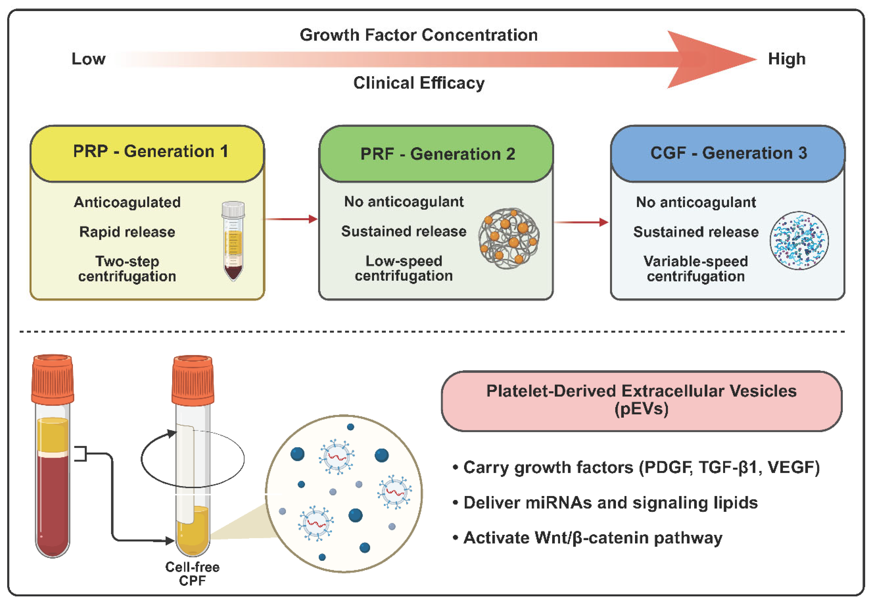

Table 1.
Comparison of different generations of autologous platelet concentrates.
| Parameter | PRP (Gen 1) | PRF (Gen 2) | CGF (Gen 3) |
| Growth factor concentration | ~3× WB | ~5× WB | ~8× WB |
|
Leukocyte concentration |
Low | High | High |
| Growth factor release profile | Rapid (<1 day) | Sustained (7–14 days) |
Sustained (7-14 days) |
| Fibrin matrix | Loose fibrin network |
Porous three-dimensional fibrin network |
Dense three-dimensional fibrin network |
| Stem cell concentration | Negligible | Negligible | High (CD34+) |
| Key active components | Soluble growth factors | Growth factors, Fibrin matrix, Leukocytes | Concentrated growth factors, CD34+ cells |
| Additives | Anticoagulants or external activators | None | None |
| Centrifugation protocol | Two-step, Low and high speed |
Single-step, Low-speed (≤1500rpm) |
Variable-speed (2400-3300rpm) |
| Activation method | Activation through calcium chloride and thrombin | Endogenous activation | Endogenous activation |
Note: Values are approximate and can vary depending on the specific centrifugation protocols.
Disclaimer/Publisher’s Note: The statements, opinions and data contained in all publications are solely those of the individual author(s) and contributor(s) and not of MDPI and/or the editor(s). MDPI and/or the editor(s) disclaim responsibility for any injury to people or property resulting from any ideas, methods, instructions or products referred to in the content. |
© 2026 by the authors. Licensee MDPI, Basel, Switzerland. This article is an open access article distributed under the terms and conditions of the Creative Commons Attribution (CC BY) license (http://creativecommons.org/licenses/by/4.0/).
Copyright: This open access article is published under a Creative Commons CC BY 4.0 license, which permit the free download, distribution, and reuse, provided that the author and preprint are cited in any reuse.


