

Submitted:
17 July 2026
Posted:
20 July 2026
You are already at the latest version
Abstract
While the value of XR has been broadly demonstrated in the literature, there has been little empirical work seeking to establish where AR could effectively be placed within the training pathway. This is particularly true in radiology, where training relies heavily on anatomical comprehension and AR has been proposed as a tool to support this through enhanced 3D visualisation of complex anatomical structures. A series of seven interviews with radiologists and radiology registrars was conducted, using AR anatomy tasks with the Microsoft HoloLens 2 and Meta Quest 3 to engage participants in a rich dialogue around the educational value of AR in radiological training. Reflexive thematic analysis was employed to interpret the resulting data. Four themes were generated, revealing an expertise reversal effect whereby AR’s value is greatest for early-stage trainees and diminishes with experience, alongside a need for AR to integrate with, rather than replace, the traditional 2D methods used in practice. These themes were mapped to two design considerations for AR-enabled radiological curricula that stand as the contribution of this work; prioritising early-stage training through a structured graduation pathway into traditional practice, and positioning AR as a supplement to existing training rather than a replacement for it.
Keywords:
augmented reality
; mixed reality
; radiology
; interaction
; reflexive thematic analysis
; medical education
1. Introduction
Augmented reality (AR), and extended reality (XR) more generally, have been investigated across the literature for medical training with a range of studies comparing the pedagogical effectiveness of XR systems to their traditional counterparts. Studies have largely shown positive results in terms of engagement and knowledge retention but there is little empirical work suggesting the role that AR could play in medical training to generate maximum impact. The work described in this paper qualitatively investigates this question in terms of the interaction value of AR and radiological training. Seven participants were recruited for a series of in-depth interviews, in which they were presented with examples of AR technology in the form of the Microsoft HoloLens 2 and the Meta Quest 3 with two pieces of software developed by GigXR [1]. Participants then completed tasks designed to simulate aspects of radiological training and practice using these head-mounted displays (HMDs), which aimed to prompt conversation around various aspects of potential AR usage in the radiological training pathway. The interactions that AR provides were the focus of these conversations with interest around whether they could improve comprehension for radiologists in training, enabling some direction towards a where AR could effectively be deployed.
The `think aloud’ approach was used in the interviews in order to keep the focus on the participants’ experiences and thoughts with reflexive thematic analysis used to analyse the interview video and audio recordings [2,3]. The reflexive thematic analysis produced four themes, with sub-themes beneath, capturing individual attitudes and concepts from the interview data. These themes were then mapped to three design considerations which stand as the contribution of this study. These design considerations are intended to be useful to future designers and researchers when conducting further research in this area or developing an AR tool for radiological education by directing attention towards specific applications and the specific value AR could afford.
For this study there were two research questions:
- 1.
- How could the interaction advantages of AR afford the opportunity for more efficient radiological training through better comprehension or other means?
- 2.
- What are the restrictions on modern AR for radiological education?
2. Background
The clinical potential of AR has been discussed since the early 1980s [4] and this has only expanded with the increased interest and improvement of technology [5,6,7]. The potential of AR spans the breadth of healthcare specialties with applications suggested across fields such as surgical guidance, post-operative care, medical training, and radiology [6,8,9]. While few AR applications have been deployed into regular use within healthcare, medical training is an important and promising area being explored. In current practice, simulation is a very widely used technique across many aspects of medical education and training, manikin-based training being a key example [10]. Other traditional methods such as cadaver-based and lecture-based education still lead anatomy education but challenged resources such as cost and time open the door for XR to fill the gaps and provide more besides [11,12].
XR has been put forward for many applications within education and training including anatomy education, medical emergency simulation, surgical training, emotionally realistic training scenarios, and professional skills [7,10,12,13]. Many positives have been acknowledged within these studies such as safer, more suitable, cheaper, more standardised and replicable, and more accessible training [12,14]. There is great potential value across medical education acknowledged in the literature but this purported value centres around taking up the slack where resources are challenged and improving learning outcomes through increased engagement, reduced cognitive load, and more accessible material.
In terms of anatomy education many studies have evaluated XR and demonstrated a significant improvement in engagement and broadly positive results in regard to knowledge retention. Studies have found an increase in anatomy recall combined with a lower cognitive load [12]. With the value of XR in radiological education acknowledged, Shanahan [15] highlight some issues that may interrupt wide spread adoption, for example the availability of high-quality content [7]. However, the the main question now, as Curran et al. [14] suggest, is not comparing XR to current methods but investigating where and how it should be deployed for maximum impact. It is on this question that there is little empirical work, with most studies focusing on quantitatively comparing effectiveness of XR to traditional methods. In addition there is little work that aims to empirically understand where XR technologies fit into training pathways, most present the technology as a disruptor and intervention rather than suggesting how it may be used as a component of the curriculum. Answering RQ2 in this study aims to contribute to answering this wider question, and RQ1 places a focus on gaining an empirical understanding of the value of AR interactions in this context.
As context to current systems, the training pathway for radiologists in the UK is typically the five year medical degree, then two years rotational training as a junior doctor, followed by five years as a radiology registrar doing specialty training, before completing training and become a consultant radiologist. These five years of specialty training as a radiology registrar is split into three years of core radiology training then two years of sub-specialty training in whatever niche a given registrar has chosen, for example neuroradiology.
In sum, the potential value of XR compared to traditional methods in radiology education is acknowledged and the literature no longer calls for comparative studies [14]. Understanding where and how AR could be deployed effectively with empirical study is the step forward that this work takes. The interaction advantages for diagnostic radiological education have been somewhat discussed but how these advantages could be integrated into the education pathway is an open question, and one which this study aims to begin to answer.
3. Methodology
This work was granted ethical approval by Newcastle University ethics committee ref: 61693/2023. Participants gave their informed consent to the interviews and it was made clear that they could withdraw their participation at any time. The interviews described here were run to investigate two lines of research as two studies, one of which is detailed here. One series of interviews was used for both studies because of the overlap in potential participants and the similarity of the activities that could be used in the interview. Aspects specific to each study and broader questions that applied to both were asked throughout the interviews. This yielded raw data that could then be interpreted and analysed for each study. While the interviews described in this study are shared with another, the analysis, results, and contribution from this work stand alone.
3.1. Recruitment Process and Participants
For this work, seven radiologists were recruited, four registrars, and three consultants from North Eastern NHS trusts in England. Six of these participants were male which illustrates the issues with recruiting women in a male dominated field such as this [16]. This is discussed in more depth in the Limitations Section (Section 5.3). The consultants were associated with a variety of sub-specialties and while registrars are general, their interests were varied. Five participants sat within the 25-34 age bracket while two consultants were in the next two brackets. Participant demographic details and summarised in Table 1. Participants were asked to rate their prior experience with XR from one to five with the labels shown in Table 3. Six participants listed their experience as one or two out of five with the final participant rating their experience as five, owning a Meta Quest 3. Two of the participants participated in a previous study by Hobbs et al. [17] as part of the same thread of research, with the remaining five participants recruited through snowballing and by reaching out directly.
3.2. The Interviews
These interviews were designed to enable us to present the participants with a variety of different situations in AR in a short space of time, closely enough aligned with their current workflows and goals that meaningful discussions could be made around the opportunities and challenges these AR tools provided. Most participants had very little experience of AR and the first objective of the workshop was to demonstrate AR to the participants in a way representative of a longer more extensive AR trial. The aim here was to allow the discussions had with the participants to get beyond the initial learning curve associated with using AR, and indeed any new technology, and on to a more realistic, holistic view of the role it could play. These seven interviews were run one-on-one with the lead researcher and a participant.
The aim of these interviews was to qualitatively investigate the impact of AR interactions for the integration into radiological reporting, with the research question how could the interaction advantages of AR afford the opportunity for increased efficiency or comprehension for radiologists? Various forms of data were collected through the interviews, principally the point of view (POV) video recording of what the participant was seeing and the audio recording of the conversation. The two HMDs were set up to cast this POV to a Windows PC. A Microsoft Teams call was set up and the screen sharing and meeting recording functions were used to achieve this. The participant was seated at one end of a desk in front of a PC but space was deliberately provided along the desk to the left and around the room further to the left and behind them, should they have wanted to use the space. A GoPro was positioned with the participant in frame to record how the participant used the wider space and any bigger actions that would not be caught on the POV footage. The researcher was seated at a second desk to the right of the participant with a second monitor, keyboard, and mouse to the PC, with the cast POV from the HMD displayed throughout the workshop. Figure 1 shows the experimental set up from the view of the GoPro camera, and Figure 2 shows a floor plan diagram of the set up. The researcher made some additional observational notes throughout the interviews where necessary, generally to guide the analysis and remember key moments in interviews rather than to add another form of data to collect. Finally the NASA-TLX was used to collect initial indicative quantitative data about how difficult the participants found the tasks. The NASA-TLX is a subjective assessment designed to estimate the mental workload of a participant during a task [18].
Think aloud was the principle technique used to enable organic conversation but keep to the core focus of the interviews. Think aloud is a commonly used evaluation technique for assessing a systems usability. This technique encourages users to “verbalise their experiences, thoughts, actions, and feelings whilst interacting with the interface" [2]. While we were not looking to evaluate the applications used in the interviews, think aloud was an appropriate technique as it allows the expertise of the participant to lead the conversation with the researcher filling the gaps and driving the conversation forwards.
Two HMDs were used for the interviews with two different applications, both supplied by GigXR [1]. GigXR are a software company that make and sell XR software for medical education. Their core applications aim to facilitate clinical skills practice and anatomy learning for students. Their products are designed to enhance and extend existing pedagogical approaches to support active and collaborative learning. The Microsoft HoloLens 2 was used for the majority of the workshop with the GigXR DICOM XR Library application [19]. While the HoloLens is now an older device it was once an industry leader and has had a significant impact on multiple industries, applications, and XR research and development [13]. The GigXR DICOM XR Library application was chosen for these interviews as it is a very flexible tool with a variety of built-in features that position it well for radiological discussion. This application has a considerable number of medical cases that have been built up into 3D models and the anatomy segmented. Tools enabling the inclusion or removal of individually segmented anatomy, placing pin markers, and a cutting plane to slice through the model and see the internal structures are provided. These tools make this application valuable in demonstrating to radiologists what a 3D AR representation of the scans they look at day-to-day could look like and how they may be able to interact with them. This enables a solid platform to have in-depth conversations about issues they have or can foresee if bespoke applications were to be designed. Figure 3, Figure 4 and Figure 5 show the DICOM XR Library functionality. The Meta Quest 3 was used additionally for a small portion at the end of the workshop to contrast to the HoloLens and enable the researchers to begin to isolate which issues or advantages were associated with the HoloLens and which were resolved or provided with the newer and very different viewing mechanism of the Quest. The DICOM XR Library application is not available for the Meta Quest 3 so the GigXR HoloHuman+ application was used instead [20]. While this application is designed more explicitly for broader anatomy education with a more extensive but simplified and customisable model, it gave the participants the opportunity to experience the different viewing mechanism of the Quest, the different ways of interacting (i.e. with the controllers), and allowed for reflection regarding what they saw as advantages and disadvantages of each HMD. Figure 6 and Figure 7 show the HoloHuman+ application.
The interviews consisted of five activities with the HoloLens and two with the Quest. The first activity was preceded only by a very brief explanation for the researcher of how to put the HoloLens on, what the buttons on the HMD do, the fact that the visor tips up, and how to click or select apps. Once the participant was comfortable with the HMD they were instructed to select the Tips app and go through the hand gestures training path of the tips app provided with the HoloLens by Microsoft, the steps are shown in Table 2. This gave the participants, which had likely not used XR much before, an opportunity to understand and get used to the interactions without the medical context and before diving into the body of the workshop. This had the advantage of also triggering the HoloLens to realise a different person was using the device and completing the calibration. After this was complete the participant was told to go back the Start menu open the DICOM XR Library app and, once logged in, select a case from the library that appealed to their interests or sub-specialty. With this first model participants were invited to have a play with the model and describe what their first impressions were, and any initial thoughts about the model manipulation. Additional tools were then introduced by the researcher to continue the conversation. Following this it became quite natural with some participants when they wanted to move to a different model. Generally a new model was chosen for each task but this was left flexible to respond to the attitudes in the workshop.
With a new model the participant was then asked a question from the facilitator guide about the case in front of them, which was provided along with the GigXR DICOM XR Library software. This question was relevant to the model chosen but was often along the lines of “describe the abnormal finding" which the researcher then asked the participant to talk them through what it was like using the AR to answer this question. This was to try and explore how the participant responded to having to get clinical information about the case from the AR software. For the next activity participants were asked to role play measuring a structure that was presented on the model, often a tumour, with two or more pin markers. This activity was inspired by the common task for radiologists of measuring structures. The DICOM XR Library software does not provide a measuring tool but does allow the user to create and place pin markers to point at and mark out structures. This exercise forced the participant to manipulate objects accurately and tested the nature of precision interactions. It also often meant leaning on multiple tools which encouraged a more holistic use of the software.
The final task with the HoloLens involved role playing writing an email to a colleague on the PC about the case the participant was viewing in AR. This did not involve writing a full report of the case but the participant was asked to type out an email with the physical keyboard on the PC as if they were, for example, asking a colleague for a second opinion. This activity was used to explore how the participants found using the PC and HMD at the same time and discuss any interaction issues or opportunities that arose in that regard.
For the Quest part of the workshop there were two activities, accompanied by questions following up conversations that were had when using the HoloLens, aiming to understand continuity or discontinuity. The first activity with the Quest was much like the HoloLens i.e.having a play with the software, exploring it’s capabilities and having the participant narrate what they were doing, aiming to do, liked, and didn’t like. Secondly, while this software doesn’t having pin markers like the DICOM XR Library, the participant was asked to use one of the small finger bones to point at other structures on the model like a marker. This, like on the HoloLens, was used to explore precise interactions, interactions with small structures, and interactions with multiple virtual components colliding.
To conclude the interviews, the participants were asked if there was any further reflections or considerations they would like to put forward before completing the NASA-TLX questionnaire.
3.3. Analysis Process
The POV video and audio from the interviews were recorded via the Microsoft Teams call which the automatically produced a transcript of the conversation. The POV video along with the transcripts were then subjected to reflexive thematic analysis which allowed the realisation of trends, contradictions, and insights across the whole data corpus. Reflexive thematic analysis is an interpretive qualitative analysis approach, developed by Braun and Clarke [22] that engages critical reflection from the researchers to analyse and identify patterns across a qualitative data set. Reflexivity is integral to this analysis method and the researchers’ perspectives and biases are used as tools for analysis. As such it is important to understand these biases in order to understand the context used to support and influence the conclusions made.
The GoPro footage from the side of the workshop activity space was coded separately to the main data of the POV video and audio recordings as it allowed for a secondary perspective of the interviews. The codes from the GoPro analysis were then used to inform the theming of the core data. This GoPro footage also allowed observations to be gained around how the participants used the space. These observations are integrated into the results below to reinforce the themes built from the core data.
This study adopts Braun and Clarke’s updated version of reflexive thematic analysis [22] which builds on their original work [23]. For this study, the analysis was approached from a critical realism ontological position and utilised a contextualism epistemology. This means that the researchers could explore the meaning from the participants in context and be directed by this, constructing meaning and evidence through the analysis. This is opposed to a more traditional realist post-positivist approach where it is considered that a single objective truth exists within the data and it is the researcher’s job to find it [22,24].
An inductive coding process was used in this study which enabled the focus to be put on the participants’ experiences and opinions, allowing themes across the data to be built from participant experiences. This inductive process was coloured by inherent epistemological and ontological assumptions as “you cannot enter a theoretical vacuum when doing thematic analysis" [25]. A combination of both semantic and latent codes were used throughout the process to capture the explicit, surface-level as well as the deeper, more implicit points being made. This combination allowed for all aspects of the interviews to be captured in the analysis.
3.3.1. Six Phases of Reflexive Thematic Analysis
Braun and Clarke detail a six phase approach for reflexive thematic analysis [22]: Familiarisation, coding, initial theme generation, developing and reviewing themes, refining defining and naming themes, and writing up. For this study the familiarisation phase was achieved in two ways, firstly by facilitating the interviews there is an initial exposure to all of the data in the context it was given. Secondly, through the transcription process. The automatically generated transcript from Microsoft Teams was used as a base but the transcripts were checked against the workshop recordings. This ensured that the transcripts were accurate while also contributing to the familiarisation phase of the analysis. The coding and theme generation were primarily carried out by the lead author, with the second author offering opinions and challenging decisions after each round. Two full coding rounds were completed and theme generation was completed over two iterations with the second author contributing opinions after the first round of coding, after all interviews had been coded, and between iterations of theme development. This was an opportunity for biases and assumptions to be challenged which was a key aspect of the reflexivity within the analysis. This provoked further reflection on the codes and themes throughout the analysis process and meant that assumptions could be challenged resulting in strong, reflexively considered themes being developed.
While Nvivo is an obvious choice for thematic coding of textual data it was not appropriate for this study as there was textual and video data to code [26]. Instead a virtual whiteboard on FigJam was used with sticky notes used to represent codes [27]. Each sticky note had the code name, participant ID, timestamp from the POV or GoPro video, and often the quote from the transcript. This made the theming stages intuitive as it was easy to see the codes individually and holistically. It also meant links between themes and distinctions were easier to see as the theming process iterated. Initial groupings could then be discussed effectively between authors to challenge the assumptions and biases that helped build them, reflexively engaging with the content and building coherent themes as presented in the results below.
3.3.2. Positionality Statement
Here, the authors consider our positionality and discuss how it will have impacted this work. We are computer scientists based in Open Lab, a Human-Computer Interaction lab in the School of Computing at Newcastle University, UK. We have some knowledge of digital health and HCI health-tech, but no formal medical training. Our expertise lies in qualitative methods and designing technologies for specialist user groups. This places us as outsiders to the clinical contexts examined in this work, and our engagement with participants was shaped by this status.
During the interviews we adopted an investigative stance, using our outsider position to ask foundational or clarifying questions, including asking the “stupid question”, in order to reduce assumptions and fully understand clinical processes. A medically trained facilitator might have elicited different kinds of narratives or prioritised different aspects of the discussion, our disciplinary lens inevitably shaped the direction and content of data generation.
The theoretical positioning for this work has been based around critical realism, being attentive to how meaning was shaped through language. Our technological and systems focus also influenced the generation of codes and themes, often drawing attention toward workflow, processes, and technological touchpoints within clinical practice.
The authors engaged in reflexive practice throughout the analysis, principally through ongoing discussions in which we examined the assumptions guiding decisions and reconsidered how our positionality influenced coding and theme development. We also reflected on how our epistemological positioning shaped what we expected to find in the data, and how these expectations changed during analysis. We acknowledge that the data and the final themes are shaped by our backgrounds, disciplinary training, theoretical commitments, and presence in the research process.
4. Results
The results from the workshops for this study are presented here as the four themes. These themes are Enhanced 3D Understanding which talks about the advantages participants saw in this technology for radiological education and the context this provides for integration. Stage-Specific Utility encapsulates the group who participants believed would benefit most from AR technology and how this group might be impacted by integration of AR tools. Integrating Anatomy into Broader Training covers the holistic nature of the radiological training pathway and how AR systems could fall down if not designed with this in mind. Finally, Limitations of Precision and Practical Relevance is a criticism of the object manipulation and applicability of the technology. Figure 8 shows a theme map of the four themes described below along with their associated sub-themes.
4.1. Enhanced 3D Understanding for Anatomy
This first theme captures the core advantage of the technology for AR education as perceived by the participants, namely the interaction value of understanding complex 3D shapes, manipulating the objects freely, and the context this provides for integrating the technology into radiological education.
This sub-theme describes the educational value participants found for both sides of the interaction dialogue with the ability to understand complex 3D anatomy and the ability to view images in more than just the three standard planes. Participants suggested that they found it easier to understand the shapes of complex 3D structures in AR compared to traditional 2D slices, in addition to the enhanced understanding of relationships between these structures, i.e. how structure relate to one another. This potential utility was acknowledged more broadly than just radiology registrars but for early education in medical school and groups such as surgical registrars as well. This ability to teach certain areas of anatomy in 3D was hailed particularly for structures that are composed of complex 3D shapes which are therefore difficult to show and understand on traditional 2D slices: “you can see this being really useful in early radiology education and medical school education because it can be quite hard based off diagrams and pictures to learn the relationships between structures and being able to grab something and move it out of the way is quite handy" (participant E). This next quote from Participant A demonstrates how the 6 degrees of freedom, and not being restricted to just three planes, can be an advantage for understanding these complex structures that don’t follow any one of the three conventional planes emphasising the potential for increased speed in understanding: “It would have been much quicker for me to get my head around the anatomy of various parts of the brain, like especially, and I keep banging on about the ventricles, but because they are such a complex 3 dimensional structure and you can’t- no matter how many times you look at them in axial planes and then sagittal and then coronal, you never really quite get a grasp for, you know what they look like until you see them in three dimensions like this".
For an educational context, the extra degrees of freedom, versus the standard three planes, could afford a student or trainee the ability to gain a better understanding of the three dimensional shapes of structures so that building up a 3D mental picture of anatomy from 2D slices is easier when using traditional methods: “Sometimes it’s quite hard to visualise. Because everything we do is 2D images of the 3D structure, and sometimes it’s quite hard in your head to build that 3D picture. So it’s kind of nice to be able to do this and just cut through it to get an idea of exactly what’s going on" (participant E). Participant F went further and described the advantage of the six degrees of freedom as an “advantage for things you can’t fit on a screen", 3D representations of anatomy could be part of this, “if you could move your head around and look into things like things you can’t fit on the screen like I’m sure it’s much more useful that you can go and look and see what it would look like in real size". This references both sides of the interaction dialogue, the ability to intake 3D information better and move those 3D objects around effectively to gain the view that is desired. The observations made around how participants used the space available corroborates this, with every participant standing up and some point and many moving themselves around the virtual content. This observation aligns with this statement of value around things that don’t fit on a screen. This sub-theme aligns with the literature around the value XR could provide in medical education and works as context for how the technology could be effectively applied within the radiological training pathway.
Participants unanimously thought that the advantage of this technology in education would be within anatomy teaching, due to the enhanced comprehension of complex 3D structures and the free movement of the virtual models “it would be good for teaching, essentially for learning mostly the anatomy" (participant B), “this would be useful for like the first year medical students because like I think the availability of cadavers is horrendous" (participant D), “I can see the potential for learning and teaching anatomy" (participant A). It was clear that these interaction advantages had a direct link with anatomy education.
This theme illustrates the advantages of both sides of the interaction dialogue as found in the data collected through these interviews, the understanding of the 3D structures and the manipulation of virtual objects to gain the desired information. This theme confirms aspects of the literature discussed in Section 2 and provides radiological specific context for the next themes.
4.2. Stage-Specific Utility
This second theme covers the opinions on where in the training pathway (laid out in Section 2) AR technology could be of most value. This theme first describes how the earlier stages of radiology training stand to gain more from this technology then moves on to join this with the integration challenges.
4.2.1. Stage-Specific Value
This first sub-theme notes the limitation of the low level of detail and the implications that has on the value the technology could provide at different stages in the training pathway. While it was generally seen that a lower level of detail can be OK for training purposes, and that this example application from GigXR could immediately provide value from an educational point of view, there were still concerns about certain aspects of the lack of detail. For example the more specific, smaller anatomy was missing and the way everything had been abstracted with clear boundaries to create the 3D render proved contentious. Participant A was of the opinion that for some aspects of teaching this level of detail is adequate, “I really think as a teaching tool you know it’s there, you could implement this right now and it would be beneficial" (participant A). However, amongst other participants there was significant concern with the lack of detail that would inhibit the learning of complex anatomy, “I don’t think it gives a useful enough impression of the sort of the heterogeneity of everything. [...] it looks like it fits together like Lego when actually, it’s a lot more messy than that" (participant C), “This hasn’t helped me understand the mesentery at all. It’s a complex structure [and this doesn’t show enough detail]" (participant G).
This contributed to the implication across participants that it was clear that the earlier stages of education and training are likely to gain more from technology such as this. The low level of detail was a core factor in this but also because the intuition for 3D and AR technologies is stronger for those who are not radiologists or are less experienced. This is the suggestion that as experience in the radiological domain increases, the value gained from 3D AR, over 2D slices, decreases. There were consistent suggestions for the utility of AR technology for first year radiology registrars as a tool to support them through their anatomy exam, but also earlier than this at the university level anatomy education in medical school. Part of this anatomy education that participant G suggested that this could be useful for was anatomical variance “anatomical variance I think are- would be helpful to have. Again, so if you open up or if you’ve got a CT, or if you’re looking at imaging this, this is the average that we’re presenting here. But, what are the variations?". “So for us in radiology, when in our first year of training is when we sort of learn all the radiological anatomy" (participant B), “I think it for teaching radiology registrars, probably even surgical registrants as well" (participant A), “This is- feels more intuitive for somebody who doesn’t, who doesn’t have much experience of radiology" (participant A).
This idea that trainees will `grow out’ of AR tools and graduate on to using the traditional 2D slices that are used in practice was broadly consistent across the participant pool. This is a continuation of the idea that as experience increases, the value gained from 3D AR, over 2D slices, reduces, and goes on to suggest that at some point trainees will likely naturally make a transition to preferring traditional 2D tools despite the value they may have seen in 3D AR earlier on in their training. It was difficult to place when this feeling of growing out of the technology might occur, and would likely be different for different people, especially as some advantage could be maintained well into being a consultant. Participant A made his case against growing out of the technology completely but suggested those earlier in training would gain more from it; “even as a consultant I would probably find something like this handy to- if I I’m looking at some complex anatomy and. Yeah. It will still be a helpful tool for me to be honest. I don’t think it’s necessarily something that you would need to grow out of. But I suppose you know where its primary use would probably be registrars". Whereas when asked about growing out of the technology participant B agreed and said “Yeah, it would be good for teaching, essentially for learning mostly the anatomy. At least at its current stage".
4.2.2. Bridging 3D Learning to 2D Practice
This sub-theme continues to focus the direction of AR technology into the radiological teaching pathway. The potential seen in the technology by participants was unanimous but as discussed in the previous sub-theme this potential was linked strongly to the early stages of radiological training. In addition to the diminishing value from AR as experience increase, concerns were raised around how the technology would fit into a training program if AR is not used in practice. Participant F phrased this as “you need the training in the modality and the visual platform that you’re using to report it later on". However, when asked about this, participant A proposed that using AR could provide a faster progression and that 2D traditional methods and an AR tool could complement each other “You apply it. Even if I wasn’t using this [in practice], if I’ve gained the understanding of the anatomy, it will make it easier for me to know what’s going on viewing things the old fashioned way". This is supported by the suggestion or desire from many participants to be able to fuse the 2D slices and the 3D model, i.e. look at both 2D and 3D at the same time. This was suggested to be a solution to enable the system to provide the context and information together with an enhanced understanding: “if you can sort of see the 2D slices and then render 3D model based on that and then use this to sort of represent what it would look like in 3D because lot some people struggle going from the you know, 3D type anatomy that you will learn in medical school to what it looks like just 2D on a screen" (participant B), “it could be useful for converting both schema and real 3D space into 2D if there was a side by side comparison" (participant C), “[...] you would have all the different anatomical structures that you could turn on and off and scroll so through so that you get your textbook of anatomy and then alongside that you get the 3D model and they can kind of see how things all interact and build up that 3D view. I think this model in medical school would be easier than just the black and white CT images that we get" (participant G). This suggests that the technology in this form or without proper integration could introduce a void between the training material and the tasks and context of the real job. It would then be necessary here to acknowledge this and integrate the technology in a way where this gap can be bridged.
4.3. Integrating Anatomy into Broader Training
This theme captures the views of participants around the content of AR training tools in radiological training, and how any tool must include wider context beyond specific anatomy training. This content of AR tools is an important aspect to consider before integrating into a training pathway. The significant potential was seen for teaching anatomy but the two sub-themes here detail how any AR application would have to have links outside of anatomy and should run along-side traditional teaching methods.
Participants suggested AR could aid in the education process where resources are lacking, whether that’s a lack of time (of the educator or trainee), or physical resources such as cadavers. There is an obvious potential advantage of this technology for when resources are scarce and this is investigated in the literature which is discussed in Section 2. This theme shows how these concepts were reflected in the workshops and brings more empirical support for the technology in this area. The conversations with participants raised specific points around potential for more self-guided learning, freeing up time for educators to complete other tasks. These points add empirical weight to the discussion for using AR in radiological education. Participant G suggested that with the right application and tools to support a student AR could provide opportunities for self-guided education rather than having a group of student gather around one educator “it’s probably the sort of model that you may not need to have too much one-on-one training with an experienced trainer, because if it’s all a self-contained reality and there’s all the appropriate kind of knowledge base and ability to add the layers on. They probably don’t need to have someone else standing there. The information that could be a self-contained education package in its own right". This then has implications on time allocations for educators and provides potential opportunity for more in depth teaching from an educator where having one-on-one educator-trainee time has more impact. Similarly participant D suggested that this technology could enable trainees to gain experience more quickly, by building on the enhanced comprehension of 3D structures and their relationships discussed previously “I think this will help trainees much to like gain experience significantly faster". This point, in line with the previous theme, could suggest that an AR tool could run alongside traditional training methods to supplement resource shortfalls and provide registrars with an opportunity to continue developing.
Participant G went on to suggest how the utility could be enhanced if more context and links were provided to things such as pathologies “it would be brilliant if you could link all of this into pathologies [...] for me the education is trying to put it into that context and make it seem relevant. Why do I need to know about this? What’s an aortic aneurysm? What’s an aortic dissection? Why do I care about that? Things like that I think would be would really kind of bring this along". This demonstrates the interconnected nature of a radiology registrar’s training, and how any new way of teaching anatomy will have to incorporate at least some wider context.
The literature highlights the resources which are lacking in medical education and how XR could be used to supplement this shortfall, it also highlights the importance of high-quality content. This this theme makes the holistic nature of radiological training clear and therefore the context and links outside of anatomy specifically that will have to be included in an XR training tool.
4.4. Limitations of Precision and Practical Relevance
This final theme captures detail around the two key blockers to uptake of AR technologies in radiological education identified through these interviews, namely the object manipulation issues and a low cost-benefit ratio.
4.4.1. Interaction Imprecision
There were a lot of issues with object manipulation and precision throughout the interviews. Some of these were improved a little over the HoloLens with the Quest but many persisted. While to some extent the precision and interaction issues have less impact in an educational setting because there is no one’s life riding on them, they were clearly still considered a blocker to the technology being adopted. Specifically for education purposes, it was clear that precision and repeatability were important for both educators and trainees, as well as the use of both hands were important for anatomical demonstrations. Intuition tracks that pointing at small parts of the anatomy needs to be easy and reliable to enable efficient teaching, as does the use of two hands to both manipulate the model and point to areas of interest. These two activities were significant issues throughout the interviews. Moving to the Quest gave the advantage over the HoloLens of being able to use each hand independently but the precision was still low, particularly when letting go of a virtual object and it moving a little as the button was released. Real-life, physical models and cadavers have this advantage over AR as well as having a physicality to them and haptic feedback, another desire expressed by some participants. “If I’m demonstrating using this as somebody’s looking at the screen, I can’t trust that if I point something out, so if I go oh look at these, these vessels here. I can’t trust that I’ll get the view I want, whereas I’m much more precise with using a keyboard and mouse" (participant C), “I’d maybe want a little bit more precision than I’m able to get on this" (participant E), “I’m struggling with this a little bit, which is frustrating because this is such a good idea. Such a good thing to be able to do. It’s just that yeah, this just feels quite hard" (participant A).
4.4.2. Limited Added Value
This final sub-theme revisits the ideal of a low cost-benefit ratio but now in this educational context. There were strong opinions among some of the participants in the workshop that there would be very little gained from using AR tools in the radiology training pathway, particularly if no AR tools were used in practice. As some 3D tools are available currently for PCs as part of training, participant F particularly couldn’t see advantage in transferring this into AR “I just don’t know what you gain from it because you can 3D render on a screen, why do you need to have it floating in the air? I guess is the question, what’s the interaction- what do you gain from the interaction that you don’t gain from a 2D with a mouse click or with a 3D picture on a screen? I’m not sure what I’m gaining". Similarly participant G compared AR to 3D printing in practice “Using 3D printing as the analogy, we haven’t really found a niche whereby that improves efficiencies, safety, or whatever else on a case by case basis or in a collective kind of case series. As far as I’m aware, to justify it being embedded into practise". And opposed to the point in theme Section 4.2.2), any advantage gained from learning with AR when traditional 2D methods are used in practice was unclear for participant E “we’re mostly taught to just do stuff on a 2D image on the screen and part of the reason for doing that- [...] it works like that because that is how the images are at the minute. Learning to do this when it’s not in clinical use, I’m not sure how helpful that would be". There are some contradictions here then, but it is clear that the cost-benefit ratio of introducing this technology for education is deemed too low for some.
5. Discussion
This study has qualitatively investigated where there is opportunity for AR interactions in radiological education and how this could be implemented, with the findings laid out in the previous section. The individual phenomena identified here are not, on their own, a novel contribution. An expertise reversal effect, whereby AR is of greatest benefit to novices and of diminishing value as competency grows, has been observed elsewhere in AR-based surgical training [28], and the broader principle that AR should supplement rather than replace existing training tools is echoed across the wider medical education literature [29]. The contribution of this study lies instead in grounding these effects specifically within radiological anatomy education, a context that remains comparatively under-explored relative to surgical and general medical AR training, and in translating them into two concrete design considerations for how an AR-enabled radiology curriculum could be structured. Prioritising AR for early-stage training with a graduation pathway into traditional practice, and positioning AR as a supplement to, rather than a replacement for, existing 2D methods.
5.1. Prioritising Early-Stage Training with a Graduation Pathway
The attitudes expressed in the workshops made it clear that the value AR provided was in enhanced anatomical understanding, with the ability to see complex 3D shapes in their 3D form, and how these anatomical structures relate to one another. The six degrees of freedom afforded by viewing anatomy as a 3D hologram was seen to enable a freer appreciation of the shapes of the structures. This value was recognised on both sides of the interaction dialogue, by both trainees and educators.
However, the analysis points to this value being far from uniform across a trainee’s progression. Participants repeatedly framed AR’s benefit as weighted heavily towards the early stages of training, with diminishing returns as experience increases. This is an expertise reversal argument, where a tool that accelerates learning for a novice becomes progressively less useful, and potentially even a hindrance, once foundational anatomical knowledge is established [30]. This corroborates a similar effect observed in AR-based surgical training [28], and extends it specifically to radiological anatomy education. It points specifically to the early stages of the radiology registrar pathway as the area where AR has the potential to provide the most value, with the analysis further suggesting potential for surgical registrars and anatomy teaching in medical school, though this should be confirmed with further research into these groups specifically, since there may be idiosyncrasies that change the application’s value.
What "experience" consists of here is worth drawing out further. Participants were clear that anatomy education does not happen in isolation, it feeds into pathology and into the wider training programme, and ultimately into post-qualification practice. Placing this alongside the expertise reversal effect, this suggests one account of how the tipping point manifests. I.e. not simply anatomical familiarity on its own, but the accumulating ability to situate a structure within this wider clinical picture, recognising not just its 3D form but its relevance to pathology and to the processes that follow later in training. This connection was not made explicitly by participants themselves and is offered here as an interpretation consistent with a reflexive thematic analysis approach, but it points towards holistic clinical integration, rather than anatomical recognition alone, as a meaningful marker of where a trainee sits on that curve. It is unlikely, however, that this marker falls at the same stage for every trainee. Prior exposure to anatomy, spatial reasoning ability, and the pace of individual training programmes could all plausibly shift where this integration is reached, and the interviews conducted here were not designed to pin this down. What this study can establish is that the effect exists and roughly where it sits in the training pathway.
This directly contributes to answering RQ1 by identifying where the interactions afforded by AR can provide value, and it forms the basis of the first design consideration. This is that AR should be targeted deliberately at early-stage radiology registrars, the user group best placed to exploit the value AR provides, and that this targeting should be built around a structured `graduation pathway’ that carries trainees from AR into the traditional 2D systems and techniques a qualified radiologist uses day to day.
This graduation pathway follows directly from the expertise reversal effect. If AR’s value is highest early and recedes as experience (understood here as growing integration with the wider clinical picture) accumulates, then AR cannot simply be made available and left to be used until it stops being useful, it must be designed around that eventual withdrawal. Treating AR as scaffolding for comprehension of complex 3D structures, rather than as a permanent fixture of the training pathway, means the technology should be woven into the curriculum with an explicit route back out again, one that maps the AR representation of anatomy onto the traditional 2D interface as the trainee progresses, rather than leaving that transition to happen informally or by chance. Determining a more structured, individually responsive graduation pathway, for instance one where progression is tied to demonstrated integration of anatomical knowledge with the wider clinical picture, rather than a fixed point in the curriculum, is left as a direction for future research which could inform how a graduation pathway of this kind is built into an AR-enabled radiological curriculum.
5.2. Supplementing Rather Than Replacing Traditional Practice
The wider literature on XR in radiological education, together with the results on early-stage anatomy training presented above, support a role for AR that is additive rather than substitutive. A way of strengthening comprehension alongside existing training, rather than standing in for it.
This framing speaks directly to the scepticism raised in the interviews. What is the point of training with a tool that will not be used in practice? This is a fair question, but it is better understood as a question of how AR is integrated into training than as a reason to dismiss it altogether. AR’s value in this context lies less in replicating the clinician’s eventual working environment and more in addressing resource scarcity. Be that of educator time, trainee time, or physical resources such as cadavers. Here, self-directed or lower-contact AR teaching, combined with enhanced comprehension, could allow trainees to learn more efficiently without displacing the traditional tools used in practice. Positioned this way, AR supplements the training pathway where resources are constrained, rather than competing with the 2D methods that remain the clinical standard.
However, this supplementary role is contingent on the technology itself continuing to improve. The interaction issues discussed in the previous chapter persist as an issue in this context. Namely, problematic object manipulation and the need for higher-resolution, higher-quality models. Even though the precision demands here are lower than in clinical practice, they still constrain the training experience and must be addressed. This is not an immediate blocker to adoption, but improvements in interaction design, and newer devices that reduce symptoms such as motion sickness, will ease AR’s integration into the radiological education pathway. This directly addresses RQ2, highlighting that while hardware should continue to be developed in line with the needs of these stakeholders, the greatest challenge to adoption is one of effective integration alongside, rather than in place of, existing practice. This forms the second design consideration, that AR should be positioned to supplement training where resources or comprehension are the limiting factor, supported by continued technological development, while traditional 2D methods remain the backbone of practice.
5.3. Limitations
This qualitative analysis aims to provide a representative insight into the views and opinions of the training of radiologists in the United Kingdom and the role AR could play in this training pathway. However, we must acknowledge the limitations of both the methodology and the dataset.
Our participants were radiologists and radiology registrars from NHS trusts in the North East of England. We successfully recruited a range of participants with a range of specialisms to provide a variety of views and differing contexts, which adds strength and breadth to this work. However, a potential shortcoming of this participant pool was our ability to only recruit one woman, which could introduce bias. Where possible, we took appropriate steps to try and recruit women, but in part due to this being a very male-dominated field, we were unable to; this disparity is represented in the radiological workforce [16,31]. This will restrict the gender diversity of the perspectives presented, but it reflects the wider demographic trend in radiology. Future work should aim for a more diverse participant pool.
Our study was limited to the United Kingdom, which we acknowledge may limit the generalizability to wider audiences. However, this limitation is commensurate with the scope of this work.
6. Conclusions
In this paper we have presented the results of a study based on interviews conducted with radiologists, described in Section 3. Here, we used reflexive thematic analysis to interpret these workshops to investigate how and where AR could be integrated most effectively into radiological training. The phenomena identified are not, on their own, unprecedented, similar effects have been observed elsewhere in the AR training literature. Our contribution lies in grounding them specifically within radiological anatomy education and translating them into two concrete design considerations for how an AR-enabled curriculum could be structured, alongside direction for further research in this area.
The first design consideration suggests the targeting of early-stage radiology registrars as the user group best positioned to gain from the integration of AR as a training tool, built around a structured graduation pathway that carries trainees into traditional 2D techniques as an expertise reversal effect takes hold with experience, with further work needed to confirm the same holds for surgical registrars and medical students, and to determine how such a pathway could be made individually responsive rather than fixed. Secondly, I advocate for AR to be positioned as a supplement to, rather than a replacement for, existing radiological training, addressing scarcity in educator time, trainee time, and physical resources such as cadavers, while continued development of the technology and well-supported integration into the training pathway allow this value to be sustained as the field advances.
Beyond the design considerations themselves, these interviews give an indication of where future work is needed. This includes continued development of the technology for this particular application, principally the fiddly object manipulation, an area that may have been addressed to some degree by HMDs like the Apple Vision Pro and the Samsung Galaxy XR, but which should be explored further, along with the likely opportunity for interactions specific to this application to be created, producing a more efficient teaching and learning environment. It also includes further research into where the tipping point of AR’s usefulness falls across different trainees in order to inform a more structured, individually responsive graduation pathway that could be built into an AR-enabled radiological curriculum.
Funding
This research received no external funding.
Institutional Review Board Statement
Ethical approval granted from Newcastle University reference: 61693/2023.
Informed Consent Statement
Informed consent was obtained from all subjects involved in the study.
Data Availability Statement
The raw dataset from this study will not be made publicly available due to anonymity concerns.
Conflicts of Interest
The authors declare no conflicts of interest.
References
- GigXR. GigXR: Holographic Healthcare Training. Accessed. 2024. (accessed on 09-03-2026).
- Alhadreti, O.; Mayhew, P. Rethinking Thinking Aloud: A Comparison of Three Think-Aloud Protocols. In Proceedings of the Proceedings of the 2018 CHI Conference on Human Factors in Computing Systems, New York, NY, USA, 2018; CHI ’18, pp. 1–12. [Google Scholar] [CrossRef]
- Braun, V.; Clarke, V. Reflecting on reflexive thematic analysis. In Qualitative Research in Sport, Exercise and Health; Routledge _eprint: Publisher; Volume 11, pp. 589–597.
- Kelly, P.J.; Alker, G.J.J.; Goerss, S. Computer-assisted Stereotactic Laser Microsurgery for the Treatment of Intracranial Neoplasms. Neurosurgery 1982, 10, 324–331. [Google Scholar] [CrossRef] [PubMed]
- Caudell, T.; Mizell, D. Augmented reality: an application of heads-up display technology to manual manufacturing processes. In Proceedings of the Proceedings of the Twenty-Fifth Hawaii International Conference on System Sciences, Kauai, HI, USA, 1992; pp. 659–669 vol.2. [Google Scholar] [CrossRef]
- Iqbal, A.I.; Aamir, A.; Hammad, A.; Hafsa, H.; Basit, A.; Oduoye, M.O.; Anis, M.W.; Ahmed, S.; Younus, M.I.; Jabeen, S. Immersive Technologies in Healthcare: An In-Depth Exploration of Virtual Reality and Augmented Reality in Enhancing Patient Care, Medical Education, and Training Paradigms. In Journal of Primary Care & Community Health; SAGE Publications Inc: Publisher, 2024; Volume 15, p. 21501319241293311. [Google Scholar] [CrossRef] [PubMed]
- Paulson, S.; Figueiredo, D.; Matre, S. Understanding perception of the radiology community concerning virtual reality (VR) and augmented reality (AR) technology in radiology education. Egypt. J. Radiol. Nucl. Med. 2025, 56, 12. [Google Scholar] [CrossRef]
- Kukla, P.; Maciejewska, K.; Strojna, I.; Zapał, M.; Zwierzchowski, G.; Bąk, B. Extended Reality in Diagnostic Imaging—A Literature Review. Tomography 2023, 9, 1071–1082. [Google Scholar] [CrossRef] [PubMed]
- Sun, P.; Zhao, Y.; Men, J.; Ma, Z.R.; Jiang, H.Z.; Liu, C.Y.; Feng, W. Application of Virtual and Augmented Reality Technology in Hip Surgery: Systematic Review. In Journal of Medical Internet Research; Company: Journal of Medical Internet Research Distributor: Journal of Medical Internet Research Institution: Journal of Medical Internet Research Label: Journal of Medical Internet Research Publisher: JMIR Publications Inc.: Toronto, Canada, 2023; Volume 25, p. e37599. [Google Scholar] [CrossRef] [PubMed]
- Al Turki, S.; Skaff, D.; Mujlli, G.; Ozdemir, B.; Maghraby, N.; Iskandarani, L.; Diab, C.; Ashour, M.N.; Almass, A.; Othman, A.; et al. Virtual reality vs. Manikin based training on emergency life saving basic rescue skills: a summative evaluation. BMC Med. Educ. 2025, 25, 1375. [Google Scholar] [CrossRef] [PubMed]
- Moro, C.; Štromberga, Z.; Raikos, A.; Stirling, A. The effectiveness of virtual and augmented reality in health sciences and medical anatomy. Anat. Sci. Educ. 2017, 10, 549–559. [Google Scholar] [CrossRef] [PubMed]
- Tene, T.; Vique López, D.F.; Valverde Aguirre, P.E.; Orna Puente, L.M.; Vacacela Gomez, C. Virtual reality and augmented reality in medical education: an umbrella review. Front. Digit. Health 2024, 6, 1365345. [Google Scholar] [CrossRef] [PubMed]
- Karunaratne, D.; Whittock, J.; Moore, A.; Dasigan, K.; Chevolleau, J.; Bartholomew, B.; Kelly, N.; Cohen, C.E. Exploring the Use of an Augmented Reality Device Learning Tool for Multidisciplinary Staff Training on Domestic Abuse and Sexual Violence: Postintervention Qualitative Evaluation. JMIR Form. Res. 2025, 9, e60075. [Google Scholar] [CrossRef] [PubMed]
- Curran, V.R.; Xu, X.; Aydin, M.Y.; Meruvia-Pastor, O. Use of Extended Reality in Medical Education: An Integrative Review. Med. Sci. Educ. 2023, 33, 275–286. [Google Scholar] [CrossRef] [PubMed]
- Shanahan, M. Student perspective on using a virtual radiography simulation. Radiography 2016, 22, 217–222. [Google Scholar] [CrossRef]
- Barnes, K.L.; McGuire, L.; Dunivan, G.; Sussman, A.L.; McKee, R. Gender Bias Experiences of Female Surgical Trainees. J. Surg. Educ. 2019, 76, e1–e14. [Google Scholar] [CrossRef] [PubMed]
- Hobbs, J.; Bull, C.; Claisse, C.; Elameer, M.; Davison, R. Role of Augmented Reality in Tertiary Care: Qualitative Investigation Using Thematic Analysis. JMIR XR Spat. Comput. (JMXR) 2025, 2, e68810. [Google Scholar] [CrossRef] [PubMed]
- Cao, A.; Chintamani, K.K.; Pandya, A.K.; Ellis, R.D. NASA TLX: Software for assessing subjective mental workload. Behav. Res. Methods 2009, 41, 113–117. [Google Scholar] [CrossRef] [PubMed]
- GigXR. GigXR DICOM XR Library. Accessed. 2024. (accessed on 09-03-2026).
- GigXR. GigXR HoloHuman+. Accessed. 2024. (accessed on 09-03-2026).
- GigXR. GigXR HoloHuman+ Vimeo. Accessed. 2024. (accessed on 09-03-2026).
- Braun, V.; Clarke, V. Thematic Analysis: A Practical Guide; SAGE Publications, 2021. [Google Scholar]
- Braun, V.; Clarke, V. Using thematic analysis in psychology. In Qualitative Research in Psychology; Routledge _eprint: Publisher, 2006; Volume 3, pp. 77–101. [Google Scholar]
- Creswell, J.W.; Poth, C.N. Qualitative Inquiry and Research Design: Choosing Among Five Approaches; SAGE Publications: Google-Books-ID; DLbBDQAAQBAJ, 2016. [Google Scholar]
- Braun, V.; Clarke, V. One size fits all? What counts as quality practice in (reflexive) thematic analysis? In Qualitative Research in Psychology; Routledge _eprint, 2021; Volume 18, pp. 328–352. [Google Scholar]
- Lumivero. Nvivo by Lumivero, 2026. Accessed. (accessed on 09-03-2026).
- Figma. FigJam: The Online Collaborative Whiteboard for Teams. Accessed. 2026. (accessed on 09-03-2026).
- El Ashry, M.; El Ashry, A.; Khalique, H.; Abdalle, Y.; Yeung, T. Augmented Reality in Surgical Training: Systematic Review of Its Impact on Technical Performance in Surgical Trainees. JMIR Med. Educ. 2026, 12, e71572. [Google Scholar] [CrossRef] [PubMed]
- Moro, C.; Bu, D.; Gadgil, A.; Wright, G.; Jones, C.J. Virtual models using augmented reality may provide a suitable supplement, although not a physical specimen replacement, in pathology education. Med. Sci. Educ. 2023, 33, 879–885. [Google Scholar]
- Kalyuga, S.; Rikers, R.; Paas, F. Educational implications of expertise reversal effects in learning and performance of complex cognitive and sensorimotor skills. Educ. Psychol. Rev. 2012, 24, 313–337. [Google Scholar]
- Jessica, C.; Mary, W.; Pauline, G.; Robyn, G. R. Lessons learned the hard way: Sharing experiences from female radiologists regarding gender inequality. Curr. Probl. Diagn. Radiol. 2025, 54, 40–44. [Google Scholar]
Figure 1.
Workshop Setup - Participant Closest to Camera - Researcher to their Right.
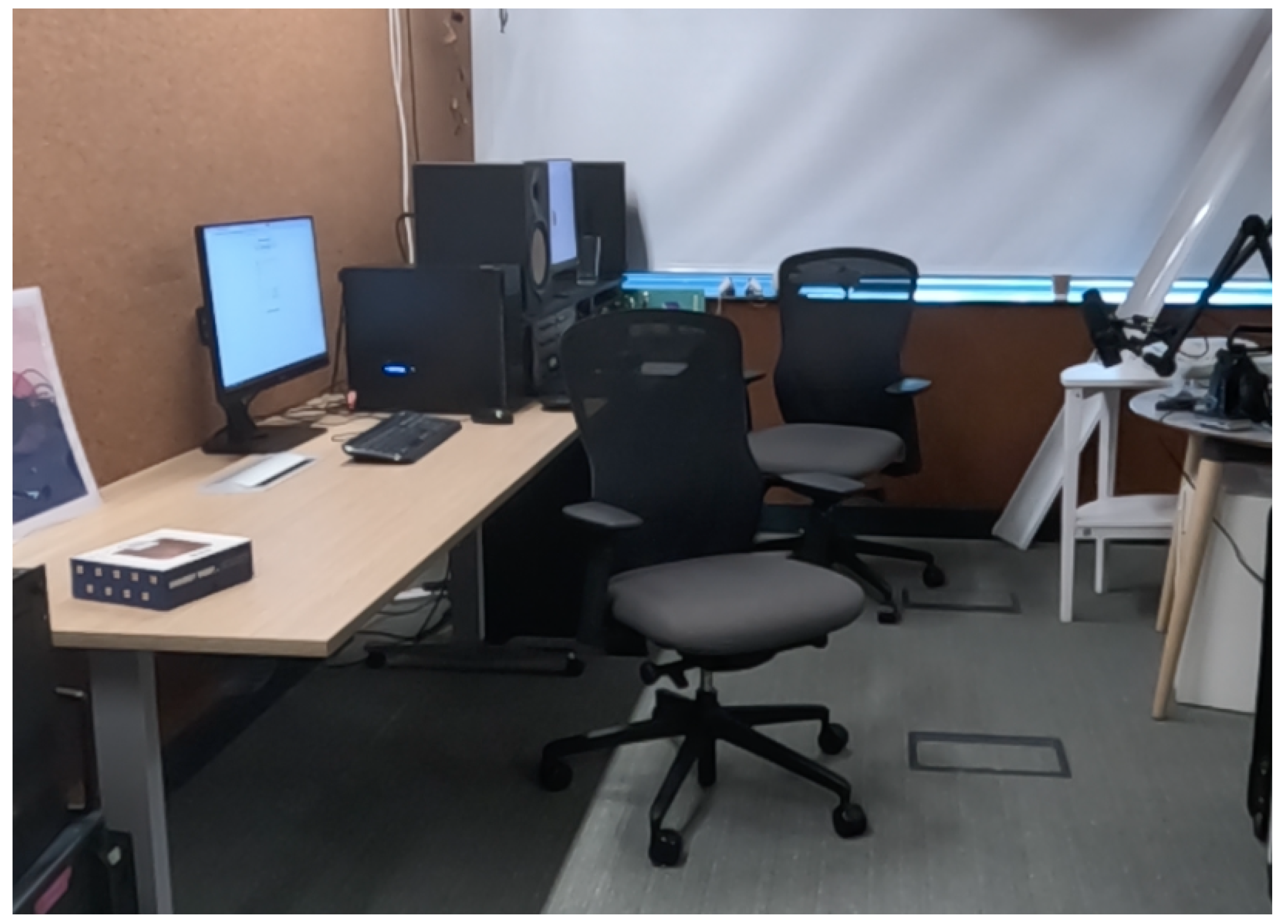
Figure 2.
Workshop Floor Plan.
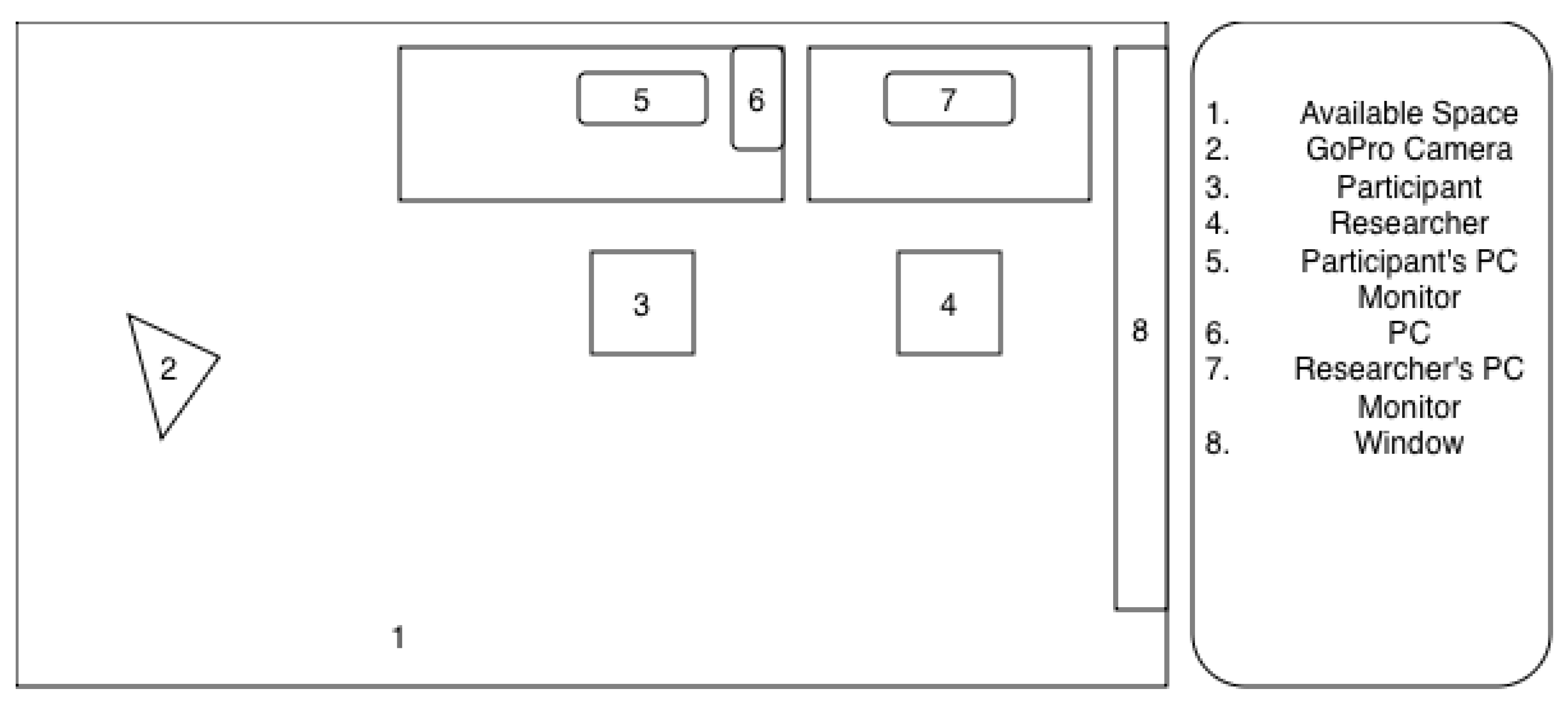
Figure 3.
GigXR DICOM XR Library Demonstrating Cut View [19].
Figure 3.
GigXR DICOM XR Library Demonstrating Cut View [19].
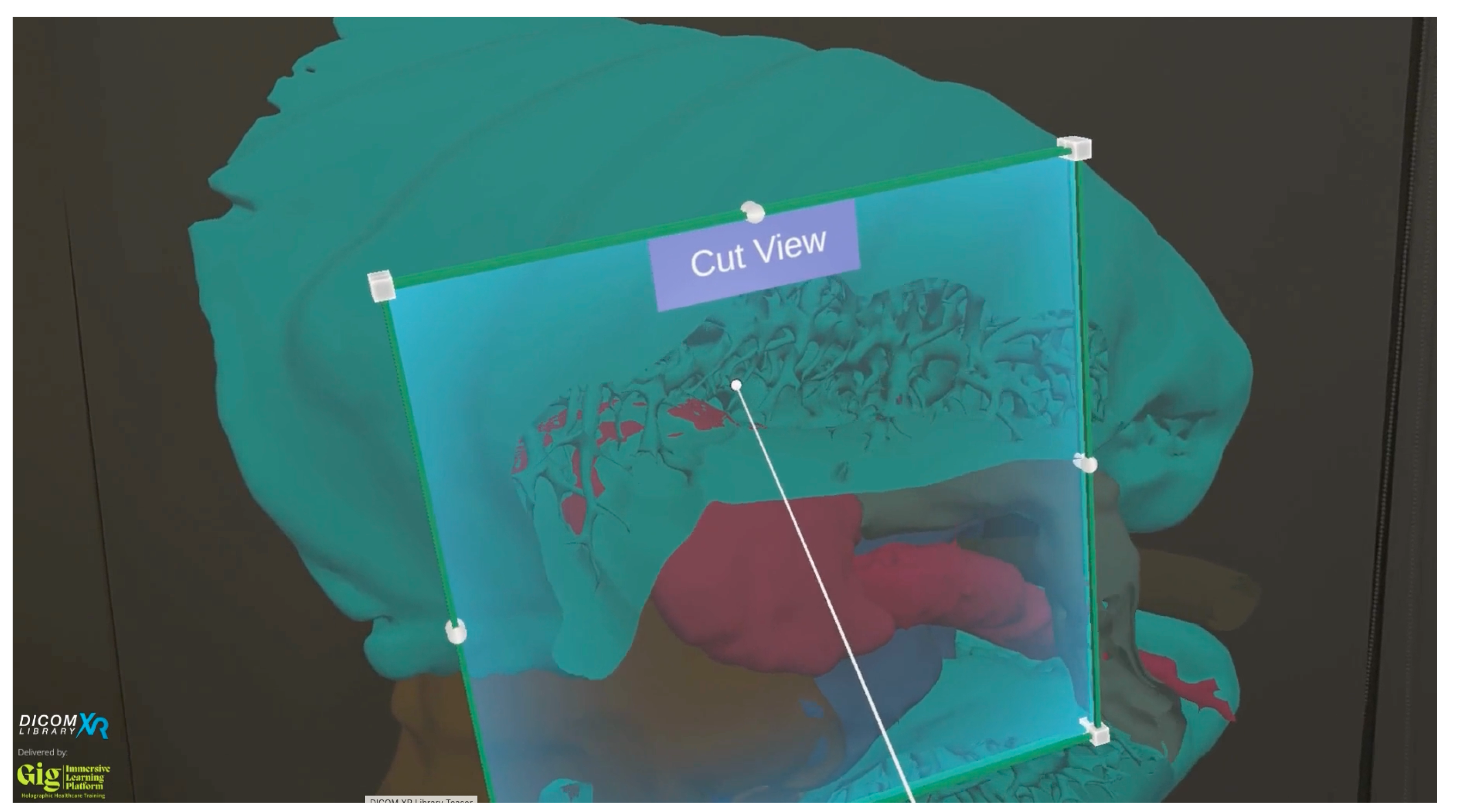
Figure 4.
Participant Manipulating Brain Model.
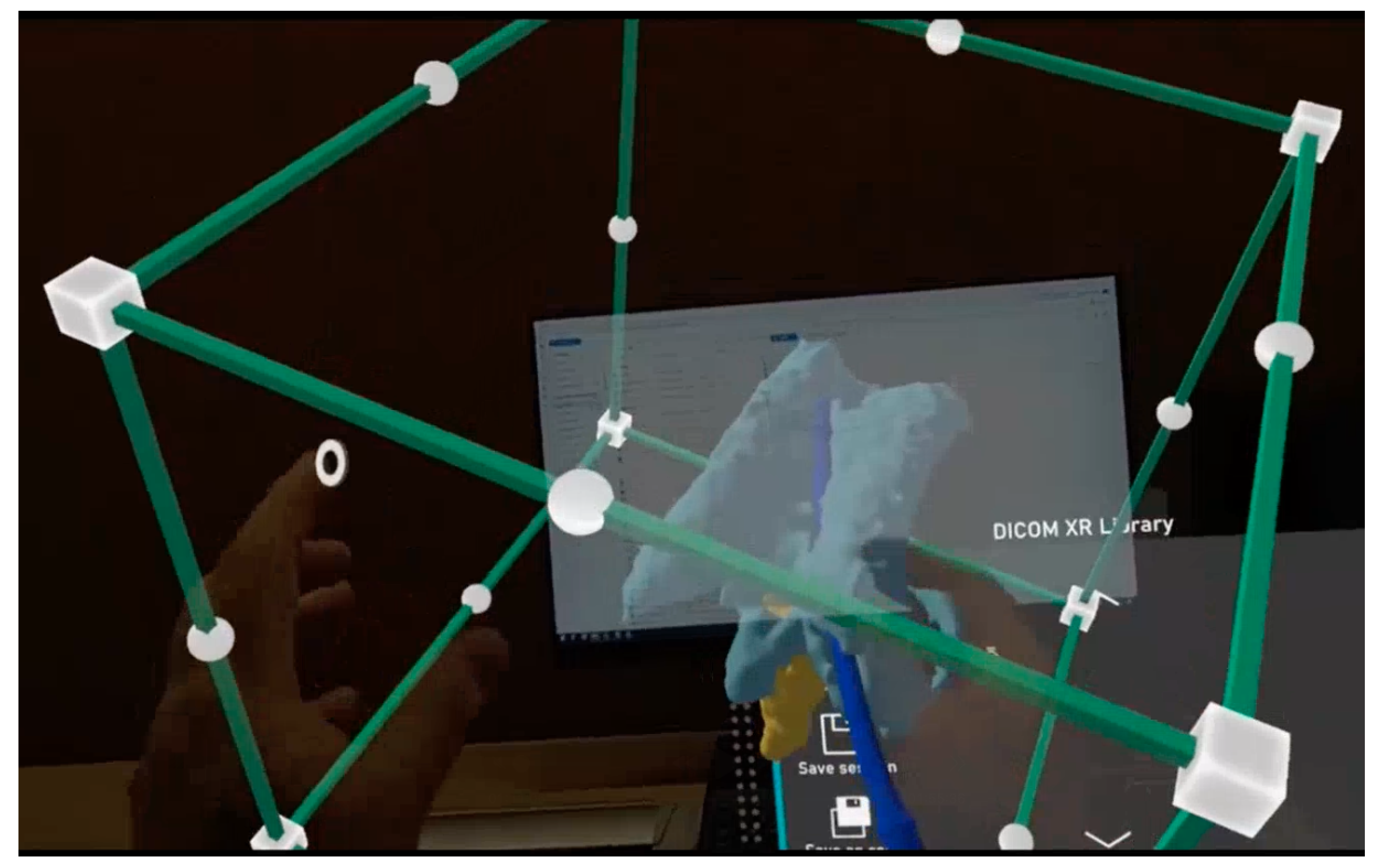
Figure 5.
GigXR DICOM XR Library Menu [19].
Figure 5.
GigXR DICOM XR Library Menu [19].
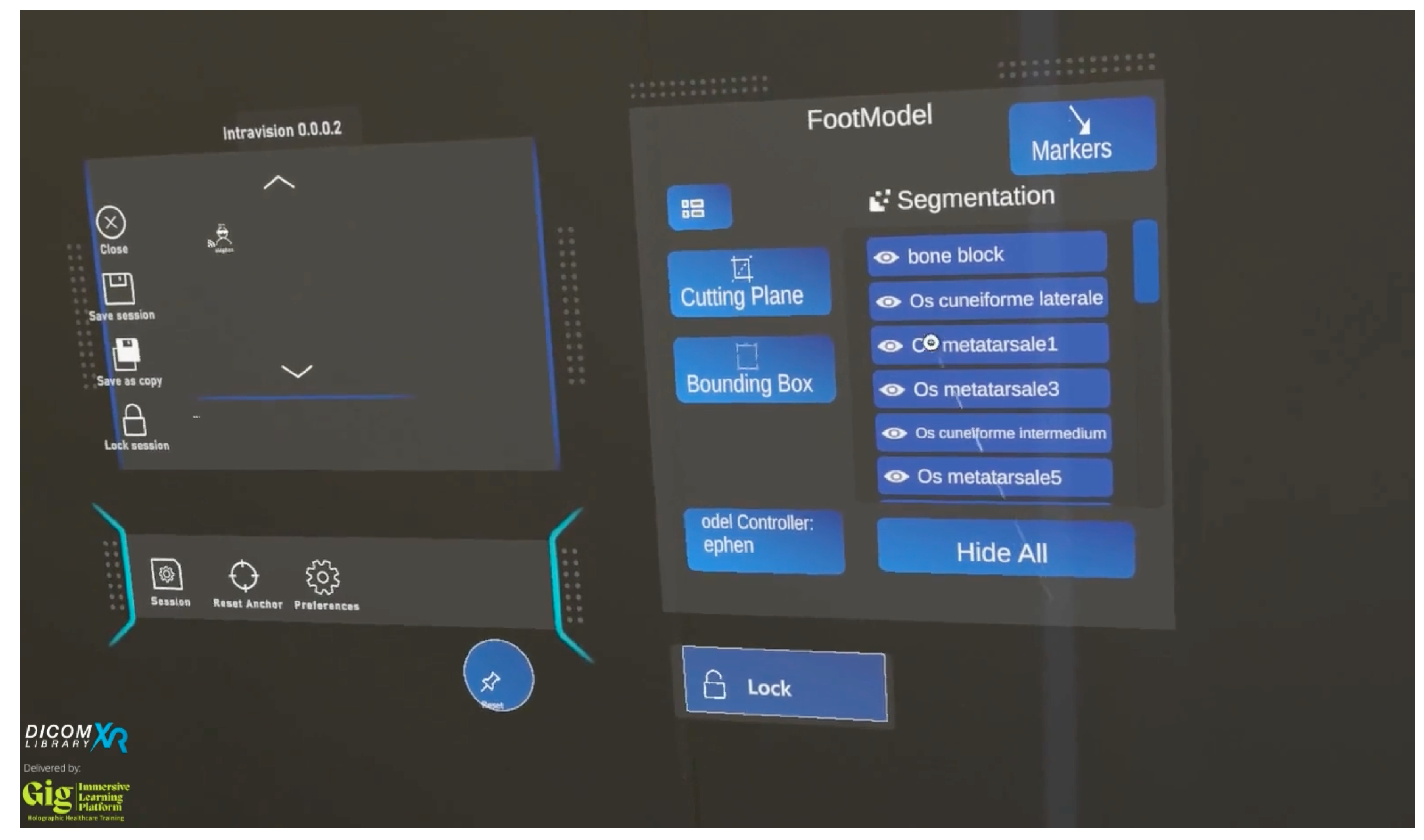
Figure 6.
Participant Exploring HoloHuman+ [20].
Figure 6.
Participant Exploring HoloHuman+ [20].
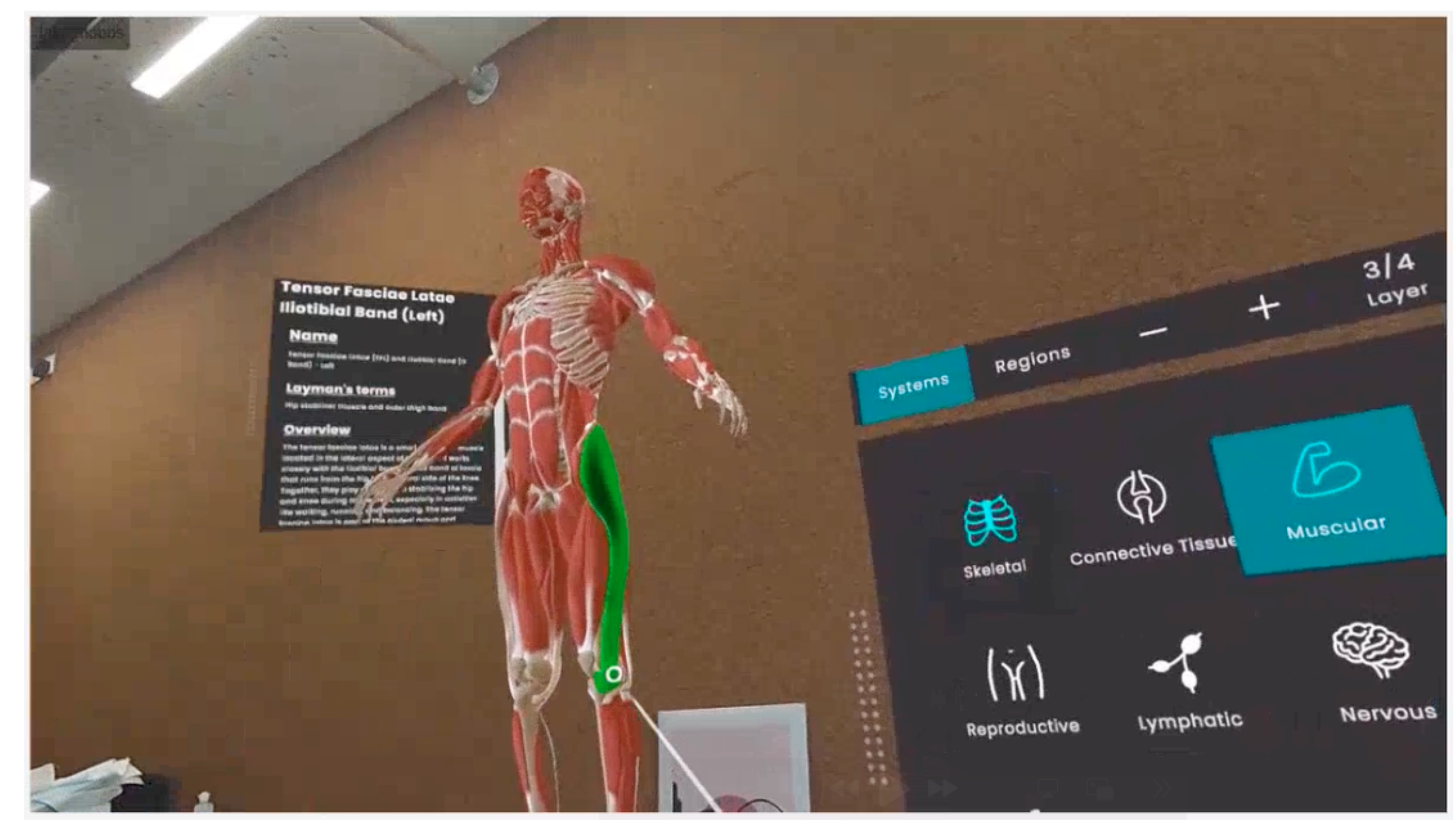
Figure 7.
GigXR HoloHuman+ Demonstration Video [21].
Figure 7.
GigXR HoloHuman+ Demonstration Video [21].
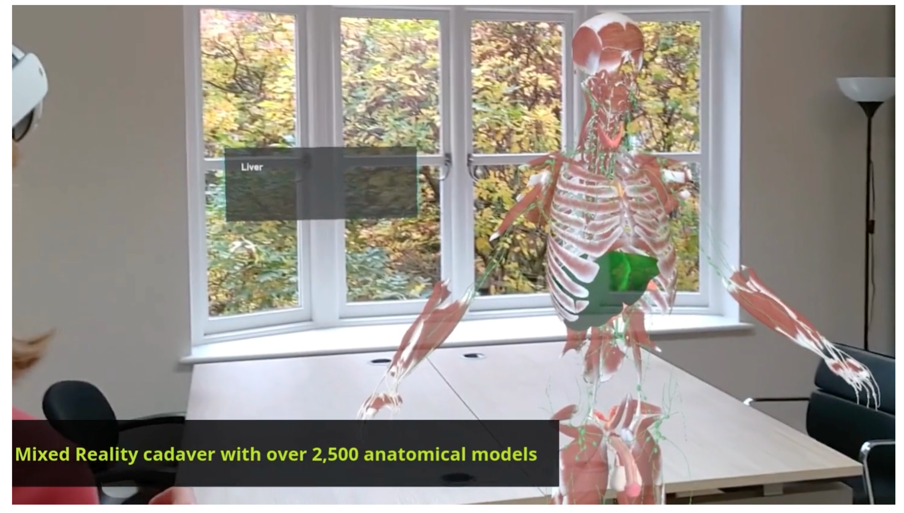
Figure 8.
Theme Map Showing Themes and Sub-Themes.
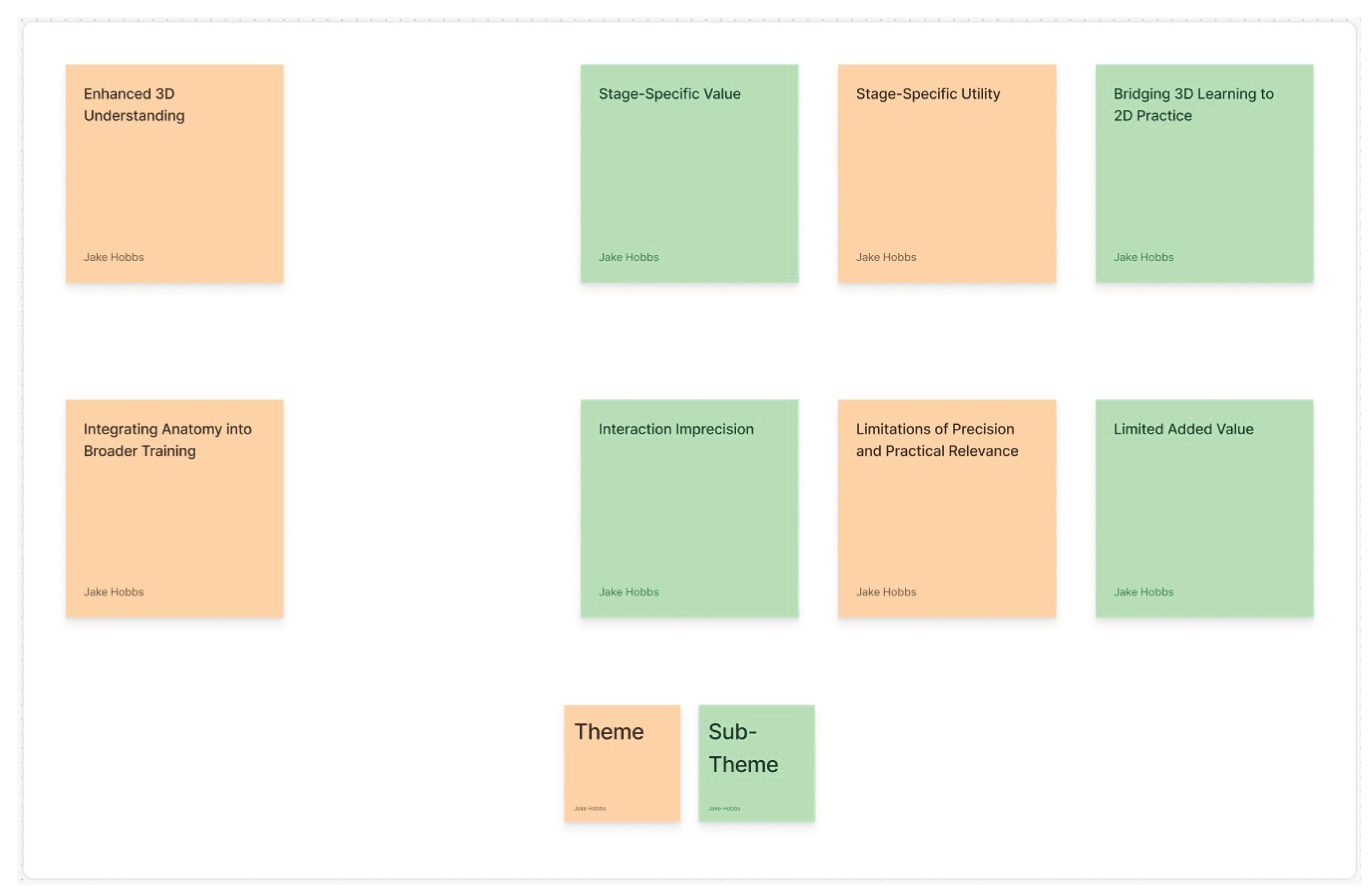

Table 1.
Workshop Studies Participant Demographic Information.
| ID | Age Range | Gender | Ethnicity | Role | Experience in Field | AR Experience |
|---|---|---|---|---|---|---|
| A | 25-34 | Male | Mixed white and Asian | Consultant Diagnostic Neuroradiologist | 3 years as consultant, 2 years fellowship in neuroradiology | 1 |
| B | 25-34 | Male | Mixed Afro-Caribbean and white British | Radiology Registrar - ST4 | 4 years | 5 |
| C | 25-34 | Male | White - Northern Irish | ST3 Clinical Radiology Registrar | 3 years speciality training/4 years as medical SHO before that | 2 |
| D | 25-34 | Male | Asian - other | Radiology Registrar | 4 years | 2 |
| E | 25-34 | Male | White British | ST4 Clinical Radiology | 4 years | 2 |
| F | 35-44 | Male | British | Radiology Consultant | 17 years | 1 |
| G | 45-54 | Female | White British | Consultant Radiologist | Consultant for 12 years. Radiologist for 20 | 2 |
Table 2.
Microsoft HoloLens 2 Tips App Hand Gestures Training Path.
| Task | Hand Gesture |
|---|---|
| Tap three holograms of gems | Select virtual objects within reach |
| `Air tap’ three holograms of gems | Select virtual objects out of reach |
| Move 3 holograms of flowers into a circle | Moving virtual objects from `A’ to `B’ |
| Make 3 holograms of flowers bigger or smaller | Change virtual object size |
| Rotate 3 holograms of flowers until they open | Rotating virtual objects |
| Open and close the start menu with two or just one hand | N/A |
| Exit an immersive app | N/A |
Table 3.
AR Experience Ratings.
| Experience Rating | Description |
|---|---|
| 1 | Heard of it but never used it |
| 2 | Experienced it once or twice; e.g. a demo |
| 3 | Occasional user |
| 4 | Frequent user |
| 5 | Own an AR or VR device |
Disclaimer/Publisher’s Note: The statements, opinions and data contained in all publications are solely those of the individual author(s) and contributor(s) and not of MDPI and/or the editor(s). MDPI and/or the editor(s) disclaim responsibility for any injury to people or property resulting from any ideas, methods, instructions or products referred to in the content. |
© 2026 by the authors. Licensee MDPI, Basel, Switzerland. This article is an open access article distributed under the terms and conditions of the Creative Commons Attribution (CC BY) license (http://creativecommons.org/licenses/by/4.0/).
Copyright: This open access article is published under a Creative Commons CC BY 4.0 license, which permit the free download, distribution, and reuse, provided that the author and preprint are cited in any reuse.