
Submitted:
17 July 2026
Posted:
20 July 2026
You are already at the latest version
Abstract
Background and Objectives: Inflammatory and nutritional impairment may contribute to adverse outcomes after permanent pacemaker implantation, but their prognostic value in degenerative complete atrioventricular block remains unclear. We evaluated the association between routinely available inflammatory and nutritional indices and long-term all-cause mortality in this population.
Materials and Methods: This retrospective single-center study included 272 patients who underwent permanent pacemaker implantation for isolated complete atrioventricular block attributed to degenerative conduction system disease between August 2020 and July 2024. Baseline laboratory values were used to calculate the prognostic nutritional index (PNI), geriatric nutritional risk index (GNRI), neutrophil-to-lymphocyte ratio, lymphocyte-to-monocyte ratio, systemic immune-inflammation index, systemic inflammation response index, and pan-immune-inflammation value. The primary endpoint was all-cause mortality during follow-up. Discriminatory performance was assessed by ROC analysis, and independent predictors were evaluated using multivariable Cox regression.
Results: During a mean follow-up of 27.8 ± 16.6 months, 69 patients (25.4%) died. Non-survivors were older and had lower hemoglobin, lymphocyte count, albumin, estimated glomerular filtration rate, GNRI, LMR, and PNI, whereas C-reactive protein, NLR, and SIRI were higher. PNI showed the highest discriminative ability for mortality prediction (AUC 0.720, 95% CI 0.648-0.793; p< 0.001), exceeding albumin, lymphocyte count, GNRI, and other inflammatory indices. In multivariable Cox regression, PNI remained independently associated with mortality (HR 0.945, 95% CI 0.909-0.983; p=0.004). Single-chamber pacemaker implantation was also associated with all-cause mortality (HR 2.137, 95% CI 1.202-3.802; p=0.010), although this should be interpreted in the context of baseline vulnerability and device-selection patterns.
Conclusions: PNI may provide useful prognostic information for mortality risk stratification in patients undergoing permanent pacemaker implantation for degenerative complete atrioventricular block.
Keywords:
complete atrioventricular block
; permanent pacemaker
; prognostic nutritional index
; geriatric nutritional risk index
; inflammatory indices
; nutritional status
; all-cause mortality
1. Introduction
Because of the increasing prevalence of degenerative conduction system disease and the expanding indications for cardiac pacing, the number of patients requiring permanent pacemaker implantation has been steadily increasing [1]. Despite pacemaker implantation, patients with advanced atrioventricular conduction disease continue to have increased mortality and morbidity compared with age-matched individuals without atrioventricular block [2]. In the literature, the CODE study demonstrated that patients with third-degree atrioventricular block had the highest mortality during follow-up [3].
Complete atrioventricular block requiring permanent pacemaker implantation may result from various causes, including medications, acute coronary syndromes, toxins, electrolyte disturbances, cardiac surgery, catheter-based interventions, and congenital conduction disorders [1]. However, in routine clinical practice, permanent pacemaker implantation is most frequently required in patients with conduction system disease secondary to degenerative processes [1]. This subgroup represents a clinically important population, as degenerative complete atrioventricular block often occurs in older and more vulnerable individuals with varying degrees of comorbidity, frailty, nutritional impairment, and systemic inflammation [4,5,6,7].
Several studies have evaluated predictors of mortality in patients with permanent pacemakers, identifying factors such as advanced age, diabetes mellitus, reduced left ventricular ejection fraction, anemia, renal dysfunction, and low physical activity [4,5,6]. In addition, serum albumin, a simple marker of nutritional status and systemic illness burden, has been suggested as a potential predictor of mortality in patients with permanent pacemakers [7]. However, serum albumin alone may not fully capture the complex interaction between nutritional reserve, immune competence, inflammatory status, and frailty, all of which may influence long-term outcomes after pacemaker implantation [7,8,9].
In this context, composite inflammatory and nutritional indices derived from routinely available laboratory parameters have gained increasing attention as prognostic markers in various clinical settings [8,9,10,11,12,13,14]. The prognostic nutritional index combines serum albumin and lymphocyte count, thereby reflecting both nutritional status and immune-inflammatory balance [8]. Similarly, the geriatric nutritional risk index has been proposed as a practical tool for assessing nutrition-related risk in older medical patients [9]. Other hematologic indices, such as the neutrophil-to-lymphocyte ratio, lymphocyte-to-monocyte ratio, systemic immune-inflammation index, systemic inflammation response index, and pan-immune-inflammation value, may reflect different aspects of systemic inflammation and immune dysregulation [10,11,12,13,14]. These indices are inexpensive, easily calculated, and widely available in daily clinical practice.
Nevertheless, the prognostic value of inflammatory and nutritional indices has not been well established in patients undergoing permanent pacemaker implantation for isolated complete atrioventricular block due to degenerative conduction system disease. Moreover, previous studies on pacemaker populations have generally included heterogeneous indications and have not specifically focused on this clinically uniform subgroup [4,5,6,7]. Therefore, the present study aimed to evaluate the association between routinely available inflammatory and nutritional indices and long-term all-cause mortality in patients undergoing permanent pacemaker implantation for degenerative complete atrioventricular block, and to determine whether these indices provide additional prognostic information beyond conventional clinical and laboratory parameters.
2. Materials and Methods
2.1. Study Population and Design
This retrospective, single-center observational study included patients who underwent permanent pacemaker implantation for complete atrioventricular block between August 2020 and July 2024. Patients were eligible for inclusion if permanent pacemaker implantation was performed for isolated complete atrioventricular block attributed to degenerative conduction system disease.
The exclusion criteria were pacemaker implantation for conduction disorders other than complete atrioventricular block; atrioventricular block attributable to reversible, secondary, iatrogenic, or congenital causes; drug-induced atrioventricular block; acute coronary syndrome or acute myocardial infarction; electrolyte disturbances; toxin-related atrioventricular block; acute systemic illness; active infection; atrioventricular block following transcatheter aortic valve implantation or cardiac surgery; post-ablation atrioventricular block; congenital heart block; age < 18 years; and the presence of rheumatic, hematologic, malignant, chronic inflammatory, or autoimmune diseases. Patients with heart failure who met standard clinical indications for cardiac resynchronization therapy were managed accordingly and were not included in the conventional pacemaker group when cardiac resynchronization therapy was required.
Degenerative conduction system disease was therefore defined clinically as isolated complete atrioventricular block requiring permanent pacemaker implantation after exclusion of the above potentially reversible, secondary, iatrogenic, congenital, inflammatory, infectious, malignant, or hematologic conditions.
Demographic characteristics, comorbidities, pacemaker type, and laboratory parameters were obtained from institutional medical records. Pacemaker type was categorized as single-chamber or dual-chamber pacemaker implantation. Heart failure, coronary artery disease, diabetes mellitus, hypertension, and atrial fibrillation were recorded according to the patients’ medical history and available clinical documentation.
Laboratory measurements and calculation of inflammatory and nutritional indices.
Baseline laboratory parameters obtained before pacemaker implantation were recorded. These included hemoglobin, white blood cell count, neutrophil count, lymphocyte count, monocyte count, platelet count, serum creatinine, estimated glomerular filtration rate, serum albumin, total protein, C-reactive protein, electrolytes, and other routine biochemical parameters. The estimated glomerular filtration rate was calculated using the Chronic Kidney Disease Epidemiology Collaboration (CKD-EPI) creatinine equation [15].
Inflammatory and nutritional indices were calculated from baseline laboratory values according to previously described formulas. The prognostic nutritional index was calculated using the following formula [8]:
PNI = serum albumin (g/L) + 5 × lymphocyte count (109/L)
The geriatric nutritional risk index was calculated as follows [9]:
GNRI = 1.489 × serum albumin (g/L) + 41.7 × (actual body weight/ideal body weight)
When actual body weight exceeded ideal body weight, the weight ratio was set to 1. The neutrophil-to-lymphocyte ratio, lymphocyte-to-monocyte ratio, systemic immune-inflammation index, systemic inflammation response index, and pan-immune-inflammation value were calculated using the following formulas [10,11,12,13,14]:
NLR = neutrophil count/lymphocyte count
LMR = lymphocyte count/monocyte count
SII = platelet count × neutrophil count/lymphocyte count
SIRI = neutrophil count × monocyte count/lymphocyte count
PIV = platelet count × neutrophil count × monocyte count/lymphocyte count
All indices were calculated using laboratory measurements obtained at baseline before pacemaker implantation.
2.2. Study Endpoint and Follow-Up
The primary endpoint of the study was all-cause mortality during follow-up. Survival status and dates of death were obtained from institutional records and available electronic health records. Follow-up duration was calculated from the date of pacemaker implantation to the date of death or the last available clinical follow-up.
2.3. Statistical Analysis
Statistical analyses were performed using IBM SPSS Statistics version 25. Categorical variables were presented as numbers and percentages, whereas continuous variables were presented as mean ± standard deviation or median (interquartile range), according to their distribution. The distribution of continuous variables was assessed using appropriate normality tests and visual inspection of histograms. Categorical variables were compared using the chi-square test or Fisher’s exact test, as appropriate. Continuous variables with normal distribution were compared using the independent samples t-test, whereas non-normally distributed variables were compared using the Mann–Whitney U test.
Conventional receiver operating characteristic (ROC) curve analysis was performed to evaluate the discriminatory performance of inflammatory, nutritional, and conventional laboratory parameters for predicting all-cause mortality. The area under the curve (AUC), 95% confidence interval, and p value were reported for each variable. For variables inversely associated with mortality, AUC values were reported according to the direction of increased risk. The optimal cut-off value for each evaluated parameter was determined using the Youden index, and the corresponding sensitivity, specificity, positive predictive value, negative predictive value, and Youden index were reported. Kaplan-Meier survival curves were generated according to the optimal PNI cut-off value and compared using the log-rank test.
Cox proportional hazards regression analysis was used to identify independent predictors of all-cause mortality. Follow-up duration was entered as the time variable, and all-cause mortality status was entered as the event variable. Variables that were statistically significant in univariate analyses or considered clinically relevant were included in the multivariable Cox regression model. To reduce the risk of model overfitting in the context of the observed number of mortality events, the multivariable model was kept parsimonious and limited to clinically relevant variables. The final model included PNI, hemoglobin, estimated glomerular filtration rate, age, pacemaker type, heart failure, and atrial fibrillation. Hazard ratios with 95% confidence intervals and p values were reported. A two-sided p value <0.05 was considered statistically significant.
Ethical approval details are provided in the Institutional Review Board Statement Section.
3. Results
A total of 272 patients who underwent permanent pacemaker implantation for degenerative complete atrioventricular block were included. During a mean follow-up of 27.8 ± 16.6 months, all-cause mortality occurred in 69 patients (25.4%). Baseline categorical characteristics, continuous laboratory parameters, ROC curve analysis, and multivariable Cox proportional hazards regression findings are presented below.

Table 1.
Baseline categorical characteristics according to mortality status.
| Variable | Survivors (n = 203) | Non-Survivors (n = 69) | p Value |
|---|---|---|---|
| Heart failure | 39/67 (58.2%) | 28/67 (41.8%) | <0.001 a |
| Coronary artery disease | 71/102 (69.6%) | 31/102 (30.4%) | 0.116 a |
| Diabetes mellitus | 67/97 (69.1%) | 30/97 (30.9%) | 0.102 a |
| Hypertension | 128/175 (73.1%) | 47/175 (26.9%) | 0.444 a |
| Atrial fibrillation | 53/80 (66.3%) | 27/80 (33.8%) | 0.033 a |
| Sex | 0.581 a | ||
| Male | 104/142 (73.2%) | 38/142 (26.8%) | |
| Female | 99/130 (76.2%) | 31/130 (23.8%) | |
| Pacemaker type | <0.001 a | ||
| Single-chamber | 32/59 (54.2%) | 27/59 (45.8%) | |
| Dual-chamber | 171/213 (80.3%) | 42/213 (19.7%) |
a Pearson chi-square test.
Patients with heart failure and atrial fibrillation had significantly higher mortality rates. Observed mortality was also significantly higher among patients implanted with single-chamber pacemakers compared with those implanted with dual-chamber pacemakers.
Table 2.
Comparison of continuous laboratory parameters and inflammatory/nutritional indices according to mortality status.
Table 2.
Comparison of continuous laboratory parameters and inflammatory/nutritional indices according to mortality status.
| Variable | Survivors (n = 203) | Non-Survivors (n = 69) | p Value |
|---|---|---|---|
| Hemoglobin, g/dL | 12.38 ± 1.93 | 11.03 ± 2.19 | <0.001 b |
| WBC, 109/L | 8.54 (3.03) | 8.56 (6.12) | 0.761 c |
| Neutrophil count, 109/L | 5.50 (3.32) | 5.76 (6.16) | 0.526 c |
| Lymphocyte count, 109/L | 1.75 (1.23) | 1.36 (1.11) | 0.001 c |
| Monocyte count, 109/L | 0.67 (0.32) | 0.85 (0.64) | 0.165 c |
| Platelet count, 109/L | 221.66 ± 65.07 | 220.98 ± 75.10 | 0.943 b |
| AST, U/L | 19.00 (11.00) | 29.00 (28.75) | 0.559 c |
| ALT, U/L | 16.00 (14.00) | 15.50 (20.25) | 0.056 c |
| Creatinine, mg/dL | 1.08 (0.60) | 1.37 (1.13) | <0.001 c |
| Total protein, g/L | 66.00 (10.00) | 64.50 (14.00) | 0.133 c |
| Albumin, g/L | 40.00 (6.00) | 34.50 (7.00) | <0.001 c |
| Total cholesterol, mg/dL | 161.00 (55.00) | 143.00 (70.00) | 0.390 c |
| Triglycerides, mg/dL | 112.00 (79.00) | 107.50 (77.00) | 0.492 c |
| Sodium, mmol/L | 139.00 (4.00) | 138.00 (6.50) | 0.094 c |
| Potassium, mmol/L | 4.45 ± 0.60 | 4.54 ± 0.59 | 0.283 b |
| Age, years | 72.00 (13.75) | 83.50 (12.25) | <0.001 c |
| BMI, kg/m2 | 28.65 (6.27) | 25.71 (7.37) | 0.125 c |
| CRP, mg/L | 5.00 (18.75) | 16.40 (30.75) | 0.016 c |
| GFR, mL/min/1.73 m2 | 60.40 (40.68) | 41.70 (32.10) | <0.001 c |
| GNRI | 111.99 ± 11.04 | 105.05 ± 13.71 | <0.001 b |
| NLR | 2.77 (2.89) | 3.53 (5.29) | 0.012 c |
| LMR | 2.69 (1.31) | 1.61 (1.82) | <0.001 c |
| PIV | 384.97 (430.20) | 504.24 (1751.68) | 0.068 c |
| PNI | 49.28 ± 7.14 | 44.00 ± 6.30 | <0.001 b |
| SII | 590.76 (671.56) | 727.02 (1468.36) | 0.061 c |
| SIRI | 1.82 (1.68) | 2.35 (6.18) | 0.019 c |
Values are presented as mean ± standard deviation for variables analyzed using the independent samples t-test and as median (interquartile range) for variables analyzed using the Mann-Whitney U test. b Independent samples t-test; c Mann-Whitney U test. ALT, alanine aminotransferase; AST, aspartate aminotransferase; BMI, body mass index; CRP, C-reactive protein; GFR, glomerular filtration rate; GNRI, geriatric nutritional risk index; LMR, lymphocyte-to-monocyte ratio; NLR, neutrophil-to-lymphocyte ratio; PIV, pan-immune-inflammation value; PNI, prognostic nutritional index; SII, systemic immune-inflammation index; SIRI, systemic inflammation response index; WBC, white blood cell count.
Non-survivors were older and had lower hemoglobin, lymphocyte count, albumin, GFR, GNRI, LMR, and PNI values. In contrast, CRP, NLR, and SIRI were significantly higher among non-survivors. PIV and SII were numerically higher in non-survivors but did not reach statistical significance.
Receiver operating characteristic curve analysis was performed to evaluate the discriminatory performance of inflammatory, nutritional, and conventional laboratory parameters for all-cause mortality. Detailed ROC-derived cut-off values and diagnostic performance metrics are presented in Table 3. Among all evaluated variables, PNI demonstrated the highest discriminative ability, with an AUC of 0.720 (95% CI: 0.648–0.793; p < 0.001). At the optimal cut-off value of ≤48.90, PNI yielded a sensitivity of 82.8%, specificity of 53.4%, PPV of 39.0%, NPV of 89.6%, and Youden index of 0.362. Albumin, GFR, hemoglobin, GNRI, age, LMR, and lymphocyte count also showed significant discriminatory performance. In contrast, SII, SIRI, NLR, and PIV had lower AUC values and did not reach statistical significance. These findings suggest that PNI may provide a more comprehensive prognostic assessment than isolated inflammatory or nutritional markers in this patient population.
In ROC curve analysis, the prognostic nutritional index (PNI) emerged as the most informative parameter for mortality prediction among the evaluated markers. The optimal PNI cut-off value, determined using the Youden index, was ≤48.90, yielding a sensitivity of 82.8% and a specificity of 53.4%. Therefore, a PNI value of 48.90 or lower was considered the optimal threshold for identifying patients at increased mortality risk. Although this cut-off showed relatively modest specificity, its high sensitivity supports the potential role of PNI as a practical screening marker for risk stratification (Figure 1).
Kaplan–Meier survival analysis demonstrated significantly lower cumulative survival in patients with a PNI ≤ 48.90 compared with those with a PNI > 48.90. The difference between the survival curves was statistically significant according to the log-rank test (χ2 = 23.435, df = 1, p < 0.001), indicating that reduced PNI was associated with an increased risk of mortality during follow-up (Figure 2).
Table 4.
Multivariable Cox proportional hazards regression analysis for all-cause mortality.
| Variable | HR | 95% CI | p Value |
|---|---|---|---|
| PNI | 0.945 | 0.909–0.983 | 0.004 |
| Single-chamber pacemaker vs. dual-chamber pacemaker | 2.137 | 1.202–3.802 | 0.010 |
| Hemoglobin, g/dL | 0.882 | 0.770–1.009 | 0.067 |
| GFR, mL/min/1.73 m2 | 0.992 | 0.982–1.002 | 0.125 |
| Age, years | 1.016 | 0.991–1.042 | 0.232 |
| Heart failure | 1.258 | 0.690–2.290 | 0.453 |
| Atrial fibrillation | 0.907 | 0.501–1.642 | 0.746 |
CI, confidence interval; GFR, estimated glomerular filtration rate; HR, hazard ratio; PNI, prognostic nutritional index. Continuous predictors are expressed per 1-unit increase.
In multivariable Cox proportional hazards regression analysis, PNI remained independently associated with all-cause mortality after adjustment for hemoglobin, renal function, age, pacemaker type, heart failure, and atrial fibrillation. Each 1-point increase in PNI was associated with a 5.5% lower risk of mortality (HR: 0.945, 95% CI: 0.909–0.983; p = 0.004). In addition, single-chamber pacemaker implantation was independently associated with all-cause mortality compared with dual-chamber pacemaker implantation (HR: 2.137, 95% CI: 1.202–3.802; p = 0.010).
In the supplementary analysis according to pacemaker type, patients receiving single-chamber pacemakers had a less favorable baseline clinical and laboratory profile than those receiving dual-chamber pacemakers. Single-chamber pacemaker recipients were older, had lower hemoglobin, albumin, PNI, and GFR values, and had higher rates of atrial fibrillation, heart failure, and all-cause mortality. These findings suggest that the association between pacemaker type and mortality should be interpreted cautiously, as it may partly reflect baseline clinical and nutritional vulnerability as well as device-selection patterns. These data are presented in Supplementary Table S1.
4. Discussion
In this retrospective study of patients undergoing permanent pacemaker implantation for degenerative complete atrioventricular block, the main finding was that inflammatory and nutritional status was closely associated with long-term all-cause mortality. Among the evaluated inflammatory, nutritional, and conventional laboratory parameters, PNI demonstrated the highest discriminative ability for mortality prediction and remained independently associated with all-cause mortality after adjustment for hemoglobin, renal function, age, pacemaker type, heart failure, and atrial fibrillation. Importantly, PNI showed greater discriminatory performance than either serum albumin or lymphocyte count alone, suggesting that an integrated immune-nutritional marker may provide more clinically relevant prognostic information than isolated laboratory parameters.
The present study focused specifically on a homogeneous population with isolated complete atrioventricular block attributed to degenerative conduction system disease. This is clinically relevant because complete atrioventricular block may result from several secondary or iatrogenic conditions, whereas degenerative conduction system disease usually occurs in older and more vulnerable patients. Previous studies evaluating mortality after permanent pacemaker implantation have identified advanced age, diabetes mellitus, reduced left ventricular ejection fraction, anemia, renal dysfunction, and low physical activity as important predictors of adverse outcomes [4,5,6]. However, these studies generally included heterogeneous pacing indications. By restricting the study population to degenerative complete atrioventricular block, the present analysis allowed a more focused evaluation of systemic vulnerability, nutritional status, and inflammation in a clinically uniform subgroup.
PNI was originally developed as a nutritional and immunological index using serum albumin and lymphocyte count [8]. Serum albumin reflects protein-calorie reserve, hepatic synthetic function, inflammation-related capillary leakage, and overall disease burden, whereas lymphocyte count reflects immune competence and may decrease in chronic inflammation, malnutrition, and physiological stress. Therefore, PNI may capture the interaction between malnutrition and immune-inflammatory dysfunction more comprehensively than albumin or lymphocyte count alone. This concept is supported by our ROC analysis, in which PNI had a higher AUC than albumin and lymphocyte count. Moreover, although albumin, lymphocyte count, GNRI, LMR, and several inflammatory markers differed between survivors and non-survivors, only PNI remained independently associated with mortality in the multivariable model.
The prognostic importance of albumin in cardiovascular disease has been increasingly recognized. In patients with permanent pacemakers, Hayiroglu et al. reported that serum albumin was associated with long-term mortality in patients with dual-chamber pacemakers [7]. In broader cardiovascular populations, low albumin has also been linked to adverse outcomes and may represent a global marker of nutritional impairment, chronic inflammation, frailty, and reduced physiological reserve [16,17]. Our findings extend these observations by showing that PNI, which integrates albumin with lymphocyte count, outperformed albumin alone for mortality prediction in patients with degenerative complete atrioventricular block. This suggests that the prognostic signal in this population may not be explained solely by hypoalbuminemia, but rather by a broader immune-nutritional impairment.
Other inflammatory and nutritional indices also provided relevant information. GNRI, LMR, NLR, and SIRI differed significantly between survivors and non-survivors, whereas SII and PIV were numerically higher among non-survivors but did not reach statistical significance. GNRI has been proposed as a practical tool for assessing nutrition-related risk in older medical patients [9], while hematologic indices such as NLR, LMR, SII, SIRI, and PIV reflect different components of systemic inflammation and immune dysregulation [10,11,12,13,14]. However, in our cohort, their discriminatory performance was lower than that of PNI. These findings suggest that, in degenerative complete atrioventricular block, the combined effect of nutritional depletion and immune-inflammatory impairment may be more prognostically relevant than isolated inflammatory activation.
Pacemaker type was also associated with mortality in the multivariable analysis. Patients implanted with single-chamber pacemakers had higher observed all-cause mortality than those implanted with dual-chamber pacemakers. Previous studies have similarly reported worse outcomes among patients receiving single-chamber pacemakers, although the magnitude and interpretation of this association may vary according to patient characteristics and pacing indication [18,19,20,21,22]. Several mechanisms may contribute to differences in outcomes between pacing modes, including loss of atrioventricular synchrony, reduced chronotropic response, right ventricular pacing-related dyssynchrony, and lower functional capacity [23]. However, this finding should not be interpreted as evidence of a direct causal effect of single-chamber pacing. In routine clinical practice, device selection is influenced by rhythm status, comorbidity burden, frailty, expected functional benefit, and physician judgment. Consistent with this selection pattern, the supplementary comparison according to pacemaker type showed that patients receiving single-chamber pacemakers were older and had higher rates of atrial fibrillation and heart failure, as well as lower hemoglobin, albumin, GFR, and PNI values. Therefore, the higher mortality observed in the single-chamber group may partly reflect baseline clinical and nutritional vulnerability and confounding by indication rather than pacing mode alone.
The clinical implication of this study is that PNI may serve as a simple, inexpensive, and widely available marker for risk stratification in patients undergoing permanent pacemaker implantation for degenerative complete atrioventricular block. As PNI can be calculated from routine preprocedural laboratory tests, it may help identify patients who require closer clinical follow-up, careful assessment of frailty and nutritional status, and optimization of modifiable systemic risk factors. However, whether interventions targeting nutritional status or systemic inflammation can improve outcomes in this population remains uncertain and should be evaluated in future prospective studies.
The mortality rate observed in our cohort may also reflect the specific characteristics of the study population. The Czech national registry reported higher long-term survival among patients undergoing pacemaker implantation [24]. In contrast, our study exclusively included patients with degenerative complete atrioventricular block and excluded secondary, iatrogenic, and congenital causes, thereby focusing on a more clinically uniform but potentially more vulnerable group. This strict selection increases the internal consistency of the cohort and supports the relevance of immune-nutritional risk stratification in this specific population.
5. Limitations
This study has several limitations. First, the retrospective and single-center design may limit the generalizability of the findings and does not allow causal inference. Second, although PNI was independently associated with mortality, frailty, nutritional status, functional capacity, and sarcopenia were not directly assessed using validated scales. Third, inflammatory and nutritional indices were calculated from baseline laboratory values only; therefore, longitudinal changes in PNI, albumin, lymphocyte count, or inflammatory markers could not be evaluated. Fourth, pacemaker type was not randomly assigned, and the association between single-chamber pacemaker implantation and mortality may be influenced by confounding by indication, baseline clinical vulnerability, and device-selection patterns. Fifth, detailed echocardiographic parameters, including left ventricular ejection fraction, chamber dimensions, and valvular variables, were not systematically available and therefore could not be included in the multivariable analysis. Sixth, data on pacing burden, device programming, and cause-specific mortality were not available in sufficient detail. Seventh, conventional ROC analysis does not fully account for time-to-event information; therefore, the ROC-derived cut-off value and discriminatory findings should be interpreted as exploratory. Finally, external validation in larger prospective cohorts is required to confirm the prognostic value of PNI in this patient population.
6. Conclusions
In patients undergoing permanent pacemaker implantation for degenerative complete atrioventricular block, inflammatory and nutritional status was closely associated with long-term all-cause mortality. Among the evaluated inflammatory, nutritional, and conventional laboratory parameters, PNI showed the highest discriminative ability and remained independently associated with mortality after adjustment for clinically relevant variables. PNI provided greater prognostic information than albumin or lymphocyte count alone, suggesting that combined immune-nutritional assessment may be particularly useful in this vulnerable population. Single-chamber pacemaker implantation was also associated with higher observed mortality; however, this relationship should be interpreted cautiously because it may partly reflect baseline frailty, clinical vulnerability, and device-selection patterns. Future prospective studies are needed to validate these findings and to determine whether targeted nutritional or frailty-oriented interventions can improve outcomes after pacemaker implantation.
Supplementary Materials
The following supporting information can be downloaded at the website of this paper posted on Preprints.org, Table S1: Baseline clinical and laboratory characteristics according to pacemaker type.
Author Contributions
Conceptualization, M.E.A.; Methodology, M.E.A. and V.P.; Software, M.E.A.; Validation, M.E.A., V.P., S.B. and T.A.; Formal Analysis, M.E.A.; Investigation, M.E.A., C.O., G.I.B.A., M.K., E.O., V.P., S.B. and T.A.; Resources, M.E.A., V.P., S.B. and T.A.; Data Curation, M.E.A., C.O., G.I.B.A., M.K. and E.O.; Writing—Original Draft Preparation, M.E.A.; Writing—Review and Editing, M.E.A., V.P., S.B. and T.A.; Visualization, M.E.A.; Supervision, V.P., S.B. and T.A.; Project Administration, M.E.A. All authors have read and agreed to the published version of the manuscript.
Funding
This research received no external funding.
Institutional Review Board Statement
The study was conducted in accordance with the Declaration of Helsinki and approved by the Ethics Committee of Istanbul Başakşehir Çam and Sakura City Hospital Training and Research Hospital (approval number: 2025-297; date of approval: 24 June 2025).
Informed Consent Statement
Patient consent was waived due to the retrospective design of the study, the use of anonymized data, and the absence of identifiable patient information.
Data Availability Statement
The data presented in this study are available from the corresponding author upon reasonable request due to privacy and ethical restrictions.
Acknowledgments
Not applicable.
Conflicts of Interest
The authors declare no conflict of interest.
References
- Glikson, M.; Nielsen, J.C.; Kronborg, M.B.; Blomström-Lundqvist, C.; Auricchio, A.; Barra, S.; et al. 2021 ESC Guidelines on cardiac pacing and cardiac resynchronization therapy. Eur. Heart J. 2021, 42, 3427–3520. [Google Scholar] [CrossRef] [PubMed]
- Simon, A.B.; Zloto, A.E. Atrioventricular block: Natural history after permanent ventricular pacing. Am. J. Cardiol. 1978, 41, 500–507. [Google Scholar] [CrossRef] [PubMed]
- Paixão, G.M.M.; Lima, E.M.; Quadros, A.B.; de Souza e Silva, C.G.; da Silva, K.R.; Gomes, P.R.; et al. Association between atrioventricular block and mortality in primary care patients: The CODE study. Arq. Bras. Cardiol. 2022, 119, 564–571. [Google Scholar] [CrossRef] [PubMed]
- Mittal, G.K.; Priya, D.; Anantharaj, A.; Sharma, V.; Singh, S.; Kumar, R.; et al. Risk factors for mortality in patients undergoing permanent pacemaker implant. Indian J. Clin. Cardiol. 2025. [Google Scholar] [CrossRef]
- Goto, T.; Mori, K.; Nakasuka, K.; Sakamoto, Y.; Tsutsui, H.; Yamamoto, K. Physical activity and mortality in older patients with a pacemaker. Geriatr. Gerontol. Int. 2020, 20, 106–111. [Google Scholar] [CrossRef] [PubMed]
- Cheng, C.W.; Wang, C.H.; Chen, W.S.; Chen, C.C.; Chen, Y.C.; Lin, L.J.; et al. Predictors of long-term survival prior to permanent pacemaker implantation in octogenarians or older. Aging Clin. Exp. Res. 2019, 31, 1001–1009. [Google Scholar] [CrossRef]
- Hayıroğlu, M.I.; Cınar, T.; Cinier, G.; Pay, L.; Şahin, M.; Selçuk, M.; et al. Prognostic value of serum albumin for long-term mortality in patients with dual-chamber permanent pacemakers. Biomark. Med. 2022, 16, 341–348. [Google Scholar] [CrossRef]
- Onodera, T.; Goseki, N.; Kosaki, G. Prognostic nutritional index in gastrointestinal surgery of malnourished cancer patients. Nihon Geka Gakkai Zasshi 1984, 85, 1001–1005. [Google Scholar] [PubMed]
- Bouillanne, O.; Morineau, G.; Dupont, C.; Coulombel, I.; Vincent, J.P.; Nicolis, I.; et al. Geriatric Nutritional Risk Index: A new index for evaluating at-risk elderly medical patients. Am. J. Clin. Nutr. 2005, 82, 777–783. [Google Scholar] [CrossRef] [PubMed]
- Walsh, S.R.; Cook, E.J.; Goulder, F.; Justin, T.A.; Keeling, N.J. Neutrophil-lymphocyte ratio as a prognostic factor in colorectal cancer. J. Surg. Oncol. 2005, 91, 181–184. [Google Scholar] [CrossRef] [PubMed]
- Nishijima, T.F.; Muss, H.B.; Shachar, S.S.; Tamura, K.; Takamatsu, Y. Prognostic value of lymphocyte-to-monocyte ratio in patients with solid tumors: A systematic review and meta-analysis. Cancer Treat. Rev. 2015, 41, 971–978. [Google Scholar] [CrossRef] [PubMed]
- Hu, B.; Yang, X.R.; Xu, Y.; Sun, Y.F.; Sun, C.; Guo, W.; et al. Systemic immune-inflammation index predicts prognosis of patients after curative resection for hepatocellular carcinoma. Clin. Cancer Res. 2014, 20, 6212–6222. [Google Scholar] [CrossRef] [PubMed]
- Qi, Q.; Zhuang, L.; Shen, Y.; Geng, Y.; Yu, S.; Chen, H.; et al. A novel systemic inflammation response index for predicting the survival of patients with pancreatic cancer after chemotherapy. Cancer 2016, 122, 2158–2167. [Google Scholar] [CrossRef] [PubMed]
- Fucà, G.; Guarini, V.; Antoniotti, C.; Morano, F.; Moretto, R.; Corallo, S.; et al. The Pan-immune-inflammation Value is a new prognostic biomarker in metastatic colorectal cancer: Results from a pooled-analysis of the Valentino and TRIBE first-line trials. Br. J. Cancer 2020, 123, 403–409. [Google Scholar] [CrossRef] [PubMed]
- Levey, A.S.; Stevens, L.A.; Schmid, C.H.; Zhang, Y.L.; Castro, A.F., 3rd; Feldman, H.I.; et al. A new equation to estimate glomerular filtration rate. Ann. Intern Med. 2009, 150, 604–612. [Google Scholar] [CrossRef] [PubMed]
- Huang, T.; An, Z.; Huang, Z.; Zhang, Y.; Liu, Y.; Wang, X.; et al. Serum albumin and cardiovascular disease: A Mendelian randomization study. BMC Cardiovasc Disord. 2024, 24, 196. [Google Scholar] [CrossRef] [PubMed]
- Vincent, J.L.; Dubois, M.J.; Navickis, R.J.; Wilkes, M.M. Hypoalbuminemia in acute illness: Is there a rationale for intervention? Ann. Surg. 2003, 237, 319–334. [Google Scholar] [CrossRef] [PubMed]
- Celik, T.; Kabil, H.; Balta, S.; Ozturk, C.; Iyisoy, A.; Cakar, M.A. Long-term follow-up of patients undergoing permanent pacemaker implantation: GATA experience. Gulhane Med. J. 2015, 57, 408–413. [Google Scholar] [CrossRef]
- Pérez-Díaz, P.; Jiménez-Díaz, J.; Higuera-Sobrino, F.; Martín-Sánchez, F.J.; Del Castillo, J.G.; Martín, A.; et al. Medium-long-term mortality and change in functional status in elderly patients with pacemaker. Arch. Cardiol. Mex. 2019, 89, 212–220. [Google Scholar] [CrossRef]
- Bodin, A.; Texier, I.; Bisson, A.; Herbert, J.; Clementy, N.; Babuty, D.; et al. Dual-chamber vs. single-chamber pacemaker in patients in sinus rhythm with an atrioventricular block: A nationwide cohort study. Europace 2024, 26, euae238. [Google Scholar] [CrossRef] [PubMed]
- Mahtani, K.; Maclean, E.; Parker, M.; Bowers, N.; Ganesan, A.; Lim, P.B.; et al. Emergency pacemaker implantation in nonagenarians with CHB: Single-versus dual-chamber pacing. Br. J. Cardiol. 2024, 31, 80. [Google Scholar] [CrossRef] [PubMed]
- Pyatt, J.R.; Somauroo, J.D.; Jackson, B.; Grayson, A.D.; Osula, S.; Aggarwal, R.K.; et al. Long-term survival after permanent pacemaker implantation: Analysis of predictors for increased mortality. Europace 2002, 4, 113–119. [Google Scholar] [CrossRef] [PubMed]
- Sweeney, M.O.; Prinzen, F.W. A new paradigm for physiologic ventricular pacing. J. Am. Coll. Cardiol. 2006, 47, 282–288. [Google Scholar] [CrossRef] [PubMed]
- Taborsky, M.; Kautzner, J.; Jarkovsky, J.; Mraz, T.; Táborský, P.; Bělohlávek, J.; et al. Mortality of patients with implanted pacemaker: Long-term follow-up data from Czech National Pacemaker Registry (REPACE). Eur. Heart J. 2023, 44, ehad655.667. [Google Scholar] [CrossRef]
Figure 1.
Receiver operating characteristic curves for prediction of all-cause mortality. Abbreviations: ROC, receiver operating characteristic; PNI, prognostic nutritional index; GNRI, geriatric nutritional risk index.
Figure 1.
Receiver operating characteristic curves for prediction of all-cause mortality. Abbreviations: ROC, receiver operating characteristic; PNI, prognostic nutritional index; GNRI, geriatric nutritional risk index.
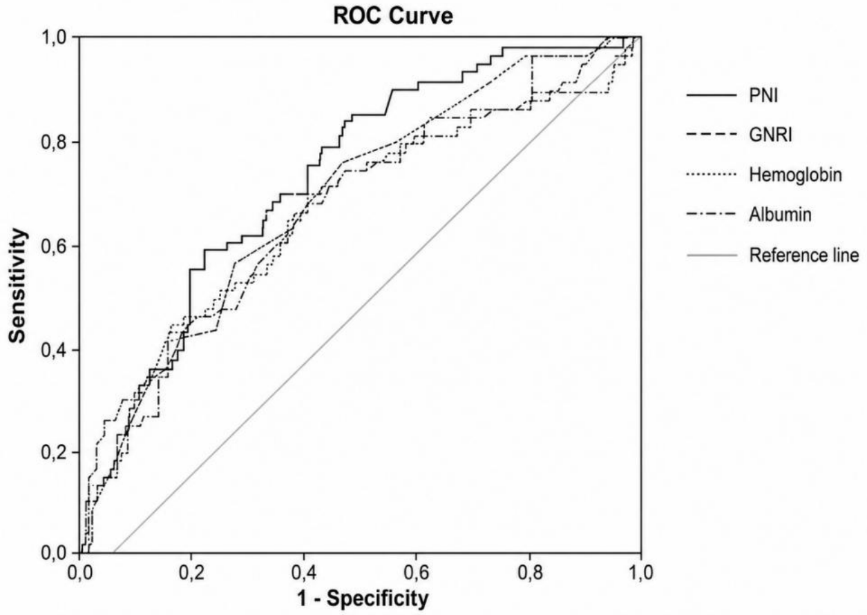
Figure 2.
Kaplan–Meier survival curves according to PNI cut-off value. Abbreviations: PNI, prognostic nutritional index.
Figure 2.
Kaplan–Meier survival curves according to PNI cut-off value. Abbreviations: PNI, prognostic nutritional index.
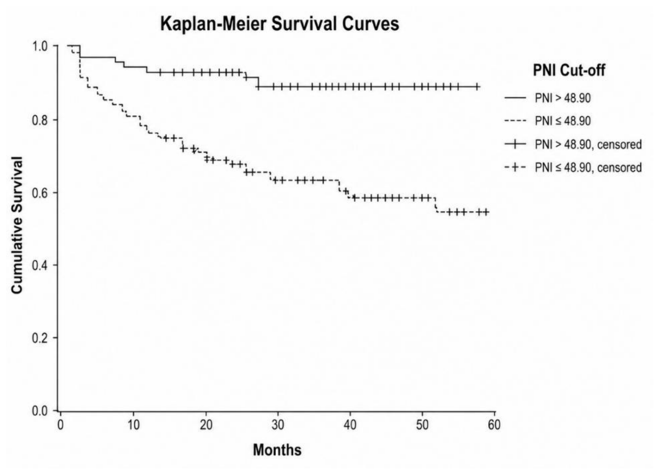
Table 3.
Receiver operating characteristic curve analysis and diagnostic performance metrics for mortality prediction.
Table 3.
Receiver operating characteristic curve analysis and diagnostic performance metrics for mortality prediction.
| Variable | AUC (95% CI) | p Value | Cut-Off | Sensitivity (%) | Specificity (%) | PPV (%) | NPV (%) | Youden Index |
|---|---|---|---|---|---|---|---|---|
| PNI | 0.720 (0.648–0.793) | <0.001 | ≤48.90 | 82.8 | 53.4 | 39.0 | 89.6 | 0.362 |
| Albumin, g/L | 0.699 (0.620–0.778) | <0.001 | ≤37.50 | 57.8 | 73.7 | 44.0 | 83.0 | 0.315 |
| GFR, mL/min/1.73 m2 | 0.686 (0.607–0.765) | <0.001 | ≤73.15 | 79.4 | 47.5 | 33.8 | 87.3 | 0.269 |
| Hemoglobin, g/dL | 0.683 (0.599–0.768) | <0.001 | ≤12.15 | 69.6 | 56.4 | 35.3 | 84.4 | 0.260 |
| GNRI | 0.677 (0.589–0.766) | <0.001 | ≤104.27 | 57.6 | 78.1 | 47.9 | 84.1 | 0.357 |
| Age, years | 0.668 (0.588–0.748) | <0.001 | ≥74.50 | 71.0 | 54.7 | 34.8 | 84.7 | 0.257 |
| LMR | 0.655 (0.568–0.743) | 0.001 | ≤2.22 | 53.0 | 74.1 | 40.2 | 82.8 | 0.271 |
| Lymphocyte count, 109/L | 0.637 (0.553–0.721) | 0.002 | ≤1.79 | 72.5 | 55.4 | 35.7 | 85.5 | 0.279 |
| SIRI | 0.580 (0.488–0.671) | 0.077 | ≥1.57 | 71.2 | 46.3 | 30.3 | 83.0 | 0.175 |
| NLR | 0.574 (0.486–0.663) | 0.099 | ≥2.48 | 71.0 | 44.1 | 30.2 | 81.7 | 0.151 |
| PIV | 0.558 (0.463–0.653) | 0.197 | ≥666.88 | 39.4 | 76.6 | 35.6 | 79.4 | 0.160 |
| SII | 0.552 (0.459–0.645) | 0.250 | ≥1616.57 | 26.1 | 91.1 | 50.0 | 78.3 | 0.172 |
Abbreviations: AUC, area under the curve; CI, confidence interval; GFR, estimated glomerular filtration rate; GNRI, geriatric nutritional risk index; LMR, lymphocyte-to-monocyte ratio; NLR, neutrophil-to-lymphocyte ratio; NPV, negative predictive value; PIV, pan-immune-inflammation value; PNI, prognostic nutritional index; PPV, positive predictive value; SII, systemic immune-inflammation index; SIRI, systemic inflammation response index. For variables inversely associated with mortality, lower values were considered to indicate increased risk, and cut-off values are presented accordingly. Sensitivity, specificity, PPV, NPV, and Youden index were calculated according to the selected ROC-derived cut-off values using available cases for each variable.
Disclaimer/Publisher’s Note: The statements, opinions and data contained in all publications are solely those of the individual author(s) and contributor(s) and not of MDPI and/or the editor(s). MDPI and/or the editor(s) disclaim responsibility for any injury to people or property resulting from any ideas, methods, instructions or products referred to in the content. |
© 2026 by the authors. Licensee MDPI, Basel, Switzerland. This article is an open access article distributed under the terms and conditions of the Creative Commons Attribution (CC BY) license (http://creativecommons.org/licenses/by/4.0/).
Copyright: This open access article is published under a Creative Commons CC BY 4.0 license, which permit the free download, distribution, and reuse, provided that the author and preprint are cited in any reuse.


