

Submitted:
17 July 2026
Posted:
20 July 2026
You are already at the latest version
Abstract
Chronic kidney disease (CKD) represents a major global health challenge, affecting more than 10% of the population and contributing substantially to morbidity and premature mortality. Growing evidence identifies oxidative stress and mitochondrial dysfunction as central drivers of renal injury and disease progression across diverse etiologies. The kidney is one of the most mitochondria-rich organs in the body, reflecting the high bioenergetic demands required for tubular reabsorption and metabolic homeostasis. Disruption of mitochondrial oxidative phosphorylation, excessive production of reactive oxygen species (ROS), and impaired mitochondrial quality control mechanisms promote tubular injury, inflammation, and fibrosis. In particular, dysfunction of the electron transport chain, activation of NADPH oxidase isoforms—especially NOX4—and alterations in mitochondrial dynamics create a vicious cycle of oxidative damage and bioenergetic failure. Emerging evidence highlights the importance of mitochondrial quality control pathways, including fusion–fission balance, PINK1/Parkin-mediated mitophagy, and mitochondrial biogenesis regulated by PGC-1α and TFAM. Recent studies have also identified additional mechanisms linking mitochondrial dysfunction to CKD progression, including ferroptosis, epigenetic regulation of mitochondrial genes, and mitochondrial DNA release acting as damage-associated molecular patterns that activate innate immune pathways. At the clinical level, redox and mitochondrial biomarkers—such as F2-isoprostanes, 8-hydroxy-2′-deoxyguanosine, advanced oxidation protein products, and circulating mitochondrial DNA—may improve early diagnosis and risk stratification. Therapeutically, both established nephroprotective drugs and emerging mitochondria-targeted interventions aim to restore mitochondrial homeostasis and represent promising strategies for slowing CKD progression.
Keywords:
chronic kidney disease (CKD)
; mitochondrial dysfunction
; mitophagy (PINK1/Parkin)
; NADPH oxidase 4 (NOX4)
; reactive oxygen species (ROS)
; mitochondria-targeted therapy
1. Introduction
The kidney is among the most metabolically active organs in the human body, characterized by high oxygen consumption and an exceptionally dense mitochondrial network that supports its complex physiological functions. In fact, the renal cortex—particularly the proximal tubular epithelium—contains one of the highest mitochondrial densities among mammalian tissues, surpassed only by the myocardium [1]. This remarkable abundance reflects the enormous bioenergetic requirements associated with maintaining renal homeostasis, including glomerular filtration, electrolyte balance, acid–base regulation, and active reabsorption of solutes and water. Approximately 180 liters of glomerular filtrate are processed by the kidneys daily, and the majority of this filtrate is reabsorbed in the proximal tubules through energy-dependent transport mechanisms that rely heavily on mitochondrial adenosine triphosphate (ATP) production [1,2,3].
Mitochondria generate ATP primarily through oxidative phosphorylation (OXPHOS), a process driven by the mitochondrial electron transport chain (ETC) located within the inner mitochondrial membrane [1,4,5]. Electrons derived from metabolic substrates are transferred through ETC complexes I–IV, ultimately reducing oxygen to water while generating a proton gradient that powers ATP synthesis. While this system is highly efficient, it also represents a major source of reactive oxygen species (ROS), which are generated as unavoidable byproducts of electron leakage from ETC complexes—particularly complexes I and III [1,6]. Under physiological conditions, low levels of ROS function as signaling molecules that regulate cellular processes such as proliferation, differentiation, and stress responses. However, excessive ROS production or impaired antioxidant defenses disrupt cellular redox homeostasis, leading to oxidative stress and damage to lipids, proteins, and nucleic acids [7,8,9].
Chronic kidney disease (CKD) is increasingly recognized as a disorder characterized by profound disturbances in mitochondrial function and redox balance. Affecting more than 750 million individuals worldwide, CKD represents a significant public health challenge and is associated with increased cardiovascular risk, reduced quality of life, and high healthcare costs [10]. Although CKD arises from diverse etiologies—including diabetes, hypertension, autoimmune disorders, and genetic abnormalities—mounting evidence suggests that mitochondrial dysfunction and oxidative stress represent common downstream pathways driving disease progression [8,11,12,13].
Mitochondrial dysfunction in renal cells manifests through multiple mechanisms, including impaired oxidative phosphorylation, mitochondrial DNA (mtDNA) damage, altered mitochondrial dynamics, and defective mitochondrial quality control pathways [6,14,15]. These alterations lead to decreased ATP production, increased ROS generation, and activation of inflammatory and fibrotic signaling pathways that contribute to tubular injury and progressive loss of renal function [6,8,14]. In addition, damaged mitochondria can release mtDNA and other mitochondrial components into the cytosol or extracellular space, where they act as damage-associated molecular patterns (DAMPs) that activate innate immune pathways such as Toll-like receptor 9 (TLR9) and the cyclic GMP–AMP synthase–stimulator of interferon genes (cGAS–STING) signaling cascade, further amplifying inflammatory responses in the kidney [16,17,18].
Another critical aspect of mitochondrial homeostasis in the kidney involves mitochondrial quality control (MQC), a coordinated network of processes that maintain mitochondrial integrity and functionality. These processes include mitochondrial dynamics (fusion and fission), mitophagy (the selective removal of damaged mitochondria), and mitochondrial biogenesis [3,6,19]. Disruption of MQC leads to the accumulation of dysfunctional mitochondria that generate excessive ROS and contribute to cellular injury, inflammation, and fibrosis—hallmarks of CKD progression [6,8,11,20].
Recent advances in molecular nephrology have revealed that mitochondrial dysfunction is not only a consequence of renal injury but also a central driver of disease initiation and progression. Emerging pathogenic mechanisms—including ferroptosis, epigenetic regulation of mitochondrial genes, and metabolic reprogramming—have further expanded our understanding of how oxidative stress and mitochondrial alterations contribute to renal pathology [4,14,15,21,22]. Moreover, novel therapeutic strategies targeting mitochondrial pathways are currently being investigated, raising the possibility that restoring mitochondrial homeostasis may represent a promising approach for preventing or slowing CKD progression [4,14,23,24].
This review aims to provide a comprehensive overview of the molecular mechanisms linking oxidative stress and mitochondrial dysfunction to chronic kidney disease. We discuss the major sources of oxidative stress in renal tissue, examine the role of mitochondrial quality control pathways, and explore emerging pathogenic mechanisms such as ferroptosis and epigenetic regulation. In addition, we review current and potential therapeutic strategies targeting mitochondrial dysfunction and highlight the clinical relevance of redox biomarkers for improving diagnosis, risk stratification, and personalized treatment of CKD.
2. Primary Sources of Oxidative Stress in the Renal Parenchyma
Oxidative stress represents a fundamental pathogenic mechanism in chronic kidney disease and is characterized by an imbalance between the production of reactive oxygen species (ROS) and the capacity of cellular antioxidant systems to neutralize them. In the kidney, several cellular and molecular processes contribute to ROS generation, including mitochondrial dysfunction, activation of NADPH oxidases, hormonal signaling pathways such as the renin–angiotensin–aldosterone system (RAAS), and the accumulation of uremic toxins associated with dysbiosis of the gut microbiota. These processes interact to create a complex network of redox dysregulation that promotes inflammation, cellular injury, and progressive renal fibrosis (Figure 1) [7,8,14,25].
2.1. Mitochondrial Electron Transport Chain Dysfunction and Superoxide Leakage
Mitochondria represent the primary endogenous source of reactive oxygen species in most mammalian cells. During oxidative phosphorylation (OXPHOS), electrons derived from metabolic substrates are transferred through the mitochondrial electron transport chain, which consists of five multiprotein complexes embedded within the inner mitochondrial membrane. Although the ETC is designed to efficiently transfer electrons to molecular oxygen, a small proportion of electrons prematurely escape the chain and react with oxygen to form the superoxide radical (O₂•⁻) [1,26,27].
Under physiological conditions, mitochondrial ROS production is tightly controlled by antioxidant defense systems such as superoxide dismutases (SODs), catalase, glutathione peroxidase, and peroxiredoxins [23,28,29,30]. However, pathological conditions associated with CKD—such as hyperglycemia, lipid overload, hypoxia, and inflammation—can impair ETC function and increase electron leakage, resulting in excessive ROS production. Complexes I and III are considered the primary sites of superoxide generation, and structural or functional alterations in these complexes have been frequently observed in experimental models of kidney disease [8,15,26,27,31].
In proximal tubular epithelial cells, mitochondrial dysfunction is particularly detrimental due to their heavy reliance on fatty acid oxidation (FAO) as a primary energy source. Disruption of FAO leads to the accumulation of lipid intermediates, including long-chain acylcarnitines, which interfere with mitochondrial metabolism and further increase ROS production. This process contributes to lipotoxicity, mitochondrial fragmentation, and the progressive decline of cellular bioenergetic capacity [32]. In addition, oxidative damage to mitochondrial DNA can impair the synthesis of essential ETC proteins encoded by the mitochondrial genome, further compromising mitochondrial respiration and establishing a self-perpetuating cycle of oxidative stress and mitochondrial dysfunction [16,31].
Mitochondrial oxidative stress also plays a critical role in acute kidney injury (AKI), where early mitochondrial damage leads to ATP depletion, tubular cell death, and inflammation. In experimental models of ischemia–reperfusion injury and nephrotoxic damage, increased mitochondrial ROS production has been shown to trigger activation of inflammatory pathways such as the NLRP3 inflammasome and promote the transition from AKI to CKD [16,33,34,35,36,37,38,39].
2.2. NADPH Oxidase Signaling: The Central Role of NOX4
While mitochondria generate ROS as byproducts of metabolism, the NADPH oxidase (NOX) family of enzymes produces ROS as its primary biological function. Among the seven NOX isoforms identified in mammals, NOX4 is the predominant isoform expressed in renal tissue and is particularly abundant in tubular epithelial cells, podocytes, and endothelial cells [40,41,42,43,44].
Unlike other NOX isoforms that primarily generate superoxide, NOX4 predominantly produces hydrogen peroxide (H₂O₂), a relatively stable ROS that can diffuse across cellular compartments and participate in redox signaling. Under physiological conditions, NOX4-derived ROS contribute to cellular signaling pathways involved in differentiation and metabolic adaptation. However, chronic activation of NOX4 has been implicated in the pathogenesis of multiple kidney diseases, including diabetic kidney disease, hypertensive nephropathy, and renal fibrosis [41,42,43,44,45,46,47].
In diabetic kidney disease, hyperglycemia and advanced glycation end products stimulate NOX4 expression in podocytes and mesangial cells, leading to increased ROS production and activation of pro-fibrotic signaling pathways such as transforming growth factor-β (TGF-β) signaling. Increased NOX4 activity has also been linked to podocyte apoptosis, glomerulosclerosis, and albuminuria in experimental models of diabetic nephropathy [42,48,49,50,51,52].
Recent studies have identified the SH3YL1 protein as an important cytosolic activator that interacts with NOX4 to enhance ROS generation in renal cells [4]. Disruption of the SH3YL1–NOX4 interaction has been shown to reduce oxidative stress and attenuate renal fibrosis in experimental models, highlighting this signaling axis as a potential therapeutic target. Furthermore, several pharmacological compounds—including polyphenols and AMPK activators—have been shown to suppress NOX4 expression and mitigate oxidative stress in diabetic kidney disease [12,53,54,55].
2.3. RAAS Activation and Redox Crosstalk
The renin–angiotensin–aldosterone system (RAAS) plays a central role in regulating blood pressure, fluid balance, and renal hemodynamics. However, chronic activation of RAAS is also a major contributor to oxidative stress and inflammation in CKD. Angiotensin II (Ang II), the principal effector peptide of RAAS, exerts multiple deleterious effects on renal tissue through activation of the angiotensin II type 1 receptor (AT₁R) [40,44,56].
Binding of Ang II to AT₁R stimulates the activation of NADPH oxidases, particularly NOX4, leading to increased ROS production and oxidative damage in renal cells. This ROS generation initiates a cascade of signaling events that activate transcription factors such as nuclear factor-κB (NF-κB) and activator protein-1 (AP-1), promoting the expression of pro-inflammatory cytokines, chemokines, and adhesion molecules. As a result, immune cells—including macrophages and T lymphocytes—are recruited to the kidney, further amplifying inflammatory responses and promoting tissue injury [40,44,56].
Moreover, Ang II-induced ROS production can impair mitochondrial function by damaging mitochondrial DNA, disrupting mitochondrial membrane potential, and promoting mitochondrial fragmentation. This interaction between RAAS signaling and mitochondrial dysfunction establishes a feedback loop in which oxidative stress perpetuates inflammation and fibrosis, ultimately contributing to progressive renal damage [8,11,31,40,44,57,58].
2.4. The Gut–Kidney Axis and Uremic Toxins
In addition to intrinsic renal sources of oxidative stress, increasing attention has been directed toward the role of the gut–kidney axis in the pathogenesis of chronic kidney disease. Alterations in intestinal microbiota composition and metabolism—collectively referred to as dysbiosis—contribute to the generation and systemic accumulation of uremic toxins that exacerbate oxidative stress and inflammation in CKD [25,59,60,61,62].
In patients with impaired renal function, the reduced clearance of microbial metabolites leads to the accumulation of several protein-bound uremic toxins, among which indoxyl sulfate (IS) and p-cresyl sulfate (pCS) are the most extensively studied. These metabolites originate from the bacterial metabolism of dietary amino acids such as tryptophan and tyrosine and undergo hepatic conjugation before entering systemic circulation. Because of their strong binding affinity to albumin, these toxins are poorly removed by conventional dialysis and progressively accumulate as kidney function declines [60,61,63,64,65].
Indoxyl sulfate has been identified as a potent inducer of oxidative stress in renal tubular epithelial cells and vascular endothelial cells. Mechanistically, IS stimulates ROS production through the activation of NADPH oxidases, particularly NOX4, and enhances mitochondrial oxidative stress. Moreover, IS interferes with endogenous antioxidant defense systems by suppressing the activity of nuclear factor erythroid 2–related factor 2 (Nrf2), a transcription factor that regulates the expression of numerous antioxidant and detoxification enzymes [63,66]. The inhibition of Nrf2 signaling results in decreased expression of protective proteins such as heme oxygenase-1 (HO-1), glutathione peroxidase, and superoxide dismutase, thereby exacerbating oxidative damage within renal tissue [28,29,66,67,68].
Beyond its direct pro-oxidant effects, indoxyl sulfate also promotes inflammatory signaling pathways that contribute to renal fibrosis. Activation of the aryl hydrocarbon receptor (AhR) by IS triggers downstream signaling cascades that stimulate the production of pro-inflammatory cytokines and profibrotic mediators, including transforming growth factor-β (TGF-β) and connective tissue growth factor (CTGF). These processes accelerate tubulointerstitial fibrosis, which represents a key determinant of CKD progression [59,63,69].
The systemic consequences of gut-derived uremic toxins extend beyond the kidney. Elevated circulating levels of IS and pCS have been strongly associated with endothelial dysfunction, vascular calcification, and increased cardiovascular risk in CKD patients. Oxidative stress induced by these toxins contributes to mitochondrial damage in vascular cells, thereby linking renal dysfunction with the heightened cardiovascular morbidity observed in CKD populations [56,59,63,70,71].
Importantly, therapeutic strategies aimed at modulating the gut microbiota—including dietary interventions, probiotics, prebiotics, and oral adsorbents—are currently being investigated as potential approaches to reduce uremic toxin burden and mitigate oxidative stress in CKD. Although clinical evidence remains limited, these strategies highlight the growing recognition of the gut–kidney axis as an important contributor to oxidative stress and mitochondrial dysfunction in chronic kidney disease [59,61,69,72].
3. Mitochondrial Quality Control (MQC) Failure
Mitochondrial homeostasis is maintained by a highly coordinated network of processes collectively referred to as mitochondrial quality control (MQC). These mechanisms ensure the preservation of mitochondrial integrity and functionality through the dynamic regulation of mitochondrial morphology, selective removal of damaged organelles, and synthesis of new mitochondria. The MQC system consists primarily of three interconnected processes: mitochondrial dynamics (fusion and fission), mitophagy, and mitochondrial biogenesis [6,19,73].
In the healthy kidney, these processes work in concert to maintain an efficient mitochondrial network capable of meeting the high metabolic demands of renal tubular cells. However, in chronic kidney disease, multiple stressors—including oxidative stress, metabolic dysregulation, and inflammatory signaling—disrupt MQC pathways, leading to the accumulation of dysfunctional mitochondria. These damaged organelles generate excessive reactive oxygen species, impair ATP production, and activate apoptotic and inflammatory pathways that contribute to progressive renal injury [4,8,11,19,31,74].
Recent studies have highlighted the central importance of MQC dysfunction in both acute kidney injury (AKI) and CKD. In AKI models, early mitochondrial fragmentation and impaired mitophagy lead to rapid ATP depletion and tubular cell death. When mitochondrial damage persists or repair mechanisms fail, maladaptive responses can promote the transition from AKI to CKD through chronic inflammation and fibrosis (Figure 2) [6,33,35,36,38].
3.1. Mitochondrial Dynamics: The Balance Between Fusion and Fission
Mitochondria are highly dynamic organelles that continuously undergo cycles of fusion and fission. This dynamic remodeling allows mitochondria to adapt to changing metabolic conditions, maintain mitochondrial DNA integrity, and segregate damaged mitochondrial components for degradation [75,76,77,78,79,80,81].
Mitochondrial fusion involves the merging of adjacent mitochondria, enabling the exchange of mitochondrial DNA, proteins, and metabolites. This process helps dilute damaged mitochondrial components and maintain mitochondrial functionality. Fusion is primarily mediated by three key proteins: mitofusin-1 (MFN1), mitofusin-2 (MFN2), and optic atrophy protein 1 (OPA1). MFN1 and MFN2 regulate the fusion of the outer mitochondrial membrane, whereas OPA1 mediates fusion of the inner mitochondrial membrane [75,76,77,78,79,80,81].
In contrast, mitochondrial fission involves the division of mitochondria into smaller organelles. This process is essential for mitochondrial distribution during cell division and for isolating damaged mitochondrial segments that require removal by mitophagy. The central mediator of mitochondrial fission is dynamin-related protein 1 (Drp1), a cytosolic GTPase that translocates to the mitochondrial outer membrane where it interacts with receptor proteins such as Fis1, Mff, and MiD49/51 to constrict and divide the mitochondrial membrane [75,77,78,81,82].
The activity of Drp1 is tightly regulated by post-translational modifications, particularly phosphorylation at specific serine residues. Phosphorylation of Drp1 at serine 616 (p-Drp1S616) promotes mitochondrial fission and has been associated with mitochondrial fragmentation and apoptosis. Conversely, phosphorylation at serine 637 (p-Drp1S637) inhibits Drp1 activity and favors mitochondrial elongation [75,82,83,84,85].
In CKD, the balance between mitochondrial fusion and fission is frequently disrupted, resulting in excessive mitochondrial fragmentation. Elevated levels of p-Drp1S616 and reduced expression of fusion proteins such as MFN2 and OPA1 have been reported in various models of renal injury. This imbalance leads to fragmented mitochondria that exhibit impaired respiratory capacity and increased ROS production, ultimately contributing to tubular cell apoptosis and renal fibrosis [8,11,75,82,83].
Furthermore, excessive mitochondrial fission has been linked to the activation of pro-fibrotic signaling pathways in kidney disease. Mitochondrial fragmentation promotes the release of cytochrome c and other pro-apoptotic factors, initiating the intrinsic apoptotic pathway and amplifying cellular injury. In addition, fragmented mitochondria produce higher levels of ROS, further exacerbating oxidative stress and inflammatory responses within renal tissue.
3.2. Mitophagy Pathways: The PINK1/Parkin Cascade
Mitophagy is the selective degradation of damaged or dysfunctional mitochondria through the autophagy–lysosome pathway. This process represents a critical defense mechanism against mitochondrial dysfunction by removing ROS-producing mitochondria before they can cause irreversible cellular damage [6,73,86,87,88,89].
Among the various mitophagy pathways described in mammalian cells, the PINK1/Parkin-mediated pathway is the most extensively characterized. Under normal conditions, PTEN-induced kinase 1 (PINK1) is continuously imported into healthy mitochondria and rapidly degraded. However, when mitochondria lose their membrane potential—a hallmark of mitochondrial damage—PINK1 accumulates on the outer mitochondrial membrane [87,90,91].
The accumulation of PINK1 triggers a series of molecular events that initiate mitophagy. PINK1 undergoes autophosphorylation and subsequently phosphorylates ubiquitin molecules and the E3 ubiquitin ligase Parkin. Activated Parkin translocates from the cytosol to the damaged mitochondria, where it ubiquitinates numerous outer mitochondrial membrane proteins, including voltage-dependent anion channels (VDACs) and mitofusins [87,90,91].
These ubiquitin tags serve as signals for the recruitment of autophagy receptors such as optineurin (OPTN) and nuclear dot protein 52 (NDP52). These receptors interact with microtubule-associated protein light chain 3 (LC3) on the surface of forming autophagosomes, enabling the engulfment of damaged mitochondria and their subsequent degradation within lysosomes [6,73,86,88,91].
Mitophagy plays a protective role in kidney injury by limiting mitochondrial ROS production and preventing activation of inflammatory pathways. Experimental studies have demonstrated that genetic deletion of PINK1 or Parkin exacerbates renal injury in models of ischemia–reperfusion and cisplatin-induced nephrotoxicity. In these models, impaired mitophagy leads to the accumulation of dysfunctional mitochondria, increased oxidative stress, and enhanced activation of inflammatory pathways such as the NLRP3 inflammasome [15,33,37,86,92].
In addition to the PINK1/Parkin pathway, receptor-mediated mitophagy pathways have also been described. Proteins such as BNIP3, NIX, and FUNDC1 contain LC3-interacting regions (LIRs) that allow them to directly recruit autophagosomes without requiring ubiquitination. These pathways are particularly important during hypoxic stress and have been implicated in renal ischemia and diabetic nephropathy [6,73,86,88,89].
Despite its protective role in acute injury, dysregulated mitophagy may contribute to chronic kidney disease when mitochondrial clearance becomes insufficient or maladaptive. Clinical studies have demonstrated that impaired mitophagy correlates with declining glomerular filtration rate (GFR) and increased renal fibrosis in CKD patients, suggesting that defective mitochondrial turnover represents a critical mechanism of disease progression [11,15,19,86].
3.3. Mitochondrial Biogenesis and the PGC-1α Signaling Axis
While mitophagy removes damaged mitochondria, mitochondrial biogenesis replenishes the mitochondrial pool by generating new organelles. This process is primarily regulated by the transcriptional coactivator peroxisome proliferator-activated receptor gamma coactivator-1 alpha (PGC-1α), which orchestrates the expression of nuclear genes encoding mitochondrial proteins [93,94,95].
PGC-1α interacts with several transcription factors, including nuclear respiratory factors 1 and 2 (NRF1 and NRF2), to stimulate the transcription of genes required for mitochondrial respiration, fatty acid oxidation, and oxidative metabolism. One of the key downstream targets of PGC-1α is mitochondrial transcription factor A (TFAM), which is essential for mitochondrial DNA replication and transcription [93,94,95,96].
In healthy renal tissue, PGC-1α plays a central role in maintaining mitochondrial abundance and metabolic flexibility. However, in CKD, PGC-1α expression is often markedly reduced due to metabolic stress, inflammation, and epigenetic modifications. Reduced PGC-1α signaling leads to impaired mitochondrial biogenesis, decreased mitochondrial mass, and diminished ATP production [8,11,93].
Sirtuin proteins, particularly SIRT1 and SIRT3, are important regulators of PGC-1α activity. These NAD⁺-dependent deacetylases enhance PGC-1α function and promote mitochondrial biogenesis under conditions of metabolic stress. However, reduced sirtuin activity in CKD—often associated with metabolic syndrome and aging—contributes to mitochondrial dysfunction and impaired cellular metabolism [12,93,97,98,99,100].
The combined failure of mitochondrial dynamics, mitophagy, and biogenesis ultimately results in the accumulation of dysfunctional mitochondria that produce excessive ROS and contribute to progressive renal injury. Consequently, restoring mitochondrial quality control has emerged as a promising therapeutic strategy for preventing CKD progression and improving renal outcomes [6,19,93].
4. Emerging Pathogenic Axes in Renal Pathology
Beyond classical oxidative stress pathways, recent advances in molecular nephrology have uncovered several additional mechanisms that connect mitochondrial dysfunction to renal injury. Among these, ferroptosis, epigenetic regulation of mitochondrial genes, and non-coding RNA signaling have emerged as critical contributors to oxidative stress amplification and disease progression in chronic kidney disease (CKD). These mechanisms integrate metabolic disturbances, mitochondrial dysfunction, and inflammatory signaling, forming complex pathogenic networks that drive tubular injury and fibrosis [6,31].
4.1. Ferroptosis: Iron-Dependent Lipid Peroxidation
Ferroptosis is a recently described form of regulated cell death that is mechanistically distinct from apoptosis, necrosis, and autophagy. It is characterized by the iron-dependent accumulation of lipid peroxides that ultimately leads to catastrophic membrane damage and cell death. Ferroptosis has gained increasing attention in kidney research due to its involvement in both acute kidney injury (AKI) and chronic kidney disease progression [6,101,102,103,104,105,106].
At the biochemical level, ferroptosis is driven by uncontrolled lipid peroxidation reactions that occur when cellular antioxidant defenses are overwhelmed. Iron plays a central role in this process through the Fenton reaction, in which ferrous iron (Fe²⁺) reacts with hydrogen peroxide to generate highly reactive hydroxyl radicals [101,104,105,107,108,109]:
Fe²⁺ + H₂O₂ → Fe³⁺ + OH⁻ + OH•
These radicals initiate lipid peroxidation by attacking polyunsaturated fatty acids (PUFAs) within cellular membranes, leading to the formation of lipid hydroperoxides and membrane destabilization [104,105,106,109].
One of the key enzymes involved in ferroptosis is acyl-CoA synthetase long-chain family member 4 (ACSL4), which promotes the incorporation of polyunsaturated fatty acids such as arachidonic acid into membrane phospholipids. These lipids serve as substrates for lipid peroxidation, thereby sensitizing cells to ferroptotic death [6,104,106,110,111]. Increased ACSL4 expression has been observed in several models of renal injury, suggesting that enhanced lipid remodeling contributes to ferroptosis susceptibility in kidney disease [102,105,112].
The primary defense against ferroptosis is mediated by glutathione peroxidase 4 (GPX4), a selenoprotein that detoxifies lipid hydroperoxides using glutathione (GSH) as a reducing agent. GPX4 converts toxic lipid hydroperoxides into non-toxic lipid alcohols, thereby preventing membrane damage and ferroptotic cell death. Loss of GPX4 activity results in uncontrolled lipid peroxidation and rapid cell death, highlighting its essential role in cellular survival (Figure 3) [6,104,106,113,114,115].
In the kidney, ferroptosis has been implicated in multiple pathological conditions, including ischemia–reperfusion injury, cisplatin-induced nephrotoxicity, and diabetic kidney disease. Experimental studies have demonstrated that inhibition of ferroptosis using pharmacological agents such as ferrostatin-1 or liproxstatin-1 can significantly attenuate renal injury and inflammation, indicating that ferroptosis represents a promising therapeutic target [34,101,102,105,112].
Another important regulatory pathway controlling ferroptosis is the nuclear factor erythroid 2–related factor 2 (Nrf2) signaling pathway. Nrf2 is a transcription factor that regulates the expression of numerous antioxidant and cytoprotective genes, including those involved in glutathione synthesis and iron metabolism. Activation of Nrf2 enhances cellular resistance to ferroptosis by increasing GPX4 expression and restoring redox balance. Conversely, impaired Nrf2 signaling—frequently observed in CKD—facilitates lipid peroxidation and ferroptotic cell death [6,28,29,66,67,68,116].
The close relationship between ferroptosis and mitochondrial dysfunction further highlights the central role of oxidative stress in renal pathology. Mitochondrial ROS production can amplify lipid peroxidation, while mitochondrial iron accumulation can increase susceptibility to ferroptotic injury. These interactions establish ferroptosis as a key mediator linking mitochondrial dysfunction with tubular cell death and progressive renal damage [6,31,103,104,106,107].
4.2. Epigenetic Regulation of Mitochondrial Function
Epigenetic mechanisms have emerged as important regulators of mitochondrial homeostasis and oxidative stress in kidney disease. These mechanisms involve reversible modifications of DNA and histone proteins that influence gene expression without altering the underlying DNA sequence [21,22,117,118,119].
Among the most extensively studied epigenetic modifications in CKD are DNA methylation, histone acetylation, and histone methylation. These modifications regulate the transcription of genes involved in mitochondrial metabolism, oxidative stress responses, and inflammatory signaling pathways [21,22,117,118,119].
DNA methylation is catalyzed by DNA methyltransferases and typically occurs at cytosine residues within CpG islands. Hypermethylation of promoter regions can suppress gene expression, whereas hypomethylation may enhance transcriptional activity. In kidney disease, altered DNA methylation patterns have been identified in several genes involved in mitochondrial function, including mitochondrial processing peptidase subunit beta (PMPCB) and mitochondrial transcription factor A (TFAM). Dysregulation of these genes can impair mitochondrial protein processing, reduce mitochondrial DNA replication, and ultimately compromise mitochondrial respiration [21,22,96,120].
Histone modifications also play a critical role in regulating mitochondrial dynamics and oxidative stress responses. For example, increased acetylation of histone H3 lysine 27 (H3K27ac) has been associated with the activation of genes involved in mitochondrial fission and fibrosis in diabetic kidney disease. Similarly, phosphorylation of the mitochondrial fission protein Drp1 at serine 616 promotes mitochondrial fragmentation and has been linked to renal fibroblast activation and tubulointerstitial fibrosis [21,83,117,119].
Histone methyltransferases such as enhancer of zeste homolog 2 (EZH2) also contribute to mitochondrial dysfunction in CKD. EZH2-mediated trimethylation of histone H3 lysine 27 (H3K27me3) has been shown to repress antioxidant gene expression, thereby promoting oxidative stress and renal injury. Interestingly, mitochondrial metabolites such as S-adenosylmethionine (SAM) serve as methyl donors for histone methylation reactions, establishing a direct link between cellular metabolism and epigenetic regulation [22,117,118,119].
These findings highlight the existence of a metabolic–epigenetic feedback loop in which mitochondrial dysfunction alters cellular metabolism, which in turn modifies epigenetic landscapes that regulate mitochondrial gene expression. This cycle contributes to the persistence of oxidative stress and fibrosis in chronic kidney disease [21,22,117,118].
4.3. Non-Coding RNAs and Mitochondrial Regulation
Non-coding RNAs, particularly microRNAs (miRNAs), represent another important layer of regulation linking oxidative stress and mitochondrial dysfunction in kidney disease. MicroRNAs are short RNA molecules that regulate gene expression by binding to complementary sequences in target mRNAs, leading to translational repression or mRNA degradation [121,122,123].
Several microRNAs have been identified as regulators of mitochondrial dynamics and metabolism in renal cells. For instance, miR-668 has been shown to inhibit mitochondrial fission by targeting mitochondrial protein 18 (MTP18), thereby protecting renal tubular cells from apoptosis during ischemic injury. By suppressing excessive mitochondrial fragmentation, miR-668 helps preserve mitochondrial function and cellular viability [122,123].
Conversely, miR-17 has been implicated in the suppression of peroxisome proliferator-activated receptor alpha (PPAR-α), a transcription factor involved in fatty acid oxidation and mitochondrial metabolism. Downregulation of PPAR-α by miR-17 impairs mitochondrial energy metabolism and promotes cyst formation in polycystic kidney disease models [93,121,122,123].
These regulatory networks demonstrate how epigenetic and post-transcriptional mechanisms converge to influence mitochondrial function and oxidative stress responses. Understanding these pathways may provide new opportunities for therapeutic intervention aimed at restoring mitochondrial homeostasis in CKD [121,122,123,124,125].
5. Organelle Crosstalk: Mitochondria–Endoplasmic Reticulum Interactions
Mitochondria do not function as isolated organelles but instead participate in complex interactions with other cellular compartments. Among these interactions, communication between mitochondria and the endoplasmic reticulum (ER) plays a particularly important role in maintaining cellular homeostasis [76,80].
These interactions occur at specialized contact sites known as mitochondria-associated membranes (MAMs). MAMs facilitate the transfer of lipids, calcium ions, and metabolic intermediates between the ER and mitochondria, thereby coordinating cellular metabolism, calcium signaling, and stress responses [76,80,81].
In renal cells, MAMs are essential for regulating mitochondrial calcium uptake. Calcium ions released from the ER are transferred directly to mitochondria through channels such as the inositol 1,4,5-trisphosphate receptor (IP3R), voltage-dependent anion channel (VDAC), and mitochondrial calcium uniporter (MCU). Controlled calcium transfer stimulates mitochondrial metabolism by activating dehydrogenases involved in the tricarboxylic acid (TCA) cycle [4,76,80].
However, excessive calcium transfer from the ER to mitochondria can lead to mitochondrial calcium overload, which disrupts mitochondrial membrane potential and triggers opening of the mitochondrial permeability transition pore (mPTP). This event results in mitochondrial swelling, release of pro-apoptotic factors, and activation of cell death pathways [31,76,126].
Disruption of MAM signaling has been implicated in various forms of kidney disease. Oxidative stress and mitochondrial dysfunction can alter ER–mitochondria communication, leading to abnormal calcium signaling and increased susceptibility to apoptosis [31,76,126].
Another important consequence of mitochondrial dysfunction is the release of mitochondrial DNA into the cytoplasm. Because mtDNA resembles bacterial DNA in structure, it can be recognized by innate immune receptors as a danger signal. One of the most important pathways activated by cytosolic mtDNA is the cyclic GMP–AMP synthase–stimulator of interferon genes (cGAS–STING) pathway [17,126,127,128,129].
Activation of cGAS by mtDNA leads to the production of cyclic GMP–AMP (cGAMP), which activates STING on the ER membrane. STING signaling subsequently induces the production of type I interferons and pro-inflammatory cytokines, promoting inflammatory responses within the kidney [17,127].
Persistent activation of the cGAS–STING pathway has been implicated in chronic kidney disease and renal fibrosis. In experimental models, inhibition of STING signaling reduces inflammation and attenuates kidney injury, highlighting the importance of mitochondrial–immune crosstalk in renal pathology [17,127].
6. Disease-Specific Redox Mechanisms
Although oxidative stress and mitochondrial dysfunction represent common pathogenic mechanisms across different kidney diseases, the molecular pathways involved vary according to the underlying etiology, the dominant injured renal compartment, and the nature of the initiating insult [11,31]. In some settings, redox imbalance arises primarily from metabolic overload and mitochondrial substrate excess; in others, it is driven by podocyte injury, complement activation, chronic inflammation, or genetically determined alterations in calcium handling and cellular bioenergetics. Consequently, oxidative stress should not be regarded as a uniform downstream event, but rather as a disease-modifying process that acquires distinct molecular features in different nephropathies.
6.1. Diabetic Kidney Disease (DKD)
Diabetic kidney disease is the leading cause of chronic kidney disease worldwide and accounts for a substantial proportion of end-stage renal disease cases [12,130,131,132]. In this setting, hyperglycemia promotes a persistent state of metabolic oversupply that enhances mitochondrial electron flux, increases superoxide production, and favors oxidative injury to mitochondrial DNA, respiratory proteins, and membrane lipids [12,49,50,51,53,130,133,134,135,136,137]. However, the redox phenotype of DKD cannot be explained by mitochondrial ROS generation alone. Advanced glycation, lipotoxic stress, intrarenal RAAS activation, and inflammatory cytokines converge on ROS-generating systems, particularly NOX4, thereby amplifying oxidative injury and linking metabolic stress to structural renal damage.
In diabetic kidneys, excessive glucose exposure suppresses the activity of AMP-activated protein kinase (AMPK), a key metabolic sensor that normally supports mitochondrial biogenesis, fatty acid oxidation, and antioxidant defense [12,49,53,54,133,135,136,137]. Reduced AMPK signaling favors NOX4 upregulation and thereby intensifies ROS production in podocytes and tubular cells [12,48,53,54,55]. This interaction is mechanistically relevant because NOX4-derived oxidant signaling contributes not only to cellular injury but also to activation of profibrotic pathways, including transforming growth factor-β signaling, extracellular matrix deposition, and albuminuria progression. Experimental observations indicating that metformin restores AMPK activity and attenuates oxidative stress further support the view that mitochondrial dysfunction in DKD reflects defective adaptation to metabolic overload rather than a passive consequence of hyperglycemia alone [12,48,53,54,55].
Another important feature of DKD is the close integration between redox balance and mitochondrial quality control. Hyperglycemia suppresses the Sirt1–FOXO1 signaling axis, which normally promotes antioxidant gene expression, autophagy, and resistance to apoptosis [12,98,99,134,135]. Loss of this protective pathway renders renal cells more vulnerable to oxidative injury, especially in podocytes and proximal tubular epithelial cells, where sustained mitochondrial stress rapidly translates into cytoskeletal instability, slit diaphragm dysfunction, and fibrogenic signaling. Thus, the disease-specific redox signature of DKD is best understood as the combined result of mitochondrial ROS overproduction, impaired AMPK-dependent metabolic adaptation, NOX4 amplification, and defective Sirt1-mediated stress resistance.
6.2. Focal Segmental Glomerulosclerosis (FSGS)
Focal segmental glomerulosclerosis is characterized by progressive podocyte injury and segmental glomerular scarring [138,139]. Because podocytes are highly specialized cells with limited regenerative capacity and substantial bioenergetic requirements, mitochondrial dysfunction has particularly severe structural consequences in this disease. ATP depletion, excessive ROS generation, and impaired mitochondrial dynamics destabilize the actin cytoskeleton, promote detachment from the glomerular basement membrane, and accelerate podocyte loss, a central event in the transition from reversible glomerular stress to irreversible sclerosis [138,139].
The oxidative phenotype of FSGS is shaped by both mitochondrial and non-mitochondrial ROS sources. In addition to mitochondrial dysfunction, high NOX activity contributes to increased oxidant burden, inflammation, and fibrosis [140,141]. Persistent mtROS accumulation can damage lipids, proteins, and nucleic acids, thereby worsening mitochondrial bioenergetics and reinforcing podocyte vulnerability [142,143]. This process is clinically relevant because podocyte depletion is strongly associated with lesion progression, and once a critical threshold of podocyte loss is reached, segmental sclerosis becomes self-propagating.
Several signaling pathways connect redox stress to disease progression in FSGS. Nrf2-dependent antioxidant defense appears to have a protective role, and compounds such as citral, antroquinonol, and THSG have been shown experimentally to attenuate oxidative stress and glomerular injury through activation of the Nrf2/Keap1 pathway [138,139,144]. By contrast, HIF-1α-dependent signaling may aggravate damage under conditions of glomerular stress and hypoxia. Increased expression of angiopoietin-like protein 4 (ANGPTL4), a downstream target of HIF-1α, has been linked to oxidative stress and podocyte apoptosis, whereas epigallocatechin-3-gallate attenuates these changes through inhibition of the HIF-1α–ANGPTL4 axis [138,139,144,145]. In addition, microRNA-mediated pathways may intensify both oxidative injury and fibrosis, as illustrated by the HIF-1/miR-155-5p/Nrf2 axis, which has been implicated in inflammatory damage and progression of glomerular scarring [145]. Altogether, FSGS represents a disease in which oxidative stress is tightly linked to the structural fragility of the podocyte and to the progression from focal injury to permanent glomerular remodeling.
6.3. Autosomal Dominant Polycystic Kidney Disease (ADPKD)
Autosomal dominant polycystic kidney disease is a genetic disorder caused by mutations in PKD1 or PKD2, resulting in altered calcium signaling, cyst formation, and progressive kidney enlargement [146]. In contrast to DKD and FSGS, oxidative stress in ADPKD is integrated with cystic metabolism, epithelial proliferation, and chloride-dependent fluid secretion. Mitochondrial dysfunction develops early in cystic tissue and contributes to reduced oxidative phosphorylation, altered ATP handling, and abnormal redox signaling that favor cyst expansion rather than simply reflecting advanced structural injury [146,147,148].
Experimental studies have shown that oxidative stress can precede overt cyst enlargement and may actively support disease progression. Nrf2 appears to be particularly relevant in this context. Reduced antioxidant defense promotes ROS accumulation and lipid peroxidation, whereas pharmacologic activation of Nrf2 attenuates oxidative damage and slows cyst growth in experimental models [28,29,66,67,68,148,149]. These findings are consistent with observations that compounds such as obacunone can limit lipid peroxide accumulation through the Nrf2/GPX4 axis and thereby restrain cyst progression [149,150].
The relationship between redox stress and epithelial fluid secretion is also central in ADPKD. Abnormal activation of chloride channels drives cyst fluid accumulation, and CFTR has long been considered a major mediator of this process [151]. AMPK activation has emerged as a potentially protective mechanism because it can suppress cell growth pathways and inhibit chloride-dependent secretion [152,153,154,155]. Agents such as metformin, PF-06409577, and steviol have been shown experimentally to activate AMPK, inhibit mTOR signaling, and reduce cystic expansion [151,152,153,154,155]. At the same time, recent work suggests that calcium-activated chloride conductances, including TMEM16A, may interact with or even dominate over CFTR in some cystic settings, indicating that the downstream secretory phenotype may be more heterogeneous than previously assumed. In this regard, ADPKD illustrates a distinctive redox-metabolic disease model in which oxidative stress is coupled to calcium dysregulation, proliferative reprogramming, and abnormal epithelial transport.
6.4. Membranous Nephropathy (MN)
Membranous nephropathy is an autoimmune glomerular disease and a major cause of adult nephrotic syndrome, characterized by subepithelial immune complex deposition and diffuse thickening of the glomerular basement membrane [156,157,158]. In this setting, oxidative stress is closely linked to complement-mediated podocyte injury rather than to metabolic substrate excess or primary mitochondrial disease. Immune complex deposition at the podocyte surface perturbs intracellular signaling, promotes inflammatory activation, and contributes to mitochondrial dysfunction, thereby amplifying structural damage to the filtration barrier. Experimental data increasingly support the concept that redox injury is an active component of the effector phase of MN rather than a nonspecific by-product of glomerular injury [159,160,161,162]. Recent primary evidence has further shown that complement stimulation can induce podocyte pyroptosis in MN and that mitochondrial dysfunction with ROS generation is a key component of this process.
The antioxidant response appears to be relevant in both experimental and potentially translational terms. Activation of the Nrf2/HO-1 pathway has been associated with attenuation of renal injury in experimental MN, and agents such as Sanqi oral solution, crocin, and curcumin have been shown to reduce oxidative stress and improve podocyte injury through Nrf2-centered pathways [159,160,161,162]. In parallel, autophagy-related mechanisms intersect with redox regulation. Salvianolic acid B and the combination of metformin with rapamycin have been reported to enhance autophagic responses and improve podocyte injury, supporting the view that mitochondrial quality control and oxidative stress are mechanistically linked in MN [163,164]. Ferroptotic mechanisms may also participate in disease progression. Passive Heymann nephritis studies have shown altered GPX4, GSH, and ACSL4 expression together with renal iron deposition, suggesting that ferritinophagy-associated ferroptosis may represent an additional oxidative injury pathway in MN [165,166,167,168]. Thus, in MN, oxidative stress appears to function as a bridge between immune complex/complement injury, mitochondrial dysfunction, and downstream podocyte loss.
6.5. Lupus Nephritis (LN)
Lupus nephritis is one of the most severe manifestations of systemic lupus erythematosus and reflects the convergence of autoimmune inflammation, complement activation, immune cell infiltration, and intrinsic renal cell injury [169,170,171,172,173,174,175,176,177]. In contrast to DKD or ADPKD, the redox signature of LN is not confined to a single renal compartment but involves podocytes, mesangial cells, infiltrating leukocytes, and the systemic inflammatory milieu. Oxidative stress is increased in active disease and correlates with inflammatory burden and renal injury severity [175,176]. In this setting, mitochondrial dysfunction appears to be both a consequence of inflammatory injury and an amplifier of tissue damage through ROS generation, mitochondrial fragmentation, and inflammasome activation.
Recent evidence indicates that altered mitochondrial dynamics may be particularly important in podocyte injury in LN. Kidney biopsy-based studies have described abnormal mitochondrial morphology and increased expression of fission-related proteins such as Drp1 and Fis1 in podocytes, supporting a role for excessive mitochondrial fission in proteinuria and glomerular damage [174]. More recent primary work has shown that the C5a–C5aR1 axis can promote Drp1-mediated mitochondrial fission in podocytes, thereby linking complement activation directly to mitochondrial injury and structural glomerular damage in LN.
Several antioxidant and redox-regulating pathways have been implicated in disease modulation. Nrf2 suppresses lupus nephritis by limiting oxidative injury and restraining NF-κB- and TGF-β-related inflammatory signaling [177]. Citral and EGCG have both been shown experimentally to reduce oxidative stress and inflammatory activation through Nrf2-associated mechanisms [178,179]. Sirt1 has also emerged as a relevant protective factor because it can inhibit ROS-dependent activation of TRPM2-mediated Ca2+ influx and thereby limit NLRP3 inflammasome activity [169]. This is mechanistically important because ROS accumulation, calcium dysregulation, and inflammasome activation form a mutually reinforcing loop in active LN [169,171,172,173]. Clinical observations with N-acetylcysteine, although limited, also support the biological relevance of redox-targeted strategies in selected patients [170,180]. Overall, LN represents a disease in which mitochondrial dysfunction acts as an intermediary between immune activation and tissue injury, making oxidative stress central to both disease amplification and therapeutic vulnerability.
7. Clinical Evaluation: Redox and Mitochondrial Biomarkers in CKD
Traditional clinical markers used to monitor kidney disease—such as serum creatinine, estimated glomerular filtration rate (eGFR), and albuminuria—reflect functional decline rather than the underlying molecular mechanisms driving renal injury. Consequently, there is growing interest in identifying biomarkers capable of detecting oxidative stress and mitochondrial dysfunction at earlier stages of disease progression [24,71,181].
Oxidative stress biomarkers can be broadly classified into three categories: markers of lipid peroxidation, DNA oxidation, and protein oxidation. In addition, emerging biomarkers reflecting mitochondrial damage have attracted significant attention as potential tools for precision nephrology (Table 1) [24,71,181,182].
7.1. Lipid Peroxidation Markers
Lipid peroxidation is a hallmark of oxidative stress and occurs when reactive oxygen species attack polyunsaturated fatty acids within cellular membranes. Among the most reliable biomarkers of lipid peroxidation are F2-isoprostanes, which are prostaglandin-like compounds generated through free radical–mediated oxidation of arachidonic acid [183,184].
F2-isoprostanes are widely considered the gold standard for assessing in vivo oxidative stress due to their chemical stability and specificity. Elevated levels of F2-isoprostanes have been detected in plasma and urine samples from patients with chronic kidney disease and are associated with increased cardiovascular risk and accelerated renal decline [2,24,71,181,183,184].
The major urinary metabolite, 15-F₂t-isoprostane (15F₂tIsoPM), reflects systemic oxidative stress and provides a non-invasive indicator of lipid peroxidation. Accurate quantification of F2-isoprostanes typically requires mass spectrometry–based methods such as gas chromatography–mass spectrometry (GC–MS) or liquid chromatography–tandem mass spectrometry (LC–MS/MS). Although enzyme-linked immunosorbent assays (ELISA) are more accessible, they often overestimate concentrations due to cross-reactivity with related compounds [183,184].
7.2. DNA Oxidation Markers
Oxidative damage to DNA represents another important consequence of redox imbalance in CKD. One of the most widely studied biomarkers of DNA oxidation is 8-hydroxy-2′-deoxyguanosine (8-OHdG), which results from ROS-induced modification of guanine residues within DNA.
Elevated levels of 8-OHdG have been detected in both urine and plasma of CKD patients and are associated with increased mortality and cardiovascular complications. Urinary 8-OHdG reflects systemic oxidative DNA damage and has been proposed as an independent predictor of renal disease progression [2].
However, interpretation of 8-OHdG measurements requires caution because levels may be influenced by factors such as smoking, inflammation, and metabolic disorders. Moreover, variability in assay methodologies has limited its widespread clinical adoption.
7.3. Protein Oxidation Markers
Advanced oxidation protein products (AOPPs) represent another class of oxidative stress biomarkers relevant to CKD. AOPPs are formed through oxidative modification of plasma proteins, particularly albumin, by chlorinated oxidants generated during inflammatory processes [56,64,65,181,185].
Elevated circulating levels of AOPPs have been reported in CKD patients and are associated with increased oxidative stress, endothelial dysfunction, and cardiovascular complications. AOPPs can also stimulate inflammatory responses by activating monocytes and macrophages, thereby contributing to the vicious cycle of oxidative stress and inflammation in CKD [56,71,181,185].
7.4. Mitochondrial Biomarkers
Beyond traditional oxidative stress markers, increasing attention has been directed toward biomarkers reflecting mitochondrial damage. Among these, circulating mitochondrial DNA (mtDNA) has emerged as a promising indicator of mitochondrial dysfunction [127,128,182].
Mitochondrial DNA can be released into the circulation during cellular stress, apoptosis, or necrosis. Because mtDNA resembles bacterial DNA in structure, its presence in extracellular fluids can activate innate immune receptors and promote inflammatory responses [17,127,128].
Clinical studies have demonstrated that elevated levels of circulating or urinary mtDNA correlate with renal injury and disease severity in both acute kidney injury and chronic kidney disease. In particular, increased mtDNA copy number and fragmentation have been observed in patients with early renal dysfunction, suggesting that mtDNA may serve as an early biomarker of mitochondrial damage [186].
8. Therapeutic Strategies Targeting Oxidative Stress and Mitochondrial Dysfunction
Given the central role of oxidative stress and mitochondrial dysfunction in CKD progression, therapeutic strategies aimed at restoring redox balance and mitochondrial homeostasis have attracted considerable attention. These approaches range from established nephroprotective drugs with pleiotropic metabolic effects to novel mitochondria-targeted therapies and emerging biotechnological interventions (Figure 4, Table 2) [8,12,23,187,188].
8.1. Current CKD Therapies Targeting Oxidative Stress and Mitochondrial Dysfunction
Several drugs currently used in CKD management exert beneficial effects on mitochondrial metabolism and oxidative stress pathways [8,12,23,187,188].
Among these, sodium–glucose cotransporter-2 (SGLT2) inhibitors have emerged as one of the most important therapeutic breakthroughs in nephrology. Originally developed as glucose-lowering agents for type 2 diabetes, SGLT2 inhibitors have demonstrated remarkable nephroprotective and cardioprotective effects in multiple large-scale clinical trials [189,190,191,192].
Beyond their metabolic effects, SGLT2 inhibitors improve mitochondrial function by activating nutrient-deprivation signaling pathways such as AMP-activated protein kinase (AMPK) and sirtuin 1 (SIRT1). Activation of these pathways suppresses the mammalian target of rapamycin (mTOR) signaling cascade, thereby promoting autophagy and mitochondrial quality control [12,53,54,55].
Renin–angiotensin–aldosterone system (RAAS) inhibitors—including angiotensin-converting enzyme inhibitors and angiotensin receptor blockers—also exhibit antioxidant effects by reducing angiotensin II–mediated ROS production. By limiting activation of NADPH oxidases, these agents reduce oxidative stress and slow the progression of renal fibrosis [40,44,56,193].
8.2. Mitochondria-Targeted Antioxidants
Conventional antioxidants have generally shown limited success in clinical trials due to poor mitochondrial targeting. As a result, considerable effort has been directed toward developing antioxidants specifically designed to accumulate within mitochondria [8,23,187,188].
One of the most extensively studied compounds is MitoQ, a mitochondria-targeted derivative of coenzyme Q10 that selectively accumulates within the inner mitochondrial membrane. By neutralizing ROS at their primary site of production, MitoQ has demonstrated protective effects in experimental models of kidney injury [23,187,188,194].
Another promising compound is SS-31 (elamipretide), a mitochondria-targeted tetrapeptide that binds to cardiolipin, a phospholipid located in the inner mitochondrial membrane. By stabilizing cardiolipin and improving electron transport chain efficiency, SS-31 enhances mitochondrial respiration and reduces ROS generation [187,188,195,196].
Notably, elamipretide recently received regulatory approval for the treatment of Barth syndrome, providing proof-of-concept for the clinical viability of mitochondria-targeted therapies [186].
8.3. Activation of the Nrf2 Antioxidant Pathway
The Nrf2 signaling pathway represents one of the most important cellular defense mechanisms against oxidative stress. Activation of Nrf2 promotes the transcription of numerous antioxidant and cytoprotective genes, including heme oxygenase-1 (HO-1), glutathione synthesis enzymes, and detoxification proteins [28,29,66,67,68,116].
8.4. Emerging Therapeutic Approaches
In addition to pharmacological interventions, several innovative strategies are being explored to restore mitochondrial function in kidney disease [187,188].
Mitochondrial transplantation represents a novel approach involving the delivery of functional mitochondria into injured tissues. Experimental studies have shown that transplantation of healthy mitochondria can restore cellular bioenergetics and reduce inflammation in models of kidney injury [187,188].
Nanotechnology-based drug delivery systems also offer promising opportunities for targeted therapy. Nanoparticles designed to accumulate in renal mitochondria can deliver antioxidants or therapeutic molecules directly to sites of oxidative damage while minimizing systemic side effects [187,188].
These approaches remain in early stages of development but illustrate the rapidly evolving landscape of mitochondrial medicine.
9. Lifestyle, Metabolism, and Hormonal Influences
Lifestyle factors and metabolic conditions play important roles in modulating oxidative stress and mitochondrial function in the kidney [202].
Obesity and metabolic syndrome are major risk factors for chronic kidney disease and are associated with increased mitochondrial oxidative stress. Experimental studies have demonstrated that high-fat diets induce a biphasic mitochondrial response in renal tissue. In early stages, compensatory increases in antioxidant defenses may temporarily maintain mitochondrial function. However, prolonged exposure to metabolic stress leads to mitochondrial dysfunction, reduced mitochondrial DNA copy number, and increased lipid peroxidation [55,186,203,204].
Hormonal factors also influence mitochondrial homeostasis. Estrogen has been shown to exert protective effects on mitochondrial function by enhancing antioxidant enzyme activity and regulating mitochondrial calcium handling. These effects may partly explain the lower incidence of CKD observed in premenopausal women [202].
Thyroid hormones represent another important regulator of mitochondrial metabolism. Hypothyroidism, which is common in advanced CKD, is associated with reduced mitochondrial respiration and impaired energy metabolism, potentially contributing to accelerated renal decline [202,205,206,207].
Dietary interventions aimed at improving mitochondrial metabolism—such as caloric restriction, ketogenic diets, and increased intake of antioxidant-rich foods—have been proposed as complementary strategies for slowing CKD progression. Although clinical evidence remains limited, these approaches highlight the importance of metabolic health in maintaining renal mitochondrial function [185,203,208].
10. Conclusions and Future Perspectives
Chronic kidney disease is increasingly recognized as a disorder driven by profound disturbances in mitochondrial metabolism and redox homeostasis. The kidney’s exceptional reliance on mitochondrial oxidative phosphorylation renders it particularly vulnerable to bioenergetic failure, making mitochondrial dysfunction a central driver of disease progression.
Disruption of mitochondrial quality control mechanisms—including impaired mitochondrial dynamics, defective mitophagy, and reduced mitochondrial biogenesis—leads to the accumulation of dysfunctional mitochondria that generate excessive reactive oxygen species. These processes trigger inflammation, cellular apoptosis, and fibrosis, ultimately contributing to progressive loss of renal function.
Emerging pathogenic mechanisms such as ferroptosis, epigenetic regulation of mitochondrial genes, and mitochondrial DNA–mediated immune activation have further expanded our understanding of the complex interactions linking oxidative stress and kidney disease.
From a clinical perspective, the identification of reliable biomarkers reflecting oxidative stress and mitochondrial injury may enable earlier detection of disease and facilitate personalized therapeutic strategies. At the same time, the development of mitochondria-targeted therapies—including targeted antioxidants, mitophagy modulators, and mitochondrial transplantation—offers exciting opportunities for future interventions.
Ultimately, translating these mechanistic insights into effective therapies will require multidisciplinary collaboration integrating molecular biology, clinical nephrology, and systems medicine. By targeting the fundamental mechanisms of mitochondrial dysfunction, it may be possible to shift the paradigm of CKD treatment from symptomatic management toward mechanism-based precision medicine capable of improving long-term renal outcomes.
Author Contributions
Conceptualization, G.S.N., F.D.L. and D.T.; methodology, F.D.L. and D.T.; software, V.C., G.L., F.G. and R.C.; validation, B.I.; formal analysis, V.C., G.L., F.G. and R.C.; investigation, V.C., G.L., F.G. and R.C.; resources, F.D.L. and D.T.; data curation, F.D.L. and D.T.; writing—original draft preparation, F.D.L. and D.T.; writing—review and editing, B.I., G.S., E.R. and G.S.N.; visualization, G.S.N.; supervision, G.S.N.; project administration, E.R. and G.S.N.; funding acquisition, G.S.N. and F.D.L. All authors have read and agreed to the published version of the manuscript.
Funding
This work was supported by grant funding from Italian Ministry of Health (Ricerca Finalizzata, grant number RF-2024-12378673 granted to G.S.N.) and from University of Foggia (University Research Projects 2019 “PRA 2019” and 2021 “PRA 2021” granted to G.S.N., University Research Projects 2025 “PRA 2025” granted to F.D.L.).
Institutional Review Board Statement
Not applicable.
Informed Consent Statement
Not applicable.
Data Availability Statement
No new data were created or analyzed in this study. Data sharing is not applicable to this article.
Conflicts of Interest
The authors declare no conflicts of interest.
Abbreviations
The following abbreviations are used in this manuscript:
| 8-OHdG | 8-hydroxy-2′-deoxyguanosine |
| ACSL4 | Acyl-CoA synthetase long-chain family member 4 |
| ADPKD | Autosomal Dominant Polycystic Kidney Disease |
| AhR | Aryl hydrocarbon receptor |
| AKI | Acute Kidney Injury |
| AMPK | AMP-activated protein kinase |
| AOPPs | Advanced Oxidation Protein Products |
| AP-1 | Activator Protein-1 |
| ATP | Adenosine Triphosphate |
| AT₁R | Angiotensin II Type 1 Receptor |
| BNIP3 | BCL2 Interacting Protein 3 |
| cGAMP | Cyclic GMP–AMP |
| cGAS | Cyclic GMP–AMP Synthase |
| CFTR | Cystic Fibrosis Transmembrane Conductance Regulator |
| CKD | Chronic Kidney Disease |
| CTGF | Connective Tissue Growth Factor |
| DKD | Diabetic Kidney Disease |
| DAMPs | Damage-Associated Molecular Patterns |
| DNA | Deoxyribonucleic Acid |
| Drp1 | Dynamin-related Protein 1 |
| eGFR | Estimated Glomerular Filtration Rate |
| ELISA | Enzyme-Linked Immunosorbent Assay |
| ER | Endoplasmic Reticulum |
| ETC | Electron Transport Chain |
| EZH2 | Enhancer of Zeste Homolog 2 |
| F2-IsoPs | F2-isoprostanes |
| FAO | Fatty Acid Oxidation |
| FSGS | Focal Segmental Glomerulosclerosis |
| FUNDC1 | FUN14 Domain Containing 1 |
| GC–MS | Gas Chromatography–Mass Spectrometry |
| GFR | Glomerular Filtration Rate |
| GPX4 | Glutathione Peroxidase 4 |
| GSH | Glutathione |
| H₂O₂ | Hydrogen Peroxide |
| H3K27ac | Histone H3 Lysine 27 Acetylation |
| H3K27me3 | Histone H3 Lysine 27 Trimethylation |
| HIF-1α | Hypoxia-Inducible Factor 1-alpha |
| HO-1 | Heme Oxygenase-1 |
| IP3R | Inositol 1,4,5-trisphosphate Receptor |
| IS | Indoxyl Sulfate |
| LC3 | Microtubule-Associated Protein Light Chain 3 |
| LC–MS/MS | Liquid Chromatography–Tandem Mass Spectrometry |
| MAMs | Mitochondria-Associated Membranes |
| MFN1 | Mitofusin 1 |
| MFN2 | Mitofusin 2 |
| miRNAs | MicroRNAs |
| MitoQ | Mitochondria-targeted Coenzyme Q10 derivative |
| mPTP | Mitochondrial Permeability Transition Pore |
| MPT18 | Mitochondrial Protein 18 |
| MQC | Mitochondrial Quality Control |
| mtDNA | Mitochondrial DNA |
| mTOR | Mammalian Target of Rapamycin |
| NADPH | Nicotinamide Adenine Dinucleotide Phosphate |
| NDP52 | Nuclear Dot Protein 52 |
| NF-κB | Nuclear Factor-kappa B |
| NIX | NIP3-like Protein X |
| NLRP3 | NLR Family Pyrin Domain Containing 3 |
| NOX | NADPH Oxidase |
| NOX4 | NADPH Oxidase 4 |
| Nrf2 | Nuclear Factor Erythroid 2–Related Factor 2 |
| NRF1 | Nuclear Respiratory Factor 1 |
| NRF2 | Nuclear Respiratory Factor 2 |
| O₂•⁻ | Superoxide Radical |
| OH• | Hydroxyl Radical |
| OPA1 | Optic Atrophy Protein 1 |
| OPTN | Optineurin |
| OXPHOS | Oxidative Phosphorylation |
| pCS | p-cresyl Sulfate |
| p-Drp1S616 | Phosphorylated Drp1 at Serine 616 |
| p-Drp1S637 | Phosphorylated Drp1 at Serine 637 |
| PGC-1α | Peroxisome Proliferator-Activated Receptor Gamma Coactivator-1 Alpha |
| PINK1 | PTEN-Induced Kinase 1 |
| PKD1 | Polycystic Kidney Disease 1 |
| PKD2 | Polycystic Kidney Disease 2 |
| PMPCB | Mitochondrial Processing Peptidase Subunit Beta |
| PPAR-α | Peroxisome Proliferator-Activated Receptor Alpha |
| PUFAs | Polyunsaturated Fatty Acids |
| qPCR | Quantitative Polymerase Chain Reaction |
| RAAS | Renin–Angiotensin–Aldosterone System |
| RNA | Ribonucleic Acid |
| ROS | Reactive Oxygen Species |
| SAM | S-adenosylmethionine |
| SGLT2 | Sodium–Glucose Cotransporter-2 |
| SH3YL1 | SH3 Domain Containing YSC84-Like 1 |
| SIRT1 | Sirtuin 1 |
| SIRT3 | Sirtuin 3 |
| SODs | Superoxide Dismutases |
| SS-31 | Szeto–Schiller 31 peptide (elamipretide) |
| STING | Stimulator of Interferon Genes |
| TCA | Tricarboxylic Acid Cycle |
| TFAM | Mitochondrial Transcription Factor A |
| TGF-β | Transforming Growth Factor-beta |
| TLR9 | Toll-Like Receptor 9 |
| VDAC | Voltage-Dependent Anion Channel |
References
- Bhargava, P.; Schnellmann, R.G. Mitochondrial Energetics in the Kidney. Nat. Rev. Nephrol. 2017, 13, 629–646. [Google Scholar] [CrossRef] [PubMed]
- Yao, L.; Liang, X.; Qiao, Y.; Chen, B.; Wang, P.; Liu, Z. Mitochondrial Dysfunction in Diabetic Tubulopathy. Metabolism 2022, 131, 155195. [Google Scholar] [CrossRef] [PubMed]
- Zhang, L.; Miao, M.; Xu, X.; Bai, M.; Wu, M.; Zhang, A. From Physiology to Pathology: The Role of Mitochondria in Acute Kidney Injuries and Chronic Kidney Diseases. Kidney Dis. 2023, 9, 342–357. [Google Scholar] [CrossRef] [PubMed]
- Guo, Y.; Che, R.; Wang, P.; Zhang, A. Mitochondrial Dysfunction in the Pathophysiology of Renal Diseases. Am. J. Physiol.-Ren. Physiol. 2024, 326, F768–F779. [Google Scholar] [CrossRef] [PubMed]
- Che, R.; Yuan, Y.; Huang, S.; Zhang, A. Mitochondrial Dysfunction in the Pathophysiology of Renal Diseases. Am. J. Physiol.-Ren. Physiol. 2014, 306, F367–F378. [Google Scholar] [CrossRef] [PubMed]
- Tang, C.; Cai, J.; Yin, X.-M.; Weinberg, J.M.; Venkatachalam, M.A.; Dong, Z. Mitochondrial Quality Control in Kidney Injury and Repair. Nat. Rev. Nephrol. 2020, 17, 299–318. [Google Scholar] [CrossRef] [PubMed]
- Ho, H.-J.; Shirakawa, H. Oxidative Stress and Mitochondrial Dysfunction in Chronic Kidney Disease. Cells 2022, 12, 88. [Google Scholar] [CrossRef] [PubMed]
- Fontecha-Barriuso, M.; Lopez-Diaz, A.M.; Guerrero-Mauvecin, J.; Miguel, V.; Ramos, A.M.; Sanchez-Niño, M.D.; Ruiz-Ortega, M.; Ortiz, A.; Sanz, A.B. Tubular Mitochondrial Dysfunction, Oxidative Stress, and Progression of Chronic Kidney Disease. Antioxidants 2022, 11, 1356. [Google Scholar] [CrossRef] [PubMed]
- Santangelo, L.; Gigante, M.; Netti, G.S.; Diella, S.; Puteo, F.; Carbone, V.; Grandaliano, G.; Giordano, M.; Gesualdo, L. A Novel SMARCAL1 Mutation Associated with a Mild Phenotype of Schimke Immuno-Osseous Dysplasia (SIOD). BMC Nephrol. 2014, 15. [Google Scholar] [CrossRef] [PubMed]
- Levey, A.S.; Eckardt, K.-U.; Tsukamoto, Y.; Levin, A.; Coresh, J.; Rossert, J.; Zeeuw, D.D.E.; Hostetter, T.H.; Lameire, N.; Eknoyan, G. Definition and Classification of Chronic Kidney Disease: A Position Statement from Kidney Disease: Improving Global Outcomes (KDIGO). Kidney Int. 2005, 67, 2089–2100. [Google Scholar] [CrossRef] [PubMed]
- Galvan, D.L.; Green, N.H.; Danesh, F.R. The Hallmarks of Mitochondrial Dysfunction in Chronic Kidney Disease. Kidney Int. 2017, 92, 1051–1057. [Google Scholar] [CrossRef] [PubMed]
- Takasu, M.; Kishi, S.; Nagasu, H.; Kidokoro, K.; Brooks, C.R.; Kashihara, N. The Role of Mitochondria in Diabetic Kidney Disease and Potential Therapeutic Targets. Kidney Int. Rep. 2025, 10, 328–342. [Google Scholar] [CrossRef] [PubMed]
- Rotondi, M.; Netti, G.S.; Lazzeri, E.; Stallone, G.; Bertoni, E.; Chiovato, L.; Grandaliano, G.; Gesualdo, L.; Salvadori, M.; Schena, F.P.; et al. High Pretransplant Serum Levels of CXCL9 Are Associated with Increased Risk of Acute Rejection and Graft Failure in Kidney Graft Recipients. Transpl. Int. 2010, 23. [Google Scholar] [CrossRef] [PubMed]
- Srivastava, A.; Tomar, B.; Sharma, D.; Rath, S.K. Mitochondrial Dysfunction and Oxidative Stress: Role in Chronic Kidney Disease. Life Sci. 2023, 319, 121432. [Google Scholar] [CrossRef] [PubMed]
- Jia, X.; Zhu, L.; Zhu, Q.; Zhang, J. The Role of Mitochondrial Dysfunction in Kidney Injury and Disease. Autoimmun. Rev. 2024, 23, 103576. [Google Scholar] [CrossRef] [PubMed]
- Zhao, M.; Wang, Y.; Li, L.; Liu, S.; Wang, C.; Yuan, Y.; Yang, G.; Chen, Y.; Cheng, J.; Lu, Y.; et al. Mitochondrial ROS Promote Mitochondrial Dysfunction and Inflammation in Ischemic Acute Kidney Injury by Disrupting TFAM-Mediated mtDNA Maintenance. Theranostics 2021, 11, 1845–1863. [Google Scholar] [CrossRef] [PubMed]
- Decout, A.; Katz, J.D.; Venkatraman, S.; Ablasser, A. The cGAS–STING Pathway as a Therapeutic Target in Inflammatory Diseases. Nat. Rev. Immunol. 2021, 21, 548–569. [Google Scholar] [CrossRef] [PubMed]
- Infante, B.; Mercuri, S.; Dello Strologo, A.; Franzin, R.; Catalano, V.; Troise, D.; Cataldo, E.; Pontrelli, P.; Alfieri, C.; Binda, V.; et al. Unraveling the Link between Interferon-α and Systemic Lupus Erythematosus: From the Molecular Mechanisms to Target Therapies. IJMS 2022, 23, 15998. [Google Scholar] [CrossRef] [PubMed]
- Srivastava, S.P.; Kanasaki, K.; Goodwin, J.E. Loss of Mitochondrial Control Impacts Renal Health. Front. Pharmacol. 2020, 11. [Google Scholar] [CrossRef] [PubMed]
- Stasi, A.; Castellano, G.; Ranieri, E.; Infante, B.; Stallone, G.; Gesualdo, L.; Netti, G.S. SARS-CoV-2 and Viral Sepsis: Immune Dysfunction and Implications in Kidney Failure. J. Clin. Med. 2020, 9. [Google Scholar] [CrossRef] [PubMed]
- Swan, E.J.; Maxwell, A.P.; McKnight, A.J. Distinct Methylation Patterns in Genes That Affect Mitochondrial Function Are Associated with Kidney Disease in Blood-derived DNA from Individuals with Type 1 Diabetes. Diabet. Med. 2015, 32, 1110–1115. [Google Scholar] [CrossRef] [PubMed]
- F. C. Lopes, A. Mitochondrial Metabolism and DNA Methylation: A Review of the Interaction between Two Genomes. Clin. Epigenet. 2020, 12. [Google Scholar] [CrossRef] [PubMed]
- Bhatia, D.; Capili, A.; Choi, M.E. Mitochondrial Dysfunction in Kidney Injury, Inflammation, and Disease: Potential Therapeutic Approaches. Kidney Res. Clin. Pract. 2020, 39, 244–258. [Google Scholar] [CrossRef] [PubMed]
- Mizdrak, M.; Kumrić, M.; Kurir, T.T.; Božić, J. Emerging Biomarkers for Early Detection of Chronic Kidney Disease. J. Pers. Med. 2022, 12, 548. [Google Scholar] [CrossRef] [PubMed]
- Rocchetti, M.T.; Cosola, C.; di Bari, I.; Magnani, S.; Galleggiante, V.; Scandiffio, L.; Dalfino, G.; Netti, G.S.; Atti, M.; Corciulo, R.; et al. Efficacy of Divinylbenzenic Resin in Removing Indoxyl Sulfate and P-Cresol Sulfate in Hemodialysis Patients: Results from an in Vitro Study and an in Vivo Pilot Trial (Xuanro4-Nature 3.2). Toxins 2020, 12. [Google Scholar] [CrossRef] [PubMed]
- Murphy, M.P. How Mitochondria Produce Reactive Oxygen Species. Biochem. J. 2008, 417, 1–13. [Google Scholar] [CrossRef] [PubMed]
- Brand, M.D. The Sites and Topology of Mitochondrial Superoxide Production. Exp. Gerontol. 2010, 45, 466–472. [Google Scholar] [CrossRef] [PubMed]
- Nguyen, T.; Nioi, P.; Pickett, C.B. The Nrf2-Antioxidant Response Element Signaling Pathway and Its Activation by Oxidative Stress. J. Biol. Chem. 2009, 284, 13291–13295. [Google Scholar] [CrossRef] [PubMed]
- Bellezza, I.; Giambanco, I.; Minelli, A.; Donato, R. Nrf2-Keap1 Signaling in Oxidative and Reductive Stress. Biochim. Et. Biophys. Acta (BBA) -Mol. Cell Res. 2018, 1865, 721–733. [Google Scholar] [CrossRef] [PubMed]
- Sies, H.; Jones, D.P. Reactive Oxygen Species (ROS) as Pleiotropic Physiological Signalling Agents. Nat. Rev. Mol. Cell Biol. 2020, 21, 363–383. [Google Scholar] [CrossRef] [PubMed]
- Tirichen, H.; Yaigoub, H.; Xu, W.; Wu, C.; Li, R.; Li, Y. Mitochondrial Reactive Oxygen Species and Their Contribution in Chronic Kidney Disease Progression Through Oxidative Stress. Front. Physiol. 2021, 12. [Google Scholar] [CrossRef] [PubMed]
- Kang, H.M.; Ahn, S.H.; Choi, P.; Ko, Y.-A.; Han, S.H.; Chinga, F.; Park, A.S.D.; Tao, J.; Sharma, K.; Pullman, J.; et al. Defective Fatty Acid Oxidation in Renal Tubular Epithelial Cells Has a Key Role in Kidney Fibrosis Development. Nat. Med. 2014, 21, 37–46. [Google Scholar] [CrossRef] [PubMed]
- Funk, J.A.; Schnellmann, R.G. Persistent Disruption of Mitochondrial Homeostasis after Acute Kidney Injury. Am. J. Physiol.-Ren. Physiol. 2012, 302, F853–F864. [Google Scholar] [CrossRef] [PubMed]
- Huang, R.; Zhang, C.; Xiang, Z.; Lin, T.; Ling, J.; Hu, H. Role of Mitochondria in Renal Ischemia–Reperfusion Injury. FEBS J. 2024, 291, 5365–5378. [Google Scholar] [CrossRef] [PubMed]
- Noble, R.A.; Lucas, B.J.; Selby, N.M. Long-Term Outcomes in Patients with Acute Kidney Injury. Clin. J. Am. Soc. Nephrol. 2020, 15, 423–429. [Google Scholar] [CrossRef] [PubMed]
- Hoste, E.A.J.; Kellum, J.A.; Selby, N.M.; Zarbock, A.; Palevsky, P.M.; Bagshaw, S.M.; Goldstein, S.L.; Cerdá, J.; Chawla, L.S. Global Epidemiology and Outcomes of Acute Kidney Injury. Nat. Rev. Nephrol. 2018, 14, 607–625. [Google Scholar] [CrossRef] [PubMed]
- Lin, Q.; Li, S.; Jiang, N.; Shao, X.; Zhang, M.; Jin, H.; Zhang, Z.; Shen, J.; Zhou, Y.; Zhou, W.; et al. PINK1-Parkin Pathway of Mitophagy Protects against Contrast-Induced Acute Kidney Injury via Decreasing Mitochondrial ROS and NLRP3 Inflammasome Activation. Redox Biol. 2019, 26, 101254. [Google Scholar] [CrossRef] [PubMed]
- Yao, C.; Li, Z.; Sun, K.; Zhang, Y.; Shou, S.; Jin, H. Mitochondrial Dysfunction in Acute Kidney Injury. Ren. Fail. 2024, 46. [Google Scholar] [CrossRef] [PubMed]
- Gelardi, M.; Netti, G.S.; Giancaspro, R.; Spadaccino, F.; Pennella, A.; Fiore, V.; Gatta, E.L.; Grilli, G.M.; Cassano, M.; Ranieri, E. Chronic Rhinosinusitis with Nasal Polyposis (CRSwNP): The Correlation between Expression of Galectin-10 and Clinical-Cytological Grading (CCG). Am. J. Rhinol. Allergy 2022, 36. [Google Scholar] [CrossRef] [PubMed]
- Sedeek, M.; Hébert, R.L.; Kennedy, C.R.; Burns, K.D.; Touyz, R.M. Molecular Mechanisms of Hypertension: Role of Nox Family NADPH Oxidases. Curr. Opin. Nephrol. Hypertens. 2009, 18, 122–127. [Google Scholar] [CrossRef] [PubMed]
- Gorin, Y. Nox4 as a Potential Therapeutic Target for Treatment of Uremic Toxicity Associated to Chronic Kidney Disease. Kidney Int. 2013, 83, 541–543. [Google Scholar] [CrossRef] [PubMed]
- Block, K.; Gorin, Y.; Abboud, H.E. Subcellular Localization of Nox4 and Regulation in Diabetes. Proc. Natl. Acad. Sci. 2009, 106, 14385–14390. [Google Scholar] [CrossRef] [PubMed]
- Babelova, A.; Avaniadi, D.; Jung, O.; Fork, C.; Beckmann, J.; Kosowski, J.; Weissmann, N.; Anilkumar, N.; Shah, A.M.; Schaefer, L.; et al. Role of Nox4 in Murine Models of Kidney Disease. Free Radic. Biol. Med. 2012, 53, 842–853. [Google Scholar] [CrossRef] [PubMed]
- Sedeek, M.; Nasrallah, R.; Touyz, R.M.; Hébert, R.L. NADPH Oxidases, Reactive Oxygen Species, and the Kidney. J. Am. Soc. Nephrol. 2013, 24, 1512–1518. [Google Scholar] [CrossRef] [PubMed]
- Netti, G.S.; Infante, B.; Spadaccino, F.; Godeas, G.; Corallo, M.G.; Prisciandaro, C.; Croce, L.; Rotondi, M.; Gesualdo, L.; Stallone, G.; et al. Serum Levels of BAFF and APRIL Predict Clinical Response in Anti-PLA2R-Positive Primary Membranous Nephropathy. J. Immunol. Res. 2019, 2019. [Google Scholar] [CrossRef] [PubMed]
- Netti, G.S.G.S.; Franzin, R.; Stasi, A.; Spadaccino, F.; Strologo, A.D.; Infante, B.; Gesualdo, L.; Castellano, G.; Ranieri, E.; Stallone, G. Role of Complement in Regulating Inflammation Processes in Renal and Prostate Cancers. Cells 2021, 10, 2426. [Google Scholar] [CrossRef] [PubMed]
- Netti, G.S.; Prattichizzo, C.; Montemurno, E.; Simone, S.; Cafiero, C.; Rascio, F.; Stallone, G.; Ranieri, E.; Grandaliano, G.; Gesualdo, L. Exposure to Low- Vs Iso-Osmolar Contrast Agents Reduces NADPH-Dependent Reactive Oxygen Species Generation in a Cellular Model of Renal Injury. Free Radic. Biol. Med. 2014, 68. [Google Scholar] [CrossRef] [PubMed]
- Jha, J.C.; Gray, S.P.; Barit, D.; Okabe, J.; El-Osta, A.; Namikoshi, T.; Thallas-Bonke, V.; Wingler, K.; Szyndralewiez, C.; Heitz, F.; et al. Genetic Targeting or Pharmacologic Inhibition of NADPH Oxidase Nox4 Provides Renoprotection in Long-Term Diabetic Nephropathy. J. Am. Soc. Nephrol. 2014, 25, 1237–1254. [Google Scholar] [CrossRef] [PubMed]
- Forbes, J.M.; Coughlan, M.T.; Cooper, M.E. Oxidative Stress as a Major Culprit in Kidney Disease in Diabetes. Diabetes 2008, 57, 1446–1454. [Google Scholar] [CrossRef] [PubMed]
- Sifuentes-Franco, S.; Padilla-Tejeda, D.E.; Carrillo-Ibarra, S.; Miranda-Díaz, A.G. Oxidative Stress, Apoptosis, and Mitochondrial Function in Diabetic Nephropathy. Int. J. Endocrinol. 2018, 2018, 1–13. [Google Scholar] [CrossRef] [PubMed]
- Reidy, K.; Kang, H.M.; Hostetter, T.; Susztak, K. Molecular Mechanisms of Diabetic Kidney Disease. J. Clin. Investig. 2014, 124, 2333–2340. [Google Scholar] [CrossRef] [PubMed]
- Gigante, M.; D’Altilia, M.; Montemurno, E.; Diella, S.; Bruno, F.; Netti, G.S.; Ranieri, E.; Stallone, G.; Infante, B.; Grandaliano, G.; et al. Branchio-Oto-Renal Syndrome (BOR) Associated with Focal Glomerulosclerosis in a Patient with a Novel EYA1 Splice Site Mutation. BMC Nephrol. 2013, 14. [Google Scholar] [CrossRef] [PubMed]
- Wei, P.Z.; Szeto, C.C. Mitochondrial Dysfunction in Diabetic Kidney Disease. Clin. Chim. Acta 2019, 496, 108–116. [Google Scholar] [CrossRef] [PubMed]
- Dugan, L.L.; You, Y.-H.; Ali, S.S.; Diamond-Stanic, M.; Miyamoto, S.; DeCleves, A.-E.; Andreyev, A.; Quach, T.; Ly, S.; Shekhtman, G.; et al. AMPK Dysregulation Promotes Diabetes-Related Reduction of Superoxide and Mitochondrial Function. J. Clin. Investig. 2013, 123, 4888–4899. [Google Scholar] [CrossRef] [PubMed]
- Declèves, A.-E.; Mathew, A.V.; Cunard, R.; Sharma, K. AMPK Mediates the Initiation of Kidney Disease Induced by a High-Fat Diet. J. Am. Soc. Nephrol. 2011, 22, 1846–1855. [Google Scholar] [CrossRef] [PubMed]
- Cachofeiro, V.; Goicochea, M.; de Vinuesa, S.G.; Oubiña, P.; Lahera, V.; Luño, J. Oxidative Stress and Inflammation, a Link between Chronic Kidney Disease and Cardiovascular Disease. Kidney Int. 2008, 74, S4–S9. [Google Scholar] [CrossRef] [PubMed]
- Giordano, P.; Netti, G.S.; Santangelo, L.; Castellano, G.; Carbone, V.; Torres, D.D.; Martino, M.; Sesta, M.; Cuonzo, F.D.; Resta, M.C.; et al. A Pediatric Neurologic Assessment Score May Drive the Eculizumab-Based Treatment of Escherichia Coli-Related Hemolytic Uremic Syndrome with Neurological Involvement. Pediatr. Nephrol. 2019, 34. [Google Scholar] [CrossRef] [PubMed]
- Santangelo, L.; Netti, G.S.; Giordano, P.; Carbone, V.; Martino, M.; Torres, D.D.; Rossini, M.; Palma, A.M.D.; Gesualdo, L.; Giordano, M. Indications and Results of Renal Biopsy in Children: A 36-Year Experience. World J. Pediatr. 2018, 14. [Google Scholar] [CrossRef] [PubMed]
- Koppe, L.; Soulage, C.O. The Impact of Dietary Nutrient Intake on Gut Microbiota in the Progression and Complications of Chronic Kidney Disease. Kidney Int. 2022, 102, 728–739. [Google Scholar] [CrossRef] [PubMed]
- Vaziri, N.D.; Wong, J.; Pahl, M.; Piceno, Y.M.; Yuan, J.; DeSantis, T.Z.; Ni, Z.; Nguyen, T.-H.; Andersen, G.L. Chronic Kidney Disease Alters Intestinal Microbial Flora. Kidney Int. 2013, 83, 308–315. [Google Scholar] [CrossRef] [PubMed]
- Evenepoel, P.; Poesen, R.; Meijers, B. The Gut–Kidney Axis. Pediatr. Nephrol. 2016, 32, 2005–2014. [Google Scholar] [CrossRef] [PubMed]
- Xu, C.; Snelson, M.; Marques, F.Z. Cardiovascular–Kidney–Metabolic Syndrome through the Lens of Gut-derived Uremic Toxins. Gut Microbes 2026, 18, 2685906. [Google Scholar] [CrossRef] [PubMed]
- Lekawanvijit, S.; Kompa, A.R.; Wang, B.H.; Kelly, D.J.; Krum, H. Cardiorenal Syndrome. Circ. Res. 2012, 111, 1470–1483. [Google Scholar] [CrossRef] [PubMed]
- Himmelfarb, J.; Stenvinkel, P.; Ikizler, T.A.; Hakim, R.M. The Elephant in Uremia: Oxidant Stress as a Unifying Concept of Cardiovascular Disease in Uremia. Kidney Int. 2002, 62, 1524–1538. [Google Scholar] [CrossRef] [PubMed]
- Himmelfarb, J.; Ikizler, T.A. Hemodialysis. N. Engl. J. Med. 2010, 363, 1833–1845. [Google Scholar] [CrossRef] [PubMed]
- Aranda-Rivera, A.K.; Cruz-Gregorio, A.; Pedraza-Chaverri, J.; Scholze, A. Nrf2 Activation in Chronic Kidney Disease: Promises and Pitfalls. Antioxidants 2022, 11, 1112. [Google Scholar] [CrossRef] [PubMed]
- Cuadrado, A.; Manda, G.; Hassan, A.; Alcaraz, M.J.; Barbas, C.; Daiber, A.; Ghezzi, P.; León, R.; López, M.G.; Oliva, B.; et al. Transcription Factor NRF2 as a Therapeutic Target for Chronic Diseases: A Systems Medicine Approach. Pharmacol. Rev. 2018, 70, 348–383. [Google Scholar] [CrossRef] [PubMed]
- Ruiz, S.; Pergola, P.E.; Zager, R.A.; Vaziri, N.D. Targeting the Transcription Factor Nrf2 to Ameliorate Oxidative Stress and Inflammation in Chronic Kidney Disease. Kidney Int. 2013, 83, 1029–1041. [Google Scholar] [CrossRef] [PubMed]
- Huang, Y.; Xin, W.; Xiong, J.; Yao, M.; Zhang, B.; Zhao, J. The Intestinal Microbiota and Metabolites in the Gut-Kidney-Heart Axis of Chronic Kidney Disease. Front. Pharmacol. 2022, 13. [Google Scholar] [CrossRef] [PubMed]
- Dou, L.; Sallée, M.; Cerini, C.; Poitevin, S.; Gondouin, B.; Jourde-Chiche, N.; Fallague, K.; Brunet, P.; Calaf, R.; Dussol, B.; et al. The Cardiovascular Effect of the Uremic Solute Indole-3 Acetic Acid. J. Am. Soc. Nephrol. 2015, 26, 876–887. [Google Scholar] [CrossRef] [PubMed]
- Duni, A.; Liakopoulos, V.; Rapsomanikis, K.-P.; Dounousi, E. Chronic Kidney Disease and Disproportionally Increased Cardiovascular Damage: Does Oxidative Stress Explain the Burden? Oxidative Med. Cell. Longev. 2017. [Google Scholar] [CrossRef] [PubMed]
- Rossi, M.; Johnson, D.W.; Morrison, M.; Pascoe, E.M.; Coombes, J.S.; Forbes, J.M.; Szeto, C.-C.; McWhinney, B.C.; Ungerer, J.P.J.; Campbell, K.L. Synbiotics Easing Renal Failure by Improving Gut Microbiology (SYNERGY). Clin. J. Am. Soc. Nephrol. 2016, 11, 223–231. [Google Scholar] [CrossRef] [PubMed]
- Pickles, S.; Vigié, P.; Youle, R.J. Mitophagy and Quality Control Mechanisms in Mitochondrial Maintenance. Curr. Biol. 2018, 28, R170–R185. [Google Scholar] [CrossRef] [PubMed]
- Stallone, G.; Netti, G.S.G.S.; Cormio, L.; Castellano, G.; Infante, B.; Pontrelli, P.; Divella, C.; Selvaggio, O.; Spadaccino, F.; Ranieri, E.; et al. Modulation of Complement Activation by Pentraxin-3 in Prostate Cancer. 2020, 10, 18400. [Google Scholar] [CrossRef] [PubMed]
- Zhan, M.; Brooks, C.; Liu, F.; Sun, L.; Dong, Z. Mitochondrial Dynamics: Regulatory Mechanisms and Emerging Role in Renal Pathophysiology. Kidney Int. 2013, 83, 568–581. [Google Scholar] [CrossRef] [PubMed]
- Filadi, R.; Pendin, D.; Pizzo, P. Mitofusin 2: From Functions to Disease. Cell Death Dis. 2018, 9. [Google Scholar] [CrossRef] [PubMed]
- Wai, T.; Langer, T. Mitochondrial Dynamics and Metabolic Regulation. Trends Endocrinol. Metab. 2016, 27, 105–117. [Google Scholar] [CrossRef] [PubMed]
- Archer, S.L. Mitochondrial Dynamics — Mitochondrial Fission and Fusion in Human Diseases. N. Engl. J. Med. 2013, 369, 2236–2251. [Google Scholar] [CrossRef] [PubMed]
- Chan, D.C. Mitochondrial Dynamics and Its Involvement in Disease. Annu. Rev. Pathol. Mech. Dis. 2020, 15, 235–259. [Google Scholar] [CrossRef] [PubMed]
- Friedman, J.R.; Nunnari, J. Mitochondrial Form and Function. Nature 2014, 505, 335–343. [Google Scholar] [CrossRef] [PubMed]
- Mishra, P.; Chan, D.C. Metabolic Regulation of Mitochondrial Dynamics. J. Cell Biol. 2016, 212, 379–387. [Google Scholar] [CrossRef] [PubMed]
- Brooks, C.; Wei, Q.; Cho, S.-G.; Dong, Z. Regulation of Mitochondrial Dynamics in Acute Kidney Injury in Cell Culture and Rodent Models. J. Clin. Investig. 2009, 119, 1275–1285. [Google Scholar] [CrossRef] [PubMed]
- Jiang, Y.; Krantz, S.; Qin, X.; Li, S.; Gunasekara, H.; Kim, Y.-M.; Zimnicka, A.; Bae, M.; Ma, K.; Toth, P.T.; et al. Caveolin-1 Controls Mitochondrial Damage and ROS Production by Regulating Fission - Fusion Dynamics and Mitophagy. Redox Biol. 2022, 52, 102304. [Google Scholar] [CrossRef] [PubMed]
- Wang, Y.; Lu, M.; Xiong, L.; Fan, J.; Zhou, Y.; Li, H.; Peng, X.; Zhong, Z.; Wang, Y.; Huang, F.; et al. Drp1-Mediated Mitochondrial Fission Promotes Renal Fibroblast Activation and Fibrogenesis. Cell Death Dis. 2020, 11. [Google Scholar] [CrossRef] [PubMed]
- Qin, L.; Xi, S. The Role of Mitochondrial Fission Proteins in Mitochondrial Dynamics in Kidney Disease. IJMS 2022, 23, 14725. [Google Scholar] [CrossRef] [PubMed]
- Wang, Y.; Cai, J.; Tang, C.; Dong, Z. Mitophagy in Acute Kidney Injury and Kidney Repair. Cells 2020, 9, 338. [Google Scholar] [CrossRef] [PubMed]
- Youle, R.J.; Narendra, D.P. Mechanisms of Mitophagy. Nat. Rev. Mol. Cell Biol. 2010, 12, 9–14. [Google Scholar] [CrossRef] [PubMed]
- Palikaras, K.; Lionaki, E.; Tavernarakis, N. Mechanisms of Mitophagy in Cellular Homeostasis, Physiology and Pathology. Nat. Cell Biol. 2018, 20, 1013–1022. [Google Scholar] [CrossRef] [PubMed]
- Ashrafi, G.; Schwarz, T.L. The Pathways of Mitophagy for Quality Control and Clearance of Mitochondria. Cell Death Differ. 2012, 20, 31–42. [Google Scholar] [CrossRef] [PubMed]
- Narendra, D.; Tanaka, A.; Suen, D.-F.; Youle, R.J. Parkin Is Recruited Selectively to Impaired Mitochondria and Promotes Their Autophagy. J. Cell Biol. 2008, 183, 795–803. [Google Scholar] [CrossRef] [PubMed]
- Lazarou, M.; Sliter, D.A.; Kane, L.A.; Sarraf, S.A.; Wang, C.; Burman, J.L.; Sideris, D.P.; Fogel, A.I.; Youle, R.J. The Ubiquitin Kinase PINK1 Recruits Autophagy Receptors to Induce Mitophagy. Nature 2015, 524, 309–314. [Google Scholar] [CrossRef] [PubMed]
- Wang, Y.; Tang, C.; Cai, J.; Chen, G.; Zhang, D.; Zhang, Z.; Dong, Z. PINK1/Parkin-Mediated Mitophagy Is Activated in Cisplatin Nephrotoxicity to Protect against Kidney Injury. Cell Death Dis. 2018, 9. [Google Scholar] [CrossRef] [PubMed]
- Fontecha-Barriuso, M.; Martin-Sanchez, D.; Martinez-Moreno, J.; Monsalve, M.; Ramos, A.; Sanchez-Niño, M.; Ruiz-Ortega, M.; Ortiz, A.; Sanz, A. The Role of PGC-1α and Mitochondrial Biogenesis in Kidney Diseases. Biomolecules 2020, 10, 347. [Google Scholar] [CrossRef] [PubMed]
- Puigserver, P.; Spiegelman, B.M. Peroxisome Proliferator-Activated Receptor-γ Coactivator 1α (PGC-1α): Transcriptional Coactivator and Metabolic Regulator. Endocr. Rev. 2003, 24, 78–90. [Google Scholar] [CrossRef] [PubMed]
- Tran, M.; Tam, D.; Bardia, A.; Bhasin, M.; Rowe, G.C.; Kher, A.; Zsengeller, Z.K.; Akhavan-Sharif, M.R.; Khankin, E.V.; Saintgeniez, M.; et al. PGC-1α Promotes Recovery after Acute Kidney Injury during Systemic Inflammation in Mice. J. Clin. Invest. 2011, 121, 4003–4014. [Google Scholar] [CrossRef] [PubMed]
- Lv, T.; Zhang, Y.; Ji, X.; Sun, S.; Xu, L.; Ma, W.; Liu, Y.; Wan, Q. GCN5L1-Mediated TFAM Acetylation at K76 Participates in Mitochondrial Biogenesis in Acute Kidney Injury. J. Transl. Med. 2022, 20. [Google Scholar] [CrossRef] [PubMed]
- Ralto, K.M.; Rhee, E.P.; Parikh, S.M. NAD+ Homeostasis in Renal Health and Disease. Nat. Rev. Nephrol. 2019, 16, 99–111. [Google Scholar] [CrossRef] [PubMed]
- Kitada, M.; Koya, D. SIRT1 in Type 2 Diabetes: Mechanisms and Therapeutic Potential. Diabetes Metab. J. 2013, 37, 315. [Google Scholar] [CrossRef] [PubMed]
- Hasegawa, K.; Wakino, S.; Simic, P.; Sakamaki, Y.; Minakuchi, H.; Fujimura, K.; Hosoya, K.; Komatsu, M.; Kaneko, Y.; Kanda, T.; et al. Renal Tubular Sirt1 Attenuates Diabetic Albuminuria by Epigenetically Suppressing Claudin-1 Overexpression in Podocytes. Nat. Med. 2013, 19, 1496–1504. [Google Scholar] [CrossRef] [PubMed]
- Franzin, R.; Stasi, A.; Ranieri, E.; Netti, G.S.G.S.; Cantaluppi, V.; Gesualdo, L.; Stallone, G.; Castellano, G. Targeting Premature Renal Aging: From Molecular Mechanisms of Cellular Senescence to Senolytic Trials. 2021, 12, 630419. [Google Scholar] [CrossRef] [PubMed]
- Borawski, B.; Malyszko, J. Iron, Ferroptosis, and New Insights for Prevention in Acute Kidney Injury. Adv. Med. Sci. 2020, 65, 361–370. [Google Scholar] [CrossRef] [PubMed]
- Ni, L.; Yuan, C.; Wu, X. Targeting Ferroptosis in Acute Kidney Injury. Cell Death Dis. 2022, 13. [Google Scholar] [CrossRef] [PubMed]
- Otasevic, V.; Vucetic, M.; Grigorov, I.; Martinovic, V.; Stancic, A. Ferroptosis in Different Pathological Contexts Seen through the Eyes of Mitochondria. Oxidative Med. Cell. Longev. 2021, 2021. [Google Scholar] [CrossRef] [PubMed]
- Tang, D.; Chen, X.; Kang, R.; Kroemer, G. Ferroptosis: Molecular Mechanisms and Health Implications. Cell Res. 2020, 31, 107–125. [Google Scholar] [CrossRef] [PubMed]
- Hu, Z.; Zhang, H.; Yang, S.; Wu, X.; He, D.; Cao, K.; Zhang, W. Emerging Role of Ferroptosis in Acute Kidney Injury. Oxidative Med. Cell. Longev. 2019, 2019, 1–8. [Google Scholar] [CrossRef] [PubMed]
- Stockwell, B.R. Ferroptosis Turns 10: Emerging Mechanisms, Physiological Functions, and Therapeutic Applications. Cell 2022, 185, 2401–2421. [Google Scholar] [CrossRef] [PubMed]
- Scindia, P.; Yogesh; Leeds, M.; Joseph; Swaminathan, M. Sundararaman Iron Homeostasis in Healthy Kidney and Its Role in Acute Kidney Injury. Semin. Nephrol. 2019, 39, 76–84. [Google Scholar] [CrossRef] [PubMed]
- Swaminathan, S. Iron Homeostasis Pathways as Therapeutic Targets in Acute Kidney Injury. Nephron 2018, 140, 156–159. [Google Scholar] [CrossRef] [PubMed]
- Dixon, S.J.; Lemberg, K.M.; Lamprecht, M.R.; Skouta, R.; Zaitsev, E.M.; Gleason, C.E.; Patel, D.N.; Bauer, A.J.; Cantley, A.M.; Yang, W.S.; et al. Ferroptosis: An Iron-Dependent Form of Nonapoptotic Cell Death. Cell 2012, 149, 1060–1072. [Google Scholar] [CrossRef] [PubMed]
- Su, L.; Jiang, X.; Yang, C.; Zhang, J.; Chen, B.; Li, Y.; Yao, S.; Xie, Q.; Gomez, H.; Murugan, R.; et al. Pannexin 1 Mediates Ferroptosis That Contributes to Renal Ischemia/Reperfusion Injury. J. Biol. Chem. 2019, 294, 19395–19404. [Google Scholar] [CrossRef] [PubMed]
- Yuan, H.; Li, X.; Zhang, X.; Kang, R.; Tang, D. Identification of ACSL4 as a Biomarker and Contributor of Ferroptosis. Biochem. Biophys. Res. Commun. 2016, 478, 1338–1343. [Google Scholar] [CrossRef] [PubMed]
- Martin-Sanchez, D.; Ruiz-Andres, O.; Poveda, J.; Carrasco, S.; Cannata-Ortiz, P.; Sanchez-Niño, M.D.; Ruiz Ortega, M.; Egido, J.; Linkermann, A.; Ortiz, A.; et al. Ferroptosis, but Not Necroptosis, Is Important in Nephrotoxic Folic Acid–Induced AKI. J. Am. Soc. Nephrol. 2016, 28, 218–229. [Google Scholar] [CrossRef] [PubMed]
- Friedmann Angeli, J.P.; Schneider, M.; Proneth, B.; Tyurina, Y.Y.; Tyurin, V.A.; Hammond, V.J.; Herbach, N.; Aichler, M.; Walch, A.; Eggenhofer, E.; et al. Inactivation of the Ferroptosis Regulator Gpx4 Triggers Acute Renal Failure in Mice. Nat. Cell Biol. 2014, 16, 1180–1191. [Google Scholar] [CrossRef] [PubMed]
- Yang, W.S.; SriRamaratnam, R.; Welsch, M.E.; Shimada, K.; Skouta, R.; Viswanathan, V.S.; Cheah, J.H.; Clemons, P.A.; Shamji, A.F.; Clish, C.B.; et al. Regulation of Ferroptotic Cancer Cell Death by GPX4. Cell 2014, 156, 317–331. [Google Scholar] [CrossRef] [PubMed]
- Cormio, L.; Lucarelli, G.; Netti, G.S.; Stallone, G.; Selvaggio, O.; Troiano, F.; Fino, G.D.; Sanguedolce, F.; Bufo, P.; Grandaliano, G.; et al. Post-Void Residual Urinary Volume Is an Independent Predictor of Biopsy Results in Men at Risk for Prostate Cancer. Anticancer Res. 2015, 35. [Google Scholar] [PubMed]
- Cuadrado, A.; Rojo, A.I.; Wells, G.; Hayes, J.D.; Cousin, S.P.; Rumsey, W.L.; Attucks, O.C.; Franklin, S.; Levonen, A.-L.; Kensler, T.W.; et al. Therapeutic Targeting of the NRF2 and KEAP1 Partnership in Chronic Diseases. Nat. Rev. Drug Discov. 2019, 18, 295–317. [Google Scholar] [CrossRef] [PubMed]
- Susztak, K. Understanding the Epigenetic Syntax for the Genetic Alphabet in the Kidney. J. Am. Soc. Nephrol. 2014, 25, 10–17. [Google Scholar] [CrossRef] [PubMed]
- Ko, Y.-A.; Susztak, K. Epigenomics: The Science of No-Longer-Junk DNA. Why Study It in Chronic Kidney Disease? Semin. Nephrol. 2013, 33, 354–362. [Google Scholar] [CrossRef] [PubMed]
- Kato, M.; Natarajan, R. Diabetic Nephropathy—Emerging Epigenetic Mechanisms. Nat. Rev. Nephrol. 2014, 10, 517–530. [Google Scholar] [CrossRef] [PubMed]
- Wing, M.R.; Devaney, J.M.; Joffe, M.M.; Xie, D.; Feldman, H.I.; Dominic, E.A.; Guzman, N.J.; Ramezani, A.; Susztak, K.; Herman, J.G.; et al. DNA Methylation Profile Associated with Rapid Decline in Kidney Function: Findings from the CRIC Study. Nephrol. Dial. Transplant. 2014, 29, 864–872. [Google Scholar] [CrossRef] [PubMed]
- Kato, M.; Natarajan, R. MicroRNAs in Diabetic Nephropathy: Functions, Biomarkers, and Therapeutic Targets. Ann. N. Y. Acad. Sci. 2015, 1353, 72–88. [Google Scholar] [CrossRef] [PubMed]
- Gomez, I.G.; Nakagawa, N.; Duffield, J.S. MicroRNAs as Novel Therapeutic Targets to Treat Kidney Injury and Fibrosis. Am. J. Physiol.-Ren. Physiol. 2016, 310, F931–F944. [Google Scholar] [CrossRef] [PubMed]
- Bhatt, K.; Mi, Q.-S.; Dong, Z. microRNAs in Kidneys: Biogenesis, Regulation, and Pathophysiological Roles. Am. J. Physiol.-Ren. Physiol. 2011, 300, F602–F610. [Google Scholar] [CrossRef] [PubMed]
- Wang, B.; Komers, R.; Carew, R.; Winbanks, C.E.; Xu, B.; Herman-Edelstein, M.; Koh, P.; Thomas, M.; Jandeleit-Dahm, K.; Gregorevic, P.; et al. Suppression of microRNA-29 Expression by TGF-Β1 Promotes Collagen Expression and Renal Fibrosis. J. Am. Soc. Nephrol. 2012, 23, 252–265. [Google Scholar] [CrossRef] [PubMed]
- Kölling, M.; Kaucsar, T.; Schauerte, C.; Hübner, A.; Dettling, A.; Park, J.-K.; Busch, M.; Wulff, X.; Meier, M.; Scherf, K.; et al. Therapeutic miR-21 Silencing Ameliorates Diabetic Kidney Disease in Mice. Mol. Ther. 2017, 25, 165–180. [Google Scholar] [CrossRef] [PubMed]
- Ralto, K.M.; Parikh, S.M. Mitochondria in Acute Kidney Injury. Semin. Nephrol. 2016, 36, 8–16. [Google Scholar] [CrossRef] [PubMed]
- Jin, L.; Yu, B.; Armando, I.; Han, F. Mitochondrial DNA-Mediated Inflammation in Acute Kidney Injury and Chronic Kidney Disease. Oxidative Med. Cell. Longev. 2021. [Google Scholar] [CrossRef] [PubMed]
- Jansen, M.P.B.; Pulskens, W.P.; Butter, L.M.; Florquin, S.; Juffermans, N.P.; Roelofs, J.J.T.H.; Leemans, J.C. Mitochondrial DNA Is Released in Urine of SIRS Patients With Acute Kidney Injury and Correlates With Severity of Renal Dysfunction. Shock 2018, 49, 301–310. [Google Scholar] [CrossRef] [PubMed]
- de Martino, M.; Gigante, M.; Cormio, L.; Prattichizzo, C.; Cavalcanti, E.; Gigante, M.; Ariano, V.; Netti, G.S.; Montemurno, E.; Mancini, V.; et al. JAK3 in Clear Cell Renal Cell Carcinoma: Mutational Screening and Clinical Implications. Urol. Oncol. Semin. Orig. Investig. 2013, 31. [Google Scholar] [CrossRef] [PubMed]
- Sagoo, M.K.; Gnudi, L. Diabetic Nephropathy: An Overview. Methods Mol. Biol. 2019, 3–7. [Google Scholar] [CrossRef] [PubMed]
- Kassebaum, N.J.; Arora, M.; Barber, R.M.; Bhutta, Z.A.; Brown, J.; Carter, A.; Casey, D.C.; Charlson, F.J.; Coates, M.M.; Coggeshall, M.; et al. Global, Regional, and National Disability-Adjusted Life-Years (DALYs) for 315 Diseases and Injuries and Healthy Life Expectancy (HALE), 1990–2015: A Systematic Analysis for the Global Burden of Disease Study 2015. The Lancet 2016, 388, 1603–1658. [Google Scholar] [CrossRef] [PubMed]
- Zelnick, L.R.; Weiss, N.S.; Kestenbaum, B.R.; Robinson-Cohen, C.; Heagerty, P.J.; Tuttle, K.; Hall, Y.N.; Hirsch, I.B.; de Boer, I.H. Diabetes and CKD in the United States Population, 2009–2014. Clin. J. Am. Soc. Nephrol. 2017, 12, 1984–1990. [Google Scholar] [CrossRef] [PubMed]
- Coughlan, M.T.; Sharma, K. Challenging the Dogma of Mitochondrial Reactive Oxygen Species Overproduction in Diabetic Kidney Disease. Kidney Int. 2016, 90, 272–279. [Google Scholar] [CrossRef] [PubMed]
- Forbes, J.M.; Thorburn, D.R. Mitochondrial Dysfunction in Diabetic Kidney Disease. Nat. Rev. Nephrol. 2018, 14, 291–312. [Google Scholar] [CrossRef] [PubMed]
- Hallan, S.; Sharma, K. The Role of Mitochondria in Diabetic Kidney Disease. Curr. Diabetes Rep. 2016, 16. [Google Scholar] [CrossRef] [PubMed]
- Sharma, K.; Karl, B.; Mathew, A.V.; Gangoiti, J.A.; Wassel, C.L.; Saito, R.; Pu, M.; Sharma, S.; You, Y.-H.; Wang, L.; et al. Metabolomics Reveals Signature of Mitochondrial Dysfunction in Diabetic Kidney Disease. J. Am. Soc. Nephrol. 2013, 24, 1901–1912. [Google Scholar] [CrossRef] [PubMed]
- Czajka, A.; Malik, A.N. Hyperglycemia Induced Damage to Mitochondrial Respiration in Renal Mesangial and Tubular Cells: Implications for Diabetic Nephropathy. Redox Biol. 2016, 10, 100–107. [Google Scholar] [CrossRef] [PubMed]
- Yang, S.-M.; Hua, K.-F.; Lin, Y.-C.; Chen, A.; Chang, J.-M.; Kuoping Chao, L.; Ho, C.-L.; Ka, S.-M. Citral Is Renoprotective for Focal Segmental Glomerulosclerosis by Inhibiting Oxidative Stress and Apoptosis and Activating Nrf2 Pathway in Mice. PLoS ONE 2013, 8, e74871. [Google Scholar] [CrossRef] [PubMed]
- Tsai, P.-Y.; Ka, S.-M.; Chao, T.-K.; Chang, J.-M.; Lin, S.-H.; Li, C.-Y.; Kuo, M.-T.; Chen, P.; Chen, A. Antroquinonol Reduces Oxidative Stress by Enhancing the Nrf2 Signaling Pathway and Inhibits Inflammation and Sclerosis in Focal Segmental Glomerulosclerosis Mice. Free Radic. Biol. Med. 2011, 50, 1503–1516. [Google Scholar] [CrossRef] [PubMed]
- Li, Y.; Fan, J.; Zhu, W.; Niu, Y.; Zhang, A. Therapeutic Potential Targeting Podocyte Mitochondrial Dysfunction in Focal Segmental Glomerulosclerosis. Kidney Dis. 2023, 9, 254–264. [Google Scholar] [CrossRef] [PubMed]
- Irazabal, M.V.; Torres, V.E. Reactive Oxygen Species and Redox Signaling in Chronic Kidney Disease. Cells 2020, 9, 1342. [Google Scholar] [CrossRef] [PubMed]
- Cowley, A.W.; Yang, C.; Zheleznova, N.N.; Staruschenko, A.; Kurth, T.; Rein, L.; Kumar, V.; Sadovnikov, K.; Dayton, A.; Hoffman, M.; et al. Evidence of the Importance of Nox4 in Production of Hypertension in Dahl Salt-Sensitive Rats. Hypertension 2016, 67, 440–450. [Google Scholar] [CrossRef] [PubMed]
- Gujarati, N.A.; Vasquez, J.M.; Bogenhagen, D.F.; Mallipattu, S.K. The Complicated Role of Mitochondria in the Podocyte. Am. J. Physiol.-Ren. Physiol. 2020, 319, F955–F965. [Google Scholar] [CrossRef] [PubMed]
- Lin, E.; Bayarsengee, U.; Wang, C.; Chiang, Y.; Cheng, C. The Natural Compound 2,3,5,4′-tetrahydroxystilbene-2-O-β-d Glucoside Protects against Adriamycin-induced Nephropathy through Activating the Nrf2-Keap1 Antioxidant Pathway. Environ. Toxicol. 2017, 33, 72–82. [Google Scholar] [CrossRef] [PubMed]
- Liu, G.; He, L.; Yang, X.; Tang, L.; Shi, W.; She, J.; Wei, J. MicroRNA-155-5p Aggravates Adriamycin-Induced Focal Segmental Glomerulosclerosis through Targeting Nrf2. Nephron 2022, 147, 108–119. [Google Scholar] [CrossRef] [PubMed]
- Chapin, H.C.; Caplan, M.J. The Cell Biology of Polycystic Kidney Disease. J. Cell Biol. 2010, 191, 701–710. [Google Scholar] [CrossRef] [PubMed]
- Andries, A.; Daenen, K.; Jouret, F.; Bammens, B.; Mekahli, D.; Van Schepdael, A. Oxidative Stress in Autosomal Dominant Polycystic Kidney Disease: Player and/or Early Predictor for Disease Progression? Pediatr. Nephrol. 2018, 34, 993–1008. [Google Scholar] [CrossRef] [PubMed]
- Ishimoto, Y.; Inagi, R.; Yoshihara, D.; Kugita, M.; Nagao, S.; Shimizu, A.; Takeda, N.; Wake, M.; Honda, K.; Zhou, J.; et al. Mitochondrial Abnormality Facilitates Cyst Formation in Autosomal Dominant Polycystic Kidney Disease. Mol. Cell. Biol. 2017, 37. [Google Scholar] [CrossRef] [PubMed]
- Lu, Y.; Sun, Y.; Liu, Z.; Lu, Y.; Zhu, X.; Lan, B.; Mi, Z.; Dang, L.; Li, N.; Zhan, W.; et al. Activation of NRF2 Ameliorates Oxidative Stress and Cystogenesis in Autosomal Dominant Polycystic Kidney Disease. Sci. Transl. Med. 2020, 12. [Google Scholar] [CrossRef] [PubMed]
- Qiu, Z.; He, J.; Shao, G.; Hu, J.; Li, X.; Zhou, H.; Li, M.; Yang, B. Obacunone Retards Renal Cyst Development in Autosomal Dominant Polycystic Kidney Disease by Activating NRF2. Antioxidants 2021, 11, 38. [Google Scholar] [CrossRef] [PubMed]
- Su, L.; Yuan, H.; Zhang, H.; Wang, R.; Fu, K.; Yin, L.; Ren, Y.; Liu, H.; Fang, Q.; Wang, J.; et al. PF-06409577 Inhibits Renal Cyst Progression by Concurrently Inhibiting the mTOR Pathway and CFTR Channel Activity. FEBS Open Bio 2022, 12, 1761–1770. [Google Scholar] [CrossRef] [PubMed]
- Takiar, V.; Nishio, S.; Seo-Mayer, P.; King, J.D.; Li, H.; Zhang, L.; Karihaloo, A.; Hallows, K.R.; Somlo, S.; Caplan, M.J. Activating AMP-Activated Protein Kinase (AMPK) Slows Renal Cystogenesis. Proc. Natl. Acad. Sci. 2011, 108, 2462–2467. [Google Scholar] [CrossRef] [PubMed]
- Inoki, K.; Zhu, T.; Guan, K.-L. TSC2 Mediates Cellular Energy Response to Control Cell Growth and Survival. Cell 2003, 115, 577–590. [Google Scholar] [CrossRef] [PubMed]
- Davidow, C.J.; Maser, R.L.; Rome, L.A.; Calvet, J.P.; Grantham, J.J. The Cystic Fibrosis Transmembrane Conductance Regulator Mediates Transepithelial Fluid Secretion by Human Autosomal Dominant Polycystic Kidney Disease Epithelium in Vitro. Kidney Int. 1996, 50, 208–218. [Google Scholar] [CrossRef] [PubMed]
- Yuajit, C.; Muanprasat, C.; Gallagher, A.-R.; Fedeles, S.V.; Kittayaruksakul, S.; Homvisasevongsa, S.; Somlo, S.; Chatsudthipong, V. Steviol Retards Renal Cyst Growth through Reduction of CFTR Expression and Inhibition of Epithelial Cell Proliferation in a Mouse Model of Polycystic Kidney Disease. Biochem. Pharmacol. 2014, 88, 412–421. [Google Scholar] [CrossRef] [PubMed]
- Arghiani, M.; Zamani, B.H.; Nazemian, F.; Samadi, S.; Afsharian, M.S.; Habibzadeh, M.; Eslami, S.; Sabbagh, M.G. A Cohort Study of Membranous Nephropathy, Primary or Secondary. BMC Nephrol. 2021, 22. [Google Scholar] [CrossRef] [PubMed]
- Xu, X.; Wang, G.; Chen, N.; Lu, T.; Nie, S.; Xu, G.; Zhang, P.; Luo, Y.; Wang, Y.; Wang, X.; et al. Long-Term Exposure to Air Pollution and Increased Risk of Membranous Nephropathy in China. J. Am. Soc. Nephrol. 2016, 27, 3739–3746. [Google Scholar] [CrossRef] [PubMed]
- Ruggenenti, P.; Remuzzi, G. A First Step toward a New Approach to Treating Membranous Nephropathy. N. Engl. J. Med. 2019, 381, 86–88. [Google Scholar] [CrossRef] [PubMed]
- Sutariya, B.; Taneja, N.; Saraf, M. Betulinic Acid, Isolated from the Leaves of Syzygium Cumini (L.) Skeels, Ameliorates the Proteinuria in Experimental Membranous Nephropathy through Regulating Nrf2/NF-κB Pathways. Chem.-Biol. Interact. 2017, 274, 124–137. [Google Scholar] [CrossRef] [PubMed]
- Wang, X.; Liu, J.; Tian, R.; Zheng, B.; Li, C.; Huang, L.; Lu, Z.; Zhang, J.; Mao, W.; Liu, B.; et al. Sanqi Oral Solution Mitigates Proteinuria in Rat Passive Heymann Nephritis and Blocks Podocyte Apoptosis via Nrf2/HO-1 Pathway. Front. Pharmacol. 2021, 12. [Google Scholar] [CrossRef] [PubMed]
- Liu, H.; Cheng, H.; Wang, H.; Wang, Q.; Yuan, J. Crocin Improves the Renal Autophagy in Rat Experimental Membranous Nephropathy via Regulating the SIRT1/Nrf2/HO-1 Signaling Pathway. Ren. Fail. 2023, 45. [Google Scholar] [CrossRef] [PubMed]
- Di Tu, Q.; Jin, J.; Hu, X.; Ren, Y.; Zhao, L.; He, Q. Curcumin Improves the Renal Autophagy in Rat Experimental Membranous Nephropathy via Regulating the PI3K/AKT/mTOR and Nrf2/HO-1 Signaling Pathways. BioMed Res. Int. 2020, 2020. [Google Scholar] [CrossRef] [PubMed]
- Chen, J.; Hu, Q.; Luo, Y.; Luo, L.; Lin, H.; Chen, D.; Xu, Y.; Liu, B.; He, Y.; Liang, C.; et al. Salvianolic Acid B Attenuates Membranous Nephropathy by Activating Renal Autophagy via microRNA-145-5p/Phosphatidylinositol 3-Kinase/AKT Pathway. Bioengineered 2022, 13, 13956–13969. [Google Scholar] [CrossRef] [PubMed]
- Ma, M.; Pan, Y.; Zhang, Y.; Yang, M.; Xi, Y.; Lin, B.; Hao, W.; Liu, J.; Wu, L.; Liu, Y.; et al. Metformin Combined with Rapamycin Ameliorates Podocyte Injury in Idiopathic Membranous Nephropathy through the AMPK/mTOR Signaling Pathway. J. Cell Commun. Signal. 2023, 17, 1405–1415. [Google Scholar] [CrossRef] [PubMed]
- Mancias, J.D.; Wang, X.; Gygi, S.P.; Harper, J.W.; Kimmelman, A.C. Quantitative Proteomics Identifies NCOA4 as the Cargo Receptor Mediating Ferritinophagy. Nature 2014, 509, 105–109. [Google Scholar] [CrossRef] [PubMed]
- Hou, W.; Xie, Y.; Song, X.; Sun, X.; Lotze, M.T.; Zeh, H.J.; Kang, R.; Tang, D. Autophagy Promotes Ferroptosis by Degradation of Ferritin. Autophagy 2016, 12, 1425–1428. [Google Scholar] [CrossRef] [PubMed]
- Li, N.; Wang, W.; Zhou, H.; Wu, Q.; Duan, M.; Liu, C.; Wu, H.; Deng, W.; Shen, D.; Tang, Q. Ferritinophagy-Mediated Ferroptosis Is Involved in Sepsis-Induced Cardiac Injury. Free Radic. Biol. Med. 2020, 160, 303–318. [Google Scholar] [CrossRef] [PubMed]
- Santana-Codina, N.; Gikandi, A.; Mancias, J.D. The Role of NCOA4-Mediated Ferritinophagy in Ferroptosis. Adv. Exp. Med. Biol. 2021, 41–57. [Google Scholar] [CrossRef] [PubMed]
- Tian, J.; Huang, T.; Chen, J.; Wang, J.; Chang, S.; Xu, H.; Zhou, X.; Yang, J.; Xue, Y.; Zhang, T.; et al. SIRT1 Slows the Progression of Lupus Nephritis by Regulating the NLRP3 Inflammasome through ROS/TRPM2/Ca2+ Channel. Clin. Exp. Med. 2023, 23, 3465–3478. [Google Scholar] [CrossRef] [PubMed]
- Lai, Z.; Hanczko, R.; Bonilla, E.; Caza, T.N.; Clair, B.; Bartos, A.; Miklossy, G.; Jimah, J.; Doherty, E.; Tily, H.; et al. N -acetylcysteine Reduces Disease Activity by Blocking Mammalian Target of Rapamycin in T Cells from Systemic Lupus Erythematosus Patients: A Randomized, Double-blind, Placebo-controlled Trial. Arthritis Rheum. 2012, 64, 2937–2946. [Google Scholar] [CrossRef] [PubMed]
- Rossol, M.; Pierer, M.; Raulien, N.; Quandt, D.; Meusch, U.; Rothe, K.; Schubert, K.; Schöneberg, T.; Schaefer, M.; Krügel, U.; et al. Extracellular Ca2+ Is a Danger Signal Activating the NLRP3 Inflammasome through G Protein-Coupled Calcium Sensing Receptors. Nat. Commun. 2012, 3. [Google Scholar] [CrossRef] [PubMed]
- Ding, R.; Yin, Y.-L.; Jiang, L.-H. Reactive Oxygen Species-Induced TRPM2-Mediated Ca2+ Signalling in Endothelial Cells. Antioxidants 2021, 10, 718. [Google Scholar] [CrossRef] [PubMed]
- Li, X.; Zhang, X.; Pan, Y.; Shi, G.; Ren, J.; Fan, H.; Dou, H.; Hou, Y. mTOR Regulates NLRP3 Inflammasome Activation via Reactive Oxygen Species in Murine Lupus. Acta Biochim. Et. Biophys. Sin. 2018, 50, 888–896. [Google Scholar] [CrossRef] [PubMed]
- Halfon, M.; Tankeu, A.T.; Ribi, C. Mitochondrial Dysfunction in Systemic Lupus Erythematosus with a Focus on Lupus Nephritis. Int. J. Mol. Sci. 2024, 25, 6162. [Google Scholar] [CrossRef] [PubMed]
- Li, Y.; Raman, I.; Du, Y.; Yan, M.; Min, S.; Yang, J.; Fang, X.; Li, W.; Lu, J.; Zhou, X.J.; et al. Kallikrein Transduced Mesenchymal Stem Cells Protect against Anti-GBM Disease and Lupus Nephritis by Ameliorating Inflammation and Oxidative Stress. PLoS ONE 2013, 8, e67790. [Google Scholar] [CrossRef] [PubMed]
- Bona, N.; Pezzarini, E.; Balbi, B.; Daniele, S.M.; Rossi, M.F.; Monje, A.L.; Basiglio, C.; Pelusa, H.F.; Arriaga, S.M.M. Oxidative Stress, Inflammation and Disease Activity Biomarkers in Lupus Nephropathy. Lupus 2020, 29, 311–323. [Google Scholar] [CrossRef] [PubMed]
- Jiang, T.; Tian, F.; Zheng, H.; Whitman, S.A.; Lin, Y.; Zhang, Z.; Zhang, N.; Zhang, D.D. Nrf2 Suppresses Lupus Nephritis through Inhibition of Oxidative Injury and the NF-κB-Mediated Inflammatory Response. Kidney Int. 2014, 85, 333–343. [Google Scholar] [CrossRef] [PubMed]
- Ka, S.-M.; Lin, J.-C.; Lin, T.-J.; Liu, F.-C.; Chao, L.K.; Ho, C.-L.; Yeh, L.-T.; Sytwu, H.-K.; Hua, K.-F.; Chen, A. Citral Alleviates an Accelerated and Severe Lupus Nephritis Model by Inhibiting the Activation Signal of NLRP3 Inflammasome and Enhancing Nrf2 Activation. Arthritis Res. Ther. 2015, 17. [Google Scholar] [CrossRef] [PubMed]
- Tsai, P.-Y.; Ka, S.-M.; Chang, J.-M.; Chen, H.-C.; Shui, H.-A.; Li, C.-Y.; Hua, K.-F.; Chang, W.-L.; Huang, J.-J.; Yang, S.-S.; et al. Epigallocatechin-3-Gallate Prevents Lupus Nephritis Development in Mice via Enhancing the Nrf2 Antioxidant Pathway and Inhibiting NLRP3 Inflammasome Activation. Free Radic. Biol. Med. 2011, 51, 744–754. [Google Scholar] [CrossRef] [PubMed]
- LI, M.; GAO, W.; MA, J.; ZHU, Y.; LI, X. Early-Stage Lupus Nephritis Treated with N-Acetylcysteine: A Report of Two Cases. Exp. Ther. Med. 2015, 10, 689–692. [Google Scholar] [CrossRef] [PubMed]
- Daenen, K.; Andries, A.; Mekahli, D.; Van Schepdael, A.; Jouret, F.; Bammens, B. Oxidative Stress in Chronic Kidney Disease. Pediatr. Nephrol. 2018, 34, 975–991. [Google Scholar] [CrossRef] [PubMed]
- Eirin, A.; Lerman, A.; Lerman, L.O. Mitochondrial Injury and Dysfunction in Hypertension-Induced Cardiac Damage. Eur. Heart J. 2014, 35, 3258–3266. [Google Scholar] [CrossRef] [PubMed]
- ROBERTS, L.J.; Morrow, J.D. MEASUREMENT OF F2-ISOPROSTANES AS AN INDEX OF OXIDATIVE STRESS IN VIVO. Bio-Assays Oxidative Stress Status 2001, 65–73. [Google Scholar] [CrossRef]
- Milne, G.L.; Musiek, E.S.; Morrow, J.D. F2-Isoprostanes as Markers of Oxidative Stressin Vivo: An Overview. Biomarkers 2005, 10, 10–23. [Google Scholar] [CrossRef] [PubMed]
- Rapa, S.F.; Di Iorio, B.R.; Campiglia, P.; Heidland, A.; Marzocco, S. Inflammation and Oxidative Stress in Chronic Kidney Disease—Potential Therapeutic Role of Minerals, Vitamins and Plant-Derived Metabolites. Int. J. Mol. Sci. 2019, 21, 263. [Google Scholar] [CrossRef] [PubMed]
- Pavlović, N.; Križanac, M.; Kumrić, M.; Vukojević, K.; Božić, J. Mitochondrial Dysfunction: The Silent Catalyst of Kidney Disease Progression. Cells 2025, 14, 794. [Google Scholar] [CrossRef] [PubMed]
- Granata, S.; Dalla Gassa, A.; Tomei, P.; Lupo, A.; Zaza, G. Mitochondria: A New Therapeutic Target in Chronic Kidney Disease. Nutr. Metab. 2015, 12. [Google Scholar] [CrossRef] [PubMed]
- Granata, S.; Votrico, V.; Spadaccino, F.; Catalano, V.; Netti, G.S.; Ranieri, E.; Stallone, G.; Zaza, G. Oxidative Stress and Ischemia/Reperfusion Injury in Kidney Transplantation: Focus on Ferroptosis, Mitophagy and New Antioxidants. Antioxidants 2022, 11, 769. [Google Scholar] [CrossRef] [PubMed]
- Heerspink, H.J.L.; Stefánsson, B.V.; Correa-Rotter, R.; Chertow, G.M.; Greene, T.; Hou, F.-F.; Mann, J.F.E.; McMurray, J.J.V.; Lindberg, M.; Rossing, P.; et al. Dapagliflozin in Patients with Chronic Kidney Disease. N. Engl. J. Med. 2020, 383, 1436–1446. [Google Scholar] [CrossRef] [PubMed]
- The EMPA-KIDNEY Collaborative Group Empagliflozin in Patients with Chronic Kidney Disease. N Engl. J. Med. 2023, 388, 117–127. [CrossRef] [PubMed]
- Wanner, C.; Inzucchi, S.E.; Lachin, J.M.; Fitchett, D.; von Eynatten, M.; Mattheus, M.; Johansen, O.E.; Woerle, H.J.; Broedl, U.C.; Zinman, B. Empagliflozin and Progression of Kidney Disease in Type 2 Diabetes. N. Engl. J. Med. 2016, 375, 323–334. [Google Scholar] [CrossRef] [PubMed]
- Perkovic, V.; Jardine, M.J.; Neal, B.; Bompoint, S.; Heerspink, H.J.L.; Charytan, D.M.; Edwards, R.; Agarwal, R.; Bakris, G.; Bull, S.; et al. Canagliflozin and Renal Outcomes in Type 2 Diabetes and Nephropathy. N. Engl. J. Med. 2019, 380, 2295–2306. [Google Scholar] [CrossRef] [PubMed]
- Declèves, A.-E.; Sharma, K. New Pharmacological Treatments for Improving Renal Outcomes in Diabetes. Nat. Rev. Nephrol. 2010, 6, 371–380. [Google Scholar] [CrossRef] [PubMed]
- Xiao, L.; Xu, X.; Zhang, F.; Wang, M.; Xu, Y.; Tang, D.; Wang, J.; Qin, Y.; Liu, Y.; Tang, C.; et al. The Mitochondria-Targeted Antioxidant MitoQ Ameliorated Tubular Injury Mediated by Mitophagy in Diabetic Kidney Disease via Nrf2/PINK1. Redox Biol. 2017, 11, 297–311. [Google Scholar] [CrossRef] [PubMed]
- Chicco, A.J.; Sparagna, G.C. Role of Cardiolipin Alterations in Mitochondrial Dysfunction and Disease. Am. J. Physiol.-Cell Physiol. 2007, 292, C33–C44. [Google Scholar] [CrossRef] [PubMed]
- Szeto, H.H.; Liu, S.; Soong, Y.; Wu, D.; Darrah, S.F.; Cheng, F.-Y.; Zhao, Z.; Ganger, M.; Tow, C.Y.; Seshan, S.V. Mitochondria-Targeted Peptide Accelerates ATP Recovery and Reduces Ischemic Kidney Injury. J. Am. Soc. Nephrol. 2011, 22, 1041–1052. [Google Scholar] [CrossRef] [PubMed]
- Pergola, P.E.; Raskin, P.; Toto, R.D.; Meyer, C.J.; Huff, J.W.; Grossman, E.B.; Krauth, M.; Ruiz, S.; Audhya, P.; Christ-Schmidt, H.; et al. Bardoxolone Methyl and Kidney Function in CKD with Type 2 Diabetes. N. Engl. J. Med. 2011, 365, 327–336. [Google Scholar] [CrossRef] [PubMed]
- Nangaku, M.; Kanda, H.; Takama, H.; Ichikawa, T.; Hase, H.; Akizawa, T. Randomized Clinical Trial on the Effect of Bardoxolone Methyl on GFR in Diabetic Kidney Disease Patients (TSUBAKI Study). Kidney Int. Rep. 2020, 5, 879–890. [Google Scholar] [CrossRef] [PubMed]
- Toto, R.D. Bardoxolone—the Phoenix? J. Am. Soc. Nephrol. 2018, 29, 360–361. [Google Scholar] [CrossRef] [PubMed]
- de Zeeuw, D.; Akizawa, T.; Audhya, P.; Bakris, G.L.; Chin, M.; Christ-Schmidt, H.; Goldsberry, A.; Houser, M.; Krauth, M.; Lambers Heerspink, H.J.; et al. Bardoxolone Methyl in Type 2 Diabetes and Stage 4 Chronic Kidney Disease. N. Engl. J. Med. 2013, 369, 2492–2503. [Google Scholar] [CrossRef] [PubMed]
- Chin, M.P.; Wrolstad, D.; Bakris, G.L.; Chertow, G.M.; de Zeeuw, D.; Goldsberry, A.; Linde, P.G.; McCullough, P.A.; McMurray, J.J.; Wittes, J.; et al. Risk Factors for Heart Failure in Patients With Type 2 Diabetes Mellitus and Stage 4 Chronic Kidney Disease Treated With Bardoxolone Methyl. J. Card. Fail. 2014, 20, 953–958. [Google Scholar] [CrossRef] [PubMed]
- Takemura, K.; Nishi, H.; Inagi, R. Mitochondrial Dysfunction in Kidney Disease and Uremic Sarcopenia. Front. Physiol. 2020, 11. [Google Scholar] [CrossRef] [PubMed]
- Agil, A.; Chayah, M.; Visiedo, L.; Navarro-Alarcon, M.; Rodríguez Ferrer, J.M.; Tassi, M.; Reiter, R.J.; Fernández-Vázquez, G. Melatonin Improves Mitochondrial Dynamics and Function in the Kidney of Zücker Diabetic Fatty Rats. J. Clin. Med. 2020, 9, 2916. [Google Scholar] [CrossRef] [PubMed]
- Tsuboi, N.; Okabayashi, Y.; Shimizu, A.; Yokoo, T. The Renal Pathology of Obesity. Kidney Int. Rep. 2017, 2, 251–260. [Google Scholar] [CrossRef] [PubMed]
- Rotondi, M.; Netti, G.S.; Rosati, A.; Mazzinghi, B.; Magri, F.; Ronconi, E.; Becherucci, F.; Pradella, F.; Salvadori, M.; Serio, M.; et al. Pretransplant Serum FT3 Levels in Kidney Graft Recipients Are Useful for Identifying Patients with Higher Risk for Graft Failure. Clin. Endocrinol. 2008, 68. [Google Scholar] [CrossRef] [PubMed]
- Rotondi, M.; Leporati, P.; Lodigiani, S.; Netti, G.S.; Precerutti, S.; Magri, F.; Rosati, A.; Pradella, F.; Salvadori, M.; Serio, M.; et al. Pretransplant Positivity for Circulating Thyroid Antibodies and Graft Survival in Patients Undergoing Kidney Transplant. Horm. Res. 2009, 71. [Google Scholar] [CrossRef] [PubMed]
- Netti, G.S.; Rotondi, M.; Lorenzo, A.D.; Papantonio, D.; Teri, A.; Schirone, M.; Spadaccino, F.; Croce, L.; Infante, B.; Perulli, R.; et al. Nocturnal Haemodialysis Is Associated with a Reduced Occurrence of Low Triiodothyronine Serum Levels in Haemodialysed Patients. Clin. Kidney J. 2020, 13. [Google Scholar] [CrossRef] [PubMed]
- Puchalska, P.; Crawford, P.A. Multi-Dimensional Roles of Ketone Bodies in Fuel Metabolism, Signaling, and Therapeutics. Cell Metab. 2017, 25, 262–284. [Google Scholar] [CrossRef] [PubMed]
Figure 1.
Sources of oxidative stress in chronic kidney disease. Major contributors to oxidative stress in CKD include mitochondrial electron transport chain dysfunction, activation of NADPH oxidases—particularly NOX4—renin–angiotensin–aldosterone system (RAAS) activation, and gut–kidney axis dysregulation with accumulation of uremic toxins. These pathways converge to amplify reactive oxygen species generation, inflammation, and renal injury.
Figure 1.
Sources of oxidative stress in chronic kidney disease. Major contributors to oxidative stress in CKD include mitochondrial electron transport chain dysfunction, activation of NADPH oxidases—particularly NOX4—renin–angiotensin–aldosterone system (RAAS) activation, and gut–kidney axis dysregulation with accumulation of uremic toxins. These pathways converge to amplify reactive oxygen species generation, inflammation, and renal injury.
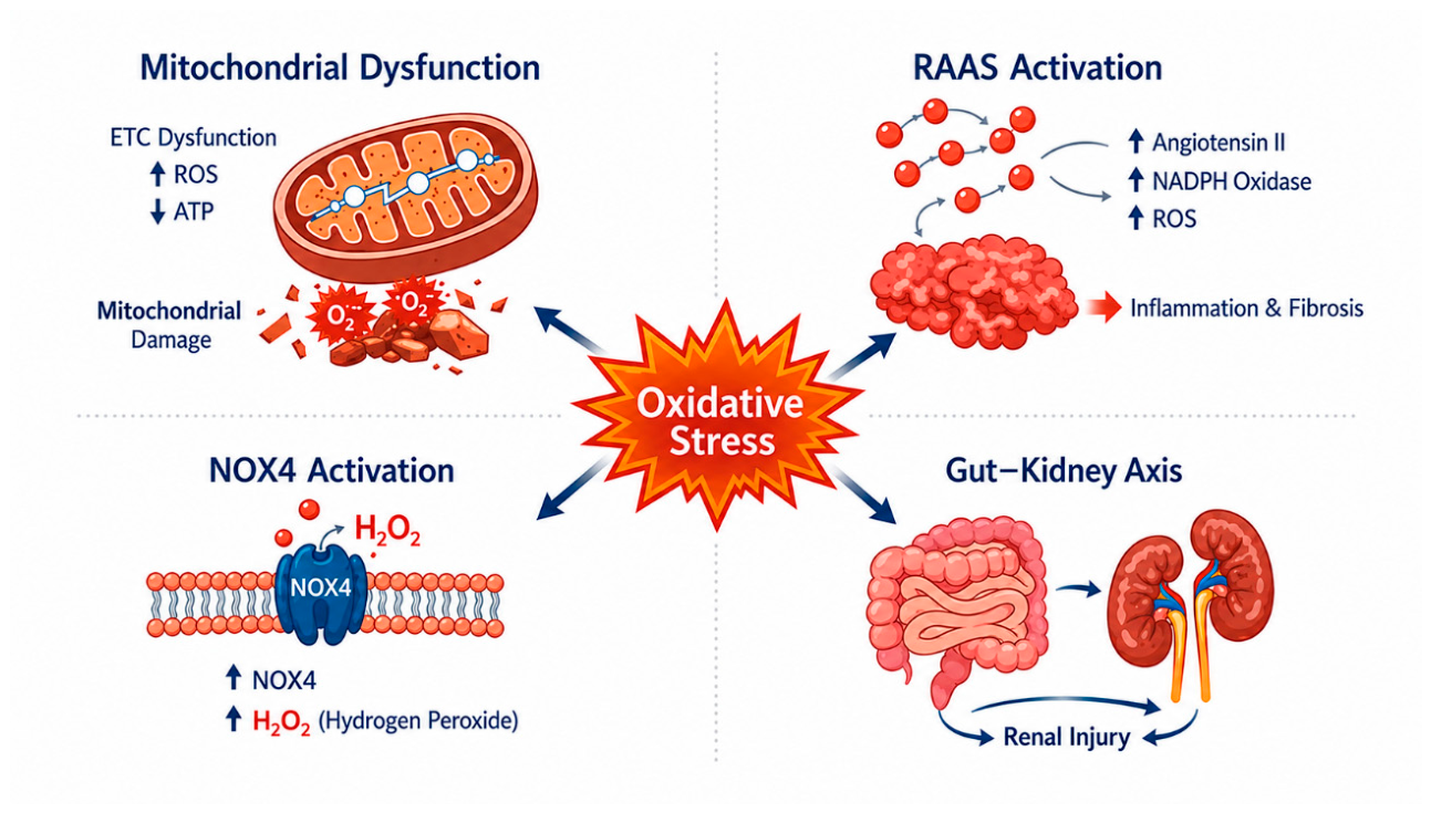
Figure 2.
Mitochondrial quality control pathways in chronic kidney disease. Mitochondrial quality control (MQC) is maintained through the coordinated regulation of mitochondrial dynamics (fusion and fission), mitophagy, and mitochondrial biogenesis. Key mediators include MFN1/2, OPA1, Drp1, the PINK1/Parkin pathway, receptor-mediated mitophagy pathways, and the PGC-1α/NRF1/2/TFAM axis. Additional regulatory inputs include SIRT1/3-, AMPK-, and redox-sensitive signaling pathways. Failure of MQC promotes mitochondrial fragmentation, ATP depletion, oxidative stress, apoptosis, inflammation, and fibrosis.
Figure 2.
Mitochondrial quality control pathways in chronic kidney disease. Mitochondrial quality control (MQC) is maintained through the coordinated regulation of mitochondrial dynamics (fusion and fission), mitophagy, and mitochondrial biogenesis. Key mediators include MFN1/2, OPA1, Drp1, the PINK1/Parkin pathway, receptor-mediated mitophagy pathways, and the PGC-1α/NRF1/2/TFAM axis. Additional regulatory inputs include SIRT1/3-, AMPK-, and redox-sensitive signaling pathways. Failure of MQC promotes mitochondrial fragmentation, ATP depletion, oxidative stress, apoptosis, inflammation, and fibrosis.
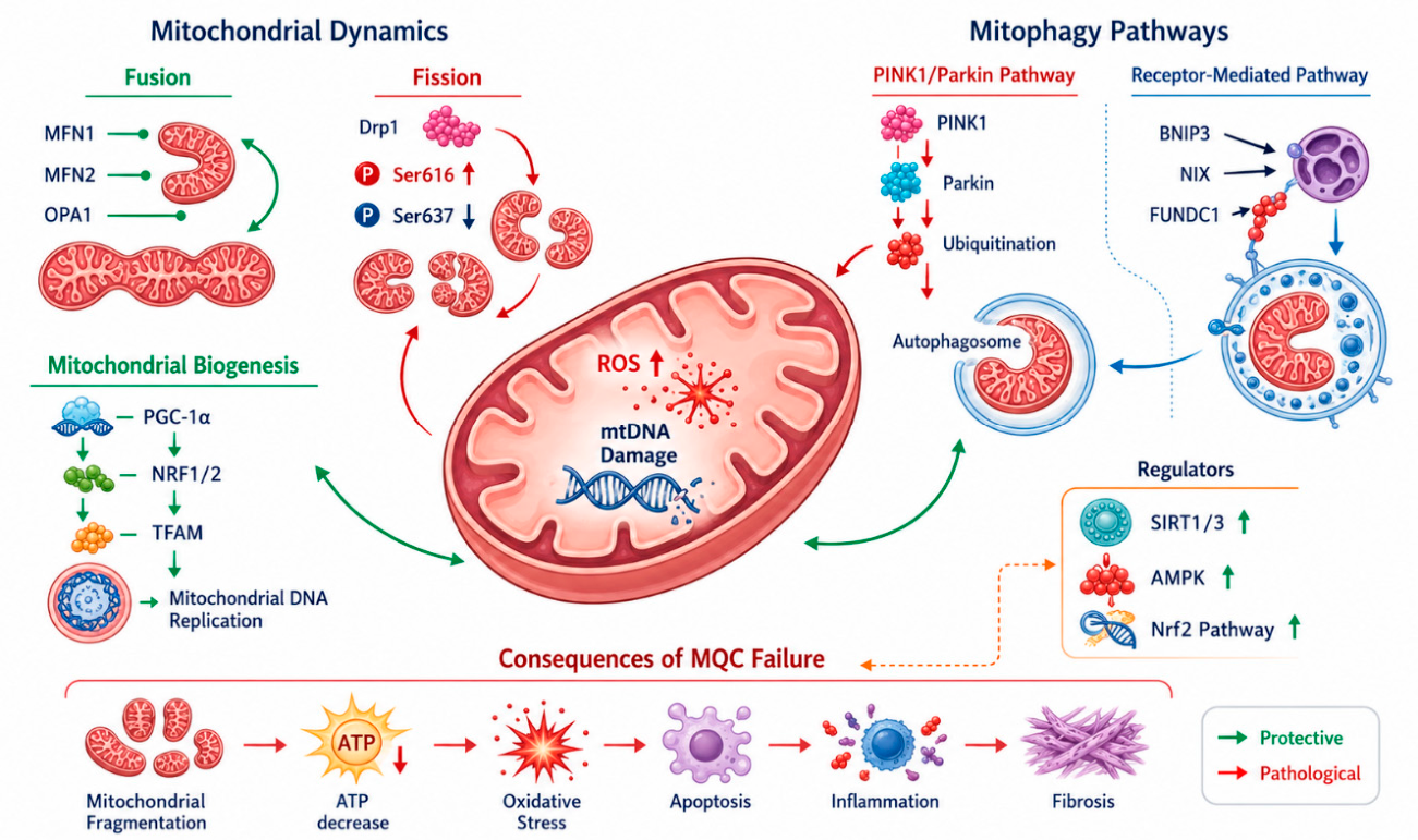
Figure 3.
Molecular mechanisms of ferroptosis in renal cells. Ferroptosis is an iron-dependent form of regulated cell death driven by uncontrolled lipid peroxidation. Transferrin-mediated iron uptake and intracellular Fe²⁺/Fe³⁺ redox cycling promote the Fenton reaction, generating hydroxyl radicals that initiate peroxidation of polyunsaturated fatty acids incorporated into membrane phospholipids (PUFA-PL). ACSL4 facilitates PUFA incorporation into membrane phospholipids, thereby increasing susceptibility to lipid hydroperoxide formation. The major anti-ferroptotic defense is the System Xc⁻/cystine/GSH/GPX4 axis, in which GPX4 uses glutathione to reduce toxic lipid hydroperoxides to non-toxic lipid alcohols. When GPX4 activity is reduced or lost, lipid ROS and lipid peroxides accumulate, leading to membrane damage and ferroptotic cell death.
Figure 3.
Molecular mechanisms of ferroptosis in renal cells. Ferroptosis is an iron-dependent form of regulated cell death driven by uncontrolled lipid peroxidation. Transferrin-mediated iron uptake and intracellular Fe²⁺/Fe³⁺ redox cycling promote the Fenton reaction, generating hydroxyl radicals that initiate peroxidation of polyunsaturated fatty acids incorporated into membrane phospholipids (PUFA-PL). ACSL4 facilitates PUFA incorporation into membrane phospholipids, thereby increasing susceptibility to lipid hydroperoxide formation. The major anti-ferroptotic defense is the System Xc⁻/cystine/GSH/GPX4 axis, in which GPX4 uses glutathione to reduce toxic lipid hydroperoxides to non-toxic lipid alcohols. When GPX4 activity is reduced or lost, lipid ROS and lipid peroxides accumulate, leading to membrane damage and ferroptotic cell death.
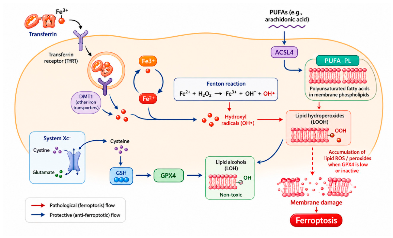
Figure 4.
Selected therapeutic approaches targeting mitochondrial dysfunction in chronic kidney disease. Therapeutic strategies targeting mitochondrial dysfunction in CKD include established nephroprotective agents and emerging mitochondria-directed interventions. These include SGLT2 inhibitors, Nrf2 activation by bardoxolone methyl, mitochondria-targeted antioxidants such as MitoQ and SS-31 (elamipretide), and experimental approaches such as mitochondrial transplantation. Collectively, these strategies aim to reduce oxidative stress, improve mitochondrial bioenergetics, and limit renal injury progression.
Figure 4.
Selected therapeutic approaches targeting mitochondrial dysfunction in chronic kidney disease. Therapeutic strategies targeting mitochondrial dysfunction in CKD include established nephroprotective agents and emerging mitochondria-directed interventions. These include SGLT2 inhibitors, Nrf2 activation by bardoxolone methyl, mitochondria-targeted antioxidants such as MitoQ and SS-31 (elamipretide), and experimental approaches such as mitochondrial transplantation. Collectively, these strategies aim to reduce oxidative stress, improve mitochondrial bioenergetics, and limit renal injury progression.
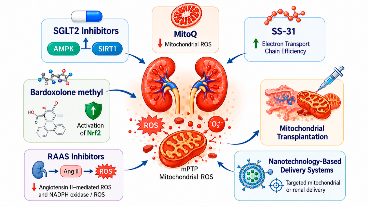

Table 1.
Redox and mitochondrial biomarkers in chronic kidney disease: biological meaning, specimen type, analytical methods, and clinical relevance.
Table 1.
Redox and mitochondrial biomarkers in chronic kidney disease: biological meaning, specimen type, analytical methods, and clinical relevance.
| Biomarker | Biological matrix | Pathophysiological domain reflected | Main analytical method | Potential clinical use | Main limitations |
| F2-isoprostanes | Plasma; urine | Global lipid peroxidation and in vivo oxidative stress | GC–MS; LC–MS/MS | Assessment of systemic oxidative stress burden; research-level risk stratification | Limited routine availability; mass spectrometry often required; values may be influenced by pre-analytical variability |
| 15-F₂t-isoprostane | Urine | Systemic lipid peroxidation | LC–MS/MS; GC–MS | Non-invasive monitoring of oxidative stress in CKD | Less widely standardized than routine clinical biomarkers; interpretation may vary across assays |
| 8-OHdG | Urine; plasma | Oxidative DNA damage | ELISA; HPLC; LC–MS/MS | Potential indicator of oxidative DNA injury and disease progression | Affected by smoking, inflammation, and metabolic comorbidities; assay heterogeneity limits comparability |
| Advanced oxidation protein products (AOPPs) | Serum; plasma | Protein oxidation and inflammation-associated oxidative damage | Spectrophotometric assay; ELISA-based methods | Potential marker of oxidative-inflammatory burden and CKD progression | Lower specificity; influenced by systemic inflammation and albumin oxidation |
| Circulating mtDNA | Plasma; serum | Mitochondrial injury, cell stress, and DAMP-mediated inflammation | qPCR; digital PCR | Early indication of mitochondrial damage; possible risk stratification tool | Pre-analytical variability; lack of standardized reference ranges; may reflect extra-renal sources |
| Urinary mtDNA | Urine | Renal mitochondrial injury and tubular stress | qPCR; digital PCR | Non-invasive marker of renal mitochondrial damage; potentially useful in AKI-to-CKD transition and CKD monitoring | Urine handling strongly affects results; normalization strategies are not fully standardized |
Abbreviations: AOPPs, advanced oxidation protein products; GC–MS, gas chromatography–mass spectrometry; HPLC, high-performance liquid chromatography; LC–MS/MS, liquid chromatography–tandem mass spectrometry; mtDNA, mitochondrial DNA; qPCR, quantitative polymerase chain reaction.
Table 2.
Therapeutic strategies targeting oxidative stress and mitochondrial dysfunction in chronic kidney disease.
Table 2.
Therapeutic strategies targeting oxidative stress and mitochondrial dysfunction in chronic kidney disease.
|
Therapy / intervention |
Main target / pathway | Expected mitochondrial / redox effect | Disease setting / evidence context | Main limitation or caveat |
| SGLT2 inhibitors | AMPK/SIRT1 signaling; metabolic reprogramming; autophagy-related pathways | Reduce oxidative stress, improve mitochondrial efficiency, and support mitochondrial quality control | Strong clinical evidence in CKD and diabetic kidney disease; established nephroprotective therapy | Not a dedicated mitochondria-targeted therapy; mechanistic effects are partly indirect |
| RAAS inhibitors | Angiotensin II signaling; NADPH oxidase activation | Lower Ang II-mediated ROS production and limit fibrosis-associated redox injury | Established CKD therapy with broad clinical use | Antioxidant effects are secondary to hemodynamic/nephroprotective action |
| Bardoxolone methyl / Nrf2 activation | Nrf2-dependent antioxidant transcriptional response | Enhance antioxidant defenses and redox resilience | Clinical trials in diabetic kidney disease and related CKD settings | Safety concerns, especially fluid overload/heart failure risk in selected populations |
| MitoQ | Mitochondria-targeted antioxidant activity within the inner mitochondrial membrane | Directly scavenges mitochondrial ROS and may reduce oxidative damage | Mainly preclinical or translational evidence in kidney injury models | Limited CKD-specific clinical validation |
| SS-31 (elamipretide) | Cardiolipin stabilization; electron transport chain function | Improves mitochondrial bioenergetics and reduces ROS generation | Preclinical and translational evidence; broader proof-of-concept from mitochondrial disease settings | Evidence in CKD remains limited; availability and regulatory use are context-dependent |
| Mitochondrial transplantation | Replacement of damaged mitochondria with functional mitochondria | Restores cellular bioenergetics and may reduce inflammation and oxidative injury | Experimental / early-stage preclinical approach | Currently not standardized for routine clinical nephrology |
| Nanotechnology-based delivery systems | Targeted delivery of antioxidants or bioactive compounds to renal tissue or mitochondria | Increase local drug concentration while limiting systemic toxicity | Early experimental / proof-of-concept stage | Technological complexity, translational barriers, and limited human data |
Abbreviations: AOPPs, advanced oxidation protein products; GC–MS, gas chromatography–mass spectrometry; HPLC, high-performance liquid chromatography; LC–MS/MS, liquid chromatography–tandem mass spectrometry; mtDNA, mitochondrial DNA; qPCR, quantitative polymerase chain reaction.
Disclaimer/Publisher’s Note: The statements, opinions and data contained in all publications are solely those of the individual author(s) and contributor(s) and not of MDPI and/or the editor(s). MDPI and/or the editor(s) disclaim responsibility for any injury to people or property resulting from any ideas, methods, instructions or products referred to in the content. |
© 2026 by the authors. Licensee MDPI, Basel, Switzerland. This article is an open access article distributed under the terms and conditions of the Creative Commons Attribution (CC BY) license (http://creativecommons.org/licenses/by/4.0/).
Copyright: This open access article is published under a Creative Commons CC BY 4.0 license, which permit the free download, distribution, and reuse, provided that the author and preprint are cited in any reuse.


