
Submitted:
16 July 2026
Posted:
20 July 2026
You are already at the latest version
Abstract
Canine mammary gland tumors are widely recognized as a spontaneous and translational model for breast cancer research. However, the lack of robust ex vivo culture systems capable of preserving tissue architecture and tumor microenvironment limits their experimental use. The aim of this study was to develop and characterize an ex vivo histoculture model of canine mammary tumors using heterologous equine platelet-rich plasma as a culture substrate. Fresh tumor specimens obtained during surgical excision were sectioned and cultured in physiological saline solution or platelet-rich plasma for up to 21 days. Tissue preservation was evaluated through macroscopic inspection, histological assessment, and immunohistochemical analysis of epithelial, mesenchymal, proliferative, and hypoxia–related markers. Tumor explants cultured with platelet-rich plasma showed superior macroscopic integrity, preservation of native tissue architecture, and reduced autolytic changes compared with controls. Immunohistochemical findings demonstrated sustained epithelial and stromal cell integrity, maintained proliferative activity, and limited hypoxic signaling. Overall, platelet-rich plasma provided a biologically supportive microenvironment that prolonged tissue viability ex vivo. This histoculture model represents a cost–effective and ethically refined platform for translational oncology, supporting drug testing, tumor biology studies, and reduction of animal use in accordance with the principles of replacement, reduction, and refinement.
Keywords:
canine mammary tumor
; ex vivo culture
; patient–derived explants
; platelet–rich plasma
; histoculture
; tumor microenvironment
; translational oncology
; veterinary oncology
1. Introduction
Biomedical research applied to oncology requires urgent and necessary preclinical models that accurately replicate the native architecture and microenvironment of primary solid tumors to improve understanding of tumor biology and facilitate the development of improved therapeutic approaches [1]. The use of in vitro models with cultures of immortalized human cell lines, for the study of new therapies in vitro or in vivo, is convenient but also one of the main reasons for the high failure of new drugs entering clinical trials. To move to more clinically relevant model systems, researchers have been adopting patient–derived approaches such as organoids [1,2,3] and patient-derived xenografts (PDX) [1,4,5,6].
However, several technical issues, e.g. cultivation times and high costs, limit the use of these models [7]. In human medicine, several studies recognize the potential of tissues cultured ex vivo [7,8,9,10], but in veterinary medicine it is under-explored. Patient–derived explants (PDEs) are ex vivo tumor models created by directly implanting fresh tumor tissue from patients into a supportive matrix or culture system, preserving the original tumor microenvironment. Unlike traditional 2D cultures or patient–derived xenografts (PDXs), PDEs retain the heterogeneity, architecture, and stromal interactions of the original tumor, making them highly relevant for drug testing and personalized medicine. PDEs allow for short-term screening of therapeutic responses, including immunotherapies, by maintaining immune cell populations and extracellular matrix components, which are often lost in other models. Studies have demonstrated that PDEs can predict patient responses to chemotherapy, targeted agents, and immunotherapies more accurately than cell–line–derived models [1,11]. Moreover, PDEs are being utilized to identify biomarkers for treatment stratification, particularly in aggressive cancers such as colorectal cancers [12]. However, challenges are still, including maintaining viable tissue for extended periods, ensuring reproducibility, and standardizing culture conditions. Although PDEs have a long history, general recognition of the potential of ex vivo cultured tissues to increase the clinical relevance of laboratory research [8,13,14,15,16], the PDE method has not been widely adopted to study solid tumors. Human breast tissue explant cultures provide important information for the study of breast cancer structure and phenotype, as they include the context of the surrounding microenvironment, allowing for the complete exploration of patient heterogeneity. However, the main limitation of currently available techniques remains the short–term viability of the tissue due to the loss of structural integrity [17].
PRP is a blood-derived plasma product that is easily obtained from peripheral blood samples and contains a platelet concentration approximately three to five times higher than that of whole blood. This provides a wide range of highly concentrated bioactive growth factors [18,19,20], including transforming growth factor (TGF-ß1 and TGF-ß2), platelet-derived growth factor (PDGF-AA, -AB, and -BB), vascular endothelial growth factor (VEGF-A and VEGF-C), insulin-like growth factor (IGF-1), and epidermal growth factor (EGF)These. which promote local angiogenesis, stem cell homing, cell migration, proliferation, and differentiation, along with the deposition of proteins such as collagen, which are essential for restoring normal tissue structure and function [21,22,23]. Due to the high content of growth factors in platelets, PRP has been shown to enhance cell growthoftensurpassing that of standard cultures supplemented with Fetal Calf Serum (FCS) [24]. In this study, preliminary trials used only PRP as a culture medium, with the addition of calcium gluconate to initiate the coagulation cascade and trigger growth factor release. Calcium gluconate gives PRP a gel-like consistency. After the gel retracts, PRP clot (CPRP) and PRP serum (SPRP) are obtained [25], with the latter used as a supplement to the remaining PRP.
Several studies have shown that PRP contributes to reducing regeneration times and improving tissue quality [20,26,27,28] and have promoted its wide use in clinical practice [29]. Lucarelli et al. [30] investigated the ex vivo influence of 1% up to 10% PRP as platelet gel on bone marrow stromal stem cells, showing a dose-dependent effect of PRP on cell proliferation.
Canine mammary cancer is considered by many researchers to be an excellent translational model due to the numerous similarities with breast cancer, although many aspects are still to be investigated and deepened. Canine mammary cancer is one of the main cancers diagnosed in whole. This also represents an excellent translational model of breast cancer, showing similarities both in the risk factors of onset and in terms of progression of breast cancer disease, which can be the cause of death in both women and dogs. The study of tumor in animal models allows to deepen the knowledge on prognostic factors that could also represent possible therapeutic targets both in human medicine and veterinary.
This study demonstrates that ex vivo culture derived from cancer samples obtained during surgery provides a low-cost, high-throughput model that maintains native tissue architecture, microenvironment, cell viability, and major oncogenic factors, all enhanced using PRP as a culture medium, even at low production cost, but high potential.
2. Materials and Methods
Blood was collected, using a blood bag (Terumo Blood Bags, CPDA-1) containing an anticoagulant solution (CPDA-1), from the jugular vein of a client-owned horse as part of a routine therapeutic procedure for the preparation of autologous PRP intended for the treatment of endometritis. No blood samples were collected specifically for research purposes. Residual PRP remaining after completion of the clinical treatment was subsequently used for the in vitro study. After the first centrifugation (1000–1200 rpm for 10 min, temperature at 4–10 °C), three layers were evident: the lower layer composed of red blood cells, the middle layer composed of platelets and white blood cells (buffy coat) and the upper layer of plasma. Fifty percent of the supernatant was discarded up to the buffy coat and, to obtain a platelet-enriched plasma fraction, a second centrifugation was performed. For this purpose, the plasma obtained after the first centrifugation was aspirated using a sterile syringe and transferred into 50 mL Falcon tubes. One tube was set aside to obtain fibrin–rich plasma (PRF). After the second centrifugation (2500–3000 rpm for 10 min, temperature at 4–10 °C), the upper fraction corresponded to platelet-poor plasma (PPP), whereas the lower fraction represented platelet-rich plasma (PRP). PPP was removed, while previously obtained autologous thrombin was added to PRP, thus activating the coagulation cascade. To obtain autologous thrombin, a PRP tube was isolated in which calcium gluconate (10%, Braun) was added. The mixture was incubated for 30–40 min at 37 °C and the PRP clot (CPRP) and PRP serum (SPRP) were obtained. The serum was filtered to remove cells and fibrin. The latter was used as a supplement to the remaining part of PRP obtained, as it contains significant thrombin activity.
Two canine mammary gland tumors, from patients who underwent total or partial mastectomy, were transported on ice to the laboratory of the Unit of Pathology, of the Department of Veterinary Sciences, University of Messina, within 15 min and cut into 5 x 5 x 2 mm contiguous pieces. From the first tumor, a sample was placed directly in 10% formalin (T0) while an additional four (4) were cultured in 0.9% saline solution (control group, SS) and equine heterologous PRP (PS group) for 3 and 7 days, ensuring that the tissue fragments were completely submerged in the culture medium. After each incubation period, a gross examination was performed to assess changes in volume, shape, and color, and samples were fixed in formalin for histological analysis. Hematoxylin and Eosin stain (H&E) was performed in each specimen and immunohistochemistry was carried out for cytokeratin panCK, AE1/AE3, Vimentin, PCNA. From the second tumor a sample was placed directly in 10% formalin (T0) while an additional six (6) were cultured in 0.9% saline solution (control group, SS) and equine heterologous PRP (PS group) for 7, 14 and 21 days. In this second setting, after a preliminary analysis with H&E, immunohistochemistry was carried out for Cytokeratin, Vimentin, β-catenin, , HIF–1 alpha, Ki-67, PCNA Epo (erythropoietin), EpoR (erythropoietin receptor), P63(Table 1).
Briefly, for immunohistochemical staining, samples were fixed in formalin and embedded in paraffin. Sections of 5 µm thickness on poly-L-lysine coated slides underwent microwave oven antigen unmasking in citrate buffer at pH 6 or EDTA buffer at pH 8. Hydrogen peroxide to block the endogenous peroxidases and a blocking substrate (ChemCruz—UltraCruz Blocking Reagent, Santa Cruz Biotechnology)) to block non-specific protein reactions were applied over. The sections were then incubated with the selected primary antibodies (as reported in Table 1), overnight at 4 °C. As revelation method, were then applied biotin- or HRP-conjugated secondary antibodies followed by an incubation with acetylated Streptavidin (Biospa antibiotic products, Milan; for biotin-conjugated antibody only). Diaminobenzidine (DAB) enzyme substrate, followed by nuclear counterstain with Haematoxylin, were used to stain the immune reaction.
Each round of IHC included negative reagent controls and internal controls. Each antibody was tested under different conditions as probing with and without AR, detection systems with biotin and HRP conjugated secondary Abs. The use of multiple antibodies of the same isotype and similar concentrations represented a set of irrelevant reagent controls. Immunohistochemical positivity was assessed as membranous, cytoplasmic or nuclear.
3. Results
H&E stain revealed that the first sample was a grade 1 tubular carcinoma (T0) (Figure 1).
Cultured canine mammary carcinoma explants exhibited markedly superior preservation in all incubation intervals when supplemented with PRP, as evidenced by the sustained bright pink coloration observed macroscopically for up to 21 days. In contrast, specimens maintained in physiological saline displayed a progressive shift toward a yellow hue after approximately 5 days, indicative of advancing autolytic degeneration until showing signs of putrefaction at 21 days. These macroscopic observations were corroborated by histopathological evaluation using H&E staining, which confirmed improved maintenance of tissue architecture and reduced autolytic artifacts in the PRP cultured samples.
Cytokeratin (AE1/AE3) immunolabeling demonstrated a coherent expression within luminal epithelial cell populations, showing increased immunoreactivity and intensity in PRP cultured fragments (P3–P7) compared with uncultured baseline tissue (T0). This suggests enhanced preservation of epithelial lineage markers and cellular differentiation status under PRP enriched conditions
Vimentin immunostaining highlighted consistent mesenchymal labeling across both fresh (T0) and cultured (T3–T7) samples, showing sustained stromal viability, fibroblastic cytoskeletal integrity, and preservation of epithelial–mesenchymal interactions (Figure 2). Histomorphological evaluation further revealed that PDEs maintained on gelatin scaffolds for up to 7 days retained reproducible tissue polarity, cohesive tumor cell nests, closely mirroring their uncultured counterparts. Multifocal hypercellular areas were suggestive that a proliferation had occurred, and this was further proven by the PCNA staining (Figure 3).
In the second experimental setting, H&E revealed a grade 3 tubular carcinoma (T0). At T7, T14, and T21, only PS showed marked preservation of epithelial and stromal architecture along with sustained proliferative activity. Numerous mitotic figures were consistently observed, mainly at the tumor invasion front, defined in patient-derived explant cultures as the preserved tumor–stroma interface originally present in vivo, where malignant epithelial cells are in direct contact with and infiltrate the surrounding stromal compartment. These morphological features were kept throughout the culture period up to 21 days (Figure 4).
Tumor cells remained embedded within a structurally recognizable stromal matrix, showing retention of the native tumor microenvironment in the explant model. In contrast, PS exhibited a change in the histological pattern, having more malignant characteristics and appearing with a solid appearance and increased infiltration of the surrounding stroma.
Immunohistochemical evaluation of hypoxia-inducible factor-1α (HIF-1α), Erythropoietin (Epo) and Erythropoietin receptor (EpoR) demonstrated persistent nuclear positivity in PRP samples up to T21. HIF-1α expression was mainly detected in tumor cells localized at the invasion front and in adjacent stromal cells, including spindle-shaped cells and residual immune cells. A comparable immunoreactivity was detected for Epo and EpoR, mirroring HIF-1α expression. This co-localization is compatible with activation of hypoxia-driven signaling pathways and indicates preservation of HIF-1α–dependent transcriptional activity within the patient-derived explant microenvironment.
Epithelial integrity and viability were further confirmed by cytokeratin (CK), p63 and Beta catenin immunostaining. CK was strongly expressed in cell forming tubuli and in cells multifocally distributed in the neoplastic solid growth. Beta catenin expression was continuous and homogeneous highlighting the cell boundaries in a few cells. In most neoplastic cells showed a granular, patchy and discontinuous expression along the cytoplasmic membrane, similarly to what observed in the T0 sample (Figure 5). P63 was strongly expressed in the nuclei of numerous epithelial cells, consistent with its role as a marker of epithelial stem and myoepithelial cells Ki-67 immunoreactivity was mainly located at the invasion front, where several mitoses were detectable (insets), and supported the presence of ongoing proliferative activity, in line with its known association with multiple phases of the cell cycle (Figure 6).
4. Discussion
Ex vivo cultures derived from surgically excised spontaneous canine mammary tumors is a robust, reproducible, and highly translational model for cancer research. By preserving native tissue architecture and tumor microenvironment, patient-derived explant (PDE) cultures provide a valuable alternative to conventional in vitro and in vivo models, supporting the principles of Replacement, Reduction, and Refinement (3Rs) [31]. In this context, PDEs offer a cost-effective, ethically sound platform to investigate tumor biology, drug responses, and microenvironment-dependent mechanisms with high predictive value. The development and appropriate fine-tuning of methods for preservation of ex vivo tumor cultures using PRP, or other blood components could also represent an economic model that potentially leads to the development and filing of patents. The ex vivo model is also called the "organotypic model" because it maintains the three-dimensional structure of the organ and is a model that combines the advantages of in vitro and in vivo cultures and allows the biology of cancer to be studied on tissue in its entire composition [32]. The aim is also to investigate immunoediting and the long-term effects of drugs in a set-up that closely resembles the original tumor microenvironment. It has been demonstrated that the interaction of epithelial and stromal tissues is important for epithelial maintenance and plays an important role in malignant transition to cancer [17].
In patient-derived explant cultures, the tumor invasion front corresponds to the preserved tumor–stroma interface originally present in vivo, where malignant epithelial cells remain in direct contact with, and infiltrate into, the surrounding stromal compartment. This region retains key structural, cellular, and functional features of tumor invasion, including active proliferation, stromal interaction, and spatially confined signaling responses. The persistence of a defined invasion front in patient-derived explant cultures underscores the ability of this model to faithfully preserve the dynamic tumor–stroma interface that governs invasion and disease progression in vivo, supporting its relevance for studying microenvironment-dependent tumor behavior and therapeutic responses.
In the present study, we developed and optimized an original histoculture protocol for canine mammary tumors using heterologous (equine) platelet-rich plasma (PRP) as the culture medium. Gel-like PRP matrix provided mechanical support comparable to surrounding tissue in vivo, maintaining tissue compactness while allowing diffusion of nutrients and soluble factors. This structural support was associated with sustained tissue viability and retained proliferative activity for up to three weeks, indicating the establishment of a stable and long-lasting organotypic culture.
The prolonged preservation of tissue integrity observed in our model is likely attributable to the combined effects of PRP supplementation and low-temperature storage. PRP provides a concentrated source of growth factors and bioactive molecules that support cell survival, proliferation, and tissue repair, while hypothermic conditions reduce metabolic demand, enzymatic activity, and stress-induced damage. The synergy between these two factors likely contributed to limiting apoptotic and necrotic pathways, thereby preserving tissue architecture and microenvironmental complexity over time.
The invasion front of tumor explants was characterized by the spatial convergence of proliferative, hypoxia-related, and adhesion-associated markers. Ki-67 immunoreactivity was predominantly localized at the tumor–stroma interface, consistent with active cell cycling and increased mitotic activity in this region, as previously described for invasive tumor compartments. This proliferative pattern was accompanied by sustained HIF-1α expression, supporting the presence of localized hypoxic signaling known to promote invasive growth and metabolic adaptation. In parallel, the discontinuous and patchy membranous distribution of β-catenin observed in neoplastic cells at the invasion front is compatible with altered cell–cell adhesion dynamics associated with tumor–stroma interactions and invasive behavior. Finally, the persistence of epithelial markers such as cytokeratin and p63 within this compartment indicates that invasive activity occurs in the context of preserved epithelial identity rather than complete dedifferentiation. Collectively, the coordinated expression of these markers supports the ability of the patient-derived explant model to retain biologically relevant features of the native tumor invasion front ex vivo.
Together, these findings demonstrate that the PRP-based PDE model faithfully preserves the structural, cellular, and functional features of the native tumor invasion front ex vivo. By maintaining epithelial–stromal interactions, hypoxic signaling, and proliferative activity within an intact three-dimensional context, this organotypic culture system represents a powerful platform for studying tumor progression, immunoediting, and long-term therapeutic effects. Given its simplicity, low cost, and high translational relevance, this approach holds significant potential for both veterinary and comparative oncology, as well as for future diagnostic, therapeutic, and innovation-driven applications.
5. Conclusions
Ex vivo culture of canine mammary tumors using platelet-rich plasma represents a reliable and cost-effective histoculture approach that preserves tissue architecture, cellular viability, and tumor microenvironment for extended periods. The proposed model maintains key epithelial and stromal components, supporting its suitability for studying tumor biology and microenvironmental interactions under controlled conditions. Importantly, this methodology aligns with the principles of replacement, reduction, and refinement by maximizing biological information obtained from surgically excised clinical samples.
This platform holds significant translational potential for veterinary and comparative oncology, particularly for short-term drug screening, biomarker assessment, and evaluation of tumor–stroma interactions. Further optimization and validation studies are warranted to assess predictive value for therapeutic responses and to extend culture longevity. Overall, platelet–rich plasm-based histoculture provides a promising experimental tool bridging conventional in vitro systems and in vivo models
Author Contributions
Conceptualization, A.S., R.P.; methodology, A.S., G.G., S.D.G.; validation, A.S. and G.G.; formal analysis, A.S.; investigation, G.G.; resources, A.S., C.V.; data curation, A.S.; writing—original draft preparation, A.S., G.M.; writing—review and editing, A.S., G.M.; visualization, G.G., S.D.G.; supervision, A.S.; project administration, A.S; funding acquisition, R.P. All authors have read and agreed to the published version of the manuscript.
Funding
This research was funded by, Istituto Zooprofilattico Sperimentale della Sicilia, ItalianMinistry of Health Ricerca Corrente 2023, grant number H77G23000330001.
Institutional Review Board Statement
Blood samples used for platelet-rich plasma (PRP) preparation were obtained from client-owned horses undergoing routine therapeutic treatment for endometritis at the Veterinary Teaching Hospital. Blood collection was performed exclusively for clinical purposes as part of the preparation of autologous PRP for treatment. No additional blood samples were collected for research purposes, and no additional procedures, restraint, or handling beyond routine veterinary care were performed. Residual PRP remaining after completion of the clinical procedure was subsequently used for in vitro experiments.Canine mammary tumour tissues were obtained from client-owned dogs undergoing clinically indicated surgical excision as part of routine veterinary care.The study involved exclusively the use of residual biological materials derived from routine clinical procedures and did not require any additional interventions on live animals for research purposes.
Informed Consent Statement
Written informed consent was obtained from the owners of all client-owned animals included in the study prior to the collection and use of biological materials for research purposes.
Data Availability Statement
Most relevant data are presented, additional data can be asked to authors.
Acknowledgments
Preliminary findings from this study were previously presented at the ESVP/ECVP 2025 Annual Congress and published in abstract form [33]. The present manuscript reports the complete study, including expanded experimental data and analyses.
Conflicts of Interest
The authors declare no conflicts of interest.
References
- Powley, I.R.; Patel, M.; Miles, G.; Pringle, H.; Howells, L.; Thomas, A.; Kettlebrough, C.; Bryans, J.; Hammonds, T.; MacFarlane, M.; Pritchard, C. Patient–derived explants (PDEs) as a powerful preclinical platform for anti–cancer drug and biomarker discovery. Br. J. Cancer 2020, 122, 735–744. [Google Scholar] [CrossRef] [PubMed]
- Gao, D.; Vela, I.; Sboner, A.; Iaquinta, P.J.; Karthaus, W.R.; Gopalan, A.; Dowling, C.; Wanjala, J.N.; Undvall, E.A.; Arora, V.K.; Wongvipat, J.; Kossai, M.; Ramazanoglu, S.; Barboza, L.P.; Di, W.; Cao, Z.; Zhang, Q.F.; Sirota, I.; Ran, L.; MacDonald, T.Y.; Beltran, H.; Mosquera, J.M.; Touijer, K.A.; Scardino, P.T.; Laudone, V.P.; Curtis, K.R.; Rathkopf, D.E.; Morris, M.J.; Danila, D.C.; Slovin, S.F.; Solomon, S.B.; Eastham, J.A.; Chi, P.; Carver, B.; Rubin, M.A.; Scher, H.I.; Clevers, H.; Sawyers, C.L.; Chen, Y. Organoid cultures derived from patients with advanced prostate cancer. Cell 2014, 159, 176–187. [Google Scholar] [CrossRef] [PubMed]
- Drost, J.; Karthaus, W.R.; Gao, D.; Driehuis, E.; Sawyers, C.L.; Chen, Y.; Clevers, H. Organoid culture systems for prostate epithelial and cancer tissue. Nat. Protoc. 2016, 11, 347–358. [Google Scholar] [CrossRef] [PubMed]
- Wang, Y.; Revelo, M.P.; Sudilovsky, D.; Cao, M.; Chen, W.G.; Goetz, L.; Xue, H.; Sadar, M.; Shappell, S.B.; Cunha, G.R.; Hayward, S.W. Development and characterization of efficient xenograft models for benign and malignant human prostate tissue. Prostate 2005, 64, 149–159. [Google Scholar] [CrossRef] [PubMed]
- Lawrence, M.S.; Stojanov, P.; Mermel, C.H.; Robinson, J.T.; Garraway, L.A.; Golub, T.R.; Meyerson, M.; Gabriel, S.B.; Lander, E.S.; Getz, G. Discovery and saturation analysis of cancer genes across 21 tumour types. Nature 2014, 505, 495–501. [Google Scholar] [CrossRef] [PubMed]
- Whittle, J.R.; Lewis, M.T.; Lindeman, G.J.; Visvader, J.E. Patient–derived xenograft models of breast cancer and their predictive power. Breast Cancer Res. 2015, 17, 17. [Google Scholar] [CrossRef] [PubMed]
- Centenera, M.M.; Hickey, T.E.; Jindal, S.; Ryan, N.K.; Ravindranathan, P.; Mohammed, H.; Robinson, J.L.; Schiewer, M.J.; Ma, S.; Kapur, P.; Sutherland, P.D.; Hoffmann, C.E.; Roehrborn, C.G.; Gomella, L.G.; Carroll, J.S.; Birrell, S.N.; Knudsen, K.E.; Raj, G.V.; Butler, L.M.; Tilley, W.D. A patient–derived explant (PDE) model of hormone–dependent cancer. Mol. Oncol. 2018, 12, 1608–1622. [Google Scholar] [CrossRef] [PubMed]
- Centenera, M.M.; Raj, G.V.; Knudsen, K.E.; Tilley, W.D.; Butler, L.M. Ex vivo culture of human prostate tissue and drug development. Nat. Rev. Urol. 2013, 10, 483–487. [Google Scholar] [CrossRef] [PubMed]
- McClurg, U.L.; McCracken, S.R.; Butler, L.; Riabowol, K.T.; Binda, O. Ex vivo culture and lentiviral transduction of benign prostatic hyperplasia (BPH) samples. Bio Protoc. 2018, 8, e3075. [Google Scholar] [CrossRef] [PubMed]
- Ricciardelli, C.; Lokman, N.A.; Sabit, I.; Gunasegaran, K.; Bonner, W.M.; Pyragius, C.E.; Macpherson, A.M.; Oehler, M.K. Novel ex vivo ovarian cancer tissue explant assay for prediction of chemosensitivity and response to novel therapeutics. Cancer Lett. 2018, 421, 51–58. [Google Scholar] [CrossRef] [PubMed]
- Hidalgo, M.; Amant, F.; Biankin, A.V.; Budinská, E.; Byrne, A.T.; Caldas, C.; Clarke, R.B.; de Jong, S.; Jonkers, J.; Mælandsmo, G.M.; Roman–Roman, S.; Seoane, J.; Trusolino, L.; Villanueva, A. Patient–derived xenograft models: An emerging platform for translational cancer research. Cancer Discov. 2014, 4, 998–1013. [Google Scholar] [CrossRef] [PubMed]
- Mata, S.; Franchi-Mendes, T.; Abreu, S.; Filipe, B.; Morgado, S.; Mesquita, M.; Albuquerque, C.; Fonseca, R.; Santo, V.E.; Boghaert, E.R.; et al. Patient-Derived Explants of Colorectal Cancer: Histopathological and Molecular Analysis of Long-Term Cultures. Cancers 2021, 13, 4695. [Google Scholar] [CrossRef] [PubMed]
- Vescio, R.A.; Connors, K.M.; Kubota, T.; Hoffman, R.M. Correlation of histology and drug response of human tumors grown in native–state three–dimensional histoculture and in nude mice. Proc. Natl. Acad. Sci. USA 1991, 88, 5163–5166. [Google Scholar] [CrossRef] [PubMed]
- Pretlow, T.G.; Yang, B.; Pretlow, T.P. Organ culture of benign, aging, and hyperplastic human prostate. Microsc. Res. Tech. 1995, 30, 271–281. [Google Scholar] [CrossRef] [PubMed]
- Kim, J.B. Three–dimensional tissue culture models in cancer biology. Semin. Cancer Biol. 2005, 15, 365–377. [Google Scholar] [CrossRef] [PubMed]
- Risbridger, G.P.; Toivanen, R.; Taylor, R.A. Preclinical models of prostate cancer: Patient–derived xenografts, organoids, and other explant models. Cold Spring Harb. Perspect. Med. 2018, 8, a030536. [Google Scholar] [CrossRef] [PubMed]
- Koch, M.K.; Ravichandran, A.; Murekatete, B.; Clegg, J.; Joseph, M.T.; Hampson, M.; Jenkinson, M.; Bauer, H.S.; Snell, C.; Liu, C.; Gough, M.; Thompson, E.W.; Werner, C.; Hutmacher, D.W.; Haupt, L.M.; Bray, L.J. Exploring the potential of PEG–heparin hydrogels to support long–term ex vivo culture of patient–derived breast explant tissues. Adv. Healthc. Mater. 2023, 12, 2202202. [Google Scholar] [CrossRef] [PubMed]
- Marx, R.E. Platelet-rich plasma (PRP): What is PRP and what is not PRP? Implant Dent. 2001, 10, 225–228. [Google Scholar] [CrossRef] [PubMed]
- Marx, R.E. Platelet–rich plasma: Evidence to support its use. J. Oral Maxillofac. Surg. 2004, 62, 489–496. [Google Scholar] [CrossRef] [PubMed]
- El–Sharkawy, H.; Kantarci, A.; Deady, J.; Hasturk, H.; Liu, H.; Alshahat, M.; Van Dyke, T.E. Platelet–rich plasma: Growth factors and pro– and anti–inflammatory properties. J. Periodontol. 2007, 78, 661–669. [Google Scholar] [CrossRef] [PubMed]
- Harrison, P.; Cramer, E.M. Platelet alpha–granules. Blood Rev. 1993, 7, 52–62. [Google Scholar] [CrossRef] [PubMed]
- Coppinger, J.A.; Cagney, G.; Toomey, S.; Kislinger, T.; Belton, O.; McRedmond, J.P.; Cahill, D.J.; Emili, A.; Fitzgerald, D.J.; Maguire, P.B. Characterization of the proteins released from activated platelets leads to localization of novel platelet proteins in human atherosclerotic lesions. Blood 2004, 103, 2096–2104. [Google Scholar] [CrossRef] [PubMed]
- Nurden, A.T. Platelets, inflammation and tissue regeneration. Thromb. Haemost. 2011, 105, S13–S33. [Google Scholar] [CrossRef] [PubMed]
- Lang, S.; Loibl, M.; Herrmann, M. Platelet-rich plasma in tissue engineering: Hype and hope. Eur. Surg. Res. 2018, 59, 265–275. [Google Scholar] [CrossRef] [PubMed]
- Ferraz, D.C.; Tatsui, N.H.; Rodrigues, L.R.; Lombello, C.B. Plasma rico em plaquetas como suplemento e arcabouço para a cultura de células. In Proceedings of the Congresso Brasileiro de Hematologia, Hemoterapia e Terapia Celular—HEMO 2019 Hematol. Transfus. Cell Ther., Rio de Janeiro, Brazil, 6–9 November 2019; 2019; 41, p. S507. [Google Scholar]
- Weibrich, G.; Hansen, T.; Kleis, W.; Buch, R.; Hitzler, W.E. Effect of platelet concentration in platelet-rich plasma on peri–implant bone regeneration. Bone 2004, 34, 665–671. [Google Scholar] [CrossRef] [PubMed]
- Carneiro, M.O.; Barbieri, C.H.; Barbieri Neto, J. Platelet-rich plasma gel promotes regeneration of articular cartilage in knees of sheeps. Acta Ortop. Bras. 2013, 21, 80–86. [Google Scholar] [CrossRef] [PubMed]
- Pinto, J.M.; Pizani, N.S.; Kang, H.C.; Silva, L.A. Application of platelet-rich plasma in the treatment of chronic skin ulcer—case report. An. Bras. Dermatol. 2014, 89, 638–640. [Google Scholar] [CrossRef] [PubMed]
- Tresoldi, M.M.; Faga, A.; Nicoletti, G. Experimental assessment of regenerative properties of platelet rich plasma on the human skin—A review. Plast. Aesthet. Res. 2022, 9, 12. [Google Scholar] [CrossRef]
- Lucarelli, E.; Beccheroni, A.; Donati, D.; Sangiorgi, L.; Cenacchi, A.; Del Vento, A.M.; Meotti, C.; Bertoja, A.Z.; Giardino, R.; Fornasari, P.M.; Mercuri, M.; Picci, P. Platelet–derived growth factors enhance proliferation of human stromal stem cells. Biomaterials 2003, 24, 3095–3100. [Google Scholar] [CrossRef] [PubMed]
- Piwocka, O.; Suchorska, W.M.; Kulcenty, K. Empowering personalized medicine: Unleashing the potential of patient-derived explants in clinical practice. EXCLI J. 2024, 23, 81–91. [Google Scholar] [CrossRef] [PubMed]
- Urzì, O.; Gasparro, R.; Costanzo, E.; De Luca, A.; Giavaresi, G.; Fontana, S.; Alessandro, R. Three-Dimensional Cell Cultures: The Bridge between In Vitro and In Vivo Models. Int. J. Mol. Sci. 2023, 24, 12046. [Google Scholar] [CrossRef] [PubMed]
- Sfacteria, A.; Giambrone, G.; Di Giorgio, S.; Marino, G.; Catone, G.; Puleio, R. Patient-derived explants from canine mammary gland cancer using heterologous platelet-rich plasma as culture medium. J. Comp. Pathol. Abstract published in conference proceedings. 2025, 220. [Google Scholar] [CrossRef]
Figure 1.
Histological and immunohistochemical features of the uncultured canine mammary tumor sample at baseline (T0), and at day 3 (T3) and day 7 (T7) both in saline solution sample (SS) and PRP sample (PS). Hematoxylin and eosin (H&E) staining shows the native histological architecture of the tumor and the modification occurred during the culture. (H&E, SS T3, 5X. SS T7/PS T3/PS T7, 10X. T0, 20X).
Figure 1.
Histological and immunohistochemical features of the uncultured canine mammary tumor sample at baseline (T0), and at day 3 (T3) and day 7 (T7) both in saline solution sample (SS) and PRP sample (PS). Hematoxylin and eosin (H&E) staining shows the native histological architecture of the tumor and the modification occurred during the culture. (H&E, SS T3, 5X. SS T7/PS T3/PS T7, 10X. T0, 20X).
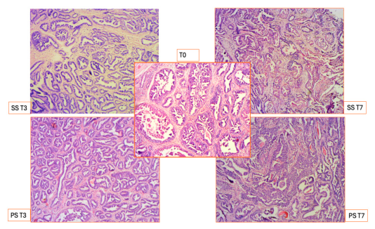
Figure 2.
IHC revealed that PS regained a better preservation. As evidenced by panCK and VIM stains, at T3 both SS and PS don’t show appreciable histological differences compared with the T0 sample. At T7 SS had several areas of epithelial alteration with detachment from the basal membrane and sloughing. On the contrary, PS showed an intact epithelium and stroma as highlighted by the immunostain. panCK and VIM in T7 PS have multifocal hypercellular areas of proliferation. (IHC, DAB, 5X).
Figure 2.
IHC revealed that PS regained a better preservation. As evidenced by panCK and VIM stains, at T3 both SS and PS don’t show appreciable histological differences compared with the T0 sample. At T7 SS had several areas of epithelial alteration with detachment from the basal membrane and sloughing. On the contrary, PS showed an intact epithelium and stroma as highlighted by the immunostain. panCK and VIM in T7 PS have multifocal hypercellular areas of proliferation. (IHC, DAB, 5X).
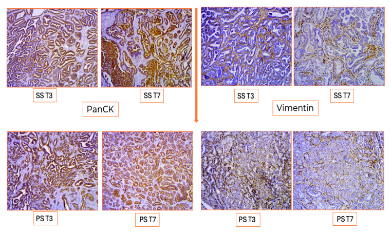
Figure 3.
PCNA staining shows an active proliferation in T7 PS compared to SS. (IHC, DAB, SS T7/PS T7, 10x. T0, 20X).
Figure 3.
PCNA staining shows an active proliferation in T7 PS compared to SS. (IHC, DAB, SS T7/PS T7, 10x. T0, 20X).
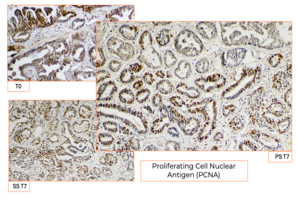
Figure 4.
At T14 and T21 only PS showed wide areas of preserved epithelial, stromal and proliferative activity with several mitoses especially at the so called “invasion front” (H&E, T0, 5X.SS T7/PS T7/PS T21/SS T21, 10X. SS T14/PS T14, 20x. Insets 40X).
Figure 4.
At T14 and T21 only PS showed wide areas of preserved epithelial, stromal and proliferative activity with several mitoses especially at the so called “invasion front” (H&E, T0, 5X.SS T7/PS T7/PS T21/SS T21, 10X. SS T14/PS T14, 20x. Insets 40X).
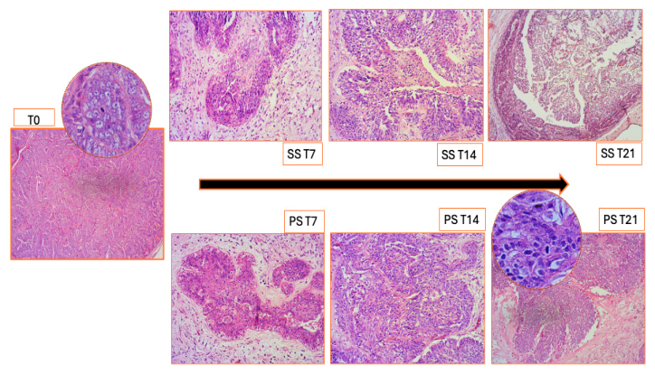
Figure 5.
T21 PS. CK and Beta catenin immunostaining highlights epithelial integrity, Vim shows a structurally recognizable stromal matrix and retention of the native tumor microenvironment. HIF-1, Epo, and Epor were expressed both in epithelium, spindle and inflammatory stromal cells. (IHC, DAB, from left to right 10x, insets 20x).
Figure 5.
T21 PS. CK and Beta catenin immunostaining highlights epithelial integrity, Vim shows a structurally recognizable stromal matrix and retention of the native tumor microenvironment. HIF-1, Epo, and Epor were expressed both in epithelium, spindle and inflammatory stromal cells. (IHC, DAB, from left to right 10x, insets 20x).
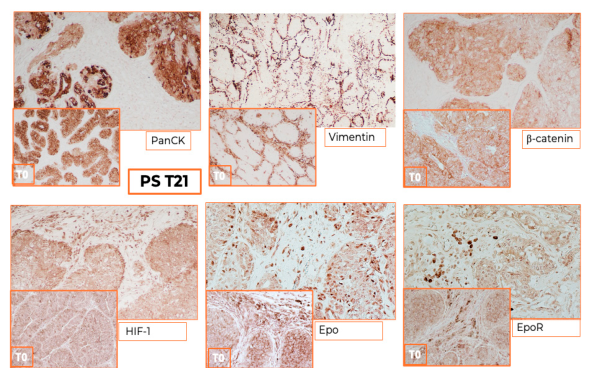
Figure 6.
T21 PS. p63 is located in the nuclei of numerous epithelial cells. Ki-67 immunoreactivity is expressed in the nuclei of cells at the invasion front, where several mitoses were detectable and indicative of ongoing proliferative activity. (IHC, DAB, 20X).
Figure 6.
T21 PS. p63 is located in the nuclei of numerous epithelial cells. Ki-67 immunoreactivity is expressed in the nuclei of cells at the invasion front, where several mitoses were detectable and indicative of ongoing proliferative activity. (IHC, DAB, 20X).
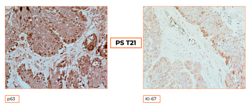

Table 1.
Primary antibodies used for immunohistochemical analysis.
| Primary Antibody | Clone | Dilution | Brand |
|---|---|---|---|
| Cytokeratin | AE1/AE3 | 1/100 | Santa Cruz Biotechnology, Inc. |
| Vimentin | VIM 3B4 | 1/100 | Santa Cruz Biotechnology, Inc. |
| β-catenin | 6F9 | 1/100 | Santa Cruz Biotechnology, Inc. |
| HIF-1 alpha | PA1–16601 | 1/400 | Invitrogen/Thermo-Fisher Scientific |
| KI-67 | KI–67 | 1/100 | Santa Cruz Biotechnolog, Inc. |
| PCNA | FL–261 | 1/50 | Santa Cruz Biotechnology, Inc. |
| Epo | 7D10 | 1/400 | Santa Cruz Biotechnology, Inc. |
| EpoR | D-5 | 1/400 | Santa Cruz Biotechnology, Inc. |
| P63 | (D-9) | 1/100 | Santa Cruz Biotechnology, Inc. |
| Secondary Antibody | |||
| Biotinylated Goat anti-mouse | 1/100 | Vector Laboratories | |
| Biotinylated Goat anti-rabbit | 1/50 | Vector Laboratories |
Disclaimer/Publisher’s Note: The statements, opinions and data contained in all publications are solely those of the individual author(s) and contributor(s) and not of MDPI and/or the editor(s). MDPI and/or the editor(s) disclaim responsibility for any injury to people or property resulting from any ideas, methods, instructions or products referred to in the content. |
© 2026 by the authors. Licensee MDPI, Basel, Switzerland. This article is an open access article distributed under the terms and conditions of the Creative Commons Attribution (CC BY) license (http://creativecommons.org/licenses/by/4.0/).
Copyright: This open access article is published under a Creative Commons CC BY 4.0 license, which permit the free download, distribution, and reuse, provided that the author and preprint are cited in any reuse.


