

Submitted:
17 July 2026
Posted:
17 July 2026
You are already at the latest version
Abstract
A novel chiral three-dimensional conjugated polymer with aggregation-induced emission (AIE) was constructed via asymmetric Suzuki–Miyaura cross-coupling polymerization. Comprehensive photophysical characterizations confirmed its stable AIE activity, excitation-tunable luminescence, and solvatochromism in biomimetic media including water, simulated body fluid, and simulated urine. Ion sensing studies revealed that the chiral framework specifically chelates Ag⁺ through multiple coordination sites, producing significant fluorescence enhancement with a linear response from 0–900 μM and a micromolar detection limit, alongside excellent anti-interference capability. The material also exhibits differentiated dual-channel optical responses toward Fe³⁺ and Cr⁶⁺. Electrochemical analysis elucidated an electron-rich conjugated backbone, supporting a charge-transfer-mediated recognition mechanism. Benefiting from its unique multilayer three-dimensional cavity topology, the polymer demonstrates superior ion capture capacity and specificity over conventional linear or monolayer systems. This work presents a new molecular design strategy for high-performance fluorescent probes, with substantial practical potential for environmental and biological monitoring applications such as industrial wastewater treatment, surface water heavy-metal screening, and trace ion analysis in biological fluids.
Keywords:
AIE
; multialyer 3D polymer
1. Introduction
In recent years, the ecological risks and threats to human health arising from heavy metal pollution in water bodies have attracted increasing public attention. Developing highly sensitive and specific fluorescent detection materials represents a core technical route for rapid in-situ monitoring of heavy metals. [1] Conjugated polymers with aggregation-induced emission (AIE) characteristics possess distinctive luminescence mechanisms and have become a crucial research frontier in fluorescence sensing and trace heavy metal detection in the environment. [2,3,4,5,6] Conventional organic fluorophores and linear conjugated luminescent materials generally suffer from aggregation-caused quenching (ACQ): tight π–π stacking upon molecular aggregation greatly accelerates non-radiative energy dissipation and leads to severe luminescence attenuation, which largely restricts their practical detection applications under high ion concentrations and complicated aqueous environments. [7,8,9,10] In sharp contrast, AIE-active polymers display weak emission when dispersed as single molecules. Upon molecular aggregation, intramolecular rotations and vibrations are restricted by steric hindrance, non-radiative transition pathways are substantially blocked, radiative recombination efficiency is greatly improved, and luminescence intensity is significantly boosted. [11] Such features perfectly meet the requirements of ion enrichment and aggregation-based fluorescence detection in water, demonstrating irreplaceable advantages in qualitative identification and quantitative analysis of trace metal ions.
Among existing conjugated luminescent polymer systems, linear conjugated polymers feature ordered molecular chains yet single spatial structures with limited chelating sites for metal ions, resulting in insufficient recognition specificity when facing complicated water samples containing multiple coexisting metal ions. In comparison, multi-layer three-dimensional topological polymers naturally construct abundant interconnected nano-cavities inside the framework, and high-density heteroatom coordination sites are distributed on the framework surface and inner cavity walls. [12] Specific capture of target metal ions can be strengthened via multi-chelation and electrostatic synergistic effects, simultaneously improving recognition selectivity and detection sensitivity of the sensing system from the perspective of molecular structure. Furthermore, covalently incorporating chiral building units into three-dimensional AIE networks enables precise regulation of intermolecular stacking modes, intermolecular dipole–dipole interactions and charge transfer efficiency, further achieving multiple characteristic photophysical behaviors such as solvatochromism and excitation-wavelength-dependent tunable luminescence. [13] The synergistic effect of these multiple structural merits endows chiral three-dimensional AIE polymers with strong environmental adaptability, allowing them to stably maintain optical sensing performance in biomimetic media with abundant ion species and complex polarity, such as simulated body fluid (SBF) and simulated urine (SU). [14] They can directly simulate detection matrices of real biological fluids, industrial wastewater and surface water, exhibiting broad application prospects in biological sample analysis and environmental water quality monitoring.
Benzofuran fused aromatic units possess rigid planar conjugated skeletons and oxygen-containing heteroatom active sites. [15,16] With strong molecular rigidity and easy accessibility for polymerization modification, the heteroatoms can provide abundant coordination sites, making them ideal building blocks for constructing high-performance AIE luminescent polymers. The well-established palladium-catalyzed Suzuki–Miyaura cross-coupling polymerization proceeds under mild conditions with broad substrate compatibility and high polymerization conversion efficiency, which can efficiently link benzofuran aromatic monomers to form continuous large π-conjugated three-dimensional polymer networks. [17,18] Most reported AIE sensing materials to date adopt simple linear chains or monolayer three-dimensional skeletons. [19] Such materials contain scarce effective coordination sites available for interaction with metal ions and are prone to non-specific binding when various interfering cations coexist in water samples, leading to poor anti-interference ability and difficulty in satisfying accurate detection requirements for practical complex water bodies. Therefore, the precise design and synthesis of chiral multi-layer three-dimensional AIE conjugated polymers with regular topological structures, abundant pore channels and excellent specific recognition toward silver ions constitute a critical technical challenge to be addressed in the field of polymeric fluorescent probe design and heavy metal sensing. [20,21,22]
Based on the above research background and drawbacks of existing materials, two functional aromatic monomers were designed in this work. N- [3,5-bis(4,4,5,5-tetramethyl-1,3,2-dioxaborolan-2-yl)phenyl] benzamide and 4,6-dibromodibenzo [b,d] furan were selected as polymerization raw materials. Chiral bisphosphine-coordinated palladium complex Pd [S-BINAP] Cl₂ served as the asymmetric catalytic center, and a novel chiral multi-layer three-dimensional AIE polymer was constructed via one-pot asymmetric Suzuki cross-coupling polymerization. [23] Comprehensive photophysical characterizations including steady-state photoluminescence and UV–vis absorption were performed. The typical aggregation-induced emission effect, solvatochromism and excitation-tunable luminescence of the polymer were systematically verified in three types of organic–aqueous mixtures: tetrahydrofuran/deionized water, tetrahydrofuran/simulated body fluid and tetrahydrofuran/simulated urine. [24,25] A series of metal ion selective fluorescence sensing tests demonstrate that the chiral three-dimensional polymer generates prominent fluorescence enhancement signals only in the presence of Ag⁺, while nearly no obvious optical responses are observed toward various common coexisting metal cations including Cu²⁺, Fe²⁺, Hg²⁺, Mn²⁺ and Pd²⁺. Multiple-ion competitive anti-interference experiments further confirm that various interfering cations cannot suppress or block the specific coordination recognition between the polymer and silver ions, delivering excellent stability of sensing signals. Gradient silver ion fluorescence titration reveals that the fluorescence intensity shows favorable positive linear correlation with Ag⁺ concentration within 0–900 μM, enabling micromolar-level quantitative detection of silver ions. The outstanding Ag⁺ recognition selectivity essentially originates from the unique chiral multi-layer three-dimensional topological skeleton: interconnected multi-layer cavities and uniformly distributed heteroatom binding sites can form multiple stable coordination bonds with Ag⁺ to realize specific capture of target ions. This work proposes a facile and efficient one-step synthetic strategy for chiral three-dimensional AIE fluorescent probes, clarifies the structure–optical sensing performance relationship, provides new insights into the fabrication of high-performance, anti-interference fluorescent materials for heavy metal detection in the environment, and yields novel luminescent materials with practical application potential for real-time quantitative monitoring of silver ion pollutants in industrial wastewater and natural water bodies. [26]
2. Experimental Section
2.1. Materials and Instruments
All synthetic operations were carried out under anhydrous and oxygen-free nitrogen atmosphere to eliminate side reactions induced by water vapor and oxygen. All organic solvents were strictly dried in advance, and glass reaction vessels were baked under vacuum at high temperature to remove adsorbed residual moisture. After each polymerization and small-molecule synthesis reaction, volatile solvents were removed from crude products via vacuum rotary evaporation, followed by purification through silica gel column chromatography. Appropriate eluents were adopted to separate target polymers from unreacted monomers, catalytic residues and low-molecular-weight byproducts.
A variety of standard characterization techniques were employed to systematically investigate the chemical structure, molecular weight distribution, optical properties, electrochemical characteristics and aggregation behavior of the chiral multi-layer AIE polymer. Proton nuclear magnetic resonance spectroscopy (¹H NMR) was used to confirm the precise molecular structure of the polymer and monomer polymerization ratio. Gel permeation chromatography (GPC) was applied to determine key polymer parameters including number-average molecular weight, weight-average molecular weight and dispersity. Steady-state UV–vis absorption and photoluminescence (PL) spectra were recorded to characterize intrinsic optical properties and AIE-related photophysical behaviors of the polymer in mixed solvents with different compositions. Cyclic voltammetry (CV) was utilized to analyze electrochemical energy levels and redox properties of the conjugated backbone. Dynamic light scattering (DLS) quantitatively measured hydrodynamic diameters of polymer aggregates in organic–aqueous mixtures to assist the verification of AIE mechanism.
2.2. Raw Materials and Synthetic Procedures
Into a dried 100 mL round-bottom flask were sequentially added N- [3,5-bis(4,4,5,5-tetramethyl-1,3,2-dioxaborolan-2-yl)phenyl] benzamide 1 (876 mg, 2 mmol, 1 eq.), 4,6-dibromodibenzo [b,d] furan 2 (978 mg, 3 mmol, 1.5 eq.), chiral catalyst Pd [S-BINAP] Cl₂ (80 mg, 0.1 mmol, 5 mol%) and potassium carbonate (1105 mg, 8 mmol, 4 eq.). 20 mL tetrahydrofuran (THF) and 4 mL deionized water were added into the solid mixture. The flask was vacuumed and degassed, then refilled with nitrogen to maintain inert atmosphere. The reaction system was heated to 85 °C and stirred constantly for more than 4 days.
Upon reaction completion, heating was terminated and the mixture was cooled to room temperature. The suspension was filtered with a Buchner funnel. The filtrate was transferred to a separatory funnel and extracted with ethyl acetate and saturated brine. All organic phases were combined and concentrated under reduced pressure using a rotary evaporator. The solid residue was repeatedly washed with methanol and deionized water to finally obtain pale yellow powdered solid product.
¹H NMR spectroscopy was adopted to verify the chemical structure of the target polymer; the NMR spectrum is presented in Figure S1 of Supporting Information. GPC characterization was conducted to measure the molecular weight and distribution of the chiral three-dimensional polymer, and the GPC elution curve together with molecular weight data are displayed in Figure S2 of Supporting Information.
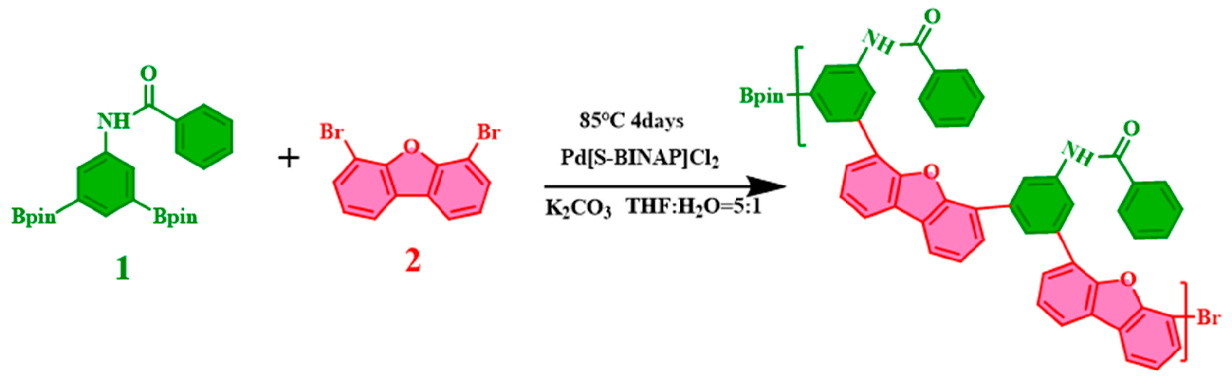
Figure 1.
Synthetic procedure of aimed Polymer.
3. Results and Discussion
3.1. Photoluminescence and Aggregation-Induced Emission Performance of Chiral Three-Dimensional Polymer
Figure 2 summarizes comprehensive photoluminescence (PL) characterizations of the chiral multi-layer three-dimensional polymer at a concentration of 0.05 mg/mL, fully demonstrating three optical features: excitation-wavelength-dependent luminescence, aggregation-induced emission (AIE) and solvatochromism. Figure 2A displays serial PL spectra collected under different excitation wavelengths in pure THF. As a good solvent, THF enables sufficient stretching of polymer chains without steric constraints. Unrestricted intramolecular rotations and vibrations drastically aggravate non-radiative energy loss, leading to weak intrinsic fluorescence of the material. Variation of excitation wavelength induces regular changes in emission maximum, fluorescence intensity and full width at half maximum. Long-wavelength excitation activates low-energy electron transition channels, accompanied by red-shifted emission peaks and slight alteration of peak width. Such excitation-tunable luminescence originates from multi-level conjugated unit energy levels distributed within the chiral three-dimensional skeleton, achieving wavelength-controllable luminescence with promising applications in multi-channel bioimaging and multi-wavelength anti-counterfeiting.
With excitation wavelength fixed at 280 nm, Figure 2B records PL spectra in THF/deionized water mixtures with different water volume fractions, exploring the AIE effect and solvatochromism of the material without ion interference. At extremely low water fractions, the polymer is uniformly dispersed as single molecules and non-radiative relaxation dominates, generating very weak fluorescence signals. Continuous increase of water content elevates solvent polarity while reducing solubility. Hydrophobic interaction drives polymer aggregation, activating the restriction of intramolecular rotation (RIR) mechanism. Non-radiative dissipation is greatly suppressed and fluorescence intensity rises exponentially, directly verifying typical AIE characteristics. Meanwhile, persistent polarity variation of microenvironment surrounding aggregated chromophores changes the dipole moment difference between ground state and excited state, leading to gradual red-shift of emission peaks. The luminescence color slowly shifts from blue region to yellow-green region owing to continuously enhanced intermolecular dipole–dipole interaction and π–π stacking. Deionized water contains no additional electrolytes, eliminating ion disturbance on aggregation kinetics and independently clarifying the internal law of luminescence regulated synergistically by solvent polarity and molecular aggregation.
Figure 2C shows PL curves of THF/simulated body fluid mixtures excited at 280 nm. Spectral results confirm that the chiral three-dimensional polymer can still stably exhibit AIE and solvatochromic responses in ionic physiological fluids without interference from inorganic salts and buffer components in body fluid, suitable for intracellular fluorescence sensing and biological labeling. As illustrated in Figure 2D, PL measurements of THF/simulated urine mixtures were synchronously implemented under 280 nm excitation. The results reveal that the polymer maintains stable optical performance even in complex urine matrices rich in polar metabolites, applicable to urine fluorescence detection and in vitro biological sample analysis.
Supporting Information Figure S3 summarizes complete CIE 1931 chromaticity coordinates and color variation trajectories corresponding to Figure 2A–D. Chromaticity points shift linearly along a single axis with varying excitation wavelength in Figure 2A. For the three biomimetic solvent systems in Figure 2B, C and D, chromaticity points form continuous gradient trajectories upon increasing water fraction, visually demonstrating the whole process of luminescence color regulated synergistically by aggregation degree and polarity. Meanwhile, the tunable color gamut in different biological media is quantified, providing intuitive color references for visual fluorescent biological detection.
3.2. Fluorescence Sensing Performance of Chiral Three-Dimensional AIE Polymer Toward Silver Ions
The skeleton of this chiral multi-layer three-dimensional polymer is densely equipped with intrinsic coordination cavities and exposed heteroatom binding sites. The distinctive topological network endows it with great potential as a fluorescent chemical sensor.
Figure 3A records PL spectra of 0.05 mg/mL polymer THF solution incubated with various common metal cations. After introducing different metal ions into polymer solution, remarkable and distinguishable fluorescence enhancement is observed exclusively for the Ag⁺ system. Nearly no luminescence intensity variation is detected for Ag⁺, Al³⁺, Ba²⁺, Ca²⁺, Cr³⁺, Cr⁶⁺, Cu²⁺, Fe³⁺, Hg²⁺, Mg²⁺, Mn²⁺, Na⁺, Ni²⁺, Pb²⁺, Pd²⁺, Zn²⁺, Au³⁺, Fe²⁺ and K⁺, whose spectral curves almost overlap with blank samples. Such differentiated optical responses preliminarily confirm that the chiral three-dimensional probe possesses ultrahigh recognition selectivity toward Ag⁺ over various interfering transition metals and alkaline earth metals.
To evaluate the practical applicability of the sensing material in complex real water samples containing coexisting ions, parallel anti-interference experiments were designed and conducted. Figure 3B quantitatively records fluorescence intensity variation at the characteristic emission wavelength of 380 nm in multi-ion coexisting systems. In each group, silver ions were mixed with one competing ion separately. The fluorescence enhancement amplitude originating from Ag⁺ coordination is barely affected by foreign coexisting ions, and fluorescence values of all mixed systems remain basically consistent. This fully proves the excellent anti-interference performance for silver ion detection; other metal ions in complex matrices cannot block or weaken the specific recognition and binding between polymer and Ag⁺. Meanwhile, Figure 3C records signal changes of the probe toward Cr⁶⁺ at 420 nm in identical multi-ion systems. Similar to Ag⁺ sensing behavior, Cr⁶⁺ can effectively bind with the polymer skeleton and induce fluorescence enhancement, and this response is also free from interference of various competing ions, further reflecting the outstanding universal anti-interference ion recognition capability of the topological polymer.
Gradient concentration titration experiments were carried out to quantitatively characterize the detection sensitivity and linear range of the polymer toward Ag⁺. Figure 3D presents continuous evolution of PL spectra as Ag⁺ concentration gradually increases from 0 μM to 900 μM. With gradual addition of Ag⁺, more coordination sites on the chiral three-dimensional network are occupied, restricting intramolecular conformational rotations and vibrations of chromophores, suppressing non-radiative loss and activating radiative transitions. Fluorescence intensity rises steadily and monotonously with increasing Ag⁺ dosage.
Data extracted from titration spectra were adopted to plot the fitting curve between maximum fluorescence intensity and Ag⁺ concentration, as shown in Figure 3E. Satisfactory positive linear correlation between fluorescence intensity and Ag⁺ concentration is achieved within 0–900 μM. The linear fitting equation is Y=26.43+0.1618X with correlation coefficient R=0.85359, enabling reliable linear quantitative response within this concentration window. The limit of detection was calculated to be 123 μM according to standard signal-to-noise ratio method for fluorescent sensors. Such detection performance verifies that the material realizes micromolar-level quantitative detection of silver ions, meeting the fundamental requirements for routine monitoring of heavy metal residues in industrial wastewater and surface water.
3.3. Silver Ion Recognition Mechanism: Combined Analysis of Fluorescence Spectroscopy and Dynamic Light Scattering
Characterization data for Ag⁺ sensing of the chiral multi-layer three-dimensional polymer include normalized PL spectra and gradient-concentration DLS measurements, jointly verifying the sensing selectivity and microscopic interaction mechanism between Ag⁺ and the polymer.
Figure A displays normalized PL spectra of polymer (0.05 mg/mL in THF) incubated with different metal ion solutions. All spectra were normalized against baseline intensity of blank samples to eliminate interference from solvent dilution and systematic errors. It can be clearly observed that only the addition of Ag⁺ leads to obvious elevation of normalized fluorescence intensity at the characteristic emission wavelength of 380 nm; all other measured metal cations exert negligible influence on emission intensity and peak position at this wavelength. This result provides rigorous evidence for the superior selectivity of the polymer toward Ag⁺ at the characteristic emission band.
Figure B to Figure G show DLS particle size distribution curves of polymer systems upon successive increase of Ag⁺ concentration with a constant concentration interval of 180 μM between adjacent experimental groups. Figure B corresponds to blank system without Ag⁺ (0 μM), where the average hydrodynamic particle size of polymer aggregates is approximately 700 nm. Figure C to Figure G correspond to Ag⁺ concentrations of 180 μM, 360 μM, 540 μM, 720 μM and 900 μM, respectively. The average hydrodynamic particle size of polymer aggregates continuously and steadily increases with rising Ag⁺ concentration. When Ag⁺ concentration reaches 900 μM (Figure G), the average aggregate particle size exceeds 10000 nm, demonstrating evident concentration-dependent particle growth behavior.
Such gradual enlargement of aggregates arises from complexation-induced aggregation triggered by silver ions. Abundant electron-rich coordination sites are distributed on the multi-layer three-dimensional skeleton of the polymer, including oxygen atoms on benzofuran heterocycles and amide groups on side chains. As soft acid metal ions, Ag⁺ can act as multidentate coordination centers and simultaneously chelate multiple coordination sites on different polymer chains, serving as intermolecular bridging units to drive crosslinking assembly of originally dispersed polymer chains. With elevated Ag⁺ concentration, more intermolecular bridging sites form in the system, continuously improving crosslinking degree of aggregates and promoting aggregate size growth from nanoscale to microscale.
Notably, this concentration-dependent particle size evolution highly matches the continuous fluorescence enhancement trend observed in Ag⁺ titration experiments. Larger aggregates impose stronger restriction on intramolecular rotations and vibrations of aromatic units on polymer backbones, further inhibiting non-radiative energy dissipation and boosting radiative luminescence intensity via the core mechanism of AIE, namely restriction of intramolecular motions (RIM). The DLS results provide direct microscopic evidence for the fluorescence-enhanced sensing mechanism toward Ag⁺, confirming that specific complexation between Ag⁺ and polymer skeleton is the intrinsic driving force for aggregate growth and luminescence enhancement.
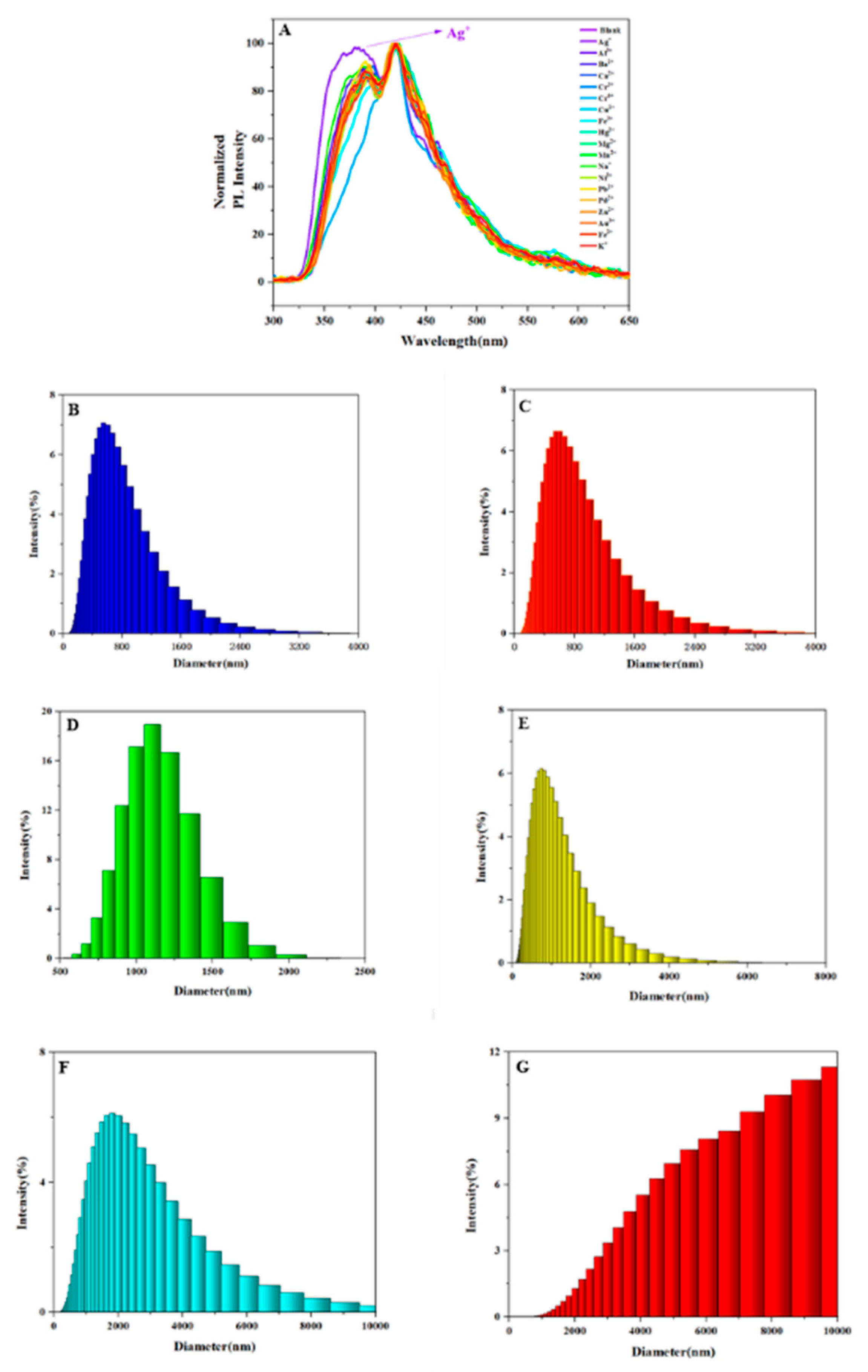
Figure 4.
A .Normalized PL spectra of polymer (0.05 mg/mL in THF) after the addition of various metal ion solutions.B-G .Dynamic light scattering (DLS) spectra at different Ag⁺ concentrations (0–900 μM).
Figure 4.
A .Normalized PL spectra of polymer (0.05 mg/mL in THF) after the addition of various metal ion solutions.B-G .Dynamic light scattering (DLS) spectra at different Ag⁺ concentrations (0–900 μM).
3.4. UV–Vis Absorption Responses of Chiral Three-Dimensional AIE Polymer Toward Metal Ions
Fluorescence sensing results in the previous section confirm that the chiral multi-layer three-dimensional polymer enables differentiated identification of Ag⁺ and Cr⁶⁺. UV–vis absorption spectroscopy can monitor ground-state electronic transition variations of conjugated skeletons induced by metal ion coordination, serving as a complementary characterization technique to reveal intrinsic selective binding between the polymer and various metal ions. Therefore, comprehensive UV–vis absorption tests were implemented, and spectral data are summarized in Figure 5 to further corroborate fluorescence sensing conclusions.
Figure 5A records absorption spectra of 0.05 mg/mL polymer THF solution incubated with Ag⁺, Al³⁺, Ba²⁺, Ca²⁺, Cr³⁺, Cr⁶⁺, Cu²⁺, Fe³⁺, Hg²⁺, Mg²⁺, Mn²⁺, Na⁺, Ni²⁺, Pb²⁺, Pd²⁺, Zn²⁺, Au³⁺, Fe²⁺ and K⁺. Compared with blank polymer, almost no visible shift occurs in overall absorption profile, peak position or absorbance, and curves nearly overlap. It indicates that the above ions cannot form strong ground-state coordination with polymer conjugated backbones and fail to alter intrinsic electron distribution and π–π transition bandgap of chromophores. In sharp contrast, only the Fe³⁺ system induces significant red-shift of characteristic absorption peaks and obvious absorbance variation of the polymer. Exclusive spectral perturbation triggered by Fe³⁺ directly proves the existence of specific ground-state intermolecular coordination between the chiral three-dimensional polymer and Fe³⁺, whose binding mode is completely different from weak interactions with Ag⁺, Cr⁶⁺ and other ions.
Gradient Fe³⁺ titration UV tests within 0–450 μM were performed to quantitatively track ground-state absorption signal evolution driven by Fe³⁺ coordination and clarify concentration-dependent rules. Serial spectra are shown in Figure 5B. Upon gradual addition of Fe³⁺, more coordination sites on the chiral topological network bind with Fe³⁺, modifying electron cloud distribution of conjugated fragments and narrowing the energy gap between ground state and excited state. The red-shift magnitude of absorption peaks continuously increases accompanied by regular variation of absorbance. The inset in Figure 5B magnifies subtle long-wavelength spectral differences, intuitively presenting the gradual red-shift trend of absorption peaks upon increasing Fe³⁺ concentration.
Combined with previous fluorescence sensing data of Ag⁺ and Cr⁶⁺, UV–vis absorption results systematically elucidate the differentiated metal ion recognition mechanism of the chiral multi-layer three-dimensional polymer: strong coordination with Fe³⁺ induces ground-state absorption red-shift; Ag⁺ and Cr⁶⁺ only alter excited states and trigger fluorescence enhancement without affecting ground-state absorption curves. Such differentiated responses originate from abundant cavities and diversified heteroatom coordination sites of the chiral three-dimensional skeleton, which can output distinguishable specific binding signals toward Fe³⁺, Ag⁺ and Cr⁶⁺ respectively. Dual-channel responses of fluorescence and UV–vis spectra provide reliable strategies for simultaneous detection of multiple metal ions, laying structural and experimental foundations for applying this AIE polymer in synchronous monitoring of multiple heavy metals in environmental water samples.
3.5. Electrochemical Properties of Chiral Three-Dimensional AIE Polymer
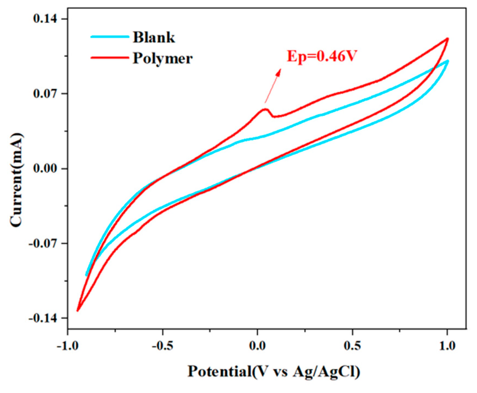
Figure 6 displays cyclic voltammetry (CV) curves of the as-prepared chiral multi-layer three-dimensional polymer using Ag/AgCl as reference electrode with potential scanning range from −1.0 V to 1.0 V. Curves of blank electrolyte and polymer sample were measured simultaneously for comparison.
As observed from voltammetric curves, the current curve of blank electrolyte is nearly flat without characteristic redox peaks across the whole scanning potential window. It demonstrates that supporting electrolyte and electrode setup exhibit no electrochemical activity interference under test conditions, guaranteeing accurate assignment of subsequent electrochemical signals. In contrast, the polymer sample shows a well-resolved anodic oxidation peak at oxidation potential Ep=0.46 V, while no corresponding obvious reduction peak appears during reverse scanning, proving that electrochemical oxidation of this three-dimensional polymer is an irreversible process under current test conditions.
This oxidation behavior is intrinsically correlated with the molecular structure of chiral multi-layer three-dimensional polymer. The polymer backbone is constructed from benzofuran heterocycles and amide-substituted aromatic rings, both of which are electron-rich structural units. The oxidation peak at 0.46 V corresponds to electrochemical oxidation of the above conjugated electron-donating units, where electrons escape from delocalized π-electron systems of the polymer. This result directly confirms the electron-donating nature of the synthesized three-dimensional polymer, providing critical structural basis for its specific interaction with metal ions via charge transfer.
From the perspective of sensing mechanism, this electrochemical characterization further supports the fluorescence response mechanism of the polymer toward target metal ions such as Ag⁺. Target metal ions act as electron acceptors upon coordination with polymer skeletons and induce charge redistribution of conjugated backbones, which fundamentally accounts for regulation of fluorescence signals in sensing experiments. The characteristic oxidation potential at 0.46 V can be adopted as a standard parameter for subsequent research on electrochemical behavior variation after ion binding. Meanwhile, it offers experimental support for expanding this material from single fluorescence sensing to photoelectric dual-mode detection.
4. Conclusions
In this work, a chiral multi-layer three-dimensional aggregation-induced emission polymer was fabricated via Suzuki cross-coupling polymerization. Systematic photophysical characterizations verify that the material possesses multiple optical properties including typical aggregation-induced emission, excitation-dependent luminescence and solvatochromism in aqueous solutions and biomimetic media. Fluorescence experiments confirm that the polymer can selectively recognize Ag⁺ accompanied by remarkable fluorescence enhancement with excellent anti-interference capability, realizing micromolar-level linear quantitative detection within 0–900 μM. Cr⁶⁺ can also induce fluorescence enhancement, and the recognition signal is free from interference of various coexisting ions. UV–vis absorption spectroscopy reveals that only Fe³⁺ triggers red-shift of characteristic absorption peaks of the polymer, and the red-shift magnitude continuously increases with elevated Fe³⁺ concentration. Cyclic voltammetry measurements demonstrate an irreversible oxidation peak at 0.46 V, confirming its electron-rich conjugated skeleton and explaining the binding mechanism with metal ions from the perspective of charge transfer. Benefiting from abundant cavities and high-density heteroatom coordination sites brought by chiral three-dimensional topological structure, the polymer achieves differentiated ion recognition via dual channels of fluorescence and UV–vis absorption, and its photoelectric synergistic property enables potential extension to photoelectric dual-mode sensing systems. This study clarifies the structure–ion recognition performance relationship of chiral multi-layer three-dimensional skeletons, provides new insights into designing anti-interference AIE fluorescent probes, and exhibits considerable practical application prospects in real-time monitoring of heavy metals in environmental water such as surface water and industrial wastewater.
Supplementary Materials
The following supporting information can be downloaded at the website of this paper posted on Preprints.org.
References
- Rasin, P.; Kumar, V. S.; Malappuram, K. M.; Sreekanth, A. Fluorescent Materials for Latent Fingerprint Detection. Curr. Anal. Chem. 2023, 19 (10), 693–703. [CrossRef]
- Yang, S.-Y.; Zhang, L.; Kong, F.-C.; Chen, Y.; Li, W.-J.; Wang, F.; Liu, C.; He, X.; Xiao, X.; Wang, J.; Sun, J.; Chow, P. C. Y.; Kwok, R. T. K.; Lam, J. W. Y.; Tang, B. Z. Scintillators with Aggregation-Induced Emission. Chem 2025, 11 (5), 102534. [CrossRef]
- Yoon, K.-Y.; Dong, G. Multicomponent Polymerization for π-Conjugated Polymers. Macromol. Rapid Commun. 2021, 42 (6), e2000646. [CrossRef]
- Chornodolskyy, Y.; Demkiv, T.; Demchenko, P.; Kurlyak, V.; Kotlov, A.; Gloskovskii, A.; Salapak, V.; Gektin, A.; Voloshinovskii, A. Quenching Mechanisms of CeF3 Luminescence. J. Lumin. 2025, 280 (121120), 121120. [CrossRef]
- Lakowicz, J. R.; Gryczynski, I.; Gryczynski, Z.; Tolosa, L.; Rao, G.; Dattelbaum, J.; Eichorn, L. Novel Methods in Fluorescence Sensing. Microsc. Microanal. 1999, 5 (S2), 506–507. [CrossRef]
- Saikrithika, S.; Kim, Y.-J. Biochar-Derived Electrochemical Sensors: A Green Route for Trace Heavy Metal Detection. Chemosensors (Basel) 2025, 13 (8), 278. [CrossRef]
- Lei, Y.; Wang, Y.; Hill, S. K.; Cheng, Z.; Song, Q.; Perrier, S. Supra-Fluorophores: Ultrabright Fluorescent Supramolecular Assemblies Derived from Conventional Fluorophores in Water. Adv. Mater. 2024, 36 (25), e2401346. [CrossRef]
- Liu, M.; Wang, W.; Su, P.; Ren, J.; Peng, Q. A Molecular Descriptor for Characterizing the Strength of Through-Space Conjugation for Luminescent Property in Organic Materials. J. Phys. Chem. Lett. 2025, 16 (34), 8731–8738. [CrossRef]
- Hu, J.; Zhao, B.; Wen, R.; Zhang, X.; Zhang, Y.; Kohane, D. S.; Liu, Q. Squaraine Dye-Sensitized Upconversion with Enhanced Stability and Minimized Aggregation-Caused Quenching. Nano Lett. 2023, 23 (11), 5209–5216. [CrossRef]
- Wang, Z.; Wu, L.; Liang, H.; Cai, W.; Zhang, Z.; Jiang, Z. Controllable Synthesis of Bifunctional NaYF4:Yb3+/Ho3+@SiO2/Au Nanoparticles with Upconversion Luminescence and High X-Ray Attenuation. J. Alloys Compd. 2011, 509 (37), 9144–9149. [CrossRef]
- Kim, Y.-T.; Kim, D.; Park, S.; Zhexembekova, A.; Byeon, M.; Hong, T. E.; Lee, J.; Lee, C. Y. Aqueous Microlenses for Localized Collection and Enhanced Raman Spectroscopy of Gaseous Molecules. Adv. Opt. Mater. 2021, 9 (22), 2101209. [CrossRef]
- Goto, T.; Teshima, T. F.; Sakai, K.; Yamaguchi, M. Three-Dimensional Self-Folding Assembly of Multi-Layer Graphene at the Interface with a Polymeric Film. AIP Adv. 2022, 12 (7), 075002. [CrossRef]
- Wu, Y.; Chen, C.; Meng, R.; Chen, Y.; Ba, L.; Liu, Y.; Huang, W.; Yang, F.; Cheng, J.; Wang, Y.; Yi, X. The Interfacial Polymerization Reaction Induced by Intermolecular Dipole–Dipole Interaction for the Preparation of Nanofiltration Membrane under the Low Concentration of the Aqueous Monomers. Sep. Purif. Technol. 2025, 362 (131750), 131750. [CrossRef]
- Rempel, S. V.; Valeeva, A. A.; Rempel, A. A. Solubility of ТiOy/HAp Nanocomposites in Body Simulated Fluid. Ceram. Int. 2022, 48 (17), 25213–25218. [CrossRef]
- Kim, S.-J.; Kim, T.-H.; Ahmad, I.; Noh, H.-J.; Jung, S.-M.; Im, Y.-K.; Mahmood, J.; Bae, Y.-S.; Baek, J.-B. Fused Aromatic Networks with the Different Spatial Arrangement of Structural Units. Cell Rep. Phys. Sci. 2021, 2 (7), 100502. [CrossRef]
- Bautista, M. V.; Varni, A. J.; Ayuso-Carrillo, J.; Carson, M. C.; Noonan, K. J. T. Pairing Suzuki–Miyaura Cross-Coupling and Catalyst Transfer Polymerization. Polym. Chem. 2021, 12 (10), 1404–1414. [CrossRef]
- Uva, A.; Michailovich, S.; Hsu, N. S. Y.; Tran, H. Degradable π-Conjugated Polymers. J. Am. Chem. Soc. 2024, 146 (18), 12271–12287. [CrossRef]
- Pan, M.; Li, J.; Pan, B. Identifying the Active Sites of Heteroatom Graphene as a Conductive Membrane for the Electrochemical Filtration of Organic Contaminants. Int. J. Mol. Sci. 2022, 23 (23), 14967. [CrossRef]
- Liu, G.; Gao, Y. Study on Micro-Mechanical Mechanisms of Aggregate Skeleton Based on Three-Dimensional Discrete Element Method and Force Chain Theory. Int. J. Pavement Eng. 2025, 26 (1). [CrossRef]
- (20) Martinez-Monge, A.; Pastor, I.; Bustamante, C.; Manosas, M.; Ritort, F. Measurement of the Specific and Non-Specific Binding Energies of Mg2+ to RNA. Biophys. J. 2022, 121 (16), 3010–3022. [CrossRef]
- Xia, Q.; Qin, A.; Tang, B. Z. Recent Advances in Chiral AIE Polymers. J. Nanopart. Res. 2023, 25 (1). [CrossRef]
- Fu, R.; Chen, X.; Yan, X.; Li, H.; Hu, T.; Wei, L.; Qu, Y.; Cheng, T. Optical Fiber Sensors for Heavy Metal Ion Sensing. J. Mater. Sci. Technol. 2024, 189, 110–131. [CrossRef]
- Gupta, P. K.; Kumar, N.; Ranganath, K. V. S. Asymmetric Suzuki Cross-coupling Reactions Catalyzed by Chiral Surfactant-stabilized Palladium Nanoparticles. Asian J. Org. Chem. 2022, 11 (8).
- Vidal, M.; Morales, S.; Mera-Adasme, R.; Aliaga, C.; Domínguez, M. Solvatochromism of π-Extended Pyridinium-Thienylene-Phenolates. J. Org. Chem. 2025, 90 (34), 12117–12125. [CrossRef]
- Tassanov, A.; Lee, H.; Spainhour, D. W.; Xia, Y.; Hodges, J. M. Structure-Optical Property Relationships in AMM’Q3 Chalcogenides. Chem. Mater. 2026, 38 (12), 5938–5946. [CrossRef]
Figure 2.
A.PL spectroscopy of polymer excited at different wavelengths (0.05mg/mL in THF).B. PL spectra of polymer in different volume fractions of DI water (0.05mg/mL in THF/DI water mixture, excitation wavelength: 280nm).C. PL spectra of polymer in different volume fractions of DI water (0.05mg/mL in THF/SBF mixture, excitation wavelength: 280nm).D. PL spectra of polymer in different volume fractions of DI water (0.05mg/mL in THF/SU mixture, excitation wavelength: 280nm).
Figure 2.
A.PL spectroscopy of polymer excited at different wavelengths (0.05mg/mL in THF).B. PL spectra of polymer in different volume fractions of DI water (0.05mg/mL in THF/DI water mixture, excitation wavelength: 280nm).C. PL spectra of polymer in different volume fractions of DI water (0.05mg/mL in THF/SBF mixture, excitation wavelength: 280nm).D. PL spectra of polymer in different volume fractions of DI water (0.05mg/mL in THF/SU mixture, excitation wavelength: 280nm).
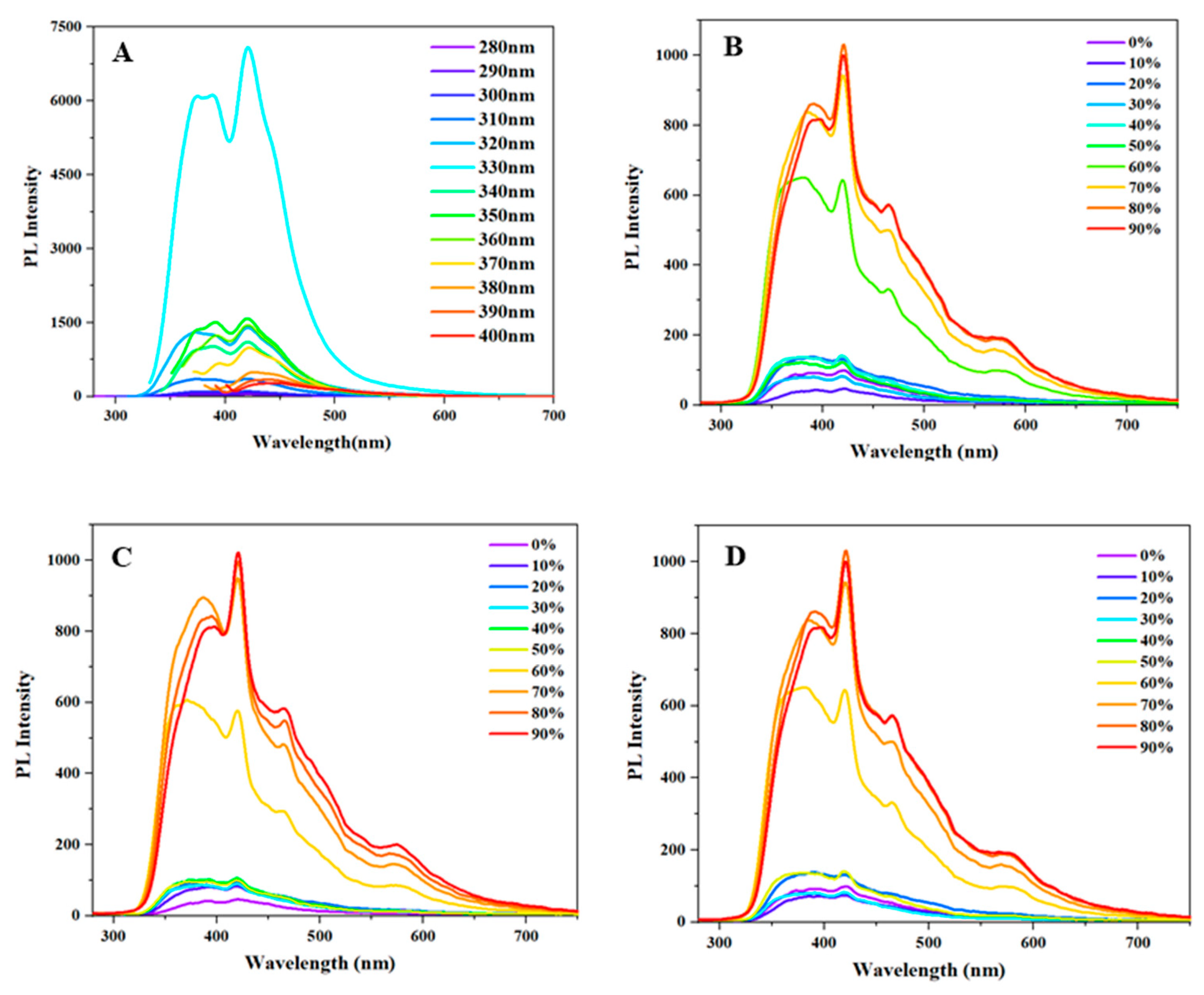
Figure 3.
A. PL spectra of polymer (0.05mg/mL) upon addition of various metal ion solutions in THF.B. Fluorescence intensity changes of polymer (0.05mg/mL in THF) at 380nm after adding Ag⁺ in the presence of various competing cations.C. Fluorescence intensity changes of polymer (0.05mg/mL in THF) at 420nm after adding Cr⁶⁺ in the presence of various competing cations.D. Fluorescence spectra of polymer (0.05mg/mL in THF) with successive addition of Ag⁺ at different concentrations (0–900μM).E. Linear relationship between the fluorescence intensity of polymer (0.05mg/mL in THF) and Ag⁺ concentration (0–900μM).
Figure 3.
A. PL spectra of polymer (0.05mg/mL) upon addition of various metal ion solutions in THF.B. Fluorescence intensity changes of polymer (0.05mg/mL in THF) at 380nm after adding Ag⁺ in the presence of various competing cations.C. Fluorescence intensity changes of polymer (0.05mg/mL in THF) at 420nm after adding Cr⁶⁺ in the presence of various competing cations.D. Fluorescence spectra of polymer (0.05mg/mL in THF) with successive addition of Ag⁺ at different concentrations (0–900μM).E. Linear relationship between the fluorescence intensity of polymer (0.05mg/mL in THF) and Ag⁺ concentration (0–900μM).
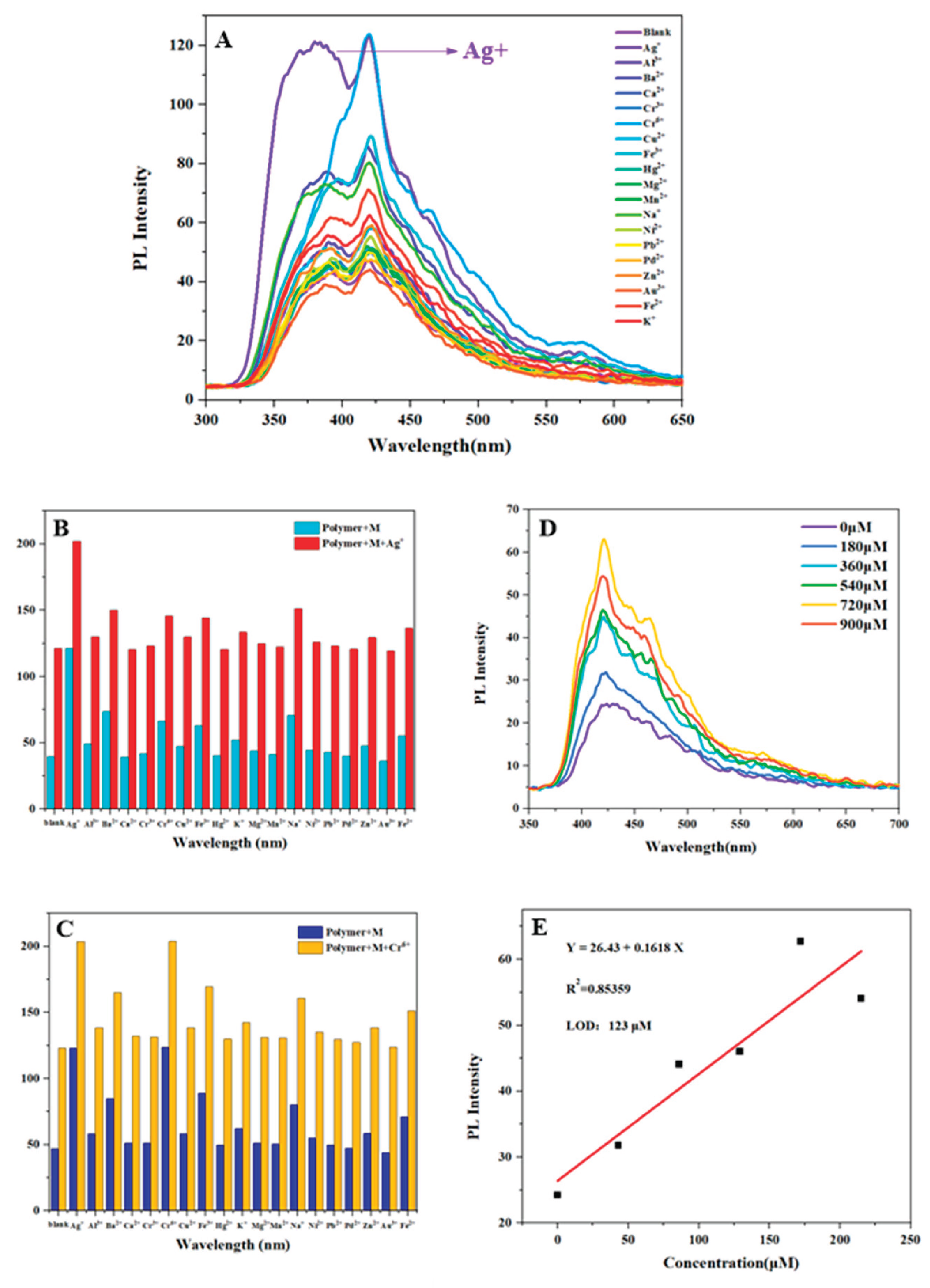
Figure 5.
A. UV–vis absorption spectra of polymer (0.05mg/mL in THF) upon addition of various test cations. B. Concentration-dependent UV–vis absorption spectra of polymer upon the addition of Fe³⁺ with different concentrations (0–450μM).
Figure 5.
A. UV–vis absorption spectra of polymer (0.05mg/mL in THF) upon addition of various test cations. B. Concentration-dependent UV–vis absorption spectra of polymer upon the addition of Fe³⁺ with different concentrations (0–450μM).
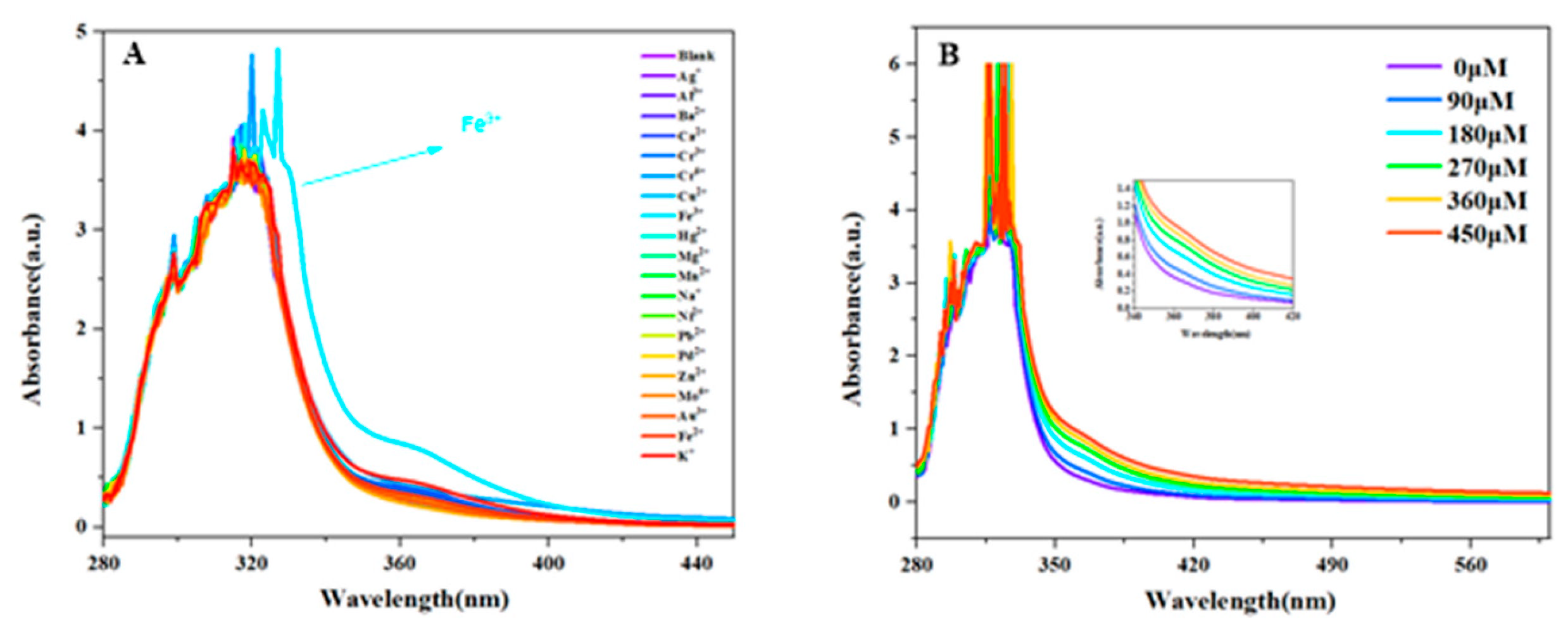
Figure 6.
Cyclic voltammetry curves of the polymer with Ag/AgCl as the reference electrode.
Disclaimer/Publisher’s Note: The statements, opinions and data contained in all publications are solely those of the individual author(s) and contributor(s) and not of MDPI and/or the editor(s). MDPI and/or the editor(s) disclaim responsibility for any injury to people or property resulting from any ideas, methods, instructions or products referred to in the content. |
© 2026 by the authors. Licensee MDPI, Basel, Switzerland. This article is an open access article distributed under the terms and conditions of the Creative Commons Attribution (CC BY) license (http://creativecommons.org/licenses/by/4.0/).
Copyright: This open access article is published under a Creative Commons CC BY 4.0 license, which permit the free download, distribution, and reuse, provided that the author and preprint are cited in any reuse.