

Submitted:
16 July 2026
Posted:
17 July 2026
You are already at the latest version
Abstract
Fungal infections are an increasingly important yet underrecognized global health threat. Climate change, environmental disruption, and intensified human–animal interactions are shifting fungal ecology, enabling pathogens to push beyond traditional geographic and thermal boundaries. Increasing temperature, shifting humidity, and extreme weather events have favoured the emergence and reemergence of fungal disease, as reflected in the rapid global dissemination of Candida auris, in a manner defying conventional models of pathogen spread. The environmental drive to fungal adaptation includes increased thermotolerance and survival under hostile conditions for the infection of endothermic hosts. At the human-animal-environment interface, the zoonotic spillover accelerates fungal evolution toward increased virulence and antifungal resistance. Resistance to all major classes of antifungal drugs is increasingly observed and “seriously challenges treatment, leading to persistent infections and a greater healthcare burden.” Despite advances in molecular diagnostics and genomic surveillance, significant gaps in our understanding of fungal transmission, host adaptation, and resistance mechanisms remain. This review focuses on climate-driven fungal emergence, its zoonotic potential, antifungal resistance, and current detection strategies that call for immediate action through integrated One Health surveillance and coordinated global responses.
Keywords:
Fungal infections
; Climatic change
; Emerging pathogens
; Antifungal resistance
; Zoonosis
; One Health
; Candida auris
; Global health.
1. Introduction
Fungal diseases are an increasing, yet often ignored, global health issue (Rio et al., 2023; Anupama, 2024). The World Health Organization (WHO) released its first-ever fungal priority pathogens list (FPPL) in 2022, identifying 19 fungi that pose major risks to public health due to rising antifungal resistance and increasing incidence. (Figure 1) shows the global list of priority endemic pathogens. Once viewed mainly as opportunistic infections with little significance, fungal pathogens are now recognized as organisms that can quickly change, spread to new areas, and find new hosts (Corrêa-Junior et al., 2025; Kruithoff et al., 2023). The rising rates of invasive, superficial, and allergic mycoses worldwide show clear links to changes in climate, land use, urban development, medical practices, and global movement (Halak, 2025; Sztandera-Tymoczek, 2023; Petrucelli et al., 2020). These factors have shifted the ecological and evolutionary landscape of fungi. As a result, we need to pay more attention to how fungi spread and how new diseases emerge globally.
Recent years have revealed unusual global patterns of fungal emergence. These show the almost simultaneous appearance of different genetic lineages across various continents (Garcia-Bustos, 2024; Phan-Canh et al., 2024). Such trends challenge traditional outbreak models that focus only on local spread. A key example is Candida auris, which appears to have distinct clades emerging nearly at the same time due to strong environmental pressures (Garcia-Bustos, 2024; Phan-Canh et al., 2024). These findings highlight how ecological changes, environmental reservoirs, and adaptive evolution allow fungi to spread beyond typical geographic locations and host types.
Climate change is a major factor driving these trends. It affects fungal ecology by raising temperatures, changing humidity, and increasing extreme weather events (Anupama, 2024; Rio et al., 2023; Cuartero et al., 2024). These changes expand fungal habitats and promote the development of traits like heat tolerance and stress resistance, which help fungi thrive in animal hosts (Phan-Canh et al., 2024; Corrêa-Junior et al., 2025). This climate-driven adaptation lowers the usual barriers that restrict fungi’s ability to cause disease in humans and animals. As a result, there is a greater chance of new or returning fungal diseases.
At the same time, increasing interactions between humans, animals, and the environment have raised opportunities for zoonotic spillover and host adaptation (Gupta et al., 2025; Cabañes, 2000; Chang et al., 2022). Domestic animals, wildlife, and human-altered environments are becoming interconnected reservoirs under a One Health approach (Ganesan et al., 2025). Evidence of fungal infections in animals raises concerns about transmission between species, lasting reservoirs, and the potential increase in virulence or drug resistance through host switching (Gupta et al., 2025; Chang et al., 2022). These processes are not yet well understood, but they may play a key role in ongoing fungal transmission.
A troubling aspect of modern fungal epidemiology is the rapid rise of antifungal resistance. This has become a global problem affecting many types of fungi, making both first-choice and alternative treatments less effective (Kruithoff et al., 2023; Lass-Flörl & Cuenca-Estrella, 2017; Hwang et al., 2014). Several factors contribute to this crisis, including the limited number of antifungal drug classes, the widespread use of azole compounds in agriculture, inappropriate or long-term antifungal use in hospitals, and environmental stressors that promote genetic and epigenetic changes (Phan-Canh et al., 2024; Cuartero et al., 2024). Resistance occurs through various mechanisms, such as mutations at target sites, overexpression of efflux pumps, biofilm formation, and stress-induced genomic changes, allowing fungi to withstand both environmental pressures and treatments (Phan-Canh et al., 2024; Kruithoff et al., 2023).
The impact of antifungal resistance on human health is serious. Infections from drug-resistant fungi often lead to delayed diagnoses, treatment failures, ongoing colonization, longer hospital stays, and higher death rates (Lass-Flörl & Cuenca-Estrella, 2017; Anupama, 2024; Kruithoff et al., 2023). Invasive infections from resistant fungi particularly affect immunocompromised individuals, critically ill patients, and those needing long-term medical care. Additionally, resistance complicates efforts to control infections, raises healthcare costs, and limits treatment options, highlighting a significant gap in global readiness for fungal diseases.
Despite progress in molecular biology, we still face significant knowledge gaps in fungal epidemiology, genomics, and surveillance (Phan-Canh et al., 2024; Cuartero et al., 2024; García-Martín et al., 2024). Many areas lack standardized diagnostic procedures, routine antifungal susceptibility tests, and integrated genomic monitoring systems (Petrucelli et al., 2020; Kruithoff et al., 2023). Although tools like polymerase chain reaction tests, whole-genome sequencing, MALDI-TOF MS, and environmental metabarcoding have improved how we detect and study fungi, their use is inconsistent worldwide (Petrucelli et al., 2020; García-Martín et al., 2024; Amin & Bertelsen, 2025). Methodological issues such as underreporting, diagnostic uncertainty, incomplete reference databases, and a lack of integration of human, animal, and environmental data continue to obscure our understanding of transmission routes and the full scope of antifungal resistance.
This review brings together current evidence on global patterns of fungal transmission and geographic spread, the impact of climate change on fungal emergence, zoonotic spillover, host adaptation, and the growing issue of antifungal resistance, including its origins, mechanisms, and effects on human health. It also evaluates current detection methods and available protocols globally and identifies key methodological and knowledge gaps that must be addressed to improve surveillance, diagnosis, and management of fungal diseases during rapid environmental change.
2. Global Patterns of Fungal Transmission and Geographic Expansion
Global fungal transmission results from long-term ecological factors and modern human activities. Unlike fungi that only infect humans, these organisms can thrive without hosts. They can survive in soil, water, plant material, built environments, and animal reservoirs (Lass-Flörl et al., 2017; Vitasse et al., 2021; Corrêa-Junior et al., 2025). This ability allows fungi to quietly expand their geographic reach over long periods. Their endurance in the environment lets fungi cross political and ecological boundaries without direct human-to-human spread.
Recent phylogenomic studies indicate that fungal expansion usually occurs from multiple independent emergence events rather than a single source (Garcia-Bustos, 2024; Phan-Canh et al., 2024). The nearly simultaneous appearance of different strains of Candida auris across continents strongly supports this idea (Garcia-Bustos, 2024; Phan-Canh et al., 2024). We see similar patterns with dermatophytes, where migration, international travel, movement of people, and changes in hygiene and lifestyle have shifted which species dominate in various regions (Petrucelli et al., 2020; Belmokhtar et al., 2024; Kruithoff et al., 2023; Gupta et al., 2025).
Environmental sources like wastewater systems, hospital waste, swimming pools, and soil disrupted by urbanization connect clinical and environmental fungal populations (Garcia-Bustos, 2024; Cuartero et al., 2024; Amin & Bertelsen, 2025). Together, these findings suggest that we should view fungal transmission as a global ecological process and not just a clinical issue.
3. Rapid Emergence of Antifungal Resistance
Antifungal resistance has become a major global issue in medical and environmental mycology. It has fundamentally changed how we handle fungal diseases (Kruithoff et al., 2023; Lass-Flörl & Cuenca-Estrella, 2017; Hwang et al., 2014). Unlike bacterial pathogens, fungi have similar cellular and metabolic processes to humans. This limits the number of safe antifungal drug targets and reduces treatment options to a few drug classes, mainly azoles, echinocandins, and polyenes (Lass-Flörl & Cuenca-Estrella, 2017). Echinocandins (such as caspofungin, micafungin, and anidulafungin) have similar spectra and safety profiles but share susceptibility to the same resistance mechanisms. Amphotericin B, the only widely used polyene, is limited by significant toxicity. Although three drug classes are available, resistance to even one can substantially restrict therapeutic options, particularly in the presence of adverse effects or drug interactions (Logan et al., 2022). Reports show that resistance to one or more of these classes is now common among yeasts, dermatophytes, and filamentous fungi (Kruithoff et al., 2023; Lass-Flörl & Cuenca-Estrella, 2017; Gupta et al., 2025).
A key feature of antifungal resistance is that it often develops independently in different geographic areas (Garcia-Bustos, 2024; Hwang et al., 2014). Genomic studies suggest that resistant strains usually emerge locally due to selective pressure rather than spreading from a single ancestral strain (Garcia-Bustos, 2024; Phan-Canh et al., 2024). A clear example is Candida auris, where genetically distinct groups evolved independently on different continents. (Table 1) presents the antifungal resistance patterns and evolutionary trajectories of the major fungal pathogens.
The rise of antifungal resistance is driven by a combination of environmental, ecological, and clinical pressures (Kruithoff et al., 2023; Phan-Canh et al., 2024). The widespread use of azole fungicides in agriculture is a major environmental factor (Lass-Flörl & Cuenca-Estrella, 2017; Kruithoff et al., 2023). Climate change worsens this pressure by encouraging fungal growth, extending growth seasons, and increasing the use of antifungal chemicals in farming (Sztandera-Tymoczek, 2023; Halak, 2025). Environmental stresses like temperature changes, humidity shifts, chemical exposure, and nutrient shortages provoke adaptive genetic responses in fungi (Cuartero et al., 2024; Phan-Canh et al., 2024).
Clinical practices also significantly contribute to the development of resistance. Inappropriate prescribing of antifungals, prolonged or repeated treatments, empirical therapies without lab confirmation, and inadequate dosing promote the selection of resistant strains (Dogra & Narang, 2017; Kruithoff et al., 2023). Additionally, the lack of regular antifungal susceptibility testing and standardized monitoring systems in many areas adds to these problems (Petrucelli et al., 2020; Hwang et al., 2014).
Microbial resistance encompasses both primary resistances, in which strains exhibit intrinsic reduced susceptibility to a specific antifungal agent, and secondary resistance, where previously susceptible strains develop resistance following antifungal exposure (Cowen et al., 2014). On a molecular level, antifungal resistance occurs through various mechanisms (Phan-Canh et al., 2024; Kruithoff et al., 2023). These include point mutations in antifungal target genes, overexpression of efflux pumps, and biofilm formation as represented in (Figure 2). Fungi also show noteworthy genetic flexibility, such as aneuploidy and stress-induced chromosomal changes (Phan-Canh et al., 2024; Cuartero et al., 2024).
4. Effect of Antifungal Resistance
The impact of antifungal resistance extends beyond microbiological failure. It affects clinical outcomes, healthcare systems, and public health (Lass-Flörl & Cuenca-Estrella, 2017; Anupama, 2024; Kruithoff et al., 2023). Drug-resistant fungal infections often lead to treatment failures, prolonged colonization, repeated disease, and a higher risk of transmission (Dogra & Narang, 2017; Gupta et al., 2025). In cases of invasive fungal infections, resistance increases morbidity and mortality rates, especially among immunocompromised individuals (Anupama, 2024; Lass-Flörl & Cuenca-Estrella, 2017).
In superficial and chronic mycoses, antifungal resistance has turned manageable infections into ongoing and debilitating conditions (Dogra & Narang, 2017; Kruithoff et al., 2023). Patients often face extensive disease, chronic inflammation, relapses, and a significant decline in their quality of life. This frequently requires long or repeated courses of antifungal therapy. These clinical challenges create a large economic burden, including increased healthcare visits, repeated tests, longer treatment times, and the use of second-line drugs (Anupama, 2024; Kruithoff et al., 2023).
From a public health standpoint, antifungal resistance makes controlling outbreaks harder and skews disease burden estimates. The inconsistency between in vitro antifungal susceptibility testing and clinical outcomes adds to the difficulty of making treatment decisions and conducting surveillance (Dogra et al., 2017; Petrucelli et al., 2024). Furthermore, finding resistant fungal strains in the environment and animal hosts shows that resistance extends beyond healthcare facilities. It is part of larger ecological systems, which emphasizes the need for surveillance and intervention strategies based on a One Health approach (Garcia-Bustos, 2024; Chang et al., 2022).
5. Climate Change as a Driver of Emerging and Re-Emerging Fungal Diseases
Climate change plays a significant role in influencing fungal diversity, distribution, and their ability to cause disease (Anupama, 2024; Rio et al., 2023; Cuartero et al., 2024; Corrêa-Junior et al., 2025). Changes in global temperatures, humidity, rainfall patterns, and extreme weather events create constant pressure on fungal populations in the environment (Sztandera-Tymoczek, 2023; Halak, 2025; Amin & Bertelsen, 2025). This favors traits like heat tolerance, salt tolerance, resistance to oxidative stress, and adaptability (Phan-Canh et al., 2024; Corrêa-Junior et al., 2025). These traits are also essential for fungi to survive in mammals, indicating that climate change helps fungi that cause diseases to emerge (Phan-Canh et al., 2024; Anupama, 2024). The potential effects of climate change on different pathogenic fungi and fungal diseases are illustrated in (Figure 3).
Longer warm seasons and irregular rainfall extend the growth period for fungi, increasing human and animal exposure to them (Halak, 2025; Sztandera-Tymoczek, 2023). Research on airborne fungi shows that spores are appearing earlier in the year and lasting longer into fall (Halak, 2025). This disrupts traditional seasonal patterns and raises the risk of cumulative exposure. Extreme weather events like floods, storms, hurricanes, and dust storms can spread fungal spores over long distances, introducing them into areas that typically do not support these fungi (Sztandera-Tymoczek, 2023; Cuartero et al., 2024). These changes enable both harmful and allergenic fungi to expand their geographic range, even into temperate and high-altitude areas (Vitasse et al., 2021; Rio et al., 2023).
Climate change also affects the vulnerability of hosts (Anupama, 2024; Rio et al., 2023). Heat, food shortages, malnutrition, and environmental damage weaken the immune responses of humans and animals. This increases the risk of opportunistic fungal infections (Anupama, 2024). Rising respiratory diseases, damage to skin barriers, and ongoing health problems add to this vulnerability. Additionally, shifts in indoor environments, such as increased dampness, poor ventilation, and higher humidity, lead to more indoor fungal growth (Amin & Bertelsen, 2025). This further increases exposure to harmful molds.
In agricultural and environmental settings, climate change has heightened the threat from fungal plant diseases (Mawar & Saranya, 2023; Cuartero et al., 2024). This has resulted in more and longer use of azole fungicides (Kruithoff et al., 2023; Lass-Flörl & Cuenca-Estrella, 2017). These substances share structural and functional similarities with medical azoles, creating environmental sources of antifungal resistance that overlap with clinical antifungal treatments (Lass-Flörl & Cuenca-Estrella, 2017; Kruithoff et al., 2023). This connection highlights an ecological link between climate change, farming practices, and the rise of antifungal resistance that is crucial for health. Overall, these findings reveal that climate change is not just a background factor; it is a major driver of the emergence, adaptation, and resurgence of fungi that impact humans and animals (Anupama, 2024; Corrêa-Junior et al., 2025).
6. Zoonotic Spillover and Host Adaptation in Fungal Pathogens
Zoonotic spillover has gained attention as a key factor in the emergence and persistence of fungal pathogens (Gupta et al., 2025; Chang et al., 2022). Many fungi have great ecological flexibility and low host specificity. This allows them to infect humans, domestic animals, livestock, wildlife, and environmental sources such as soil and water (Cabañes et al., 2000; Chang et al., 2022; Gupta et al., 2025). Animals often act as silent carriers, keeping fungal populations alive over time and allowing unnoticed transmission between different host species and regions (Gupta et al., 2025; Cabañes et al., 2000). (Table 2) shows global annual incidence of zoonotic transfer of various fungal pathogens. Recent epidemiological surveillance according to Denning (2024) shows a sharp uptick in fungal “alerts” and incidence. According to ProMED data, fungal disease alerts increased from 1% to 7% of all recorded outbreaks between 1995 and 2024, with animal-to-human spillover events specifically rising by approximately 1.4%.
Fungi adapt to their hosts through a combination of phenotypic flexibility and quick genetic changes. Processes like morphogenetic switching, metabolic reprogramming, stress-response activation, and genomic rearrangements help fungi efficiently take advantage of new hosts and environments (Phan-Canh et al., 2024; Corrêa-Junior et al., 2025). For example, evidence of Candida auris colonizing dogs and reports of dermatophyte transmission from pets and livestock to humans suggest that fungi might cycle back and forth between humans and animals (Garcia-Bustos, 2024; Chang et al., 2022; Gupta et al., 2025). These cycles may support longer persistence, greater exposure to antifungal agents, and the selection of stress-resistant and drug-resistant traits (Gupta et al., 2025; Kruithoff et al., 2023).
Climate change increases the risk of zoonotic spillover by altering animal habitats, migration patterns, and population densities. This change leads to more interactions between humans and animals (Anupama, 2024; Ganesan et al., 2025). Thermal stress and environmental disruptions can weaken animal immune systems, making them more vulnerable to fungal colonization and shedding (Ganesan et al., 2025). In farming and peri-urban areas, intensified livestock production and close living conditions between humans and animals present more opportunities for fungal exchange. Wildlife moving due to habitat loss and extreme climate conditions might also introduce new fungal species into human-dominated areas, increasing the risk of host switching (Gupta et al., 2025). (Figure 4) shows a proposed scheme for the emergence of Candida auris, a multidrug-resistant, often fatal fungus identified by the WHO as a “critical” priority pathogen on its Fungal Priority Pathogens List (FPPL).
Despite growing evidence of zoonotic roles in fungal epidemiology, monitoring fungal pathogens in animal populations remains uneven (Gupta et al., 2025; Cabañes et al., 2000). Most monitoring systems focus on bacterial and viral zoonoses, leaving fungal spillover less recognized and reported. The absence of connected One Health monitoring frameworks makes it difficult to find new animal reservoirs, transmission routes, and key evolutionary areas (Gupta et al., 2025). Closing these gaps is crucial for understanding how host adaptation, zoonotic spillover, and environmental changes affect the future of fungal diseases.
7. Diagnostic Strategies and Standardized Protocols for Fungal Detection
Fungal detection and identification rely on a combination of conventional, molecular, and genomic methods. Each method has its own strengths and weaknesses (Petrucelli et al., 2020; García-Martín et al., 2024; Kruithoff et al., 2023). Direct microscopy and culture are the primary methods for diagnosing fungi worldwide because they are inexpensive and widely used (Petrucelli et al., 2020). However, these techniques often have low sensitivity, long turnaround times, and frequent false-negative results, especially in superficial and invasive fungal infections (Petrucelli et al., 2020; Dogra & Narang, 2017). Morphological similarities among different fungal species and slow growth make accurate identification more difficult (Petrucelli et al., 2020).
Histopathology uses stains like periodic acid-Schiff and Gomori methenamine silver to show tissue invasion. However, it usually does not help with identification at the species level (García-Martín et al., 2024). As a result, many diagnoses remain presumptive, which can delay specific treatments.
Molecular diagnostic techniques have greatly improved fungal detection (Petrucelli et al., 2020; Kruithoff et al., 2023). PCR-based tests, including conventional and real-time PCR focused on the internal transcribed spacer (ITS) regions, offer higher sensitivity and specificity. They also allow for quick species identification directly from clinical samples (Petrucelli et al., 2020). MALDI-TOF MS has become a useful tool for routine fungal identification in clinical labs, but its accuracy depends on having comprehensive reference spectral libraries, which are often limited for many fungal species (Petrucelli et al., 2020; Hwang et al., 2014).
Whole-genome sequencing (WGS) is the most complete method for identifying fungi, investigating outbreaks, analyzing relationships between species, and profiling antifungal resistance (Garcia-Bustos, 2024; Phan-Canh et al., 2024). WGS has helped reveal independent emergence events, clade structures, and resistance mechanisms in new fungi. However, its use is mostly confined to research and reference labs due to costs, required infrastructure, and bioinformatics expertise (Phan-Canh et al., 2024).
Environmental surveillance methods, such as DNA metabarcoding and wastewater monitoring, are becoming important for detecting fungal populations and giving early warnings about fungal circulation outside clinical settings (Cuartero et al., 2024; Amin & Bertelsen, 2025). These methods provide insights into environmental reservoirs and transmission patterns, but they are not yet standardized for regular public health use.
For antifungal resistance, international bodies like the Clinical and Laboratory Standards Institute (CLSI) and the European Committee on Antimicrobial Susceptibility Testing (EUCAST) provide standardized testing protocols (Kruithoff et al., 2023; Hwang et al., 2014). These protocols set reference methods and interpretive guidelines, but their consistent use varies worldwide, especially in low- and middle-income countries (Hwang et al., 2014). Furthermore, there is often little correlation between lab susceptibility results and patient outcomes, complicating interpretation and clinical decisions (Kruithoff et al., 2023).
In summary, while many diagnostic tools and protocols are available globally, unequal access, a lack of standardization, and limited integration across human, animal, and environmental sectors present significant challenges (García-Martín et al., 2024; Gupta et al., 2025). To improve global fungal detection, it is necessary to standardize protocols, expand reference databases, enhance lab capabilities, and create coordinated One Health surveillance systems. This approach will help ensure timely detection and an effective response to emerging fungal threats.
8. Current Knowledge Gaps and Methodological Limitations in Fungal Epidemiology and Genomics
Despite growing awareness of fungal diseases as a global health problem, significant gaps remain in our understanding of fungal epidemiology, surveillance, and genomics (Phan-Canh et al., 2024; Cuartero et al., 2024; García-Martín et al., 2024). One main limitation is the consistent underestimation of the burden of fungal diseases. This stems from underreporting, misdiagnosis, and inadequate routine surveillance in many areas (Gupta et al., 2025; Petrucelli et al., 2020). Unlike bacterial and viral infections, fungal diseases are often left out of national and international surveillance systems. This results in incomplete and fragmented epidemiological data (Anupama, 2024).
A significant knowledge gap exists regarding how fungi are transmitted and their sources in the environment (Cuartero et al., 2024; Amin & Bertelsen, 2025). While we are becoming more aware of environmental, animal, and healthcare-related sources, we still do not fully understand the significance of each pathway. Long-term data linking environmental fungal populations to clinical samples is limited. This restricts our understanding of how climate change, land use, and human actions contribute to the emergence and spread of these diseases (Rio et al., 2023). Additionally, the dynamics of zoonotic transmission have not been studied in depth. Fungal surveillance in animals rarely connects with human health data (Gupta et al., 2025).
From a genomic perspective, whole-genome sequencing has changed our understanding of fungal populations (Phan-Canh et al., 2024). However, the datasets we have are still mostly biased towards clinical samples from wealthy countries. Environmental and animal samples are often absent, which limits comparative studies and our understanding of how fungi evolve, adapt to hosts, and develop resistance (Cuartero et al., 2024; Gupta et al., 2025). Many studies mainly focus on describing genomic data. There is a lack of research validating genetic variants linked to virulence or resistance to antifungal treatments. This limits the practical applications of the research (Phan-Canh et al., 2024).
Methodological challenges also include the absence of standardized genomic and phenotypic measurements (García-Martín et al., 2024; Phan-Canh et al., 2024). Variability within species, epigenetic factors, and genomic changes make it difficult to link genotypes to phenotypes. This is particularly true when comparing samples from different regions and labs. The lack of universally accepted reference strains and consistent analytical methods complicates efforts to ensure reproducibility and comparability worldwide (Phan-Canh et al., 2024; Cuartero et al., 2024). Together, these issues hinder effective risk assessments, early detection of new pathogens, and predictions regarding resistance trends.
9. Conclusions
Fungal diseases are no longer rare or limited to specific areas. Fungi are increasingly spreading across different regions and hosts due to environmental change, human activity, and their own remarkable ability to adapt. The old view of fungal infections as opportunistic and localized is being replaced by a clearer understanding of fungi as dynamic pathogens influenced by global ecological factors. Climate change is a key factor in this shift. Rising temperatures, changed rainfall patterns, and extreme weather events create conditions that support fungal growth, persistence, and spread. These environmental changes also favor traits like heat tolerance and stress resistance, which help fungi survive in humans and animals. Changes in climate that affect host immunity and the increased use of antifungals in agriculture further link environmental change to the rise of hard-to-treat fungal infections. Zoonotic spillover adds more complexity. Many fungi can easily move between the environment, animals, and humans, often without causing clear disease in animal hosts. This silent circulation lets fungi persist, adapt, and sometimes return to humans in more resilient or resistant forms. Growing evidence of two-way transmission shows the importance of viewing fungal diseases through a One Health perspective.
One concerning trend is the rapid rise of antifungal resistance. With only a few classes of antifungal drugs available, resistance leads to serious issues. Resistant infections are harder to treat, tend to return, and result in higher rates of illness, death, and healthcare costs. The detection of resistant fungi outside hospitals indicates that this problem goes beyond clinical settings and is closely linked to larger environmental pressures. Despite progress in molecular diagnostics and genomics, there are significant gaps in how fungal diseases are detected, monitored, and understood globally. Limited surveillance, uneven access to diagnostic tools, and a lack of integrated human, animal, and environmental data mean that many fungal threats are recognized too late. Coordinated global surveillance, better antifungal management, and stronger One Health approaches will be crucial to reduce the impact of fungal diseases in a rapidly changing world.
References
- Alexander EB, Coleman RG, Keeler-Wolf T, Harrison SP: Serpentine geoecology of western North America: Geology, soils, and vegetation. Oxford: Oxford University Press; 2007.
- Ali-Shtayeh MS, Khaleel TK, Jamous RM: Ecology of dermatophytes and other keratinophilic fungi in swimming pools and polluted and unpolluted streams. Mycopathologia 2002, 156:193-205. [CrossRef]
- Amin H, Bertelsen RJ: Climate change and indoor biological exposures: A hidden risk to immune health. Front Public Health 2025, 13:1597881. [CrossRef]
- Aneke CI, Rhimi W, Hubka V, Otranto D, Cafarchia C: Virulence and antifungal susceptibility of Microsporum canis strains from animals and humans. Antibiotics 2021, 10:296. [CrossRef]
- Anupama YJ: Climate change and human health: Time to recognize the new threat. APIK J Intern Med 2025, 13:13-22. [CrossRef]
- Araya S, Tesfaye B, Fente D: Epidemiology of dermatophyte and non-dermatophyte fungi infection in Ethiopia. Clin Cosmet Investig Dermatol 2020, 13:291-297. [CrossRef]
- Aruna GL, Bellibatlu R: Dermatophytes: Keratin eaters. Kavaka 2024, 60:1-8.
- Bazzicalupo A: Local adaptation in fungi. FEMS Microbiol Rev 2022, 46:fuac026. [CrossRef]
- Belmokhtar Z, Djaroud S, Matmour D, Merad Y: Atypical and unpredictable superficial mycosis presentations: A narrative review. J Fungi 2024, 10:295. [CrossRef]
- Bhatia VK, Sharma PC: Epidemiological studies on dermatophytosis in human patients in Himachal Pradesh, India. SpringerPlus 2014, 3:134. [CrossRef]
- Bidartondo MI, Ellis C, Kauserud H, Kennedy PG, Lilleskov EA, Suz LM, Andrew C: Climate change: Fungal responses and effects. In State of the World’s Fungi 2018. 2018.
- Bontems O, Fratti M, Salamin K, Guenova E, Monod M: Epidemiology of dermatophytoses in Switzerland. J Fungi 2020, 6:95. [CrossRef]
- Brescini L, Fioriti S, Morroni G, Barchiesi F: Antifungal combinations in dermatophytes. J Fungi 2021, 7:727. [CrossRef]
- Britto Martins de Oliveira J, Barbieri M, Corrêa-Junior D, Schmitt M, Santos LLR, Bahia AC, Parente CET, Frases S: Urban mangroves under threat: Metagenomic analysis reveals a surge in human and plant pathogenic fungi. Pathogens 2025, 14:759.
- Brooks RR: Serpentine and its vegetation: A multidisciplinary approach. Oregon: Dioscorides Press; 1987. [CrossRef]
- Burstein VL, Beccacece I, Guasconi L, Mena CJ, Cervi L, Chiapello LS: Skin immunity to dermatophytes. Front Immunol 2020, 11:605644. [CrossRef]
- Cabañes F: Dermatophytes in domestic animals. Rev Iberoam Micol 2000, 17:104-108.
- Casadevall A, Kontoyiannis DP, Robert V: On the emergence of Candida auris: Climate change, azoles, swamps, and birds. mBio 2019, 10:e01397-19.
- Chang CC, Wechtaisong W, Chen SY, Cheng MC, Chung CS, Lin LS, Lien YY, Tsai YL: Prevalence and risk factors of zoonotic dermatophyte infection in pet rabbits in northern Taiwan. J Fungi 2022, 8:627. [CrossRef]
- Chanyachailert P, Leeyaphan C, Bunyaratavej S: Cutaneous fungal infections caused by dermatophytes and non-dermatophytes. J Fungi 2023, 9:669. [CrossRef]
- Cheng KW, Yang JI, Srimongkol P, Ariyawansa HA, Stadler M, Karnchanatat A: Fungal frontiers in toxic terrain. IMA Fungus 2025, 16:e155308. [CrossRef]
- Cowen LE, Sanglard D, Howard SJ, Rogers PD, Perlin DS: Mechanisms of antifungal drug resistance. Cold Spring Harb Perspect Med 2014, 5:a019752.
- Day AM, McNiff MM, da Silva Dantas A, Gow NAR, Quinn J: Hog1 regulates stress tolerance and virulence in the emerging fungal pathogen Candida auris. mSphere 2018, 3:e00506-18. [CrossRef]
- De Hoog GS, Dukik K, Monod M, et al.: Toward a novel multilocus phylogenetic taxonomy for the dermatophytes. Mycopathologia 2017, 182:5-31. [CrossRef]
- Deng R, Wang X, Li R: Dermatophyte infection: From fungal pathogenicity to host immune responses. Front Immunol 2023, 14:1285887. [CrossRef]
- Denning DW: Global incidence and mortality of severe fungal diseases. Lancet Infect Dis 2024, 24:e428-e438. [CrossRef]
- Djaroud S, Belmokhtar Z, Merad Y, Nassour K, Belkacemi M, Matmour D, Merad Z: The influence of gaseous ozone on the growth of fungi isolated from clinical and environmental samples. Environ Health Eng Manag 2023, 10:261-265. [CrossRef]
- Dogra S, Narang T: Emerging atypical and unusual presentations of dermatophytosis in India. Clin Dermatol Rev 2017, 1(Suppl 1):S12-S18.
- Du Y, Fu X, Chu Y, Wu P, Liu Y, Ma L, Tian H, Zhu B: Biosynthesis and the roles of plant sterols in development and stress responses. Int J Mol Sci 2022, 23:2332. [CrossRef]
- Ebrahimi M, Zarrinfar H, Naseri A, Najafzadeh MJ, Fata A, Parian M, Khorsand I, Novak Babič M: Epidemiology of dermatophytosis in northeastern Iran: A subtropical region. Curr Med Mycol 2019, 5:16-21.
- Fernando LD, Pérez-Llano Y, Dickwella Widanage MC, Jacob A, Martínez-Ávila L, Lipton AS, Batista-García RA, Gunde-Cimerman N, Latgé JP, Wang T: Structural adaptation of fungal cell wall in hypersaline environment. Nat Commun 2023, 14:7082. [CrossRef]
- Fernández-Bravo A, Camuña-Pardo L, Sanchis M, Ahmiane Y, Capilla J, Gené J: Pathogenic characterization of Phialophora submersa, a new black yeast isolated from freshwater sediments in Spain. Front Cell Infect Microbiol 2025, 15:1620047. [CrossRef]
- Folasole A: Data analytics and predictive modelling approaches for identifying emerging zoonotic infectious diseases: Surveillance techniques, prediction accuracy, and public health implications. Int J Eng Technol Res Manag 2023, 7:292-308.
- Galuppi R, Gambarara A, Bonoli C, Ostanello F, Tampieri MP: Antimycotic effectiveness against dermatophytes: Comparison of two in vitro tests. Vet Res Commun 2010, 34(Suppl 1):S57-S61. [CrossRef]
- Ganesan PI, Sravani G, Archana C: Climate change–shifts in dermatophytes, fungi and yeasts activities: A field level retrospective study in domestic animals. Int J Adv Biochem Res 2025, 9(Suppl 6):200-204. [CrossRef]
- Garcia-Bustos V: Is Candida auris the first multidrug-resistant fungal zoonosis emerging from climate change? mBio 2024, 15:e00146-24. [CrossRef]
- García-Martín JM, Muro A, Fernández-Soto P: Diagnosis of human endemic mycoses caused by thermally dimorphic fungi: From classical to molecular methods. J Fungi 2024, 10:637. [CrossRef]
- Gawdzik A, Nowogrodzka K, Hryncewicz-Gwozdz A, Szepietowski J, Maj J, Jankowska-Konsur A: Epidemiology of dermatophytoses in paediatric population in Southwestern Poland, 2011–2016. Adv Dermatol Allergol 2021, 38:91-95. [CrossRef]
- George ME, Gaitor TT, Cluck DB, Henao-Martínez AF, Sells NR, Chastain DB: The impact of climate change on the epidemiology of fungal infections: Implications for diagnosis, treatment, and public health strategies. Ther Adv Infect Dis 2025, 12:1-24. [CrossRef]
- Gómez-De-Anda FR, Flores-Jiménez NG, de-la-Rosa-Arana JL, Zepeda-Velázquez AP: Isolation and identification of filamentous fungi and yeasts with zoonotic potential obtained from cattle egret (Bubulcus ibis) droppings. Vet Res Forum 2023, 14:525-530. [CrossRef]
- Gusa A, Williams JD, Cho J, Averette AF, Sun S, Shouse EM, Heitman J, Alspaugh JA, Jinks-Robertson S: Transposon mobilization in the human fungal pathogen Cryptococcus is mutagenic during infection and promotes drug resistance in vitro. Proc Natl Acad Sci USA 2020, 117:9973-9980. [CrossRef]
- Gupta AK, Thornbush M, Wang T: Climate change, natural disasters, and cutaneous fungal infections. Int J Dermatol 2025, 64:1349-1355.
- Gupta CM, Dhanvijay AG, Tripathi K, Nema S: Current trends of clinicomycological profile of dermatophytosis in Central India. IOSR J Dent Med Sci 2014, 13:23-26. [CrossRef]
- Halak K: Allergenic fungal spores, climate change, urbanization, and socioeconomic disparities in Lithuania: A non-systematic literature review. Slauga Mokslas ir Praktika 2025, 6:63-70. [CrossRef]
- Hasso-Agopsowicz M, Hwang A, Hollm-Delgado MG, Umbelino-Walker I, Karron RA, Rao R, Asante KP, Sheel M, Sparrow E, Giersing B: Identifying WHO global priority endemic pathogens for vaccine research and development (R&D) using multi-criteria decision analysis (MCDA): An objective of the Immunization Agenda 2030. EBioMedicine 2024, 110:105424. [CrossRef]
- Hayette MP, Sacheli R: Dermatophytosis: Trends in epidemiology and diagnostic approach. Curr Fungal Infect Rep 2015, 9:164-179. [CrossRef]
- Hwang SY, Lim YK, Choe KW, Choi YH, Lee MK: Seasonality and epidemiological trends in species distribution and antifungal susceptibility of Candida species isolated from various clinical specimens conducted during 2011–2022, Korea: A retrospective surveillance study. Ann Clin Microbiol 2024, 27:185-196. [CrossRef]
- Ibrahim SY, Abd El-Salam MM: Anti-dermatophyte efficacy and environmental safety of some essential oils commercial and in vitro extracted pure and combined against four keratinophilic pathogenic fungi. Environ Health Prev Med 2015, 20:279-286.
- Ibe C, Pohl CH: Climatic and endothermic thermal stress adaptation may be a major driver of emerging and emergent multidrug resistant fungi. Front Microbiol 2025, 16:1575755. [CrossRef]
- Jain N, Sharma M, Sharma M, Saxena V: Spectrum of dermatophytoses in Jaipur, India. Afr J Microbiol Res 2014, 8:237-243.
- Janardhan B, Vani G: Clinico mycological study of dermatophytosis. Int J Res Med Sci 2016, 5:31-39.
- Jartarkar SR, Patil A, Goldust Y, Cockerell CJ, Schwartz RA, Grabbe S, Goldust M: Pathogenesis, immunology and management of dermatophytosis. J Fungi 2022, 8:39.
- Khosravi A, Kordbacheh P, Bokaee S: An epidemiological approach to the zoophilic dermatophytoses in Iran. Med J Islam Repub Iran 1994, 7:253-257.
- Kruckeberg AR: Plant life of western North American ultramafics. In: Roberts BA, Proctor J, eds. The ecology of areas with serpentinized rocks: A world view. Dordrecht: Kluwer Academic Publishers; 1992:31-74.
- Kruithoff C, Gamal A, McCormick TS, Ghannoum MA: Dermatophyte infections worldwide: Increase in incidence and associated antifungal resistance. Life 2024, 14:1.
- Kukhar Y, Kiyan V, Smagulova A, Nikulina A: Identification of dermatomycetes isolated from people and animals with dermatophytoses on the territory of Kazakhstan. Adv Anim Vet Sci 2019, 7(Suppl 1):21-27.
- Kunert J: Physiology of keratinophilic fungi. Department of Biology, Faculty of Medicine, Palacky University, Olomouc, Czech Republic.
- Kumar P, Ramachandran S, Das S, Bhattacharya SN, Taneja B: Insights into changing dermatophyte spectrum in India through analysis of cumulative 161,245 cases between 1939 and 2021. Mycopathologia 2023, 188:183-202.
- Kumar V, Sarma VV, Thambugala KM, Huang JJ, Li XY, Hao GF: Ecology and evolution of marine fungi with their adaptation to climate change. Front Microbiol 2021, 12:719000.
- Kwaśna H, Małecka M, Sierota Z, Jaworski T: Effects of sawdust amendment on forest soil fungal community and infestation by cockchafers. Dendrobiology 2016, 75:87-97. [CrossRef]
- Lass-Flörl C, Cuenca-Estrella M: Changes in the epidemiological landscape of invasive mould infections and disease. J Antimicrob Chemother 2017, 72(Suppl 1):i5-i11. [CrossRef]
- Lee SC, Ni M, Li W, Shertz C, Heitman J: The evolution of sex: A perspective from the fungal kingdom. Microbiol Mol Biol Rev 2010, 74:298-340. [CrossRef]
- Li J, Li X, Guo S, Xi J, Shao Y, Chen Y, Yuan Z: Soil microorganism distributions depend on habitat partitioning of topography in a temperate mountain forest. Microbiol Spectr 2025, 13:e02056-24. [CrossRef]
- Liu B, Fu R, Wu B, Liu X, Xiang M: Rock-inhabiting fungi: Terminology, diversity, evolution and adaptation mechanisms. Mycology 2022, 13:1-31. [CrossRef]
- Liu S, Xiong C, Lin L, Keyhani NO, Zhu M, Zhao Z, Zhang W, Yang C, Su H, Liu P, Guan X, Qiu J: Assessing the structure and diversity of fungal community in plant soil under different climatic and vegetation conditions. Front Microbiol 2023, 14:1288066. [CrossRef]
- Logan A, Wolfe A, Williamson JC: Antifungal resistance and the role of new therapeutic agents. Curr Infect Dis Rep 2022, 24:105-116. [CrossRef]
- Maggi O, Tosi S, Angelova M, Lagostina E, Fabbri AA, Pecoraro L, Altobelli E, Picco AM, Savino E, Branda E, Turchetti B, Zotti M, Vizzini A, Buzzini P: Adaptation of fungi, including yeasts, to cold environments. Plant Biosyst 2013, 147:247-258. [CrossRef]
- Martinez-Rossi NM, Peres NTA, Bitencourt TA, Martins MP, Rossi A: State-of-the-art dermatophyte infections: Epidemiology aspects, pathophysiology, and resistance mechanisms. J Fungi 2021, 7:629.
- Mawar R, Saranya R: Bio diversity of fungal pathogens in Indian arid region: Impact of climate change. Biodivers Online J 2023, 4:000586.
- Mazzotta E, Lucchese L, Corrò M, Ceglie L, Danesi P, Capello K, Natale A: Zoonoses in dog and cat shelters in north-east Italy: Update on emerging, neglected and known zoonotic agents. Front Vet Sci 2024, 11:1490649. [CrossRef]
- Metcalf R, Akinbobola A, Woodford L, et al.: Thermotolerance, virulence, and drug resistance of human pathogenic Candida species colonising plastic pollution in aquatic ecosystems. Environ Sci Pollut Res 2025, 32:14993-15005. [CrossRef]
- Metin B, Heitman J: Sexual reproduction in dermatophytes. Mycopathologia 2017, 182:45-55.
- Mina S, Yaakoub H, Razafimandimby B, Dwars E, Wéry M, Papon N, Meyer W, Bouchara JP: First environmental survey of Scedosporium species in Lebanon. Front Cell Infect Microbiol 2025, 15:1547800.
- Moallaei H, Zaini F, Pihet M, Mahmoudi M, Hashemi J: Isolation of keratinophilic fungi from soil samples of forests and farm yards. Iran J Public Health 2006, 35:62-69.
- Money NP: Fungal thermotolerance revisited and why climate change is unlikely to be supercharging pathogenic fungi (yet). Fungal Biol 2024, 128:1638-1641. [CrossRef]
- Motta JC, Rivas-Pinedo P, Onate JM: Changing climate, changing Candida: Environmental and social pressures on invasive candidiasis and antifungal resistance in Latin America. J Fungi 2025, 11:609. [CrossRef]
- Narayanasamy S, Dat VQ, Thanh NT, Ly VT, Chan JF, Yuen KY, Ning C, Liang H, Li L, Chowdhary A, Youngchim S, Supparatpinyo K, Aung NM, Hanson J, Andrianopoulos A, Dougherty J, Govender NP, Denning DW, Chiller T, et al.: A global call for talaromycosis to be recognised as a neglected tropical disease. Lancet Glob Health 2021, 9:e1618-e1622. [CrossRef]
- Naranjo-Ortiz MA, Gabaldón T: Fungal evolution: Major ecological adaptations and evolutionary transitions. Biol Rev 2019, 94:1443-1476.
- Nnadi NE, Carter DA: Climate change and the emergence of fungal pathogens. PLoS Pathog 2021, 17: e1009503. [CrossRef]
- Nonzom S, Sumbali G: Fate of mitosporic soil fungi in cold deserts: A review. Am Int J Res Formal Appl Nat Sci 2015, 9:1-9.
- Noffsinger C, Cripps CL, Horak E: A 200-year history of arctic and alpine fungi in North America: Early sailing expeditions to the molecular era. Arct Antarct Alp Res 2020, 52:323-340. [CrossRef]
- Oliveira MME, Lima VFM, Costa GL, Baptista BO, Augusto JA, Hauser-Davis RA: First report of the emerging pathogenic yeast Candida palmioleophila in commercially relevant fish from southeastern Brazil. Front Fungal Biol 2025, 6:1545572. [CrossRef]
- Onofri S, Belisario A: Diversity of form, function and adaptation in fungi. In: Biological science fundamentals and systematics – Vol. II: Diversity of form, function and adaptation in fungi. Encyclopedia of Life Support Systems (EOLSS).
- Park JM, Hong JW, Lee W, Lee BH, You YH: Geographical isolation and root-associated fungi in the marine terrains: A step toward establishing a strategy for acquiring unique microbial resources. Mycobiology 2021, 49:235-248. [CrossRef]
- Park JM, Kwak TW, Hong JW, You YH: Root-layer fungi native to four volcanic topographies on conserved ocean islands: Another clue to facilitate access to newer natural microbial resources in extreme terrains. Sustainability 2023, 15:12824. [CrossRef]
- Perrone G, Ferrara M, Medina A, Pascale M, Magan N: Toxigenic fungi and mycotoxins in a climate change scenario: Ecology, genomics, distribution, prediction and prevention of the risk. Microorganisms 2020, 8:1496. [CrossRef]
- Petrucelli MF, Abreu MH, Cantelli BAM, Segura GG, Nishimura FG, Bitencourt TA, Marins M, Fachin AL: Epidemiology and diagnostic perspectives of dermatophytoses. J Fungi 2020, 6:310. [CrossRef]
- Phan-Canh T, Kuchler K: Do morphogenetic switching and intraspecies variation enhance virulence of Candida auris? PLoS Pathog 2024, 20:e1012559.
- Purvis OW: Adaptation and interaction of saxicolous crustose lichens with metals. Bot Stud 2014, 55:23. [CrossRef]
- Rao RM, Mahale R, Shivappa S, Anantharajaurs T, Kulkarni M: Mycological study of dermatophytosis in a part of South India. Malays J Microbiol 2017:1-5. [CrossRef]
- Rio P, Caldarelli M, Gasbarrini A, Gambassi G, Cianci R: The impact of climate change on immunity and gut microbiota in the development of disease. Diseases 2024, 12:118. [CrossRef]
- Rossi L, Sorrentino A, Signoretto C, Gaibani P: Epidemiology and genomic characterization of Trichophyton mentagrophytes over a period of 4 years in Northern Italy. J Fungi 2025, 11:566. [CrossRef]
- Roth MG, Westrick NM, Baldwin TT: Fungal biotechnology: From yesterday to tomorrow. Front Fungal Biol 2023, 4:1135263. [CrossRef]
- Sacheli R, Hayette MP: Antifungal resistance in dermatophytes: Genetic considerations, clinical presentations and alternative therapies. J Fungi 2021, 7:983. [CrossRef]
- Schwiesow MJW, Farinella LA, Ruzic M, Leinas JT, Elde NC, Hilbert ZA: Distinct routes to thermotolerance in the fungal pathogen Cryptococcus neoformans. Genetics 2025, 231:iyaf165. [CrossRef]
- Selbmann L, Egidi E, Isola D, Onofri S, Zucconi L, de Hoog GS, Chinaglia S, Testa L, Tosi S, Balestrazzi A, Lantieri A, Compagno R, Tigini V, Varese GC: Biodiversity, evolution and adaptation of fungi in extreme environments. Plant Biosyst 2013, 147:237-246.
- Segal E, Elad D: Human and zoonotic dermatophytoses: Epidemiological aspects. Front Microbiol 2021, 12:713532. [CrossRef]
- Shankar T: Dermatophytosis and its homoeopathic management in poor socioeconomic patients. Int J Sci Res 2024, 13:231-232. [CrossRef]
- Simpanya M: Dermatophytes: Their taxonomy, ecology and pathogenicity. Rev Iberoam Micol 2000, 17.
- Smith DFQ, Kulkarni M, Bencomo A, Faiez TS, Hardwick JM, Casadevall A: Environmental fungi from cool and warm neighborhoods in the heat island of Baltimore City show differences in thermal susceptibility and pigmentation. ISME Commun 2025, 5:ycaf177. [CrossRef]
- Sousa MGT, Santana GB, Criado PR, Benard G: Chronic widespread dermatophytosis due to Trichophyton rubrum: A syndrome associated with a Trichophyton-specific functional defect of phagocytes. Front Microbiol 2015, 6:801. [CrossRef]
- Sztandera-Tymoczek M, Szuster-Ciesielska A: Fungal aeroallergens—The impact of climate change. J Fungi 2023, 9:544. [CrossRef]
- Teklebirhan G, Bitew A: Prevalence of dermatophytic infection and the spectrum of dermatophytes in patients attending a tertiary hospital in Addis Ababa, Ethiopia. Int J Microbiol 2015, 2015:653419. [CrossRef]
- Thakur R, Kalsi AS: Updates on genital dermatophytosis. Clin Cosmet Investig Dermatol 2020, 13:743-750. [CrossRef]
- Upadhyay V, Kumar A, Singh AK, Pandey J: Epidemiological characterization of dermatophytes at a tertiary care hospital in Eastern Uttar Pradesh, India. Curr Med Mycol 2019, 5:1-6. [CrossRef]
- Van Nuland ME, Averill C, Stewart JD, Prylutskyi O, Corrales A, van Galen LG, Manley BF, Qin C, Lauber T, Mikryukov V, Dulia O, Furci G, Marín C, Sheldrake M, Weedon JT, Peay KG, Cornwallis CK, Větrovský T, Kohout P, et al.: Global hotspots of mycorrhizal fungal richness are poorly protected. Nature 2025. [CrossRef]
- Vitasse Y, Ursenbacher S, Klein G, Bohnenstengel T, Chittaro Y, Delestrade A, Monnerat C, Rebetez M, Rixen C, Strebel N, Schmidt B, Wipf S, Wohlgemuth T, Yoccoz N, Lenoir J: Phenological and elevational shifts of plants, animals and fungi under climate change in the European Alps. Biol Rev 2021. [CrossRef]
- Vivas Hernando A, Sun W, Abitbol T: “You are what you eat”: How fungal adaptation can be leveraged toward myco-material properties. Glob Chall 2023:2300140.
- Wang M, Tian J, Xiang M, Liu X: Living strategy of cold-adapted fungi with reference to several representative species. Mycology 2017, 8:178-188. [CrossRef]
- Williams SL, Toda M, Chiller T, Brunkard JM, Litvintseva AP: Effects of climate change on fungal infections. PLoS Pathog 2024, 20:e1012219.
- Xu N, Min X, Wu K, La T, Cao B: Environmental transmission from bamboo rats: Mapping current and future talaromycosis risk in China under climate change. One Health 2025, 21:101264. [CrossRef]
- Yao Y, Gao S, Ding X, Zhang Q, Li P: Topography effect on Aspergillus flavus occurrence and aflatoxin B1 contamination associated with peanut. Curr Res Microb Sci 2021, 2:100021. [CrossRef]
- Yu W, Xing H, Wang C, Cui X, Wu X, Liu Y: Assessing soil fungal diversity under different sampling schemes in conjunction with remote sensing technologies in a subtropical forest. Geoderma 2024.
- Zhang Y, Xie Y, Ma H, Zhang J, Jing L, Wang Y, Li J: The influence of climate warming and humidity on plant diversity and soil bacteria and fungi diversity in desert grassland. Plants 2021, 10:2580. [CrossRef]
- Zakharova K, Marzban G, de Vera JP, Lorek A, Sterflinger K: Protein patterns of black fungi under simulated Mars-like conditions. Sci Rep 2014, 4:5114.
- Zheng W, Qiao F, Yang X, Tang X, Lin C, Chen Q, Pan Z, Chen R: Tinea capitis in Hainan: A prospective study. Front Cell Infect Microbiol 2025, 15:1590315.
- Zhou X, Qin S, Zhu J, Sun X, He X: Effects of alpine environments on wheat mycobiomes: Diversity, composition and functional adaptations. J Plant Ecol 2025, 18:rtaf 094. [CrossRef]
Figure 1.
Global Priority Pathogens as per the WHO fungal priority pathogens list. Dots indicate where each pathogen appears on regional top 10 lists. Regional results are intended to inform deliberations and should not be read as regional priorities in themselves. ∗Cytomegalovirus results are provisional due to lack of systematic burden estimates. Reproduced from Hasso-Agopsowicz et al. licensed under CC BY 4.0.
Figure 1.
Global Priority Pathogens as per the WHO fungal priority pathogens list. Dots indicate where each pathogen appears on regional top 10 lists. Regional results are intended to inform deliberations and should not be read as regional priorities in themselves. ∗Cytomegalovirus results are provisional due to lack of systematic burden estimates. Reproduced from Hasso-Agopsowicz et al. licensed under CC BY 4.0.
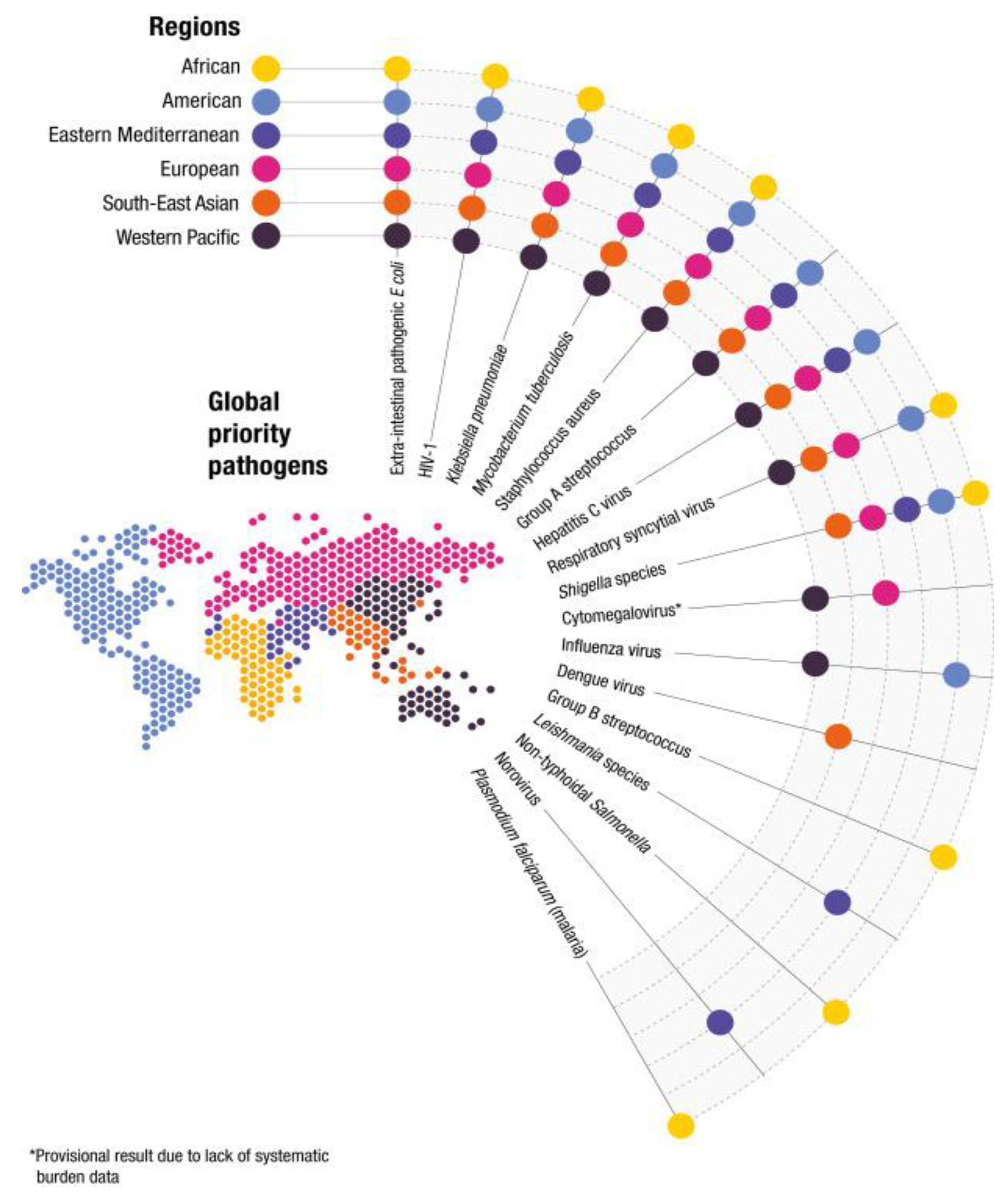
Figure 2.
Pictorial representation of mechanisms of antifungal resistance. Reproduced from Logan et al. with permission from Springer Nature.
Figure 2.
Pictorial representation of mechanisms of antifungal resistance. Reproduced from Logan et al. with permission from Springer Nature.
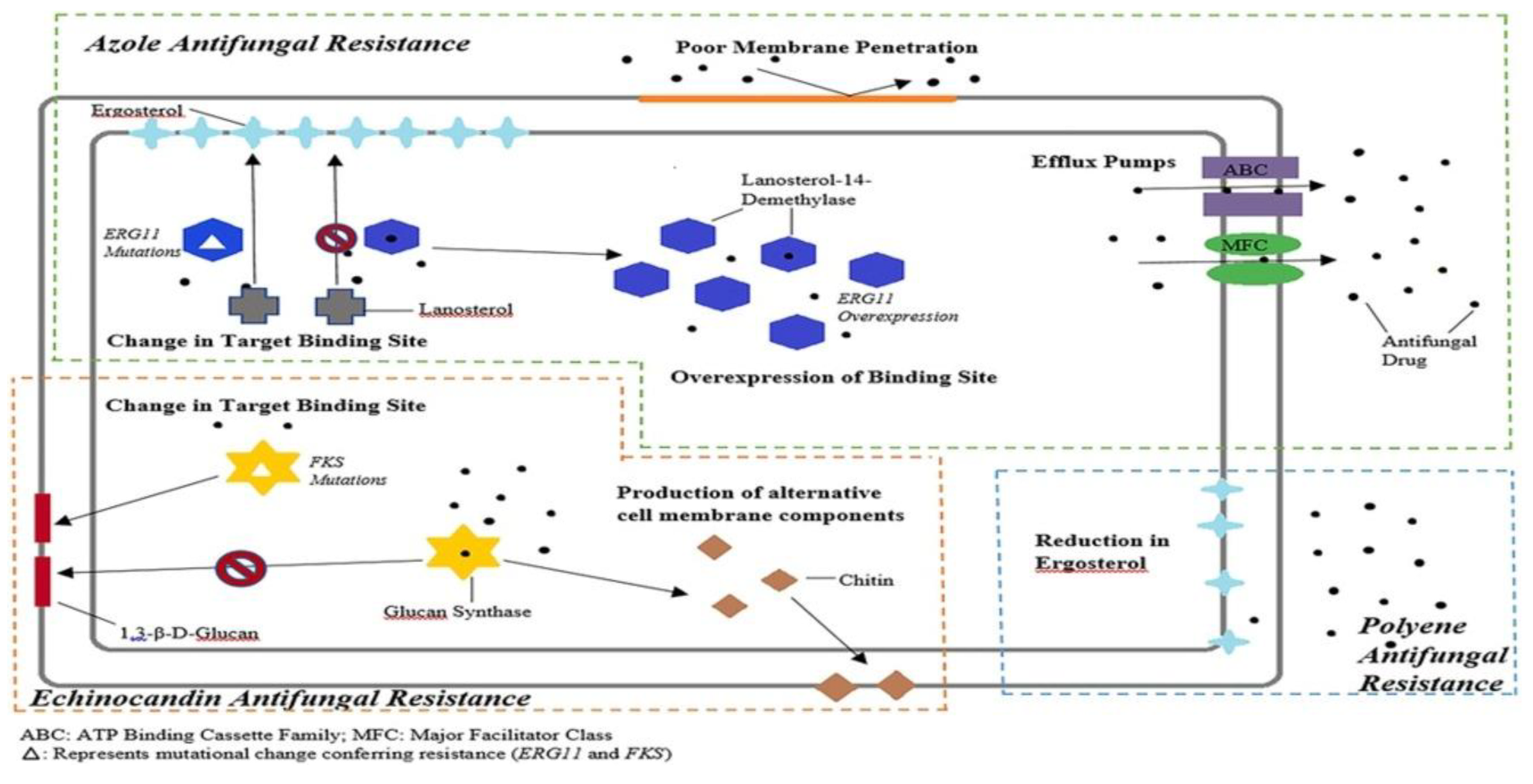
Figure 3.
Potential effects of climate change on pathogenic fungi and fungal diseases. Reproduced from Nnadi and Carter licensed under CC BY 4.0.
Figure 3.
Potential effects of climate change on pathogenic fungi and fungal diseases. Reproduced from Nnadi and Carter licensed under CC BY 4.0.
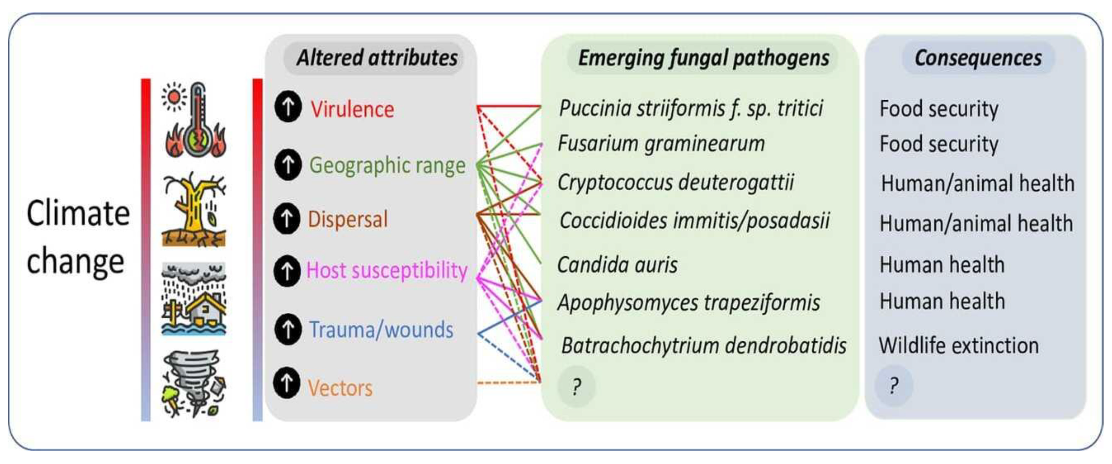
Figure 4.
Proposed scheme for the emergence of C. auris. Reproduced from Casadevall et al. licensed under CC BY 4.0.
Figure 4.
Proposed scheme for the emergence of C. auris. Reproduced from Casadevall et al. licensed under CC BY 4.0.
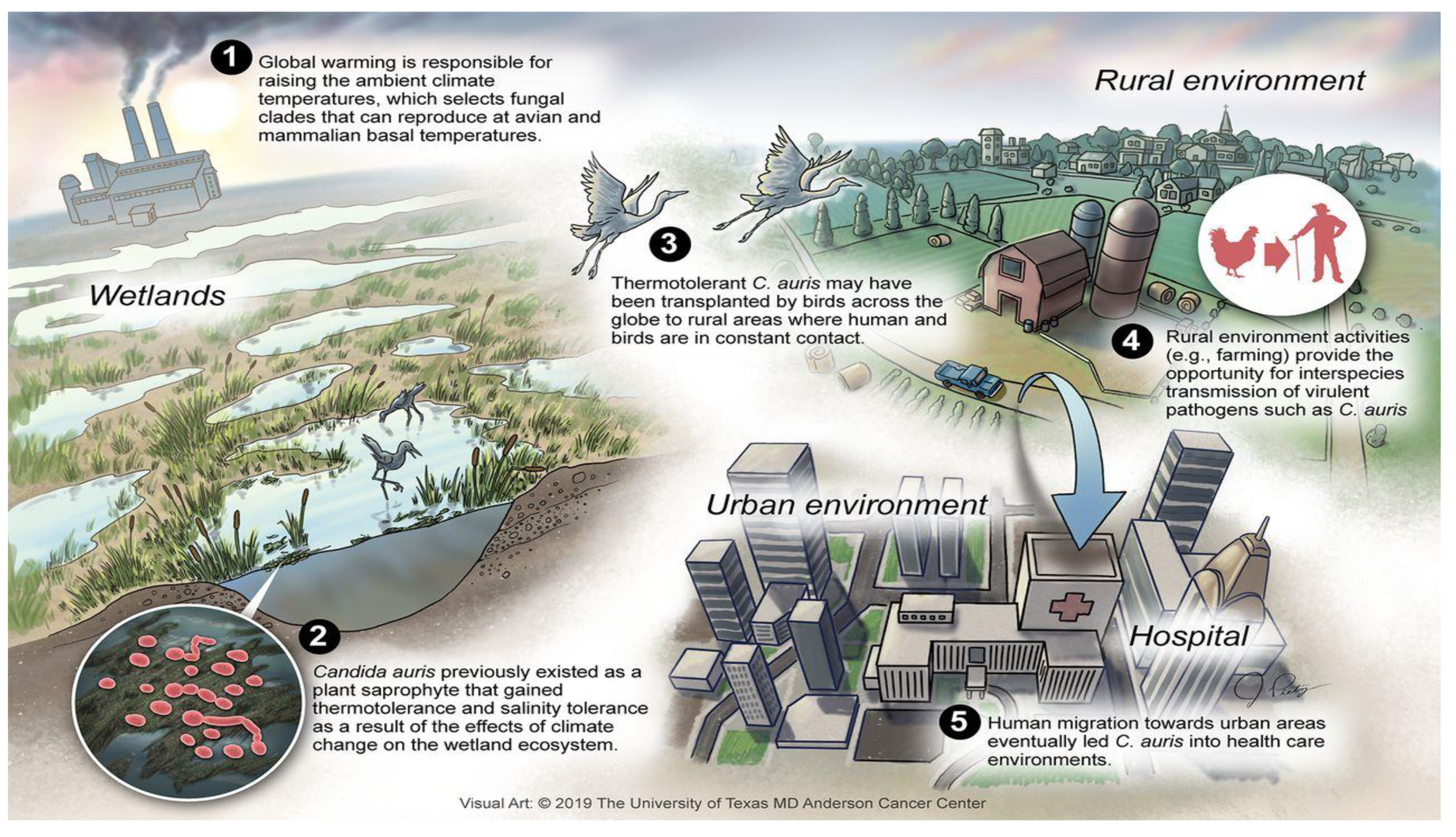

Table 1.
Patterns of Antifungal Resistance and Evolutionary Dynamics of Major Fungal Pathogens, Source: Motta et al. (2025).
Table 1.
Patterns of Antifungal Resistance and Evolutionary Dynamics of Major Fungal Pathogens, Source: Motta et al. (2025).
| Species/Complex | Resistance Pattern (Drug class) | Key molecular mechanisms | Evolutionary/Epidemiological Features | Clinical & Public Health Implications |
| Candida parapsilosis | FCZ: up to 63% (regional outbreaks) VCZ: cross-resistance (~44%) Emerging echinocandin resistance AmB: <3% resistance | ERG11 mutations (Y132F, K143R) Silent T591C mutation Efflux pump overexpression FKS1 hotspot mutations (F652S, R658G, S656P) Natural P660A polymorphism | Clonal nosocomial outbreaks Resistance without prior exposure Adaptive mutation + hospital persistence | Persistent candidemia Reduced echinocandin efficacy AmB as salvage therapy Requires molecular surveillance |
| Candida tropicalis | Azoles: ~10% resistance Echinocandins: ~0.4% AmB: highly active | ERG11 mutations (Y132F, Y257H/N) FKS1 mutation (S659P) | High genotypic diversity Environmental reservoirs (e.g., beach sand) Long-distance clonal spread (>1400 km) | Potential unrecognized outbreaks Strong candidate for One Health surveillance |
| Nakaseomyces glabratus | FCZ: 4–30% (variable regionally) Echinocandins: up to 10% Polyenes: rare resistance | PDR1 mutations FKS1/FKS2 hotspot mutations Biofilm formation EPA adhesins | High adaptability under drug pressure Prior echinocandin exposure selects mutants Intracellular persistence | Multidrug resistance risk Elevated MICs compromise therapy AmB critical in salvage regimens |
| Pichia kudriavzevii | Intrinsic FCZ resistance (~70%) VCZ: ~25% Echinocandins: <5% baseline AmB: >95% susceptible | FKS1 hotspot mutations (treatment-emergent) | Intrinsic azole resistance Resistance may emerge during therapy Regional variability | Requires echinocandin first-line Transition to AmB in breakthrough infection |
| Candida haemulonii complex | Multidrug resistance FCZ & AmB resistance common | Not fully characterized Misidentification common | Emerging invasive pathogen Frequently misidentified Hospital-associated | Requires MALDI-TOF/molecular ID Surveillance essential |
| Candida duobushaemulonii | Multidrug resistant | Limited genomic data Likely shared mechanisms with haemulonii complex | Underdiagnosed nosocomial spread Misidentified during surveillance | Hidden burden Requires reference-lab confirmation |
| Meyerozyma guilliermondii complex | Azole resistance: 40–70% Reports of AmB & echinocandin resistance | Genetic diversity Clade-specific azole resistance | High taxonomic complexity Clade spread (e.g., M. caribbica) | Individualized therapy needed Strengthened molecular diagnostics required |
|
Candida rugosa |
FCZ resistance: ~65% VCZ reduced sensitivity AmB resistance reported | Not fully defined | Higher regional prevalence ICU-associated outbreaks | Azoles unreliable Therapeutic risk with empirical treatment |
| Candidozyma auris | FCZ: >90% (Clade IV) AmB: 30–60% Echinocandins: up to 5% MDR common | ERG11 mutations (F126L, Y132F, K143R) TAC1B involvement FKS1 mutations Transcription factor mutations (e.g., FLO8 homolog S108N) Biofilm formation | Independent clade emergence (I–IV) Local transmission foci Environmental persistence (marine, freshwater) Thermal adaptation hypothesis Nosocomial colonization | High mortality (30–60%) Prolonged colonization Hospital outbreaks Requires mandatory reporting & genomic surveillance One Health relevance |
Abbreviation: FCZ, Fluconazole; VCZ, Voriconazole; AmB, Amphotericin B; MIC, Minimum Inhibitory Concentration; ERG11, Gene encoding lanosterol 14-α-demethylase; FKS1 / FKS2, Genes encoding subunits of β-1,3-D-glucan synthase; PDR1, Pleotropic Drug Resistance 1 transcription factor; EPA, Epithelial Adhesin proteins.
Table 2.
Global annual incidence of fungal spillover of various species. Source: Denning (2024).
| Pathogen Category | Global Annual Incidence | Mortality Rate | Primary Source/Reservoir |
| Invasive Fungal Infections | ~6.5 Million | ~58% | Multi-environmental |
| Cryptococcal Meningitis | ~194,000 | ~75% | Bird guano, Trees, Bats |
| Zoonotic Dermatophytosis | >2.1 Billion (All types) | Low (but rising resistance) | Cats, Dogs, Livestock |
| Candida auris | Rapidly Increasing | 30% – 60% | Environmental/Hospital |
Disclaimer/Publisher’s Note: The statements, opinions and data contained in all publications are solely those of the individual author(s) and contributor(s) and not of MDPI and/or the editor(s). MDPI and/or the editor(s) disclaim responsibility for any injury to people or property resulting from any ideas, methods, instructions or products referred to in the content. |
© 2026 by the authors. Licensee MDPI, Basel, Switzerland. This article is an open access article distributed under the terms and conditions of the Creative Commons Attribution (CC BY) license (http://creativecommons.org/licenses/by/4.0/).
Copyright: This open access article is published under a Creative Commons CC BY 4.0 license, which permit the free download, distribution, and reuse, provided that the author and preprint are cited in any reuse.


