

Submitted:
16 July 2026
Posted:
17 July 2026
You are already at the latest version
Abstract
Prostate cancer is strongly associated with aging, but the biological mechanisms linking aging to tumor progression remain incompletely defined. Beyond accumulated genetic alterations, aging reshapes the prostate tissue microenvironment through cellular senescence, chronic low-grade inflammation, immune dysfunction, stromal remodeling, metabolic stress, and impaired tissue repair. These processes contribute to inflammaging, a persistent inflammatory state that may create a permissive microenvironment for prostate tumor initiation, progression, immune evasion, and treatment resistance. Senescent epithelial and stromal cells can secrete cytokines, chemokines, growth factors, matrix-remodeling enzymes, and extracellular vesicles through the senescence-associated secretory phenotype (SASP). In parallel, immune aging alters T-cell subsets, myeloid cells, macrophages, and other immune populations, affecting anti-tumor surveillance and tumor-promoting inflammation. This review summarizes current knowledge of cellular senescence and inflammaging in prostate cancer, with emphasis on SASP, Th17/Treg imbalance, IL-17/IL-23-related inflammatory signaling, myeloid remodeling, stromal aging, metabolic stress, and immune–stromal–epithelial crosstalk. We also discuss how aging-associated inflammatory networks may influence tumor progression, therapeutic response, and emerging opportunities for cytokine modulation, senescence-directed therapy, metabolic intervention, and biomarker-guided approaches. Understanding the aging prostate microenvironment may reveal new strategies to prevent or delay aggressive prostate cancer progression in older men.
Keywords:
prostate cancer
; aging
; inflammaging
; cellular senescence
; senescence-associated secretory phenotype
; tumor microenvironment
; immune aging
; Th17/Treg balance
; IL-17/IL-23 axis
; metabolic inflammation
1. Introduction: Aging as an Active Biological Context for Prostate Cancer
Prostate cancer is one of the most common malignancies in men and is strongly associated with aging. Although this relationship has traditionally been attributed to the gradual accumulation of genetic and epigenetic alterations over time, it is increasingly clear that aging also changes the biological context in which prostate cancer develops. The aged prostate is not simply an older version of young tissue. Rather, it is characterized by altered epithelial homeostasis, stromal remodeling, chronic inflammatory signaling, immune dysfunction, metabolic stress, vascular changes, and impaired tissue repair. These age-associated alterations may influence tumor initiation, local progression, immune escape, therapeutic response, and the emergence of aggressive disease [1,2,3,4,5,6,7].
A key feature of biological aging is chronic, low-grade inflammation, commonly referred to as inflammaging. Inflammaging is driven by multiple cellular and systemic stressors, including cellular senescence, mitochondrial dysfunction, oxidative stress, endogenous danger signals, altered immune-cell composition, metabolic imbalance, and impaired resolution of inflammation. Unlike acute inflammation, which is typically transient and protective, inflammaging is persistent and can gradually remodel tissue structure and function. In the prostate, this chronic inflammatory state may create a permissive microenvironment for tumor development and progression by altering the behavior of epithelial cells, stromal fibroblasts, endothelial cells, immune cells, and extracellular matrix (ECM) components [8,9,10,11].
Cellular senescence is one of the major biological processes linking aging to chronic tissue inflammation [12,13,14]. Senescent cells undergo stable growth arrest in response to stressors such as telomere dysfunction, DNA damage, oncogene activation, oxidative stress, and therapy-induced injury [13,15,16,17,18]. While senescence can prevent the proliferation of damaged cells and thereby function as an important tumor-suppressive mechanism, senescent cells remain metabolically active and can secrete a broad spectrum of inflammatory and tissue-remodeling factors. This secretory program, known as the senescence-associated secretory phenotype (SASP), includes cytokines, chemokines, growth factors, proteases, ECM-modifying enzymes, and extracellular vesicles [15,19,20,21,22,23]. Persistent accumulation of senescent cells with age may therefore shift senescence from a protective response toward a chronic inflammatory and tumor-supportive process.
The aging immune system further contributes to inflammaging in the prostate tumor microenvironment. Aging is associated with reduced naïve T-cell diversity, accumulation of memory and exhausted T-cell populations, altered regulatory T-cell activity, myeloid skewing, impaired antigen presentation, and increased production of inflammatory cytokines [24,25,26,27,28]. These immune changes may weaken anti-tumor surveillance while increasing tumor-promoting inflammation. In prostate cancer, inflammatory immune circuits involving myeloid cells, macrophages, T-cell subsets, and stromal cells may support angiogenesis, matrix remodeling, immune suppression, and resistance to therapy [29,30,31,32,33,34,35,36,37,38]. Among adaptive immune populations, changes in T-helper cell balance, including Th17/Treg-associated inflammation, are particularly relevant because these cells can regulate epithelial and stromal responses through cytokine networks [39,40,41,42,43,44,45,46,47,48,49,50,51,52,53].
The IL-17/IL-23 axis represents one important inflammatory pathway that may connect immune aging to prostate cancer biology. IL-17 family cytokines can activate NF-κB, STAT3, chemokine expression, and tissue-remodeling programs in epithelial, stromal, endothelial, and immune cells. IL-23, produced primarily by myeloid and antigen-presenting cells, supports the maintenance of Th17-associated inflammatory responses. Together, IL-17- and IL-23-related signaling can sustain chronic inflammatory loops within tissues. In prostate cancer, these pathways may contribute to tumor-promoting inflammation, altered immune-cell recruitment, stromal remodeling, and disease progression. However, the biological effects of Th17-associated inflammation are context dependent and may vary according to tumor stage, tissue environment, immune composition, and treatment status [39,40,41,42,43,44,45,46,54].
In addition to immune-cell remodeling, stromal aging and metabolic stress are important components of the aging prostate microenvironment [29,30,55,56,57]. Aging fibroblasts and senescent stromal cells can produce inflammatory mediators, ECM proteins, matrix metalloproteinases (MMPs), and growth factors that alter epithelial behavior and tissue architecture [58,59,60]. Age-associated vascular dysfunction may affect hypoxia, nutrient delivery, Immune-cell trafficking, and angiogenesis [61,62,63,64,65,66]. Metabolic changes, including mitochondrial dysfunction, oxidative stress, obesity-associated inflammation, insulin resistance, and lipid dysregulation, can further amplify inflammatory signaling [67,68,69,70,71,72,73,74,75]. These stromal and metabolic alterations may cooperate with immune aging to generate a tissue environment that supports tumor progression and therapeutic resistance [28,29,67,68,69,70,71].
Importantly, aging-associated inflammation does not act through a single cell type or pathway. Instead, it reflects multicellular crosstalk among epithelial cells, immune cells, fibroblasts, endothelial cells, adipose-associated signals, ECM components, and systemic metabolic factors. This complexity has important implications for prostate cancer research. Many preclinical studies rely on young animal models, which may not accurately represent the aged tissue and immune environments in which human prostate cancer most commonly arises. Age-defined and temporally controlled prostate cancer models, including controlled Pten-knockout systems, provide a more relevant approach for studying how host age affects tumor initiation and progression [76,77,78]. Similarly, clinical studies that treat age as a simple demographic variable may overlook important biological differences among patients with distinct inflammatory, immune, senescent, and metabolic aging states.
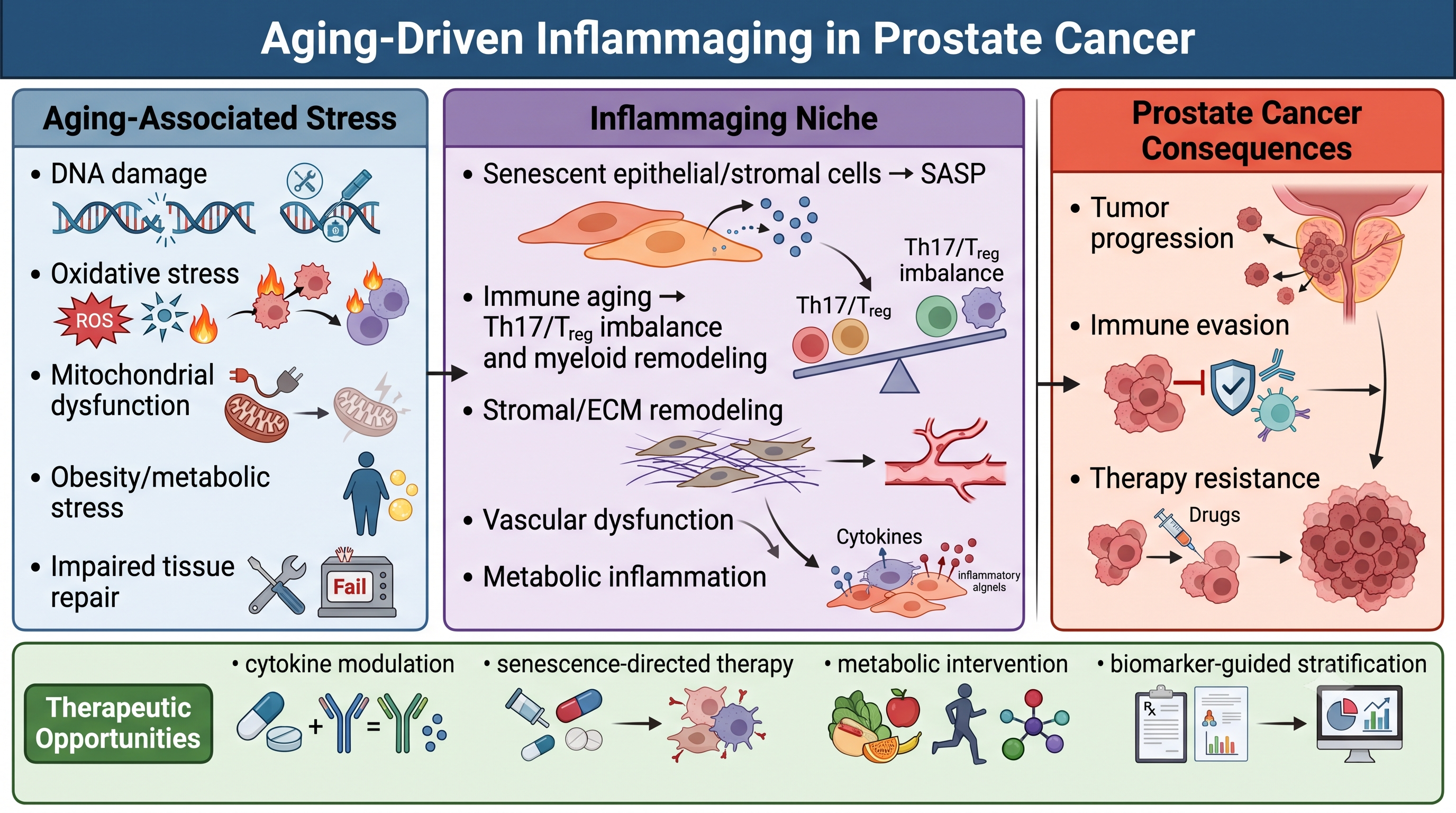
This review summarizes current knowledge of cellular senescence and inflammaging in prostate cancer, with a focus on the aging tumor microenvironment, immune remodeling, stromal dysfunction, metabolic stress, and therapeutic implications. We discuss SASP programs, immune aging, Th17/Treg imbalance, IL-17/IL-23-related inflammation, myeloid remodeling, ECM remodeling, and metabolic inflammatory signaling. We also highlight how these aging-associated processes may influence prostate cancer progression, immune evasion, and response to therapy. Finally, we discuss emerging opportunities to target inflammaging using cytokine modulation, senescence-directed approaches, metabolic intervention, and biomarker-guided strategies. By focusing on aging as an active biological process rather than a passive clinical variable, this review aims to provide a framework for understanding how the aged prostate microenvironment contributes to prostate cancer progression and how this knowledge may inform future therapeutic development [1,5,6,8,28,29] (Figure 1).
2. Cellular Senescence in the Aging Prostate Microenvironment
2.1. Biological Features of Cellular Senescence
Cellular senescence is a stress-associated cell state characterized by stable proliferative arrest, altered metabolism, resistance to apoptosis, chromatin remodeling, and acquisition of a complex secretory phenotype. Senescence can be triggered by multiple intrinsic and extrinsic stressors, including telomere shortening, DNA damage, oncogene activation, oxidative stress, mitochondrial dysfunction, epigenetic disruption, inflammatory signaling, and therapeutic injury [13,14,16,17,18]. In young or acutely injured tissues, senescence can serve beneficial functions by preventing the expansion of damaged cells, limiting malignant transformation, promoting wound healing, and facilitating immune-mediated clearance of abnormal cells. However, with aging, senescent cells may accumulate because of persistent tissue stress and declining immune clearance. This accumulation can contribute to chronic inflammation, altered tissue architecture, and impaired regenerative capacity.
Senescent cells are commonly associated with activation of the p53–p21CIP1 and p16INK4a–RB tumor suppressor pathways, which enforce cell-cycle arrest through inhibition of cyclin-dependent kinase activity and suppression of E2F-dependent transcription [13,15,18,21,79,80]. These pathways are not mutually exclusive and may be activated to different degrees depending on the inducing stress, cell type, and tissue context. In addition to growth arrest, senescent cells often display persistent DNA damage response signaling, increased lysosomal activity, senescence-associated β-galactosidase activity, altered mitochondrial function, increased reactive oxygen species (ROS), changes in nuclear morphology, and formation of senescence-associated heterochromatin foci. Because no single marker universally defines senescence across all tissues and disease states, rigorous identification typically requires multiple complementary markers and functional evidence.
In the prostate, cellular senescence is particularly relevant because the gland undergoes lifelong hormonal regulation, repeated inflammatory exposure, age-associated stromal remodeling, and progressive changes in epithelial homeostasis [81,82,83,84]. Prostate epithelial cells, fibroblasts, endothelial cells, and immune cells may all acquire senescence-like features under chronic stress. These senescent or senescence-associated cell states may influence prostate tissue structure and tumor behavior through both cell-autonomous and non-cell-autonomous mechanisms. For example, senescence within epithelial cells may initially suppress malignant expansion by blocking proliferation of damaged cells, whereas senescence in surrounding stromal or immune compartments may alter epithelial behavior through paracrine inflammatory and growth-regulatory signals.
A key challenge in studying senescence in prostate cancer is distinguishing protective senescence from persistent, maladaptive senescence. Oncogene-induced senescence can prevent early tumor development by arresting premalignant cells. Therapy-induced senescence may also limit tumor-cell proliferation after DNA-damaging treatment or androgen pathway inhibition. However, when senescent cells are not efficiently cleared, their prolonged survival and secretory activity may create a tissue environment that supports tumor progression. Thus, senescence should not be viewed as a single fixed process, but as a dynamic biological state whose consequences depend on timing, cell type, immune context, and tissue microenvironment [17,19,85,86,87,88,89,90,91].
2.2. Senescence-Associated Secretory Phenotype
One of the most important features of senescent cells is the SASP. The SASP consists of a heterogeneous mixture of soluble and insoluble factors, including inflammatory cytokines, chemokines, growth factors, proteases, ECM components, lipid mediators, and extracellular vesicles [15,19,20,21,22,23]. Major SASP-associated factors frequently include IL-6, IL-8/CXCL8, CCL2, CXCL1, CXCL2, TNF-α, TGF-β, GM-CSF, VEGF, MMPs, and other tissue-remodeling molecules [15,19,20,21,22,23]. The precise composition of the SASP varies according to cell type, senescence trigger, duration of senescence, tissue environment, and immune context.
The SASP can reinforce senescence in an autocrine manner and transmit stress signals to neighboring cells in a paracrine manner. Through cytokines and chemokines, senescent cells can recruit immune cells that may eliminate damaged cells and restore tissue homeostasis. In this context, the SASP can be beneficial and may support tissue repair or tumor suppression. However, persistent SASP production during aging can have detrimental effects. Chronic secretion of inflammatory mediators may promote immune-cell dysfunction, fibroblast activation, ECM remodeling, angiogenesis, epithelial stress responses, and altered tissue architecture [14,15,19,20,21,22,23]. In the prostate, these effects may be particularly important because epithelial cells are embedded within a hormonally responsive and stromally active microenvironment.
Several signaling pathways regulate SASP induction and maintenance. DNA damage response signaling, NF-κB activation, C/EBPβ, p38 MAPK, mTOR, cGAS–STING signaling, inflammasome activation, and mitochondrial dysfunction have all been implicated in SASP regulation [15,21,79,80,92]. NF-κB and C/EBPβ are especially important transcriptional regulators of inflammatory SASP components, including IL-6 and IL-8/CXCL8. Mitochondrial dysfunction and cytosolic DNA sensing may further amplify inflammatory signaling through ROS and innate immune pathways. These mechanisms provide a biological connection between aging-associated cellular stress and chronic inflammatory remodeling of tissue.
In prostate cancer, SASP factors may influence several aspects of tumor biology. IL-6 and related cytokines can activate STAT3 signaling, which has been linked to proliferation, survival, inflammatory adaptation, and treatment resistance in multiple cancer types. Chemokines such as CCL2 and CXCL family members can recruit monocytes, macrophages, neutrophils, and other immune cells, thereby reshaping the tumor immune microenvironment. MMPs and other proteases can remodel ECM and facilitate tissue invasion. Growth factors and angiogenic mediators can support epithelial growth, stromal activation, and vascular remodeling [81,82,89,90,91,93,94]. Although these processes are not unique to prostate cancer, they are highly relevant to aging-associated prostate tumor progression because they provide plausible mechanisms through which senescent cells can influence non-senescent tumor and stromal cells.
The SASP also has important implications for therapy. Cancer therapies that induce DNA damage, oxidative stress, androgen deprivation, or cellular stress may generate senescent tumor or stromal cells. While therapy-induced senescence can suppress proliferation, persistent senescent cells may contribute to a pro-inflammatory and pro-survival environment. This dual effect has led to growing interest in senescence-directed strategies, including senolytics that eliminate senescent cells and senomorphics that suppress harmful SASP programs without necessarily killing senescent cells [81,82,93,94,95,96,97,98,99,100,101]. For prostate cancer, such approaches remain investigational, but they may be particularly relevant for older patients whose tissues already contain elevated senescence and inflammatory burden.
2.3. Tumor-Suppressive Versus Tumor-Promoting Roles of Senescence
The relationship between senescence and cancer is complex because senescence can either suppress or promote tumor development depending on biological context. As a tumor-suppressive mechanism, senescence prevents damaged or oncogene-stressed cells from continuing to proliferate [17,85,86,89,90]. This is particularly important during early tumorigenesis, when activation of oncogenic pathways or loss of normal growth control can trigger a protective senescence response. In this setting, senescence functions as a barrier to malignant transformation by enforcing durable cell-cycle arrest and facilitating immune-mediated clearance of abnormal cells.
In prostate tissue, tumor-suppressive senescence may occur in response to oncogenic stress, DNA damage, oxidative injury, or other cellular insults. Activation of p53, p21CIP1, p16INK4a, and RB-associated pathways can limit uncontrolled proliferation and maintain tissue integrity. This protective role is consistent with the broader concept that senescence is an important anti-cancer defense mechanism [83,84,89,90,91,102]. Indeed, failure to establish or maintain senescence programs may permit damaged epithelial cells to bypass growth arrest and acquire additional malignant features.
However, senescence can also promote tumor progression when senescent cells persist and accumulate. In aged tissues, senescent cells may not be efficiently cleared because of impaired immune surveillance, reduced phagocytic function, T-cell dysfunction, or chronic inflammatory desensitization. Persistent senescent cells can produce SASP factors that alter neighboring cells and the surrounding microenvironment. In cancer, this can create a paradox: senescent cells themselves may not proliferate, yet their secreted factors can support the proliferation, survival, migration, invasion, and therapy resistance of nearby premalignant or malignant cells [19,20,22,87,88,91,98].
This duality is especially relevant in the prostate tumor microenvironment, where epithelial, stromal, endothelial, and immune compartments interact closely. Senescent fibroblasts may produce inflammatory cytokines, growth factors, and matrix-remodeling enzymes that influence epithelial cell behavior. Senescent epithelial cells may reinforce local inflammation and attract immune cells. Senescent endothelial cells may contribute to vascular dysfunction and altered immune trafficking. Senescent immune cells may exhibit impaired effector function while maintaining inflammatory mediator production. Together, these cell-type-specific senescence programs can generate a multicellular environment that supports tumor adaptation even when individual senescent cells remain growth arrested [13,14,81,82,93,94].
The timing of senescence is also important. Acute senescence followed by immune clearance may be protective, whereas chronic senescence with persistent SASP production may be harmful. Similarly, senescence within tumor cells may have different consequences from senescence within non-malignant stromal cells. Tumor-cell senescence may initially limit tumor expansion, but therapy-induced senescent tumor cells may survive as residual disease and contribute to relapse under certain conditions. Stromal senescence may provide paracrine support for tumor growth, especially in aged tissues. Therefore, the biological outcome of senescence depends on whether senescence is transient or persistent, whether senescent cells are cleared or retained, and which cell types are affected [13,14,81,82,93,94].
This complexity has important therapeutic implications. Complete suppression of senescence may be undesirable because senescence can prevent malignant transformation and contribute to treatment response. Conversely, persistent senescent cells and chronic SASP activity may promote inflammation and tumor progression. Therapeutic strategies must therefore distinguish beneficial senescence from maladaptive senescence. In prostate cancer, this may require identifying which senescent cell populations accumulate during aging, which SASP factors are most relevant to tumor progression, and which patients exhibit senescence-associated inflammatory signatures that could be therapeutically targeted [95,96,97,98,99,100,101].
2.4. Senescence in Prostate Epithelial and Stromal Compartments
The prostate microenvironment is composed of epithelial cells, basal and luminal cell populations, stromal fibroblasts, smooth muscle cells, endothelial cells, immune cells, ECM, nerves, and systemic hormonal and metabolic inputs [55,56,57,103,104,105]. Aging can affect each of these compartments. Therefore, senescence in the prostate should be considered a tissue-level process rather than a feature of one cell type alone. The interaction between senescent epithelial cells and senescent or activated stromal cells may be particularly important for prostate cancer development and progression.
In the epithelial compartment, senescence may arise in response to DNA damage, oxidative stress, oncogenic signaling, inflammation, androgen-related stress, or therapeutic injury. Senescence in premalignant epithelial cells may initially function as a protective barrier by preventing proliferation of damaged cells [83,84,89,90,91,102]. However, senescent epithelial cells can also produce inflammatory mediators and tissue-remodeling factors that alter local immune and stromal responses. In aging prostate tissue, repeated cycles of epithelial stress, injury, inflammation, and repair may contribute to accumulation of senescence-associated programs. These changes may influence epithelial differentiation, barrier function, secretory activity, and communication with surrounding stromal cells.
The stromal compartment is equally important. Prostate stromal fibroblasts play essential roles in epithelial development, tissue architecture, androgen responsiveness, ECM production, and paracrine growth regulation [29,30,31,55,56,57,103,104,105,106,107,108,109,110]. With aging, stromal fibroblasts can acquire senescence-like or activated phenotypes characterized by altered secretory activity, inflammatory signaling, ECM remodeling, and reduced capacity to maintain normal epithelial homeostasis. Senescent stromal fibroblasts may produce IL-6, CXCL chemokines, CCL2, TGF-β family members, growth factors, and MMPs. These factors can influence epithelial proliferation, survival, inflammatory recruitment, and stromal remodeling.
Senescence-associated stromal changes may also interact with cancer-associated fibroblast biology. Although senescent fibroblasts and cancer-associated fibroblasts (CAFs) are not identical, they may share features such as inflammatory cytokine production, matrix remodeling, paracrine growth signaling, and altered tissue stiffness. In aged prostate tissue, senescent stromal cells may create conditions that favor fibroblast activation and tumor-supportive remodeling. Increased ECM deposition, altered collagen organization, and enhanced matrix-degrading enzyme activity may contribute to changes in tissue mechanics and local invasion [29,30,31,104,105,106,107,108,109,110]. These processes provide a plausible connection between aging stromal biology and prostate cancer progression.
Endothelial and vascular senescence may further shape the aging prostate microenvironment. Senescent endothelial cells can contribute to vascular dysfunction, altered permeability, impaired angiogenic balance, hypoxia, and modified immune-cell trafficking. Because tumor progression depends on nutrient delivery, oxygen availability, immune-cell access, and angiogenic remodeling, endothelial aging may influence both tumor growth and therapeutic response. In addition, vascular dysfunction can amplify tissue hypoxia and oxidative stress, thereby reinforcing inflammatory and senescence-associated signaling [61,62,63,64,65,66].
Immune-cell senescence and exhaustion add another layer of complexity. Aging can reduce effective anti-tumor immunity while increasing chronic inflammatory signaling. Senescence-like features in T cells, altered macrophage function, myeloid skewing, and impaired antigen presentation may collectively reduce immune surveillance. At the same time, inflammatory immune cells can reinforce SASP-like signaling and stromal remodeling. This bidirectional relationship between senescent tissue cells and aging immune cells is central to inflammaging [24,25,26,27,28,111,112,113,114].
Taken together, senescence in the aging prostate microenvironment involves coordinated changes in epithelial, stromal, endothelial, and immune compartments. These changes may influence prostate cancer biology through direct effects on epithelial cells and indirect effects mediated by cytokines, chemokines, ECM remodeling, immune-cell recruitment, vascular dysfunction, and metabolic stress. Understanding the cell-type-specific sources and consequences of senescence will be essential for developing biomarkers and therapeutic strategies that target harmful aging-associated inflammation while preserving the protective functions of senescence (Table 1).
3. Immune Aging and Inflammaging in the Prostate Cancer
3.1. Immunosenescence and Impaired Immune Surveillance
The immune system undergoes substantial remodeling during aging, a process commonly referred to as immunosenescence. This process affects both innate and adaptive immune compartments and is characterized by reduced immune diversity, altered immune-cell function, chronic inflammatory activation, and impaired immune resolution [24,25,26,27]. In the adaptive immune system, aging is associated with thymic involution, reduced generation of naïve T cells, contraction of T-cell receptor diversity, expansion of memory and terminally differentiated T-cell populations, and increased features of T-cell dysfunction or exhaustion [32,33,34]. These changes may limit the ability of the aged immune system to recognize and eliminate emerging malignant cells.
Effective anti-tumor immunity requires coordinated antigen presentation, T-cell priming, cytotoxic effector function, immune-cell trafficking, and immune memory [24,25,26,27,32,33,34]. Aging can disrupt each of these steps. Dendritic cells and other antigen-presenting cells may show altered antigen uptake, processing, migration, and co-stimulatory function. CD8+ T cells may exhibit reduced proliferative capacity, impaired cytokine production, diminished cytotoxic activity, and increased expression of inhibitory receptors. CD4+ T-cell subsets may shift in frequency and function, altering the balance between anti-tumor immunity and tumor-promoting inflammation. Together, these age-associated changes may reduce immune surveillance and allow prostate epithelial lesions or established tumor cells to persist under reduced immune pressure.
At the same time, immunosenescence does not simply represent immune deficiency. Aging is also associated with increased basal inflammatory tone, sometimes described as sterile chronic inflammation or inflammaging. Therefore, the aged immune system can be simultaneously less effective at tumor elimination and more prone to chronic inflammatory activation [8,9,10,11,25,26,27,28,35,114]. This paradox is highly relevant to prostate cancer, where chronic immune-cell infiltration and inflammatory cytokine production may coexist with weak anti-tumor cytotoxicity. In this setting, immune aging may support tumor progression not only by reducing protective surveillance, but also by generating inflammatory signals that remodel the tumor microenvironment.
In prostate cancer, the immune microenvironment is typically complex and heterogeneous. Tumors may contain T cells, macrophages, myeloid-derived suppressor cells, dendritic cells, neutrophils, natural killer cells, mast cells, and B cells, with variable functional states depending on tumor stage, tissue location, treatment history, and host factors [29,30,31,32,33,34,36,37,38]. In older patients, these immune populations are further shaped by systemic aging, metabolic status, comorbidities, prior inflammation, and therapy exposure. Thus, understanding prostate cancer immunity requires considering not only tumor-intrinsic immune escape mechanisms, but also the aged host immune environment in which the tumor develops.
3.2. Chronic Inflammation and Cytokine Imbalance
Inflammaging is characterized by persistent, low-grade inflammatory signaling that occurs in the absence of acute infection. This chronic inflammatory state is driven by multiple aging-associated processes, including cellular senescence, mitochondrial dysfunction, oxidative stress, endogenous damage-associated molecular patterns, altered tissue repair, metabolic stress, and impaired clearance of damaged cells [8,9,11,25,27]. In the prostate microenvironment, chronic inflammation may influence epithelial behavior, stromal activation, immune-cell recruitment, angiogenesis, ECM remodeling, and therapeutic response.
Several cytokine and chemokine pathways have been implicated in aging-associated inflammation and prostate cancer progression. IL-6 is one of the best-studied inflammatory cytokines in cancer biology and can activate JAK/STAT3 signaling, which has been linked to tumor-cell survival, proliferation, inflammatory adaptation, angiogenesis, and treatment resistance [19,20,21,92]. TNF-α can activate NF-κB and other stress-response pathways, contributing to inflammatory gene expression, immune-cell recruitment, and stromal remodeling. IL-1 family cytokines can amplify innate immune activation and inflammatory tissue injury. TGF-β has context-dependent roles, functioning in some settings as a suppressor of epithelial proliferation and in others as a driver of immune suppression, fibroblast activation, ECM remodeling, and invasive behavior [29,30,32,33,34,36,37,38].
Chemokines are also central mediators of the inflammatory tumor microenvironment. CCL2, CXCL1, CXCL2, CXCL8/IL-8, CXCL12, and related chemokine pathways can regulate recruitment of monocytes, macrophages, neutrophils, lymphocytes, and other immune-cell populations [29,30,31]. In aged tissues, persistent chemokine production may reinforce immune-cell infiltration and chronic inflammation. In prostate cancer, chemokine signaling may also influence stromal remodeling, angiogenesis, tumor-cell migration, and immune suppression. These pathways provide important links between senescence-associated secretory programs, immune-cell recruitment, and tumor-supportive inflammation.
IL-17- and IL-23-related inflammatory signaling represents another pathway of interest in prostate cancer. IL-17 family cytokines can activate inflammatory and tissue-remodeling programs in epithelial, stromal, endothelial, and immune cells. IL-23, produced primarily by myeloid and antigen-presenting cells, supports the maintenance and pathogenic activity of Th17-associated immune responses [39,40,41,42,43,44,45,46,54]. Together, IL-17 and IL-23 may sustain chronic inflammatory circuits in tissue microenvironments. However, the effects of these pathways in cancer are context dependent and may vary according to tumor type, stage, immune composition, and local cytokine networks.
Importantly, cytokine imbalance in the aged prostate microenvironment should not be viewed as the effect of one isolated mediator. Instead, inflammatory cytokines operate as interconnected networks. IL-6, IL-17, TNF-α, IL-1, IL-23, TGF-β, and chemokines may cooperate or counter-regulate one another depending on cellular context. These pathways can also intersect with androgen receptor signaling, metabolic stress, senescence-associated programs, and stromal activation [29,30,32,33,34,36,37,38,39,40,41,42,43,44,45,46,47,48,49,50,51,52,53,54]. Therefore, therapeutic targeting of inflammaging in prostate cancer will likely require careful patient stratification and a detailed understanding of dominant inflammatory circuits in each biological context.
3.3. Th17/Treg Balance in Aging and Prostate Cancer
CD4+ T-cell subsets are important regulators of tissue inflammation and tumor immunity. Among these subsets, Th17 cells and regulatory T cells are particularly relevant to chronic inflammatory diseases and cancer. Th17 cells are characterized by production of IL-17A, IL-17F, IL-21, IL-22, and other inflammatory mediators, whereas regulatory T cells suppress immune activation and maintain immune tolerance through mechanisms involving FOXP3, IL-10, TGF-β, CTLA-4, and other suppressive pathways [47,48,49,50,51,54]. The balance between Th17-associated inflammation and Treg-mediated immune suppression can shape both inflammatory tissue damage and anti-tumor immunity.
Aging may alter the balance, differentiation, and function of Th17 and Treg populations. Chronic antigenic stimulation, inflammatory cytokines, metabolic stress, microbial changes, and altered antigen-presenting cell function can all influence CD4+ T-cell polarization during aging [24,25,26,27,42,111,112,113,114]. Increased inflammatory tone may favor Th17-associated responses in some contexts, whereas accumulation or altered function of Tregs may suppress anti-tumor effector immunity. The resulting immune state may therefore combine chronic inflammation with reduced tumor immune surveillance.
In prostate cancer, both Th17 cells and Tregs have been implicated in shaping the tumor immune microenvironment. Tregs can suppress cytotoxic T-cell responses and may contribute to immune evasion. Th17 cells can have context-dependent effects, with evidence from different cancer types supporting both pro-tumor and anti-tumor functions [39,40,41,42,43,44,45,46]. Pro-tumor mechanisms may include promotion of inflammatory cytokine production, angiogenesis, chemokine-mediated immune-cell recruitment, stromal activation, and tissue remodeling. Anti-tumor effects may occur when Th17-related responses enhance immune-cell recruitment, support effector T-cell function, or contribute to tumor rejection. These divergent effects highlight the importance of tissue context and disease stage.
The IL-23 pathway is particularly important for sustaining Th17 inflammatory programs. While early Th17 differentiation involves cytokines such as IL-6, TGF-β, IL-1β, and IL-21, IL-23 can stabilize and expand pathogenic Th17-like states in chronic inflammation [43,44,54]. In the prostate tumor microenvironment, myeloid cells and antigen-presenting cells may provide IL-23 or related inflammatory signals that support persistent Th17-associated activity. This type of immune circuit may be especially relevant in aged tissues, where chronic inflammatory signals and altered myeloid function are common features.
Th17/Treg imbalance is best reviewed as a broad immune-aging mechanism relevant to prostate cancer rather than as a single deterministic pathway. Published studies support the importance of T-cell subset balance, inflammatory cytokines, and IL-17/IL-23-related signaling in prostate cancer biology [39,40,41,42,43,44,45,46]. Additional work is needed to define how aging modifies these immune circuits across disease stages, treatment settings, and patient populations. A clearer understanding of Th17/Treg balance may help identify inflammatory biomarkers and guide immune-modulatory strategies.
3.4. Myeloid Remodeling During Aging
Myeloid cells are major contributors to inflammaging and the tumor microenvironment. Aging is associated with myeloid-biased hematopoiesis, altered monocyte and macrophage function, impaired antigen presentation, increased basal inflammatory activation, and reduced resolution of tissue inflammation [24,25,26,27,35,111,112,113,114]. These changes can promote persistent cytokine production and inflammatory remodeling in multiple tissues. In cancer, age-associated myeloid remodeling may support immune suppression, angiogenesis, matrix remodeling, tumor-cell survival, and resistance to therapy.
Tumor-associated macrophages are among the most abundant immune populations in many solid tumors, including prostate cancer. These cells can display diverse functional states rather than a simple binary M1/M2 classification. Depending on microenvironmental cues, macrophages may produce inflammatory cytokines, growth factors, angiogenic mediators, proteases, immunosuppressive molecules, and chemokines [29,30,31]. In prostate cancer, macrophage-rich microenvironments have been associated with tumor progression, immune suppression, and therapy resistance in several studies. Aging may further influence macrophage function by altering metabolic state, phagocytic capacity, cytokine production, and responsiveness to tissue damage.
Myeloid-derived suppressor cells are another important population in tumor-associated immune suppression. These cells can inhibit T-cell activation and effector function through mechanisms involving arginine metabolism, ROS, nitric oxide, immunosuppressive cytokines, and checkpoint-related pathways [29,31,32,33,34,36,37,38,43]. In aged hosts, expansion of immature or suppressive myeloid populations may be favored by chronic inflammation, altered hematopoiesis, and tumor-derived factors. Their accumulation in prostate cancer may contribute to poor anti-tumor immunity and limited response to immunotherapy.
Dendritic cells may also be affected by aging and tumor-associated inflammation. Effective dendritic cell function is required for antigen presentation and T-cell priming. Aging can impair dendritic cell migration, antigen presentation, cytokine production, and co-stimulatory signaling [24,25,26,27,32,33,34,111,112,113,114]. Within the prostate tumor microenvironment, suppressive cytokines, metabolic stress, tumor-derived factors, and chronic inflammation may further weaken dendritic cell function. This may reduce productive anti-tumor T-cell priming and promote immune tolerance.
Neutrophils and inflammatory monocytes may also contribute to the aged prostate tumor microenvironment. These cells can produce cytokines, proteases, ROS, and extracellular traps, and may influence angiogenesis, matrix remodeling, and tumor-cell invasion [24,25,26,27,29,30,111,112,113,114]. Although their roles in prostate cancer require further study, they represent important components of innate immune remodeling. Because aging is associated with altered innate immune responsiveness and persistent inflammatory activation, these myeloid populations may help sustain inflammaging-associated tumor-promoting niches.
Overall, myeloid remodeling provides a major link between aging, inflammation, and prostate cancer progression. Myeloid cells can produce cytokines such as IL-6, IL-1β, TNF-α, IL-10, TGF-β, and IL-23; recruit additional immune cells through chemokines; suppress T-cell responses; remodel ECM; and support angiogenesis. These functions place myeloid cells at the center of aging-associated inflammatory crosstalk in the prostate tumor microenvironment [29,31,32,33,34,36,37,38,43].
3.5. Transcriptional Control of Inflammatory Immune States
Immune aging and inflammaging are regulated not only by changes in immune-cell abundance, but also by changes in transcriptional and epigenetic programs that determine immune-cell identity and function. T-cell differentiation, macrophage polarization, dendritic cell activation, and inflammatory cytokine production are controlled by coordinated transcription factor networks. In aging tissues, chronic inflammatory stimuli, metabolic stress, antigen exposure, and altered cytokine environments may reshape these transcriptional programs and produce persistent inflammatory or dysfunctional immune states [24,25,26,27,111,112,113,114].
In T cells, transcription factors such as T-bet, GATA3, RORγt, FOXP3, STAT3, STAT5, IRF4, and AP-1 family members regulate differentiation into effector, regulatory, and inflammatory subsets. Th17 differentiation requires coordinated cytokine signaling and transcriptional programming involving STAT3, RORγt, IRF4, BATF, and related factors [47,48,49,50,51,54]. Tregs depend on FOXP3 and associated regulatory networks. The balance between these programs is influenced by cytokines such as IL-6, TGF-β, IL-1β, IL-21, IL-23, IL-2, and inflammatory metabolic cues. Aging-associated changes in these signals may therefore alter T-helper cell balance and inflammatory potential.
BATF is an established transcriptional regulator involved in Th17 differentiation and inflammatory T-cell programming. In the context of this review, BATF can be discussed as an example of how transcriptional regulation shapes immune-cell inflammatory states. Published studies have shown that BATF-dependent Th17 programs and IL-23/IL-23R-related signaling can contribute to prostate cancer-associated inflammation and tumor progression [39,43,54]. However, the broader relationship among immune-cell transcriptional states, biological aging, metabolic stress, and prostate tumor behavior remains an active area of investigation. For this reason, this review does not present BATF as a single central driver of aging-associated prostate cancer, but rather as one component of a broader inflammatory transcriptional network.
In myeloid cells, transcriptional regulators such as NF-κB, STAT1, STAT3, STAT6, HIF-1α, C/EBPβ, IRF family members, and nuclear receptors can control inflammatory activation, immune suppression, antigen presentation, and metabolic adaptation. Chronic activation of NF-κB and STAT3 pathways is particularly relevant to inflammaging because these pathways integrate signals from cytokines, pattern-recognition receptors, oxidative stress, senescence-associated mediators, and tumor-derived factors [25,29,31]. Persistent activation of these pathways may promote cytokine production, chemokine expression, myeloid recruitment, and immune suppression in the prostate tumor microenvironment.
Epigenetic regulation also contributes to immune-cell aging. DNA methylation changes, histone modifications, chromatin accessibility, and non-coding RNAs can affect immune-cell differentiation, inflammatory memory, and exhaustion-like states [24,25,26,27,111,112,113,114]. Trained immunity in innate immune cells and chronic antigen stimulation in adaptive immune cells may establish durable immune states that persist even after the original stimulus has changed. In prostate cancer, such stable inflammatory or suppressive immune states may influence tumor progression and treatment response.
Understanding transcriptional control of immune aging has practical implications. Cell-type-specific immune signatures may help distinguish patients with inflammatory, immunosuppressive, senescent, or metabolically stressed tumor microenvironments. These signatures could support biomarker development and guide selection of therapies targeting cytokines, immune checkpoints, myeloid cells, senescence-associated inflammation, or metabolic pathways [32,33,34,36,37,38]. Future studies using single-cell transcriptomics, spatial profiling, epigenomic analysis, and age-stratified models will be important for defining how immune transcriptional states shape the aging prostate tumor microenvironment.
Section Summary
Immune aging links impaired surveillance with persistent inflammatory activation in prostate cancer. Changes in T-cell diversity, Th17/Treg balance, myeloid activity, antigen presentation, and cytokine signaling may promote chronic inflammation, immune suppression, stromal remodeling, and therapy resistance. These pathways operate as an integrated multicellular network rather than isolated drivers, and cell-type-specific studies will be essential for biomarkers and therapeutic development (Table 2).
4. Stromal Aging, Extracellular Matrix Remodeling, and Prostate Tumor Progression
4.1. Aging Fibroblasts and Cancer-Associated Fibroblasts
The prostate is a highly stromal organ in which epithelial cells are embedded within a complex microenvironment composed of fibroblasts, smooth muscle cells, endothelial cells, immune cells, nerves, ECM, and soluble paracrine factors. During aging, this stromal compartment undergoes progressive remodeling that can alter epithelial homeostasis and influence prostate cancer development. Stromal aging is characterized by changes in fibroblast function, ECM organization, inflammatory signaling, tissue stiffness, paracrine growth regulation, and response to injury [55,56,57,103,104,105]. These changes may create a permissive tissue environment in which premalignant or malignant epithelial cells acquire growth and survival advantages.
Fibroblasts are central regulators of prostate tissue architecture. In normal prostate tissue, stromal fibroblasts and smooth muscle cells help maintain epithelial differentiation, androgen responsiveness, basement membrane integrity, and paracrine growth control. With aging, fibroblasts may acquire senescence-associated or activated phenotypes marked by altered cytokine production, growth factor secretion, ECM remodeling, and reduced ability to maintain normal epithelial organization [55,56,57,103,104,105]. These changes can disrupt stromal–epithelial communication and contribute to chronic tissue inflammation.
Senescent stromal fibroblasts can produce a broad range of SASP factors, including IL-6, IL-8/CXCL8, CCL2, CXCL1, TGF-β family members, growth factors, and MMPs [13,14,55,56,57,103,104,105]. These mediators can influence epithelial proliferation, immune-cell recruitment, angiogenesis, and ECM remodeling. Although senescence can initially function as a protective response to cellular stress, persistent accumulation of senescent stromal cells may promote a chronic inflammatory state that supports tumor progression. Thus, stromal senescence represents an important mechanism through which aging may affect prostate cancer biology.
CAFs are another major component of the prostate tumor microenvironment. CAFs are functionally heterogeneous and can arise from local fibroblasts, smooth muscle cells, pericytes, mesenchymal progenitors, or other stromal sources depending on tissue context [29,30,104,105,106,110]. In prostate cancer, CAFs can produce growth factors, cytokines, chemokines, ECM proteins, and proteases that support tumor-cell survival, local invasion, angiogenesis, immune suppression, and therapeutic resistance. CAFs can also alter androgen receptor signaling and epithelial differentiation through paracrine mechanisms, although the precise effects depend on tumor stage and stromal subtype.
Aging may influence CAF biology in several ways [28,29,30,55,56,57]. First, aged fibroblasts may be more prone to inflammatory activation or SASP programs. Second, chronic low-grade inflammation may promote fibroblast activation and ECM remodeling. Third, age-associated changes in tissue mechanics and matrix composition may reinforce CAF phenotypes. Fourth, impaired immune clearance of senescent or damaged stromal cells may allow tumor-supportive stromal niches to persist. Together, these processes suggest that aging may not only affect epithelial tumor cells directly but also reshape the stromal context in which tumor cells evolve.
The distinction between senescent fibroblasts and CAFs is important but not absolute. Senescent fibroblasts are typically growth arrested and defined by stress-associated cell-cycle arrest and SASP production, whereas CAFs are activated stromal cells with diverse functional states that may or may not be senescent. Nevertheless, these populations can share overlapping features, including inflammatory cytokine production, ECM remodeling, growth factor secretion, and paracrine support of epithelial cells. In aged prostate tissue, senescent stromal cells and CAF-like cells may coexist and cooperate to promote chronic inflammation and tumor-supportive remodeling [13,14,29,30,31,104,105,106,107,108,109,110].
Understanding stromal aging in prostate cancer is clinically relevant because the tumor stroma can influence tumor growth, invasion, immune-cell exclusion, drug delivery, and therapeutic response. Stromal signatures have been associated with prostate cancer aggressiveness in multiple studies, and stromal-rich or desmoplastic tumor regions may create physical and biochemical barriers to effective treatment [29,30,31,105,106,107,108,109,110]. Therefore, defining how aging alters stromal fibroblast states may provide new opportunities for biomarker development and therapeutic intervention.
Extracellular Matrix Remodeling
The ECM is a dynamic structural and signaling network that regulates tissue architecture, cell adhesion, growth factor availability, mechanical tension, migration, and cellular differentiation. In the prostate, the ECM provides essential support for epithelial organization and stromal–epithelial communication. Aging can alter ECM composition, crosslinking, stiffness, proteolytic remodeling, and spatial organization, thereby changing the biochemical and mechanical properties of the tissue microenvironment [58,59,60,115,116,117].
ECM remodeling is a hallmark of cancer progression. In prostate cancer, changes in collagen deposition, basement membrane integrity, fibronectin expression, laminin organization, hyaluronan accumulation, and matrix stiffness can influence tumor-cell behavior [55,58,59,115,116]. Increased matrix stiffness can activate mechanotransduction pathways involving integrins, focal adhesion kinase, SRC family kinases, YAP/TAZ, Rho GTPases, and cytoskeletal remodeling. These pathways can regulate cell survival, migration, invasion, and resistance to therapy [58,59,115,116,117]. Thus, the ECM is not merely a passive scaffold but an active regulator of tumor biology.
Aging can promote ECM remodeling through several mechanisms. Senescent fibroblasts and activated stromal cells can produce MMPs, cathepsins, lysyl oxidases, collagens, fibronectin, and other matrix-associated factors. Chronic inflammation can further stimulate matrix remodeling through cytokines such as IL-6, TNF-α, TGF-β, and IL-1 family members. Oxidative stress and glycation can alter matrix crosslinking and stiffness. Impaired tissue repair may lead to disorganized matrix deposition after injury or inflammation [28,59,60,83,117]. Together, these age-associated changes may modify the physical and signaling environment surrounding prostate epithelial and tumor cells.
MMPs are particularly important mediators of ECM remodeling. MMPs can degrade basement membrane and interstitial matrix components, release matrix-bound growth factors, and generate bioactive matrix fragments. In prostate cancer, MMP activity has been associated with invasion, angiogenesis, and metastatic potential in many studies [55,58,59,60,105,117]. In aging tissues, increased inflammatory signaling and senescent stromal activity may enhance MMP production, contributing to tissue remodeling and local invasion. However, MMPs also have context-dependent roles, and broad inhibition of MMP activity has historically been challenging in cancer therapy. A more precise understanding of cell-type-specific MMP regulation may be needed for future therapeutic approaches.
TGF-β signaling is another major regulator of stromal and ECM remodeling. In normal tissues and early tumorigenesis, TGF-β can suppress epithelial proliferation and maintain tissue homeostasis. In later-stage cancer or chronically inflamed tissue, TGF-β may promote fibroblast activation, immune suppression, ECM deposition, epithelial stress responses, and invasive behavior [29,104,105,107,108]. Aging-associated increases in TGF-β activity may therefore contribute to fibrotic remodeling and tumor-supportive stromal changes. In prostate cancer, TGF-β-related stromal signaling may influence tumor progression, immune exclusion, and response to therapy.
ECM remodeling also affects immune-cell trafficking and function. Dense or disorganized matrix can restrict T-cell infiltration, alter macrophage localization, and create physical barriers that limit drug delivery. Matrix components can also signal directly to immune cells through integrins and other receptors, influencing immune activation or suppression [29,58,59,60,108,117]. In the aged prostate tumor microenvironment, ECM remodeling may therefore cooperate with immune aging to create niches that support immune evasion. This is especially relevant because prostate cancer often exhibits limited response to immune checkpoint blockade in unselected patients, and stromal or matrix barriers may contribute to this resistance.
In addition, ECM remodeling can influence tumor-cell adaptation to therapeutic stress. Changes in matrix stiffness and integrin signaling can activate survival pathways that reduce sensitivity to androgen receptor-directed therapy, chemotherapy, radiation, or targeted agents. Matrix-rich tumor regions may also show altered vascularization and hypoxia, which can further affect therapeutic response [58,59,60,105,110,117]. Therefore, age-associated ECM remodeling may contribute not only to local invasion but also to treatment resistance.
Taken together, ECM remodeling represents a key link between stromal aging and prostate cancer progression. Aging-associated changes in fibroblast activity, inflammatory signaling, matrix composition, stiffness, protease activity, and tissue architecture may create a microenvironment that supports tumor growth, invasion, immune evasion, and therapy resistance. Future studies using spatial profiling, matrix imaging, single-cell transcriptomics, and mechanical characterization of aged prostate tissues will be important for defining how ECM remodeling contributes to prostate cancer in older patients [29,30,31,58,59,60,105,106,107,108,109,110,115,116,117].
Endothelial Aging and Angiogenesis
The vascular compartment is an essential component of the prostate tumor microenvironment. Endothelial cells regulate oxygen and nutrient delivery, immune-cell trafficking, tissue perfusion, angiogenesis, vascular permeability, and responses to therapy. During aging, endothelial cells undergo functional decline characterized by oxidative stress, mitochondrial dysfunction, impaired nitric oxide signaling, increased inflammatory activation, altered barrier integrity, reduced regenerative capacity, and senescence-associated changes [65,66]. These vascular changes may influence prostate tissue homeostasis and tumor progression.
Endothelial aging can contribute to chronic inflammation through increased expression of adhesion molecules, inflammatory cytokines, chemokines, and pro-thrombotic mediators. These changes may facilitate recruitment of immune cells into tissues but may also promote persistent inflammatory activation and vascular dysfunction. In the prostate tumor microenvironment, altered endothelial function may affect the composition and localization of immune-cell infiltrates, including T cells, macrophages, monocytes, and neutrophils. Thus, vascular aging may indirectly shape tumor immunity by regulating immune-cell access and inflammatory trafficking [32,33,34,36,37,38,65,66].
Angiogenesis is required for tumor growth beyond a limited size and contributes to progression by supplying oxygen, nutrients, and routes for dissemination. Prostate cancer angiogenesis is regulated by tumor cells, stromal fibroblasts, macrophages, endothelial cells, hypoxia, inflammatory cytokines, and growth factors such as VEGF [61,62,63]. Aging-associated inflammation and senescence-associated secretory programs can increase production of pro-angiogenic mediators, including VEGF, IL-6, IL-8/CXCL8, and matrix-remodeling enzymes. These factors may support abnormal vascular remodeling within the tumor microenvironment.
However, tumor-associated vasculature is often structurally and functionally abnormal. Tumor vessels may be tortuous, leaky, poorly organized, and inefficient at oxygen delivery. This can produce hypoxic regions that activate HIF-1α and other stress-response pathways [62,63,64]. Hypoxia can promote angiogenesis, metabolic adaptation, immune suppression, ECM remodeling, and resistance to therapy. In aged tissues, pre-existing vascular dysfunction may further intensify hypoxic stress and inflammatory signaling, thereby contributing to tumor progression.
Endothelial senescence may also influence therapeutic response. Impaired vascular function can affect drug delivery, radiation response, immune-cell infiltration, and tissue repair after therapy. Radiation and chemotherapy can themselves induce endothelial stress or senescence, potentially amplifying inflammatory tissue injury [63,64,65,66]. In older patients, baseline vascular aging and comorbidities such as obesity, diabetes, hypertension, and metabolic syndrome may further modify treatment tolerance and tumor response. Although these relationships require additional investigation in prostate cancer, they highlight the importance of considering vascular aging as part of the tumor microenvironment.
The vascular compartment also interacts closely with stromal and immune cells. Perivascular macrophages, fibroblasts, pericytes, and endothelial cells can form specialized niches that regulate immune-cell entry, matrix remodeling, angiogenesis, and tumor-cell survival. In aging tissues, these perivascular niches may be altered by chronic inflammation, senescence, oxidative stress, and metabolic dysfunction. This may affect not only local tumor growth but also the ability of tumor cells to invade surrounding tissues and access vascular routes for dissemination [29,30,63,64,65,66].
Targeting angiogenesis in prostate cancer has produced mixed clinical outcomes, suggesting that vascular biology in prostate cancer is complex and may depend on tumor stage, molecular subtype, stromal context, and host factors [63,64]. Aging may be one underappreciated host factor that influences vascular remodeling and therapeutic response. Future studies should examine whether age-associated endothelial dysfunction, vascular senescence, or inflammatory angiogenic signatures identify subsets of prostate tumors that are more dependent on vascular remodeling or more resistant to standard therapies.
Overall, endothelial aging and angiogenesis represent important but incompletely understood components of the aging prostate tumor microenvironment. Age-associated vascular dysfunction can contribute to hypoxia, inflammatory trafficking, altered perfusion, angiogenesis, immune modulation, and therapy response. Integrating vascular aging into studies of prostate cancer may help explain how older tissue environments influence tumor progression and treatment outcomes.
Section Summary
Stromal aging is a major component of the prostate tumor microenvironment. Aging fibroblasts, senescent stromal cells, CAFs, remodeled ECM, and dysfunctional endothelial cells can collectively alter epithelial behavior, immune-cell trafficking, tissue mechanics, angiogenesis, and therapeutic response. These stromal changes may cooperate with cellular senescence and immune aging to create a chronic inflammatory niche that supports prostate cancer progression. Understanding stromal aging will be essential for defining biomarkers and therapeutic strategies that target the aging tumor microenvironment without disrupting normal tissue repair and homeostasis (Table 3).
5. Metabolic Stress and Inflammatory Metabolism in Aging-Associated Prostate Cancer
5.1. Mitochondrial Dysfunction and Oxidative Stress
Metabolic stress is a major feature of biological aging and is closely linked to chronic inflammation. Among aging-associated metabolic alterations, mitochondrial dysfunction and oxidative stress are particularly important because they can influence epithelial homeostasis, immune-cell activity, stromal remodeling, and tumor progression. Mitochondria regulate ATP production, redox balance, apoptosis, calcium signaling, innate immune activation, and biosynthetic metabolism. With aging, mitochondrial function may decline because of accumulated mitochondrial DNA damage, impaired mitophagy, altered mitochondrial dynamics, reduced respiratory efficiency, and increased production of ROS [1,74]. These changes can generate persistent cellular stress and promote inflammatory signaling within tissues.
ROS can have both physiological and pathological roles. At moderate levels, ROS participate in normal signaling, host defense, and tissue adaptation. However, excessive or persistent ROS can damage DNA, proteins, lipids, and organelles, thereby promoting genomic instability, senescence, and inflammatory activation [74,75]. In the prostate, chronic oxidative stress may contribute to epithelial injury, stromal dysfunction, immune-cell recruitment, and altered tissue repair. Oxidative DNA damage may also cooperate with age-associated decline in DNA repair capacity to increase mutational burden or cellular stress responses.
Mitochondrial dysfunction can promote inflammaging through several mechanisms. Damaged mitochondria can release mitochondrial DNA, cardiolipin, and other mitochondrial danger-associated molecular patterns that activate innate immune pathways, including cGAS–STING, inflammasome signaling, and NF-κB-associated inflammatory programs [74,79,80]. Impaired mitophagy may further increase accumulation of dysfunctional mitochondria and sustain inflammatory activation. These mechanisms link metabolic aging to chronic sterile inflammation and may contribute to the inflammatory prostate tumor microenvironment.
In prostate cancer, mitochondrial metabolism is complex and context dependent. Normal prostate epithelial cells have unique metabolic features related to citrate production and secretion, whereas malignant transformation is associated with altered energy metabolism, lipid biosynthesis, oxidative phosphorylation, and redox regulation [68,71,112]. During cancer progression, tumor cells may adapt their mitochondrial function to support survival, proliferation, stress resistance, and response to therapy. Aging-associated mitochondrial dysfunction in stromal and immune cells may further influence the tumor microenvironment by altering cytokine production, immune-cell polarization, and tissue remodeling.
Oxidative stress can also interact with cellular senescence. Persistent ROS can induce senescence through DNA damage response activation, p53–p21CIP1 signaling, p16INK4a–RB pathway activation, and mitochondrial stress responses. Senescent cells, in turn, can produce additional ROS and inflammatory mediators, generating a feed-forward loop between oxidative stress, senescence, and chronic inflammation [15,19,74,79,80]. In the aging prostate microenvironment, this loop may involve epithelial cells, fibroblasts, endothelial cells, and immune cells.
Mitochondrial dysfunction may also influence immune-cell function. T cells, macrophages, dendritic cells, and myeloid-derived suppressor cells rely on metabolic programming to regulate activation, differentiation, effector function, and immune suppression. Aging-associated mitochondrial stress may impair cytotoxic T-cell activity, alter macrophage inflammatory states, reduce antigen presentation, and promote immune exhaustion-like phenotypes [24,25,26,27,74,111,112,113,114]. Therefore, mitochondrial aging may contribute to prostate cancer progression not only through effects on epithelial cells but also through remodeling of the immune microenvironment.
Therapeutically, mitochondrial stress and oxidative inflammation represent potential intervention points, but they require careful consideration. Broad antioxidant strategies have shown inconsistent benefits in cancer prevention and treatment, likely because ROS have both tumor-suppressive and tumor-promoting functions depending on context. More targeted approaches that restore mitochondrial quality control, improve mitophagy, modulate redox-sensitive inflammatory pathways, or reduce harmful senescence-associated oxidative stress may be more promising. In prostate cancer, future studies should define which cell types exhibit mitochondrial dysfunction during aging and how these changes affect tumor progression and therapy response [74,75,95,96,97,98,99,100,101].
5.2. Obesity, Aging, and Systemic Inflammation
Obesity frequently coexists with aging and can amplify systemic inflammation, metabolic dysfunction, and cancer risk. Although aging and obesity are biologically distinct, they share several inflammatory and metabolic features, including adipose tissue inflammation, insulin resistance, altered lipid metabolism, mitochondrial stress, oxidative injury, immune-cell dysfunction, and increased circulating cytokines [67,118,119,120,121,122]. These overlapping processes may converge in older men to create a systemic environment that influences prostate cancer progression.
Adipose tissue is an active endocrine and immune organ. During obesity, adipocytes can become hypertrophic and stressed, leading to altered secretion of adipokines, cytokines, free fatty acids, and metabolic mediators. Obese adipose tissue often contains increased macrophage infiltration, inflammatory cytokine production, hypoxia, fibrosis, and impaired metabolic regulation [67,73,118]. These changes contribute to chronic low-grade inflammation and may affect distant organs, including the prostate. In older individuals, age-associated immune remodeling and senescence may further intensify obesity-related inflammatory signaling.
Insulin resistance and altered insulin/IGF signaling are important metabolic consequences of obesity and aging. Hyperinsulinemia and increased IGF pathway activity may influence cell growth, survival, metabolism, and inflammatory signaling [67,68,73,118,119,120,121,122]. In prostate cancer, insulin/IGF-related pathways have been studied in relation to cancer risk, progression, and treatment response. Although findings across epidemiological and mechanistic studies are not always uniform, metabolic syndrome and obesity-associated endocrine changes may contribute to a tumor-supportive environment in at least some patient subsets.
Obesity can also alter sex hormone metabolism, adipokine balance, and systemic inflammatory tone. Leptin, adiponectin, resistin, and other adipose-derived factors can influence immune-cell function, inflammation, angiogenesis, and cancer-cell behavior [67,73,120]. Increased leptin and reduced adiponectin, often observed in obesity, may favor inflammatory and growth-promoting signaling. In prostate cancer, the biological effects of adipokines are likely context dependent and may vary according to tumor stage, androgen status, metabolic health, and local tissue environment.
The relationship between obesity and prostate cancer is complex. Some studies suggest that obesity is associated with more aggressive disease, higher risk of biochemical recurrence, increased prostate cancer mortality, and worse treatment outcomes, whereas associations with overall prostate cancer incidence are less consistent [67,118,119,120,121,122]. Potential explanations include metabolic inflammation, altered hormone signaling, delayed detection, differences in tumor biology, and treatment-related factors. From a biological perspective, obesity may be especially important when considered together with aging, because both conditions can increase inflammatory burden and impair immune homeostasis.
In the aging prostate microenvironment, obesity-associated systemic inflammation may interact with local senescence, stromal remodeling, myeloid activation, and T-cell dysfunction. Circulating metabolic mediators may affect prostate epithelial cells, immune cells, endothelial cells, and fibroblasts. Conversely, local tumor-derived factors may influence systemic metabolism and inflammation. This bidirectional interaction between systemic metabolic state and local tumor microenvironment is an important area for future investigation [67,68,69,70,71,72,73].
Obesity can be framed as a broad inflammatory and metabolic modifier of aging-associated prostate cancer. Aging and obesity may converge on cytokine imbalance, myeloid activation, oxidative stress, insulin/IGF signaling, lipid dysregulation, and immune dysfunction [67,73,118,119,120,121,122]. This framework supports the importance of metabolic health while avoiding premature attribution to any single mechanism.
5.3. Lipid Metabolism and Inflammatory Signaling
Lipid metabolism is central to both prostate biology and immune regulation. Lipids serve as structural components of membranes, energy substrates, signaling molecules, and regulators of inflammation. Prostate cancer cells frequently display altered lipid uptake, de Figure 1. Cellular senescence and inflammaging reshape the aging prostate tumor microenvironment. Aging-associated cellular stress, including DNA damage, oxidative stress, mitochondrial dysfunction, metabolic stress, obesity-associated systemic inflammation, and impaired tissue repair, promotes cellular senescence and chronic inflammation. Senescent epithelial and stromal cells release SASP factors that interact with immune cells, myeloid suppressor cells, fibroblasts, endothelial cells, and soluble mediators, including IL-6, IL-17, IL-23, TNF-α, ROS, chemokines, and MMPs. Together with ECM remodeling and metabolic imbalance, these signals create an inflammaging niche that may support prostate tumor growth, local invasion, stromal remodeling, immune evasion, therapy resistance, and disease progression. Selected key references: [8,13,14,20,24,25,26,27,28,29,67,74]. aging-associated prostate cancer [68,69,70,71,72]. For the purposes of this review, lipid dysregulation can be presented as one important component of metabolic inflammaging, rather than as a single dominant mechanism.
5.4. Nutrient-Sensing Pathways
Nutrient-sensing pathways integrate information about energy availability, growth factors, amino acids, glucose, oxygen, and cellular stress. These pathways are central regulators of aging, metabolism, inflammation, and cancer [1,5,68,71]. In the aging prostate microenvironment, altered nutrient sensing may influence epithelial cell growth, immune-cell function, senescence, stromal remodeling, and therapeutic [1,68,92,126].
The mechanistic target of rapamycin pathway is one of the best-characterized nutrient-sensing systems. mTOR integrates signals from amino acids, growth factors, oxygen, and energy status to regulate protein synthesis, autophagy, lipid metabolism, mitochondrial function, and cell growth [1,68,92,126]. Increased or dysregulated mTOR activity has been associated with aging, cellular senescence, inflammatory signaling, and cancer progression. In senescent cells, mTOR can contribute to SASP production, and mTOR inhibition has been explored as a strategy to modulate aging-associated inflammation. In prostate cancer, PI3K–AKT–mTOR signaling is particularly relevant because PTEN loss and pathway activation are common events in tumor progression.
AMP-activated protein kinase is another major metabolic regulator. AMPK senses cellular energy stress and promotes catabolic processes that restore energy balance while inhibiting anabolic growth pathways [68,71,73]. AMPK activation can suppress mTOR signaling, promote autophagy, improve mitochondrial homeostasis, and reduce inflammatory signaling in some contexts. Because aging and metabolic disease are associated with altered energy sensing, AMPK-related pathways may influence both prostate epithelial biology and immune-cell function. Metformin and exercise-associated metabolic effects have often been discussed in relation to AMPK activation, although their clinical impact in prostate cancer remains an active area of investigation.
Insulin and IGF signaling link systemic metabolic state to cellular growth and survival. These pathways can activate PI3K–AKT–mTOR, MAPK, and other downstream signaling networks [67,68,73,118,119,120,121,122]. In obesity and metabolic syndrome, hyperinsulinemia and altered IGF signaling may contribute to inflammatory and growth-promoting environments. In prostate cancer, insulin/IGF-related pathways may interact with androgen receptor signaling, tumor metabolism, and treatment response. However, the relationship is complex and may depend on tumor genotype, metabolic status, and therapy context.
Nuclear receptors also play important roles in metabolic inflammation. Peroxisome proliferator-activated receptors regulate lipid metabolism, adipogenesis, insulin sensitivity, macrophage function, and inflammatory responses [68,69,70,71,72,73]. Liver X receptors, farnesoid X receptor, and other lipid-sensing nuclear receptors can influence cholesterol metabolism, bile acid signaling, immune-cell activity, and tissue inflammation. In the aging prostate microenvironment, nuclear receptor pathways may provide links between systemic metabolism, immune-cell function, and local inflammatory signaling.
Autophagy is closely connected to nutrient sensing, mitochondrial quality control, senescence, and immune regulation. Aging is often associated with impaired autophagy, which can lead to accumulation of damaged organelles, increased oxidative stress, altered antigen presentation, and inflammatory activation [1,5,68,71]. In cancer, autophagy has context-dependent roles. It can suppress tumor initiation by maintaining cellular homeostasis, but established tumors may use autophagy to survive metabolic stress and therapy. In prostate cancer, autophagy may influence tumor-cell survival, androgen deprivation response, immune-cell function, and senescence-associated inflammation.
These nutrient-sensing pathways interact extensively with each other [68,69,70,71,72,73,74,75]. mTOR, AMPK, insulin/IGF signaling, nuclear receptors, and autophagy form interconnected networks that regulate growth, metabolism, inflammation, and stress adaptation. Aging and obesity can shift these networks toward chronic inflammatory and tumor-supportive states. However, because these pathways also maintain normal tissue function, therapeutic targeting requires careful balance. For example, broad suppression of mTOR or inflammatory metabolism may have different effects in tumor cells, immune cells, stromal cells, and normal tissues.
Future studies should define how nutrient-sensing pathways operate in specific cell populations within the aging prostate microenvironment [68,69,70,71,72,73,74,75]. Single-cell and spatial approaches may help determine whether metabolic stress is primarily localized to epithelial cells, fibroblasts, immune cells, endothelial cells, or distinct tumor regions. Such studies could identify metabolic biomarkers and guide interventions that reduce harmful inflammaging while preserving protective immune and tissue-repair functions.
Section Summary
Metabolic stress is an important component of aging-associated prostate cancer biology. Mitochondrial dysfunction, oxidative stress, obesity-associated inflammation, lipid dysregulation, and altered nutrient-sensing can interact with senescence, immune aging, stromal activation, vascular dysfunction, and therapeutic stress. Integrating metabolomics, lipidomics, spatial profiling, and cell-type-specific functional studies will be important for identifying safe strategies to target metabolic inflammaging (Table 4).
6. Consequences of Inflammaging for Prostate Cancer Progression and Therapy Response (Figure 2)
6.1. Tumor Initiation and Early Progression
Aging-associated inflammaging may influence prostate cancer development from the earliest stages of epithelial transformation. The aged prostate microenvironment is characterized by chronic inflammatory signaling, cellular senescence, oxidative stress, stromal remodeling, immune dysfunction, and altered tissue repair. These changes may create a permissive environment in which genetically or epigenetically altered epithelial cells are more likely to survive, expand, and interact with tumor-supportive stromal and immune compartments [1,5,8,9,10,11,12,28,89,90,91,127]. Although prostate cancer initiation is driven in part by tumor-intrinsic alterations, the surrounding aged tissue environment may determine whether early lesions remain indolent or progress toward clinically significant disease.
Figure 2.
Multicellular crosstalk links senescence, immune aging, stromal remodeling, vascular dysfunction, and metabolic stress in prostate cancer. The aging prostate tumor microenvironment is shaped by interactions among senescent epithelial/stromal cells, cancer-associated fibroblasts, ECM, macrophages/MDSCs, T-cell subsets, endothelial cells, systemic metabolic inputs, and prostate tumor/epithelial cells. SASP mediators, cytokines, chemokines, growth factors, ECM remodeling, altered Th17/Treg balance, immune exhaustion, hypoxia, and insulin/lipid stress can activate NFκB, STAT3, TGF-β/SMAD, PI3K-AKT-mTOR, and ROS/mitochondrial stress pathways. These interconnected signals may promote tumor growth, local invasion, immune evasion, stromal remodeling, and therapy resistance. Selected key references: [19,24,28,29,58,67,68].
Figure 2.
Multicellular crosstalk links senescence, immune aging, stromal remodeling, vascular dysfunction, and metabolic stress in prostate cancer. The aging prostate tumor microenvironment is shaped by interactions among senescent epithelial/stromal cells, cancer-associated fibroblasts, ECM, macrophages/MDSCs, T-cell subsets, endothelial cells, systemic metabolic inputs, and prostate tumor/epithelial cells. SASP mediators, cytokines, chemokines, growth factors, ECM remodeling, altered Th17/Treg balance, immune exhaustion, hypoxia, and insulin/lipid stress can activate NFκB, STAT3, TGF-β/SMAD, PI3K-AKT-mTOR, and ROS/mitochondrial stress pathways. These interconnected signals may promote tumor growth, local invasion, immune evasion, stromal remodeling, and therapy resistance. Selected key references: [19,24,28,29,58,67,68].
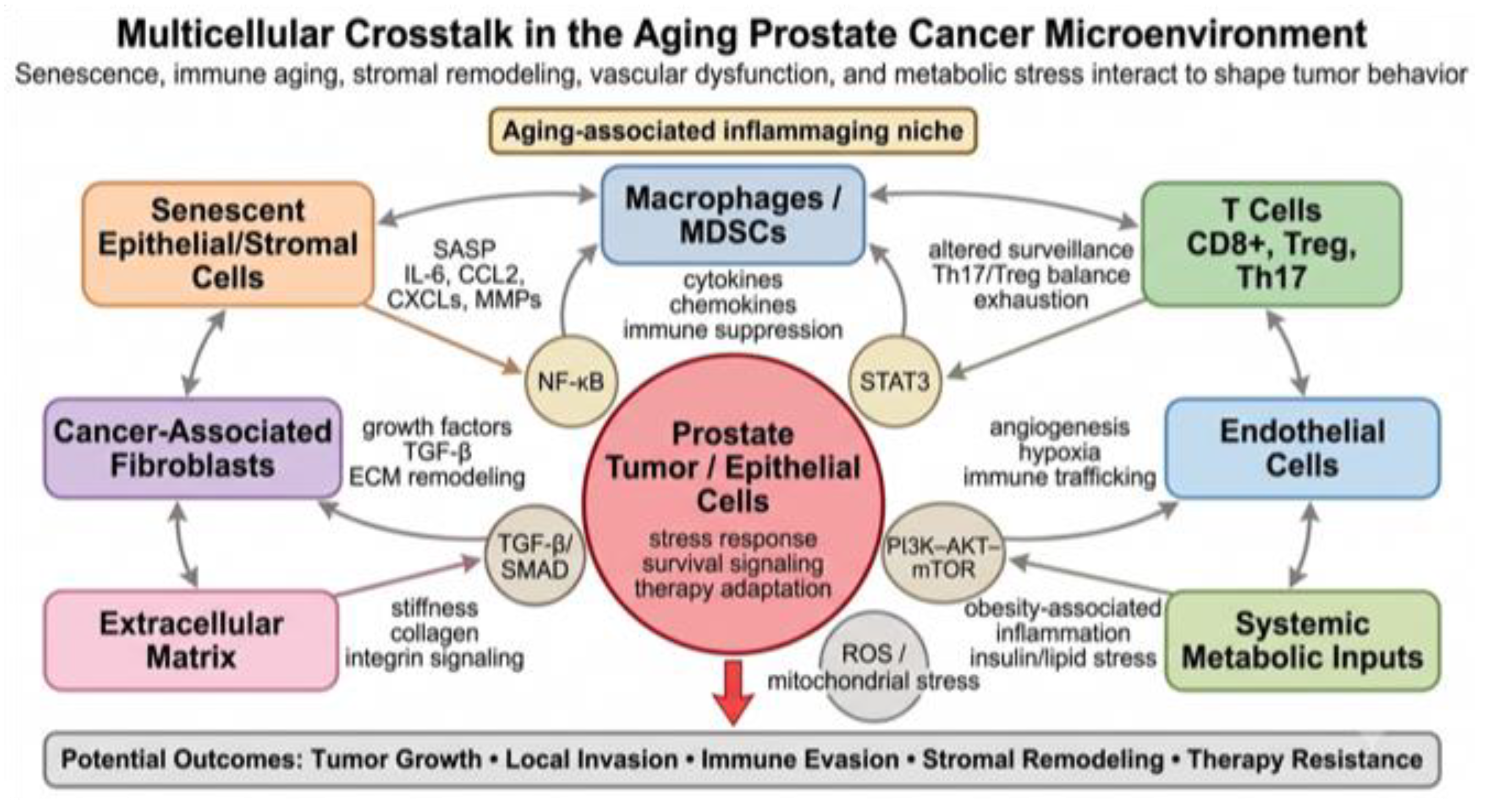
Chronic inflammation can promote early tumorigenesis through several mechanisms. Inflammatory cytokines and ROS can induce DNA damage, epigenetic alterations, and stress-response signaling in epithelial cells. Senescent epithelial and stromal cells can produce SASP factors that alter local growth control, recruit immune cells, and remodel ECM. Inflammatory immune cells can release cytokines, chemokines, proteases, and growth factors that support tissue remodeling and epithelial adaptation. Together, these processes may disturb normal epithelial homeostasis and increase the likelihood that premalignant cells acquire growth advantages [15,19,20,21,22,23,39,40,41,42,43,44,45,46,52,53,54,89,90,91].
The prostate is particularly sensitive to stromal–epithelial communication [55,56,57,103,104,105]. During normal development and adult homeostasis, stromal cells provide signals that regulate epithelial differentiation, androgen responsiveness, and tissue architecture. Aging can disrupt these regulatory interactions. Senescent or activated fibroblasts may produce inflammatory and growth-promoting mediators, while remodeled ECM may alter epithelial polarity, adhesion, and mechanotransduction. These stromal changes may cooperate with epithelial alterations to support early tumor expansion.
Immune surveillance is also critical during early tumor development [24,25,26,27,32,33,34,111,112,113,114]. In younger or immunologically intact tissues, emerging abnormal cells may be recognized and eliminated by cytotoxic T cells, natural killer cells, macrophages, and antigen-presenting cells. Aging may impair these protective responses through reduced antigen presentation, diminished cytotoxic function, T-cell exhaustion-like features, and myeloid-mediated immune suppression [25,26,27,32,34]. Thus, immune aging may allow abnormal epithelial cells to persist while chronic inflammatory signaling simultaneously promotes tissue remodeling.
Inflammaging may also affect early prostate cancer progression by altering the balance between tumor-suppressive and tumor-promoting senescence. Acute senescence in premalignant epithelial cells can limit tumor initiation by enforcing growth arrest. However, persistent accumulation of senescent cells in the surrounding tissue may generate chronic SASP activity that supports non-senescent neighboring cells. This distinction is important because the same biological process—senescence—can restrain or promote early tumor progression depending on duration, cell type, and immune clearance [13,14,17,19,20,85,86,87,88,89,90,91].
Therefore, prostate tumor initiation and early progression should be considered not only as cell-intrinsic genetic events but also as tissue-level processes influenced by aging. The aged microenvironment may provide inflammatory, stromal, metabolic, and immune signals that increase the probability of tumor expansion. This concept highlights the importance of age-appropriate experimental models and human studies that examine both tumor cells and the surrounding aging tissue context [76,77,78].
6.2. Local Invasion and Tumor Progression
As prostate cancer progresses, tumor cells interact more extensively with stromal fibroblasts, immune cells, endothelial cells, ECM, and soluble inflammatory mediators [29,30,31,106,107,108,109,110]. Inflammaging may contribute to local invasion and disease progression by promoting matrix remodeling, fibroblast activation, immune-cell recruitment, angiogenesis, and stress-adaptive signaling. These processes may allow tumor cells to breach normal tissue boundaries, invade surrounding stroma, and adapt to hostile microenvironmental conditions.
ECM remodeling is a major mechanism through which inflammaging may support invasion. Senescent stromal cells, cancer-associated fibroblasts, macrophages, and inflammatory epithelial cells can produce MMPs and other proteases that degrade basement membrane and interstitial matrix components [55]. This remodeling can create physical paths for tumor-cell movement and release matrix-bound growth factors. Changes in collagen deposition, fibronectin expression, crosslinking, and tissue stiffness can also activate mechanotransduction pathways that promote migration, survival, and invasive behavior.
Inflammatory cytokines can directly affect tumor-cell behavior. IL-6, TNF-α, IL-1 family cytokines, IL-17-related signaling, TGF-β, and chemokines may activate pathways such as NF-κB, STAT3, MAPK, SMAD, and PI3K–AKT signaling [29,30,39,40,41,42,43,44,45,46,52,53,54]. These pathways can regulate survival, motility, stress tolerance, and interaction with stromal cells. Although the effects of individual cytokines are context dependent, persistent exposure to inflammatory mediators may help tumor cells adapt to the aging tissue environment.
Myeloid cells are particularly important in invasion-supportive inflammatory niches. Tumor-associated macrophages, inflammatory monocytes, neutrophils, and myeloid-derived suppressor cells can produce cytokines, chemokines, angiogenic mediators, proteases, and ROS [29,31,32,33,34,43]. These cells may support matrix remodeling, tumor-cell survival, vascular remodeling, and immune suppression. Aging-associated myeloid remodeling may therefore amplify tumor-promoting inflammation and support local progression.
Stromal fibroblasts also contribute to local invasion [29,30,55,56,57]. Aging fibroblasts and cancer-associated fibroblasts can produce growth factors, cytokines, ECM proteins, and matrix-remodeling enzymes that alter epithelial behavior. Fibroblast-derived factors may increase epithelial proliferation, motility, survival, and resistance to stress. In addition, fibroblast-mediated matrix remodeling can increase tissue stiffness and create invasion-supportive tracks within the tumor microenvironment.
Endothelial aging and angiogenesis may further support tumor progression. Abnormal vasculature can create hypoxic regions that activate HIF-1α and other stress-response pathways. Hypoxia can promote angiogenesis, inflammatory signaling, metabolic adaptation, and resistance to therapy [61,62,63,64,65,66]. In aged tissues, pre-existing vascular dysfunction may worsen hypoxia and impair immune-cell trafficking, thereby contributing to tumor progression and treatment resistance.
Importantly, local invasion is not caused by inflammation alone. Tumor-intrinsic alterations, androgen receptor signaling changes, tumor suppressor loss, epigenetic remodeling, and therapeutic pressure all contribute to prostate cancer progression [6,128,129,130,131,132,133]. However, inflammaging may provide a permissive microenvironment that supports the consequences of these tumor-intrinsic changes. This tissue-level perspective is important for understanding why similar genetic alterations may lead to different clinical outcomes depending on host age, immune status, metabolic health, and stromal context.
6.3. Immune Evasion and Treatment Resistance
Immune evasion is a central feature of cancer progression and may be strongly influenced by aging. The aged immune system is often characterized by reduced naïve T-cell diversity, impaired cytotoxic function, altered antigen presentation, myeloid skewing, chronic inflammatory activation, and increased immune-suppressive signaling [24,25,26,27,28,32,33,34,36,37,38,111,112,113,114]. These changes can weaken anti-tumor immunity while allowing inflammatory programs that support tumor survival and adaptation.
In prostate cancer, immune evasion can involve multiple mechanisms. Tumors may reduce antigen presentation, recruit suppressive myeloid cells, expand regulatory T-cell activity, exclude effector T cells, increase checkpoint ligand expression, and establish cytokine environments that impair cytotoxic function. Aging may intensify these mechanisms by reducing immune-cell plasticity and promoting chronic suppressive or exhausted immune states. As a result, older tumor microenvironments may contain immune cells that are present but functionally ineffective.
Myeloid-mediated immune suppression is especially relevant. Macrophages and myeloid-derived suppressor cells can inhibit T-cell activation and function through arginine depletion, ROS, nitric oxide, IL-10, TGF-β, prostaglandins, and checkpoint-related pathways [29,31,32,33,34,36,37,38,43]. These suppressive myeloid populations can also promote angiogenesis, tissue remodeling, and resistance to therapy. Aging-associated myeloid skewing may therefore contribute simultaneously to immune evasion and tumor progression.
T-cell dysfunction may also limit therapeutic response. Aging and chronic antigen exposure can increase exhaustion-like features in T cells, including impaired proliferation, reduced cytokine production, and altered expression of inhibitory receptors. In prostate cancer, limited responsiveness to immune checkpoint blockade in unselected patients may reflect multiple barriers, including low tumor immunogenicity, immunosuppressive myeloid cells, stromal exclusion, and insufficient effector T-cell function [32,33,34,36,37,38,134,135,136,137,138]. Inflammaging may add another layer of resistance by creating a chronically inflamed yet poorly cytotoxic immune environment.
Inflammatory signaling can also affect response to androgen receptor-directed therapies, radiation, chemotherapy, and targeted treatments. Cytokines such as IL-6, TNF-α, TGF-β, and IL-17-related mediators can activate survival and stress-response pathways that may help tumor cells withstand therapy [81,82,93,94,98,99,100,101]. Senescent tumor or stromal cells generated by treatment may also produce SASP factors that alter the local microenvironment. While therapy-induced senescence can contribute to growth arrest, persistent senescent cells may support inflammatory adaptation and residual disease under some conditions.
Stromal and vascular aging may further modify treatment response. Dense ECM can impair drug penetration and immune-cell infiltration. Hypoxia can reduce radiation sensitivity and promote adaptive stress responses. Endothelial dysfunction can alter perfusion and tissue repair. Metabolic dysfunction can influence both tumor-cell survival and immune-cell activity. Therefore, therapy resistance in aging-associated prostate cancer may arise from interactions among tumor cells, senescent cells, immune populations, stromal architecture, vasculature, and systemic metabolic status [58,59,60,61,62,63,64,65,66,68,71,115,116,117].
These observations suggest that effective treatment of prostate cancer in older patients may require more than targeting tumor cells alone. Therapeutic strategies that modulate inflammatory cytokines, suppress harmful SASP activity, reprogram myeloid cells, improve immune surveillance, normalize stromal barriers, or correct metabolic inflammation may enhance response to conventional therapies [32,33,34,36,37,38,95,96,97,98,99,100,101,134,135,136,137,138]. However, these approaches require careful development because inflammation and senescence can also have protective roles in tissue repair and tumor suppression.
6.4. Phenotypic Adaptation Under Inflammatory and Therapeutic Stress
Prostate cancer cells can adapt to changing microenvironmental and therapeutic conditions by altering signaling pathways, differentiation state, metabolic programs, and stress-response mechanisms [15,19,20,21,22,23,81,82,93,94,98]. Chronic inflammatory signaling may contribute to this adaptation by activating pathways involved in survival, repair, motility, immune interaction, and treatment tolerance. In the aging prostate microenvironment, persistent cytokine exposure, oxidative stress, stromal remodeling, hypoxia, and metabolic dysfunction may collectively create selective pressures that favor more adaptable tumor-cell states.
Inflammatory pathways such as NF-κB, STAT3, MAPK, PI3K–AKT, and TGF-β/SMAD signaling can regulate cell survival, stress adaptation, and epithelial behavior [29,68,79,80,92,104,105,126]. These pathways can be activated by cytokines, chemokines, growth factors, oxidative stress, matrix signaling, and therapy-induced injury. Persistent activation may allow tumor cells to survive inflammatory stress and adapt to treatment. However, the effects of these pathways are highly context dependent and may differ according to tumor genotype, stage, androgen receptor status, immune composition, and treatment history.
Therapeutic stress can also modify the tumor microenvironment. Androgen deprivation therapy, androgen receptor pathway inhibitors, radiation, chemotherapy, and other treatments can induce cellular stress, inflammation, senescence, vascular changes, and immune remodeling [37,81,82,93,94,98,99,100,101]. These therapy-induced changes may be beneficial when they enhance tumor-cell death or immune recognition. However, they may also contribute to residual inflammatory niches that support surviving tumor cells. In older tissues, pre-existing inflammaging may influence how the microenvironment responds to therapy-induced injury.
Chronic inflammation has been associated with altered epithelial differentiation, survival signaling, migratory behavior, and treatment response in multiple cancers. In prostate cancer, inflammatory and stromal signals may contribute to disease progression and resistance, but the precise mechanisms linking aging-associated inflammation to specific aggressive phenotypes remain under active investigation. Thus, inflammaging is best viewed as a permissive tissue context that supports tumor-cell adaptation rather than as a single deterministic pathway [6,32,33,34,36,37,38,128,129,130,131,132,133].
Future studies should determine how inflammatory and therapeutic stress interact in aged prostate tissues [6,34,38,128,129,130,131,132,133]. Key questions include which cell types produce therapy-induced inflammatory mediators, how senescent cells persist or are cleared after treatment, how aged immune cells respond to tumor injury, and whether biological aging markers predict treatment response. Answering these questions may help identify patients whose tumors are shaped by inflammaging and may benefit from microenvironment-targeted therapeutic strategies [32,33,34,95,96,97,98,99,100,101,134,135,136,137,138].
Section Summary
Inflammaging may influence prostate cancer initiation, local invasion, immune evasion, therapy resistance, and tumor-cell adaptation. These effects arise from interactions among senescent cells, cytokines, immune dysfunction, myeloid remodeling, stromal activation, ECM remodeling, vascular dysfunction, metabolic stress, and tumor intrinsic alterations. Defining these multicellular interactions will be essential for biomarkers and interventions targeting aging-associated prostate cancer progression.
7. Therapeutic Implications of Targeting Inflammaging in Prostate Cancer
7.1. Cytokine-Targeted Approaches
Because inflammaging is characterized by persistent cytokine and chemokine signaling, inflammatory pathways represent potential therapeutic targets in aging-associated prostate cancer. Cytokines such as IL-6, IL-17, IL-23, TNF-α, IL-1 family members, and TGF-β can influence immune-cell recruitment, stromal remodeling, epithelial stress responses, angiogenesis, and treatment resistance [32,33,34,39,40,41,42,43,44,45,46,52,53,54]. These pathways are not specific to aging, but they may become increasingly relevant in older tissues where chronic inflammatory tone, senescent-cell burden, immune dysfunction, and metabolic stress are elevated.
The IL-6/JAK/STAT3 pathway is one of the most extensively studied inflammatory signaling axes in cancer. IL-6 can be produced by tumor cells, stromal fibroblasts, macrophages, senescent cells, adipose-associated cells, and other immune populations. In prostate cancer, IL-6-related signaling has been linked to tumor-cell survival, inflammatory adaptation, angiogenesis, androgen receptor pathway interactions, and resistance to therapy [29,30,32,33,34,36,37,38,92]. Because IL-6 is also a common SASP component, it may connect cellular senescence with tumor-promoting inflammation. Therapeutic strategies targeting IL-6, IL-6 receptor, JAK kinases, or STAT3 may therefore have relevance in inflammatory prostate tumor microenvironments, although patient selection and toxicity remain important considerations.
IL-17- and IL-23-related signaling may also represent therapeutic opportunities. IL-17 can activate NF-κB, MAPK, chemokine production, and stromal inflammatory programs in multiple cell types. IL-23 supports the maintenance of Th17-associated immune responses and may contribute to chronic tissue inflammation [39,40,41,42,43,44,45,46,54]. In cancer, the IL-17/IL-23 axis has context-dependent roles, with both tumor-promoting and immune-regulatory functions described depending on tissue type and disease stage. In prostate cancer, targeting this pathway may be most relevant in tumors or patient subsets with clear evidence of IL-17/IL-23-associated inflammatory activity. However, because these pathways also participate in host defense and mucosal immunity, therapeutic modulation would require careful biomarker-guided evaluation.
TNF-α, IL-1, and NF-κB-associated inflammatory pathways are also central components of chronic inflammation. TNF-α and IL-1 family cytokines can promote cytokine cascades, chemokine production, myeloid recruitment, endothelial activation, and stromal remodeling [8,9,11,79,80,92]. NF-κB functions as a major transcriptional hub integrating signals from cytokines, pattern-recognition receptors, oxidative stress, DNA damage, and senescence-associated mediators. In aging tissues, persistent NF-κB activity may help sustain inflammaging. Therapeutically, broad inhibition of these pathways may be limited by systemic toxicity and immune-suppressive effects, but selective targeting of dominant inflammatory circuits may be useful in defined biological contexts.
TGF-β signaling represents another important but complex therapeutic target. TGF-β can suppress epithelial proliferation in some contexts, but it can also promote immune suppression, fibroblast activation, ECM remodeling, and invasive behavior in established tumors [29,104,105,108,110]. In aged tissues, TGF-β may contribute to fibrosis, stromal remodeling, and immune exclusion. Targeting TGF-β signaling may therefore have potential in stromal-rich or immune-excluded prostate tumors, but the dual functions of TGF-β require careful consideration.
Overall, cytokine-targeted therapy in prostate cancer should not be approached as a uniform anti-inflammatory strategy. Inflammation can be protective or harmful depending on timing, location, cell type, and immune context. The goal should be to identify dominant tumor-promoting inflammatory circuits while preserving effective anti-tumor immunity and normal tissue repair. Biomarkers such as circulating cytokines, tissue cytokine signatures, immune-cell composition, SASP markers, and spatial immune–stromal organization may help identify patients most likely to benefit from cytokine-targeted approaches [6,34,38,128,129,130,131,132,133].
7.2. Senescence-Directed Therapies
Cellular senescence is a major contributor to aging-associated tissue inflammation, making senescence-directed therapy an attractive strategy for modifying the aged tumor microenvironment. Two broad approaches have emerged: senolytics, which aim to selectively eliminate senescent cells, and senomorphics, which aim to suppress harmful senescence-associated secretory programs without necessarily killing senescent cells [95,96,97,98,99,100,101]. These strategies may be relevant to prostate cancer because senescent epithelial, stromal, endothelial, immune, or therapy-induced tumor cells may contribute to chronic inflammatory signaling and tissue remodeling.
Senolytic strategies are based on the concept that senescent cells depend on specific pro-survival pathways that allow them to resist apoptosis. By targeting these senescence-associated survival networks, senolytics may reduce senescent-cell burden and decrease SASP-mediated inflammation [95,96,97,98,99]. Several classes of senolytic agents have been studied in preclinical aging and disease models, including BCL-2 family inhibitors, dasatinib plus quercetin, and other agents targeting senescent-cell vulnerabilities. In cancer, senolytics may be especially relevant after therapies that induce senescence, because persistent senescent cells may contribute to residual inflammatory niches.
Senomorphic approaches aim to reduce harmful SASP activity while preserving beneficial aspects of senescence, such as tumor suppression and tissue repair. Pathways such as mTOR, JAK/STAT, p38 MAPK, NF-κB, cGAS–STING, and inflammasome signaling have been implicated in SASP regulation [79,80,92,95,96,97,98,99,100,101]. Modulating these pathways may reduce inflammatory cytokine production, chemokine release, and matrix remodeling. In prostate cancer, senomorphic strategies may be useful in older patients with high inflammatory or senescence-associated signatures, but additional work is needed to identify which SASP components are most clinically relevant.
A major challenge is that senescence has both beneficial and harmful roles. Acute senescence can prevent malignant transformation and contribute to therapeutic response by enforcing growth arrest in damaged cells. Eliminating or suppressing senescent cells indiscriminately could interfere with tumor suppression, wound healing, immune clearance, or normal tissue repair. Conversely, allowing persistent senescent cells to remain may promote chronic inflammation and tumor-supportive remodeling [13,14,17,19,85,86,87,88,89,90,91]. Therefore, the timing, cell type, and disease stage of senescence-directed therapy are critical.
In prostate cancer, senescence-directed therapy may have several potential applications. It could be used to reduce age-associated inflammatory burden before or during cancer progression, to suppress harmful SASP after radiation or systemic therapy, or to improve the tumor microenvironment for immune-based treatments [81,82,93,94,95,96,97,98,99,100,101]. However, these strategies remain investigational and require careful preclinical validation in age-appropriate models. It will be important to determine whether senescent cells in prostate cancer are primarily located in tumor cells, stromal fibroblasts, endothelial cells, immune cells, or surrounding benign tissue, because the therapeutic implications may differ substantially.
Biomarker development will be essential for senescence-directed approaches. Potential biomarkers include p16INK4a, p21CIP1, DNA damage markers, senescence-associated β-galactosidase activity, SASP cytokines, senescence-related transcriptomic signatures, spatial localization of senescent cells, and circulating inflammatory mediators [13,14,18,38,81,82,93,94]. Because no single marker definitively identifies senescence in all contexts, combined biomarker panels will likely be needed. Spatial profiling may be especially useful for determining whether senescent cells are located in epithelial, stromal, vascular, or immune compartments.
7.3. Metabolic and Lifestyle Interventions
Metabolic stress is closely linked to inflammaging, and interventions that improve metabolic health may reduce chronic inflammatory burden. In older men, obesity, insulin resistance, metabolic syndrome, mitochondrial dysfunction, altered lipid metabolism, and systemic inflammation may influence prostate cancer biology [67,68,69,70,71,72,73,74,75,118,119,120,121,122,123,125,126]. Therefore, metabolic and lifestyle interventions may have relevance not only for general health but also for modifying the tumor-promoting effects of the aging microenvironment.
Exercise is one of the most broadly beneficial interventions for aging-associated metabolic and inflammatory dysfunction. Regular physical activity can improve insulin sensitivity, reduce adipose inflammation, enhance mitochondrial function, modulate immune-cell activity, improve vascular health, and reduce systemic inflammatory markers [67,73,118,119,120,121,122]. In prostate cancer patients, exercise may also improve fatigue, muscle mass, metabolic health, cardiovascular function, and quality of life during treatment. Although exercise should not be presented as a stand-alone cancer therapy, it may help reduce systemic conditions that contribute to inflammaging.
Weight management and dietary interventions may also influence aging-associated inflammation. Obesity is associated with adipose tissue inflammation, altered adipokines, insulin resistance, lipid dysregulation, and increased circulating cytokines. Reducing excess adiposity may lower systemic inflammatory tone and improve metabolic health [67,73,118,119,120,121,122]. Dietary patterns that improve cardiometabolic function may also affect inflammatory mediators, gut microbiota, lipid metabolism, and oxidative stress. However, dietary interventions vary widely, and more prostate cancer-specific studies are needed to define which approaches are most effective and biologically relevant.
Metformin has received attention because of its effects on insulin sensitivity, AMPK activation, mitochondrial metabolism, and inflammatory signaling [68,71]. Epidemiological and preclinical studies have explored whether metformin may influence cancer risk, progression, or treatment response, including in prostate cancer. However, clinical findings have been mixed, and effects may depend on patient metabolic status, tumor biology, treatment context, and study design. In the context of aging-associated prostate cancer, metformin is best discussed as a potential metabolic and inflammatory modulator rather than as an established prostate cancer therapy.
Nutrient-sensing pathways such as mTOR, AMPK, insulin/IGF signaling, autophagy, and nuclear receptor pathways may provide additional therapeutic opportunities. mTOR inhibition can modulate growth signaling, metabolism, autophagy, and SASP activity. AMPK activation may improve energy homeostasis and reduce inflammatory signaling in some contexts. PPAR pathway modulation may influence lipid metabolism, insulin sensitivity, macrophage function, and inflammatory tone [1,5,68,71,92,126]. However, these pathways are active in normal tissues, immune cells, stromal cells, and tumor cells, so systemic targeting may produce complex effects.
Lifestyle and metabolic interventions may be most effective when integrated into biomarker-guided strategies [6,34,38,128,129,130,131,132,133]. For example, patients with obesity, insulin resistance, elevated inflammatory cytokines, high senescence burden, or specific metabolic signatures may be more likely to benefit from interventions targeting metabolic inflammaging. Future studies should integrate clinical metabolic data with tissue immune profiling, serum cytokines, metabolomics, lipidomics, and treatment outcomes. Such approaches may help determine whether metabolic health modifies prostate cancer progression through measurable changes in the tumor microenvironment.
7.4. Biomarker-Guided Patient Stratification
A major challenge in targeting inflammaging is patient heterogeneity [6,34,38,128,129,130,131,132,133]. Chronological age alone is unlikely to identify patients with biologically aged or inflamed prostate tumor microenvironments. Two men of the same age may differ substantially in immune function, senescence burden, metabolic health, stromal remodeling, systemic inflammation, and treatment tolerance. Therefore, biomarker-guided stratification will be essential for translating inflammaging biology into prostate cancer prevention and therapy.
Potential biomarkers of inflammaging include circulating cytokines, chemokines, SASP factors, immune-cell subsets, myeloid signatures, Th17/Treg balance, senescence-associated markers, metabolic indicators, and tissue-level inflammatory signatures [6,34,38,128,129,130,131,132,133]. Serum or plasma markers may provide minimally invasive information about systemic inflammation, but they may not fully capture the local prostate tumor microenvironment. Tissue-based biomarkers may provide more direct information about senescent cells, immune infiltration, stromal remodeling, and spatial organization, but they require biopsy or surgical specimens.
Multiplex immunofluorescence, spatial transcriptomics, single-cell RNA sequencing, proteomics, and metabolomics can help define inflammaging-associated tumor microenvironment states [6,34,38,128,129,130,131,132,133]. These approaches may reveal whether inflammatory signals originate from senescent stromal cells, myeloid cells, T-cell subsets, endothelial cells, tumor cells, or surrounding benign tissue. Spatial approaches are particularly important because the biological effect of inflammatory cells may depend on their proximity to tumor epithelium, stromal barriers, blood vessels, or immune-suppressive niches.
Biomarkers may also help identify therapeutic vulnerabilities [34,38,95,96,97,98,99,100,101,134,135,136,137,138]. Tumors with high IL-6/STAT3 signaling may be candidates for inflammatory pathway modulation. Tumors with strong SASP signatures may be candidates for senescence-directed strategies. Tumors with myeloid-rich immunosuppressive environments may require myeloid reprogramming or combination immunotherapy. Patients with metabolic inflammation may benefit from metabolic intervention combined with standard therapy. However, these possibilities require prospective validation.
Another important goal is distinguishing biological aging from chronological age. Biological aging biomarkers may include epigenetic clocks, inflammatory signatures, immune-cell composition, senescence markers, metabolic profiles, and functional measures of tissue resilience [1,5,8,24,25,26,27,28,111,112,113,114]. In prostate cancer, integrating these markers with tumor grade, stage, genomic alterations, treatment history, and clinical comorbidities may improve risk stratification. This approach may be especially valuable for older patients, who are often underrepresented in clinical trials and may have heterogeneous treatment tolerance.
Ultimately, biomarker-guided strategies should aim to identify which patients have tumors shaped by inflammaging and which specific components of inflammaging are therapeutically actionable [6,34,38,128,129,130,131,132,133]. This will require integration of clinical data, biospecimen analysis, spatial biology, and age-appropriate preclinical modeling.
7.5. Combination Strategies and Clinical Translation
Because inflammaging involves multiple interacting cell types and pathways, single-agent targeting may be insufficient in many cases. Rational combination strategies may be needed to modify the inflammatory tumor microenvironment while also controlling tumor-cell growth. Potential combinations include cytokine modulation with androgen receptor-directed therapy, senescence-directed therapy with radiation or systemic therapy, metabolic intervention with immune modulation, and stromal-targeted approaches with conventional treatment [32,33,34,36,37,38,95,96,97,98,99,100,101,134,135,136,137,138]. However, combination therapy must be designed carefully to avoid excessive toxicity or impairment of protective immune functions.
Inflammation-targeted therapies may enhance response to standard prostate cancer treatments by reducing survival signals, stromal barriers, immune suppression, or SASP-mediated adaptation. For example, modulating IL-6/JAK/STAT3, NF-κB-related inflammation, TGF-β signaling, or myeloid suppressive pathways could theoretically improve therapeutic sensitivity in selected tumors. Similarly, senescence-directed therapies may help reduce persistent inflammatory niches after therapy-induced senescence. However, the timing of intervention is likely critical: eliminating senescent cells too early may interfere with tumor-suppressive senescence, whereas targeting persistent senescent cells after therapy may be more beneficial [81,82,93,94,95,96,97,98,99,100,101].
Immunotherapy combinations are also of interest. Prostate cancer has generally shown limited response to immune checkpoint blockade in unselected patients, likely due to multiple factors including low tumor immunogenicity, myeloid suppression, stromal exclusion, and insufficient effector T-cell activity [32,33,34,36,37,38,134,135,136,137,138]. Inflammaging may further complicate this landscape by producing chronic inflammation without effective cytotoxic immunity. Strategies that reduce suppressive myeloid cells, improve antigen presentation, modulate cytokine networks, or normalize stromal barriers may help create a more permissive environment for immunotherapy in selected patients.
Clinical translation will require careful patient selection, appropriate endpoints, and age-aware trial design. Older patients often have comorbidities, altered pharmacology, different immune function, and variable tolerance to combination therapy. Trials targeting inflammaging should consider biological age, metabolic health, inflammatory biomarkers, immune status, and functional reserve, rather than relying solely on chronological age [1,5,24,25,26,27,28,111,112,113,114]. Safety will be particularly important because inflammatory and senescence pathways participate in host defense, wound healing, and tissue repair.
Age-appropriate preclinical models are also needed. Many prostate cancer studies use young animals, which may not reflect the aged immune, stromal, vascular, and metabolic environments of human disease [76,77]. Testing inflammaging-targeted therapies in aged models, obesity-associated models, senescence-enriched settings, and human tissue-based systems may improve translational relevance. Organoids, explants, co-culture systems, and spatial profiling of human specimens may also help bridge the gap between mechanistic studies and clinical application.
In summary, therapeutic targeting of inflammaging in prostate cancer is promising but complex. The goal should not be broad suppression of inflammation, but precise modulation of harmful aging-associated inflammatory circuits while preserving protective immunity and tissue homeostasis. Biomarker-guided patient selection, cell-type-specific mechanistic studies, and age-appropriate models will be essential for developing safe and effective therapies.
Section Summary
Targeting inflammaging offers therapeutic opportunities in prostate cancer, including cytokine modulation, senescence-directed therapy, metabolic intervention, immune remodeling, and biomarker-guided treatment selection. Because inflammaging includes both protective and harmful processes, successful translation will require identifying dominant inflammatory circuits and tailoring combinations to patient age, immune status, metabolic health, and tumor microenvironment composition (Figure 3) (Table 5).
8. Knowledge Gaps and Future Directions
8.1. Distinguishing Chronological Age from Biological Aging
A major challenge in studying aging-associated prostate cancer is distinguishing chronological age from biological aging. Chronological age is easy to measure, but it does not fully capture the molecular, cellular, immune, metabolic, and functional changes that occur during aging. Individuals of the same chronological age may have very different levels of cellular senescence, immune dysfunction, metabolic stress, systemic inflammation, vascular aging, and tissue repair capacity [1,5,8,24,25,26,27,28,111,112,113,114]. Therefore, simply comparing younger and older patients may overlook important biological heterogeneity.
Biological aging reflects the cumulative functional decline of cells, tissues, and organ systems. In the prostate tumor microenvironment, biological aging may include increased senescent-cell burden, altered SASP activity, myeloid remodeling, T-cell dysfunction, stromal activation, ECM remodeling, mitochondrial stress, and systemic metabolic inflammation. These processes may occur at different rates among individuals and may be influenced by genetics, lifestyle, obesity, comorbidities, environmental exposures, microbiota, and prior therapies.
Future studies should incorporate biomarkers of biological aging into prostate cancer research. Potential biomarkers include epigenetic aging clocks, senescence-associated markers, inflammatory cytokine profiles, immune-cell composition, metabolic signatures, mitochondrial function, and tissue-level measures of stromal remodeling. These biomarkers may help identify patients whose tumors are developing within biologically aged or inflamed microenvironments, even when chronological age alone is not informative.
Distinguishing chronological age from biological aging may also improve clinical decision-making. Older patients with low inflammatory burden and preserved immune function may respond differently to therapy than younger patients with obesity-associated inflammation, high senescence burden, or metabolic dysfunction. Therefore, biological aging markers could eventually help guide risk stratification, treatment selection, and supportive care strategies in prostate cancer.
8.2. Need for Age-Appropriate Experimental Models
Many preclinical prostate cancer studies are performed in young animals or rapidly growing tumor models. While these systems are useful for defining tumor-intrinsic mechanisms, they may not accurately represent the aged tissue environment in which human prostate cancer most commonly develops. Young animals often lack the immune remodeling, stromal aging, senescence burden, vascular dysfunction, and metabolic alterations characteristic of older hosts. This limitation may partly explain why some therapies that appear effective in preclinical models show limited benefit in older or clinically heterogeneous patient populations.
Age-appropriate models are needed to understand how the aged prostate microenvironment influences tumor initiation, progression, immune evasion, and treatment response. A spatially and temporally controlled Pten-null prostate cancer mouse model generated at different adult ages provides direct evidence that host age can accelerate PI3K/AKT/mTOR signaling and prostate cancer onset and progression [76]. Such models should consider not only tumor genotype but also host age, immune status, stromal context, metabolic state, and treatment timing. Comparing tumors that arise in young versus aged hosts may help separate tumor-intrinsic effects from microenvironmental effects. In addition, age-defined models may clarify how senescence, inflammaging, and immune dysfunction influence prostate cancer biology over time.
Modeling aging is technically challenging because aged animal studies require longer timelines, higher cost, larger cohorts, and careful control of comorbidities. Aging animals may also show increased variability, which can complicate interpretation. Nevertheless, these models are essential for studying aging-associated prostate cancer. Whenever possible, studies should include both young and aged cohorts, sex- and strain-appropriate controls, longitudinal analysis, and careful evaluation of immune, stromal, and metabolic features.
In addition to animal models, human tissue-based systems can improve translational relevance. Patient-derived organoids, prostate tissue explants, stromal–epithelial co-cultures, immune-cell co-cultures, and three-dimensional ECM systems may help model specific components of the aging microenvironment. However, these systems should ideally incorporate aged stromal cells, aged immune cells, senescent-cell components, or metabolic stress conditions when studying aging-related questions. Combining age-appropriate animal models with human tissue systems may provide a more complete understanding of inflammaging in prostate cancer.
8.3. Defining Cell-Type-Specific Mechanisms
Inflammaging is a multicellular process, and one of the major knowledge gaps is identifying which cell types initiate, maintain, and respond to aging-associated inflammatory signals in prostate cancer. Senescent epithelial cells, stromal fibroblasts, macrophages, myeloid-derived suppressor cells, T-cell subsets, endothelial cells, adipose-associated cells, and tumor cells may all contribute to the inflammatory microenvironment. However, their relative contributions likely vary by disease stage, tumor genotype, treatment exposure, metabolic status, and host age.
Cell-type-specific analysis is important because the same inflammatory mediator may have different effects depending on its source and target cell [29,30,34,38]. For example, cytokines produced by senescent stromal cells may influence epithelial growth and matrix remodeling, while cytokines produced by myeloid cells may affect T-cell suppression and immune-cell recruitment. Similarly, inflammatory signaling within tumor cells may promote survival or stress adaptation, whereas inflammatory signaling within immune cells may alter anti-tumor function. Without cell-type resolution, it is difficult to determine which pathways are therapeutically actionable.
Future studies should use single-cell RNA sequencing, single-cell ATAC-seq, spatial transcriptomics, multiplex immunofluorescence, imaging mass cytometry, proteomics, and functional co-culture assays to map aging-associated cell states in prostate cancer. These approaches can help identify senescent-cell populations, inflammatory immune niches, activated stromal subsets, vascular changes, and tumor-cell responses. Importantly, spatial information is essential because cell proximity often determines biological function. For example, inflammatory myeloid cells located near tumor epithelium may have different effects from those located in stromal or perivascular regions.
Cell-type-specific studies should also examine dynamic changes over time. Inflammaging may influence early tumor initiation differently from advanced disease or therapy-resistant states. Similarly, senescence may be protective at one stage and harmful at another. Longitudinal models and serial human biospecimens may help determine how aging-associated inflammatory niches evolve during prostate cancer progression and treatment.
8.4. Integrating Multi-Omics and Spatial Biology
The complexity of inflammaging requires integrated analytical approaches. No single assay can fully capture the interactions among senescence, immune aging, stromal remodeling, metabolic stress, and tumor-cell adaptation. Multi-omics approaches, including transcriptomics, proteomics, metabolomics, lipidomics, epigenomics, and spatial profiling, can provide complementary information about aging-associated prostate cancer biology [34,38,128,129,130,131,132,133].
Transcriptomic profiling can identify inflammatory signatures, immune-cell states, senescence-associated programs, and stromal activation patterns. Proteomics can reveal changes in cytokines, signaling proteins, ECM components, and secreted factors. Metabolomics and lipidomics can define systemic and local metabolic alterations associated with aging, obesity, mitochondrial dysfunction, or inflammatory metabolism. Epigenomic profiling can identify stable regulatory changes that shape immune-cell dysfunction, stromal activation, or epithelial stress responses.
Spatial biology is particularly valuable because the tumor microenvironment is organized into localized niches. Senescent stromal cells, inflammatory macrophages, T-cell subsets, blood vessels, ECM barriers, and tumor epithelial cells may form distinct spatial arrangements that influence disease behavior. Spatial transcriptomics and multiplex imaging can help determine whether inflammaging-associated signals are distributed broadly throughout tissue or concentrated in specific tumor regions. Such information may be critical for identifying biologically meaningful biomarkers.
Integrating multi-omics data with clinical variables will be essential. Patient age, race, body mass index, metabolic status, comorbidities, tumor grade, stage, treatment history, and outcomes should be analyzed together with tissue and blood-based molecular data. This integration may help define clinically relevant inflammaging subtypes of prostate cancer. Such subtypes could eventually guide patient stratification, therapeutic selection, and biomarker development.
However, multi-omics studies also require careful design. Aging, obesity, treatment exposure, tumor grade, and tissue composition can all confound interpretation. Studies should include adequately powered cohorts, appropriate controls, validation datasets, and functional follow-up experiments. Without functional validation, omics signatures may remain descriptive. Therefore, future research should combine discovery-based profiling with mechanistic studies in age-appropriate models.
8.5. Translating Inflammaging Biology into Prevention and Therapy
A major goal of inflammaging research is to translate biological insights into improved prevention, diagnosis, and treatment. Because prostate cancer often progresses slowly and is common in older men, interventions that reduce harmful aging-associated inflammation may have potential to delay disease progression or improve treatment response. However, translation requires careful attention to safety, timing, patient selection, and biological context [32,33,34,36,37,38,95,96,97,98,99,100,101,134,135,136,137,138].
One important opportunity is prevention or risk reduction. If specific inflammatory, senescent, stromal, or metabolic signatures are associated with aggressive prostate cancer, they may help identify patients at higher risk of progression. Lifestyle interventions, metabolic optimization, anti-inflammatory approaches, or senescence-targeted strategies could potentially be evaluated in selected populations. However, such strategies must be evidence-based and should avoid broad suppression of inflammation, because immune responses are also necessary for host defense and tumor surveillance.
Another opportunity is improving treatment response. Inflammaging-targeted approaches may be most useful when combined with standard prostate cancer therapies. For example, reducing harmful SASP activity, reprogramming suppressive myeloid cells, improving immune-cell function, modifying stromal barriers, or correcting metabolic inflammation could potentially enhance response to androgen receptor-directed therapy, radiation, chemotherapy, or immunotherapy. These possibilities require rigorous preclinical and clinical validation.
Clinical translation will require biomarkers that identify patients most likely to benefit. Candidate biomarkers include circulating cytokines, immune-cell profiles, SASP signatures, myeloid markers, senescence-associated tissue markers, stromal signatures, metabolic profiles, and spatial immune–stromal organization. Biomarker-guided clinical trials may be especially important because inflammaging is heterogeneous and may not be present in all older patients or all prostate tumors.
Safety is a major consideration. Senescence, inflammation, and immune activation can be protective in certain contexts. Senescence can suppress tumor initiation, inflammation can support tissue repair and immune surveillance, and immune activation can promote tumor control. Therefore, therapies targeting inflammaging must avoid eliminating beneficial responses. The goal should be precise modulation of harmful chronic inflammation rather than broad immune suppression.
8.6. Future Conceptual Framework
Future studies should view prostate cancer as a disease shaped by both tumor-intrinsic alterations and host tissue aging. Genetic and epigenetic changes within tumor cells are essential drivers of prostate cancer, but the aged microenvironment may determine how these tumor cells behave, how they interact with immune and stromal compartments, and how they respond to therapy. This framework emphasizes the importance of studying the tumor and host together [1,5,6,8,28,30].
A useful future model is that biological aging creates a permissive prostate microenvironment through senescence, SASP activity, immune dysfunction, stromal remodeling, vascular aging, and metabolic stress. These processes do not act independently; they interact to generate chronic inflammatory niches. Within these niches, prostate tumor cells may receive signals that support survival, growth, immune evasion, and treatment adaptation. The specific outcome likely depends on tumor genotype, stage, therapy exposure, host immune status, and systemic metabolic health.
This framework also suggests that aging-associated prostate cancer may not represent one disease state. Instead, different patients may have distinct inflammaging phenotypes. Some may be dominated by senescent stromal inflammation, others by myeloid suppression, others by metabolic inflammation, and others by vascular or matrix remodeling. Defining these phenotypes could help identify personalized strategies for prevention and therapy.
Importantly, this review does not propose that inflammaging is the only driver of prostate cancer progression. Rather, inflammaging should be considered one important biological context that interacts with established tumor-intrinsic mechanisms. Understanding this interaction may help explain why prostate cancer behavior varies among patients and why some tumors become aggressive or treatment resistant despite similar clinical features.
Section Summary
Major knowledge gaps remain in understanding how aging-associated inflammation shapes prostate cancer. Future studies should distinguish chronological from biological aging, use age-appropriate models, define cell-type-specific mechanisms, integrate multi-omics and spatial biology, and develop biomarker-guided translational strategies. Addressing these gaps may reveal new ways to prevent or delay aggressive prostate cancer progression in older men.
9. Conclusions
Prostate cancer is strongly associated with aging, but the biological relationship between aging and prostate cancer progression extends beyond chronological time and accumulated genetic alterations. Aging reshapes the prostate tissue environment through cellular senescence, chronic low-grade inflammation, immune dysfunction, stromal remodeling, ECM alteration, vascular changes, metabolic stress, and impaired tissue repair [1,5,6,8]. These interconnected processes create an inflammaging-associated microenvironment that may influence tumor initiation, local progression, immune evasion, treatment response, and disease outcome.
Cellular senescence is a central component of this aging-associated microenvironment. While senescence can function as a tumor-suppressive mechanism by preventing proliferation of damaged cells, persistent accumulation of senescent epithelial, stromal, endothelial, and immune cells may promote chronic inflammation through the senescence-associated secretory phenotype. SASP factors, including inflammatory cytokines, chemokines, growth factors, matrix-remodeling enzymes, and extracellular vesicles, can alter stromal–epithelial communication, recruit immune cells, remodel ECM, and sustain tissue inflammation. Therefore, the biological consequences of senescence in prostate cancer depend strongly on timing, cell type, immune clearance, and tissue context.
Immune aging further contributes to prostate cancer-associated inflammaging by weakening anti-tumor surveillance while promoting chronic inflammatory signaling. Age-associated changes in T-cell diversity, T-helper cell balance, regulatory T-cell function, myeloid-cell activity, antigen presentation, and cytokine networks may create a tumor microenvironment that is inflamed but not effectively tumoricidal. Th17/Treg imbalance, IL-17/IL-23-related inflammation, myeloid remodeling, and persistent NF-κB and STAT3 signaling represent important mechanisms through which immune aging may affect prostate cancer biology. However, these pathways should be understood as parts of a complex multicellular network rather than as isolated drivers.
Stromal and metabolic aging add additional layers of regulation. Aging fibroblasts, cancer-associated fibroblasts, remodeled ECM, endothelial dysfunction, mitochondrial stress, oxidative injury, obesity-associated inflammation, lipid dysregulation, and altered nutrient-sensing pathways can all contribute to chronic inflammation and tumor microenvironment remodeling. These factors may affect tumor growth, invasion, immune-cell trafficking, angiogenesis, and therapeutic response. Importantly, systemic metabolic health and local prostate tissue aging may interact, suggesting that prostate cancer progression in older men is shaped by both local and organismal aging processes.
Therapeutically, inflammaging presents both opportunities and challenges. Cytokine-targeted approaches, senescence-directed therapies, metabolic interventions, stromal modulation, myeloid reprogramming, and biomarker-guided immune strategies may eventually help reduce harmful aging-associated inflammation in selected patients. However, inflammation and senescence can also have protective roles in immune defense, tissue repair, and tumor suppression. Therefore, future therapeutic strategies should aim to modulate maladaptive chronic inflammation while preserving beneficial immune and tissue-protective responses.
A major priority for the field is to distinguish chronological age from biological aging. Age alone is unlikely to identify patients whose tumors are shaped by senescence burden, inflammatory cytokine networks, immune dysfunction, metabolic stress, or stromal remodeling. Integrated biomarkers combining tissue profiling, circulating inflammatory markers, immune signatures, senescence markers, metabolomic or lipidomic features, and spatial organization may be needed to define clinically meaningful inflammaging phenotypes. Such biomarkers could improve risk stratification and guide therapeutic selection.
Future studies should incorporate age-appropriate experimental models, human aging-stratified cohorts, single-cell and spatial technologies, functional co-culture systems, and multi-omics approaches. These strategies will be essential for identifying which cell types initiate and maintain inflammaging, how aging-associated inflammatory niches evolve during prostate cancer progression, and which components of the aging microenvironment are therapeutically actionable. By viewing aging as an active biological context rather than a passive demographic variable, the field may uncover new mechanisms and interventions relevant to prostate cancer in older men.
In summary, cellular senescence and inflammaging provide a useful framework for understanding how the aging prostate microenvironment may contribute to prostate cancer progression and therapeutic resistance. A deeper understanding of senescent-cell biology, immune aging, stromal remodeling, metabolic stress, and multicellular tissue crosstalk may reveal new opportunities to prevent, delay, or treat aggressive prostate cancer while improving care for the aging male population.
Author Contributions
Conceptualization, Q.Z.; writing—original draft preparation, Q.Z.; writing—review and editing, Q.Z., K.S. and S.L.; supervision and funding acquisition, Q.Z. All authors have read and agreed to the published version of the manuscript.
Funding
This research was funded by the National Cancer Institute of the National Institutes of Health (NIH) under Award Number R01CA255802, the National Institute of General Medical Sciences of the NIH under Award Numbers P20GM103629, a grant from the Board of Regents Support Fund (BoRSF), State of Louisiana, under Contract Number (LEQSF(2024-25)-RD-A-29), the Committee on Research (COR) [Research Fellowship Grant] and the Carol Lavin Bernick Faculty Grants from Tulane University. The views and opinions expressed are only those of the authors and do not necessarily reflect those of the funders.
Institutional Review Board Statement
Not applicable.
Informed Consent Statement
Not applicable.
Data Availability Statement
No new data were created or analyzed in this study.
Acknowledgments
The Figures were partly created using the Gemini (Google AI Pro). All outputs were critically reviewed, modified, and assembled by the authors to ensure scientific accuracy.
Conflicts of Interest
The authors declare no conflicts of interest.
Abbreviations
The following abbreviations are used in this manuscript:
AMPK, AMP-activated protein kinase; AP-1, activator protein 1; BATF, basic leucine zipper ATF-like transcription factor; CAFs, cancer-associated fibroblasts; CCL, C-C motif chemokine ligand; CD, cluster of differentiation; cGAS-STING, cyclic GMP-AMP synthase-stimulator of interferon genes; CTLA-4, cytotoxic T-lymphocyte-associated protein 4; CXCL, C-X-C motif chemokine ligand; DNA, deoxyribonucleic acid; ECM, extracellular matrix; FAK, focal adhesion kinase; FOXP3, forkhead box P3; GM-CSF, granulocyte-macrophage colony-stimulating factor; HIF-1α, hypoxia-inducible factor 1-alpha; IGF, insulin-like growth factor; IL, interleukin; IRF, interferon regulatory factor; JAK, Janus kinase; MAPK, mitogen-activated protein kinase; MDSCs, myeloid-derived suppressor cells; MMPs, matrix metalloproteinases; mtDNA, mitochondrial DNA; mTOR, mechanistic target of rapamycin; NF-κB, nuclear factor kappa B; NK, natural killer; PI3K-AKT, phosphoinositide 3-kinase-protein kinase B; PPARs, peroxisome proliferator-activated receptors; PTEN, phosphatase and tensin homolog; RB, retinoblastoma protein; ROS, reactive oxygen species; SA-β-gal, senescence-associated β-galactosidase; SASP, senescence-associated secretory phenotype; SMAD, mothers against decapentaplegic homolog; STAT, signal transducer and activator of transcription; TGF-β, transforming growth factor beta; Th17, T helper 17; TME, tumor microenvironment; TNF-α, tumor necrosis factor alpha; Tregs, regulatory T cells; VEGF, vascular endothelial growth factor; YAP/TAZ, yes-associated protein/transcriptional coactivator with PDZ-binding motif.
References
- López-Otín, C.; Blasco, M.A.; Partridge, L.; Serrano, M.; Kroemer, G. Hallmarks of aging: An expanding universe. Cell 2023, 186, 243–278. [Google Scholar] [CrossRef]
- Bray, F.; Laversanne, M.; Sung, H.; Ferlay, J.; Siegel, R.L.; Soerjomataram, I.; Jemal, A. Global cancer statistics 2022: GLOBOCAN estimates of incidence and mortality worldwide for 36 cancers in 185 countries. CA Cancer J. Clin. 2024, 74, 229–263. [Google Scholar] [CrossRef] [PubMed]
- Siegel, R.L.; Kratzer, T.B.; Wagle, N.S.; Sung, H.; Jemal, A. Cancer statistics, 2026. CA A Cancer J. Clin. 2026, 76, e70043. [Google Scholar] [CrossRef]
- Kratzer, T.B.; Mazzitelli, N.; Star, J.; Dahut, W.L.; Jemal, A.; Siegel, R.L. Prostate cancer statistics, 2025. CA A Cancer J. Clin. 2025, 75, 485–497. [Google Scholar] [CrossRef]
- López-Otín, C.; Blasco, M.A.; Partridge, L.; Serrano, M.; Kroemer, G. The hallmarks of aging. Cell 2013, 153, 1194–1217. [Google Scholar] [CrossRef] [PubMed]
- Rebello, R.J.; Oing, C.; Knudsen, K.E.; Loeb, S.; Johnson, D.C.; Reiter, R.E.; Gillessen, S.; Van der Kwast, T.; Bristow, R.G. Prostate cancer. Nat. Rev. Dis. Prim. 2021, 7, 9. [Google Scholar] [CrossRef] [PubMed]
- Surveillance, Epidemiology, and End Results Program, National Cancer Institute. Cancer Stat Facts: Prostate Cancer. Available online: https://seer.cancer.gov/statfacts/html/prost.html (accessed on 6 July 2026).
- Franceschi, C.; Garagnani, P.; Parini, P.; Giuliani, C.; Santoro, A. Inflammaging: a new immune-metabolic viewpoint for age-related diseases. Nat. Rev. Endocrinol. 2018, 14, 576–590. [Google Scholar] [CrossRef] [PubMed]
- Franceschi, C.; Bonafè, M.; Valensin, S.; Olivieri, F.; De Luca, M.; Ottaviani, E.; De Benedictis, G. Inflamm-aging. An evolutionary perspective on immunosenescence. Ann. N Y Acad. Sci. 2000, 908, 244–254. [Google Scholar] [CrossRef] [PubMed]
- Fulop, T.; Larbi, A.; Dupuis, G.; Le Page, A.; Frost, E.H.; Cohen, A.A.; Witkowski, J.M.; Franceschi, C. Immunosenescence and Inflamm-Aging As Two Sides of the Same Coin: Friends or Foes? Front Immunol. 2017, 8, 1960. [Google Scholar] [PubMed]
- Furman, D.; Campisi, J.; Verdin, E.; Carrera-Bastos, P.; Targ, S.; Franceschi, C.; Ferrucci, L.; Gilroy, D.W.; Fasano, A.; Miller, G.W.; et al. Chronic inflammation in the etiology of disease across the life span. Nat. Med. 2019, 25, 1822–1832. [Google Scholar] [CrossRef] [PubMed]
- Campisi, J. Aging, cellular senescence, and cancer. Annu Rev. Physiol. 2013, 75, 685–705. [Google Scholar] [CrossRef] [PubMed]
- Gorgoulis, V.; Adams, P.D.; Alimonti, A.; Bennett, D.C.; Bischof, O.; Bishop, C.; Campisi, J.; Collado, M.; Evangelou, K.; Ferbeyre, G.; et al. Cellular Senescence: Defining a Path Forward. Cell 2019, 179, 813–827. [Google Scholar] [CrossRef] [PubMed]
- Tchkonia, T.; Zhu, Y.; van Deursen, J.; Campisi, J.; Kirkland, J.L. Cellular senescence and the senescent secretory phenotype: therapeutic opportunities. J. Clin. Invest 2013, 123, 966–972. [Google Scholar] [CrossRef] [PubMed]
- Rodier, F.; Coppe, J.P.; Patil, C.K.; Hoeijmakers, W.A.; Munoz, D.P.; Raza, S.R.; Freund, A.; Campeau, E.; Davalos, A.R.; Campisi, J. Persistent DNA damage signalling triggers senescence-associated inflammatory cytokine secretion. Nat. Cell Biol. 2009, 11, 973–979. [Google Scholar] [CrossRef] [PubMed]
- Hayflick, L.; Moorhead, P.S. The serial cultivation of human diploid cell strains. Exp. Cell Res. 1961, 25, 585–621. [Google Scholar] [CrossRef] [PubMed]
- Serrano, M.; Lin, A.W.; McCurrach, M.E.; Beach, D.; Lowe, S.W. Oncogenic ras provokes premature cell senescence associated with accumulation of p53 and p16INK4a. Cell 1997, 88, 593–602. [Google Scholar] [CrossRef] [PubMed]
- Dimri, G.P.; Lee, X.; Basile, G.; Acosta, M.; Scott, G.; Roskelley, C.; Medrano, E.E.; Linskens, M.; Rubelj, I.; Pereira-Smith, O.; et al. A biomarker that identifies senescent human cells in culture and in aging skin in vivo. Proc. Natl. Acad. Sci. U S A 1995, 92, 9363–9367. [Google Scholar] [CrossRef] [PubMed]
- Coppe, J.P.; Patil, C.K.; Rodier, F.; Sun, Y.; Munoz, D.P.; Goldstein, J.; Nelson, P.S.; Desprez, P.Y.; Campisi, J. Senescence-associated secretory phenotypes reveal cell-nonautonomous functions of oncogenic RAS and the p53 tumor suppressor. PLoS Biol. 2008, 6, 2853–2868. [Google Scholar] [CrossRef] [PubMed]
- Coppe, J.P.; Desprez, P.Y.; Krtolica, A.; Campisi, J. The senescence-associated secretory phenotype: the dark side of tumor suppression. Annu Rev. Pathol. 2010, 5, 99–118. [Google Scholar] [CrossRef] [PubMed]
- Orjalo, A.V.; Bhaumik, D.; Gengler, B.K.; Scott, G.K.; Campisi, J. Cell surface-bound IL-1alpha is an upstream regulator of the senescence-associated IL-6/IL-8 cytokine network. Proc. Natl. Acad. Sci. U S A 2009, 106, 17031–17036. [Google Scholar] [CrossRef] [PubMed]
- Kuilman, T.; Michaloglou, C.; Vredeveld, L.C.; Douma, S.; van Doorn, R.; Desmet, C.J.; Aarden, L.A.; Mooi, W.J.; Peeper, D.S. Oncogene-induced senescence relayed by an interleukin-dependent inflammatory network. Cell 2008, 133, 1019–1031. [Google Scholar] [CrossRef] [PubMed]
- Acosta, J.C.; O’Loghlen, A.; Banito, A.; Guijarro, M.V.; Augert, A.; Raguz, S.; Fumagalli, M.; Da Costa, M.; Brown, C.; Popov, N.; et al. Chemokine signaling via the CXCR2 receptor reinforces senescence. Cell 2008, 133, 1006–1018. [Google Scholar] [CrossRef] [PubMed]
- Nikolich-Zugich, J. The twilight of immunity: emerging concepts in aging of the immune system. Nat. Immunol. 2018, 19, 10–19. [Google Scholar] [CrossRef] [PubMed]
- Liu, Z.; Liang, Q.; Ren, Y.; Guo, C.; Ge, X.; Wang, L.; Cheng, Q.; Luo, P.; Zhang, Y.; Han, X. Immunosenescence: molecular mechanisms and diseases. Signal Transduct. Target Ther. 2023, 8, 200. [Google Scholar] [CrossRef] [PubMed]
- Liu, Q.; Li, J.; Sun, X.; Lin, J.; Yu, Z.; Xiao, Y.; Li, D.; Sun, B.; Bao, H.; Liu, Y. Immunosenescence and cancer: molecular hallmarks, tumor microenvironment remodeling, and age-specific immunotherapy challenges. J. Hematol. Oncol. 2025, 18, 81. [Google Scholar] [CrossRef] [PubMed]
- Yousefzadeh, M.J.; Flores, R.R.; Zhu, Y.; Schmiechen, Z.C.; Brooks, R.W.; Trussoni, C.E.; Cui, Y.; Angelini, L.; Lee, K.A.; McGowan, S.J.; et al. An aged immune system drives senescence and ageing of solid organs. Nature 2021, 594, 100–105. [Google Scholar] [CrossRef] [PubMed]
- Fane, M.; Weeraratna, A.T. How the ageing microenvironment influences tumour progression. Nat. Rev. Cancer 2020, 20, 89–106. [Google Scholar] [PubMed]
- Chen, L.; Xu, Y.X.; Wang, Y.S.; Ren, Y.Y.; Dong, X.M.; Wu, P.; Xie, T.; Zhang, Q.; Zhou, J.L. Prostate cancer microenvironment: multidimensional regulation of immune cells, vascular system, stromal cells, and microbiota. Mol. Cancer 2024, 23, 229. [Google Scholar] [CrossRef] [PubMed]
- Pakula, H.; Pederzoli, F.; Fanelli, G.N.; Nuzzo, P.V.; Rodrigues, S.; Loda, M. Deciphering the Tumor Microenvironment in Prostate Cancer: A Focus on the Stromal Component. Cancers 2024, 16. [Google Scholar] [CrossRef] [PubMed]
- Gu, Q.; Qi, A.; Wang, N.; Zhou, Z.; Zhou, X. Macrophage dynamics in prostate cancer: Molecular to therapeutic insights. Biomed. Pharmacother. 2024, 177, 117002. [Google Scholar] [CrossRef] [PubMed]
- Stultz, J.; Fong, L. How to turn up the heat on the cold immune microenvironment of metastatic prostate cancer. Prostate Cancer Prostatic Dis. 2021, 24, 697–717. [Google Scholar] [CrossRef] [PubMed]
- Huang, J.; Ojo, A.; Tsao, S.; Horowitz, A.; Kyprianou, N.; Tsao, C.K. Overcoming Immune Evasion in the Prostate Tumor Microenvironment: Novel Targeted Strategies to Improve Treatment Outcomes. Cancers 2025, 17. [Google Scholar] [CrossRef] [PubMed]
- Li, M.; Kwantwi, L.B.; Wang, C.; Xiao, Q. Tumor microenvironment-mediated immune evasion and resistance in prostate cancer: mechanisms, cross-talk, and therapeutic opportunities. Clin. Exp. Med. 2025, 26, 60. [Google Scholar] [CrossRef] [PubMed]
- Salminen, A. Activation of immunosuppressive network in the aging process. Ageing Res. Rev. 2020, 57, 100998. [Google Scholar] [CrossRef] [PubMed]
- Rastogi, I.; McNeel, D.G. Prostate tumor immune microenvironment changes following immunotherapy shared by patients who developed anti-tumor response or immune-related adverse events. Oncoimmunology 2025, 14, 2595788. [Google Scholar] [CrossRef] [PubMed]
- Paal, K.; Stranz, B.; Thurner, E.M.; Niedrist, T.; Renner, W.; Langsenlehner, T. Radiotherapy and inflammaging: the influence of prostate cancer radiotherapy on systemic inflammation. World J. Urol. 2024, 43, 35. [Google Scholar] [CrossRef] [PubMed]
- Majewska, Z.; Zajkowska, M.; Paczek, S.; Nowinski, A.R.; Sokolska, W.; Gryko, M.; Orywal, K. The Clinical Relevance of Tumor Biomarkers in Prostate Cancer-A Review. Cancers 2025, 17. [Google Scholar] [CrossRef] [PubMed]
- Liu, S.; Rivero, S.L.; Zhang, B.; Shen, K.; Li, Z.; Niu, T.; Rowan, B.G.; Jazwinski, S.M.; Abdel-Mageed, A.B.; Steele, C.; et al. BATF-dependent Th17 cells act through the IL-23R pathway to promote prostate adenocarcinoma initiation and progression. J. Natl. Cancer Inst. 2024, 116, 1598–1611. [Google Scholar] [CrossRef] [PubMed]
- Zhang, Q.; Liu, S.; Xiong, Z.; Wang, A.R.; Myers, L.; Melamed, J.; Tang, W.W.; You, Z. Interleukin-17 promotes development of castration-resistant prostate cancer potentially through creating an immunotolerant and pro-angiogenic tumor microenvironment. Prostate 2014, 74, 869–879. [Google Scholar] [CrossRef] [PubMed]
- Zhang, Q.; Liu, S.; Ge, D.; Cunningham, D.M.; Huang, F.; Ma, L.; Burris, T.P.; You, Z. Targeting Th17-IL-17 Pathway in Prevention of Micro-Invasive Prostate Cancer in a Mouse Model. Prostate 2017, 77, 888–899. [Google Scholar] [CrossRef] [PubMed]
- Liu, S.; Liu, F.; Zhang, B.; Yan, P.; Rowan, B.G.; Abdel-Mageed, A.B.; Steele, C.; Jazwinski, S.M.; Moroz, K.; Norton, E.B.; et al. CD4+ T helper 17 cell response of aged mice promotes prostate cancer cell migration and invasion. Prostate 2020, 80, 764–776. [Google Scholar] [CrossRef] [PubMed]
- Calcinotto, A.; Spataro, C.; Zagato, E.; Di Mitri, D.; Gil, V.; Crespo, M.; De Bernardis, G.; Losa, M.; Mirenda, M.; Pasquini, E.; et al. IL-23 secreted by myeloid cells drives castration-resistant prostate cancer. Nature 2018, 559, 363–369. [Google Scholar] [CrossRef] [PubMed]
- Mirlekar, B. Impact of IL-23 in prostate cancer. Asian J. Androl. 2022, 24, 223–224. [Google Scholar] [CrossRef] [PubMed]
- Radej, S.; Szewc, M.; Maciejewski, R. Prostate Infiltration by Treg and Th17 Cells as an Immune Response to Propionibacterium acnes Infection in the Course of Benign Prostatic Hyperplasia and Prostate Cancer. Int. J. Mol. Sci. 2022, 23, 8849. [Google Scholar] [CrossRef] [PubMed]
- Kiełb, P.; Kaczorowski, M.; Kowalczyk, K.; Piotrowska, A.; Nowak, Ł.; Krajewski, W.; Chorbińska, J.; Dudek, K.; Dzięgiel, P.; Hałoń, A.; et al. Role of IL-17A and IL-17RA in Prostate Cancer with Lymph Nodes Metastasis: Expression Patterns and Clinical Significance. Cancers 2023, 15. [Google Scholar] [CrossRef] [PubMed]
- Harrington, L.E.; Hatton, R.D.; Mangan, P.R.; Turner, H.; Murphy, T.L.; Murphy, K.M.; Weaver, C.T. Interleukin 17-producing CD4+ effector T cells develop via a lineage distinct from the T helper type 1 and 2 lineages. Nat. Immunol. 2005, 6, 1123–1132. [Google Scholar] [CrossRef] [PubMed]
- Park, H.; Li, Z.; Yang, X.O.; Chang, S.H.; Nurieva, R.; Wang, Y.H.; Wang, Y.; Hood, L.; Zhu, Z.; Tian, Q.; et al. A distinct lineage of CD4 T cells regulates tissue inflammation by producing interleukin 17. Nat. Immunol. 2005, 6, 1133–1141. [Google Scholar] [CrossRef] [PubMed]
- Ivanov, I.I.; McKenzie, B.S.; Zhou, L.; Tadokoro, C.E.; Lepelley, A.; Lafaille, J.J.; Cua, D.J.; Littman, D.R. The orphan nuclear receptor RORgammat directs the differentiation program of proinflammatory IL-17+ T helper cells. Cell 2006, 126, 1121–1133. [Google Scholar] [CrossRef] [PubMed]
- Bettelli, E.; Carrier, Y.; Gao, W.; Korn, T.; Strom, T.B.; Oukka, M.; Weiner, H.L.; Kuchroo, V.K. Reciprocal developmental pathways for the generation of pathogenic effector TH17 and regulatory T cells. Nature 2006, 441, 235–238. [Google Scholar] [CrossRef] [PubMed]
- Veldhoen, M.; Hocking, R.J.; Atkins, C.J.; Locksley, R.M.; Stockinger, B. TGFbeta in the context of an inflammatory cytokine milieu supports de novo differentiation of IL-17-producing T cells. Immunity 2006, 24, 179–189. [Google Scholar] [CrossRef] [PubMed]
- Kryczek, I.; Banerjee, M.; Cheng, P.; Vatan, L.; Szeliga, W.; Wei, S.; Huang, E.; Finlayson, E.; Simeone, D.; Welling, T.H.; et al. Phenotype, distribution, generation, and functional and clinical relevance of Th17 cells in the human tumor environments. Blood 2009, 114, 1141–1149. [Google Scholar] [CrossRef] [PubMed]
- Martin-Orozco, N.; Muranski, P.; Chung, Y.; Yang, X.O.; Yamazaki, T.; Lu, S.; Hwu, P.; Restifo, N.P.; Overwijk, W.W.; Dong, C. T helper 17 cells promote cytotoxic T cell activation in tumor immunity. Immunity 2009, 31, 787–798. [Google Scholar] [CrossRef] [PubMed]
- Langowski, J.L.; Zhang, X.; Wu, L.; Mattson, J.D.; Chen, T.; Smith, K.; Basham, B.; McClanahan, T.; Kastelein, R.A.; Oft, M. IL-23 promotes tumour incidence and growth. Nature 2006, 442, 461–465. [Google Scholar] [CrossRef] [PubMed]
- Tuxhorn, J.A.; Ayala, G.E.; Smith, M.J.; Smith, V.C.; Dang, T.D.; Rowley, D.R. Reactive stroma in human prostate cancer: induction of myofibroblast phenotype and extracellular matrix remodeling. Clin. Cancer Res. 2002, 8, 2912–2923. [Google Scholar] [PubMed]
- Ayala, G.; Tuxhorn, J.A.; Wheeler, T.M.; Frolov, A.; Scardino, P.T.; Ohori, M.; Wheeler, M.; Spitler, J.; Rowley, D.R. Reactive stroma as a predictor of biochemical-free recurrence in prostate cancer. Clin. Cancer Res. 2003, 9, 4792–4801. [Google Scholar] [PubMed]
- Olumi, A.F.; Grossfeld, G.D.; Hayward, S.W.; Carroll, P.R.; Tlsty, T.D.; Cunha, G.R. Carcinoma-associated fibroblasts direct tumor progression of initiated human prostatic epithelium. Cancer Res. 1999, 59, 5002–5011. [Google Scholar] [PubMed]
- Pickup, M.W.; Mouw, J.K.; Weaver, V.M. The extracellular matrix modulates the hallmarks of cancer. EMBO Rep. 2014, 15, 1243–1253. [Google Scholar] [CrossRef] [PubMed]
- Luthold, C.; Hallal, T.; Labbé, D.P.; Bordeleau, F. The Extracellular Matrix Stiffening: A Trigger of Prostate Cancer Progression and Castration Resistance? Cancers 2022, 14. [Google Scholar] [CrossRef] [PubMed]
- Hawthorne, L.; Yang, J.; Zorlutuna, P. Aging and the extracellular matrix: A tumor-permissive microenvironment driving cancer progression. Curr. Opin. BioMed Eng. 2025, 36. [Google Scholar]
- Folkman, J. Tumor angiogenesis: therapeutic implications. N Engl. J. Med. 1971, 285, 1182–1186. [Google Scholar] [CrossRef] [PubMed]
- Hanahan, D.; Folkman, J. Patterns and emerging mechanisms of the angiogenic switch during tumorigenesis. Cell 1996, 86, 353–364. [Google Scholar] [CrossRef] [PubMed]
- Carmeliet, P.; Jain, R.K. Molecular mechanisms and clinical applications of angiogenesis. Nature 2011, 473, 298–307. [Google Scholar] [CrossRef] [PubMed]
- Erler, J.T.; Bennewith, K.L.; Nicolau, M.; Dornhofer, N.; Kong, C.; Le, Q.T.; Chi, J.T.; Jeffrey, S.S.; Giaccia, A.J. Lysyl oxidase is essential for hypoxia-induced metastasis. Nature 2006, 440, 1222–1226. [Google Scholar] [CrossRef] [PubMed]
- Eelen, G.; de Zeeuw, P.; Treps, L.; Harjes, U.; Wong, B.W.; Carmeliet, P. Endothelial Cell Metabolism. Physiol. Rev. 2018, 98, 3–58. [Google Scholar] [CrossRef] [PubMed]
- Ungvari, Z.; Tarantini, S.; Donato, A.J.; Galvan, V.; Csiszar, A. Mechanisms of Vascular Aging. Circ. Res. 2018, 123, 849–867. [Google Scholar] [CrossRef] [PubMed]
- Iyengar, N.M.; Gucalp, A.; Dannenberg, A.J.; Hudis, C.A. Obesity and Cancer Mechanisms: Tumor Microenvironment and Inflammation. J. Clin. Oncol. 2016, 34, 4270–4276. [Google Scholar] [CrossRef] [PubMed]
- Ahmad, F.; Cherukuri, M.K.; Choyke, P.L. Metabolic reprogramming in prostate cancer. Br. J. Cancer 2021, 125, 1185–1196. [Google Scholar] [CrossRef] [PubMed]
- Siltari, A.; Syvälä, H.; Lou, Y.R.; Gao, Y.; Murtola, T.J. Role of Lipids and Lipid Metabolism in Prostate Cancer Progression and the Tumor’s Immune Environment. Cancers 2022, 14. [Google Scholar] [CrossRef] [PubMed]
- Scheinberg, T.; Mak, B.; Butler, L.; Selth, L.; Horvath, L.G. Targeting lipid metabolism in metastatic prostate cancer. Ther. Adv. Med. Oncol. 2023, 15, 17588359231152839. [Google Scholar] [CrossRef] [PubMed]
- Pujana-Vaquerizo, M.; Bozal-Basterra, L.; Carracedo, A. Metabolic adaptations in prostate cancer. Br. J. Cancer 2024, 131, 1250–1262. [Google Scholar] [CrossRef] [PubMed]
- Parupathi, P.; Devarakonda, L.S.; Francois, E.; Amjed, M.; Kumar, A. Reprogrammed Lipid Metabolism-Associated Therapeutic Vulnerabilities in Prostate Cancer. Int. J. Mol. Sci. 2025, 26. [Google Scholar] [CrossRef] [PubMed]
- Murphy, A.; Shyanti, R.K.; Mishra, M. Targeting obesity, metabolic syndrome, and prostate cancer: GLP-1 agonists as emerging therapeutic agents. Discov. Oncol. 2025, 16, 258. [Google Scholar] [CrossRef] [PubMed]
- Xu, X.; Pang, Y.; Fan, X. Mitochondria in oxidative stress, inflammation and aging: from mechanisms to therapeutic advances. Signal Transduct. Target Ther. 2025, 10, 190. [Google Scholar] [CrossRef] [PubMed]
- Song, Y.; Hou, Z.; Zhu, L.; Chen, Y.; Li, J. Oxidative stress as a catalyst in prostate cancer progression: unraveling molecular mechanisms and exploring therapeutic interventions. Discov. Oncol. 2025, 16, 457. [Google Scholar] [CrossRef] [PubMed]
- Liu, S.; Zhang, B.; Rowan, B.G.; Jazwinski, S.M.; Abdel-Mageed, A.B.; Steele, C.; Wang, A.R.; Sartor, O.; Niu, T.; Zhang, Q. A Novel Controlled PTEN-Knockout Mouse Model for Prostate Cancer Study. Front Mol. Biosci. 2021, 8, 696537. [Google Scholar] [CrossRef] [PubMed]
- Liu, S.; Shen, K.; Li, Z.; Rivero, C.; Zhang, Q. Temporally and Spatially Controlled Age-Related Prostate Cancer Model in Mice. Bio-protocol 2025, 15, e5144. [Google Scholar] [CrossRef] [PubMed]
- Zhang, Q.; Jazwinski, S.M. A Novel Strategy to Model Age-Related Cancer for Elucidation of the Role of Th17 Inflammaging in Cancer Progression. Cancers 2022, 14. [Google Scholar] [CrossRef] [PubMed]
- Gluck, S.; Guey, B.; Gulen, M.F.; Wolter, K.; Kang, T.W.; Schmacke, N.A.; Bridgeman, A.; Rehwinkel, J.; Zender, L.; Ablasser, A. Innate immune sensing of cytosolic chromatin fragments through cGAS promotes senescence. Nat. Cell Biol. 2017, 19, 1061–1070. [Google Scholar] [CrossRef] [PubMed]
- Dou, Z.; Ghosh, K.; Vizioli, M.G.; Zhu, J.; Sen, P.; Wangensteen, K.J.; Simithy, J.; Lan, Y.; Lin, Y.; Zhou, Z.; et al. Cytoplasmic chromatin triggers inflammation in senescence and cancer. Nature 2017, 550, 402–406. [Google Scholar] [CrossRef] [PubMed]
- Xing, Y.; Qin, G.; Lin, X.; Chen, S.; Su, Y.; Lin, T.; Chen, G. The paradox of cellular senescence in prostate cancer: from tumor suppression to tumor promotion. Front Oncol. 2026, 16, 1743564. [Google Scholar] [CrossRef] [PubMed]
- Bacca, L.; Brandariz, J.; Zacchi, F.; Zivi, A.; Mateo, J.; Labbe, D.P. Therapy-induced senescence in prostate cancer: mechanisms, therapeutic strategies, and clinical implications. Gene 2025, 972, 149774. [Google Scholar] [CrossRef] [PubMed]
- Manda, K.R.; Tripathi, P.; Hsi, A.C.; Ning, J.; Ruzinova, M.B.; Liapis, H.; Bailey, M.; Zhang, H.; Maher, C.A.; Humphrey, P.A.; et al. NFATc1 promotes prostate tumorigenesis and overcomes PTEN loss-induced senescence. Oncogene 2016, 35, 3282–3292. [Google Scholar] [PubMed]
- Parisotto, M.; Grelet, E.; El Bizri, R.; Dai, Y.; Terzic, J.; Eckert, D.; Gargowitsch, L.; Bornert, J.M.; Metzger, D. PTEN deletion in luminal cells of mature prostate induces replication stress and senescence in vivo. J. Exp. Med. 2018, 215, 1749–1763. [Google Scholar] [CrossRef] [PubMed]
- Collado, M.; Gil, J.; Efeyan, A.; Guerra, C.; Schuhmacher, A.J.; Barradas, M.; Benguria, A.; Zaballos, A.; Flores, J.M.; Barbacid, M.; et al. Tumour biology: senescence in premalignant tumours. Nature 2005, 436, 642. [Google Scholar] [CrossRef] [PubMed]
- Michaloglou, C.; Vredeveld, L.C.; Soengas, M.S.; Denoyelle, C.; Kuilman, T.; van der Horst, C.M.; Majoor, D.M.; Shay, J.W.; Mooi, W.J.; Peeper, D.S. BRAFE600-associated senescence-like cell cycle arrest of human naevi. Nature 2005, 436, 720–724. [Google Scholar] [CrossRef] [PubMed]
- Baker, D.J.; Wijshake, T.; Tchkonia, T.; LeBrasseur, N.K.; Childs, B.G.; van de Sluis, B.; Kirkland, J.L.; van Deursen, J.M. Clearance of p16Ink4a-positive senescent cells delays ageing-associated disorders. Nature 2011, 479, 232–236. [Google Scholar] [CrossRef] [PubMed]
- Baker, D.J.; Childs, B.G.; Durik, M.; Wijers, M.E.; Sieben, C.J.; Zhong, J.; Saltness, R.A.; Jeganathan, K.B.; Verzosa, G.C.; Pezeshki, A.; et al. Naturally occurring p16(Ink4a)-positive cells shorten healthy lifespan. Nature 2016, 530, 184–189. [Google Scholar] [CrossRef] [PubMed]
- Chen, Z.; Trotman, L.C.; Shaffer, D.; Lin, H.K.; Dotan, Z.A.; Niki, M.; Koutcher, J.A.; Scher, H.I.; Ludwig, T.; Gerald, W.; et al. Crucial role of p53-dependent cellular senescence in suppression of Pten-deficient tumorigenesis. Nature 2005, 436, 725–730. [Google Scholar] [CrossRef] [PubMed]
- Alimonti, A.; Nardella, C.; Chen, Z.; Clohessy, J.G.; Carracedo, A.; Trotman, L.C.; Cheng, K.; Varmeh, S.; Kozma, S.C.; Thomas, G.; et al. A novel type of cellular senescence that can be enhanced in mouse models and human tumor xenografts to suppress prostate tumorigenesis. J. Clin. Invest 2010, 120, 681–693. [Google Scholar] [CrossRef] [PubMed]
- Di Mitri, D.; Toso, A.; Chen, J.J.; Sarti, M.; Pinton, S.; Jost, T.R.; D’Antuono, R.; Montani, E.; Garcia-Escudero, R.; Guccini, I.; et al. Tumour-infiltrating Gr-1+ myeloid cells antagonize senescence in cancer. Nature 2014, 515, 134–137. [Google Scholar] [CrossRef] [PubMed]
- Laberge, R.M.; Sun, Y.; Orjalo, A.V.; Patil, C.K.; Freund, A.; Zhou, L.; Curran, S.C.; Davalos, A.R.; Wilson-Edell, K.A.; Liu, S.; et al. MTOR regulates the pro-tumorigenic senescence-associated secretory phenotype by promoting IL1A translation. Nat. Cell Biol. 2015, 17, 1049–1061. [Google Scholar] [CrossRef] [PubMed]
- Jin, C.; Liao, S.; Lu, G.; Geng, B.D.; Ye, Z.; Xu, J.; Ge, G.; Yang, D. Cellular senescence in metastatic prostate cancer: A therapeutic opportunity or challenge (Review). Mol. Med. Rep. 2024, 30. [Google Scholar] [CrossRef] [PubMed]
- Xu, M.Y.; Xia, Z.Y.; Sun, J.X.; Liu, C.Q.; An, Y.; Xu, J.Z.; Zhang, S.H.; Zhong, X.Y.; Zeng, N.; Ma, S.Y.; et al. A new perspective on prostate cancer treatment: the interplay between cellular senescence and treatment resistance. Front Immunol. 2024, 15, 1395047. [Google Scholar] [CrossRef] [PubMed]
- Zhu, Y.; Tchkonia, T.; Pirtskhalava, T.; Gower, A.C.; Ding, H.; Giorgadze, N.; Palmer, A.K.; Ikeno, Y.; Hubbard, G.B.; Lenburg, M.; et al. The Achilles’ heel of senescent cells: from transcriptome to senolytic drugs. Aging Cell 2015, 14, 644–658. [Google Scholar] [CrossRef] [PubMed]
- Chang, J.; Wang, Y.; Shao, L.; Laberge, R.M.; Demaria, M.; Campisi, J.; Janakiraman, K.; Sharpless, N.E.; Ding, S.; Feng, W.; et al. Clearance of senescent cells by ABT263 rejuvenates aged hematopoietic stem cells in mice. Nat. Med. 2016, 22, 78–83. [Google Scholar] [CrossRef] [PubMed]
- Yosef, R.; Pilpel, N.; Tokarsky-Amiel, R.; Biran, A.; Ovadya, Y.; Cohen, S.; Vadai, E.; Dassa, L.; Shahar, E.; Condiotti, R.; et al. Directed elimination of senescent cells by inhibition of BCL-W and BCL-XL. Nat. Commun. 2016, 7, 11190. [Google Scholar] [CrossRef] [PubMed]
- Demaria, M.; O’Leary, M.N.; Chang, J.; Shao, L.; Liu, S.; Alimirah, F.; Koenig, K.; Le, C.; Mitin, N.; Deal, A.M.; et al. Cellular Senescence Promotes Adverse Effects of Chemotherapy and Cancer Relapse. Cancer Discov. 2017, 7, 165–176. [Google Scholar] [CrossRef] [PubMed]
- Xu, M.; Pirtskhalava, T.; Farr, J.N.; Weigand, B.M.; Palmer, A.K.; Weivoda, M.M.; Inman, C.L.; Ogrodnik, M.B.; Hachfeld, C.M.; Fraser, D.G.; et al. Senolytics improve physical function and increase lifespan in old age. Nat. Med. 2018, 24, 1246–1256. [Google Scholar] [CrossRef] [PubMed]
- Suda, M.; Paul, K.H.; Tripathi, U.; Minamino, T.; Tchkonia, T.; Kirkland, J.L. Targeting Cell Senescence and Senolytics: Novel Interventions for Age-Related Endocrine Dysfunction. Endocr. Rev. 2024, 45, 655–675. [Google Scholar] [CrossRef] [PubMed]
- Meguro, S.; Makabe, S.; Yaginuma, K.; Onagi, A.; Tanji, R.; Matsuoka, K.; Hoshi, S.; Koguchi, T.; Kayama, E.; Hata, J.; et al. Targeting Senescence in Oncology: An Emerging Therapeutic Avenue for Cancer. Curr. Oncol. 2025, 32. [Google Scholar]
- Revandkar, A.; Perciato, M.L.; Toso, A.; Alajati, A.; Chen, J.; Gerber, H.; Dimitrov, M.; Rinaldi, A.; Delaleu, N.; Pasquini, E.; et al. Inhibition of Notch pathway arrests PTEN-deficient advanced prostate cancer by triggering p27-driven cellular senescence. Nat. Commun. 2016, 7, 13719. [Google Scholar] [CrossRef] [PubMed]
- Cunha, G.R.; Hayward, S.W.; Wang, Y.Z.; Ricke, W.A. Role of the stromal microenvironment in carcinogenesis of the prostate. Int. J. Cancer 2003, 107, 1–10. [Google Scholar] [CrossRef] [PubMed]
- Kalluri, R. The biology and function of fibroblasts in cancer. Nat. Rev. Cancer 2016, 16, 582–598. [Google Scholar] [CrossRef] [PubMed]
- Barron, D.A.; Rowley, D.R. The reactive stroma microenvironment and prostate cancer progression. Endocr. Relat. Cancer 2012, 19, R187–R204. [Google Scholar] [CrossRef] [PubMed]
- Pacheco-Torres, J.; Sharma, R.K.; Mironchik, Y.; Wildes, F.; Brennen, W.N.; Artemov, D.; Krishnamachary, B.; Bhujwalla, Z.M. Prostate fibroblasts and prostate cancer associated fibroblasts exhibit different metabolic, matrix degradation and PD-L1 expression responses to hypoxia. Front Mol. Biosci. 2024, 11, 1354076. [Google Scholar] [CrossRef] [PubMed]
- Di Carlo, E.; Sorrentino, C. The multifaceted role of the stroma in the healthy prostate and prostate cancer. J. Transl. Med. 2024, 22, 825. [Google Scholar] [CrossRef] [PubMed]
- Thomas, R.; Jerome, J.M.; Krieger, K.L.; Ashraf, N.; Rowley, D.R. The reactive stroma response regulates the immune landscape in prostate cancer. J. Transl. Genet. Genom. 2024, 8, 249–277. [Google Scholar] [CrossRef]
- Chen, H.; Fang, S.; Zhu, X.; Liu, H. Cancer-associated fibroblasts and prostate cancer stem cells: crosstalk mechanisms and implications for disease progression. Front Cell Dev. Biol. 2024, 12, 1412337. [Google Scholar] [CrossRef] [PubMed]
- Chen, P.; Chen, J.; Zhan, P.; Ye, X.; Zhao, L.; Zhang, Z.; Zuo, J.; Shi, H.; Li, X.; Wu, S.; et al. Targeting Cancer-Associated Fibroblasts in Prostate Cancer: Recent Advances and Therapeutic Opportunities. Cancers 2025, 18. [Google Scholar] [CrossRef] [PubMed]
- Goronzy, J.J.; Weyand, C.M. Understanding immunosenescence to improve responses to vaccines. Nat. Immunol. 2013, 14, 428–436. [Google Scholar] [CrossRef] [PubMed]
- Pawelec, G. Age and immunity: What is "immunosenescence"? Exp. Gerontol. 2018, 105, 4–9. [Google Scholar] [CrossRef] [PubMed]
- Aiello, A.; Farzaneh, F.; Candore, G.; Caruso, C.; Davinelli, S.; Gambino, C.M.; Ligotti, M.E.; Zareian, N.; Accardi, G. Immunosenescence and Its Hallmarks: How to Oppose Aging Strategically? A Review of Potential Options for Therapeutic Intervention. Front Immunol. 2019, 10, 2247. [Google Scholar] [CrossRef] [PubMed]
- Wang, L.; Tang, D. Immunosenescence promotes cancer development: from mechanisms to treatment strategies. Cell Commun. Signal 2025, 23, 128. [Google Scholar] [CrossRef] [PubMed]
- Paszek, M.J.; Zahir, N.; Johnson, K.R.; Lakins, J.N.; Rozenberg, G.I.; Gefen, A.; Reinhart-King, C.A.; Margulies, S.S.; Dembo, M.; Boettiger, D.; et al. Tensional homeostasis and the malignant phenotype. Cancer Cell 2005, 8, 241–254. [Google Scholar] [CrossRef] [PubMed]
- Levental, K.R.; Yu, H.; Kass, L.; Lakins, J.N.; Egeblad, M.; Erler, J.T.; Fong, S.F.; Csiszar, K.; Giaccia, A.; Weninger, W.; et al. Matrix crosslinking forces tumor progression by enhancing integrin signaling. Cell 2009, 139, 891–906. [Google Scholar] [CrossRef] [PubMed]
- Mai, Z.; Lin, Y.; Lin, P.; Zhao, X.; Cui, L. Modulating extracellular matrix stiffness: a strategic approach to boost cancer immunotherapy. Cell Death Dis. 2024, 15, 307. [Google Scholar] [CrossRef] [PubMed]
- Calle, E.E.; Rodriguez, C.; Walker-Thurmond, K.; Thun, M.J. Overweight, obesity, and mortality from cancer in a prospectively studied cohort of U.S. adults. N Engl. J. Med. 2003, 348, 1625–1638. [Google Scholar] [CrossRef] [PubMed]
- Freedland, S.J.; Aronson, W.J. Obesity and prostate cancer. Urology 2005, 65, 433–439. [Google Scholar] [CrossRef] [PubMed]
- Allott, E.H.; Masko, E.M.; Freedland, S.J. Obesity and prostate cancer: weighing the evidence. Eur. Urol. 2013, 63, 800–809. [Google Scholar] [CrossRef] [PubMed]
- Discacciati, A.; Orsini, N.; Wolk, A. Body mass index and incidence of localized and advanced prostate cancer--a dose-response meta-analysis of prospective studies. Ann. Oncol. 2012, 23, 1665–1671. [Google Scholar] [CrossRef] [PubMed]
- Freedland, S.J.; Grubb, K.A.; Yiu, S.K.; Humphreys, E.B.; Nielsen, M.E.; Mangold, L.A.; Isaacs, W.B.; Partin, A.W. Obesity and risk of biochemical progression following radical prostatectomy at a tertiary care referral center. J. Urol. 2005, 174, 919–922. [Google Scholar] [CrossRef] [PubMed]
- Swinnen, J.V.; Esquenet, M.; Goossens, K.; Heyns, W.; Verhoeven, G. Androgens stimulate fatty acid synthase in the human prostate cancer cell line LNCaP. Cancer Res. 1997, 57, 1086–1090. [Google Scholar] [PubMed]
- Ettinger, S.L.; Sobel, R.; Whitmore, T.G.; Akbari, M.; Bradley, D.R.; Gleave, M.E.; Nelson, C.C. Dysregulation of sterol response element-binding proteins and downstream effectors in prostate cancer during progression to androgen independence. Cancer Res. 2004, 64, 2212–2221. [Google Scholar] [CrossRef] [PubMed]
- Migita, T.; Ruiz, S.; Fornari, A.; Fiorentino, M.; Priolo, C.; Zadra, G.; Inazuka, F.; Grisanzio, C.; Palescandolo, E.; Shin, E.; et al. Fatty acid synthase: a metabolic enzyme and candidate oncogene in prostate cancer. J. Natl. Cancer Inst. 2009, 101, 519–532. [Google Scholar] [CrossRef] [PubMed]
- Zadra, G.; Ribeiro, C.F.; Chetta, P.; Ho, Y.; Cacciatore, S.; Gao, X.; Syamala, S.; Bango, C.; Photopoulos, C.; Huang, Y.; et al. Inhibition of de novo lipogenesis targets androgen receptor signaling in castration-resistant prostate cancer. Proc. Natl. Acad. Sci. U S A 2019, 116, 631–640. [Google Scholar] [PubMed]
- Kennedy, B.K.; Berger, S.L.; Brunet, A.; Campisi, J.; Cuervo, A.M.; Epel, E.S.; Franceschi, C.; Lithgow, G.J.; Morimoto, R.I.; Pessin, J.E.; et al. Geroscience: linking aging to chronic disease. Cell 2014, 159, 709–713. [Google Scholar] [CrossRef] [PubMed]
- Taylor, B.S.; Schultz, N.; Hieronymus, H.; Gopalan, A.; Xiao, Y.; Carver, B.S.; Arora, V.K.; Kaushik, P.; Cerami, E.; Reva, B.; et al. Integrative genomic profiling of human prostate cancer. Cancer Cell 2010, 18, 11–22. [Google Scholar] [CrossRef] [PubMed]
- Network CGAR. The Molecular Taxonomy of Primary Prostate Cancer. Cell 2015, 163, 1011–1025. [Google Scholar] [CrossRef] [PubMed]
- Robinson, D.; Van Allen, E.M.; Wu, Y.M.; Schultz, N.; Lonigro, R.J.; Mosquera, J.M.; Montgomery, B.; Taplin, M.E.; Pritchard, C.C.; Attard, G.; et al. Integrative Clinical Genomics of Advanced Prostate Cancer. Cell 2015, 162, 454. [Google Scholar] [CrossRef] [PubMed]
- Armenia, J.; Wankowicz, S.A.M.; Liu, D.; Gao, J.; Kundra, R.; Reznik, E.; Chatila, W.K.; Chakravarty, D.; Han, G.C.; Coleman, I.; et al. The long tail of oncogenic drivers in prostate cancer. Nat. Genet 2018, 50, 645–651. [Google Scholar] [CrossRef] [PubMed]
- Abida, W.; Cyrta, J.; Heller, G.; Prandi, D.; Armenia, J.; Coleman, I.; Cieslik, M.; Benelli, M.; Robinson, D.; Van Allen, E.M.; et al. Genomic correlates of clinical outcome in advanced prostate cancer. Proc. Natl. Acad. Sci. U S A 2019, 116, 11428–11436. [Google Scholar] [CrossRef] [PubMed]
- Tan, S.H.; Petrovics, G.; Srivastava, S. Prostate Cancer Genomics: Recent Advances and the Prevailing Underrepresentation from Racial and Ethnic Minorities. Int. J. Mol. Sci. 2018, 19. [Google Scholar] [CrossRef] [PubMed]
- Kantoff Philip, W.; Higano Celestia, S.; Shore Neal, D.; Berger, E.R.; Small Eric, J.; Penson David, F.; Redfern Charles, H.; Ferrari Anna, C.; Dreicer, R.; Sims Robert, B.; et al. Sipuleucel-T Immunotherapy for Castration-Resistant Prostate Cancer. N. Engl. J. Med. 363, 411–422. [CrossRef] [PubMed]
- Kwon, E.D.; Drake, C.G.; Scher, H.I.; Fizazi, K.; Bossi, A.; van den Eertwegh, A.J.; Krainer, M.; Houede, N.; Santos, R.; Mahammedi, H.; et al. Ipilimumab versus placebo after radiotherapy in patients with metastatic castration-resistant prostate cancer that had progressed after docetaxel chemotherapy (CA184-043): a multicentre, randomised, double-blind, phase 3 trial. Lancet Oncol. 2014, 15, 700–712. [Google Scholar] [CrossRef] [PubMed]
- Beer, T.M.; Kwon, E.D.; Drake, C.G.; Fizazi, K.; Logothetis, C.; Gravis, G.; Ganju, V.; Polikoff, J.; Saad, F.; Humanski, P.; et al. Randomized, Double-Blind, Phase III Trial of Ipilimumab Versus Placebo in Asymptomatic or Minimally Symptomatic Patients With Metastatic Chemotherapy-Naive Castration-Resistant Prostate Cancer. J. Clin. Oncol. 2017, 35, 40–47. [Google Scholar] [CrossRef] [PubMed]
- Antonarakis, E.S.; Piulats, J.M.; Gross-Goupil, M.; Goh, J.; Ojamaa, K.; Hoimes, C.J.; Vaishampayan, U.; Berger, R.; Sezer, A.; Alanko, T.; et al. Pembrolizumab for Treatment-Refractory Metastatic Castration-Resistant Prostate Cancer: Multicohort, Open-Label Phase II KEYNOTE-199 Study. J. Clin. Oncol. 2020, 38, 395–405. [Google Scholar] [CrossRef] [PubMed]
- Sharma, P.; Pachynski, R.K.; Narayan, V.; Flechon, A.; Gravis, G.; Galsky, M.D.; Mahammedi, H.; Patnaik, A.; Subudhi, S.K.; Ciprotti, M.; et al. Nivolumab Plus Ipilimumab for Metastatic Castration-Resistant Prostate Cancer: Preliminary Analysis of Patients in the CheckMate 650 Trial. Cancer Cell 2020, 38, 489–499.e483. [Google Scholar] [CrossRef] [PubMed]
Figure 1.
Cellular senescence and inflammaging reshape the aging prostate tumor microenvironment. Aging-associated cellular stress, including DNA damage, oxidative stress, mitochondrial dysfunction, metabolic stress, obesity-associated systemic inflammation, and impaired tissue repair, promotes cellular senescence and chronic inflammation. Senescent epithelial and stromal cells release SASP factors that interact with immune cells, myeloid suppressor cells, fibroblasts, endothelial cells, and soluble mediators, including IL-6, IL-17, IL-23, TNF-α, ROS, chemokines, and MMPs. Together with ECM remodeling and metabolic imbalance, these signals create an inflammaging niche that may support prostate tumor growth, local invasion, stromal remodeling, immune evasion, therapy resistance, and disease progression. Selected key references: [8,13,14,20,24,25,26,27,28,29,67,74].
Figure 1.
Cellular senescence and inflammaging reshape the aging prostate tumor microenvironment. Aging-associated cellular stress, including DNA damage, oxidative stress, mitochondrial dysfunction, metabolic stress, obesity-associated systemic inflammation, and impaired tissue repair, promotes cellular senescence and chronic inflammation. Senescent epithelial and stromal cells release SASP factors that interact with immune cells, myeloid suppressor cells, fibroblasts, endothelial cells, and soluble mediators, including IL-6, IL-17, IL-23, TNF-α, ROS, chemokines, and MMPs. Together with ECM remodeling and metabolic imbalance, these signals create an inflammaging niche that may support prostate tumor growth, local invasion, stromal remodeling, immune evasion, therapy resistance, and disease progression. Selected key references: [8,13,14,20,24,25,26,27,28,29,67,74].
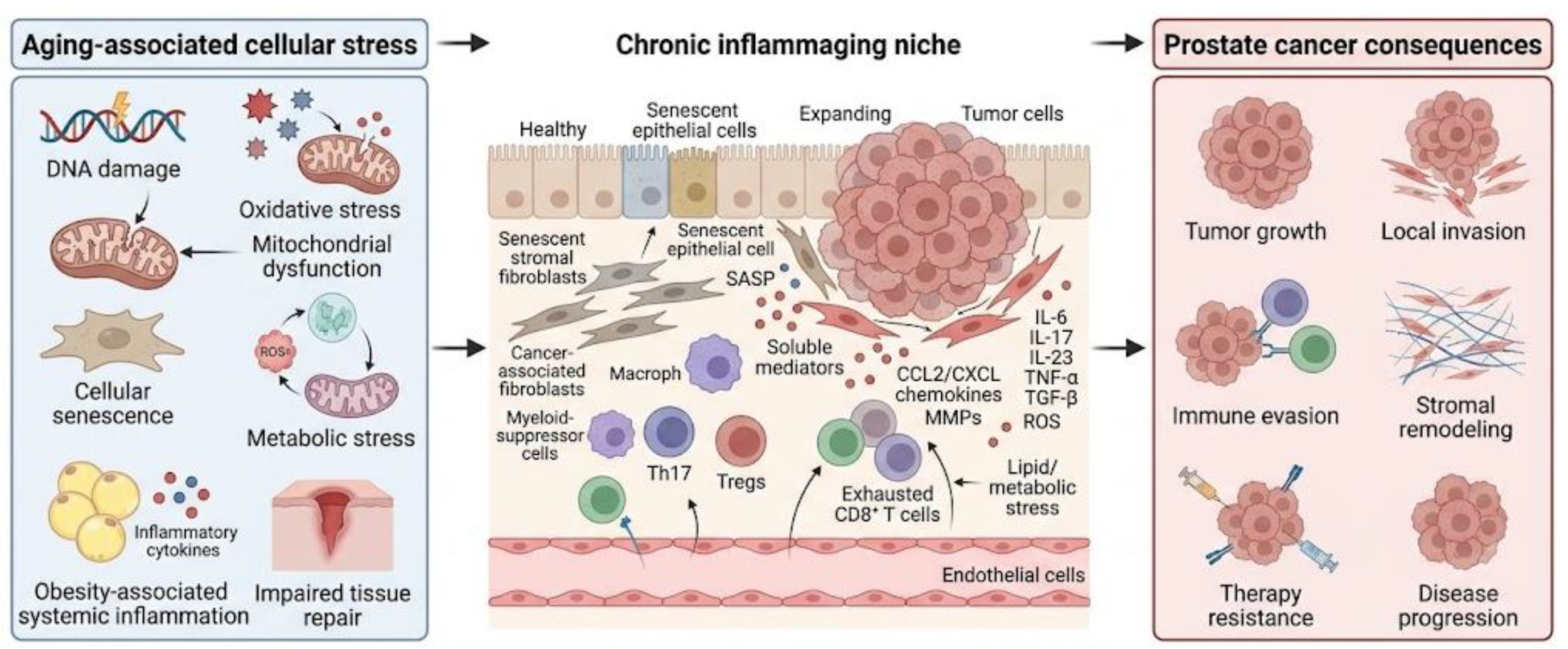
Figure 3.
Therapeutic opportunities for targeting inflammaging-associated prostate cancer progression. The figure summarizes an aged/inflamed prostate tumor microenvironment characterized by dysfunctional T/NK cells, suppressive myeloid cells, senescent cells with SASP release, leaky vasculature, and metabolic stress. Major targetable processes include cytokine imbalance, persistent senescent cells/SASP, immune dysfunction, myeloid suppression, stromal/ECM remodeling, vascular dysfunction, and metabolic stress. Potential strategies include cytokine-pathway modulation, senolytics or senomorphics, immune microenvironment remodeling, stromal/ECM modulation, metabolic and lifestyle interventions, and biomarker-guided patient stratification. These approaches aim to reduce chronic inflammation, improve immune surveillance, reduce stromal barriers, improve therapeutic response, and potentially delay progression. Selected key references: [32,34,54,95,98,99,134,138].
Figure 3.
Therapeutic opportunities for targeting inflammaging-associated prostate cancer progression. The figure summarizes an aged/inflamed prostate tumor microenvironment characterized by dysfunctional T/NK cells, suppressive myeloid cells, senescent cells with SASP release, leaky vasculature, and metabolic stress. Major targetable processes include cytokine imbalance, persistent senescent cells/SASP, immune dysfunction, myeloid suppression, stromal/ECM remodeling, vascular dysfunction, and metabolic stress. Potential strategies include cytokine-pathway modulation, senolytics or senomorphics, immune microenvironment remodeling, stromal/ECM modulation, metabolic and lifestyle interventions, and biomarker-guided patient stratification. These approaches aim to reduce chronic inflammation, improve immune surveillance, reduce stromal barriers, improve therapeutic response, and potentially delay progression. Selected key references: [32,34,54,95,98,99,134,138].
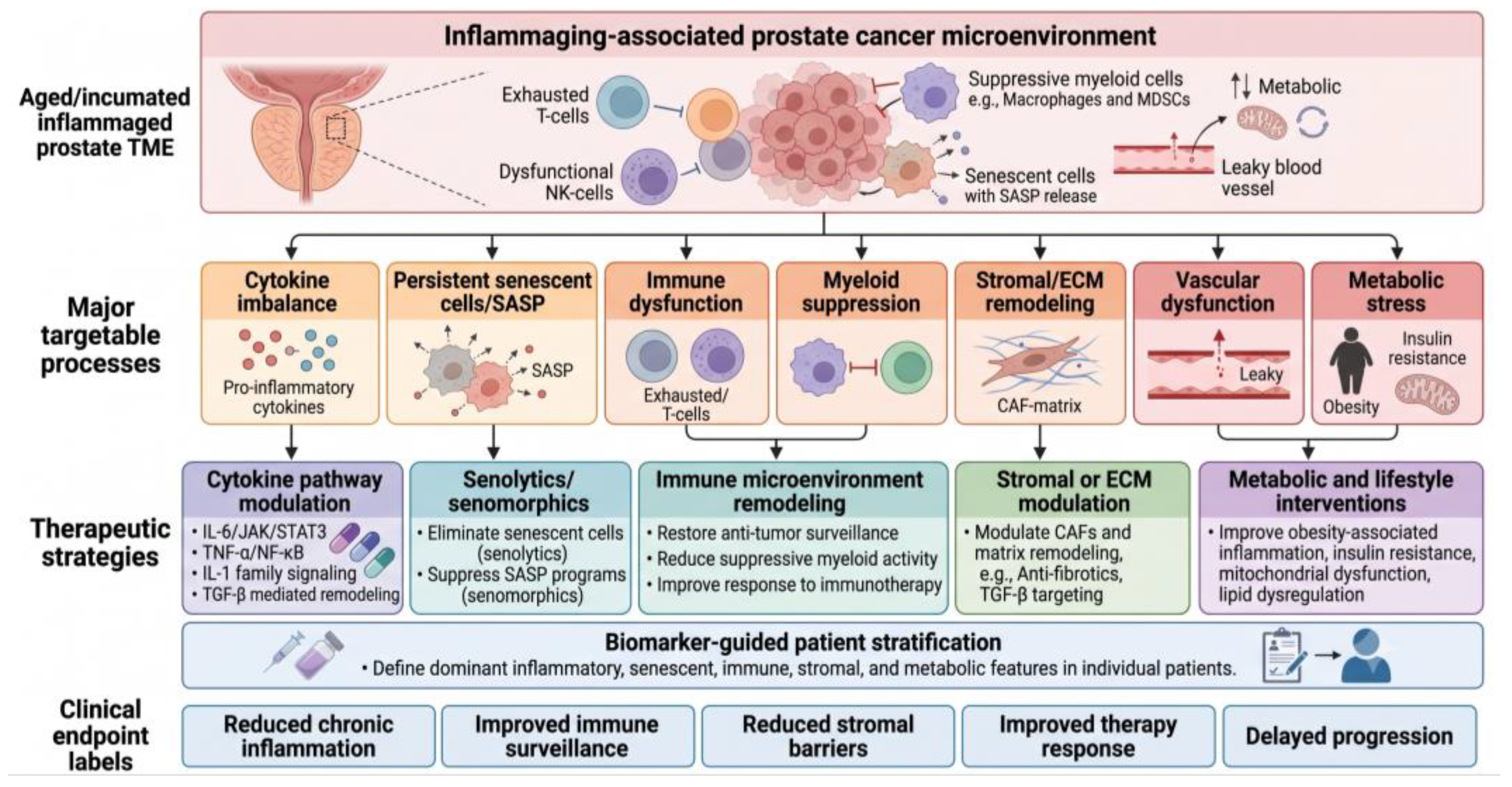

Table 1.
Senescence-associated cell types and potential effects in the aging prostate microenvironment (selected key references: [12,13,14,81,89,95]).
| Cell type | Senescence-associated features | Potential impact on prostate cancer |
| Prostate epithelial cells | Growth arrest, DNA damage response, inflammatory secretome | Tumor suppression early; inflammatory remodeling if persistent |
| Stromal fibroblasts | SASP, growth factors, MMPs, ECM remodeling | Paracrine epithelial support, stromal activation, invasion-supportive niche |
| Endothelial cells | Vascular dysfunction, altered permeability, angiogenic imbalance | Hypoxia, immune trafficking changes, therapy response |
| Immune cells | Reduced surveillance, exhaustion-like states, inflammatory cytokines | Immune escape, chronic inflammation, impaired clearance of senescent cells |
| Tumor cells after therapy | Therapy-induced senescence, residual survival, SASP-like signaling | Initial growth arrest; possible relapse-supportive inflammatory niche |
Table 2.
Immune-aging mechanisms relevant to prostate cancer (selected key references: [24,26,34,39,43,111]).
| Immune-aging feature | Major cell types | Representative mediators/pathways | Potential prostate cancer relevance |
| Reduced immune surveillance | CD8+ T cells, dendritic cells, NK cells | Exhaustion markers, impaired antigen presentation, reduced cytotoxicity | Tumor persistence, immune escape |
| Chronic cytokine imbalance | T cells, macrophages, stromal cells, senescent cells | IL-6, TNF-α, IL-1, IL-17, IL-23, TGF-β | Inflammation, stromal remodeling, therapy resistance |
| Th17/Treg imbalance | CD4+ T cells | IL-17, IL-23, FOXP3, IL-10, TGF-β | Chronic inflammation and immune suppression |
| Myeloid remodeling | Macrophages, MDSCs, monocytes, dendritic cells | IL-6, IL-10, TGF-β, IL-23, CCL2, CXCL chemokines | Immune suppression, angiogenesis, matrix remodeling |
| Altered immune transcriptional states | T cells, myeloid cells | NF-κB, STAT3, RORγt, FOXP3, BATF, IRF family factors | Persistent inflammatory or suppressive immune programs |
Table 3.
Stromal aging mechanisms in prostate cancer (selected key references: [29,55,57,58,65,104]).
| Stromal feature | Major cell type/component | Key mediators | Potential consequence |
| Senescent fibroblast activation | Fibroblasts, smooth muscle cells | IL-6, CXCL8, CCL2, TGF-β, MMPs | Chronic inflammation, paracrine epithelial support |
| CAF remodeling | Cancer-associated fibroblasts | Growth factors, cytokines, ECM proteins | Tumor growth, invasion, immune modulation |
| ECM stiffness/remodeling | Collagen, fibronectin, basement membrane | Integrins, FAK/SRC, YAP/TAZ, MMPs | Migration, invasion, treatment resistance |
| Vascular aging | Endothelial cells, pericytes | VEGF, HIF-1α, ROS, adhesion molecules | Hypoxia, altered immune trafficking, angiogenesis |
| Stromal–immune crosstalk | Fibroblasts, macrophages, T cells | CCL2, CXCL chemokines, TGF-β, IL-6 | Immune suppression, inflammatory niche formation |
Table 4.
Metabolic stress pathways in aging-associated prostate cancer (selected key references: [1,67,68,71,74]).
| Metabolic feature | Major mechanisms | Potential inflammatory effect | Possible prostate cancer relevance |
| Mitochondrial dysfunction | mtDNA damage, impaired mitophagy, altered respiration | ROS, cGAS–STING, inflammasome activation | Senescence, inflammation, therapy response |
| Oxidative stress | Excess ROS, lipid peroxidation, DNA damage | NF-κB activation, tissue injury, SASP amplification | Epithelial stress, stromal remodeling |
| Obesity-associated inflammation | Adipose inflammation, insulin resistance, adipokine imbalance | Systemic cytokines, myeloid activation | Tumor progression, immune dysfunction |
| Lipid dysregulation | Altered fatty acids, cholesterol, oxidized lipids, eicosanoids | Immune-cell modulation, inflammatory lipid signaling | Tumor metabolism, immune remodeling |
| Nutrient-sensing changes | mTOR, AMPK, insulin/IGF, PPARs, autophagy | Altered SASP, immune metabolism, stress adaptation | Growth signaling, metabolic adaptation, resistance |
Table 5.
Therapeutic strategies targeting inflammaging in prostate cancer (selected key references: [32,34,54,95,98,99,134,138]).
| Therapeutic strategy | Main target | Rationale | Potential benefit | Major caution |
| Cytokine modulation | IL-6/JAK/STAT3; IL-17/IL-23; TNF-α/NF-κB; IL-1; TGF-β | Chronic cytokine signaling can drive inflammation, stromal remodeling, immune suppression, and resistance | May reduce protumor inflammation and improve treatment response | Broad inhibition may impair protective immunity |
| Senolytics | Persistent senescent cells | Senescent cells can sustain SASP-driven inflammation | May reduce senescent-cell burden and chronic inflammatory niches | Senescence can also be protective; timing is critical |
| Senomorphics | SASP pathways (mTOR, JAK/STAT, p38 MAPK, NF-κB, cGAS–STING) | Suppress harmful SASP without eliminating all senescent cells | May preserve growth arrest while reducing inflammatory remodeling | Optimal targets and patient selection remain unclear |
| Metabolic/lifestyle intervention | Obesity, insulin resistance, mitochondrial dysfunction, lipid dysregulation | Metabolic stress can amplify inflammaging and alter immune/stromal function | May improve systemic inflammation, metabolic health, and treatment tolerance | Likely supportive rather than stand-alone therapy |
| Immune/myeloid remodeling | Myeloid cells, T-cell dysfunction, antigen presentation, immune checkpoints | Aged tumor microenvironments may be inflamed but poorly tumoricidal | May improve immune surveillance and immunotherapy response | Requires biomarker-guided selection; combinations may increase toxicity |
| Stromal/ECM modulation | CAFs, ECM stiffness, TGF-β-related remodeling, matrix proteases | Stromal remodeling can promote invasion, immune exclusion, hypoxia, and drug resistance | May improve immune-cell infiltration, drug delivery, and tissue organization | May interfere with normal tissue integrity and wound healing |
| Vascular normalization | Endothelial dysfunction, angiogenesis, hypoxia | Vascular dysfunction can impair immune trafficking and treatment delivery | May improve perfusion, oxygenation, immune access, and therapy response | Effects are context-dependent |
| Biomarker-guided stratification | Cytokines, SASP markers, immune signatures, metabolic profiles, spatial features | Inflammaging is heterogeneous and not present in all patients | May identify patients most likely to benefit from targeted strategies | Biomarkers still require clinical validation |
Disclaimer/Publisher’s Note: The statements, opinions and data contained in all publications are solely those of the individual author(s) and contributor(s) and not of MDPI and/or the editor(s). MDPI and/or the editor(s) disclaim responsibility for any injury to people or property resulting from any ideas, methods, instructions or products referred to in the content. |
© 2026 by the authors. Licensee MDPI, Basel, Switzerland. This article is an open access article distributed under the terms and conditions of the Creative Commons Attribution (CC BY) license.
Copyright: This open access article is published under a Creative Commons CC BY 4.0 license, which permit the free download, distribution, and reuse, provided that the author and preprint are cited in any reuse.