

Submitted:
11 July 2026
Posted:
16 July 2026
You are already at the latest version
Abstract
Age-related macular degeneration (nAMD) is a significant cause of visual impairment globally.1 This single-centre retrospective study evaluated real-world outcomes of faricimab in patients with neovascular age-related macular degeneration (nAMD) who responded suboptimally to other anti-VEGF treatment. Patients with nAMD who were switched to faricimab between September and December 2022 with a minimum of 12 months’ follow-up were included. Main outcomes were best-corrected visual acuity (BCVA), central subfield thickness (CST), and treatment intervals at 12 and 18 months. A total of 136 eyes received at least one intravitreal faricimab injection, of which 71 eyes (62 patients) with sufficient follow-up data were included in the analysis. Median BCVA changed from 68 ETDRS letters (IQR 57-74) to 66 (55-75, p=0.119) at 12 months and 67 (56-73, p=0.009) at 18 months. Median CST changed from 296 µm (IQR 270-339) at baseline to 273 µm (245-313, p=0.0001) at 12 months and 263 µm (240-300, p<0.000) at 18 months. 5.6% of eyes had a dry macula at baseline, compared to 26.8% (p=0.002) and 31.7% (p=0.001) at 12 and 18 months, respectively. Mean treatment interval changed from 5.2 ± 1.6 weeks at baseline, to 7.4 ± 2.8 weeks (p<0.0000) at 12 months, and 7.5 ± 3.1 weeks (p<0.0000) at 18 months. Adverse events included one case of hypertensive anterior uveitis and one post-injection hypotony. Switching to faricimab for nAMD achieved stable vision, improved anatomical outcomes and extended treatment intervals over 18 months.
Keywords:
faricimab
; anti-VEGF therapy
; age-related macular degeneration
; real-world evidence
1. Introduction
The Age-related macular degeneration (nAMD) is a significant cause of visual impairment globally.1 Intravitreal anti-vascular endothelial growth factor (anti-VEGF) is effective in reducing the risk of visual loss.2 Faricimab (Vabysmo, Roche/Genetech, Basel, Switzerland) is a bispecific antibody targeting both angiopoietin-2 (ANG-2) and VEGF-A.3 ANG-2 is a key growth factor in regulating angiogenesis and vascular permeability.4 Simultaneous inhibition of ANG-2 and VEGF-A is postulated to enhance treatment durability. TENAYA and LUCERNE demonstrated comparable vision gains between faricimab and aflibercept in nAMD, with most patients on faricimab achieving extended dosing intervals.5 This retrospective study evaluates the efficacy and safety of faricimab in a cohort of nAMD patients with suboptimal response or high treatment burden on other anti-VEGF therapies.
2. Materials and Methods
Study design and patient selection
This observational study was conducted at Torbay Hospital, Devon, UK, in accordance with the Declaration of Helsinki. Given its retrospective nature, no ethical approval was required.
Consecutive patients aged ≥50 years switched to faricimab for nAMD between September and December 2022 were included. Patients were switched if they had a poor morphological response to their anti-VEGF therapy, and/or a high treatment burden. Poor morphological response was defined as disease activity including the persistence of subretinal fluid (SRF) and/or intraretinal fluid (IRF), and/or new macular haemorrhage despite optimal treatment. Minimal, unchanged, or worsening disease activity immediately following a loading phase of anti-VEGF therapy is also considered poor response. High treatment burden is defined as treatment intervals ≤8 weeks to maintain dry macular status or stable disease activity.
Exclusion criteria were as follows: (1) treatment-naïve status; (2) second eye of bilateral treatment unless both met the inclusion criteria; (3) receipt of other treatment affecting vision such as cataract surgery during follow-up; (4) less than 12 months’ follow-up. All patients who received at least one dose of faricimab were included in the safety analysis to minimise bias. Electronic healthcare data were collected retrospectively.
Treatment protocol and outcome measures
Patients were treated with the following clinician-directed regimen: (1) full loading phase consisting of 4 monthly injections; (2) partial loading phase consisting of two to three monthly injections; or (3) a treat-and-extend (T&E) regimen starting with an interval of 4-6 weeks. At each visit, the treatment interval was adjusted based on disease activity such as change in macular fluid (SRF and/or IRF) and any accompanying visual acuity change. Treatment intervals were reduced by up to 4 weeks to a minimum 4-week interval if disease activity increased, or extended by up to 4 weeks to a maximum of 16 weeks if stable.
Baseline data collected included sex, age and eye laterality, number of the last intravitreal anti-VEGF injections, anti-VEGF drug before switching, pre-switch treatment interval, visual acuity, and macular anatomical status including central subfield thickness (CST). CST was defined as the mean thickness measured from internal limiting membrane to retinal pigment epithelium within the 1-mm diameter circle centred around fovea on optical coherence tomography (OCT) (Spectralis, Heidelberg Engineering Inc, Franklin, Massachusetts, USA). Endpoint data including visual acuity, OCT anatomical status and treatment interval were collected within prespecified time windows; at 12 months (2 weeks before to 14 weeks after) and 18 months (2 weeks before to 14 weeks after). Time windows were specified to account for asynchronous follow-up in a real-world cohort. The number of faricimab injections received at each endpoint was recorded within the respective 12-month and 18-month periods.
A complete case analysis (CCA) was performed, including only eyes with complete data for each outcome variable at each prespecified follow-up visit.
Statistics
Descriptive statistics summarised patient characteristics. Continuous data were reported as mean ± standard deviation (SD) or median (interquartile range [IQR]) depending on data normality. Data were entered into Excel (Microsoft, Redmond, Washington, USA) and analysed using R (R Foundation for Statistical Computing version 4.2.2, Vienna, Austria). Data normality was assessed with skewness, kurtosis, histograms and Q-Q plots. Wilcoxon signed-rank and McNemar tests were used to analyse non-parametric continuous and dichotomous data, respectively. A p value of <0.05 was deemed statistically significant.
3. Results
Baseline characteristics and treatment history
Data from 71 eyes (62 patients) and 60 eyes (54 patients) were analysed at 12 and 18 months, respectively. Mean age at baseline was 82.29 ± 7.04 years (range 65-98 years). 80.1% (50/62) of patients were female and all were Caucasian.
Prior to switching, 97.2% (69/71) of eyes received aflibercept 2 mg (Eylea, Regeneron Pharmaceuticals, NY, USA) and 2.8% (2/71) Ranibizumab 0.5 mg (Lucentis, Genentech, CA, USA). Patients had received a median of 20 injections (IQR: 9-33.5) per eye of their last anti-VEGF therapy over a mean of 2.99 ± 2.36 years (range 0.07-8.67). The mean treatment interval of patients’ prior anti-VEGF treatment was 5.18 ± 1.57 weeks. Immediately before switching to faricimab, 97.2% (69/71) of eyes were on treatment intervals ≤8 weeks.
At baseline, 94.4% (67/71) of eyes showed suboptimal response to prior anti-VEGF treatment. After faricimab switch, 7.0% (5/71) of eyes received a full loading course of four monthly injections, 8.5% (6/71) a partial loading course of three monthly injections, 1.4% (1/71) two monthly injections, and 83.1% (59/71) were switched directly to a T&E regimen with an initial interval of 4-8 weeks.
A mean of 8.97 ± 1.97 faricimab injections were administered over a mean period of 18.3 ± 2.1 months (range: 11.6-21.2).
Patients’ demographics and baseline characteristics are summarised in Table 1.
Visual acuity outcomes
At baseline, median best corrected visual acuity (BCVA) was 68 ETDRS letters (IQR 57-74). Median BCVA was 66 letters (IQR 55-75, p = 0.119) and 67 letters (IQR 56-73, p = 0.009) at 12 and 18 months, respectively.
At 12 months, the median BCVA change was a loss of two ETDRS letters (IQR: 7 letter loss to 3.5 letter gain). 5.6% (4/71) of eyes gained 5-9 ETDRS letters, 9.9% (7/71) gained 10-14 letters, 5.6% (4/71) gained ≥15 letters. Among those experiencing a loss, 25.4% (18/71) of eyes lost 5-9 letters, 9.9% (7/71) lost 10-14 letters, 5.6% (4/71) lost ≥15 letters.
At 18 months, the median BCVA change was a loss of three ETDRS letters (IQR: 8 letter loss to 2 letter gain). 8.3% (5/60) of eyes gained 5-9 letters, 5.0% (3/60) gained 10-14 letters, 3.3% (2/60) gained ≥15 letters. Among those with a loss, 16.7% (10/60) of eyes lost 5-9 letters, 11.7% (7/60) lost 10-14 letters, 10.0% (6/60) lost ≥15 letters. Table 2 shows BCVA change over time.
BCVA best-corrected visual acuity.
For the 83.1% of eyes (59/71) switched directly to a T&E regimen, median baseline BCVA was 69 letters (IQR 60-74), changing to 66 letters (IQR 59-75, p = 0.0316) and 67 letters (IQR 56-73, p = 0.0065) at 12 and 18 months, respectively.
Anatomical outcomes
There was a statistically significant CST change from a baseline median value of 296 µm (IQR 270-339) to 273 µm (IQR 245-313, p = 0.0001) and 263 µm (IQR 240-300, p < 0.000) at 12 and 18 months, respectively. Table 3 shows CST results.

Table 4.
CST change over time.
| Number of eyes | CST, median (IQR), µm | p value | |
| Baseline | 69 | 296 | |
| 12 months | 69 | 273 | 0.0001 |
| 18 months | 60 | 263 | <0.0000 |
CST central subfield thickness, IQR interquartile range.
At baseline, 5.6% (4/71) of eyes had a dry macula. After the first faricimab injection, 33.9% (21/62) of eyes had a dry macula (p = 0.0002). Since some eyes received partial or full loading regimens and were reviewed only afterwards, macular fluid status after the first injection could not be assessed in all cases. Compared with baseline, the proportion of eyes with a dry macula was higher at 26.8% (19/71) (p = 0.0023) and 31.7% (19/60) (p = 0.0008) at 12 and 18 months, respectively.
Of the four eyes with a dry macula at baseline, one developed a macular haemorrhage at the 18-month visit whilst on a 16-week treatment interval. Another eye had trace IRF on OCT at 18 months whilst receiving 8-weekly faricimab injections. The remaining two eyes maintained a dry macula at 18 months.
Table 5.
CST change in eyes switched directly onto T&E regimen.
| Number of eyes | CST, median (IQR), µm | p value | |
| Baseline | 57 | 289 | |
| 12 months | 57 | 271 | 0.0025 |
| 18 months | 49 | 261 | <0.0000 |
CST central subfield thickness, IQR interquartile range.
In patients switched directly to a T&E regimen, median CST was 289 µm (IQR 270-326) at baseline, 271 µm (IQR 245-309, p = 0.0025) at 12 months, and 261 µm (IQR 240-299, p < 0.0000) at 18 months.
Treatment interval
At the switching point, the mean treatment interval was 5.2 ± 1.6 weeks. This increased to 7.4 ± 2.8 weeks (p < 0.0000), and 7.5 ± 3.1 weeks (p < 0.0001) at 12 and 18 months, respectively.
9.9% (7/71) and 13.3 (8/60) of eyes achieved an extended treatment interval of ≥12 weeks (≥Q12W) at 12 and 18 months, respectively. A treatment interval extension of at least 4 weeks was achieved in 28.2% (20/71) and 36.7% (22/60) of eyes at 12 and 18 months, respectively. 46.5% (33/71) and 46.7% (28/60) of eyes achieved a treatment interval ≥Q8W at 12 and 18 months, respectively. Figure 1 shows the distribution of dosing intervals at 12 and 18 months.
Q4-6W (4-6 weekly interval), Q8-10W (8-10 weekly interval), ≥Q12W (12 weekly interval).
Treatment intervals were reduced to 4 weeks in 2.8% (2/71) and 3.3% (2/60) of eyes due to suboptimal treatment response at 12 and 18 months, respectively. At 12 months, 5.6% (4/71) of eyes were switched from faricimab to aflibercept 8 mg due to inadequate response based on clinician judgement.
In eyes switched directly to a T&E faricimab regimen, mean treatment interval was 5.2 ± 1.4 weeks at baseline, 7.2 ± 2.8 weeks (p < 0.000) at 12 months and 7.3 ± 2.9 weeks (p < 0.000) at 18 months. In this subgroup, 8.5% (5/59) and 11.8% (6/51) of eyes achieved ≥Q12W treatment interval at 12 and 18 months, respectively. A treatment interval extension of at least 4 weeks was achieved in 30.5% (18/59) and 33.3% (17/51) of eyes at 12 and 18 months, respectively. 42.4% (25/59) and 43.1% (22/51) of these eyes achieved ≥Q8W treatment interval at 12 and 18 months, respectively. Figure 2 illustrates the distribution of dosing intervals at 12 and 18 months.
Q4-6W (4-6 weekly interval), Q8-10W (8-10 weekly interval), ≥Q12W (12 weekly interval).
Safety outcomes
In this study, one eye developed a flare of hypertensive acute anterior uveitis (AAU) two days after faricimab injection. It was subsequently treated with oral antiviral therapy, topical steroids, and topical and oral carbonic anhydrase inhibitors. Another eye developed hypotony after faricimab injection without permanent sequelae. No cases of post-injection endophthalmitis were recorded.
4. Discussion
We present our real-world experience of one of the early nAMD cohorts switched to faricimab after a suboptimal response to prior anti-VEGF treatment. This retrospective study has one of the longest follow-up periods to date. Our results demonstrated stable visual acuity, significant improvement in anatomical outcomes, and extension of treatment intervals in a cohort in which the majority required dosing intervals ≤8 weeks on prior anti-VEGF treatment. Approximately one third of eyes (36.7%) had treatment intervals extended by at least 4 weeks at 18 months.
Real-world visual acuity outcomes are often inferior to those reported in randomised controlled trials such as TENAYA and LUCERNE.5 In real-world faricimab switching studies, visual acuity has predominantly been reported as stable rather than demonstrating gains in previously treated cohorts.6-17 Studies including TRUCKEE, FARWIDE-nAMD and FARETINA-AMD reported smaller improvements or stable visual acuity in previously treated subgroups compared with treatment-naïve cohorts.6,18,19 Other switching studies reported no significant BCVA change at 12 months.14-16 Another retrospective study reported a statistically significant decline in median BCVA at 12 months in a cohort of 215 eyes characterised by higher treatment burden and resistance.17
At 12 months, there was no statistically significant change in median BCVA in our cohort (p = 0.140). There was a statistically, but not clinically, meaningful change in median BCVA at 18 months. 39.4% (28/71) and 45% (27/60) of eyes had a visual acuity change of <5 letters from baseline at 12 and 18 months, respectively. Although more eyes experienced a loss than a gain in acuity at both follow-up time points, most lost <15 letters from baseline. Overall, 94.4% (67/71) and 90.0% (54/60) of eyes maintained stable visual acuity, avoiding a loss of ≥15 ETDRS letters (≥3 LogMAR lines) at 12 and 18 months, respectively. A loss of ≥3 LogMAR lines corresponds to a doubling of the visual angle which is considered moderate visual loss, an outcome traditionally reported in clinical trials.20,21
In this study, the vast majority (94.4%) of patients had a suboptimal response to prior anti-VEGF treatment. Five of the six patients who lost >15 letters at 18 months were classified as poor responders at the point of switching. Previously treated patients may introduce selection bias towards more treatment-resistant disease and are more likely to have structural damage than treatment-naïve patients, indicating worse prognosis.23,24 None of the six patients who experienced at least moderate visual loss at 18 months received a faricimab loading regimen, as per the UK label of four monthly injections, at switching. The necessity of a fixed loading phase of faricimab, as used in TENAYA and LUCERNE, remains debated. Some suggest a fixed loading phase may be required to maximise Ang-2 inhibition for better long-term visual prognosis.11,25
Given a significant proportion of our cohort (83.1%) was switched directly to a T&E regimen, a subgroup analysis was performed. In this subgroup, there was a statistically, but not clinically, significant change in visual acuity at 12 and 18 months. The interquartile range of visual acuity change highlights heterogeneity in anti-VEGF response in previously treated eyes and underscores the need for a personalised therapeutic approach. Genetic variation has been proposed as a potential mechanism for this phenomenon.22 In this cohort, prior anti-VEGF treatment duration varied widely, from less than one year to over 8 years, representing a source of heterogeneity. Interestingly, a meta-analysis examining real-world 10-year outcomes of anti-VEGF therapy for nAMD found a slow, progressive decline in visual acuity following the first year of treatment, with progression towards macular atrophy proposed as a potential explanation.26 No further subgroup analyses were performed due to limited sample size.
Similar to other real-world studies,6-13 this cohort saw a reduction in median CST from 296 µm (IQR 270-339) at baseline to 273 µm (IQR 245-313, p = 0.000) and 263 µm (IQR 239.75-299.75, p < 0.000) at 12 and 18 months, respectively. Similar findings were observed in the subgroup switched directly to a T&E regimen. The proportion of eyes with a dry macula at 12 months in our study was 26.8% (19/71), lower than the 33.7% and 40.2% reported by Sim et al. and Janmohamed et al., respectively.15,17 Patients in these studies received an initial loading phase of four monthly faricimab injections. Goodchild et al. demonstrated that the maximum macular fluid drying effect of faricimab was achieved following the third loading injection in a cohort of 98 switched eyes.10 As the majority of eyes in our cohort did not receive a loading phase of faricimab, this may partly explain the comparatively lower proportion of dry maculae at 12 months. At 18 months, 31.7% (19/60) of eyes had a dry macula, which was more comparable to that of the aforementioned studies.15,17
Frequent anti-VEGF treatment burdens patients and places substantial strain on public healthcare resources. Our analysis demonstrated encouraging treatment interval extension. Approximately one third of eyes achieved an extension of at least four weeks at 18 months. Specifically, 9.9% (7/71) and 13.3% (8/60) of eyes achieved a ≥Q12W treatment interval at 12 and 18 months, respectively. Similar outcomes were observed in the subgroup switched directly to a T&E regimen. Nevertheless, consistent with findings of Ambati et al. at one year, approximately 50% of eyes in this cohort did not reach a ≥Q8W treatment interval at either 12 or 18 months.16 This contrasts with the results of TENAYA and LUCERNE, and may reflect selection bias towards more chronic cases, in which higher VEGF levels are associated with ongoing inflammation, and under-treatment due to real-world constraints.5,26 Pharmacological factors such as tachyphylaxis have also been proposed as a plausible mechanism contributing to reduced anti-VEGF efficacy over time.27,28
For safety, there were no cases of endophthalmitis, retinal vasculitis or systemic side effects associated with faricimab after a total of 787 intravitreal injections administered in 136 eyes that received at least one dose during the study period. One case of hypertensive acute anterior uveitis (AAU) and one case of post-injection hypotony were reported, both managed non-invasively with no long-term sequelae. Notably, the patient who developed hypertensive AAU had a previous history of herpetic uveitis. Nevertheless, faricimab-related intraocular inflammation could not be excluded in this patient. The hypotony case was likely related to scleral thinning secondary to repeated intravitreal injections in the same quadrant.
The main strength of this study is its longer follow-up compared with existing real-world studies. Limitations include its retrospective design, and the absence of predefined criteria for switching to faricimab, which may have introduced selection bias. The non-standardised treatment regimen at our centre also represents an important source of heterogeneity. Furthermore, we did not account for potential confounders such as the presence of polypoidal choroidal vasculopathy, which may reduce the efficacy of anti-VEGF monotherapy.29
5. Conclusions
In conclusion, switching nAMD patients to faricimab was safe and led to overall stable visual acuity, anatomical improvement and extended treatment intervals. These benefits were maintained after 18 months. Nevertheless, individual responses varied, and personalised treatment approaches are warranted. Future research in larger cohorts with prospective study design and longer follow-up is required to validate the durability of faricimab in nAMD treatment.
Author Contributions
TGT was responsible for study conceptualisation, writing the protocol, extracting and analysing data, interpreting results, updating reference lists and writing the manuscript. RKS was responsible for interpreting results and contributed to manuscript writing. OO was responsible for study conceptualisation, contributed to writing the manuscript and provided overall supervision of the study.
Funding
This research received funding from the Research and Development Department at Torbay and South Devon NHS Foundation Trust to support payment of the article processing charge (APC).
Informed Consent Statement
Patient consent was waived due to the retrospective nature of this study involving only anonymised data.
Data Availability Statement
Data collected in this study have not been made publicly available due to the sensitive patient information it contains. Anonymised data are available from the corresponding author on appropriate request.
Conflicts of Interest
The authors declare no conflicts of interest.
Abbreviations
The following abbreviations are used in this manuscript:
| nAMD | Neovascular age-related macular degeneration |
| BCVA | Best-corrected visual acuity |
| CST | Central subfield thickness |
| Anti-VEGF ANG-2 SRF IRF T&E OCT CCA SD IQR ETDRS AAU |
Anti-vascular endothelial growth factor Angiopoietin-2 Subretinal fluid Intraretinal fluid Treat & extend Optical coherence tomography Complete case analysis Standard deviation Interquartile range Early Treatment Diabetic Retinopathy Study Acute anterior uveitis |
References
- Li JQ, Welchowski T, Schmid M, Mauschitz MM, Holz FG, Finger RP. Prevalence and incidence of age-related macular degeneration in Europe: a systematic review and meta-analysis. Br J Ophthalmol. 2020 Aug;104(8):1077-1084. [CrossRef]
- Norridge C.F.E., Gruszka-Goh M.H., McKibbin M., Burdon M. National Ophthalmology Database Audit. The Second Report of Age-related Macular Degeneration Audit (AMD) - Patients starting treatment for neovascular AMD in the 2021 NHS year (01 April 2021 to 31 March 2022). Royal College of Ophthalmologists. March 2024.
- Vabysmo. European Medicines Agency (EMA). Available from: https://www.ema.europa.eu/en/medicines/human/EPAR/vabysmo.
- Regula JT, Lundh von Leithner P, Foxton R, et al. Targeting key angiogenic pathways with a bispecific CrossMAb optimized for neovascular eye diseases. EMBO Mol Med. 2019;11(5):e10666. [CrossRef]
- Khanani AM, Kotecha A, Chang A, Chen SJ, Chen Y, Guymer R, Heier JS, Holz FG, Iida T, Ives JA, Lim JI, Lin H, Michels S, Quezada Ruiz C, Schmidt-Erfurth U, Silverman D, Singh R, Swaminathan B, Willis JR, Tadayoni R; TENAYA and LUCERNE Investigators. TENAYA and LUCERNE: Two-Year Results from the Phase 3 Neovascular Age-Related Macular Degeneration Trials of Faricimab with Treat-and-Extend Dosing in Year 2. Ophthalmology. 2024 Aug;131(8):914-926. [CrossRef]
- Khanani, A.M., Aziz, A.A., Khan, H. et al. The real-world efficacy and safety of faricimab in neovascular age-related macular degeneration: the TRUCKEE study – 6 month results. Eye 37, 3574–3581 (2023). [CrossRef]
- Leung EH, Oh DJ, Alderson SE, Bracy J, McLeod M, Perez LI, et al. Initial real-world experience with faricimab in treatment-resistant neovascular age-related macular degeneration. Clin Ophthalmol. 2023;17:1287–93. [CrossRef]
- Kishi M, Miki A, Kamimura A, Okuda M, Matsumiya W, Imai H, et al. Short-term outcomes of faricimab treatment in aflibercept-refractory eyes with neovascular age-related macular degeneration. J Clin Med. 2023;12:5145. [CrossRef]
- Kataoka K, Itagaki K, Hashiya N, Wakugawa S, Tanaka K, Nakayama M, et al. Six-month outcomes of switching from aflibercept to faricimab in refractory cases of neovascular age-related macular degeneration. Graefes Arch Clin Exp Ophthalmol. 2023;262:43–51. [CrossRef]
- Goodchild, C., Bailey, C., Soto Hernaez, J. et al. Real world efficacy and durability of faricimab in patients with neovascular AMD (nAMD) who had sub-optimal response to prior anti-VEGF therapy. Eye 38, 3059–3064 (2024). [CrossRef]
- Borchert GA, Kiire CA, Stone NM, Akil H, Gkika T, Fischer MD, Xue K, Cehajic-Kapetanovic J, MacLaren RE, Charbel Issa P, Downes SM, De Silva SR. Real-world six-month outcomes in patients switched to faricimab following partial response to anti-VEGF therapy for neovascular age-related macular degeneration and diabetic macular oedema. Eye (Lond). 2024 Dec;38(18):3569-3577. [CrossRef]
- Ng B, Kolli H, Ajith Kumar N, Azzopardi M, Logeswaran A, Buensalido J, et al. Real-world data on faricimab switching in treatment-refractory neovascular age-related macular degeneration. Life (Basel). 2024;14:193. [CrossRef]
- Quah NQX, Javed KMAA, Arbi L, Hanumunthadu D. Real-World Outcomes of Faricimab Treatment for Neovascular Age-Related Macular Degeneration and Diabetic Macular Edema. Clin Ophthalmol. 2024 May 27;18:1479-1490. [CrossRef]
- Aljundi W, Daas L, Suffo S, Seitz B, Abdin AD. First-year real-life experience with intravitreal faricimab for refractory neovascular age-related macular degeneration. Pharmaceutics. 2024;16(4):470. [CrossRef]
- Sim SY, Chalkiadaki E, Koutsocheras G, Nicholson L, Sivaprasad S, Patel PJ, et al. Real-world 1-year outcomes of treatment-intensive neovascular age-related macular degeneration switched to faricimab. Ophthalmol Retina. 2024. [CrossRef]
- Ambati NR, Leone A, Brill D, Sisk RA. Real-world long-term outcomes of intravitreal faricimab in previously treated chronic neovascular age-related macular degeneration. Retina. 2025 Mar 1;45(3):446-453. [CrossRef]
- Janmohamed, I.K., Mushtaq, A., Kabbani, J. et al. 1-Year real-world outcomes of faricimab in previously treated neovascular age-related macular degeneration. Eye (2025). [CrossRef]
- Durga S Borkar, David Tabano, Vincent Garmo, Jacqueline K. Shaia, Blanche Kuo, Rachel Myers, Andrew LaPrise, Theodore Leng, Rishi P Singh; FARETINA-AMD- Early Treatment Patterns and Outcomes in Patients with Neovascular Age-Related Macular Degeneration Initiating Faricimab: an IRIS RegistryTM Analysis. Invest. Ophthalmol. Vis. Sci. 2023;64(8):2218.
- Deepali Varma, James Talks, Gabriella de Salvo, Praveen J Patel, Samantha R De Silva, Richard Gale, Martin McKibbin, Gloria Chi, Natalee James, Amanda Downey, Melanie Dodds, Parul Dayal; Effectiveness and safety of faricimab in eyes with nAMD in the United Kingdom: 18-month results from the FARWIDE-nAMD study. Invest. Ophthalmol. Vis. Sci. 2025;66(8):3069.
- Clinical Review Report: Dexamethasone (Ozurdex): (Allergan Inc.): Indication: For the treatment of adult patients with diabetic macular edema who are pseudophakic [Internet]. Ottawa (ON): Canadian Agency for Drugs and Technologies in Health; 2018 Nov. Appendix 5, Validity of Outcome Measures. Available from: https://www.ncbi.nlm.nih.gov/books/NBK539248/.
- Joussen AM, Lehmacher W, Hilgers RD, Kirchhof B. Is significant relevant? Validity and patient benefit of randomized controlled clinical trials on age-related macular degeneration. Surv Ophthalmol. 2007 May-Jun;52(3):266-78. [CrossRef]
- Kitchens JW, Kassem N, Wood W, Stone TW, Isernhagen R, Wood E, Hancock BA, Radovich M, Waymire J, Li L, Schneider BP. A pharmacogenetics study to predict outcome in patients receiving anti-VEGF therapy in age related macular degeneration. Clin Ophthalmol. 2013;7:1987-93. [CrossRef]
- Armendariz BG, Chakravarthy U. Fibrosis in age-related neovascular macular degeneration in the anti-VEGF era. Eye (Lond). 2024 Dec;38(17):3243-3251.
- Pandit SA, Momenaei B, Wakabayashi T, Mansour HA, Vemula S, Durrani AF, Pashaee B, Kazan AS, Ho AC, Klufas M, Regillo C, Yonekawa Y, Hsu J, Kuriyan A, Chiang A. Clinical Outcomes of Faricimab in Patients with Previously Treated Neovascular Age-Related Macular Degeneration. Ophthalmol Retina. 2024 Apr;8(4):360-366. [CrossRef]
- Machida A, Kurihara J, Hirata Y, Machida E, Murakami R, Oka A, Yoneda A, Tsuiki E, Oishi A. Flexible Loading Phase Treat-and-Extend Regimen with Faricimab for Neovascular Age-Related Macular Degeneration: A Real-World Study. Biomedicines. 2025 Nov 27;13(12):2909. [CrossRef]
- Spooner K, Broadhead G, Fraser-Bell S, Hong T, Wong JG, Chang AA. Real-World 10-Year Outcomes of Anti-VEGF Therapy for Neovascular Age-Related Macular Degeneration: A Meta-Analysis. Clin Exp Ophthalmol. 2025 Sep-Oct;53(7):773-790. [CrossRef]
- Amoaku, W., Chakravarthy, U., Gale, R. et al. Defining response to anti-VEGF therapies in neovascular AMD. Eye 29, 721–731 (2015). [CrossRef]
- Hara C, Wakabayashi T, Fukushima Y, Sayanagi K, Kawasaki R, Sato S, Sakaguchi H, Nishida K. Tachyphylaxis during treatment of exudative age-related macular degeneration with aflibercept. Graefes Arch Clin Exp Ophthalmol. 2019 Nov;257(11):2559-2569. [CrossRef]
- Cheung GCM, Lai TYY, Gomi F, Ruamviboonsuk P, Koh A, Lee WK. Anti-VEGF Therapy for Neovascular AMD and Polypoidal Choroidal Vasculopathy. Asia Pac J Ophthalmol (Phila). 2017 Nov-Dec;6(6):527-534. [CrossRef]
Figure 1.
Treatment intervals at baseline, 12 months and 18 months.
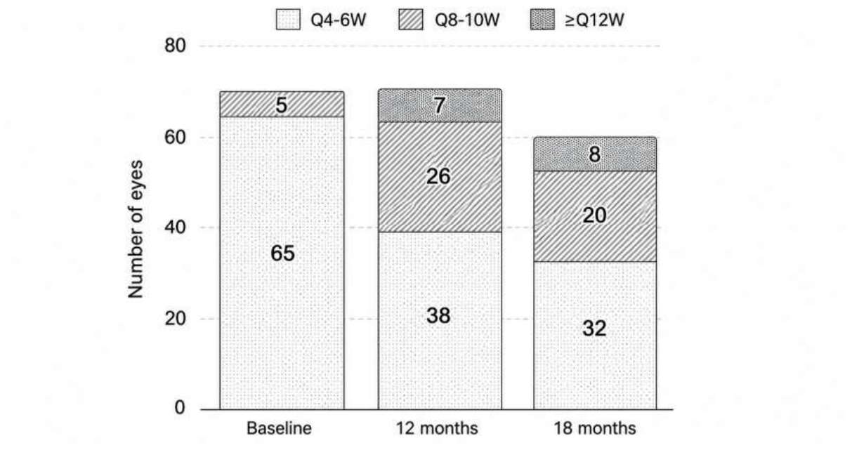
Figure 2.
Treatment intervals for eyes switched directly onto T&E regimen.
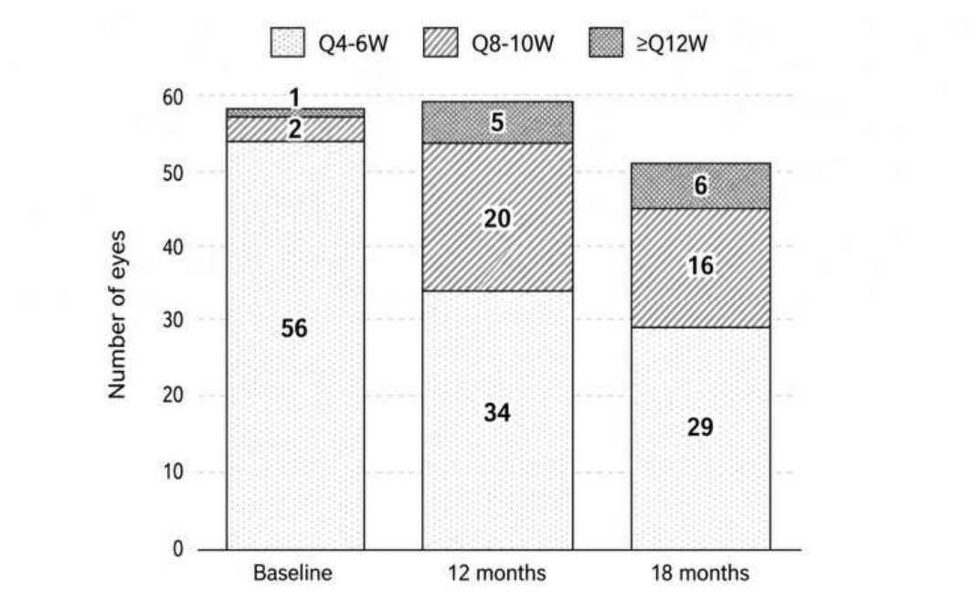
Table 1.
Patient demographics and baseline characteristics.
|
Patient demographics and baseline characteristics | ||
| Number of patients | 62 | |
| Number of eyes | 71 | |
| Age, mean (SD), years | 82.29 (7.04) | |
| Sex (female), n (%) | 50 (80.1%) | |
| Right eye, n (%) | 34 (47.9%) | |
|
Type of anti-VEGF agent prior to switch, n (%) | ||
| Aflibercept 2 mg | 69 (97.2%) | |
| Ranibizumab 0.5 mg | 2 (2.8%) | |
|
Previous anti-VEGF treatment |
Mean (SD) |
Median (IQR) |
| Number of injections of last anti-VEGF agent | 23.08 (18.29) | 20 (9-33.5) |
| Treatment interval prior to switch, weeks | 5.18 (1.57) | 4 (4-6) |
|
Baseline functional and anatomical characteristics |
Mean (SD) |
Median (IQR) |
| BCVA, ETDRS letters | 66.2 (10.3) | 68 (57-74) |
| Central subfield thickness, microns | 315.04 (79.26) | 296 (270-339) |
VEGF vascular endothelial growth factor, BCVA best-corrected visual acuity, ETDRS Early Treatment Diabetic Retinopathy Study, SD standard deviation, IQR interquartile range.
Table 2.
BCVA change over time.
| Percentage of eyes with gain of BCVA | Percentage of eyes with loss of BCVA | ||||||
| Follow-up time point | Median change (IQR) | 5-9 letters |
10-14 letters |
≥15 letters |
5-9 letters |
10-14 letters | ≥15 letters |
| 12 months |
-2 (-7, 3.5) | 5.6 | 9.9 | 5.6 |
25.4 | 9.9 |
5.6 |
| 18 months | -3 (-8, 2) |
8.3 | 5.0 | 3.3 | 16.7 |
11.7 |
10.0 |
Table 3.
BCVA change in eyes switched directly onto T&E regimen.
| Number of eyes | BCVA, median (IQR), ETDRS letters | p value | |
| Baseline | 59 | 69 | |
| 12 months | 59 | 66 | 0.0316 |
| 18 months | 51 | 67 | 0.0065 |
T&E treat-and-extend, BCVA best-corrected visual acuity, IQR interquartile range.
Disclaimer/Publisher’s Note: The statements, opinions and data contained in all publications are solely those of the individual author(s) and contributor(s) and not of MDPI and/or the editor(s). MDPI and/or the editor(s) disclaim responsibility for any injury to people or property resulting from any ideas, methods, instructions or products referred to in the content. |
© 2026 by the authors. Licensee MDPI, Basel, Switzerland. This article is an open access article distributed under the terms and conditions of the Creative Commons Attribution (CC BY) license (http://creativecommons.org/licenses/by/4.0/).
Copyright: This open access article is published under a Creative Commons CC BY 4.0 license, which permit the free download, distribution, and reuse, provided that the author and preprint are cited in any reuse.


