Preprint
Article

This version is not peer-reviewed.

Lung Ultrasound-Guided Physiotherapy in the Intensive Care Unit: A Prospective Clinical Study

Submitted:

15 July 2026

Posted:

16 July 2026

You are already at the latest version

Abstract

Pulmonary complications, particularly atelectasis and pleural effusion, are nearly universal in critically ill patients, leading to prolonged mechanical ventilation and impaired functional recovery. Traditional physical examination often lacks the sensitivity for early detection. This study aimed to evaluate the clinical utility of focused lung ultrasound (LUS) in guiding individualized physiotherapy interventions and assessing functional outcomes in the Intensive Care Unit (ICU). A prospective study was conducted involving 20 patients with respiratory failure. A focused LUS protocol targeting dorsobasal regions (zones 5 and 6) was used to monitor the effectiveness of targeted physiotherapy interventions, including manual hyperinflation, active cycle of breathing techniques, and specific positioning. Functional status was evaluated at discharge using the Functional Status Score for the ICU (FSS-ICU). Initial LUS assessment revealed a 100% prevalence of atelectasis and a 70% prevalence of pleural effusion. Targeted interventions guided by real-time LUS feedback resulted in a 65% success rate for immediate pulmonary re-aeration. The mean FSS-ICU score at discharge was 21.95 ± 8.16, with 70% of patients achieving the functional target of ≥20 points. In conclusion, focused lung ultrasound is a superior bedside diagnostic tool compared to traditional physical examination. Its integration into physiotherapy practice allows for precise, real-time monitoring and highly effective, individualized respiratory management, significantly supporting functional recovery in critically ill patients.

Keywords: 
;  ;  ;  ;  ;  

1. Introduction

The management of critically ill patients in the Intensive Care Unit (ICU) has undergone a paradigm shift with the integration of Point-of-Care Ultrasound (POCUS). Traditionally, the assessment of respiratory function and lung pathology relied heavily on physical examination—specifically auscultation and percussion—and bedside chest radiography (CXR). However, these methods often lack the sensitivity and specificity required for early detection and precise monitoring of pulmonary complications [1,2,3]. The COVID-19 pandemic further accelerated the adoption of Lung Ultrasound (LUS) as a primary diagnostic and monitoring tool, highlighting its role as a radiation-free, cost-effective, and highly accurate alternative to conventional imaging [4,5].
For physiotherapists operating within the ICU, LUS serves as “visual auscultation”, providing an immediate, real-time window into lung morphology. This capability is particularly vital given that ICU-acquired weakness and prolonged mechanical ventilation significantly impair functional outcomes. The ability to distinguish between different causes of respiratory distress allows for more targeted and safer physiotherapeutic interventions [6,7].

1.1. Pathophysiology of Atelectasis and Pleural Effusion
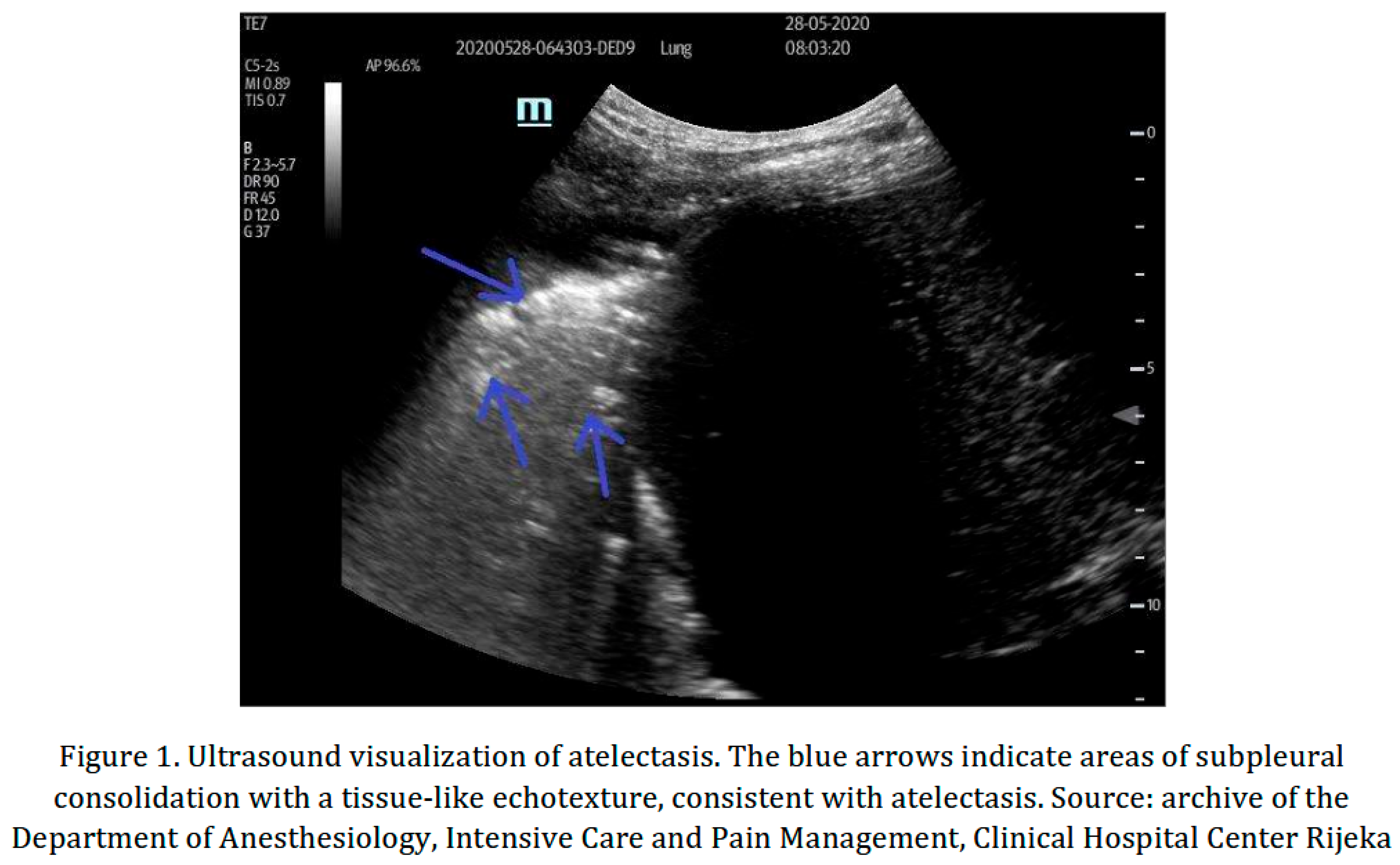
Atelectasis is one of the most frequent complications in the ICU, resulting from a combination of diaphragmatic dysfunction, compression by abdominal contents, and impaired surfactant production [6,8]. On LUS, atelectasis presents as a “tissue-like” consolidation or “lung hepatization”, often accompanied by static or dynamic air bronchograms (Figure 1). Unlike CXR, which may fail to visualize small or retrocardiac areas of collapse, LUS can detect subpleural consolidations as small as 1 mm with high sensitivity [9,10].
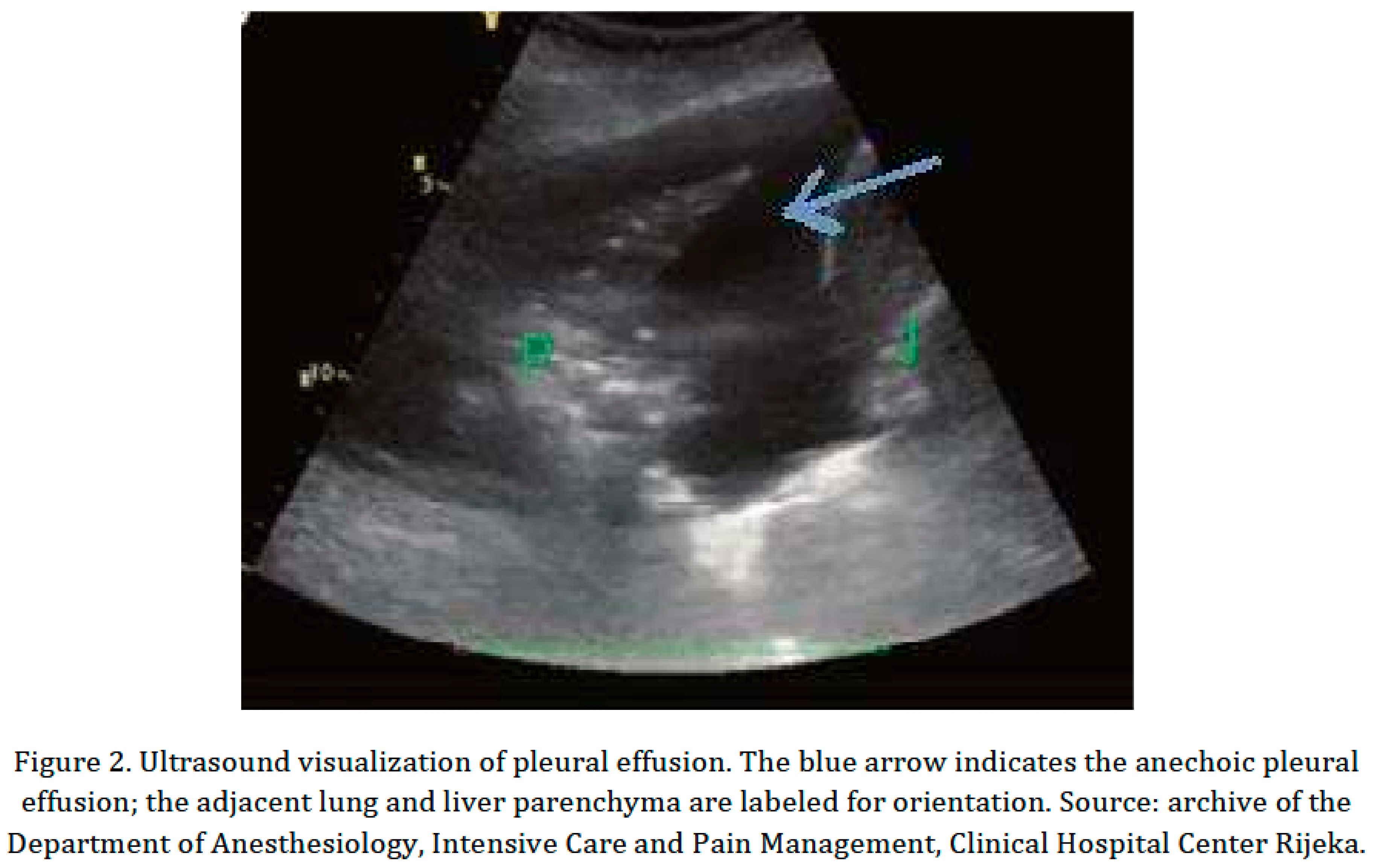
Pleural effusion represents another significant barrier to effective pulmonary rehabilitation. By increasing intrapleural pressure, it leads to compressive atelectasis and decreased lung compliance, significantly increasing the work of breathing (Figure 2). LUS is considered the “gold standard” for bedside detection of pleural fluid, enabling the clinician not only to quantify the volume but also to assess the dynamic changes during physiotherapy maneuvers such as positioning and manual hyperinflation [8,11].
Preprints 223455 i001
Preprints 223455 i002

1.2. The Role of the Physiotherapist

Modern respiratory physiotherapy in the ICU focuses on optimizing gas exchange, promoting lung re-expansion, and facilitating early mobilization. By integrating LUS, physiotherapists can move beyond “blind” interventions. They can precisely localize areas of secretion retention or collapse, apply specific manual techniques or recruitment maneuvers, and immediately evaluate the success of treatment by observing “re-aeration” signs (e.g., the reappearance of A-lines or a decrease in consolidation size). This diagnostic precision is essential for preventing the complications associated with both under-treatment and the potential barotrauma of over-aggressive recruitment [12,13].

2. Objectives and Hypotheses

2.1. Objectives of the Study

The primary objective of this study was to evaluate the clinical utility of lung ultrasound in optimizing and individualizing physiotherapy interventions for patients with respiratory failure in the ICU.
Specific objectives included:
O1: Defining the frequency of atelectasis relative to other pleural pathologies, such as pleural effusion.
O2: Determining the impact of targeted physiotherapy interventions on lung atelectasis.
O3: Evaluating the functional recovery of patients at discharge from the ICU using the FSS-ICU questionnaire.

2.2. Hypotheses

Based on the existing clinical literature and the specific aims of this research, three hypotheses were proposed:
H1: Atelectasis is the most common pulmonary pathology in patients with respiratory failure.
H2: Targeted physiotherapy interventions successfully reduce pulmonary atelectasis in patients with respiratory failure.
H3: The functional status score (FSS-ICU) at discharge is greater than or equal to 20 points.

3. Materials and Methods

3.1. Study Design and Participants

This prospective study included a convenience sample of 20 patients treated at the ICU of Clinical Hospital Center Rijeka between February and June 2025.
Inclusion criteria required ICU admission, a primary diagnosis of respiratory failure with confirmed atelectasis, SpO₂ < 90% (despite > 8 L/min O₂ therapy), and a respiratory rate > 30 breaths per minute. At the time of enrollment, depending on their clinical status, patients were managed with varying levels of respiratory support, ranging from invasive mechanical ventilation via an endotracheal tube for the most critical patients to non-invasive mechanical ventilation (NIV) and high-flow nasal oxygenation for spontaneously breathing patients. Patients undergoing thoracotomy or receiving extracorporeal membrane oxygenation (ECMO) therapy (until its removal) were excluded, as were patients with previous pleural pathology or intrathoracic malignancy.

3.2. Lung Ultrasound Protocol

A trained physiotherapist performed LUS using a FUJIFILM Sonosite Edge II device (FUJIFILM Sonosite, Inc., Bothell, WA, USA) with a convex probe (2–5 MHz). The assessment was a focused examination targeting the dorsobasal regions (zones 5 and 6) above the diaphragm.
Atelectasis was monitored and categorized into three levels of severity for each lung:
  • Level 1: No atelectasis.
  • Level 2: Presence of atelectasis in only one intercostal space (basal).
  • Level 3: Presence of atelectasis in two or more intercostal spaces.

3.3. Interventions and Success Criteria

Interventions were individualized based on real-time LUS findings. For sedated patients, LUS was performed every two days to guide positioning and manual techniques (percussion and vibration). For awake patients, LUS was performed daily to guide breathing exercises (diaphragmatic, thoracic, ACBT), incentive spirometry, PEP therapy, and cough assistance.
The success of the interventions was determined by comparing the first and last LUS findings, as follows:
  • Successful: improvement to a lower severity level in both lungs.
  • Partially successful: improvement in one lung, while the other remained stable or worsened.
  • Unsuccessful: both lungs remained at the same or a higher severity level.

3.4. Physiotherapy Interventions

The interventions included a comprehensive range of respiratory and motor rehabilitation techniques.

3.4.1. Airway Clearance Techniques (Mucociliary Clearance)

  • Manual techniques: chest vibration and percussion.
  • Postural drainage and positioning: lateral decubitus or prone positioning to facilitate secretion drainage.
  • Active Cycle of Breathing Techniques (ACBT): including breathing control, thoracic expansion exercises, and huffing.
  • Positive Expiratory Pressure (PEP) therapy.
  • Mechanical insufflation–exsufflation (cough assist) and manually assisted cough.
  • Endotracheal suctioning in conjunction with drainage and hyperinflation.

3.4.2. Lung Re-Expansion and Ventilation Optimization

  • Manual hyperinflation (MHI) using a resuscitation bag.
  • Incentive spirometry (e.g., Triflo II) to encourage maximal slow inspiration.
  • Targeted positioning: placing the patient to optimize ventilation–perfusion matching in the affected dorsobasal segments.

3.4.3. Breathing Exercises

  • Diaphragmatic breathing and thoracic expansion exercises.
  • Pursed-lip breathing and glossopharyngeal breathing for patients with severe muscle weakness.

3.4.4. Early Mobilization and Motor Rehabilitation

  • Passive and active-assisted range-of-motion (ROM) exercises in bed.
  • Progression to sitting at the edge of the bed, transfers (bed-to-chair), standing, and ambulation.
  • Balance exercises and resistance training (e.g., cycle ergometry).

3.5. Functional Status Assessment

Functional status was evaluated on the day of discharge using the Functional Status Score for the ICU (FSS-ICU). This tool assessed five functional tasks (rolling, supine-to-sit transfer, sitting at the edge of the bed, transfer to standing, and walking), with each task scored from 0 to 7. The total score ranged from 0 to 35.

3.6. Statistical Analysis

Data analysis was conducted using IBM SPSS Statistics, version 25 (IBM Corp., Armonk, NY, USA). Results are presented through descriptive statistics, including absolute frequencies, percentages, arithmetic means (M), and standard deviations (SD).

4. Results

4.1. Patient Demographics

A total of 20 patients were included in the final analysis. The study population was perfectly balanced in terms of sex (50% male, 50% female), with a mean age of 66.35 ± 12.31 years (range, 40–84 years). At the time of the initial assessment, all patients were being treated for respiratory failure. The cohort presented with varying degrees of respiratory compromise, necessitating a spectrum of support modalities ranging from non-invasive ventilation (NIV) and high-flow nasal oxygenation for spontaneously breathing patients to invasive mechanical ventilation via an endotracheal tube for the most critical cases.

4.2. Prevalence of Lung Pathologies (Hypothesis H1)

The focused LUS examination of the dorsobasal regions (zones 5 and 6) confirmed that atelectasis was present in 100% of patients (n = 20). Most patients presented with high-severity atelectasis (Level 3) in at least one lung. Additionally, pleural effusion was identified as a significant co-pathology in 70% (n = 14) of subjects. These findings strongly support H1, identifying atelectasis as the primary pulmonary complication in this cohort.

4.3. Effectiveness of Lus-Guided Physiotherapy (Hypothesis H2)
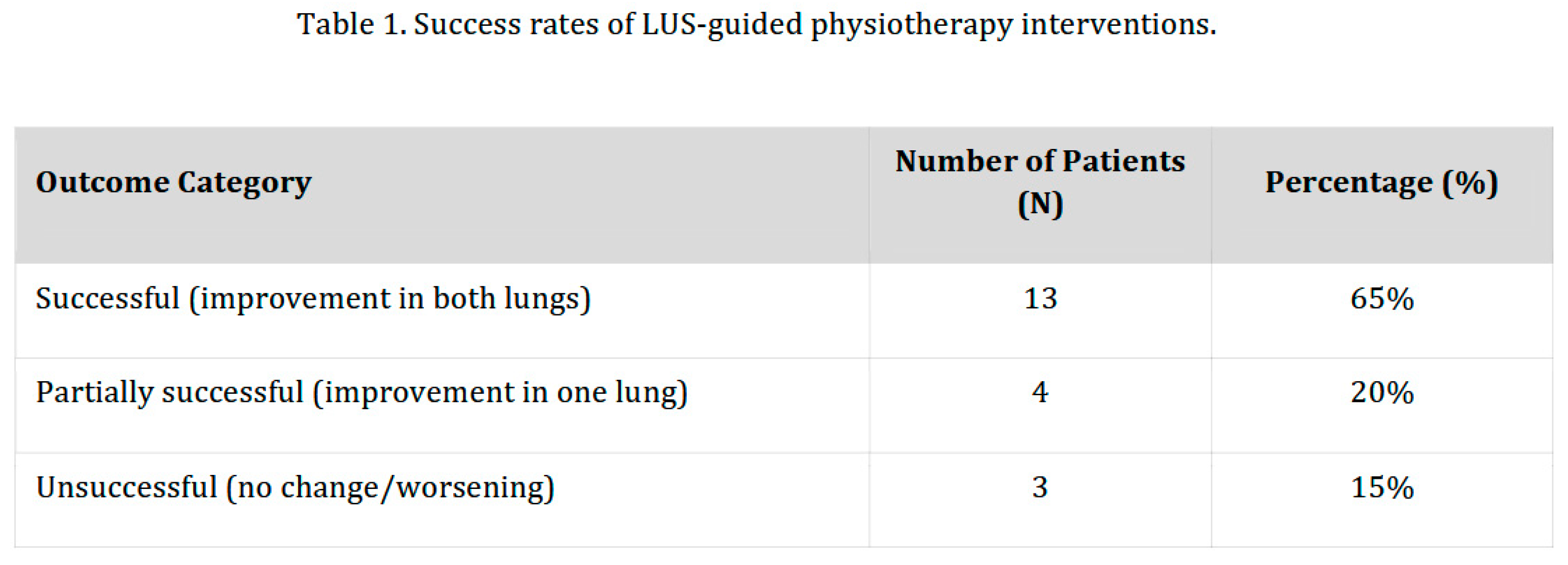
The impact of targeted physiotherapy was evaluated by comparing the baseline LUS severity levels with the findings at the end of the intervention period. Success was measured by the degree of re-aeration (reduction in severity level).
In 65% of cases, real-time feedback from LUS allowed for precise positioning and manual maneuvers that resulted in immediate morphological improvement, confirming H2 (Table 1).
Preprints 223455 i003

4.4. Functional Recovery and Discharge Status (Hypothesis H3)
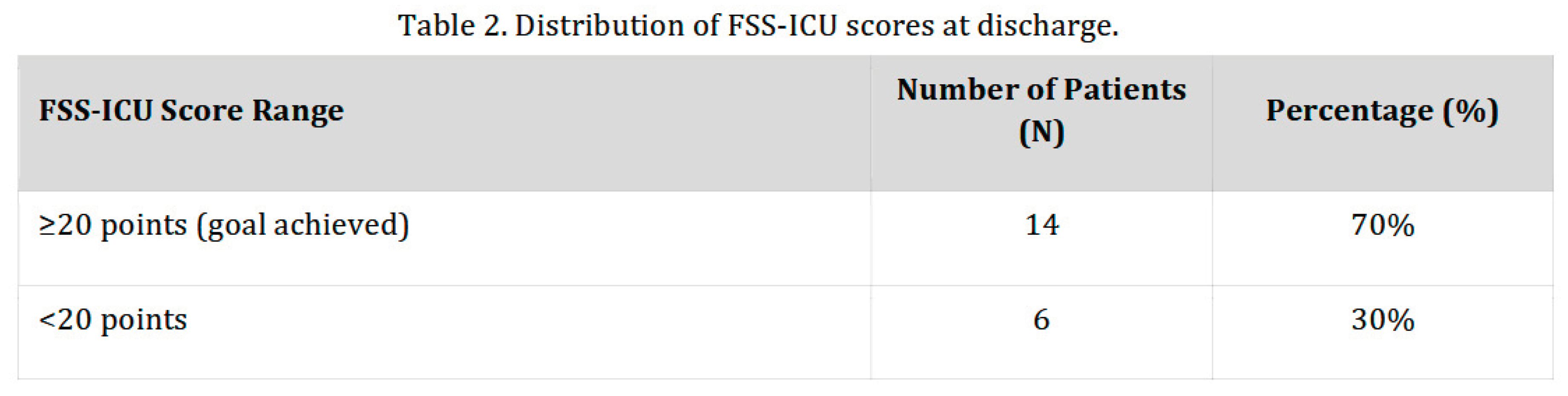
Functional status was assessed using the FSS-ICU scale. The mean score at discharge was 21.95 ± 8.16, which is significantly above the hypothesized threshold of 20 points (H3) (Table 2).
Among the individual FSS-ICU tasks, most patients achieved independence in “rolling” and “supine-to-sit” transfers, whereas “walking” remained the most challenging task, limited by the overall severity of their critical illness.
Preprints 223455 i004

5. Discussion

Lung ultrasound (LUS) is increasingly recognized as an invaluable tool in the management of critically ill patients. Because physiotherapists and respiratory therapists are integral members of the multidisciplinary ICU team, LUS is frequently highlighted as both a diagnostic and a therapeutic instrument in their daily practice. Its clinical value is particularly pronounced in that it enables timely decision-making, which can prevent both the under-treatment and over-treatment of patients [14,15,16]. In essence, LUS serves as a bridge between diagnosis and therapy, providing physiotherapists with an objective tool with which to assess, guide, and evaluate their interventions. Its portability, non-invasiveness, and complete absence of radiation exposure make it safe and highly applicable in the critical care environment [14,15,16,17].

5.1. Continuous Monitoring and Targeted Interventions (Hypothesis H2)

One of the key clinical implications of this study is the practical application of point-of-care ultrasound in the daily routine of a physiotherapist. Our results demonstrate that ultrasound assessment was used not only for initial diagnosis but primarily as a means of continuous monitoring of the immediate effects of physiotherapy interventions. This real-time evaluation allows the physiotherapist to assess the success of a specific maneuver and instantly adapt therapy based on the morphological findings. Consequently, interventions become targeted, rational, and significantly safer for the patient. This approach mitigates the risk of excessive or insufficient airway clearance, which frequently occurs when clinical decisions are based exclusively on subjective clinical impressions or traditional auscultation.
This targeted approach confirmed our second hypothesis (H2), showing a positive response to interventions in 65% of patients. This finding aligns with the current literature indicating that structured, ultrasound-guided physiotherapy can accelerate recovery and potentially reduce the duration of mechanical ventilation [18]. Similarly, a prospective cohort study by Hansell et al. demonstrated that LUS can be used effectively to monitor changes in lung aeration following respiratory physiotherapy in mechanically ventilated patients with suspected atelectasis [19].
However, interventions were partially successful or unsuccessful in a subset of patients. This finding naturally reflects the limitations of physiotherapy when faced with severe pathophysiological changes, such as massive consolidations or large pleural effusions. Furthermore, complex comorbidities and the severity of the primary illness significantly limit the success of the maneuvers applied. Future research should aim to identify predictors of therapeutic success in order to preemptively recognize patients with the highest recovery potential.

5.2. Patient Profile and Prevalence of Pathologies (Hypothesis H1)

The demographic profile of our cohort provided further insight. Most patients were of advanced age, consistent with established evidence that older patients are more vulnerable to respiratory complications and exhibit slower recovery trajectories [20]. Nevertheless, the inclusion of younger patients in our sample indicates that severe respiratory failure can affect all age groups, particularly in the context of acute conditions such as the COVID-19 pandemic or seasonal respiratory infections [21].
The extended length of ICU stay (averaging 12.75 days) further emphasizes the complexity of patients’ clinical status. Prolonged hospitalization is strongly associated with an increased risk of respiratory complications and functional decline, a correlation supported by recent studies [22]. In this context, our findings highlight the critical importance of early mobilization and respiratory physiotherapy in mitigating the detrimental effects of prolonged bed rest and mechanical ventilation.
The presence of atelectasis in 100% of patients, together with a 70% incidence of pleural effusion, firmly confirms our first hypothesis (H1). This finding demonstrates that pulmonary complications are not an exception but a standard occurrence among critically ill patients. Modern physiotherapy guidelines advocate techniques focused on improving ventilation and secretion drainage to prevent and treat these exact pathologies, validating the intervention protocols used in this study [18]. The complexity and severity of the ICU population are also starkly reflected by the fact that five initially enrolled patients were excluded from the final analysis owing to mortality.

5.3. Functional Recovery (Hypothesis H3)

The evaluation of functional recovery using the FSS-ICU scale confirmed our third hypothesis (H3). Most patients (70%) achieved a score of ≥20 points at discharge from the ICU, indicating a functional status that facilitates continued rehabilitation. This outcome supports validation studies confirming that the FSS-ICU scale is a reliable, valid, and highly sensitive tool for detecting changes in functional status in the critical care setting [23,24].
Despite these positive results, nearly a third of patients did not achieve a satisfactory functional level. This finding underscores the need for early identification of high-risk cases and the implementation of intensive rehabilitation measures during the acute phase of treatment. It also points to the need for continuous optimization of the intensity, duration, and long-term follow-up of physiotherapy interventions within the multidisciplinary team [22,25].

5.4. The Physiotherapist–Sonographer Paradigm

A distinctive and highly significant contribution of this study is that it represents, to our knowledge, the first time in the region that a physiotherapist independently performed lung ultrasound assessments. This milestone significantly broadens the competencies and scope of practice of the profession. By integrating LUS, the physiotherapist becomes a more autonomous and relevant member of the multidisciplinary team, with a direct impact on patient outcomes.
This study lays the groundwork for further promotion of physiotherapy in intensive care medicine. It opens a dialogue regarding the broader implementation of LUS in the formal education and continuous professional development of physiotherapists. Standardizing these skills through formal training would minimize institutional disparities and ensure that physiotherapists are fully equipped for the safe, independent, and responsible application of point-of-care ultrasound.

5.5. Limitations

Despite the encouraging findings, several limitations of this study must be acknowledged. The small sample size (N = 20) and the single-center design limit the generalizability of the results. Furthermore, the lack of a control group precludes direct comparison with patients who received standard care without ultrasound guidance. Future research should incorporate larger, multicenter cohorts and longitudinal follow-up after ICU discharge to comprehensively evaluate the long-term benefits of ultrasound-guided physiotherapy interventions.

6. Conclusion

The integration of focused lung ultrasound into respiratory physiotherapy practice significantly enhances the diagnostic and therapeutic management of critically ill patients in the ICU. This study demonstrates that LUS is a highly sensitive tool for the detection of dorsobasal atelectasis and pleural effusion, far surpassing the diagnostic accuracy of traditional physical examination. By providing real-time visual feedback, LUS enables physiotherapists to implement targeted, individualized interventions that lead to immediate pulmonary re-aeration and support significant functional recovery.
Furthermore, this research supports a shift in the professional paradigm, suggesting that the independent use of LUS by physiotherapists is not only feasible but essential for optimizing patient outcomes in intensive care settings. Future efforts should focus on standardizing LUS training within the physiotherapy curriculum to ensure the broad implementation of this valuable diagnostic tool.

Author Contributions

Conceptualization, M.H. and A.P.; methodology, M.H. and A.P.; investigation, M.H.; data curation, M.H.; formal analysis, M.H.; writing—original draft preparation, M.H.; writing—review and editing, A.P.; supervision, A.P. All authors have read and agreed to the published version of the manuscript.

Funding

This research received no external funding.

Institutional Review Board Statement

The study was conducted in accordance with the Declaration of Helsinki and approved by the Ethics Committee of Clinical Hospital Center Rijeka (protocol code and date of approval to be inserted by the authors).

Data Availability Statement

The data presented in this study are available on request from the corresponding author.

Acknowledgments

The authors thank the staff of the Intensive Care Unit, Department of Anesthesiology, Intensive Care and Pain Management, Clinical Hospital Center Rijeka, for their support during data collection.

Conflicts of Interest

The authors declare no conflicts of interest.

References

  1. Physiopedia. Respiratory Management of COVID-19. Available online: https://www.physio-pedia.com/Respiratory_Management_of_COVID_19 (accessed on 24 July 2024).
  2. Hayward, S.; Duncan, C. Physiotherapists’ Use of Lung Ultrasound during the COVID-19 Pandemic—A Practical Guideline on Supporting Acute Hospital Colleagues. 2020. Available online: https://www.acprc.org.uk/Data/Resource_Downloads/PhysioLUSCOVID19.pdf (accessed on 17 March 2020).
  3. Hansell, L.; Milross, M.; Delaney, A.; Tian, D.H.; Ntoumenopoulos, G. Lung ultrasound has greater accuracy than conventional respiratory assessment tools for the diagnosis of pleural effusion, lung consolidation and collapse: A systematic review. J. Physiother. 2021, 67, 41–48. [Google Scholar] [CrossRef] [PubMed]
  4. Smith, M.; Hayward, S.; Innes, S. A proposed framework for point of care lung ultrasound by respiratory physiotherapists: Scope of practice, education and governance. Ultrasound J. 2022, 14, 24. [Google Scholar] [CrossRef] [PubMed]
  5. Hayward, S.; Innes, S.; Smith, M. Challenges and opportunities in point-of-care ultrasound: A qualitative exploration of respiratory physiotherapists’ experiences of lung ultrasound training and its adoption in critical care. Ultrasound 2022, 30, 126–133. [Google Scholar] [PubMed]
  6. Physiopedia. Atelectasis. Available online: https://www.physio-pedia.com/Atelectasis (accessed on 1 October 2024).
  7. Mojoli, F.; Bouhemad, B.; Mongodi, S.; Lichtenstein, D. Lung ultrasound for critically ill patients. Am. J. Respir. Crit. Care Med. 2019, 199, 701–714. [Google Scholar] [CrossRef] [PubMed]
  8. Grott, K.; Chauhan, S.; Sanghavi, D.K.; et al. Atelectasis; StatPearls Publishing: Treasure Island, FL, USA, 2025; Available online: https://www.ncbi.nlm.nih.gov/books/NBK545316/ (accessed on 15 August 2025).
  9. Mongodi, S.; Bouhemad, B.; Iotti, G.A.; Mojoli, F. An ultrasonographic sign of intrapulmonary shunt. Intensive Care Med. 2016, 42, 912–913. [Google Scholar] [PubMed]
  10. Huang, D.; Li, Z.; Zhao, J.; Li, H.; Wang, W.; Fu, S. Lung ultrasound as a diagnostic tool for pulmonary consolidation and atelectasis after cardiac surgery. J. Thorac. Dis. 2025, 17, 4794–4802. [Google Scholar] [CrossRef] [PubMed]
  11. Zaki, H.A.; Albaroudi, B.; Shaban, E.E.; et al. Advancement in pleural effusion diagnosis: A systematic review and meta-analysis of point-of-care ultrasound versus radiographic thoracic imaging. Ultrasound J. 2024, 16, 3. [Google Scholar] [CrossRef] [PubMed]
  12. Gupta, S.; Sharma, S. An evidence-based approach to the role of physiotherapy in ICU. Int. J. Physiother. Res. 2022, 10, 4150–4161. [Google Scholar] [CrossRef]
  13. Hodgson, C.; Denehy, L.; Ntoumenopoulos, G.; Santamaria, J.; Carroll, S. An investigation of the early effects of manual lung hyperinflation in critically ill patients. Anaesth. Intensive Care 2000, 28, 255–261. [Google Scholar] [CrossRef] [PubMed]
  14. Vieira, R.; Segura-Grau, E.; Magalhães, J.; dos Santos, J.; Patrão, L. Lung ultrasound as a tool to guide respiratory physiotherapy. J. Clin. Ultrasound 2020, 48, 431–434. [Google Scholar] [CrossRef] [PubMed]
  15. Ntoumenopoulos, G.; Pizimolas, G.; Mani, S.; Hayward, S.; Lockstone, J. Lung Point of Care Ultrasound (POCUS) in cardiorespiratory physiotherapy and respiratory therapy practices: Current status and future directions. POCUS J. 2024, 9, 9–11. [Google Scholar] [CrossRef] [PubMed]
  16. Gustafson, O.; Hayward, S.; Helmsley, A.; Grant, J.; Smith, M.; Tait, C.; et al. Association of Chartered Physiotherapists in Respiratory Care position statement: Physiotherapists’ use of lung ultrasound. J. ACPRC 2021, 53, 147–150. [Google Scholar] [CrossRef]
  17. Hayward, S.; Cardinael, C.; Tait, C.; et al. Exploring the adoption of diaphragm and lung ultrasound (DLUS) by physiotherapists, physical therapists, and respiratory therapists: An updated scoping review. Ultrasound J. 2025, 17, 9. [Google Scholar] [CrossRef] [PubMed]
  18. Physiopedia. Physiotherapists’ Role in ICU. 2022. Available online: https://www.physio-pedia.com/Physiotherapists_Role_in_ICU (accessed on 25 August 2025).
  19. Hansell, L.; Milross, M.; Delaney, A.; Koo, C.M.; Tian, D.H.; Ntoumenopoulos, G. Quantification of changes in lung aeration associated with physiotherapy using lung ultrasound in mechanically ventilated patients: A prospective cohort study. Physiotherapy 2023, 119, 26–33. [Google Scholar] [CrossRef] [PubMed]
  20. Schaller, S.J.; Anstey, M.; Blobner, M.; Edrich, T.; Grabitz, S.D.; Gradwohl-Matis, I.; et al. Early, goal-directed mobilisation in the surgical intensive care unit: A randomised controlled trial. Lancet 2016, 388, 1377–1388. [Google Scholar] [CrossRef] [PubMed]
  21. Battaglini, D.; Robba, C.; Dos Santos, L.R.; et al. Rehabilitation in patients with severe respiratory failure due to COVID-19: Impact on functional recovery. J. Clin. Med. 2021, 10, 2139. [Google Scholar] [CrossRef] [PubMed]
  22. Dos Santos, J.S.F.; et al. Linking Intensive Care Unit functional scales to the International Classification of Functioning, Disability and Health. BMC Health Serv. Res. 2023, 23, 871. [Google Scholar] [CrossRef] [PubMed]
  23. Martins, G.S.; Silva, K.H.; Moraes, W.R.A.; Nakano, E.Y.; Andrade, J.M.L.; Neves, L.M.T.; Cipriano, G.F.B. Analysis of the Functional Status Score for the Intensive Care Unit and its correlation with measures of muscle strength in critically ill patients during hospitalization in the intensive care unit. Crit. Care Sci. 2025, 37, e20250197. [Google Scholar] [CrossRef] [PubMed]
  24. Wu, T.T.; Su, Q.P.; Xiong, J.; Hiser, S.; Needham, D.M.; Li, H. Reliability and validity of the Chinese version of the Functional Status Score for the ICU (FSS-ICU) after translation and cross-cultural adaptation. Disabil. Rehabil. 2025, 47, 1324–1331. [Google Scholar] [PubMed]
  25. Helms, J.; Catoire, P.; Abensur Vuillaume, L.; Bannelier, H.; Douillet, D.; Dupuis, C.; et al. Oxygen therapy in acute hypoxemic respiratory failure: Definitions and clinical recommendations. Ann. Intensive Care 2024, 14, 140. [Google Scholar] [CrossRef] [PubMed]
Disclaimer/Publisher’s Note: The statements, opinions and data contained in all publications are solely those of the individual author(s) and contributor(s) and not of MDPI and/or the editor(s). MDPI and/or the editor(s) disclaim responsibility for any injury to people or property resulting from any ideas, methods, instructions or products referred to in the content.
Copyright: This open access article is published under a Creative Commons CC BY 4.0 license, which permit the free download, distribution, and reuse, provided that the author and preprint are cited in any reuse.
Prerpints.org logo

Preprints.org is a free preprint server supported by MDPI in Basel, Switzerland.

Subscribe

© 2026 MDPI (Basel, Switzerland) unless otherwise stated

Accessibility

Disclaimer

Terms of Use

Privacy Policy

Privacy Settings