
Submitted:
14 July 2026
Posted:
16 July 2026
You are already at the latest version
Abstract
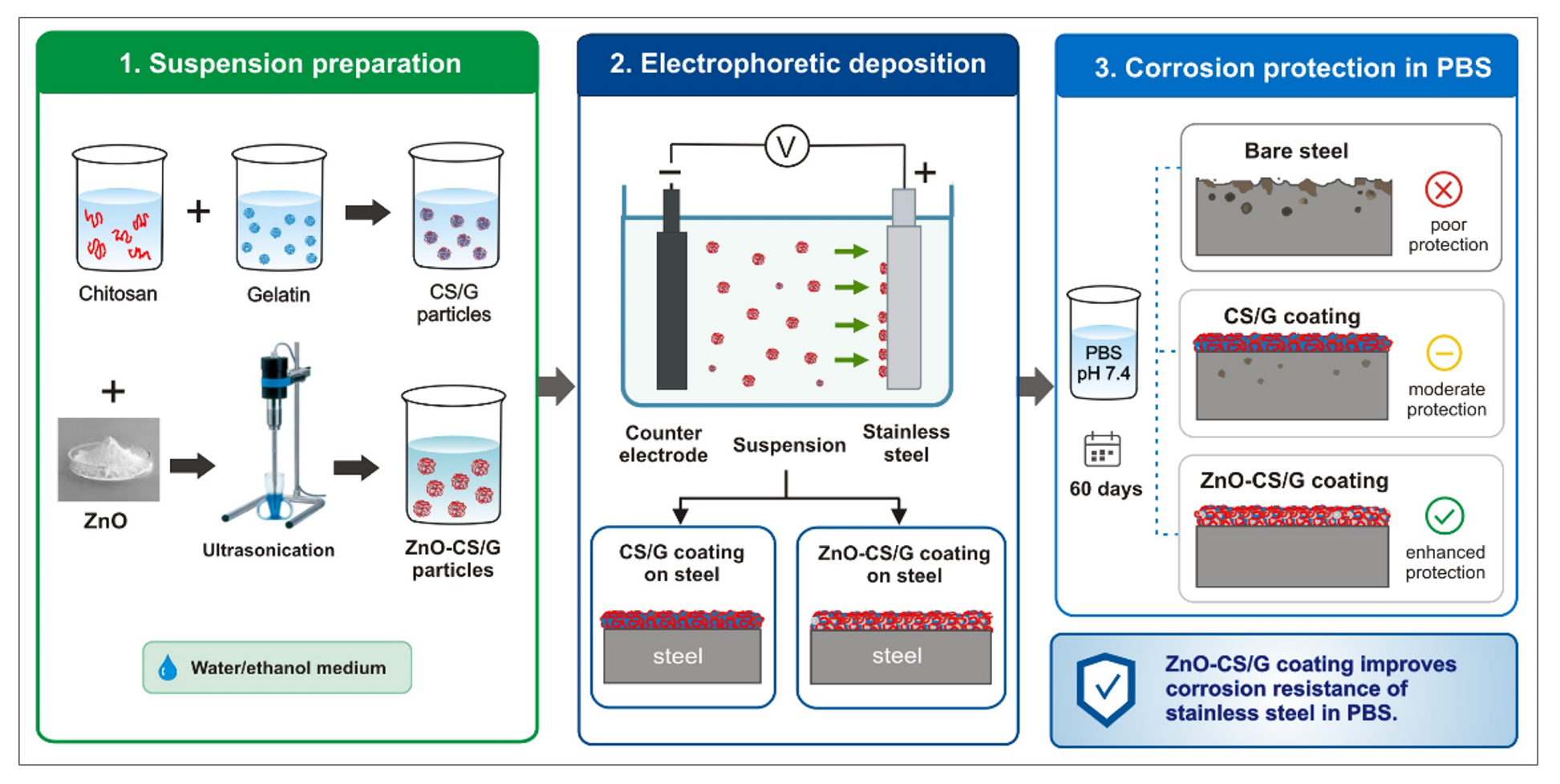
Stainless steel is widely used in biomedical and implant-related applications, but cor-rosion in physiologically relevant media can limit its long-term performance. In this study, ZnO-containing chitosan/gelatin composite coatings were fabricated on stain-less steel by electrophoretic deposition from stable water–ethanol suspensions. The coating-forming particles were obtained through chitosan/gelatin complexation in the presence of ZnO nanoparticles, enabling incorporation of an inorganic functional phase into a biopolymer matrix. The water–ethanol medium was used to suppress parasitic water electrolysis and promote the formation of continuous coatings. Sus-pension stability and electrophoretic behavior were evaluated by dynamic light scat-tering and zeta-potential measurements, while coating morphology and elemental composition were examined by scanning electron microscopy and energy-dispersive X-ray analysis. X-ray photoelectron spectroscopy was applied after 60 days of immer-sion in phosphate-buffered saline at pH 7.0 to assess the surface chemical state. Corro-sion behavior was followed by polarization resistance measurements over the same period. The ZnO-containing coating showed higher polarization resistance than both bare stainless steel and the ZnO-free chitosan/gelatin coating. The improved protection is attributed to the combined barrier effect of the biopolymer coating and the presence of ZnO within the deposited layer.
Keywords:
ZnO-chitosan/gelatin coating
; chitosan/gelatin coating
; stainless steel
; corrosion resistance
; phosphate buffer saline
1. Introduction
In recent years, a growing deal of attention has been paid to the development of biopolymer-metal oxide coatings, aiming to improve the biocompatibility and bioactivity of materials with applications in the fields of biomedical, food, agriculture and textile industries [1]. Enhancing the corrosion resistance of such materials in the human body is also of great importance. Corrosion can affect the integrity of implants and release of hazardous metallic ions into the body fluids, leading to adverse biological reactions [2].
Stainless steel is widely used in medical devices and implants due to its excellent corrosion resistance and acceptable biological properties. Interaction of reduced metal ions and electrons can provide formation of a passive oxide layer, protecting the steel structure against corrosion [3]. However, stainless steel is susceptible to localized corrosion in the presence of ions, which are generally found in the body fluids [4]. To overcome this limitation, surface modification techniques, such as coatings, are necessary [3]. Formation of an adequate biopolymer-metal oxide coating on the steel surface can protect it against corrosion, also improving biocompatibility and antibacterial activity of the coated substrate.
Biopolymers attract attention as corrosion inhibitors because they are low cost and non-toxic. Chitosan has gained considerable attention for application as a protective coating against corrosion of metallic substrates due to its specific properties such as good film-forming ability and superior adhesion to metallic surfaces. It is a polysaccharide that consists of N-acetyl glucosamine and D-glucosamine linked by beta (1-4) glycosidic bonds, derived from alkaline deacetylation of chitin [5]. The amino and hydroxyl functional groups of chitosan can form strong bonds with metal surfaces. These interactions create a protective barrier, which prevents contact of the metal substrate with the corrosion medium [6]. However, chitosan coatings suffer from high affinity towards moisture. Mechanical and barrier properties of the chitosan coatings can be improved by blending with other biopolymer and/or by reinforcing with nanomaterial. The molecular interactions between chitosan and gelatin, for example, have been found to improve the mechanical properties and stability of the polymeric material [7]. Gelatin is an ionic hydrophilic polypeptide (with –NH2 and –COOH functionalities), produced through hydrolysis of native collagen [8,9]. The adsorbed gelatin molecules can also form protective film that shields the metal surface from corrosive agents [4]. Additionally, gelatin has been found to exhibit self-healing properties, where defects in the protective films could be repaired due to migration of its molecules. When used in combination, chitosan and gelatin can exhibit synergistic effects, further enhancing their corrosion inhibition properties. The corrosion inhibition of chitosan-gelatin coating (cross-linked with glutaraldehyde) has been demonstrated for carbon steel in a 3.5 M NaCl solution [6].
Electrophoretic deposition of chitosan-gelatin (CS/GEL) coating on titanium substrate has been investigated for biomedical applications [4,10,11]. Successful electrophoretic deposition on stainless steel has been reported for CS/GEL coatings containing bioactive glass [12], silica [13] and hydroxyapatite [3,14] nanoparticles. However, the corrosion behavior of steel covered with CS/GEL coatings has been investigated only in the absence and in presence of hydroxyapatite nanoparticles: in a simulated body fluid [14] and phosphate buffer saline [3]. The coatings containing hydroxyapatite demonstrated an increase of the corrosion resistance in comparison to the bare steel samples, mainly attributed to the nanoparticles embedded in the CS/GEL matrix.
Zinc oxide (ZnO) nanoparticles have been registered to exhibit anticorrosion capacity by accepting electrons from the metal surfaces [15]. Coatings and films that contain ZnO NPs display also excellent antimicrobial activity through a number of mechanisms including the production of reactive oxygen species (ROS) and membrane disruption [16]. Apart from improving mechanical properties of the polymeric material, incorporation of ZnO nanoparticles into the polymeric matrix could allow to manage the Zn2+ ions release from the coating [17]. It is important to mention that zinc is an essential nutrient element, involved in many processes that regulate human physiology [18]. However, good biocompatibility of zinc is found at low concentrations. Ma et al. have reported, for example, that the safe daily concentration of Zn2+ ions are 12–15 mg (less than 60 μM) and the excessive Zn2+ ions are toxic [19,20]. Zinc-incorporated chitosan-gelatin coating, fabricated on titanium substrates, has demonstrated that generation of ZnO/Zn2+ complex in the coating contributed to bacteria inhibition in a concentration dependent manner [4]. Hence, to control the release of metal ions in the body is also of importance.
The aim of the present work was to develop a chitosan-gelatin hybrid coating with incorporated ZnO nanoparticles (ZnO-CS/GEL) to achieve prolonged corrosion protection of stainless steel in a simulated body fluid (PBS solution). CS/GEL nanoparticles loaded with ZnO were prepared via polyelectrolyte complexation in a water-ethanol solution. CS/GEL complexes not containing ZnO were also investigated in order to evaluate the effect of ZnO incorporation into the coating. Dynamic light scattering and electrophoresis were employed to control the formation and the size of both types of nanoparticles as well as the stability of their suspensions. CS/GEL and ZnO-CS/GEL nanoparticles were used to produce coatings on steel (cathode) substrates by electrophoretic deposition at pH 4.7 - 4.8. The morphology features of the nanoparticles as well as of the corresponding coatings were evaluated using scanning electron microscopy. X-ray photoelectron spectroscopy analysis was carried out in order to demonstrate the chemical structure of the coatings after 60 days’ immersion in a PBS buffer solution at pH 7.0. The corrosion behavior of the coatings were investigated by application of polarization resistance measurements in the PBS solution for a prolonged time interval of 60 days.
2. Materials and Methods
2.1. Materials
Chitosan (CS, Mw~50–190 kDa, pKa~6.5, 75–85% N-deacetylation), gelatin from bovine skin, Type B (GEL, 225−325 Bloom, Mw~40 −50 kDa), acetic acid (96%), and zinc oxide nanopowder (ZnO < 100 nm particle size) were purchased from Sigma Aldrich. According to previous TEM analysis, about 90% of the ZnO nanoparticles have an average size of less than 100 nm [21]. A commercial phosphate-buffered saline solution (PBS, Chem-Lab) was used for the immersion and corrosion experiments. The composition specified by the manufacturer was Na2HPO4·12H2O (18 g/L), NaCl (23 g/L), NaH2PO4·2H2O (7.8 g/L), and NaN3 (0.2 g/L), pH 7.00.
2.2. Suspension Preparation
In order to prepare CS/GEL and ZnO-CS/GEL nanoparticles for electrophoretic deposition, chitosan and gelatin solutions were prepared individually and then mixed. First, 0.05 g chitosan was dissolved in 20 mL distilled water and 1 mL acetic acid (96%) by magnetic stirring for 1 h at room temperature. Then, 79 mL ethanol was added to the chitosan solution, in order to reduce the water hydrolysis during the electrophoretic deposition process [11,12]. Separately, 0.1 g gelatin powder was added to 20 mL distilled water and 1 mL acetic acid (1%). The dissolution was achieved by magnetic stirring at 45 °C for 2h. Afterward, the solution was gently cooled down to room temperature under magnetic stirring. Finally, 79 mL ethanol was added to the cooled gelatin solution. For preparing the final suspension, equal amounts in volume of each separated solutions were used, with the aim of having 0.25 g/L chitosan and 0.50 g/L gelatin concentrations as final composition [12,13]. In order to obtain a ZnO-CS/GEL nanoparticles suspension, ZnO nanopowder was added to the mixed chitosan/gelatin suspension under ultrasound treatment (20 kHz) for 15 min to ensure the dispersion of the particles.
2.3. Electrophoretic Deposition of CS/GEL and ZnO-CS/GEL Coatings
In this research, stainless steel plates were chosen as cathode substrates with working area of 4 cm2 (2 cm x 1 cm x 0.1 cm). Inert platinum meshes were applied as anodes. The composition of the stainless steel was as follows (at %): C - 0.08 ± 0.01; N - 1.71 ± 0.10; O - 3.08 ± 0.10; V - 0.06 ± 0.06; Cr - 18.24 ± 0.35; Mn - 2.11 ± 0.15; Fe - 64.04 ± 0.88; Ni - 10.33 ± 0.53; Mo - 0.35 ± 0.03. The coatings were performed by electrophoretic deposition at a constant voltage of 25 V, supplied by a MASTECH Power Supply HY3005D, in a glass cell containing 600 mL suspension, at ambient temperature for 10 min. The concentration of ZnO nanoparticles in the suspension used to obtain the ZnO-CS/GEL coating was 0.1 g/L. After taking out the coatings from the suspensions, they were dried in air for 24 h.
2.4. Characterization Techniques
CS/GEL and ZnO-CS/GEL nanoparticles were characterized in terms of particle size (hydrodynamic diameters) and surface charge density (zeta potentials). Both parameters are measured by Dynamic Light Scattering (DLS) and Laser Doppler velocimetry, respectively (Zetasizer Pro Red Label, Malvern Panalytical Ltd., Malvern, UK). As a light source, a He-Ne laser is applied, and the intensity of the back scattered light is measured at 173°.
Electron microscopy analysis of CS/GEL and ZnO-CS/GEL nanoparticles and the coatings was performed using field-emission scanning electron microscope (FE-SEM, JEOL IT800SHL, Japan), equipped with an energy dispersive X-ray (EDX) spectrometer for elemental analysis. For the morphological analysis, the suspensions of both types of nanoparticles were drop casted onto gold coated silica surfaces and dried overnight at room temperature. Samples were sputter coated with gold prior to SEM observation to prevent the effect of charging. The elemental composition of the coatings was also studied.
The film composition and electronic structure of the corrosion products after immersion in a PBS buffer solution were investigated by X-ray photoelectron spectroscopy (XPS). The measurements were performed on a VG ESCALAB II system using AlKα radiation with energy of 1486.6 eV. The binding energies (BE) were determined with an accuracy of ± 0.1 eV utilizing the C1s line at 284.8 eV (from adventitious carbon) as a reference. The composition and chemical bonding of the samples were investigated based on the areas and binding energies of the photoelectron peaks and Scofield’s photoionization cross-sections.
The corrosion resistance and protective ability of the bare stainless steel substrate as well as of the CS/GEL and ZnO-CS/GEL coated steel samples was evaluated using polarization resistance (Rp) measurements in a corrosive PBS solution at pH 7.0. The measurements were realized in a tri-electrode electrochemical cell with a volume of 300 mL (working electrode is the coated or bare stainless steel; the reference electrode is Saturated Calomel Electrode – SCE - and a platinum wire was the counter electrode). The Rp measurements were taken at certain time intervals and have a duration of 60 days. The experimental data obtained between the individual samples showed an approximate error of ± 10%. The results are an average from the data received for five samples.
3. Results
3.1. Characterization of CS/GEL and ZnO-CS/GEL Nanoparticle Suspensions
In an acidic medium, the chitosan molecules acquire positive charge due to ionization of their amino groups (pKa of chitosan is 6.3 - 6.5) [22]. Gelatin molecules have negative as well as positive charges due to ionization of glutamic and aspartic carboxyl groups and of amine groups, respectively [23]. Thanks to their chemical structure and functional groups (–NH3+ and –OH of chitosan and –NH2, –OH and –COOH/-COO– groups of gelatin), chitosan and gelatin can form stable polyelectrolyte complexes in a pH range below the isoelectric point of chitosan solution (pH 6.5) and higher than the isoelectric point of gelatin (pH 4.7 - 5.3) [8]. At such pH values, electrostatic interactions take place between charged groups of both polymers, in addition to hydrogen bonding and van der Waals interactions [8]. When ZnO nanoparticles are introduced in the mixed chitosan-gelatin solution, they can easily bond to the polymer chains owing to electrostatic (and hydrogen bonding) interactions between the metal oxide surface and the functional groups of the polymers [17,24].
At conditions of our experiment (pH 4.7 - 4.8), chitosan and gelatin are positively charged. Yet, some side groups are oppositely charged and may interact to overcome the electrostatic repulsive forces, which leads to formation of polyelectrolyte complexes [25]. The zeta potentials of chitosan (0.5 g/L) and gelatin (1 g/L) are equal to +70.5 ± 4.9 mV and +50.9 ± 1.2 mV, respectively, when both polyelectrolytes are dissolved in 1 vol. % acetic acid solution. The dissolution of gelatin in water containing 0.01 vol. % acetic acid is found to reduce its zeta potential value to +38.1 ± 1.0 mV. The reason for the lower zeta potential is the pH increase of the solution to 5.8, leading to a decrease in the amount of positively charged NH3+ groups and an increase in the amount of negatively charged COO– groups. The blend suspension of chitosan and gelatin exhibit a positive zeta potential value at a pH of 4.5 (Table 1), which is enough high to provide stabilization of the CS/GEL suspension and electrophoretic deposition of the obtained complexes on the cathode (stainless steel) surface.
Distribution of the hydrodynamic diameters of CS/GEL and ZnO-CS/GEL nanoparticles, prepared with gelatin dissolved in a 0.01 vol% acetic acid, is presented in Figure 1. For the CS/GEL nanoparticles, a peak at 300 nm and polydispersity index (PI) of the diameters 0.20 is measured (Table 1). When the gelatin was dissolved in a 1% acetic acid, the estimated value of PI was 0.53, associated with presence of larger aggregates in the solution. The hydrodynamic diameters of the ZnO-CS/GEL nanoparticles are smaller compared to the diameters of the CS/GEL nanoparticles (Figure 1 and Table 1). A possible explanation for this decrease could be that the entrapment of ZnO nanoparticles in the CS/GEL complexes causes compaction of their structure due to additional interactions in the newly obtained structure (cross-linking process). As mentioned above, the attractive interactions between the ZnO nanoparticles and the functional groups of both polymer chains are able to entrap the ZnO nanoparticles into the CS/GEL complexes [17,24]. This is also evident from the zeta potential decrease for ZnO-CS/GEL nanoparticles (54 ± 3 mV) compared to the CS/GEL complexes (60 ± 2 mV). Small decrease in the ZnO-CS/GEL nanoparticles size could also be attributed to partial dissolution of ZnO at acidic conditions [26,27].
The SEM images show formation of well-defined spherical nanoparticles for CS/GEL and ZnO-CS/GEL samples (Figure 2). One can see nearly monodisperse spheres of similar diameters for the CS/GEL complexes with and without entrapped ZnO. Moreover, the SEM images reveal about two times lower diameters of the dried nanoparticles (compared to the hydrodynamic diameters obtained using DLS), which could be related to strong dehydration of both samples prior to the SEM analysis.
The elemental composition of the CS/GEL and ZnO-CS/GEL nanoparticles, drop casted onto a glass surface and dried overnight at room temperature, are also shown in Figure 2 (and Table 2). They display peaks related to the three main elements that constitute chitosan and gelatin (C, N and O) and a peak that is related to the presence of Si from the glass surface. The existence of Zn can be seen in the spectrum of ZnO-CS/GEL nanoparticles, which confirms the inclusion of metal oxide particles into the CS/GEL complexes.
3.2. Surface Morphology and EDS Data of CS/GEL and ZnO-CS/GEL Coatings
During the electrophoretic deposition process, the positively charged nanoparticles are driven by the electric field to cathode, where they contribute to the formation of CS/GEL and ZnO-CS/GEL coatings by coagulation on the steel surface. The morphologies and chemical composition of both coatings obtained by electrophoretic deposition on the stainless steel substrates at pH 4.7 - 4.8 are also analyzed by SEM (Figure 3 and Table 3). It is obvious that the CS/GEL and ZnO-CS/GEL coatings are homogeneous, with no cracks on the samples surface. This is reasonable attributed to the stability of the suspension, as a more stable suspension leads to a uniform appearance of the coating surface.
The EDX spectra demonstrate the presence of C, N, and O in both coatings, which is associated with the polymers (Figure 3 and Table 3). Small amounts of Cr appear in both coatings and Zn is registered in the ZnO-CS/GEL coating. The presence of Ni and Fe elements is negligible, evidencing good coverage of the steel samples at conditions of our experiment.
Figure 4 shows the cross-sectional SEM image and chemical composition of the ZnO-CS/GEL coating, which exhibits a coating thickness of ~ 1.3 - 1.5 µm. SEM image at higher magnification shows homogeneously distributed individual nanoparticles and only few of them are agglomerated.
3.3. Polarization Resistance Measurements
The coated steel substrates as well as a bare stainless steel sample were immersed in the model test medium of PBS for a period of 60 days in order to evaluate their corrosion resistance. Figure 5 shows that the bare steel sample (curve 1) had the lowest polarization resistance value (between 9 000 and 10 000 ohm.cm2) and its Rp at the end of the test period was about 1.4 – 1.9 times lower than those of both samples with protective coatings. The maximal Rp value for stainless steel was registered in the period between the 35-th and 45-th day of immersion. The steel sample with CS/GEL coating (curve 2) presented Rp values between 10 000 ohm.cm2 (at the beginning of the test) and ~ 13 000 ohm.cm2 between the 35-th and the 60-th day of the investigation. At the beginning of the test, the sample with ZnO-CS/GEL coating demonstrated relatively close Rp values to the bare steel and CS/GEL probes – about 11 500 ohm.cm2 (curve 3). Thereafter its polarization resistance gradually increased up to the 25-th day (except the value registered at the 20-th day) and remained almost unchanged until the 60-th day, demonstrating values between ~ 16 000 – 17 000 ohm.cm2. The obtained results convincingly confirm the positive effect against steel corrosion of the newly developed protective coatings for a prolonged interval of time.
3.4. XPS Studies
The surface composition and chemical state of CS/GEL and ZnO-CS/GEL coatings after prolonged immersion in the model test medium were investigated by application of XPS method. According to the obtained spectra, the surfaces consist of the elements C, O, N, P, Na and K. In addition, Zn is registered for ZnO-CS/GEL sample (Table 4). It is visible that C content is higher for CS/GEL sample compared to ZnO-CS/GEL one. The content of almost all other elements of CS/GEL sample is lower than that of ZnO-CS/GEL.
The C1s spectra of CS/GEL coating were asymmetric and corresponded to four binding energies: 284.8 eV for C-C and C-H bonds; 286.2 eV for C–O and C-N bonds, as well as to 287.5 eV for C=O bond and 288.7 eV for O-C=O and N-C=O bonds. The functional groups are attached to the polymers. In addition, one insignificant K2p peak at about 292.5 also appear nearby (Figure 6). The spectra for the same elements of ZnO-CS/GEL are practically equal although demonstrating some variations in the intensity. The presence of Zn is visible in Figure 6 (down) for ZnO-CS/GEL at 1021.8 eV.
The O1s signal for both investigated samples was characterized with one strongly expressed peak at about 532.6 eV (Figure 7). N1s spectra are also shown in Figure 7. Here, well-defined peaks were registered for both investigated samples at binding energy values of about 400 eV. The P2p signal of the tested coatings was characterized with one peak at 133.3 eV. The coatings protect the stainless steel substrate well, and this is evidenced by no iron peaks on the surface.
4. Discussion
According to the literature, the corrosion resistance of a stainless steel is a consequence of forming a natural oxide film on the surface, which consists of an inner compact film and an outer porous film [28]. The inner barrier film (generally a mixed iron-chromium oxide) has a small thickness and extremely high resistance, while the outer porous film (mainly a mixed iron–chromium hydroxide outer layer) is much thicker and has significantly lower resistance [29,30,31]. Recently, Gudic et al. have studied the electrochemical properties of oxide films on the stainless steel substrates in a PBS solution (at pH 7.0). Their results revealed that the polarization resistance of the oxide (passive) film sharply increases during the first 3 days and reaches a maximum value (in the order of kΩ.cm2) that is mostly maintained during 10 days’ exposure to the PBS solution [28].
The corrosion behavior of uncoated and chitosan-gelatin coated stainless steel substrates has recently been explored by cyclic polarization technique in a simulated body fluid. Al-Ali et al. [3] reported measurable decrease in the corrosion rate for the sample coated with chitosan-gelatin layer compared to the uncoated steel substrate (from ~ 6.3 × 10⁻² to ~ 4.4 × 10⁻² mm/py for uncoated and coated steel, respectively). In addition, the increase in the breakdown potential for the coated sample allowed them to conclude that the chitosan-gelatin coating contribute to a delayed breakdown of the passive layer on the steel surface and facilitate the rebuilding of this layer. Corrosion resistance of steel with chitosan-gelatin coating containing hydroxyapatite particles have also been investigated. Minhas et al. [14] found that the corrosion resistance of the coated stainless steel increased as compared to the uncoated one, which was attributed to predominant contribution of the hydroxyapatite particles.
Based on our measurements, we can conclude that the proposed ZnO-CS/GEL coating combines the positive anticorrosive effect of the CS/GEL layer and the benefits of the ZnO nanoparticles. Indeed, the polarization resistance measurements after 60 days’ exposure to a PBS buffer showed better protective ability and lower corrosion rate of the both coated steel samples (compared to the uncoated one) due to the presence of CS/GEL layer. However, the corrosion resistance of the ZnO-CS/GEL coated steel sample is larger compared to the one not containing ZnO. It can be hypothesized that the integration of ZnO nanoparticles in the coating enhances its physical barrier properties, but also impart a form of self-healing capability through some release of ZnO/Zn2+ ions, which can contribute an additional (galvanic) protection of steel.
According to the XPS data, the appearance of small amounts of other protective compounds could be suggested as a result of the corrosive treatment:
i) potassium nitrate (KNO3), which is a strong oxidizer but can sometimes act as inhibitor reducing the corrosion rate of iron [32];
ii) potassium phosphate (K3PO4), which is effective as a protective barrier shielding metal surfaces from moisture-induced degradation as a result of the PO43- ions adsorption (playing the role of passivating agent) [33];
iii) tetrasodium pyrophosphate (Na4P2O7), which acts as a highly effective mixed-type corrosion inhibitor by forming protective films [34].
As high dose of Zn is reported to be toxic to cells, it is worth noting that the concentration of Zn in our coating is approximately 1 mM (estimated from the initial ZnO concentration and EDS data). It is higher than the safe daily concentration of Zn2+ ions (less than 60 μM), reported by Ma et al. [19,20]. However, incorporation of oxide nanoparticles into the polymeric matrix is expected to significantly slow down the ZnO and/or Zn2+ ions release [17], thus lowering the risk of toxicity. Huang et al. [4], for example, reported that only 10 ∼ 20% of total loaded zinc was released in a PBS solution at the end of 4 weeks.
5. Conclusions
In this paper, pure chitosan/gelatin and ZnO-chitosan/gelatin nanoparticles were prepared using spontaneous chitosan-gelatin complexation and incorporation of ZnO nanoparticles into chitosan-gelatin complexes, respectively. Combined SEM and EDS analysis revealed that ZnO nanoparticles were successfully encapsulated inside the polymeric complexes. DLS and electrophoretic measurements showed good stability of the suspensions used for electrophoretic deposition of CS/GEL and ZnO-CS/GEL coatings on stainless steel substrates. Both coatings have homogeneous structure. The corrosion resistance results together with the experimental XPS data clearly demonstrate positive protective effect for steel of the CS/GEL and ZnO-CS/GEL coatings in a Phosphate Buffer Saline solution (pH 7.0) for a period of 60 days. Moreover, the results show that incorporation of small amount of ZnO (0.1 g/l loading) in the CS/GEL complexes significantly enhance the protective ability of the coating, making it suitable candidate for biological applications. Ongoing studies will address the pH-responsiveness of the ZnO-CS/GEL coating about the Zn2+ ions release under healthy and inflammatory circumstances.
Author Contributions
Conceptualization, T.R.; methodology, K.K. and N.B. (Nelly Boshkova); validation, N.B. (Nikolai Boshkov) and T.R.; formal analysis, K.K. and N.B. (Nelly Boshkova); investigation, K.K., N.B. (Nelly Boshkova), G.A. and S.A-V.; resources, N.B. (Nikolai Boshkov) and K.K.; data curation, K.K. and N.B. (Nelly Boshkova); writing—original draft preparation, K.K.; writing—review and editing, T.R. and N.B. (Nikolai Boshkov); visualization, K.K., N.B. (Nelly Boshkova), G.A. and S.A-V.; supervision, T.R.; project administration, K.K. and N.B. (Nelly Boshkova). All authors have read and agreed to the published version of the manuscript.
Funding
This work was supported by European Regional Development Fund under “Research Innovation and Digitization for Smart Transformation” program 2021-2027 under the Project BG16RFPR002-1.014-0006 “National Centre of Excellence Mechatronics and Clean Technologies”.
Data Availability Statement
The original contributions presented in this study are included in the article. Further inquiries can be directed to the corresponding author.
Acknowledgments
This work was supported by European Regional Development Fund under “Research Innovation and Digitization for Smart Transformation” program 2021-2027 under the Project BG16RFPR002-1.014-0006 “National Centre of Excellence Mechatronics and Clean Technologies”. Research equipment of the Distributed Research Infrastructure INFRAMAT, part of the Bulgarian National Roadmap for Research Infrastructures, supported by the Bulgarian Ministry of Education and Science, was used for some investigations in the present study.
Conflicts of Interest
The authors declare no conflicts of interest.
References
- Kango, S.; Kalia, S.; Celli, A.; Njuguna, J.; Habibi, Y.; Kumar, R. Surface Modification of Inorganic Nanoparticles for Development of Organic–Inorganic Nanocomposites—A Review. Prog. Polym. Sci. 2013, 8, 1232–1261. [Google Scholar] [CrossRef]
- Mohammadsadegh, A.; Allahkaram, S.R.; Gharagozlou, M. Electrophoretic Deposition of Chitosan/Gelatin/Hydroxyapatite Nanocomposite Coatings on 316 L Stainless Steel for Biomedical Applications. Biomed. Mater. 2025, 20, 015020. [Google Scholar]
- Al-Ali, N.A.; Abdulkareem, M.H.; Anoon, I.A.; Al-Amiery, A.A. Improvement of Corrosion Resistance of 316L Stainless Steel Substrate with a Composite Coating of Biopolymer Produced by Electrophoretic Deposition. Int. J. Corr. Scale Inh. 2023, 12, 913–932. [Google Scholar] [CrossRef]
- Huang, P.; Ma, K.; Cai, X.; Huang, D.; Yang, X.; Ran, J.; Wang, F.; Jiang, T. Enhanced Antibacterial Activity and Biocompatibility of Zinc-Incorporated Organic-Inorganic Nanocomposite Coatings via Electrophoretic Deposition. Colloids Surf. B Biointerfaces 2017, 160, 628–638. [Google Scholar] [CrossRef] [PubMed]
- Shukla, S.K.; Mishra, A.K.; Arotiba, O.A.; Mamba, B.B. Chitosan-Based Nanomaterials: A State-of-the-Art Review. Int. J. Biol. Macromol. 2013, 59, 46–58. [Google Scholar] [CrossRef] [PubMed]
- Mousavi, S.N.; Bahrami, A.; Sadri, M.; Alipour, A. Optimization of an Ecofriendly Coating Containing Chitosan and Gelatin as Corrosion Inhibitor of Carbon Steel Grade E by Response Surface Method. J. Part. Sci. Technol. 2019, 5, 23–31. [Google Scholar] [CrossRef]
- Kumar, S.; Mudai, A.; Roy, B.; Basumatary, I.B.; Mukherjee, A.; Dutta, I. Biodegradable Hybrid Nanocomposite of Chitosan/Gelatin and Green Synthesized Zinc Oxide Nanoparticles for Food Packaging. Foods 2020, 9, 1143–1156. [Google Scholar] [CrossRef] [PubMed]
- Sethi, S.; Medha, S.; Kaith, B.S. A Review on Chitosan-Gelatin Nanocomposites: Synthesis, Characterization and Biomedical Applications. Reac. Funct. Polym. 2022, 179, 105362. [Google Scholar] [CrossRef]
- Karahan, I.; Cetinkara, H.; Güder, H. Electro Deposition and Characterization of Zn, Zn-Fe and Zn Fe-Ni Coatings in Presence of Gelatin as Additive. T. IMF 2008, 86, 157–161. [Google Scholar] [CrossRef]
- Jiang, T.; Zhang, Z.; Zhou, Y.; Liu, Y.; Wang, Z.; Tong, H.; Shen, X.; Wang, Y. Surface Functionalization of Titanium with Chitosan/Gelatin via Electrophoretic Deposition: Characterization and Cell Behavior. Biomacromolecules 2010, 11, 1254–1260. [Google Scholar] [CrossRef] [PubMed]
- Cai, X.; Ma, K.; Zhou, Y.; Jiang, T.; Wang, Y. Surface Functionalization of Titanium with Tetracycline Loaded Chitosan-Gelatin Nanosphere Coatings via EPD: Fabrication, Characterization and Mechanism. RSC Adv. 2016, 6, 7674–7682. [Google Scholar] [CrossRef]
- Ur Rehman, M.A.; Munawar, M.A.; Schubert, D.W.; Boccaccini, A.R. Electrophoretic Deposition of Chitosan/Gelatin/Bioactive Glass Composite Coatings on 316L Stainless Steel: A Design of Experiment Study. Surf. Coat. Technol. 2019, 358, 976–986. [Google Scholar] [CrossRef]
- Aydemir, T.; Liverani, L.; Pastore, J.I.; Cere, S.M.; Goldmann, W.H.; Boccaccini, A.R.; Ballarre, J. Functional Behavior of Chitosan/Gelatin/Silica-Gentamicin Coatings by Electrophoretic Deposition on Surgical Grade Stainless Steel. Mater. Sci. Eng. 2020, 115, 111062. [Google Scholar] [CrossRef]
- Minhas, B.; Hanif, Z.; Nadeem, M.H.; Batool, S.A.; Ahmad, K.; Aizaz, A.; Manzur, J.; Ur Rehman, M.A. The Electrochemical and In-Vitro Study on Electrophoretic Deposition of Chitosan/Gelatin/Hydroxyapatite Coating on 316L Stainless Steel. Carbohyd. Polym. Technol. Appl. 2023, 5, 100322. [Google Scholar] [CrossRef]
- Quadri, T.W.; Olasunkanmi, L.O.; Fayemi, O.E.; Ebenso, E.E. Utilization of ZnO-Based Materials as Anticorrosive Agents: A Review. In Inorganic Anticorrosive Materials; Elsevier Inc.: Amsterdam, The Netherlands, 2022; pp. 161–182. [Google Scholar]
- Perelshtein, I.; Ruderman, E.; Perkas, N.; Tzanov, T.; Beddow, J.; Joyce, E.; Mason, T.J.; Blanes, M.; Molla, K.; Patlolla, A.; Frenkel, A.I.; Gedanken, A. Chitosan and Chitosan–ZnO-Based Complex Nanoparticles: Formation, Characterization, and Antibacterial Activity. J. Mater. Chem. B 2013, 1, 1968. [Google Scholar] [CrossRef] [PubMed]
- Leonardi, M.; Caruso, G.M.; Carroccio, S.C.; Boninelli, S.; Curcuruto, G.; Zambone, M.; Allegra, M.; Torrisi, B.; Ferlito, F.; Miritello, M. Smart Nanocomposites of Chitosan/Alginate Nanoparticles Loaded with Copper Oxide as Alternative Nanofertilizers. Envir. Sci. Nano 2021, 8, 174–187. [Google Scholar] [CrossRef]
- Vallee, B.L. A Role for Zinc in Gene Expression. J. Inherit. Metab. Dis. 1983, 1, 31–33. [Google Scholar] [CrossRef]
- Zhu, L.; Tong, X.; Ye, Z.; Lin, Z.; Zhou, T.; Huang, S.; Li, Y.; Lin, J.; Wen, C.; Ma, J. Zinc Phosphate, Zinc Oxide, and Their Dual-Phase Coatings on Pure Zn Foam with Good Corrosion Resistance, Cytocompatibility, and Antibacterial Ability for Potential Biodegradable Bone-Implant Applications. Chem. Eng. J. 2022, 450, 137946. [Google Scholar] [CrossRef]
- Ma, J.; Zhao, N.; Zhu, D.H. Endothelial Cellular Responses to Biodegradable Metal Zinc. ACS Biomater. Sci. Eng. 2015, 1, 1174–1182. [Google Scholar] [CrossRef] [PubMed]
- Kamburova, K.; Boshkova, N.; Boshkov, N.; Radeva, Ts. Composite Coatings with Polymeric Modified ZnO Nanoparticles and Nanocontainers with Inhibitor for Corrosion Protection of Low Carbon Steel. Colloids Surf. A Physicochem. Eng. Asp. 2021, 609, 125741. [Google Scholar] [CrossRef]
- Kumar, M.R.; Muzzarelli, R.A.A.; Muzzarelli, C.; Sashiwa, H.; Domb, A.J. Chitosan Chemistry and Pharmaceutical Perspectives. Chem. Rev. 2004, 104, 6017–6084. [Google Scholar] [CrossRef] [PubMed]
- Yin, Y.; Li, Z.; Sun, Y.; Yao, K. A Preliminary Study on Chitosan/Gelatin Polyelectrolyte Complex Formation. J. Mater. Sci. 2005, 40, 4649–4652. [Google Scholar] [CrossRef]
- Wang, Q.; Zhang, Y.; Ma, Y.; Wang, M.; Pan, G. Nano-Crosslinked Dynamic Hydrogels for Biomedical Applications. Mater. Today Bio 2023, 20, 100640. [Google Scholar] [CrossRef] [PubMed]
- Voronko, N.G.; Derkach, S.R.; Kuchina, Y.A.; Sokolan, N.I. The Chitosan-Gelatin (Bio) Polyelectrolyte Complexes Formation in an Acidic Medium. Carbohydr. Polym. 2016, 138, 265–272. [Google Scholar] [PubMed]
- Mudunkotuwa, I.; Rupasinghe, T.; Wu, M.V.; Grassian, V. Dissolution of ZnO Nanoparticles at Circumneutral pH: A Study of Size Effects in the Presence and Absence of Citric Acid. Langmuir 2012, 28, 396–403. [Google Scholar] [PubMed]
- Petkova, A.; Francesko, M.M.; Fernandes, E.; Mendoza, E.; Perelshtein, I.; Gedanken, A.; Tzanov, T. Sonochemical Coating of Textiles with Hybrid ZnO/Chitosan Antimicrobial Nanoparticles. ACS Appl. Mater. Interfaces 2014, 6, 1164–1172. [Google Scholar] [CrossRef] [PubMed]
- Gudic, S.; Nagode, A.; Simic, K.; Vrsalovic, L.; Jozic, S. Corrosion Behavior of Different Types of Stainless Steel in PBS Solution. Sustainability 2022, 14, 8935–8954. [Google Scholar] [CrossRef]
- Valero Vidal, C.; Igual Munoz, A. Electrochemical Characterization of Biomedical Alloys for Surgical Implants in Simulated Body Fluids. Corr. Sci. 2008, 50, 1954–1961. [Google Scholar] [CrossRef]
- El-Taib Heakal, F.; Shehata, O.S.; Tantawy, N.S. Integrity of Metallic Medical Implants in Physiological Solutions. Int. J. Electrochem. Sci. 2014, 9, 1986–2004. [Google Scholar] [CrossRef]
- Wang, Z.; Di-Franco, F.; Seyeux, A.; Zanna, S.; Maurice, V.; Marcus, P. Passivation-Induced Physicochemical Alterations of the Native Surface Oxide Film on 316L Austenitic Stainless Steel. J. Electrochem. Soc. 2019, 166, C3376–C3388. [Google Scholar] [CrossRef]
- Aulia, A.F.; Faizah, N.; Arifin, R.; Kaloka, A.I.; Thufailah, G.S. Effect of Inhibitor Addition, pH, and Current Density on the Corrosion Rate of Fe Metals. IPTEK J. Eng. 2025, 11, eISSN, 2807–5064. [Google Scholar]
- Xiong, Y.M.; Zhan, Q.W.; Zhang, X.; Fu, C.H.; Pan, Z.H.H. Study on Improving the Corrosion Resistance of Potassium Magnesium Phosphate Cement Coatings in Marine Environments through the Synergistic Effects of Crack Sealing and Ion Exchange. J. Build. Eng. 2025, 116, 114483. [Google Scholar] [CrossRef]
- Xu, Y.; Zhu, C.; Chen, S.; Zhang, Y.; Wu, T.; Lu, X.; Wang, M.; Feng, X. Corrosion Inhibition Effect of Sodium Pyrophosphate on Carbon Steel in Chloride Contaminated Mortar. Int. J. Electrochem. Sci. 2019, 14, 9726–9740. [Google Scholar] [CrossRef]
Figure 1.
Hydrodynamic diameter (d) distributions and zeta potential values for the ZnO-CS/GEL (1) and CS/GEL (2) nanoparticles.
Figure 1.
Hydrodynamic diameter (d) distributions and zeta potential values for the ZnO-CS/GEL (1) and CS/GEL (2) nanoparticles.
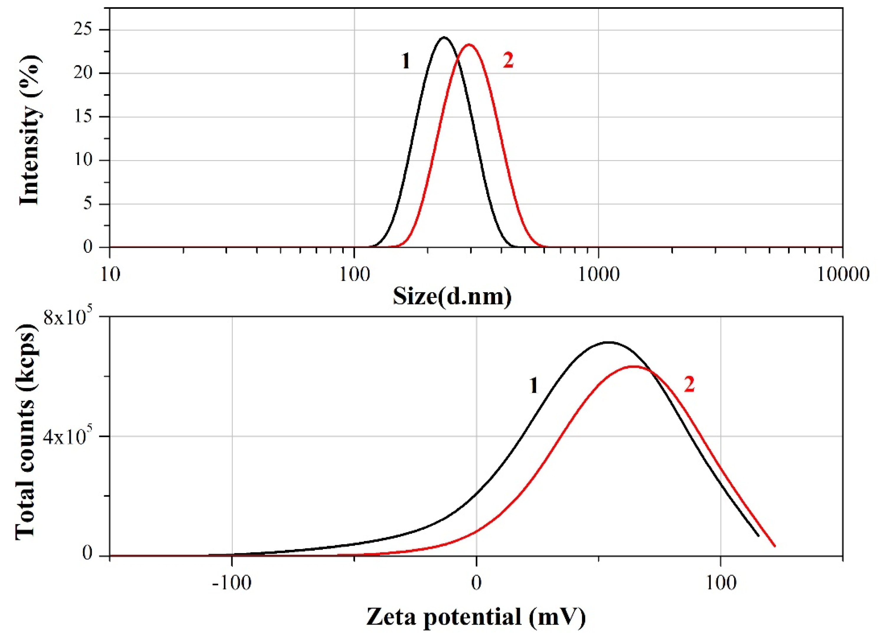
Figure 2.
SEM images, EDS spectra of CS/GEL (left) and ZnO-CS/GEL (right) nanoparticles, drop casted on a glass substrate and dried overnight at room temperature.
Figure 2.
SEM images, EDS spectra of CS/GEL (left) and ZnO-CS/GEL (right) nanoparticles, drop casted on a glass substrate and dried overnight at room temperature.
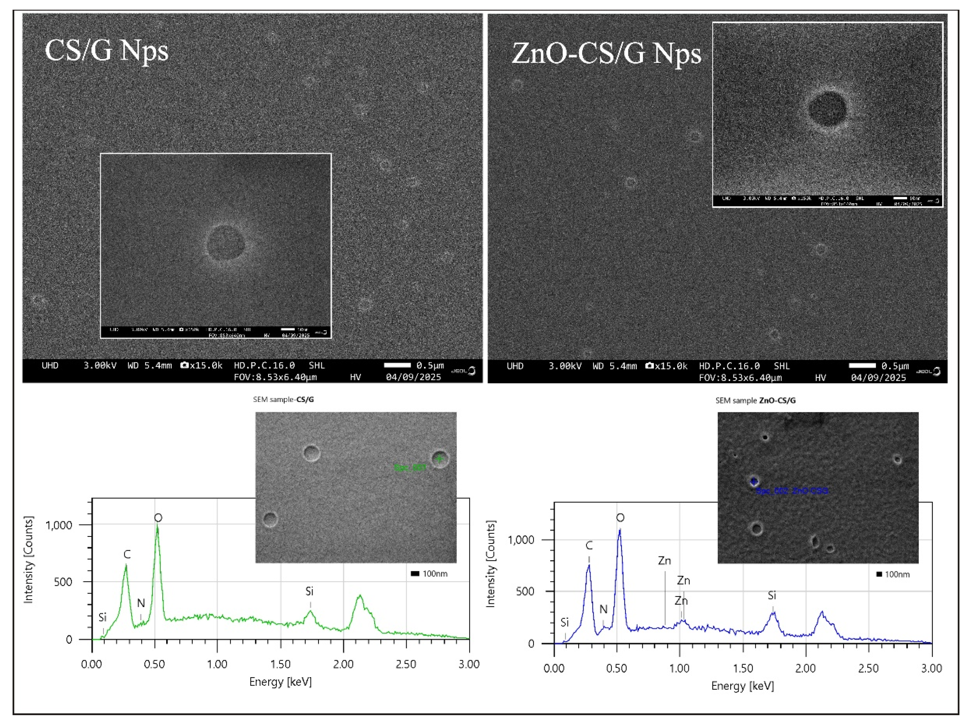
Figure 3.
SEM images of CS/GEL (right) and ZnO-CS/GEL (left) coatings obtained by electrophoretic deposition on stainless steel substrates.
Figure 3.
SEM images of CS/GEL (right) and ZnO-CS/GEL (left) coatings obtained by electrophoretic deposition on stainless steel substrates.
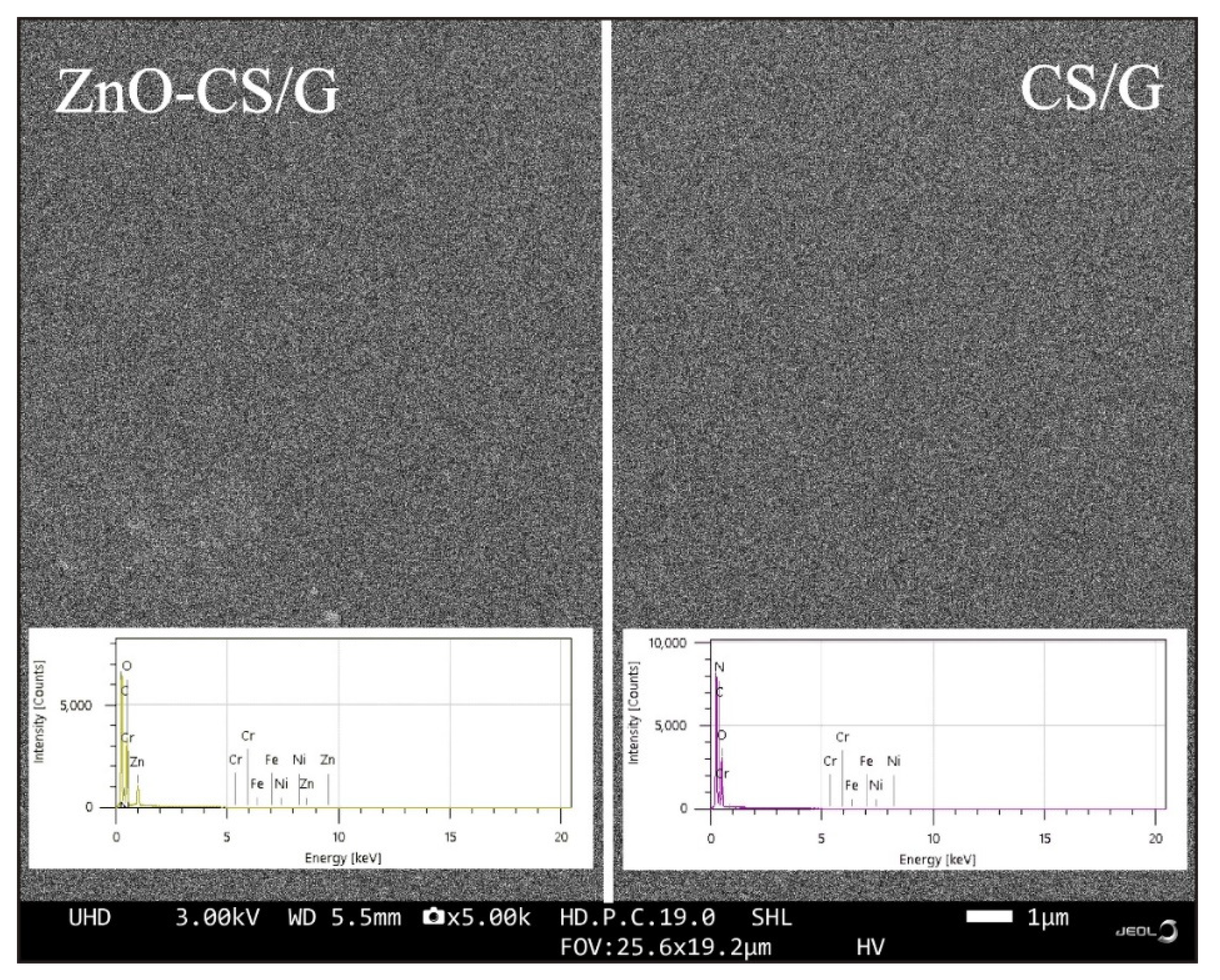
Figure 4.
SEM image of the cross-section of ZnO-CS/GEL coating. Insets: The coating at higher magnification and the result from EDS analysis.
Figure 4.
SEM image of the cross-section of ZnO-CS/GEL coating. Insets: The coating at higher magnification and the result from EDS analysis.
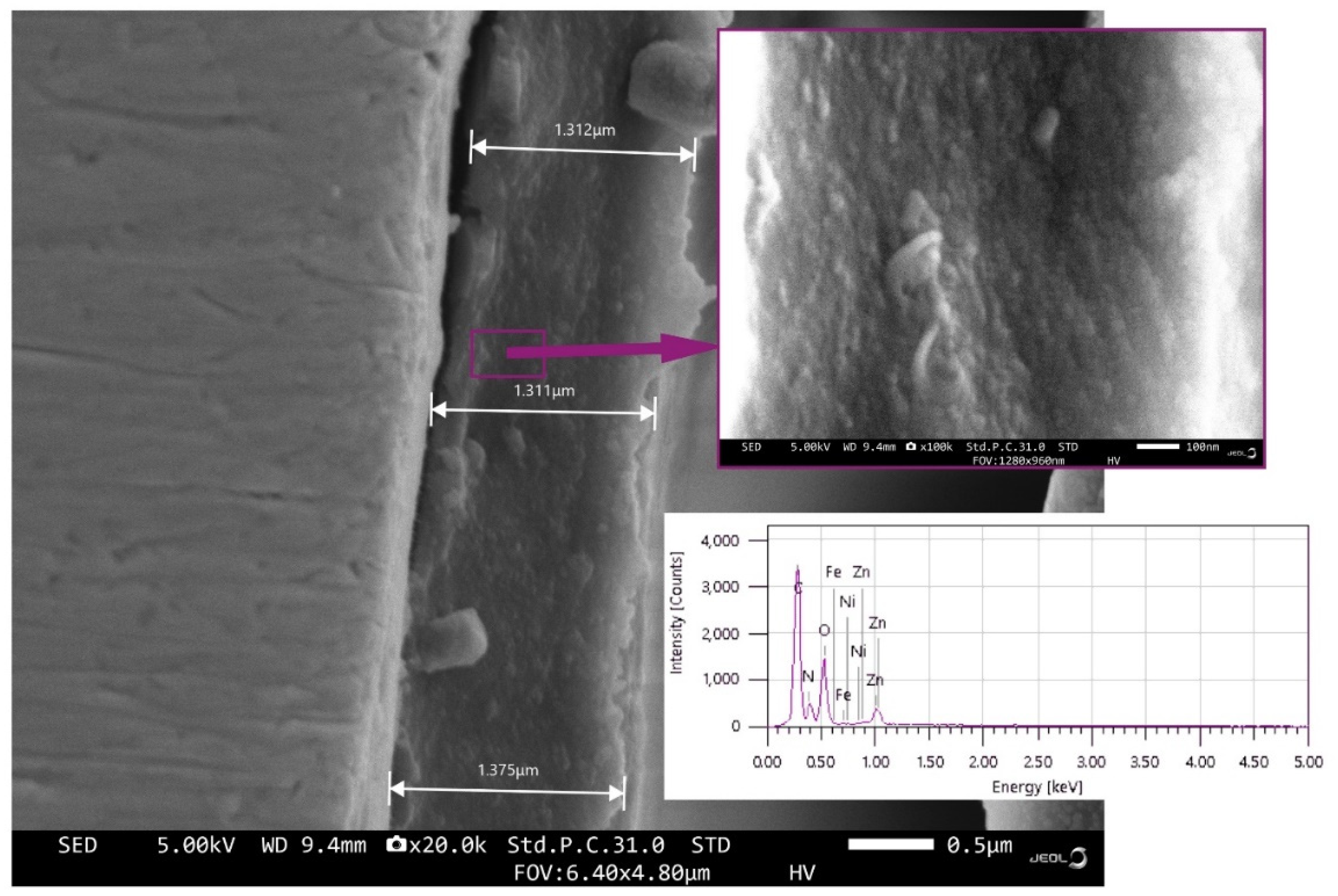
Figure 5.
Polarization resistance measurements during 60 days’ immersion in PBS at pH 7.0: 1 – Stainless steel; 2 – CS/GEL coating; 3 - ZnO-CS/GEL coating.
Figure 5.
Polarization resistance measurements during 60 days’ immersion in PBS at pH 7.0: 1 – Stainless steel; 2 – CS/GEL coating; 3 - ZnO-CS/GEL coating.
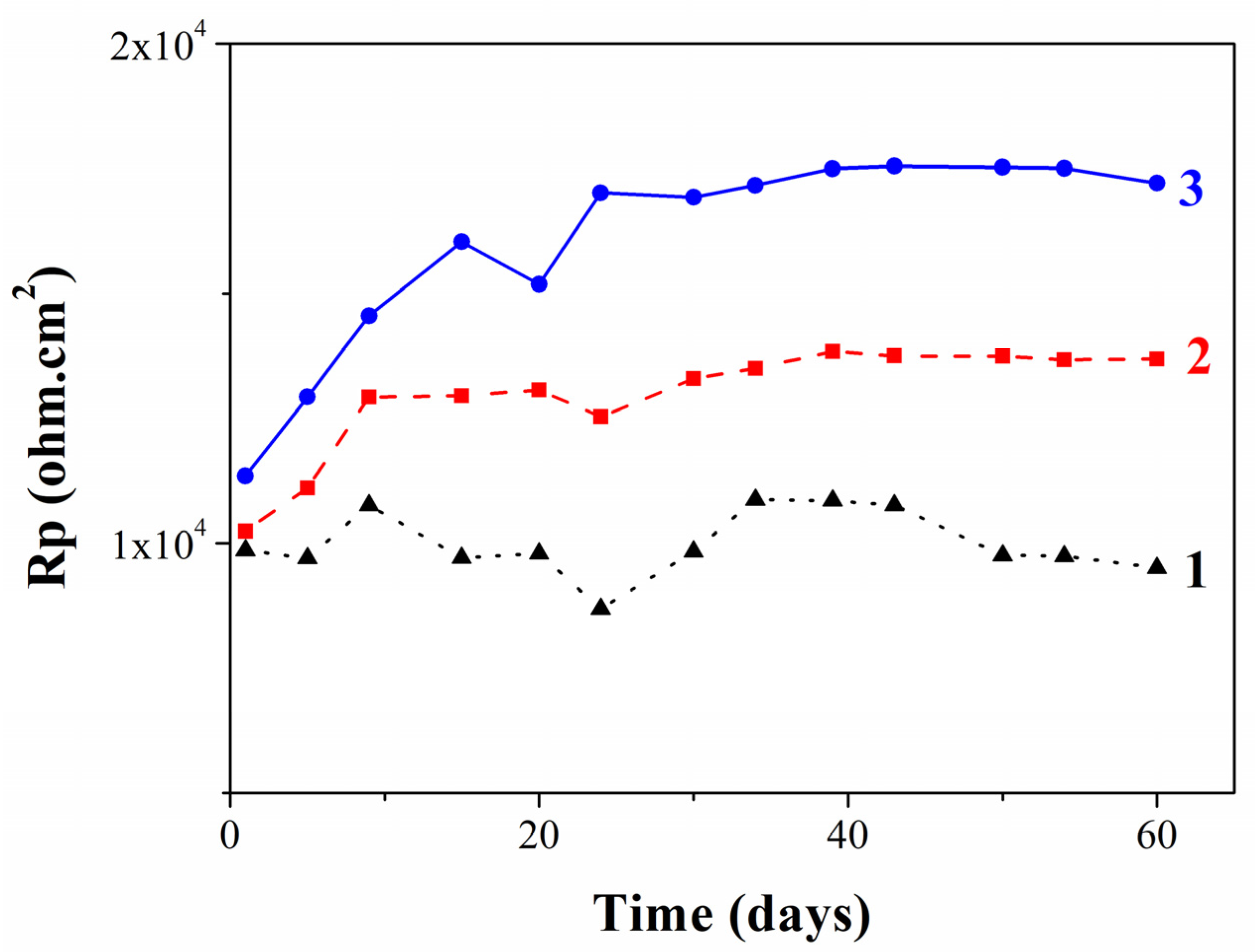
Figure 6.
Deconvolution of C1s spectra and K2p spectra of CS/GEL (left) and ZnO-CS/GEL (right) coatings. The presence of Zn is visible for ZnO-CS/GEL coating.
Figure 6.
Deconvolution of C1s spectra and K2p spectra of CS/GEL (left) and ZnO-CS/GEL (right) coatings. The presence of Zn is visible for ZnO-CS/GEL coating.
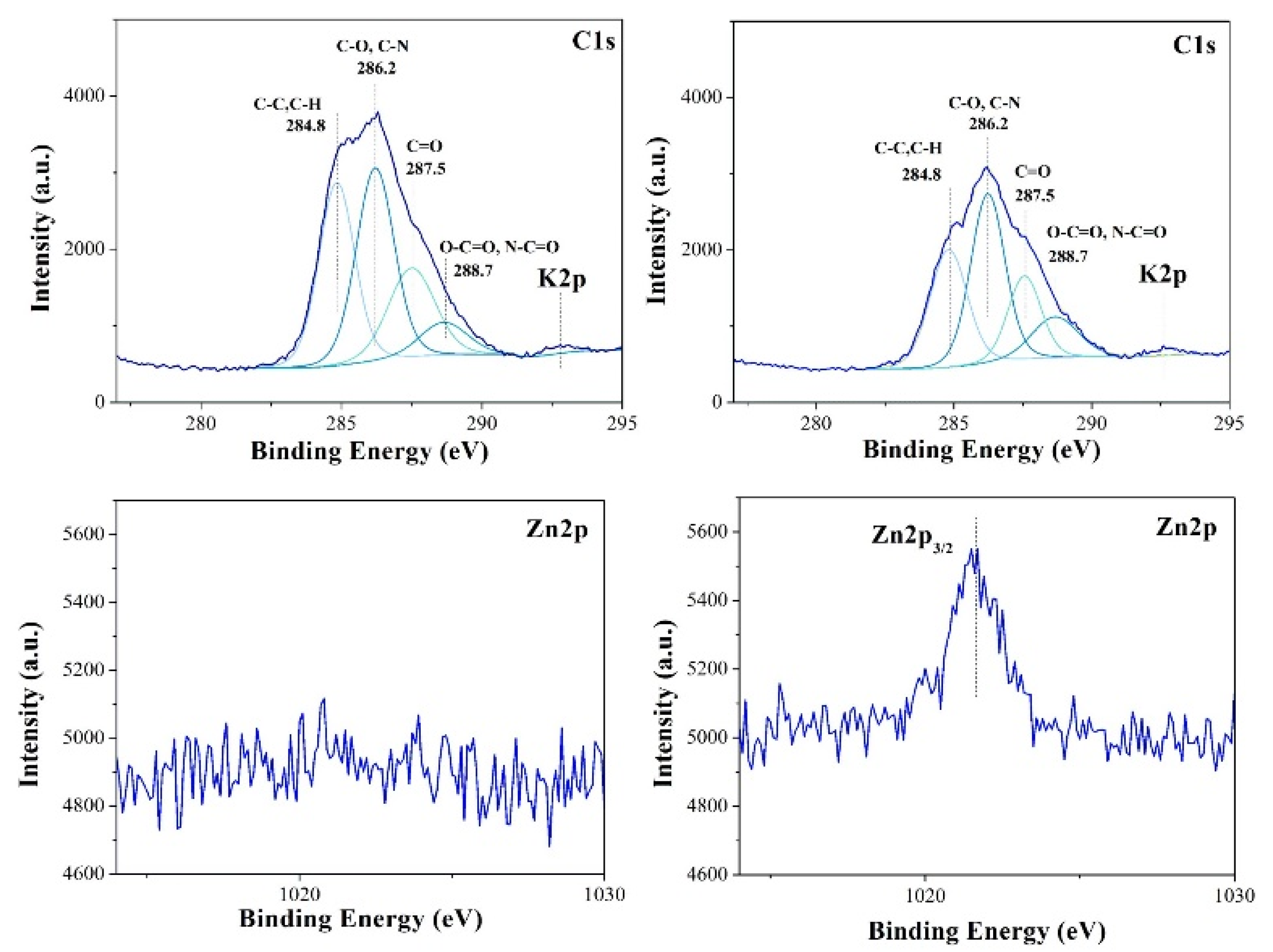
Figure 7.
XPS spectra of CS/GEL (up) and ZnO-CS/GEL (down) coatings showing the relative intensity of O, N, and P.
Figure 7.
XPS spectra of CS/GEL (up) and ZnO-CS/GEL (down) coatings showing the relative intensity of O, N, and P.
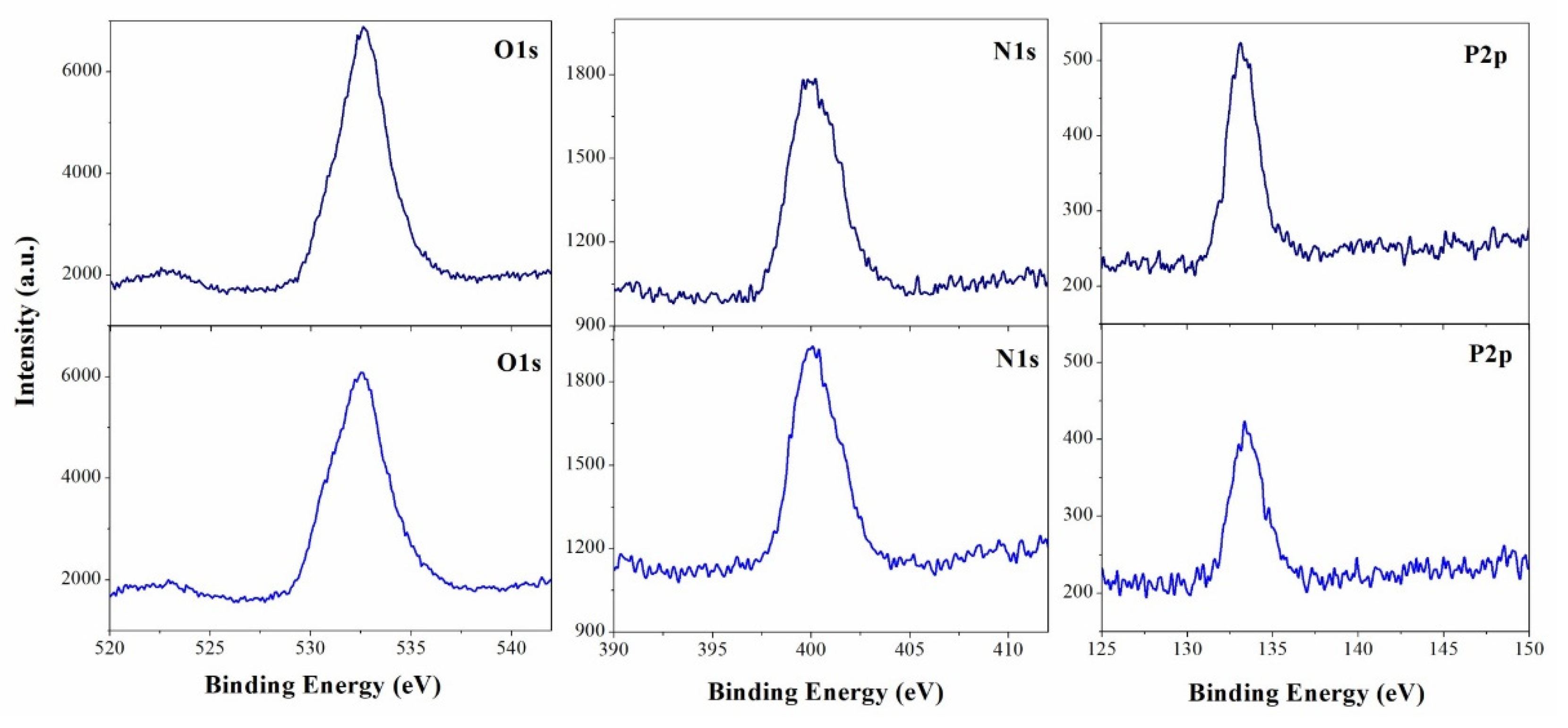

Table 1.
Zeta potential (ζ), hydrodynamic diameter (d), and index of polydispersity (PI) of CS/GEL and ZnO-CS/GEL nanoparticles.
Table 1.
Zeta potential (ζ), hydrodynamic diameter (d), and index of polydispersity (PI) of CS/GEL and ZnO-CS/GEL nanoparticles.
| CS/GEL | ZnO-CS/GEL | |
|---|---|---|
| ζ [mV] | +60 ± 2 | +54 ± 3 |
| d [nm] | 300 ± 70 | 238 ± 30 |
| PI | 0.20 | 0.18 |
Table 2.
Chemical composition (EDS measurements) of CS/GEL and ZnO-CS/GEL nanoparticles drop casted on a glass substrate.
Table 2.
Chemical composition (EDS measurements) of CS/GEL and ZnO-CS/GEL nanoparticles drop casted on a glass substrate.
| Element | CS/GEL | ZnO-CS/GEL |
|---|---|---|
| Atom % | Atom % | |
| C | 31.0 ± 0.4 | 31.7 ± 0.4 |
| N | 2.0 ± 0.3 | 4.4 ± 0.3 |
| O | 44.9 ± 0.9 | 38.1 ± 0.7 |
| Si | 22.1 ± 2.5 | 23.8 ± 1.3 |
| Zn | - | 2.0 ± 0.2 |
| Total | 100.00 | 100.00 |
Table 3.
Chemical composition (EDS measurements) of CS/GEL and ZnO-CS/GEL coatings obtained by electrophoretic deposition on stainless steel.
Table 3.
Chemical composition (EDS measurements) of CS/GEL and ZnO-CS/GEL coatings obtained by electrophoretic deposition on stainless steel.
| Element | ZnO-CS/GEL | CS/GEL |
| Atom % | Atom % | |
| C | 58.5 ± 0.2 | 61.3 ± 0.2 |
| N | 8.7 ± 0.2 | 11.0 ± 0.2 |
| O | 25.3 ± 0.2 | 24.4 ± 0.2 |
| Cr | 3.9 ± 0.2 | 3.1 ± 0.2 |
| Fe | >0.1 | >0.1 |
| Ni | >0.1 | >0.1 |
| Zn | 3.4 ± 0.1 | - |
| Total | 100.00 | 100.00 |
Table 4.
XPS results of CS/GEL and ZnO-CS/GEL coatings (at. %) after prolonged exposure in PBS solution.
Table 4.
XPS results of CS/GEL and ZnO-CS/GEL coatings (at. %) after prolonged exposure in PBS solution.
| Sample | C [at%] |
O [at%] |
N [at%] |
P [at%] |
Na [at%] |
K [at%] |
Zn [at%] |
| CS/GEL | 61.9 | 28.1 | 6.6 | 2.1 | 1.0 | 0.3 | - |
| ZnO-CS/GEL | 55.0 | 31.1 | 8.3 | 3.3 | 1.5 | 0.3 | 0.5 |
Disclaimer/Publisher’s Note: The statements, opinions and data contained in all publications are solely those of the individual author(s) and contributor(s) and not of MDPI and/or the editor(s). MDPI and/or the editor(s) disclaim responsibility for any injury to people or property resulting from any ideas, methods, instructions or products referred to in the content. |
© 2026 by the authors. Licensee MDPI, Basel, Switzerland. This article is an open access article distributed under the terms and conditions of the Creative Commons Attribution (CC BY) license (http://creativecommons.org/licenses/by/4.0/).
Copyright: This open access article is published under a Creative Commons CC BY 4.0 license, which permit the free download, distribution, and reuse, provided that the author and preprint are cited in any reuse.


