
Submitted:
14 July 2026
Posted:
15 July 2026
You are already at the latest version
Abstract
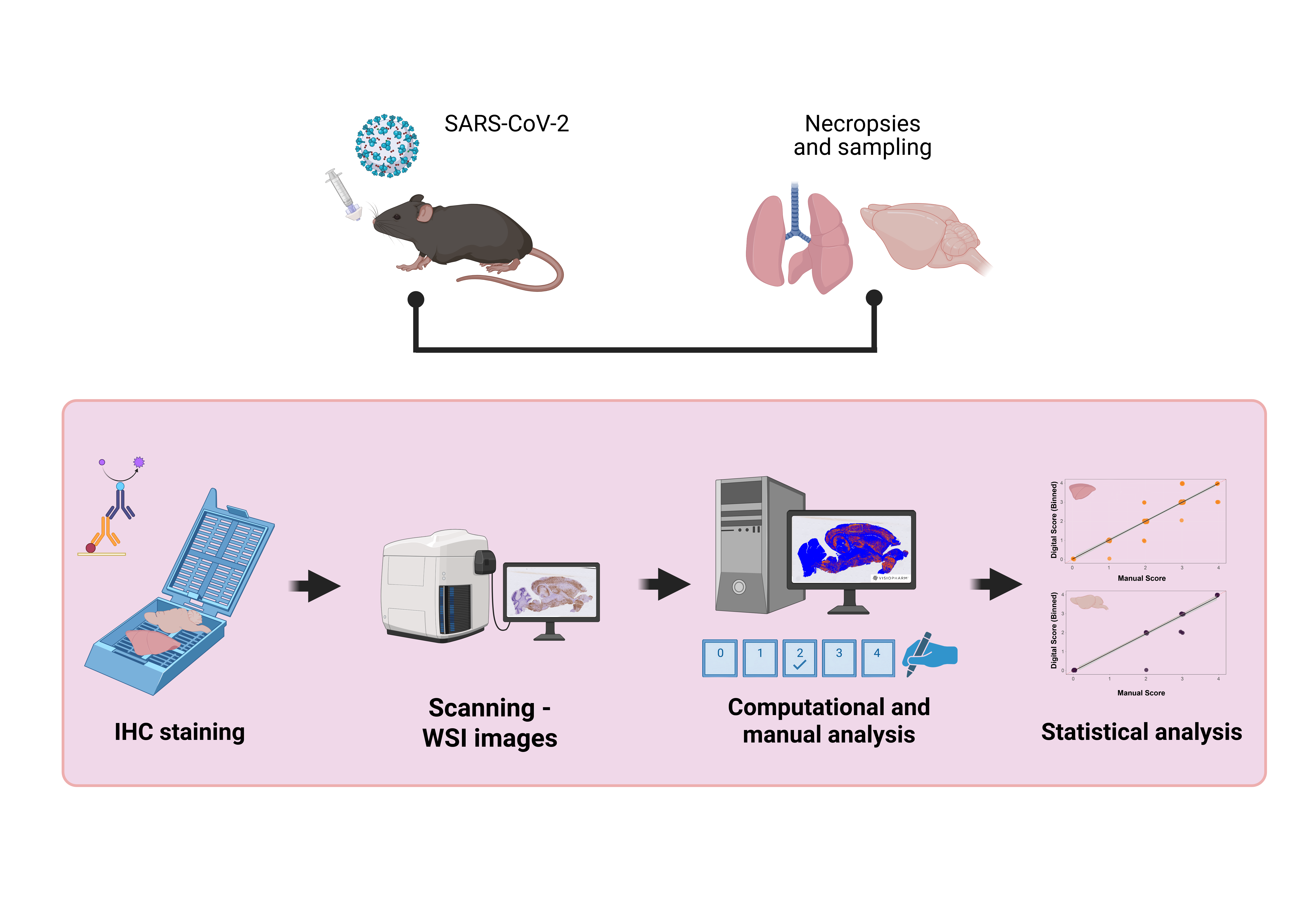
Histopathology and immunohistochemistry (IHC) are central to COVID-19 tissue evaluation. However, conventional manual scoring is limited by certain subjectivity and its semiquantitative nature. In this retrospective study of experimentally infected mice, we implemented a deep learning-based digital pathology workflow using com-mercially available software to quantitatively assess SARS-CoV-2 antigen burden in lung (n=135) and brain (n=67) tissues. The performance of digital quantification was evaluated against conventional manual scoring, and its biological relevance was as-sessed by correlation with established virological and pathological parameters across different stages of disease progression. Digital IHC quantification demonstrated near-perfect agreement with manual scoring in both lung [R=0.94, p< 0.0001, concord-ance correlation coefficient (CCC)=0.969] and brain (R=0.98, p< 0.0001; CCC=0.98) in-dicating high reproducibility and accuracy. In addition, digital antigen quantification showed significant positive correlations with viral RNA levels, infectious viral titers, and histopathological scores, indicating that it provides biologically meaningful readout of SARS-CoV2 infection. Although computational image analysis requires ad-ditional infrastructure, technical expertise, and increased analysis time, these invest-ments provide a more objective and reproducible alternative to the traditional manual gold standard while generating quantitative data that enable a more precise assess-ment of SARS-CoV-2–associated disease.
Keywords:
digital histopathology
; deep learning
; antigen quantification
; SARS-CoV-2
; immunohistochemistry
Copyright: This open access article is published under a Creative Commons CC BY 4.0 license, which permit the free download, distribution, and reuse, provided that the author and preprint are cited in any reuse.


