

Submitted:
14 July 2026
Posted:
15 July 2026
You are already at the latest version
Abstract
Background and Objectives: Rosacea is a chronic inflammatory facial dermatosis in which sensory symptoms can be as clinically important as visible erythema or inflam-matory lesions. In a subset of patients, facial dysesthesia, burning, stinging, warmth, pru-ritus, flushing, and marked trigger sensitivity dominate the disease burden and may im-pair quality of life. This narrative review aimed to reposition neurogenic rosacea as a sen-sory-predominant and translationally relevant presentation within the rosacea spectrum. Materials and Methods: A narrative literature review was conducted using Pub-Med/MEDLINE, Scopus, Web of Science, and Google Scholar through June 2026. Search terms combined rosacea, neurogenic rosacea, neuropathic rosacea, facial dysesthesia, sensory symptoms, neurogenic inflammation, mast cells, transient receptor potential channels, neuropeptides, and therapeutic approaches. Clinical, experimental, and trans-lational articles in English were selected according to relevance to the clinical phenotype, proposed mechanisms, and mechanism-based management. Results: Current clinical, experimental, and translational evidence supports a biologically plausible neurovascu-lar-immune framework linking sensory symptoms, trigger sensitivity, TRP-channel acti-vation, neuropeptide signaling, mast-cell activation, LL-37-MRGPRX2 signaling, and protease-dependent amplification in neurogenic rosacea. Conclusions: Neurogenic rosacea is best regarded as a clinically useful but incompletely validated senso-ry-predominant presentation. Its significance for clinicians and researchers lies in bridg-ing clinical symptoms such as patient-reported facial dysesthesia and quality-of-life im-pairment with plausible neurovascular-immune mechanisms and emerging therapeutic strategies. Standardized diagnostic criteria, validated symptom and quality-of-life measures, disease-relevant biomarkers, and phenotype-stratified trials are needed before neuromodulatory, mast-cell-directed, vascular-targeted, or combination approaches can be recommended as established phenotype-specific therapy.
Keywords:
neurogenic rosacea
; facial dysesthesia
; sensory symptoms
; neurogenic inflammation
; mast-cell activation
; translational therapeutics
1. Introduction
Facial burning, stinging, dysesthesia, warmth, pruritus, flushing, and trigger-induced discomfort are not merely secondary complaints in rosacea; in many patients, they represent the main source of disease burden and quality-of-life impairment. Rosacea is a chronic inflammatory disorder of the centrofacial skin with heterogeneous vascular, inflammatory, ocular, and sensory manifestations. Although rosacea has traditionally been classified according to clinical subtypes, current phenotype-based approaches allow visible signs and patient-reported symptoms to be assessed together, which is particularly important when sensory symptoms are disproportionate to visible erythema or inflammatory lesions [1,2,3,4,5,6].
The pathogenesis of rosacea is multifactorial and cannot be explained solely by vascular dysfunction. Genetic susceptibility, environmental exposures, dysregulated innate and adaptive immune responses, microbial factors, barrier impairment, oxidative stress, and neurovascular dysfunction all contribute to disease expression [3,7,8,9,10]. Among these mechanisms, neurovascular and neuroimmune pathways have gained increasing attention because they may explain several core clinical features, including flushing, persistent erythema, heightened temperature sensitivity, sensory discomfort, and exaggerated reactivity to environmental, dietary, or cosmetic triggers [7,8,11].
Neurogenic rosacea has been described as a symptom-dominant presentation in which facial dysesthesia, burning, stinging, heat sensation, pruritus, persistent flushing, marked trigger reactivity, and poor tolerance or incomplete response to standard rosacea therapies are clinically prominent [12]. More recent literature suggests that neurogenic or neuropathic rosacea may represent a sensory-predominant phenotype within the rosacea spectrum rather than only a severe form of erythematotelangiectatic rosacea [13,14]. This distinction is clinically relevant because patients may continue to experience substantial discomfort and quality-of-life impairment even when visible inflammation is limited or partially controlled [12,13,14,15].
The biological plausibility of this phenotype is supported by evidence that rosacea involves abnormal communication among sensory nerves, vascular structures, keratinocytes, immune cells, fibroblasts, and mast cells. Transient receptor potential (TRP) channels can translate thermal, chemical, mechanical, osmotic, and inflammatory stimuli into neural and vascular responses, while neuropeptides such as substance P, calcitonin gene-related peptide (CGRP), vasoactive intestinal peptide (VIP), and pituitary adenylate cyclase-activating polypeptide (PACAP) may promote vasodilation, edema, nociception, pruritus, and immune-cell activation [7,8,11]. Mast cells may represent one cellular interface within this network because their perivascular and perineural localization allows interaction with neuropeptides, antimicrobial peptides, environmental stimuli, and innate immune signals [8,11,16]. Recent translational studies have implicated fibroblasts, stromal–immune interactions, tissue remodeling, barrier dysfunction, and immune abnormalities in symptom amplification and disease persistence [10,17].
Despite growing interest, neurogenic rosacea remains incompletely defined clinically and mechanistically. Its boundaries with erythematotelangiectatic rosacea with prominent sensory symptoms, sensitive skin syndrome, facial erythromelalgia, migraine-associated facial dysesthesia, and other neuropathic or neurovascular disorders are not clearly established. The therapeutic implications of neuromodulatory, vascular-targeted, barrier-directed, and mast-cell-related approaches also remain under investigation [11,12,13]. Therefore, this narrative review translates the sensory clinical phenotype into a neurovascular-immune framework, emphasizing facial dysesthesia, quality-of-life impact, TRP-channel-mediated trigger sensitivity, neuropeptide signaling, mast-cell activation, treatment implications, and priorities for future research.
2. Materials and Methods
This narrative review was designed to synthesize clinical, experimental, and translational evidence relevant to neurogenic rosacea, with particular attention to facial dysesthesia, sensory symptoms, quality-of-life burden, proposed neurovascular-immune mechanisms, and treatment implications. Searches were conducted in PubMed/MEDLINE, Scopus, Web of Science, and Google Scholar through June 2026 using combinations of the terms rosacea, neurogenic rosacea, neuropathic rosacea, facial dysesthesia, sensory symptoms, neurogenic inflammation, transient receptor potential channels, neuropeptides, mast cells, LL-37, MRGPRX2, and therapeutic approaches.English-language original articles, clinical studies, case series, case reports, experimental and translational studies were considered. Articles were selected when they addressed the clinical phenotype of neurogenic or neuropathic rosacea, sensory symptoms in rosacea, neurovascular or immune pathways, mast-cell-related mechanisms, barrier or stromal-immune interactions, or mechanism-based therapeutic approaches. Additional sources were identified from reference lists of selected publications. Evidence was synthesized qualitatively, with emphasis on clinically relevant findings and on distinguishing direct clinical evidence from the data derived from experimental or broader rosacea studies.
3. Neurogenic Rosacea as a Clinical Phenotype
Within the phenotype-based approach, neurogenic rosacea can be framed as a rosacea presentation in which sensory dysfunction is clinically prominent. This concept is consistent with the current phenotype-based approach to rosacea, which emphasizes clinically relevant signs and symptoms rather than rigid subtype categories. In this phenotype, visible vascular findings are accompanied by symptoms suggesting exaggerated neural and neuroimmune reactivity. Patients often report facial burning or stinging, dysesthesia, heat sensation, itch, and marked intolerance to common triggers, sometimes out of proportion to the degree of visible erythema or inflammation [5,6,12,15]. This phenotype differs from classical erythematotelangiectatic rosacea by the prominence of sensory symptoms and by its often-limited response to conventional rosacea therapies. Although flushing and persistent erythema remain important clinical features, the dominant disease burden may arise from altered sensory thresholds, neurovascular hyperreactivity, and neurogenic inflammation rather than from vascular dilation alone. Therefore, neurogenic rosacea may be more appropriately discussed as a neurovascular–neuroimmune phenotype rather than simply as a severe variant of erythematotelangiectatic rosacea [12,14,18]. Patients with neurogenic rosacea may experience symptom exacerbation after common triggers such as heat, sunlight, emotional stress, exercise, spicy foods, or alcohol. Cooling may provide partial relief in some cases, further supporting the role of altered neurovascular regulation. Compared with more typical inflammatory rosacea, papules and pustules may be less prominent, whereas discomfort, dysesthesia, trigger intolerance, and treatment resistance may be more clinically relevant. These features have been emphasized in both the original description of neurogenic rosacea and subsequent clinical observations of this phenotype [5,12,14,15].
Recognition of this phenotype is important because several other neurovascular or neuropathic conditions may mimic or overlap with neurogenic rosacea. Sensitive skin syndrome, facial erythromelalgia, neuropathic facial pain, trigeminal neuralgia, carcinoid syndrome, and other systemic causes of persistent flushing or refractory facial erythema should be considered when symptoms are severe, atypical, or poorly responsive to standard therapy. Facial erythromelalgia is particularly relevant because it can present with facial erythema, warmth, swelling, burning pain, heat-induced worsening, and relief with cooling, thereby closely resembling neurogenic rosacea in selected cases [14,15,19].
The reported association between rosacea and migraine also supports a possible role for shared trigeminovascular mechanisms. Both disorders may involve neurovascular activation, sensory hypersensitivity, and neuropeptide-mediated signaling. Neuropeptides such as CGRP, substance P, VIP, and PACAP may contribute to facial flushing, dysesthesia, vascular reactivity, and neurogenic inflammation, thereby linking neurogenic rosacea with broader neurovascular pathways [15,18,20,21]. The main clinical and mechanistic features supporting neurogenic rosacea as a sensory-predominant phenotype are summarized in Table 1.
4. Neurovascular and Neuroimmune Mechanisms
Neural pathways are increasingly considered integral to the clinical expression of rosacea, particularly in patients whose disease are dominated by flushing, burning, stinging, dysesthesia, and heightened sensitivity to environmental stimuli. Earlier observations showing that neural stimulation can provoke flushing and enhance cutaneous sensitivity support the concept that rosacea cannot be explained solely by vascular dilation or visible inflammation [22].
In this context, TRP channels are relevant since they allow cutaneous cells and sensory nerves to detect a broad range of environmental and inflammatory stimuli and translate them into neural, vascular, or immune responses. Through these functions, TRP channels may influence nociception, neuropeptide release, vascular tone, immune activation, and the perception of cutaneous discomfort [23,24,25] (Figure 1).
In the skin, TRP channels form a broad sensory-signaling network rather than a pathway confined to peripheral nerves alone. Although they are expressed on sensory nerve terminals, their distribution also includes keratinocytes, endothelial cells, vascular smooth-muscle cells, and several immune-cell populations [24,25]. The expression of TRP channels in neural, vascular, epithelial, and immune compartments supports their possible involvement in rosacea-related trigger sensitivity, neurovascular reactivity, inflammatory signaling, and sensory symptoms such as burning, stinging, and cutaneous discomfort [26,27,28].
The involvement of TRP-dependent pathways in rosacea is supported by their ability to promote the release of vasoactive and proinflammatory neuropeptides from sensory nerves. Activation of these pathways can induce the liberation of substance P, CGRP, VIP, and PACAP, all of which may contribute to vasodilation, edema, pain, pruritus, and inflammatory amplification [18,28,29]. Histologic and molecular studies further support neural participation in rosacea. Erythematotelangiectatic rosacea has been associated with altered sensory innervation, and increased expression of TRP-related channels has been detected in neural, vascular, and immune compartments of rosacea-affected skin [26]. These findings suggest that rosacea may involve a lowered threshold for neurovascular activation, whereby otherwise common stimuli are more readily translated into flushing, burning, stinging, and inflammatory signaling.
5. TRP Channels and Trigger Sensitivity
Among TRP channel subtypes, activation profiles of transient receptor potential vanilloid 1 (TRPV1) and transient receptor potential ankyrin 1 (TRPA1) correspond closely to several thermal, chemical, dietary, and environmental triggers commonly implicated in rosacea exacerbation. TRPV1 is activated by noxious heat, capsaicin, ethanol, and inflammatory mediators, whereas TRPA1 responds to cold exposure and irritant chemicals, including formalin/formaldehyde-related compounds [30,31,32]. Other TRP channels may also contribute to trigger sensitivity. Transient receptor potential vanilloid 4 (TRPV4) can respond to heat, osmotic changes, and inflammatory metabolites, while transient receptor potential melastatin 8 (TRPM8) is activated by cold and menthol [27,33].
Dietary and environmental triggers provide clinically recognizable examples of this mechanism. Many patients report worsening of facial erythema, warmth, or burning after spicy foods or other dietary irritants, and these factors are widely recognized as exacerbating stimuli in rosacea [34,35]. Several exogenous compounds encountered in foods or personal care products can activate TRP channels. Capsaicin, derived from chili peppers, is a TRPV1 agonist; cinnamaldehyde, present in cinnamon, activates TRPA1; and formalin/formaldehyde exposure, including exposure related to formaldehyde-releasing preservatives in cosmetic products, may stimulate TRPA1-dependent signaling [36].
TRPV1 and TRPA1 activation may further propagate neurogenic inflammation by stimulating substance P and CGRP release. Rosacea-affected skin appears to respond to thermal and chemical stimulation at lower thresholds than non-lesional skin, which is consistent with the facial hypersensitivity frequently reported by patients. CGRP may be especially relevant to arteriolar vasodilation, whereas substance P may contribute to edema, mast-cell activation, and amplification of local inflammation [18,28,37,38,39,40]. TRP signaling may also interact with prostaglandin E2 pathways. Such reciprocal sensitization can reduce the activation threshold of sensory channels and intensify pain perception, thereby linking inflammatory mediators to heightened sensory responses [41,42].
Several TRPV channels, including TRPV1, TRPV2, TRPV3, and TRPV4, have been reported to be increased in rosacea-affected skin [27]. Experimental models have also connected cathelicidin antimicrobial peptide LL-37 (LL-37) with TRPV4 upregulation and mast-cell degranulation, suggesting that cathelicidin-derived peptides may amplify rosacea inflammation through both innate immune and neurovascular–mast-cell-dependent pathways [43,44].
6. Neuropeptides, Vasodilation, and Sensory Symptoms
Neuropeptide signaling offers a plausible mechanism through which neural activation may be translated into vascular and sensory manifestations in rosacea. Substance P may contribute to neurogenic edema and inflammatory amplification, in part through neurokinin 1 (NK1) receptor-mediated effects on postcapillary venules and mast cells. CGRP, VIP, and PACAP are strongly vasodilatory mediators and can promote vascular relaxation through actions on vascular smooth-muscle cells and endothelial pathways [45,46]. PACAP may also be released from autonomic nerve fibers and may enhance nitric oxide production by endothelial cells, thereby further supporting vasodilatory responses [46].
CGRP, substance P, VIP, and PACAP may contribute to rosacea-related symptoms through partially overlapping vascular, neural, and immune effects. CGRP is mainly associated with arteriolar vasodilation, whereas substance P can promote plasma extravasation, edema, nociceptive signaling, and mast-cell activation. VIP and PACAP may further support vasodilatory responses through actions on endothelial cells and vascular smooth-muscle cells, including nitric oxide-dependent pathways [28,45,46]. The clinical relevance of neuropeptide-mediated sensory pathways is further supported by reports in other chronic inflammatory skin diseases in which CGRP receptor blockade have improved neurogenic symptoms [47]. These mediators may therefore participate not only in transient flushing but also in facial warmth, persistent erythema, dysesthesia, and altered cutaneous sensitivity in rosacea [18,26].
The contribution of TRP-channel activation to rosacea is not limited to vascular reactivity. By promoting neuropeptide release from sensory nerves, TRP-dependent signaling may also influence immune-cell recruitment, mast-cell activation, and inflammatory amplification [18,28]. Mast cells are particularly relevant in this context because they can respond to neuropeptides and release histamine, tryptase, cytokines, chemokines, and matrix metalloproteinases, all of which may reinforce vascular changes, sensory nerve activation, and local inflammation [16,29,48]. In addition, mast-cell-derived proteases and matrix-remodeling mediators may contribute to stromal changes and tissue remodeling, suggesting that neurogenic inflammation may interact with structural alterations in rosacea skin rather than acting solely as an acute vascular reflex [17,26].
Substance P is also relevant within this neuroimmune network because it can modulate local blood flow and directly activate mast cells [29,48]. In human mast cells, substance P has been shown to induce pro-inflammatory cytokines such as tumor necrosis factor-alpha (TNF-α) and interleukin (IL)-3, as well as chemokines including C-C motif chemokine ligand (CCL) 2, C-X-C motif chemokine ligand (CXCL) 9, CXCL10, CCL5, and CXCL8/IL-8, supporting a role for neuropeptide-driven immune activation in rosacea pathophysiology [48,49]. Neuropeptides may further sustain inflammation by promoting IL-1β expression and enhancing leukocyte recruitment through increased vascular adhesion molecule expression [50,51].
Thus, neurogenic inflammation in rosacea may be considered not only as a transient vascular response but also as a process linked to immune-cell recruitment, inflammatory amplification, and persistent sensory symptoms [18,52]. Neuroimmune crosstalk may additionally involve keratinocytes, fibroblasts, endothelial cells, and mast cells, which can interact with sensory nerves through cytokines, inflammatory mediators, and neuropeptides [17,53]. Sensory neurons may also participate in innate immune signaling through the expression of pattern-recognition receptors, including Toll-like receptors, thereby providing a potential interface between microbial or damage-associated signals and neural activation [27,54].
7. Protease-Dependent Innate Immune Amplification
Protease-dependent mechanisms may provide an additional bridge between epidermal barrier dysfunction, innate immune activation, and neurogenic inflammation. Matriptase and caspase-14 participate in filaggrin processing and epidermal barrier integrity, while kallikrein (KLK) 5 and KLK7 contribute to physiological desquamation. In rosacea, increased KLK5 expression, partly related to enhanced Toll-like receptor 2 (TLR2) signaling, may promote excessive cathelicidin processing and increase the generation of bioactive peptide fragments with vasodilatory and chemotactic properties [55,56,57,58].
Enhanced serine protease activity may also influence sensory signaling through protease-activated receptors. Cross-sensitization between protease-activated receptor (PAR) 2 and TRP pathways provides a plausible mechanism by which protease activity may intensify neurogenic inflammation, pain signaling, and cutaneous hypersensitivity in rosacea [59,60]. This protease activity is consistent with an innate immune amplification loop in which TLR2 upregulation increases epidermal serine protease production and downstream inflammatory responses [57]. Reduced endogenous protease inhibition may further shift the balance toward protease-driven cutaneous activation [61].
8. Therapeutic and Translational Implications
The therapeutic relevance of neurogenic rosacea arises from the possibility that visible inflammation, vascular erythema, and sensory symptoms may be partly dissociated. A patient may show improvement in papules, pustules, or erythema but continue to experience burning, stinging, dysesthesia, warmth, pruritus, or trigger-induced discomfort. Therefore, management should not be guided by visible signs alone; it should also include systematic assessment of sensory symptoms, trigger intolerance, treatment tolerability, and quality-of-life impairment [4,13,15].
Conventional rosacea therapies, including topical metronidazole, ivermectin, azelaic acid, oral tetracyclines, topical calcineurin inhibitors, vasoconstrictive agents, and laser- or light-based modalities, remain important for inflammatory lesions and visible erythema. However, in patients with prominent neurogenic features, these treatments may need to be combined with strategies directed toward neurovascular reactivity, neuropathic-type discomfort, barrier intolerance, and trigger avoidance. Topical alpha-adrenergic agonists such as brimonidine can reduce erythema through reversible vasoconstriction, but erythema reduction alone may not fully improve burning, warmth, or dysesthesia, and rebound erythema may limit use in highly reactive skin [13,78].
Improvement reported with neuromodulatory agents, including gabapentin, pregabalin, tricyclic antidepressants, duloxetine, and related approaches, supports the clinical relevance of neuropathic pain mechanisms in selected patients [13,15]. Gabapentin may be particularly relevant when burning, stinging, or dysesthesia dominate the clinical picture, and emerging experimental evidence suggests additional anti-inflammatory effects in rosacea-like inflammation [79]. From a translational perspective, future trials should stratify patients according to sensory symptom burden and should measure both visible signs and patient-reported outcomes.
A practical mechanism-based approach would begin with identification of the dominant burden: inflammatory lesions, persistent erythema, flushing, facial dysesthesia, barrier intolerance, or overlapping neuropathic pain features. This frame work does not replace standard rosacea treatment; rather, it helps explain why some patients require individualized combinations of anti-inflammatory, vascular-targeted, barrier-supportive, neuromodulatory, procedural, or mast-cell-directed strategies. At present, these approaches should be considered adjunctive or investigational when evidence is limited, and treatment decisions should remain individualized (Figure 2).
9. Mast Cell-Targeted and Mast Cell-Related Therapies
Several current or investigational rosacea therapies may influence mast-cell-related pathways, although the strength of evidence varies substantially among agents. Cromolyn sodium is a classical mast-cell stabilizer that limits degranulation and reduces the release of mast-cell-derived mediators. In LL-37-induced experimental inflammation, cromolyn has been associated with attenuation of rosacea-like changes and reduced cutaneous inflammatory activity, including decreased MMP-related activity and inflammatory mediator expression [16,72,80]. These findings suggest that mast-cell stabilization may reduce both inflammatory amplification and tissue-remodeling signals in cathelicidin-driven rosacea models. Limited clinical evidence has also suggested improvement in facial erythema after topical 4% cromolyn sodium in some patients; however, human data remain preliminary, and cromolyn cannot yet be considered an established therapy for rosacea [72,80].
Hydroxychloroquine may also modulate mast-cell-associated inflammation in rosacea. Experimental studies have shown that hydroxychloroquine can attenuate LL-37-induced rosacea-like inflammation, reduce mast-cell infiltration, and decrease mast-cell-related mediators such as tryptase and MMP9 [81]. These effects may be relevant because tryptase and MMP-related pathways are implicated in inflammation, vascular change, and tissue remodeling. Early clinical data have reported improvement in erythematotelangiectatic and papulopustular rosacea, and a multicenter randomized pilot study suggested that hydroxychloroquine may provide clinical benefit comparable to doxycycline in selected patients [81,82].
Artemisinin and its derivatives represent another group of agents with potential relevance to inflammatory and vascular pathways in rosacea. Experimental findings indicate that artemisinin can suppress inflammation and angiogenesis in rosacea models, partly through modulation of LL-37-induced inflammatory signaling and nuclear factor kappa B (NF-κB)-related pathways [8,83]. Clinically, topical artemether emulsion has shown benefit in papulopustular rosacea in a randomized pilot study, suggesting that this pathway may have therapeutic relevance in inflammatory phenotypes [84].
Brimonidine tartrate 0.33% gel is primarily used for persistent facial erythema because of its α2-adrenergic vasoconstrictive activity. However, experimental data suggest that brimonidine may also reduce mast-cell infiltration and decrease mast-cell-associated enzymes such as tryptase and chymase in LL-37-induced rosacea-like inflammation [85]. This raises the possibility that the therapeutic effects of brimonidine may not be limited to immediate vascular constriction, although the clinical relevance of these mast-cell-related effects remains to be clarified [16,85].
Botulinum toxin has attracted interest for refractory flushing and erythema, particularly in patients with prominent neurovascular symptoms. Its benefit has usually been attributed to neuromodulatory effects, including interference with neurotransmitter release. However, mast-cell-related mechanisms may also contribute. By cleaving soluble N-ethylmaleimide-sensitive factor attachment protein receptor (SNARE) proteins, botulinum toxin may inhibit mast-cell degranulation and reduce the release of mediators that participate in neurogenic inflammation. Experimental data indicate that onabotulinum toxin A can reduce LL-37-induced erythema, mast-cell degranulation, and rosacea-associated biomarkers, including chymase, KLK5, MMP9, and TRP-channel proteins [86]. More recent experimental work has also reported reductions in mast-cell infiltration and inflammatory cytokine expression after botulinum toxin A treatment in rosacea-like inflammation [87]. These findings support the possibility that botulinum toxin may influence both neural signaling and mast-cell-mediated inflammatory amplification.
Overall, mast cells may be considered pathogenetic amplifiers and potential therapeutic targets in rosacea, but the current therapeutic evidence remains uneven. Their ability to respond to LL-37, neuropeptides, and environmental stimuli places them at an intersection between innate immunity, neurovascular activation, inflammatory remodeling, and sensory discomfort [16,43,72]. Recent work on MRGPRX2/MrgprB2 and LL-37-related mast-cell activation further supports the relevance of non-IgE-mediated mast-cell signaling in rosacea-like inflammation [73,74,75]. For clinical translation, the key question is whether mast-cell-related activity identifies patients who are more likely to benefit from mast-cell-stabilizing, anti-inflammatory, neuromodulatory, or combination strategies.
10. Discussion
Neurogenic rosacea may be regarded as a clinically recognizable but still incompletely validated sensory-predominant presentation within the rosacea spectrum. Burning, stinging, dysesthesia, warmth, pruritus, marked trigger sensitivity, flushing, and disproportionate discomfort have been emphasized in the original description of neurogenic rosacea and in subsequent clinical observations [12,13,14]. Although erythema and flushing remain important visible signs, the dominant disease burden in some patients may arise from persistent sensory symptoms that are not fully captured by conventional rosacea severity assessments. This supports the need to evaluate patient-reported discomfort, trigger intolerance, and neuropathic-type symptoms alongside visible inflammatory and vascular findings.
The available evidence suggests that TRP-channel activation and neuropeptide signaling provide plausible links between common rosacea triggers and neurogenic symptoms. Increased expression of TRP-related pathways has been demonstrated in rosacea-affected skin, supporting their relevance to neurovascular reactivity [26,27]. Thermal, chemical, dietary, cosmetic, and inflammatory stimuli may activate TRPV1, TRPA1, TRPV4, TRPM8, and related sensory pathways, leading to the release of neuropeptides such as substance P, CGRP, VIP, and PACAP. These mediators may contribute to vasodilation, edema, nociception, pruritus, mast-cell activation, and immune-cell recruitment [18,28]. However, much of this evidence is derived from experimental models, general neuroinflammatory literature, or broader rosacea studies rather than from phenotype-specific studies of neurogenic rosacea. Therefore, these pathways should be interpreted as biologically plausible rather than definitively proven mechanisms in this subgroup.
Mast cells appear to occupy an important position within this neurovascular–immune network because of their perivascular and perineural localization and their ability to respond to LL-37, neuropeptides, environmental stimuli, and innate immune signals. Experimental studies have shown that mast cells contribute to LL-37-induced rosacea-like inflammation, and they have also been positioned as cellular amplifiers within the neurovascular–immune model of rosacea [16,72]. Emerging data on the LL-37-MRGPRX2/MrgprB2 axis further support the relevance of non-IgE-mediated mast-cell activation in rosacea-like inflammatory responses [73,74,75]. Through the release of histamine, tryptase, chymase, cytokines, chemokines, and matrix metalloproteinases, mast cells may amplify vascular reactivity, sensory nerve activation, inflammation, and tissue remodeling. Nevertheless, whether mast-cell activation is specifically enriched in neurogenic rosacea compared with other rosacea phenotypes remains uncertain.
These mechanisms have therapeutic implications, but the current evidence remains preliminary. Improvement reported with neuromodulatory agents such as gabapentin and pregabalin supports the possible contribution of neuropathic mechanisms in selected patients with prominent burning, stinging, dysesthesia, or neuropathic-type discomfort [13,15]. Similarly, mast-cell-related or neurovascular-targeted approaches, including cromolyn sodium, hydroxychloroquine, brimonidine, artemisinin derivatives, and botulinum toxin, have shown varying degrees of experimental or early clinical support [71,81,84,85,86,87]. However, most available data are derived from small series, pilot studies, case reports, or experimental models, and these treatments should not yet be considered established phenotype-specific therapies for neurogenic rosacea. Overall, neurogenic rosacea remains a useful clinical and pathway-based concept, but further controlled studies are needed before it can be fully integrated into routine diagnostic and therapeutic algorithms.
11. Unmet Needs and Future Directions for Translational Research Conclusion
Several priorities need to be addressed before neurogenic rosacea can be incorporated into routine diagnostic and therapeutic algorithms. Standardized diagnostic criteria are needed to distinguish this presentation from erythematotelangiectatic rosacea with prominent sensory symptoms, sensitive skin syndrome, facial erythromelalgia, neuropathic facial pain, migraine-associated facial dysesthesia, and systemic flushing disorders. In addition, validated patient-reported outcome measures should be developed to capture burning, stinging, dysesthesia, warmth, pruritus, trigger-induced discomfort, treatment intolerance, sleep disturbance, emotional burden, and quality-of-life impairment, because conventional rosacea scales mainly emphasize visible erythema and inflammatory lesions. Furthermore, pathophysiological studies in well-characterized human cohorts should test whether TRP-channel activation, neuropeptide release, mast-cell activation, LL-37-MRGPRX2 signaling, PAR-TRP cross-sensitization, barrier dysfunction, and stromal-immune interactions are specifically enriched in the sensory-predominant phenotype. As a result, phenotype-stratified clinical trials should evaluate neuromodulatory agents, mast-cell-directed strategies, botulinum toxin, vascular-targeted therapies, barrier-directed treatments, and rational combination approaches using both objective clinician-assessed signs and patient-reported sensory outcomes [11,12,13]. These studies would help determine whether neurogenic rosacea is a distinct therapeutic subgroup, an overlap state within the rosacea spectrum, or a symptom-dominant expression of shared neurovascular-immune mechanisms.
11. Conclusion
Neurogenic rosacea may be conceptualized as a sensory-predominant presentation within the rosacea spectrum, characterized by facial dysesthesia, burning, stinging, warmth, pruritus, flushing, marked trigger sensitivity, treatment intolerance, and incomplete responses to conventional therapies. Current evidence supports a translational model in which TRP-channel activation, neuropeptide release, vascular hyperreactivity, mast-cell activation, LL-37-MRGPRX2 signaling, protease-dependent pathways, barrier dysfunction, innate immune activation, and stromal-immune interactions converge within a shared neurovascular-immune network.
Although neurogenic rosacea remains incompletely validated, recognizing this presentation has practical clinical value because it directs attention to patient-reported sensory symptoms and quality-of-life impairment that may persist despite improvement in visible inflammation. Evidence for neuromodulatory, mast-cell-directed, vascular-targeted, and combination strategies remains preliminary, but these approaches provide a rational vision for future phenotype-specific research. Better diagnostic criteria, validated sensory and quality-of-life measures, biological markers, and controlled phenotype-stratified treatment studies are needed before neurogenic rosacea can be fully integrated into routine clinical practice.
Author Contributions
S.M. was responsible for conceptualization, methodology, literature search, investigation, writing-original draft preparation, writing-review and editing, and final approval of the manuscript.
Funding
This research received no external funding.
Institutional Review Board Statement
Not applicable.
Informed Consent Statement
Not applicable.
Data Availability Statement
No new data were created or analyzed in this study. Data sharing is not applicable to this article.
Conflicts of Interest
The author declares no conflicts of interest.
Abbreviations
The following abbreviations are used in this manuscript:
| CCL, | C-C motif chemokine ligand |
| CGRP | calcitonin gene-related peptide |
| CXCL | C-X-C motif chemokine ligand |
| IL | interleukin |
| IgE | immunoglobulin E |
| IPL | intense pulsed light |
| KLK | kallikrein |
| KLK5 | kallikrein 5 |
| KLK7 | kallikrein 7 |
| LL-37 | cathelicidin antimicrobial peptide LL-37 |
| MMPs | matrix metalloproteinases |
| MMP9 | matrix metalloproteinase 9 |
| MRGPRX2 | Mas-related G protein-coupled receptor X2 |
| MrgprB2 | Mas-related G protein-coupled receptor B2 |
| NF-κB | nuclear factor kappa B |
| NK1 | neurokinin 1 |
| PACAP | pituitary adenylate cyclase-activating polypeptide |
| PAR | protease-activated receptor |
| PAR2 | protease-activated receptor 2 |
| SP | substance P |
| SNARE | soluble N-ethylmaleimide-sensitive factor attachment protein receptor |
| TLR2 | Toll-like receptor 2 |
| TNF-α | tumor necrosis factor-alpha |
| TRP | transient receptor potential |
| TRPA1 | transient receptor potential ankyrin 1 |
| TRPM | transient receptor potential melastatin 8 |
| TRPV1 | transient receptor potential vanilloid 1 |
| TRPV2 | transient receptor potential vanilloid 2 |
| TRPV3 | transient receptor potential vanilloid 3 |
| TRPV4 | transient receptor potential vanilloid 4; |
| UV | ultraviolet; |
| VIP | vasoactive intestinal peptide. |
References
- Tan, J.; Almeida, L.M.C.; Bewley, A.; Cribier, B.; Dlova, N.C.; Gallo, R.; et al. Updating the diagnosis, classification and assessment of rosacea: recommendations from the global ROSacea COnsensus (ROSCO) panel. Br. J. Dermatol. 2017, 176, 431–438. [Google Scholar] [CrossRef] [PubMed]
- Schaller, M.; Almeida, L.M.C.; Bewley, A.; Cribier, B.; Del Rosso, J.Q.; Dlova, N.C.; et al. Recommendations for rosacea diagnosis, classification and management: update from the global ROSacea COnsensus 2019 panel. Br. J. Dermatol. 2020, 182, 1269–1276. [Google Scholar] [CrossRef] [PubMed]
- van Zuuren, E.J.; Arents, B.W.M.; van der Linden, M.M.D.; Vermeulen, S.; Fedorowicz, Z.; Tan, J. Rosacea: new concepts in classification and treatment. Am. J. Clin. Dermatol. 2021, 22, 457–465. [Google Scholar] [CrossRef] [PubMed]
- Thiboutot, D.; Anderson, R.; Cook-Bolden, F.; Draelos, Z.D.; Gallo, R.L.; Granstein, R.D.; et al. Standard management options for rosacea: the 2019 update by the National Rosacea Society Expert Committee. J. Am. Acad. Dermatol. 2020, 82, 1501–1510. [Google Scholar] [CrossRef] [PubMed]
- Schaller, M.; Dirschka, T.; Lonne-Rahm, S.B.; Micali, G.; Stein Gold, L.F.; Tan, J.; et al. The importance of assessing burning and stinging when managing rosacea: a review. Acta Derm. Venereol. 2021, 101. [Google Scholar] [CrossRef] [PubMed]
- Gallo, R.L.; Granstein, R.D.; Kang, S.; Mannis, M.; Steinhoff, M.; Tan, J.; et al. Standard classification and pathophysiology of rosacea: the 2017 update by the National Rosacea Society Expert Committee. J. Am. Acad. Dermatol. 2018, 78, 148–155. [Google Scholar] [CrossRef] [PubMed]
- Geng, R.S.Q.; Bourkas, A.N.; Mufti, A.; Sibbald, R.G. Rosacea: pathogenesis and therapeutic correlates. J. Cutan. Med. Surg. 2024, 28, 178–189. [Google Scholar] [CrossRef] [PubMed]
- Tu, K.Y.; Jung, C.J.; Shih, Y.H.; Chang, A.L.S. Therapeutic strategies focusing on immune dysregulation and neuroinflammation in rosacea. Front. Immunol. 2024, 15, 1403798. [Google Scholar] [CrossRef] [PubMed]
- Galluccio, G.; D’Onghia, M.; Malvaso, D.; Lazzeri, L.; Cinotti, E.; Rubegni, G.; et al. Advances in the pathogenesis and treatment of rosacea: a phenotype-based therapeutic approach. Cosmetics 2024, 11, 11. [Google Scholar] [CrossRef]
- Chen, L.X.Y.; Hao, P.S. The role of skin barrier and immune abnormalities in the pathogenesis of Rosacea. Clin. Exp. Med. 2025, 25, 324. [Google Scholar] [CrossRef] [PubMed]
- Li, D.; Yang, W.; Chen, N.; Xu, Y. Updates in the treatment of rosacea with γ-aminobutyric acid derivatives. Clin. Cosmet. Investig. Dermatol. 2025, 18, 2207–2214. [Google Scholar] [CrossRef] [PubMed]
- Scharschmidt, T.C.; Yost, J.M.; Truong, S.V.; Steinhoff, M.; Wang, K.C.; Berger, T.G. Neurogenic rosacea: a distinct clinical subtype requiring a modified approach to treatment. Arch. Dermatol. 2011, 147, 123–126. [Google Scholar] [PubMed]
- Aedo, G.; Chahuán, M.; Gatica, E.; Herrera, I.; Parada, L.F.; Seguel, A.; et al. Managing a burning face: clinical manifestations and therapeutic approaches for neurogenic rosacea. Int. J. Mol. Sci. 2025, 26, 2366. [Google Scholar] [CrossRef] [PubMed]
- Li, M.; Tao, M.; Zhang, Y.; Pan, R.; Gu, D.; Xu, Y. Neurogenic rosacea could be a small fiber neuropathy. Front. Pain Res. 2023, 4, 1122134. [Google Scholar] [CrossRef]
- Kim, H.O.; Kang, S.Y.; Kim, K.E.; Cho, S.Y.; Kim, K.H.; Kim, I.H. Neurogenic rosacea in Korea. J. Dermatol. 2021, 48, 49–55. [Google Scholar] [PubMed]
- Wang, L.; Wang, Y.J.; Hao, D.; Wen, X.; Du, D.; He, G.; et al. The theranostic role of mast cells in the pathophysiology of rosacea. Front. Med. 2020, 6, 324. [Google Scholar] [CrossRef]
- Chen, M.; Yang, L.; Zhou, P.; Jin, S.; Wu, Z.; Tan, Z.; et al. Single-cell transcriptomics reveals aberrant skin-resident cell populations and identifies fibroblasts as a determinant in rosacea. Nat. Commun. 2024, 15, 8737. [Google Scholar] [CrossRef] [PubMed]
- Choi, J.E.; Di Nardo, A. Skin neurogenic inflammation. Semin. Immunopathol. 2018, 40, 249–259. [Google Scholar] [CrossRef] [PubMed]
- Luo, D.Q.; Zhao, Y.K.; Xu, Q.F.; He, X.Q.; Wu, L.C. Recurrent facial erythema with burning sensation and increased temperature: a variant of erythromelalgia or a new entity? Pain Med. 2014, 15, 1007–1010. [Google Scholar] [CrossRef] [PubMed]
- Wienholtz, N.K.F.; Christensen, C.E.; Coskun, H.; Zhang, D.G.; Ghanizada, H.; Egeberg, A.; et al. Infusion of pituitary adenylate cyclase-activating polypeptide-38 in patients with rosacea induces flushing and facial edema that can be attenuated by sumatriptan. J. Investig. Dermatol. 2021, 141, 1687–1698. [Google Scholar] [CrossRef] [PubMed]
- Wienholtz, N.K.F.; Christensen, C.E.; Ashina, H.; Jørgensen, N.R.; Egeberg, A.; Thyssen, J.P.; et al. Elevated plasma levels of calcitonin gene-related peptide in individuals with rosacea: a cross-sectional case-control study. J. Eur. Acad. Dermatol. Venereol. 2025, 39, 181–188. [Google Scholar] [PubMed]
- Drummond, P.D.; Su, D. Endothelial and axon reflex vasodilatation to acetylcholine in rosacea-affected skin. Arch. Dermatol. Res. 2012, 304, 133–137. [Google Scholar] [PubMed]
- Clapham, D.E. TRP channels as cellular sensors. Nature 2003, 426, 517–524. [Google Scholar] [CrossRef] [PubMed]
- Zhang, M.; Ma, Y.; Ye, X.; et al. TRP (transient receptor potential) ion channel family: structures, biological functions and therapeutic interventions for diseases. Signal Transduct. Target. Ther. 2023, 8, 261. [Google Scholar] [CrossRef] [PubMed]
- Parenti, A.; De Logu, F.; Geppetti, P.; Benemei, S. What is the evidence for the role of TRP channels in inflammatory and immune cells? Br. J. Pharmacol. 2016, 173, 953–969. [Google Scholar] [CrossRef] [PubMed]
- Holmes, A.D.; Steinhoff, M. Integrative concepts of rosacea pathophysiology, clinical presentation and new therapeutics. Exp. Dermatol. 2017, 26, 659–667. [Google Scholar] [PubMed]
- Sulk, M.; Seeliger, S.; Aubert, J.; Schwab, V.D.; Cevikbas, F.; Rivier, M.; et al. Distribution and expression of non-neuronal transient receptor potential ion channels in rosacea. J. Investig. Dermatol. 2012, 132, 1253–1262. [Google Scholar] [CrossRef] [PubMed]
- Gouin, O.; L’Herondelle, K.; Lebonvallet, N.; Le Gall-Ianotto, C.; Sakka, M.; Buhé, V.; et al. TRPV1 and TRPA1 in cutaneous neurogenic and chronic inflammation: pro-inflammatory response induced by their activation and their sensitization. Protein Cell 2017, 8, 644–661. [Google Scholar] [CrossRef] [PubMed]
- Schwab, V.D.; Sulk, M.; Seeliger, S.; Nowak, P.; Aubert, J.; Mess, C.; et al. Neurovascular and neuroimmune aspects in the pathophysiology of rosacea. J. Investig. Dermatol. Symp. Proc. 2011, 15, 53–62. [Google Scholar] [CrossRef] [PubMed]
- Shuba, Y.M. Beyond neuronal heat sensing: diversity of TRPV1 heat-capsaicin receptor-channel functions. Front. Cell. Neurosci. 2021, 14, 612480. [Google Scholar] [CrossRef] [PubMed]
- Vetter, I.; Wyse, B.D.; Roberts-Thomson, S.J.; Monteith, G.R.; Cabot, P.J. Mechanisms involved in potentiation of transient receptor potential vanilloid 1 responses by ethanol. Eur. J. Pain. 2008, 12, 441–454. [Google Scholar] [CrossRef] [PubMed]
- Kwan, K.Y.; Allchorne, A.J.; Vollrath, M.A.; Christensen, A.P.; Zhang, D.S.; Woolf, C.J.; et al. TRPA1 contributes to cold, mechanical, and chemical nociception but is not essential for hair-cell transduction. Neuron 2006, 50, 277–289. [Google Scholar] [CrossRef] [PubMed]
- Chuquilin, M.; Alghalith, Y.; Fernandez, K.H. Neurocutaneous disease: cutaneous neuroanatomy and mechanisms of itch and pain. J. Am. Acad. Dermatol. 2016, 74, 197–212. [Google Scholar] [CrossRef] [PubMed]
- Searle, T.; Ali, F.R.; Carolides, S.; Al-Niaimi, F. Rosacea and diet: what is new in 2021? J. Clin. Aesthet. Dermatol. 2021, 14, 49–54. [Google Scholar] [PubMed]
- Nguyen, C.; Kuceki, G.; Birdsall, M.; Sahni, D.R.; Sahni, V.N.; Hull, C.M. Rosacea: practical guidance and challenges for clinical management. Clin. Cosmet. Investig. Dermatol. 2024, 17, 175–190. [Google Scholar] [CrossRef] [PubMed]
- McNamara, C.R.; Mandel-Brehm, J.; Bautista, D.M.; Siemens, J.; Deranian, K.L.; Zhao, M.; et al. TRPA1 mediates formalin-induced pain. Proc. Natl. Acad. Sci. USA 2007, 104, 13525–13530. [Google Scholar] [CrossRef] [PubMed]
- Meseguer, V.; Alpizar, Y.A.; Luis, E.; Tajada, S.; Denlinger, B.; Fajardo, O.; et al. TRPA1 channels mediate acute neurogenic inflammation and pain produced by bacterial endotoxins. Nat. Commun. 2014, 5, 3125. [Google Scholar] [CrossRef] [PubMed]
- Mikkelsen, C.S.; Holmgren, H.R.; Kjellman, P.; Heidenheim, M.; Kappinnen, A.; Bjerring, P.; et al. Rosacea: a clinical review. Dermatol. Rep. 2016, 8, 6387. [Google Scholar] [CrossRef]
- Zhang, Y.; Li, T.; Zhao, H.; et al. High-sensitive sensory neurons exacerbate rosacea-like dermatitis in mice by activating γδ T cells directly. Nat. Commun. 2024, 15, 7265. [Google Scholar] [CrossRef] [PubMed]
- Brain, S.D.; Grant, A.D. Vascular actions of calcitonin gene-related peptide and adrenomedullin. Physiol. Rev. 2004, 84, 903–934. [Google Scholar] [CrossRef] [PubMed]
- Huang, S.M.; Lee, H.; Chung, M.K.; Park, U.; Yu, Y.Y.; Bradshaw, H.B.; et al. Overexpressed transient receptor potential vanilloid 3 ion channels in skin keratinocytes modulate pain sensitivity via prostaglandin E2. J. Neurosci. 2008, 28, 13727–13737. [Google Scholar] [CrossRef] [PubMed]
- Moriyama, T.; Higashi, T.; Togashi, K.; Iida, T.; Segi, E.; Sugimoto, Y.; et al. Sensitization of TRPV1 by EP1 and IP reveals peripheral nociceptive mechanism of prostaglandins. Mol. Pain 2005, 1, 3. [Google Scholar] [CrossRef] [PubMed]
- Mascarenhas, N.L.; Wang, Z.; Chang, Y.L.; Di Nardo, A. TRPV4 mediates mast cell activation in cathelicidin-induced rosacea inflammation. J. Investig. Dermatol. 2017, 137, 972–975. [Google Scholar] [CrossRef] [PubMed]
- Ahn, C.S.; Huang, W.W. Rosacea pathogenesis. Dermatol. Clin. 2018, 36, 81–86. [Google Scholar] [CrossRef] [PubMed]
- Aubdool, A.A.; Brain, S.D. Neurovascular aspects of skin neurogenic inflammation. J. Investig. Dermatol. Symp. Proc. 2011, 15, 33–39. [Google Scholar] [CrossRef] [PubMed]
- Seeliger, S.; Buddenkotte, J.; Schmidt-Choudhury, A.; Rosignoli, C.; Shpacovitch, V.; von Arnim, U.; et al. Pituitary adenylate cyclase activating polypeptide: an important vascular regulator in human skin in vivo. Am. J. Pathol. 2010, 177, 2563–2575. [Google Scholar] [CrossRef] [PubMed]
- Pereira, M.P.; Metz, M. Improvement of prurigo nodularis with erenumab. JAMA Dermatol. 2025, 161, 338–340. [Google Scholar] [CrossRef] [PubMed]
- Kulka, M.; Sheen, C.H.; Tancowny, B.P.; Grammer, L.C.; Schleimer, R.P. Neuropeptides activate human mast cell degranulation and chemokine production. Immunology 2008, 123, 398–410. [Google Scholar] [CrossRef] [PubMed]
- Gerber, P.A.; Buhren, B.A.; Steinhoff, M.; Homey, B. Rosacea: the cytokine and chemokine network. J. Investig. Dermatol. Symp. Proc. 2011, 15, 40–47. [Google Scholar] [CrossRef] [PubMed]
- Shi, X.; Wang, L.; Li, X.; Sahbaie, P.; Kingery, W.S.; Clark, J.D. Neuropeptides contribute to peripheral nociceptive sensitization by regulating interleukin-1β production in keratinocytes. Anesth. Analg. 2011, 113, 175–183. [Google Scholar] [CrossRef] [PubMed]
- Lindsey, K.Q.; Caughman, S.W.; Olerud, J.E.; Bunnett, N.W.; Armstrong, C.A.; Ansel, J.C. Neural regulation of endothelial cell-mediated inflammation. J. Investig. Dermatol. Symp. Proc. 2000, 5, 74–78. [Google Scholar] [CrossRef] [PubMed]
- Kim, Y.J.; Granstein, R.D. Roles of calcitonin gene-related peptide in the skin, and other physiological and pathophysiological functions. Brain Behav. Immun. Health 2021, 18, 100361. [Google Scholar] [CrossRef] [PubMed]
- Lowy, D.B.; Makker, P.G.S.; Moalem-Taylor, G. Cutaneous neuroimmune interactions in peripheral neuropathic pain states. Front. Immunol. 2021, 12, 660203. [Google Scholar] [CrossRef] [PubMed]
- Liu, T.; Ji, R.R. Toll-like receptors and itch. In Itch: Mechanisms and Treatment; Carstens, E., Akiyama, T., Eds.; CRC Press/Taylor & Francis: Boca Raton, FL, USA, 2014; pp. 257–269. [Google Scholar]
- List, K.; Szabo, R.; Wertz, P.W.; Segre, J.; Haudenschild, C.C.; Kim, S.Y.; et al. Loss of proteolytically processed filaggrin caused by epidermal deletion of matriptase/MT-SP1. J. Cell Biol. 2003, 163, 901–910. [Google Scholar] [CrossRef] [PubMed]
- Hoste, E.; Kemperman, P.; Devos, M.; Denecker, G.; Kezic, S.; Yau, N.; et al. Caspase-14 is required for filaggrin degradation to natural moisturizing factors in the skin. J. Investig. Dermatol. 2011, 131, 2233–2241. [Google Scholar] [CrossRef] [PubMed]
- Yamasaki, K.; Kanada, K.; Macleod, D.T.; Borkowski, A.W.; Morizane, S.; Nakatsuji, T.; et al. TLR2 expression is increased in rosacea and stimulates enhanced serine protease production by keratinocytes. J. Investig. Dermatol. 2011, 131, 688–697. [Google Scholar] [CrossRef] [PubMed]
- Two, A.M.; Del Rosso, J.Q. Kallikrein 5-mediated inflammation in rosacea: clinically relevant correlations with acute and chronic manifestations in rosacea and how individual treatments may provide therapeutic benefit. J. Clin. Aesthet. Dermatol. 2014, 7, 20–25. [Google Scholar] [PubMed]
- Amadesi, S.; Cottrell, G.S.; Divino, L.; Chapman, K.; Grady, E.F.; Bautista, F.; et al. Protease-activated receptor 2 sensitizes TRPV1 by protein kinase C epsilon- and A-dependent mechanisms in rats and mice. J. Physiol. 2006, 575, 555–571. [Google Scholar] [CrossRef] [PubMed]
- Dai, Y.; Wang, S.; Tominaga, M.; Yamamoto, S.; Fukuoka, T.; Higashi, T.; et al. Sensitization of TRPA1 by PAR2 contributes to the sensation of inflammatory pain. J. Clin. Investig. 2007, 117, 1979–1987. [Google Scholar] [CrossRef] [PubMed]
- Meyer-Hoffert, U.; Schröder, J.M. Epidermal proteases in the pathogenesis of rosacea. J. Investig. Dermatol. Symp. Proc. 2011, 15, 16–23. [Google Scholar] [CrossRef] [PubMed]
- Patel, S. A critical review on serine protease: key immune manipulator and pathology mediator. Allergol. Immunopathol. 2017, 45, 579–591. [Google Scholar] [CrossRef] [PubMed]
- Akiyama, T.; Lerner, E.A.; Carstens, E. Protease-activated receptors and itch. In Pharmacology of Itch; Cowan, A., Yosipovitch, G., Eds.; Springer: Berlin/Heidelberg, Germany, 2015; Volume 226, pp. 219–235. [Google Scholar]
- Yang, B.; Good, D.; Mosaiab, T.; Liu, W.; Ni, G.; Kaur, J.; et al. Significance of LL-37 on immunomodulation and disease outcome. Biomed. Res. Int. 2020, 2020, 8349712. [Google Scholar] [CrossRef] [PubMed]
- Ramos, R.; Silva, J.P.; Rodrigues, A.C.; Costa, R.; Guardão, L.; Schmitt, F.; et al. Wound healing activity of the human antimicrobial peptide LL-37. Peptides 2011, 32, 1469–1476. [Google Scholar] [CrossRef] [PubMed]
- Coda, A.B.; Hata, T.; Miller, J.; Audish, D.; Kotol, P.; Two, A.; et al. Cathelicidin, kallikrein 5, and serine protease activity is inhibited during treatment of rosacea with azelaic acid 15% gel. J. Am. Acad. Dermatol. 2013, 69, 570–577. [Google Scholar] [CrossRef] [PubMed]
- Kim, J.Y.; Kim, Y.J.; Lim, B.J.; Sohn, H.J.; Shin, D.; Oh, S.H. Increased expression of cathelicidin by direct activation of protease-activated receptor 2: possible implications on the pathogenesis of rosacea. Yonsei Med. J. 2014, 55, 1648–1655. [Google Scholar] [CrossRef] [PubMed]
- Aroni, K.; Tsagroni, E.; Kavantzas, N.; Patsouris, E.; Ioannidis, E. A study of the pathogenesis of rosacea: how angiogenesis and mast cells may participate in a complex multifactorial process. Arch. Dermatol. Res. 2008, 300, 125–131. [Google Scholar] [PubMed]
- Subramanian, H.; Gupta, K.; Guo, Q.; Price, R.; Ali, H. Mas-related gene X2 (MrgX2) is a novel G protein-coupled receptor for the antimicrobial peptide LL-37 in human mast cells: resistance to receptor phosphorylation, desensitization, and internalization. J. Biol. Chem. 2011, 286, 44739–44749. [Google Scholar] [CrossRef] [PubMed]
- Subramanian, H.; Gupta, K.; Ali, H. Roles of Mas-related G protein-coupled receptor X2 on mast cell-mediated host defense, pseudoallergic drug reactions, and chronic inflammatory diseases. J. Allergy Clin. Immunol. 2016, 138, 700–710. [Google Scholar] [CrossRef] [PubMed]
- Yu, Y.; Zhang, Y.; Zhang, Y.; Lai, Y.; Chen, W.; Xiao, Z.; et al. LL-37-induced human mast cell activation through G protein-coupled receptor MrgX2. Int. Immunopharmacol. 2017, 49, 6–12. [Google Scholar] [CrossRef] [PubMed]
- Muto, Y.; Wang, Z.; Vanderberghe, M.; Two, A.; Gallo, R.L.; Di Nardo, A. Mast cells are key mediators of cathelicidin-initiated skin inflammation in rosacea. J. Investig. Dermatol. 2014, 134, 2728–2736. [Google Scholar] [CrossRef] [PubMed]
- Roy, S.; Alkanfari, I.; Chaki, S.; Ali, H. Role of MrgprB2 in rosacea-like inflammation in mice: modulation by β-arrestin 2. J. Investig. Dermatol. 2022, 142, 2988–2997.e3. [Google Scholar] [CrossRef]
- Chow, B.K.C.; Choi, Y.G.; Wong, T.K.; Nawabjan, S.A.; Li, K.; Kumar, M. Therapeutic effect of an MRGPRX2/MRGPRB2 antagonist on LL-37-induced rosacea-like inflammation in mice. Inflamm. Res. 2025, 74, 174. [Google Scholar] [CrossRef]
- Fan, H.; Sun, R.; Ma, Q.; Li, X.; Liu, J.; Zhang, M.; et al. LL-37-driven mast cell degranulation and inflammation in rosacea via TLR2/JAK2/STAT3 axis. Front. Immunol. 2025, 16, 1672021. [Google Scholar] [CrossRef]
- Gupta, K.; Harvima, I.T. Mast cell-neural interactions contribute to pain and itch. Immunol. Rev. 2018, 282, 168–187. [Google Scholar] [CrossRef]
- Siiskonen, H.; Harvima, I. Mast cells and sensory nerves contribute to neurogenic inflammation and pruritus in chronic skin inflammation. Front. Cell. Neurosci. 2019, 13, 422. [Google Scholar] [CrossRef]
- Jóźwiak, W.; Pietrusiewicz, M.; Piechota-Urbańska, M.; Markowicz-Piasecka, M. Brimonidine beyond a single specialty: pharmacological profile, dermatologic applications, and advances in drug delivery systems. Int. J. Mol. Sci. 2026, 27, 1281. [Google Scholar] [CrossRef]
- Jiang, Z.; Ding, T.; Zhao, Y.; Luo, M.; Tang, Y.; Zhang, Y.; et al. Transcriptomic and metabolomic insights into gabapentin’s therapeutic role in neurogenic inflammation of rosacea. Commun. Biol. 2026, 9, 430. [Google Scholar] [CrossRef]
- Marchitto, M.C.; Chien, A.L. Mast cell stabilizers in the treatment of rosacea: a review of existing and emerging therapies. Dermatol. Ther. 2021, 11, 1541–1549. [Google Scholar] [CrossRef]
- Li, J.; Yuan, X.; Tang, Y.; et al. Hydroxychloroquine is a novel therapeutic approach for rosacea. Int. Immunopharmacol. 2020, 79, 106178. [Google Scholar] [CrossRef]
- Wang, B.; Yuan, X.; Huang, X.; et al. Efficacy and safety of hydroxychloroquine for treatment of patients with rosacea: a multicenter, randomized, double-blind, double-dummy, pilot study. J. Am. Acad. Dermatol. 2021, 84, 543–545. [Google Scholar] [CrossRef] [PubMed]
- Yuan, X.; Li, J.; Li, Y.; et al. Artemisinin, a potential option to inhibit inflammation and angiogenesis in rosacea. Biomed. Pharmacother. 2019, 117, 109181. [Google Scholar] [CrossRef] [PubMed]
- Wang, G.J.; Gao, X.Y.; Wu, Y.; et al. Evaluation of the efficacy and tolerance of artemether emulsion for the treatment of papulopustular rosacea: a randomized pilot study. J. Dermatol. Treat. 2019, 30, 809–812. [Google Scholar] [CrossRef]
- Kim, M.; Kim, J.; Jeong, S.W.; Jo, H.; Woo, Y.R.; Park, H.J. Inhibition of mast cell infiltration in an LL-37-induced rosacea mouse model using topical brimonidine tartrate 0.33% gel. Exp. Dermatol. 2017, 26, 1143–1145. [Google Scholar] [CrossRef] [PubMed]
- Choi, J.E.; Werbel, T.; Wang, Z.; Wu, C.C.; Yaksh, T.L.; Di Nardo, A. Botulinum toxin blocks mast cells and prevents rosacea-like inflammation. J. Dermatol. Sci. 2019, 93, 58–64. [Google Scholar] [CrossRef] [PubMed]
- Yoon, D.; Lee, J.O.; Jang, Y.N.; Yoo, K.H.; Kim, B.J.; Choi, S.Y. Effects of botulinum toxin A on rosacea-like inflammation in an LL-37-induced rosacea mouse model. Ann. Dermatol. 2026, 38, 226–236. [Google Scholar] [CrossRef] [PubMed]
Figure 1.
Neurovascular–immune mechanisms in neurogenic rosacea. CGRP, calcitonin gene-related peptide; IL-1β, interleukin-1 beta; KLK5, kallikrein 5; LL-37, cathelicidin antimicrobial peptide LL-37; MMPs, matrix metalloproteinases; MRGPRX2, Mas-related G protein-coupled receptor X2; PACAP, pituitary adenylate cyclase-activating polypeptide; PAR2, protease-activated receptor 2; SP, substance P; TLR2, Toll-like receptor 2; TNF-α, tumor necrosis factor-alpha; TRP, transient receptor potential; TRPA1, transient receptor potential ankyrin 1; TRPM8, transient receptor potential melastatin 8; TRPV1, transient receptor potential vanilloid 1; TRPV4, transient receptor potential vanilloid 4; UV, ultraviolet; VIP, vasoactive intestinal peptide.
Figure 1.
Neurovascular–immune mechanisms in neurogenic rosacea. CGRP, calcitonin gene-related peptide; IL-1β, interleukin-1 beta; KLK5, kallikrein 5; LL-37, cathelicidin antimicrobial peptide LL-37; MMPs, matrix metalloproteinases; MRGPRX2, Mas-related G protein-coupled receptor X2; PACAP, pituitary adenylate cyclase-activating polypeptide; PAR2, protease-activated receptor 2; SP, substance P; TLR2, Toll-like receptor 2; TNF-α, tumor necrosis factor-alpha; TRP, transient receptor potential; TRPA1, transient receptor potential ankyrin 1; TRPM8, transient receptor potential melastatin 8; TRPV1, transient receptor potential vanilloid 1; TRPV4, transient receptor potential vanilloid 4; UV, ultraviolet; VIP, vasoactive intestinal peptide.
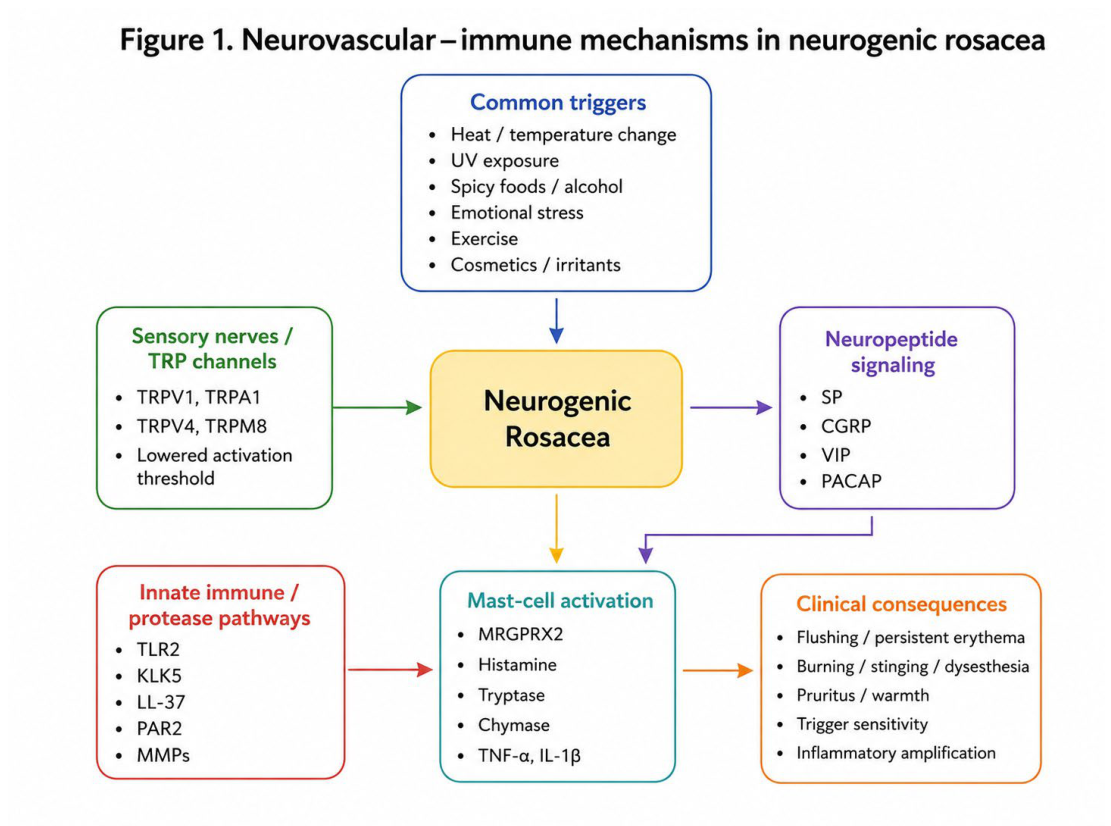
Figure 2.
Therapeutic approaches in neurogenic rosacea. This figure outlines a simplified mechanism-based management framework for neurogenic rosacea. General measures, conventional rosacea-directed therapies, vascular-targeted therapies, neuromodulatory approaches, and mast-cell- or mechanism-based strategies may address different components of the sensory, vascular, inflammatory, and neuroimmune burden. However, evidence for phenotype-specific treatment in neurogenic rosacea remains limited, and several approaches should be considered adjunctive, individualized, or investigational rather than established standards. Abbreviations: IPL, intense pulsed light; TRP, transient receptor potential; UV, ultraviolet.
Figure 2.
Therapeutic approaches in neurogenic rosacea. This figure outlines a simplified mechanism-based management framework for neurogenic rosacea. General measures, conventional rosacea-directed therapies, vascular-targeted therapies, neuromodulatory approaches, and mast-cell- or mechanism-based strategies may address different components of the sensory, vascular, inflammatory, and neuroimmune burden. However, evidence for phenotype-specific treatment in neurogenic rosacea remains limited, and several approaches should be considered adjunctive, individualized, or investigational rather than established standards. Abbreviations: IPL, intense pulsed light; TRP, transient receptor potential; UV, ultraviolet.
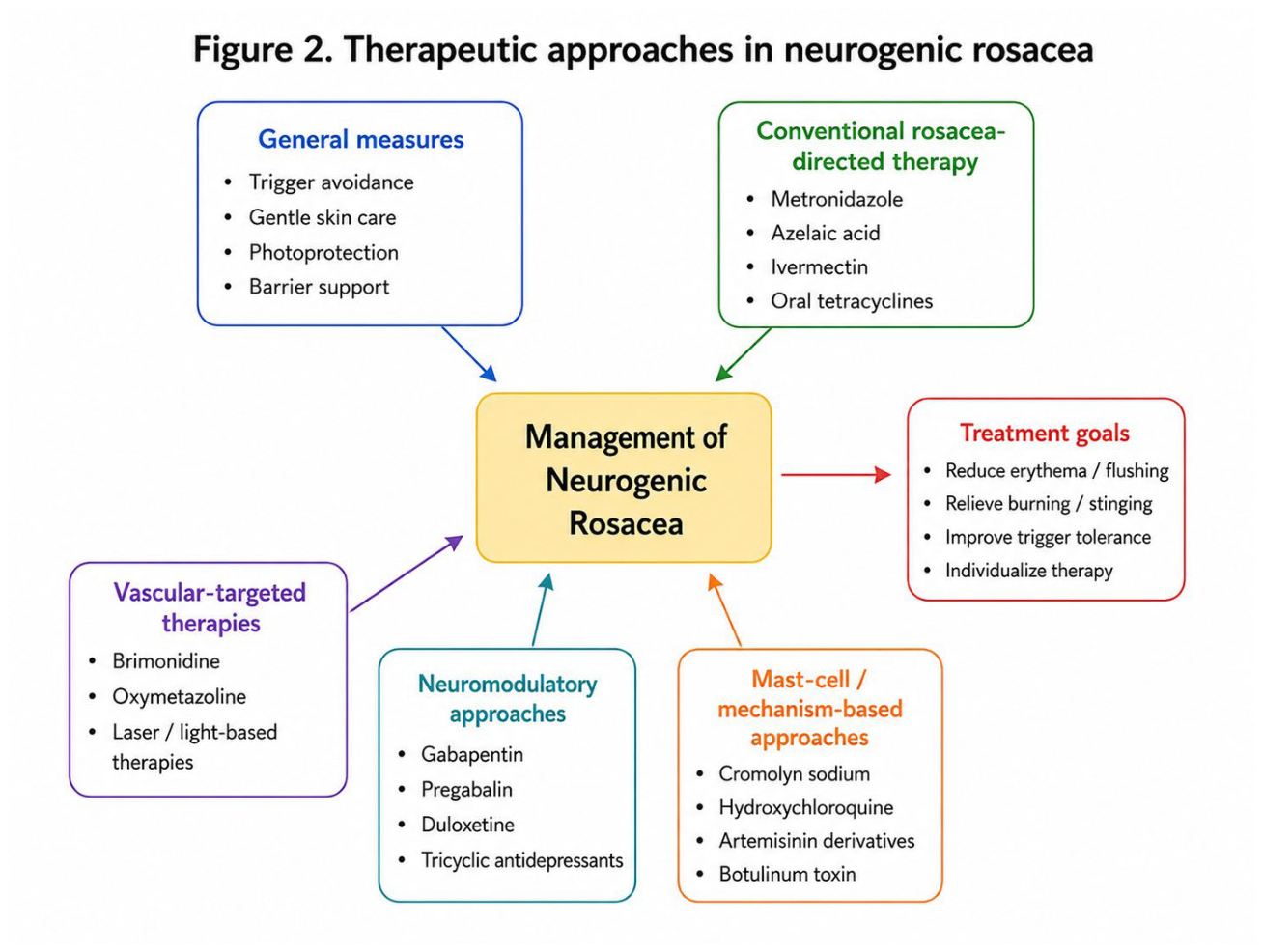

Table 1.
Clinical and pathophysiological features of neurogenic rosacea and their potential therapeutic relevance.
Table 1.
Clinical and pathophysiological features of neurogenic rosacea and their potential therapeutic relevance.
| Clinical/mechanistic aspect | Main features or mechanisms | Clinical relevance | Key references |
| Clinical phenotype | Burning, stinging, dysesthesia, warmth, pruritus, flushing, marked trigger sensitivity, and incomplete response to conventional rosacea therapy | Supports recognition of a sensory-predominant presentation within the rosacea spectrum | [12,13,14,15] |
| Trigger sensitivity and TRP signaling | Heat, UV exposure, spicy foods, alcohol, cosmetics, irritants, and inflammatory stimuli may activate TRPV1, TRPA1, TRPV4, TRPM8, and related sensory pathways | Provides a plausible link between common triggers and symptoms such as flushing, burning, stinging, and warmth | [26,27,30,31,32,33] |
| Neuropeptide-mediated neurovascular activation | SP, CGRP, VIP, and PACAP may promote vasodilation, edema, nociception, pruritus, and immune-cell activation | May explain persistent erythema, flushing, facial discomfort, and neurogenic inflammation | [18,28,45,46] |
| Mast-cell amplification | Mast cells can respond to LL-37, neuropeptides, and innate immune signals, releasing histamine, tryptase, chymase, cytokines, chemokines, and MMPs | May reinforce vascular hyperreactivity, sensory nerve activation, inflammatory amplification, and tissue remodeling | [16,43,71] |
| LL-37–MRGPRX2/MrgprB2 axis | LL-37 may activate mast cells through non-IgE-mediated pathways involving MRGPRX2/MrgprB2 | Suggests a mechanistic bridge between innate immune dysregulation and mast-cell-driven neuroinflammation | [69,70,71,72,73,74] |
| Protease-dependent amplification | TLR2, KLK5, KLK7, PAR2, MMPs, and altered protease inhibition may interact with cathelicidin processing, barrier dysfunction, and sensory signaling | May contribute to inflammation, cutaneous hypersensitivity, pruritus, pain, and persistent tissue activation | [55,56,57,58,59,60,61,62,63] |
| Therapeutic implications | Neuromodulatory agents, mast-cell-related approaches, vascular-targeted therapies, botulinum toxin, and selected anti-inflammatory agents have been explored | These approaches provide a mechanism-based rationale for individualized management, although phenotype-specific therapeutic evidence is still emerging. | [13,15,71,80,83,84,85,86] |
CGRP, calcitonin gene-related peptide; IgE, immunoglobulin E; KLK, kallikrein; LL-37, cathelicidin antimicrobial peptide LL-37; MMPs, matrix metalloproteinases; MrgprB2, Mas-related G protein-coupled receptor B2; MRGPRX2, Mas-related G protein-coupled receptor X2; PACAP, pituitary adenylate cyclase-activating polypeptide; PAR2, protease-activated receptor 2; SP, substance P; TLR2, Toll-like receptor 2; TRP, transient receptor potential; TRPA1, transient receptor potential ankyrin 1; TRPM8, transient receptor potential melastatin 8; TRPV1, transient receptor potential vanilloid 1; TRPV4, transient receptor potential vanilloid 4; UV, ultraviolet; VIP, vasoactive intestinal peptide.
Disclaimer/Publisher’s Note: The statements, opinions and data contained in all publications are solely those of the individual author(s) and contributor(s) and not of MDPI and/or the editor(s). MDPI and/or the editor(s) disclaim responsibility for any injury to people or property resulting from any ideas, methods, instructions or products referred to in the content. |
© 2026 by the authors. Licensee MDPI, Basel, Switzerland. This article is an open access article distributed under the terms and conditions of the Creative Commons Attribution (CC BY) license (http://creativecommons.org/licenses/by/4.0/).
Copyright: This open access article is published under a Creative Commons CC BY 4.0 license, which permit the free download, distribution, and reuse, provided that the author and preprint are cited in any reuse.


