

Submitted:
14 July 2026
Posted:
15 July 2026
You are already at the latest version
Abstract
Tenacibaculosis, caused by Tenacibaculum maritimum, remains a major bacterial disases affecting marine aquaculture, particularly European seabass farming. This study evaluated the immune response and efficacy of a formalin-inactivated T. maritimum vaccine administered by immersion or intraperitoneal (i.p.) injection in European seabass (Dicentrarchus labrax). Fish were challenged 36 days post-vaccination. Vaccine performance was assessed by relative percent survival (RPS), expression of immune markers (IL-1β, IL-10, IgT), and specific IgM production. Intraperitoneal vaccination provided significant protection, with 87% survival and an RPS of 67.5, accompanied by elevated serum IgM levels and strong systemic immune activation. Immersion vaccination resulted in complete survival under the experimental conditions; however, low mortality in the control group prevented reliable assessment of vaccine efficacy. Gene expression analysis revealed distinct immune profiles: immersion vaccination induced rapid but transient mucosal and systemic responses, whereas i.p. vaccination elicited stronger and more sustained systemic responses, particularly in innate and humoral pathways. Principal component analysis confirmed clear separation of immune responses according to vaccination route. Overall, i.p. vaccination induced stronger protective immunity, while optimization of immersion vaccination protocols is needed to improve protection against tenacibaculosis in European seabass.
Keywords:
Tenacibaculum maritimum
; Dicentrarchus labrax
; vaccination
; immune response
; relative percentage of survival (RPS)
1. Introduction
Tenacibaculum spp. infections are commonly referred to as marine tenacibaculosis, or simply tenacibaculosis, a term originally used to describe the ulcerative disease caused by the Gram negative filamentous bacterium Tenacibaculum maritimum [1]. Clinically, tenacibaculosis is associated with gross lesions on the body, including ulcerative and/or necrotic skin lesions, eroded or hemorrhagic mouth, frayed fins, and tail rot [1,2]. Since its first description as a gliding bacterial infection affecting fry of black seabream (Acanthopagrus schlegeli) in Japan [3], T. maritimum infection has been reported from aquaculture sites worldwide. The pathogen was described in Europe in the early 1990s, causing disease outbreaks in European sea bass (Dicentrarchus labrax) rearing facilities along the French Mediterranean coast [4]. Subsequent cases of tenacibaculosis in farmed European sea bass were reported in Italy, Greece, Turkey and Croatia [5,6,7,8], highlighting the growing concern regarding this disease in Mediterranean aquaculture. Given the increasing evidence demonstrating the pathogenicity of T. maritimum in European sea bass and the financial and environmental costs associated with prolonged antibiotic use [9], the development of an effective vaccine would be invaluable.
Vaccination represents a cost-effective and sustainable strategy for controlling infectious diseases and reducing stock losses in aquaculture [10,11,12,13]. Among the available vaccination methods, immersion vaccination is particularly attractive due to its practicality for mass immunization of small fish, which are highly susceptible to infectious agents [11,14]. However, immune responses induced by immersion vaccination are generally less potent and of shorter duration than those elicited by injection-based methods [11,13,15]. The intraperitoneal (i.p.) route is considered the most efficient vaccination method [16], as it induces strong and long-lasting systemic immune protection and immunological memory.
Nevertheless, i.p. vaccination is also associated with stress responses in fish, which may compromise vaccine-induced protection due to handling, injection procedures, and antigen exposure [17].
Effective vaccination relies on exposure of the host to a harmless form of the pathogen, leading to immune cell recruitment and antibody production by B lymphocytes. Antibodies contribute to infection control through mechanisms such as opsonization, complement activation, and toxin neutralization [18]. In teleost fish, IgM represents the main antibody isotype involved in both primary and secondary humoral immune responses, as fish lack the IgG isotype found in mammals [19,20]. Consequently, serum IgM levels are commonly used to evaluate humoral immune memory following vaccination. In addition, mucosal immune responses to T. maritimum infection involve the modulation of cytokines such as the pro-inflammatory IL-1β and the regulatory cytokine IL-10 [21]. IgT in sea bass shows high expression in mucosal organs such as the gut and gills, suggesting its role in mucosal immune responses [22]. The European sea bass (D. labrax) was the first non-salmonid marine fish species to be commercially cultured in Europe and remains, together with gilthead sea bream (Sparus aurata), one of the most important species in Mediterranean aquaculture. Given its high economic value and susceptibility to bacterial diseases, improving disease prevention strategies in this species is of major importance. Advances in molecular and physiological approaches have provided valuable tools for assessing immune responses and vaccine efficacy in cultured fish [23].
The aim of this study was to compare the immune responses and protective efficacy elicited by immersion and intraperitoneal vaccination with a formalin-inactivated T. maritimum vaccine in European seabass (D. labrax).
2. Materials and Methods
2.1. Vaccine Preparation
The vaccine was prepared using T. maritimum isolated from European seabass with clinical symptoms of tenacibaculosis. The bacterial culture was initiated using a starter inoculum and grown in a 2 L fermenter containing Marine Broth (MB) (Condalab, Spain) for 72 h at 24 °C under controlled conditions. Following incubation, the biomass was subjected to ultrasonic homogenization, and the final concentration was adjusted to an optical density of OD₆₀₀ = 3.1. Biomass purity was confirmed by plating aliquots onto Tryptic Soy Agar (TSA) (Merck, Germany) and incubating under standard conditions. The vaccine was prepared as a formalin-inactivated whole-cell bacterin by treating the biomass with 0.4% (v/v) formalin for 48 h. Complete inactivation and sterility were verified according to standard bacteriological protocols.
2.2. Fish and Experimental Conditions
European seabass fry was obtained from a marine fish farm. Upon arrival at the laboratory, the absence of T. maritimum infection was confirmed by negative bacteriological cultures of spleen, head kidney, and heart samples, as well as negative swab cultures from the skin and gills. The fish were acclimated in recirculating seawater tanks at the experimental facility for two weeks at a water temperature of 22 ± 2 °C and a dissolved oxygen concentration of 8.1 mg/L. During the acclimation period, the fish were fed daily at 1.5 % of their body weight. Before the experimental vaccination, the safety and non-toxicity of the vaccine, as well as the vaccination protocol, were preliminarily evaluated in experimental groups of 20 fish for both immersion and intraperitoneal (i.p.) vaccination. No mortality or adverse effects were observed during the 40-day observation period.
The vaccine was stored at 4 ± 2 °C and brought to room temperature before use. It was gently mixed to ensure homogeneity prior to administration.
A total of 192 clinically healthy European seabass of uniform size (60 g) were used in the experiment. In the i.p. vaccination trial, fish were randomly allocated into three treatment groups, each comprising 16 individuals (total n = 48), along with a control group consisting of 48 fish. In the immersion vaccination trial, fish were similarly distributed into three treatment groups of 16 individuals each (total n = 48), with a corresponding control group of 48 fish (Figure 1). For gene expression analysis, six fish per group, including control groups, were sampled at 4 h, 24 h, and 10 days post-vaccination. Fish were not fed for 48 h prior to handling or vaccination to minimize stress and reduce the risk of complications. All equipment used during vaccination was cleaned and disinfected before use.
2.3. Anaesthesia and Handling
Before vaccination, fish were anesthetized by immersion in a seawater solution containing 17.5 mg/L AQUI-S® Aquatic Anaesthetic (AQUI-S, Intervet International B.V., Netherlands) for 5-8 minutes, until they lost responsivness to visual and tactile stimuli while maintaining a normal respiratory rate. During anaesthesia, supplemental oxygen was provided, and oxygen saturation was maintained above 80%. Fish were returned to their tanks after vaccination and feeding was resumed two days post-vaccination. Fish sampled throughout the experimental period for gene expression analysis, as well as all surviving fish at the end of the experiment from both the infection and control groups, were euthanized by immersion in seawater containing 175 mg/L AQUI-S for at least 10 min following the cessation of opercular movements. Carcasses were disposed of in accordance with biological hazardous waste disposal protocols. All animal experimental procedures were conducted in according to the experimental research protocol approved by the National Ethics Committee of the Croatian Ministry of Agriculture Forestry and Fisheries (EP 470/2025), issued on May 16, 2025 and in accordance with Directive 2010/63/EU of the European Parliament and of the Council of 22 September 2010 on the protection of animals used for scientific purposes and amended with Consolidated text: Directive 2010/63/EU of the European Parliament and of the Council of 22 September 2010 on the protection of animals used for scientific purposes.
2.4. Vaccination
2.4.1. Immersion Vaccination
Totally 48 anesthetised fish (average body weight ~60 g) were vaccinated by immersion. The vaccine was diluted in seawater at a ratio of 1:9, resulting at a final concentration of 3.75 × 10⁶ CFU mL⁻¹. Fish were immersed in the vaccine solution for 2 min under laboratory conditions (Figure 1). Vaccinated fish were distributed into three experimental tank; 16 specimen per tank.
Additional 48 specimen were anesthetised and were immersed in 5 L of ambient seawater for 2 min and distributed into three experimental tanks to follow the experimental design.
2.4.2. Intraperitoneal (i.p.) Vaccination
Group od 48 anesthetised fish were individually vaccinated by intraperitoneal injection of 0.2 mL of vaccine per fish, containing 6.0 × 10⁷ CFU mL⁻¹. The injection was performed manually along the ventral midline (Figure 1). I.p. vaccinated fish was situated into three experimental tanks with 16 specimen per tank. As in the case immersion vaccinated fish the same batch of 48 anesthetised fish was mock vaccinated with intraperitoneal injection of 0.2 mL of sterile phosphate-buffered saline (PBS). After mock vaccination they were distributed into three experimental tanks. (Figure 1).
2.5. Sampling for Evaluation of Immune Response
2.5.1. Sampling for Gene Expression Analyses
Samples were collected from all experimental and control groups at 4 h, 24 h, and 10 days post-vaccination. At each time point, six fish per experimental group were sampled and euthanized by immersion in a overdosed seawater solution containing 175 mg/L AQUI-S® Aquatic Anaesthetic (AQUI-S, Intervet International B.V., Netherlands). Samples of gills, spleen and head kidney were collected and were placed immediately into 350 µL of RNA stabilization solution (NucleoProtect RNA, Macherey-Nagel, Germany) and stored frozen at -80oC until further analysis. Imune response elicited by vaccine administration was evaluated by analysing the expression of immune-related genes in gills, spleen, and head kidney.
2.5.2. Sampling for Detection of IgM in Sera
At 30 days post-vaccination, blood samples were collected from ten anesthetised fish per experimental group. Serum was separated, frozen, and sent to the Veterinary Pathology Laboratory, University of Udine, for quantification of T. maritimum-specific IgM using an indirect ELISA.
2.6. Challenge
Following vaccination, fish were maintained in separate experimental tanks according to treatment group at 22± 2 °C, oxygen saturation above 80% and fed daily at 1.5% of body weight. Tanks were cleaned daily and fish were monitored throughout the experimental period for behavioral changes and mortality. At 36 days post-vaccination, fish were challenged. Fish assigned to the immersion vaccination group were anesthetised and exposed to a bath containing 450 mL of bacterial suspension (1.69 × 10⁷ CFU/mL) diluted in 1050 mL of seawater for 2 min under continuous aeration.
For intraperitoneal vaccination, anesthetised fish were individually injected with 0.1 mL of bacterial suspension (7.2 × 10⁷ CFU/mL) into the peritoneal cavity using sterile syringes (Figure 1).
Immersion mock vaccinated anesthetised fish were exposed to a bath challenge containing 450 mL of bacterial suspension (1.69 × 10⁷ CFU/mL) diluted in 1050 mL of seawater for 2 min under continuous aeration (Figure 1).
I.p. mock vaccinated anesthetised fish were individually injected with 0.1 mL of bacterial suspension (7.2 × 10⁷ CFU/mL) into the peritoneal cavity using sterile syringes (Figure 1).
2.7. Relative Percent of Survival
The relative percentage of survival (RPS) is the first proposed measure where the efficiency of the vaccine can be calculated directly from the survival percentage in the group receiving the experimental vaccine and the mock-vaccinated controls [24].
It is calculated using the following formula: RPS = [1 − (A/B)] × 100 (Table 1).
The RPS value indicates the relationship between the vaccinated and the mock- vaccinated fish, where a value of 100 can be achieved if the vaccine provides 100% protection, and 0 if no protection is provided, when comparing to the mock-vaccinated. Special criteria for the successful experiment leading to a significant RPS value (p ≤ 0.01) was setup by Amend [23]. The additional criteria proposed by Amend [24] applied in our study were: 1) The challenge was performed in two replicates. In vaccinated-group with an appropriate number of fish replicate (n ≥ 25); 2) Amend [24] propose that variation between replicate groups should not be more than 20%; 3) A minimum of 60% of mock-vaccinated fish should develop disease; 4) Less than 24% of the fish vaccinated with the experimental vaccine may develop disease; 5) In all diseased fish a specific pathogen should be detected; 6) If the infection cannot be confirmed the individual fish should be censored/removed from the dataset; 7). There should be no more than 10% unspecific mortality. The RPS was the primary criterion for evaluating vaccine efficacy, and only mortalities were strictly included in the calculation. However, additional criteria recommended by Amend [24] were also considered to ensure a comprehensive assessment. All moribund fish were euthanized prior to the end of the experiment, followed by necropsy and pathogen isolation.
2.8. Evaluation of Immune Response
2.8.1. Gene Expression Analysis
Target genes included markers of humoral and adaptive immunity (IgT) and innate immunity (IL-10 and IL-1β). Ribosomal protein L13a (L13a) and 40S ribosomal protein S30 (Fau) were used as reference genes, selected according to [23] (Table 2). Oligonucleotides and probes were designed based on homologous sequences available in the NCBI database using Geneious Prime 2021.1 software.
2.8.1.1. RNA Extraction and cDNA Synthesis
Tissue disruption was performed in tubes containing sterile sand and lysis buffer using a tissue homogenizer (SpeedMill, Analytik Jena, Germany). Total RNA was extracted using the innuPREP RNA Kit – IPC16 (Analytik Jena, Germany) following the manufacturer’s instructions and stored at −20 °C until analysis. Complementary DNA (cDNA) was synthesized using the High-Capacity cDNA Reverse Transcription Kit (Thermo Fisher Scientific, USA). The thermal protocol consisted of 10 min at 25 °C, 120 min at 37 °C, and 5 min at 85 °C, performed on a ProFlex™ PCR System (Applied Biosystems, USA). Reverse transcriptase-negative controls were included to verify the absence of genomic DNA contamination.
2.8.1.2. Quantitative Real-Time PCR (qRT-PCR)
Quantitative real-time PCR reactions were carried out in a total volume of 20 µL containing 10 µL of 2× Amplifime Probe Universal Master Mix (Blirt, Poland), 1 µL of cDNA template, and primers and probes (Table 2.) at a final concentration of 0.5 µM each. FAU and L13a were used as reference genes. Primer and probe stock solutions (100 µM) were diluted to 10 µM working solutions prior to use. Thermal cycling conditions consisting of an initial denaturation step at 95 °C for 3 min, followed by 45 cycles of denaturation at 95 °C for 5 s and annealing/extension at 60 °C for 15 s were performed using a qTOWER real-time PCR system (Analytik Jena, Germany). Threshold cycle (Ct) values were automatically calculated by the real-time PCR system software. Gene expression levels were normalized to the geometric mean of the two reference genes, and relative expression was calculated using the 2⁻ΔΔCt method [25].
2.8.2. Titration of Serum IgM Against T. maritimum
The assay was performed following [26] with minor modifications. Briefly, poly-L-lysine–coated 96-well microplates (Corning Costar, USA) were coated with 100 µL/well of formalin-inactivated T. maritimum bacterin, prepared as described in Section 2.1. The bacterin was suspended in PBS at an optical density of 1 at 610 nm and sonicated immediately before use to prevent bacterial aggregation. Plates were incubated with the bacterin for 1 h at room temperature (RT), then blocked with 1% gelatin for 3 h at RT and post-blocked with 5% goat serum overnight at 4°C. Serum samples were serially diluted two-fold starting from 1:10 and incubated in duplicate for 1 h at RT. Negative control wells, containing either no serum or serum from non-immunized fish, were included. A mouse monoclonal antibody against sea bass IgM diluted 1:33 (Aquatic Diagnostic Ltd., UK) was added at 100 µL/well for 1 h at RT, followed by an HRP-conjugated goat anti-mouse IgG secondary antibody (1:4000; Sigma-Aldrich, Italy) for 1 h at RT. The enzymatic reaction was developed using 0.42 mM chromogenic substrate, TMB substrate containing 0.001% H2O2 (100 µL/well) and stopped with 50 µL/well of 2 M H2SO4. Optical density at 450 nm was measured using an automated plate reader (Sunrise, Tecan, Italy). The antibody titre was expressed as serum O.D./blank O.D. Positive antibody titre was set at exceeding the mean value of blank O.D. + three standard deviations of blank O.D. [27].
2.9. Statistical Analysis
Gene expression data were visualized using heatmap analysis in R (version 4.4.0) [28]. Expression values for IgT, IL10, and IL-1β were log-transformed using log10(x + 1) to reduce the influence of extreme values and to allow inclusion of zero values. The resulting matrix was used to generate heatmaps with the pheatmap package. In addition to absolute expression heatmaps, gene-wise Z-score normalization was applied (scaling by column) to highlight relative changes in expression of each gene across samples. Principal component analysis (PCA) was performed to explore global patterns in gene expression and to assess sample grouping. Prior to PCA, expression values of IgT, IL-10, and IL-1β were log2-transformed using log2(x + 1). PCA was conducted using the prcomp function in R with centering and scaling enabled (center = TRUE, scale. = TRUE). PCA biplos was generated using ggplot2.
Since the data did not meet the assumptions of normal distribution, non-parametric statistical analyses were applied. Differences between control and exposed groups within each vaccination type and sampling time were assessed using the Wilcoxon rank-sum test. Additionally, pairwise comparisons among sampling times were performed separately for exposed individuals within each vaccination type using pairwise Wilcoxon tests. Differences between vaccination methods (Immersion vs. i.p. vaccination) at each sampling time were also evaluated using the Wilcoxon rank-sum test. To account for multiple comparisons, p-values were adjusted using the Benjamini–Hochberg false discovery rate (FDR) correction. Statistical significance was determined based on adjusted p-values. Data manipulation and statistical analyses were conducted using the dplyr package [29].
3. Results
3.1. Survival of Vaccinated Fish After T. maritimum Challenge
In the intraperitoneal vaccination and challenge trial, cumulative mortality in the vaccinated group was 13% (4/30), corresponding to a survival rate of 87% (Figure 1, Table 1). In comparison, the unvaccinated control group exhibited significantly higher mortality, reaching 40% (12/30) following i.p. challenge while additional 10 specimen developed symptoms of the disease resulting in 73% (22/30) (Figure 2) of overal number developed diseases. The calculated RPS for the i.p. vaccinated group was 67,5.
In the immersion vaccination trial, none of the vaccinated fish (0/30) died following immersion challenge, resulting in a 100% survival rate (Figure 1, Table 1). In contrast, mortality in the unvaccinated control group reached 17% (5/30) after immersion infection. Additionally, seven fish (23%) in the vaccinated group and eight fish (23%) in the unvaccinated group developed clinical signs of the diseases. Bacterial isolation from all dead fish in each experimental group, as well as from symptomatic fish at the end of the experiment, indicated infection with T. maritimum. Based on the number of mortalities in the immersion-vaccinated group, the RPS was 100%. It should be noted that, in accordance with Amend's [24] criteria, mild clinical signs were observed in a certain number of fish in both groups immersion vaccinated and control, although these individuals did not succumb to infection. Although high survival was observed in the vaccinated group, the unexpectedly low mortality in the mock-vaccinated group resulted in limited differences between groups, making it difficult to fully assess vaccine efficacy based on the predefined criteria.
3.2. IL-1β Expression Following Vaccination Against T. maritimum
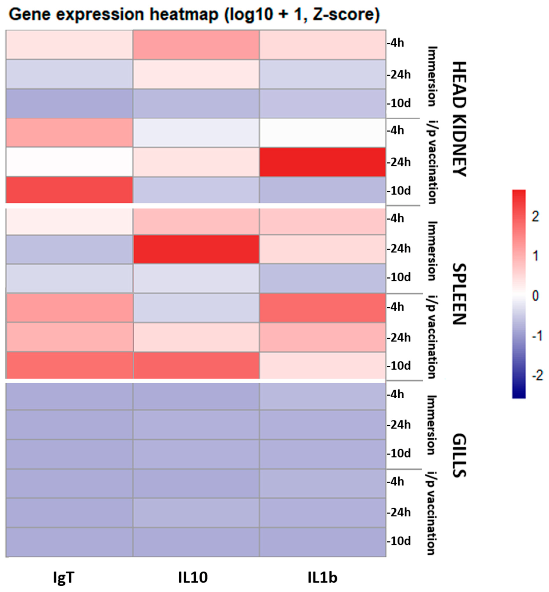
In head kidney expression increased transiently shortly after immersion vaccination, indicating early activation of systemic innate immune mechanisms. By 10 days post-vaccination, levels had decreased and were comparable to control values. The spleen showed a similar pattern to the head kidney, with transient upregulation of IL-1β shortly after immersion vaccination and a return to baseline levels by 10 days post-vaccination. In the gills, IL-1β expression was transiently upregulated at 4 h post-vaccination, reflecting an early local inflammatory response at the mucosal surface. By 24 h, expression decreased and approached control values, remaining low and comparable to controls at 10 days post-vaccination (Figure 3, Figure 6).
Following i.p. vaccination, IL-1β expression in the head kidney was strongly upregulated at 24 h, indicating rapid activation of innate immune mechanisms. By 10 days post-vaccination, expression had largely returned to baseline. The spleen showed IL-1β upregulation at 4 h post-vaccination, followed by a decrease at later time points. IL-1β expression in the gills exhibited slight upregulation at 4 h post-vaccination and remained low across subsequent time points following i.p. vaccination (Figure 3, Figure 6).
3.3. IL-10 Expression Following Vaccination Against T. maritimum
After immersion vaccination IL-10 expression in the head kidney increased at 4 h post-vaccination and gradually decreased towards 10 days, indicating an early anti-inflammatory response followed by resolution over time. In the spleen, IL-10 was upregulated after 4 h, reaching the highest levels at 24 h, and gradually decreased thereafter.
In the gills, IL-10 expression showed a slight increase at 4 h and gradually rose until 10 days post-vaccination, suggesting a sustained local anti-inflammatory response at the mucosal surface (Figure 4, Figure 6).
Following i.p. vaccination, IL-10 expression in the head kidney increased at 4 h, peaked at 24 h, and then decreased, reflecting a transient systemic anti-inflammatory response.The spleen showed peak IL-10 expression 10 days after i.p. vaccination, indicating delayed anti-inflammatory regulation compared to the head kidney.IL-10 expression levels in the gills were upregulated at 24 h post-vaccination and remained low at other time points, suggesting limited local anti-inflammatory activity following i.p. administration (Figure 4, Figure 6).
3.4. IgT Expression Following Vaccination Against T. maritimum
IgT expression after immersion vaccination showed only minor, non-significant increases at 4 h post-vaccination in all organs, followed by a decrease at later time points, indicating a limited adaptive immune response via this route (Figure 5, Figure 6).
IgT expression in the head kidney was upregulated 4 h after i.p. vaccination, then slightly decreased, reaching a peak at 10 days post-vaccination, indicating a progressive systemic adaptive immune response. A similar pattern of IgT expression was observed in the spleen, with gradual upregulation culminating at 10 days post-vaccination, mirroring the head kidney response. In the gills, IgT expression increased progressively over time. Minor changes were observed at 4 h, clearly elevated levels appeared at 24 h, and expression gradually decreased by 10 days post-vaccination, suggesting development of adaptive immune mechanisms via the systemic route what excludes gills tissue (Figure 5, Figure 6).
Figure 5.
Relative expression of the IgT gene in (A) head kidney, (B) spleen, and (C) gills at 4 h, 24 h, and 10 days following vaccination by immersion or intraperitoneal (i.p.) injection. IgT expression in vaccinated fish is shown in comparison to the control group at each time point. Asterisk above the plots indicate statistically significant differences between vaccination methods (Immersion vs. i.p. vaccination) within the same sampling time point (e.g., 4 h, 24 h, and 10 d). The raw data are provided in Supplementary Table S2.
Figure 5.
Relative expression of the IgT gene in (A) head kidney, (B) spleen, and (C) gills at 4 h, 24 h, and 10 days following vaccination by immersion or intraperitoneal (i.p.) injection. IgT expression in vaccinated fish is shown in comparison to the control group at each time point. Asterisk above the plots indicate statistically significant differences between vaccination methods (Immersion vs. i.p. vaccination) within the same sampling time point (e.g., 4 h, 24 h, and 10 d). The raw data are provided in Supplementary Table S2.
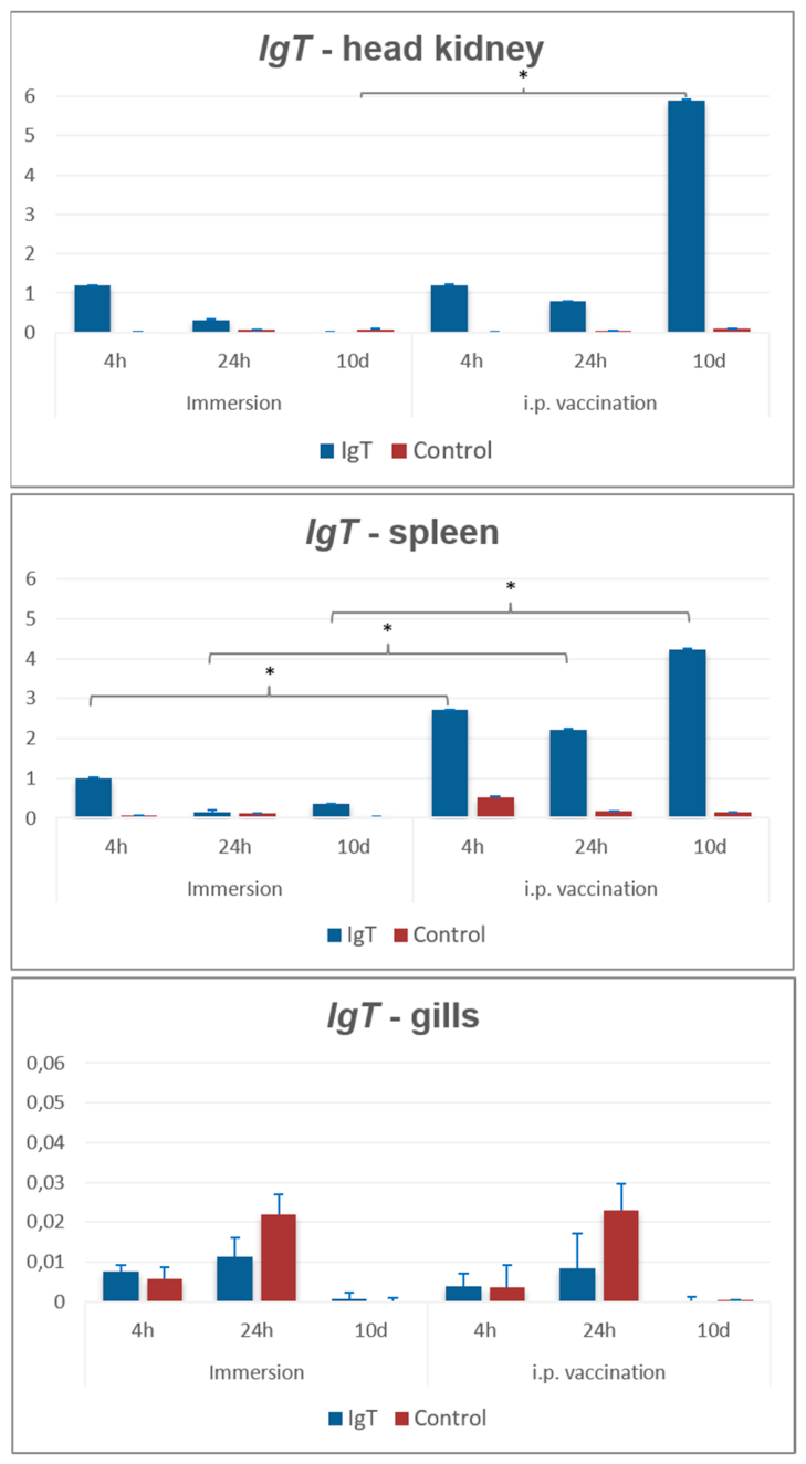
Figure 6.
Heatmap showing relative gene expression patterns of IgT, IL-10, and IL-1β across tissues, vaccination routes, and sampling time points. Expression values were log10(x + 1) transformed and normalized using gene-wise Z-score scaling. Each row represents an individual sample arranged according to the experimental design, and each column represents a gene. Color intensity reflects relative expression levels within each gene, with red indicating higher-than-average expression and blue indicating lower-than-average expression. No hierarchical clustering was applied.
Figure 6.
Heatmap showing relative gene expression patterns of IgT, IL-10, and IL-1β across tissues, vaccination routes, and sampling time points. Expression values were log10(x + 1) transformed and normalized using gene-wise Z-score scaling. Each row represents an individual sample arranged according to the experimental design, and each column represents a gene. Color intensity reflects relative expression levels within each gene, with red indicating higher-than-average expression and blue indicating lower-than-average expression. No hierarchical clustering was applied.
3.5. Result of Gene Expression
Principal component analysis (PCA) revealed a clear separation of samples according to vaccination route, indicating distinct gene expression profiles induced by immersion and i.p. vaccination. The first principal component (PC1), which explained the largest proportion of total variance in the dataset (55,5%), accounted for the dominant differences in the expression of IgT, IL-10, and IL-1β among samples. The second principal component (PC2) explained an additional proportion of variance (28,6%) and captured secondary patterns of gene expression variation.
This spatial separation suggests that the two vaccination routes consistently induce different overall immune expression profiles rather than isolated gene-specific effects. The relatively compact grouping within each ellipse further indicates a coherent response pattern associated with each vaccination route. Gene loading vectors demonstrated that all three genes contributed to the observed separation indicating distinct immune signatures between vaccination routes. The orientation and magnitude of the vectors indicate that variation along PC1 and PC2 reflects combined changes in these immune pathways, supporting the conclusion that vaccination route is a key determinant shaping the overall transcriptional immune response (Figure 7). The raw data are provided in Supplementary Table S1 while statistical comparisons (p values) among experimental groups are presented in Figure 3, Figure 4 and Figure 5 and summarized in Supplementary Table S2.
3.6. IgM Response in Vaccinated Seabass
Normality testing using the Shapiro–Wilk test indicated that the data were not normally distributed (p < 0.001). Therefore, group differences were assessed using the Kruskal–Wallis test. Post-hoc analysis using Dunn’s test with Holm correction showed significant differences between the IMM and IP groups (p = 0.009) as well as between the IP and IP_C groups (p = 2.9 × 10⁻⁶) and between the IP and IMM_C (p=0.05). No statistically significant differences were observed among the remaining pairwise comparisons after correction for multiple testing (Figure 8). Results of IgM titer after immersion vaccination against T. maritimum are the same as control vaccinated fish, while specific IgM in i.p. vaccinated fish showed significantly higher titer compared to fish vaccinated by i.p. injection of PBS. The i.p. vaccinated group showed significantly higher specific IgM anti-T. maritimum levels compared to all other experimental groups (p < 0.05).
4. Discussion
Juvenile seabass were vaccinated either by immersion or i.p. injection, and the autogenous vaccine provided significant protection against tenacibaculosis following i.p. administration, while the protection achieved by immersion vaccination was more limited. These findings suggest that the route of vaccine delivery may influence the magnitude of the protective immune response, highlighting the importance of optimizing vaccination strategies for effective disease control [30]. Gene expression responses were also evaluated to further characterize the mechanisms underlying vaccine-induced protection.
Despite the impact of tenacibaculosis in marine fish species, licensed vaccines against T. maritimum remain limited, with only one commercially available vaccine registered for turbot (Scophthalmus maximus) against serotype O2 strain LPV1.7 [31]. The lack of approved vaccines for other economically important species, including European seabass, highlights the need for improved prophylactic strategies. Although whole inactivated bacterial vaccines are already investigated, information on their protective efficacy, optimal administration routes, and the immune mechanisms underlying protection remains limited [32].
Intraperitoneal vaccination enables precise delivery of a defined antigen dose directly into the fish, resulting in a strong and long-lasting protective immune response [33]. It is therefore considered one of the most effective vaccination methods in aquaculture, particularly for larger fish, where it provides reliable and sustained protection against multiple pathogens. Immersion vaccination is a simple and non-invasive method that allows rapid immunization of large numbers of fish, particularly at early life stages when injection is not feasible. It also reduces handling stress (especially in immersion methods) and is practical for mass application despite generally requiring higher vaccine doses [11]. Although intraperitoneal injection induces primarily systemic immune responses, immersion vaccination enables antigen uptake through mucosal surfaces such as the skin and gills, thereby stimulating local mucosal immunity [34]. This route may better mimic natural infection pathways, and in some cases has been shown to confer higher mucosal immune answer compared to injection vaccination [35], as also observed in the present study.
Interleukin-1β (IL-1β) is the first identified pro-inflammatory cytokine, cleaved by caspase-1 following inflammasome activation, and plays a central role in the initiation of innate immune responses. It is primarily produced by monocytes and macrophages and is involved in activating both local and systemic immune reactions [36]. In the present study, immersion vaccination induced a rapid but transient upregulation of IL-1β expression, particularly in the gills and systemic immune organs (head kidney and spleen), suggesting an early activation of mucosal and systemic innate immune mechanisms. The pronounced response observed in the gills at 4 h post-vaccination likely reflects the primary site of antigen entry, supporting the role of mucosal surfaces in antigen uptake and early immune recognition. The subsequent decline in IL-1β expression to baseline levels by 10 days post-vaccination indicates resolution of the acute inflammatory response and restoration of immune homeostasis. In contrast, intraperitoneal vaccination resulted in a stronger and slightly delayed systemic IL-1β response, particularly in the head kidney, which is consistent with direct antigen delivery into the body cavity and rapid activation of systemic immune pathways. The weaker and transient response observed in the gills following i.p. vaccination further supports the notion that this route of administration primarily stimulates systemic rather than mucosal immunity.
Interleukin-10 (IL-10) is a multifunctional cytokine that plays an important role in both innate and adaptive immunity, primarily acting as a key anti-inflammatory mediator. In innate immune responses, IL-10 suppresses macrophage activation and inhibits the production of pro-inflammatory cytokines as well as reactive oxygen and nitrogen species [37]. Furthermore, IL-10 is essential for maintaining mucosal homeostasis, as its deficiency leads to spontaneous inflammation at mucosal surfaces [38]. In the present study, immersion vaccination induced an early and sustained IL-10 response, particularly in mucosal tissues. The gradual increase of IL-10 expression in the gills suggests prolonged local anti-inflammatory regulation, likely reflecting continuous immune modulation at the primary site of antigen entry. This pattern supports the role of IL-10 in maintaining mucosal homeostasis and preventing excessive inflammation following antigen exposure at epithelial surfaces. In systemic organs, the transient upregulation of IL-10 in the head kidney and spleen indicates a rapid regulatory response that follows early innate immune activation. In contrast, intraperitoneal vaccination elicited a predominantly systemic IL-10 response, characterized by early induction in the head kidney and a delayed peak in the spleen. This delayed anti-inflammatory response in the spleen may reflect prolonged immune activation following direct antigen delivery into the body cavity. The relatively weak and transient IL-10 expression observed in the gills further supports the limited involvement of mucosal immune regulation following i.p. administration.
Immunoglobulin T (IgT) plays a predominant role in fish mucosal immunity and represents one of the most ancient immunoglobulin classes specialized for immune protection at epithelial surfaces [39,40]. Recent studies have highlighted its key function in maintaining microbiota homeostasis and controlling pathogen colonization at mucosal sites, further supporting its central role in teleost mucosal immunity [40]. [22] showed that after nodavirus infection, IgT expression increases in the gills and spleen, and IgT⁺ cells are also found in the liver, highlighting its importance in the immune defense of sea bass.
In the present study, intraperitoneal (i.p.) vaccination induced a pronounced upregulation of IgT expression in systemic immune tissues, particularly in the head kidney and spleen, with the highest levels observed at 10 days post-vaccination. This pattern indicates a strong activation of adaptive immune mechanisms via the systemic route, with a clear time-dependent increase consistent with the development of a humoral immune response.
In contrast, immersion vaccination resulted in only minimal changes in IgT expression across all examined tissues, suggesting a limited induction of IgT-mediated adaptive immunity under the applied conditions. The lack of substantial upregulation over time further supports the weaker immunostimulatory effect of immersion delivery compared to i.p. administration.
In the gills, IgT expression remained low in both vaccination groups, with no evident induction following vaccination. Notably, IgT levels in control fish were in some cases comparable to or even higher than those in vaccinated groups, suggesting that vaccination did not induce a detectable mucosal IgT response at this site.
Vaccination in European sea bass is known to induce a strong IgM-mediated humoral response, characterized by increased levels of specific serum IgM and IgM⁺ cells in lymphoid organs such as the head kidney and spleen [26]. Similarly, elevated IgM responses accompanied by increased expression of pro-inflammatory cytokines and improved survival have been reported following vaccination with heat-killed Mycobacterium marinum [41]. In the present study, immersion vaccination against T. maritimum did not result in a significant increase in IgM titers compared to control fish. In contrast, intraperitoneal vaccination induced significantly higher IgM levels, confirming a stronger systemic humoral response via this route of administration. This difference can be explained by the mechanism of antigen delivery. Injection vaccination enables direct exposure of immune organs such as the head kidney and spleen to the antigen, thereby efficiently stimulating B cells and IgM production [42]. Immersion vaccination relies on antigen uptake through mucosal surfaces, which is generally less efficient and may limit the induction of systemic IgM responses [34]. Overall, these findings support the concept that IgM responses in fish are route-dependent, with intraperitoneal vaccination preferentially inducing systemic humoral immunity, whereas immersion vaccination may primarily stimulate mucosal immune mechanisms [43].
The RPS values obtained by both vaccination methods are consistent with the results of the selected immune-related gene expression analysis and the serological detection of IgM in the sera. The RPS value in intraperitoneally vaccinated fish is satisfactory and fully meets the criteria defined by Amend [24]. In contrast, immersion vaccination yielded an even higher RPS value (100%); however, not all criteria defined by Amend [24] were fulfilled, such as the low mortality of mock-vaccinated fish after challenge and the insufficient number of mock-vaccinated fish that developed clinical signs of the disease.
Additionally, only limited immune protection was observed following immersion vaccination, as indicated by RPS values and a modest increase in serum IgM levels, while no corresponding increase in IgT expression was detected in immunocompetent organs. Collectively, these findings suggest that immersion vaccination did not fulfil the efficacy criteria defined by Amend [24]. This research also highlights the importance of the route of administration when evaluating pathogenicity and vaccine efficacy. Similar results were obtained in a study performed on Chinook salmon juveniles vaccinated against T. maritimum via both routes in New Zealand [44].
Commercial immersion vaccines used in European sea bass are commonly applied for short exposure periods ranging from 30 seconds to 1 minute, and currently there are no scientific data demonstrating improved vaccine efficacy with prolonged immersion times. In experimental vaccination studies, immersion periods of approximately 2 minutes have also been reported. The immersion duration used in the present study was selected based on published literature and animal welfare considerations, since European sea bass are highly sensitive to manipulation and handling. Previous studies demonstrated successful immune responses after 2-minute immersion vaccination protocols against Listonella anguillarum (syn. Vibrio anguillarum) [45] and against V. anguillarum and Photobacterium piscicida subsp. piscicida [46]. Similar immersion approaches were also applied in vaccination studies against viral nervous necrosis (VNN) in European sea bass [47], although protection following immersion vaccination was limited in that case. Furthermore, the review by [34], emphasizes that immersion vaccines are generally less effective compared to injection vaccines, which is consistent with our findings.
5. Conclusions
This study demonstrates that a formalin-killed autogenous vaccine against T. maritimum induced significant protective immunity in European seabass when administered by intraperitoneal injection, as supported by RPS values, specific IgM responses, and immune-related gene expression profiles. In contrast, immersion vaccination provided limited protection under the experimental conditions, with insufficient immune activation to achieve the defined efficacy criteria. The reduced efficacy of immersion vaccination may be related to the antigen dose and single exposure protocol, highlighting the need for further optimization of vaccine formulation and delivery strategies. Improving immersion vaccination approaches remains particularly important for smaller European seabass, where injection-based vaccination is less feasible.
Supplementary Materials
The following supporting information can be downloaded at the website of this paper posted on Preprints.org. Supplementary Table S1 contains the raw gene expression data (Ct values and normalized expression levels) for IgT, IL-10, and IL-1β across all samples, including vaccination groups, tissues, and sampling time points. Supplementary Table S2 contains statistical analysis of IgT, IL-10, and IL-1β genes expression in head kidney, spleen, and gill tissues following immersion and intraperitoneal vaccination. Differences between groups were assessed using the Wilcoxon rank-sum test with Benjamini–Hochberg correction for multiple testing. Reported values include the Wilcoxon test statistic, raw p-values, adjusted p-values (p.adj), and significance levels (ns = not significant).
Author Contributions
Conceptualization, I.G.Z., S.Z. and M.G.; formal analysis, I.G.Z., L.V., D.V. and D.G.; investigation, D.O., S.Z., D.V., M.A. and I.G.Z; resources, S.Z., M.G. and D.O.; data curation, I.G.Z., D.V., L.V. and D.G; writing—original draft preparation, I.G.Z, S.Z.; writing—review and editing, S.Z. and D.O; visualization, I.G.Z. and D.G; supervision, S.Z. All authors have read and agreed to the published version of the manuscript.
Funding
This research was funded by “MARICULTURE NETWORK: Implementation of new technologies for diversified sustainable aquaculture targeting healthy society and competitive regions”-Acronym “MARINET”, within the Interreg VI A Italy–Croatia CBC Programme 2021–2027, European Union.
Institutional Review Board Statement
The study was conducted in accordance with Directive 2010/63/EU of the European Parliament and of the Council of 22 September 2010 on the protection of animals used for scientific purposes and ethical approval issued by National Ethics Committee of the Croatian Ministry of Agriculture, Forestry and Fisheries (EP 470/2025) on 16.05.2025.
Informed Consent Statement
Not applicable.
Data Availability Statement
The data presented in this study are available upon a reasonable request from corresponding author.
Acknowledgments
Not applicable
Conflicts of Interest
The authors declare no conflict of interest.
Abbreviations
The following abbreviations are used in this manuscript:
| CFU | Colony-Forming Unit |
| I.P. | Intraperitoneal |
| MB | Marine Broth |
| PBS | Phosphate-buffered saline |
| PCA | Principal component analysis |
| RPS | Relative percentage of survival |
| RT | Room temperature |
| TSA | Tryptic Soy Agar |
References
- Avendaño-Herrera, R.; Toranzo, A.E.; Magariños, B. Tenacibaculosis infection in marine fish caused by Tenacibaculum maritimum: A review. Dis. Aquat. Organ. 2006, 71, 255–266. [Google Scholar] [CrossRef] [PubMed]
- Elanco. Technical report: An overview of emerging diseases in the salmonid farming industry; Elanco Canada Ltd.: Canada, 2018. [Google Scholar]
- Masumura, K.; Wakabayashi, H. An outbreak of gliding bacterial disease in hatchery-born red seabream and gilthead fry. Fish. Pathol. 1977, 12, 171–177. [Google Scholar] [CrossRef]
- Pepin, J.F.; Emery, E. Marine cytophaga-like bacteria isolated from diseased reared sea bass (Dicentrarchus labrax L.) from the French Mediterranean coast. Bull. Eur. Assoc. Fish. Pathol. 1993, 13, 165–167. [Google Scholar]
- Salati, F.; Cubadda, C.; Viale, I.; Kusuda, R. Immune response of sea bass (Dicentrarchus labrax) to Tenacibaculum maritimum antigens. Fish. Sci. 2005, 71, 563–567. [Google Scholar] [CrossRef]
- Kolygas, M.N.; Gourzioti, E.; Vatsos, I.N.; Athanassopoulou, F. Identification of Tenacibaculum maritimum strains from marine farmed fish in Greece. Vet. Rec. 2012, 170, 623. [Google Scholar] [CrossRef] [PubMed]
- Yardımcı, E.R.; Timur, G. Isolation and identification of Tenacibaculum maritimum in farmed European sea bass in Turkey. Isr. J. Aquac. Bamidgeh 2015, 67. [Google Scholar] [CrossRef]
- Zrnčić, S.; Oraić; Račić, I.; Beck, R. Tenacibaculum maritimum isolated from sea bass (Dicentrarchus labrax) in Croatia. 16th International Conference on Diseases of Fish and Shellfish, Tampere, Sept. 2-6, 2013; p. 387. [Google Scholar]
- Kingsbury, M.V.; Hamoutene, D.; Kraska, P.; Lacoursière-Roussel, A.; Page, F.; Coyle, T.; Sutherland, T.; Gibb, O.; McKindsey, C.W.; Hartog, F.; Neil, S.; Chernoff, K.; Wong, D.; Law, B.A.; Brager, L.; Baillie, S.M.; Black, M.; Bungay, T.; Gaspard, D.; Hua, K.; Parsons, G.J. Relationship between in-feed drugs, antibiotics and organic enrichment in marine sediments at Canadian Atlantic salmon aquaculture sites. Mar. Pollut. Bull. 2023, 188, 114654. [Google Scholar] [CrossRef] [PubMed]
- Yun, S.; Giri, S.S.; Kim, H.J.; et al. Enhanced bath immersion vaccination through microbubble treatment in cyprinid loach. Fish. Shellfish Immunol. 2019, 91, 12–18. [Google Scholar]
- Plant, K.P.; LaPatra, S.E. Advances in fish vaccine delivery. Dev. Comp. Immunol. 2011, 35, 1256–1262. [Google Scholar] [CrossRef] [PubMed]
- Liu, X.H.; Khansari, A.R.; Teles, M.; Martinez-Rodriguez, G.; Zhang, Y.G.; Mancera, J.M.; Reyes-Lopez, F.E.; Tort, L. Brain and pituitary response to vaccination in gilthead seabream (Sparus aurata L.). Front. Physiol. 2019, 10, 717. [Google Scholar] [CrossRef] [PubMed]
- Muñoz-Atienza, E.; Díaz-Rosales, P.; Tafalla, C. Systemic and mucosal B and T cell responses upon mucosal vaccination of teleost fish. Front. Immunol. 2021, 11, 622377. [Google Scholar] [CrossRef] [PubMed]
- Adams, A. Progress, challenges and opportunities in fish vaccine development. Fish. Shellfish Immunol. 2019, 90, 210–214. [Google Scholar] [CrossRef] [PubMed]
- Valdenegro-Vega, V.A.; Crosbie, P.; Vincent, B.; Cain, K.D.; Nowak, B.F. Effect of immunization route on mucosal and systemic immune responses in Atlantic salmon. Vet. Immunol. Immunopathol. 2013, 151, 113–123. [Google Scholar] [CrossRef] [PubMed]
- Afonso, A.; Gomes, S.; da Silva, J.; Marques, F.; Henrique, M. Side effects in sea bass (Dicentrarchus labrax L.) due to intraperitoneal vaccination against vibriosis and pasteurellosis. Fish. Shellfish Immunol. 2005, 19, 1–16. [Google Scholar] [CrossRef] [PubMed]
- Azizi, S.; Balasch, J.C.; Cartan, S.; Jerez-Cepa, I.; Mancera, J.M.; Tort, L.; Khansari, A.R. Enhancing farmed fish welfare: Evaluating the effectiveness of plant-based stress mitigating agents as sedatives in sea bass (Dicentrarchus labrax) following intraperitoneal vaccination. Fish. Shellfish Immunol. 2025, 156, 110058. [Google Scholar] [CrossRef] [PubMed]
- Forthal, D.N. Functions of antibodies. Microbiol. Spectr. 2014, 2. [Google Scholar] [CrossRef] [PubMed]
- Dickerson, H.W.; Findly, R.C. Vertebrate adaptive immunity—Comparative insights from a teleost model. Front. Immunol. 2017, 8, 1379. [Google Scholar] [CrossRef] [PubMed]
- Semple, S.L.; Dixon, B. Salmonid antibacterial immunity: An aquaculture perspective. Biology 2020, 9, 331. [Google Scholar] [CrossRef] [PubMed]
- Ferreira, M.; Machado, M.; Mota, C.S.C.; Abreu, H.; Silva, J.; Maia, M.R.G.; Kiron, V.; Costas, B.; Valente, L.M.P. Micro- and macroalgae blend modulates the mucosal and systemic immune responses of European seabass (Dicentrarchus labrax) upon infection with Tenacibaculum maritimum. Aquaculture 2023, 566, 739222. [Google Scholar] [CrossRef]
- Buonocore, F.; Stocchi, V.; Nunez-Ortiz, N.; Randelli, E.; Gerdol, M.; Pallavicini, A.; Facchiano, A.; Bernini, C.; Guerra, L.; Scapigliati, G.; Picchietti, S. Immunoglobulin T from sea bass (Dicentrarchus labrax L.): Molecular characterization, tissue localization and expression after nodavirus infection. BMC Mol. Biol. 2017, 18, 8. [Google Scholar] [CrossRef] [PubMed]
- Mitter, K.; Kotoulas, G.; Magoulas, A.; Mulero, V.; Sepulcre, P.; Figueras, A.; Novoa, B.; Sarropoulou, E. Evaluation of candidate reference genes for qPCR during ontogenesis and in immune-relevant tissues of European seabass (Dicentrarchus labrax). Comp. Biochem. Physiol. 2009, 153, 340–347. [Google Scholar] [CrossRef] [PubMed]
- Amend, D.F. Potency testing of fish vaccines. Dev. Biol. Stand. 1981, 49, 447–454. [Google Scholar]
- Livak, K.J.; Schmittgen, T.D. Analysis of relative gene expression data using real-time quantitative PCR and the 2⁻ΔΔCt method. Methods 2001, 25, 402–408. [Google Scholar] [CrossRef] [PubMed]
- Galeotti, M.; Romano, N.; Volpatti, D.; Bulfon, C.; Brunetti, A.; Tiscar, P.G.; Mosca, F.; Bertoni, F.; Marchetti, M.G.; Abelli, L. Innovative vaccination protocol against vibriosis in Dicentrarchus labrax (L.) juveniles: Improvement of immune parameters and protection to challenge. Vaccine 2013, 31, 1224–1230. [Google Scholar] [CrossRef] [PubMed]
- Engvall, E.; Perlmann, P. Enzyme-linked immunosorbent assay (ELISA): Quantitative assay of immunoglobulin G. Immunochemistry 1971, 8, 871–874. [Google Scholar] [CrossRef] [PubMed]
- R Core Team. R: A language and environment for statistical computing. R Foundation for Statistical Computing, Vienna, Austria. 2024. Available online: https://www.R-project.org.
- Wickham, H.; François, R.; Henry, L.; Müller, K.; Vaughan, D. dplyr: A Grammar of Data Manipulation; R package version 1.1.4. 2023. Available online: https://CRAN.R-project.org/package=dplyr.
- Paolini, A.; Ridolfi, V.; Zezza, D.; Cocchietto, M.; Musa, M.; Pavone, A.; Conte, A.; Giorgetti, G. Vaccination trials of sea bass (Dicentrarchus labrax) against pasteurellosis using oral, intraperitoneal and immersion methods. Vet. Ital. 2005, 41, 137–144. [Google Scholar] [PubMed]
- Mabrok, M.; Algammal, A.M.; Sivaramasamy, E.; Hetta, H.F.; Atwah, B.; Alghamdi, S.; Fawzy, A.; Avendaño-Herrera, R.; Rodkhum, C. Tenacibaculosis caused by Tenacibaculum maritimum: Updated knowledge of this marine bacterial fish pathogen. Front. Cell. Infect. Microbiol. 2023, 13, 1068000. [Google Scholar] [CrossRef] [PubMed]
- Miccoli, A.; Saraceni, P.R.; Scapigliati, G. Vaccines and immune protection of principal Mediterranean marine fish species. Fish. Shellfish Immunol. 2019, 94, 800–809. [Google Scholar] [CrossRef] [PubMed]
- Mitchell, H. Choosing a furunculosis vaccine: Points to consider. Bull. Aquac. Assoc. Can. 1995, 95, 30–37. [Google Scholar]
- Bøgwald, J.; Dalmo, R.A. Review on immersion vaccines for fish: An update 2019. Microorganisms 2019, 7, 627. [Google Scholar] [CrossRef] [PubMed]
- Kong, W.G.; Qin, D.C.; Mu, Q.J.; Dong, Z.R.; Luo, Y.Z.; Ai, T.S.; Xu, Z. Mucosal immune responses and protective efficacy in yellow catfish after immersion vaccination with bivalent inactivated Aeromonas veronii and Edwardsiella ictaluri vaccine. Water Biol. Secur. 2022, 1, 100032. [Google Scholar] [CrossRef]
- Wang, X.; Zhang, R.; Liu, L.; Ma, G.; Zhu, H. An IL-1β homologue induced inflammation and antibacterial immune defense in Siberian sturgeon (Acipenser baeri). Fish. Shellfish Immunol. 2021, 118, 283–293. [Google Scholar] [CrossRef] [PubMed]
- Wu, S.; Duan, S.; Kong, L.; Tu, X.; Wang, L.; Guo, Z.; Ye, J. Interleukin-10 (IL-10) participates in host defense against bacterial pathogens and promotes IgM antibody production in Nile tilapia (Oreochromis niloticus). Aquaculture 2021, 531, 735829. [Google Scholar] [CrossRef]
- Rubtsov, Y.P.; Rasmussen, J.P.; Chi, E.Y.; Fontenot, J.; Castelli, L.; Ye, X.; Treuting, P.; Siewe, L.; Roers, A.; Henderson, W.R. Regulatory T cell-derived interleukin-10 limits inflammation at environmental interfaces. Immunity 2008, 28, 546–558. [Google Scholar] [CrossRef] [PubMed]
- Xu, Z.; Parra, D.; Gómez, D.; Salinas, I.; Zhang, Y.; von Gersdorff Jørgensen, L.; Heinecke, R.D.; Buchmann, K.; LaPatra, S.; Sunyer, J.O. Teleost skin, an ancient mucosal surface that elicits gut-like immune responses. Proc. Natl. Acad. Sci. USA 2013, 110, 13097–13102. [Google Scholar] [CrossRef] [PubMed]
- Salinas, I.; Fernández-Montero, Á.; Ding, Y.; Sunyer, J.O. Mucosal immunoglobulins of teleost fish: A decade of advances. Dev. Comp. Immunol. 2021, 121, 104079. [Google Scholar] [CrossRef] [PubMed]
- Ziklo, N.; Colorni, A.; Gao, L.Y.; Du, S.J.; Ucko, M. Humoral and cellular immune response of European seabass (Dicentrarchus labrax) vaccinated with heat-killed Mycobacterium marinum (iipA::kan mutant). J. Aquat. Anim. Health 2018, 30, 312–324. [Google Scholar] [CrossRef] [PubMed]
- Du, Y.; Hu, X.; Miao, L.; Chen, J. Current status and development prospects of aquatic vaccines. Front. Immunol. 2022, 13, 1040336. [Google Scholar] [CrossRef] [PubMed]
- Parra, D.; Reyes-Lopez, F.E.; Tort, L. Mucosal immunity and B cells in teleosts: Effect of vaccination and stress. Front. Immunol. 2015, 6, 354. [Google Scholar] [CrossRef] [PubMed]
- Jaramillo, D.; Busby, B.P.; Bestbier, M.; Bennett, P.; Waddington, Z. New Zealand rickettsia-like organism and Tenacibaculum maritimum vaccine efficacy study. J. Fish. Dis. 2024, 47, e13883. [Google Scholar] [CrossRef] [PubMed]
- Angelidis, P.; Karagiannis, D.; Crump, E.M. Efficacy of a Listonella anguillarum (syn. Vibrio anguillarum) vaccine for juvenile sea bass (Dicentrarchus labrax). Dis. Aquat. Org. 2006, 71, 19–24. [Google Scholar] [CrossRef] [PubMed]
- Gravningen, K.; Thorarinsson, R.; Johansen, L.H.; Nissen, B.; Rikardsen, K.S.; Greger, E.; Vigneulle, M. Bivalent vaccines for sea bass (Dicentrarchus labrax) against vibriosis and pasteurellosis. J. Appl. Ichthyol. 1998, 14, 159–162. [Google Scholar] [CrossRef]
- Núñez-Ortiz, N.; Pascoli, F.; Picchietti, S.; Buonocore, F.; Bernini, C.; Toson, M.; Scapigliati, G.; Toffan, A. A formalin-inactivated immunogen against viral encephalopathy and retinopathy (VER) disease in European sea bass (Dicentrarchus labrax): immunological and protection effects. Vet. Res. 2016, 47, 89. [Google Scholar] [CrossRef] [PubMed]
Figure 1.
Experimental design, including vaccination of fish by immersion and intraperitoneal (i.p.) injection, followed by challenge using both routes.
Figure 1.
Experimental design, including vaccination of fish by immersion and intraperitoneal (i.p.) injection, followed by challenge using both routes.
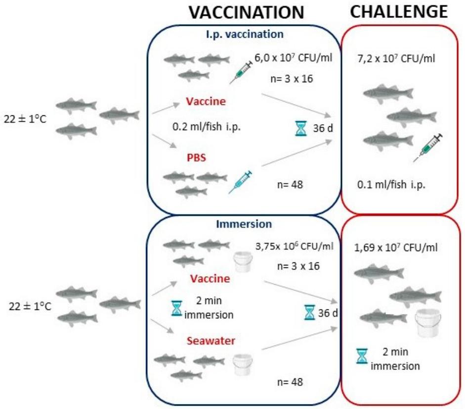
Figure 2.
Clinical signs observed in European seabass following experimental challenge with T. maritimum.
Figure 2.
Clinical signs observed in European seabass following experimental challenge with T. maritimum.
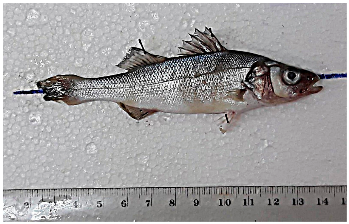
Figure 3.
Relative expression of the IL-1β gene in (A) head kidney, (B) spleen, and (C) gills at 4 h, 24 h, and 10 days following vaccination by immersion or intraperitoneal (i.p.) injection. IL-1β expression in vaccinated fish is shown in comparison to the control group at each time point. Asterisk above the plots indicate statistically significant differences between vaccination methods (Immersion vs. i.p. vaccination) within the same sampling time point (e.g., 4 h, 24 h, and 10 d). The raw data are provided in Supplementary Table S2.
Figure 3.
Relative expression of the IL-1β gene in (A) head kidney, (B) spleen, and (C) gills at 4 h, 24 h, and 10 days following vaccination by immersion or intraperitoneal (i.p.) injection. IL-1β expression in vaccinated fish is shown in comparison to the control group at each time point. Asterisk above the plots indicate statistically significant differences between vaccination methods (Immersion vs. i.p. vaccination) within the same sampling time point (e.g., 4 h, 24 h, and 10 d). The raw data are provided in Supplementary Table S2.
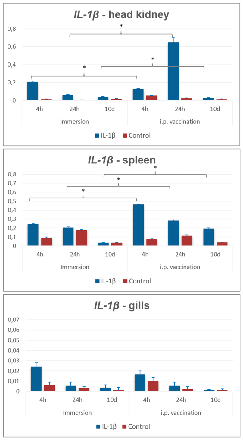
Figure 4.
Relative expression of the IL-10 gene in (A) head kidney, (B) spleen, and (C) gills at 4 h, 24 h, and 10 days following vaccination by immersion or intraperitoneal (i.p.) injection. IL-10 expression in vaccinated fish is shown in comparison to the control group at each time point. Asterisk above the plots indicate statistically significant differences between vaccination methods (Immersion vs. i.p. vaccination) within the same sampling time point (e.g., 4 h, 24 h, and 10 d). The raw data are provided in Supplementary Table S2.
Figure 4.
Relative expression of the IL-10 gene in (A) head kidney, (B) spleen, and (C) gills at 4 h, 24 h, and 10 days following vaccination by immersion or intraperitoneal (i.p.) injection. IL-10 expression in vaccinated fish is shown in comparison to the control group at each time point. Asterisk above the plots indicate statistically significant differences between vaccination methods (Immersion vs. i.p. vaccination) within the same sampling time point (e.g., 4 h, 24 h, and 10 d). The raw data are provided in Supplementary Table S2.
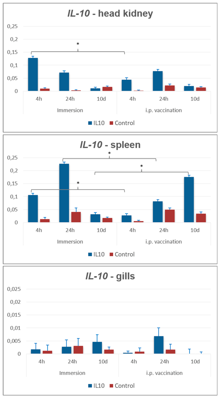
Figure 7.
Principal component analysis (PCA) of gene expression profiles (IgT, IL-10, and IL-1β) across samples following immersion and i.p. vaccination. Expression values were log2(x + 1) transformed and scaled prior to analysis. Each point represents an individual sample, colored according to vaccination route. Ellipses indicate 95% confidence intervals for each vaccination group. Arrows represent gene loading vectors, illustrating the contribution and direction of each gene to the first two principal components. The percentages shown on the axes indicate the proportion of total variance explained by PC1 and PC2.
Figure 7.
Principal component analysis (PCA) of gene expression profiles (IgT, IL-10, and IL-1β) across samples following immersion and i.p. vaccination. Expression values were log2(x + 1) transformed and scaled prior to analysis. Each point represents an individual sample, colored according to vaccination route. Ellipses indicate 95% confidence intervals for each vaccination group. Arrows represent gene loading vectors, illustrating the contribution and direction of each gene to the first two principal components. The percentages shown on the axes indicate the proportion of total variance explained by PC1 and PC2.
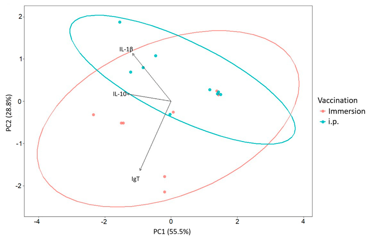
Figure 8.
Specific anti-T. maritimum IgM titers after vaccination. Immersion vaccination resulted in IgM levels comparable to controls, while i.p. vaccination induced significantly higher IgM titers than PBS-injected and other experimental groups (p < 0.05).
Figure 8.
Specific anti-T. maritimum IgM titers after vaccination. Immersion vaccination resulted in IgM levels comparable to controls, while i.p. vaccination induced significantly higher IgM titers than PBS-injected and other experimental groups (p < 0.05).
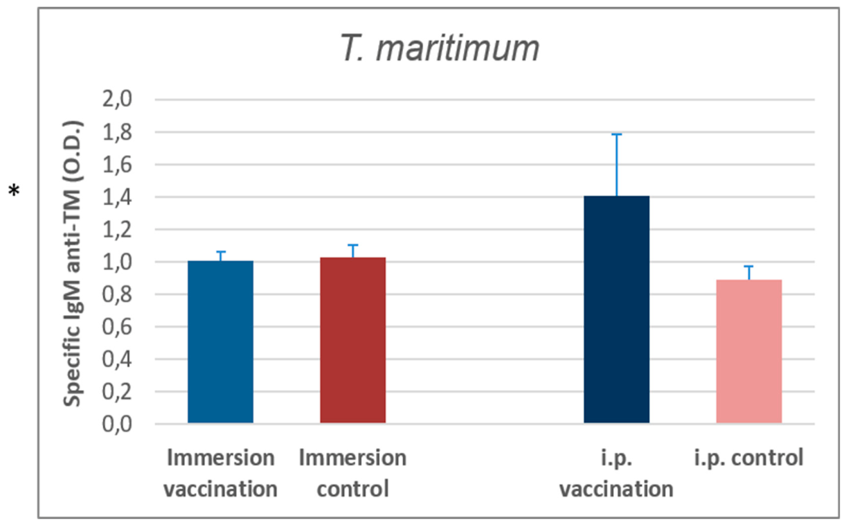

Table 1.
Survival of fish vaccinated against T. maritimum after challenge with the same bacteria.
| Experimental | Vaccination | Challenge | No. of fish per group | Mortality | Survival rate | RPS | |
|---|---|---|---|---|---|---|---|
| 2.2.1. | I.p. 0.2 mL/fish (6.0 x 10⁷ CFU/mL) |
I.p. 0.1 mL/fish 7.2 x 10⁷ CFU/mL |
10 | 1 | 87 | 67,5 | |
| 2.2.2. | 10 | 2 | |||||
| 2.2.3. | 10 | 1 | |||||
| 3.2. | I.p. mock vaccinated control |
30 | 12 | 60 | |||
| 2.1.1. | Immersion (3.75 x 10⁶ CFU/mL) | Immersion 1.69 x 10⁷ CFU/mL |
10 | 0 | 100 | 100 | |
| 2.1.2. | 10 | 0 | |||||
| 2.1.3. | 10 | 0 | |||||
| 3.1. | Immersion mock vaccinated control | 30 | 5 | 83 |
Table 2.
Primers and probe used for quantitative real-time PCR analysis of gene expression.
| Gene | Sequence (5′–3′) | |
|---|---|---|
| Fau | FOR | GACACCCAAGGTTGACAAGCAG |
| REV | GGCATTGAAGCACTTAGGAGTTG | |
| PROBE | CGCTTCGTGAATGTTGTGCCCACC | |
| L13a | FOR | TCTGGAGGACTGTCAGGGGCATGC |
| REV | AGACGCACAATCTTGAGAGCAG | |
| PROBE | CCGGCAACTTCTATCGCAACAAGCT | |
| IL10 | FOR | ATTACCCACCACCCACTGAC |
| REV | ATCTCTTCCACTATGCTCTCCAG | |
| PROBE | TCGTCTTATCTTTCTTCTGCACTGTCTGGT | |
| IL-1β | FOR | TCAGCACCCTGACGTCTGTC |
| REV | GGCACTCTCCTGGCACATCT | |
| PROBE | AGCAGAGGAGAACAACCGGCCG | |
| IgT | FOR | TGACTGTGCAGCCAGGTCAA |
| REV | TCCTTTCCCTGCAGGCTGTC | |
| PROBE | CCTGTCAGGTCTCTTATTCTGTTAGCAGCT | |
Disclaimer/Publisher’s Note: The statements, opinions and data contained in all publications are solely those of the individual author(s) and contributor(s) and not of MDPI and/or the editor(s). MDPI and/or the editor(s) disclaim responsibility for any injury to people or property resulting from any ideas, methods, instructions or products referred to in the content. |
© 2026 by the authors. Licensee MDPI, Basel, Switzerland. This article is an open access article distributed under the terms and conditions of the Creative Commons Attribution (CC BY) license (http://creativecommons.org/licenses/by/4.0/).
Copyright: This open access article is published under a Creative Commons CC BY 4.0 license, which permit the free download, distribution, and reuse, provided that the author and preprint are cited in any reuse.


