

Submitted:
12 July 2026
Posted:
15 July 2026
You are already at the latest version
Abstract
Background/Objectives: Alzheimer's disease (AD), one of the most prevalent neurodegenerative disorders in elderly, characterized by progressive cognitive loss, amyloid-β (Aβ) plaque deposition, and neurofibrillary tangle formation. Cerebral hypoxia has been reported as a complex modulator to AD pathology, with hypoxia-inducible factor-1 (HIF-1) emerging as a central molecular marker associated with neurodegeneration. This review aims to comprehensively report the roles of HIF-1 signaling in AD pathogenesis, emphasizing its neuroprotective and neurotoxic mechanisms along with demonstrating the therapeutic potential and challenges of translating this pathway for AD therapeutics. Methods: A comprehensive literature search was conducted across PubMed, Scopus, Web of Science, Embase, and Google Scholar databases for articles published between January 2000 and December 2025 on the context. Results: Under mild hypoxic conditions, HIF-1 activation enhances neuronal survival through upregulation of glucose transporters, glycolytic enzymes, angiogenic factors, erythropoietin, and antioxidant defense mechanisms. In contrast, chronic hypoxia modulates HIF-1 into a pathogenic marker through transcriptional activation of β-site amyloid precursor protein (APP) cleaving enzyme 1 (BACE1) and γ-secretase, facilitating amyloidogenic APP processing, along with facilitating tau hyperphosphorylation. Moreover, HIF-1 exacerbates neuroinflammation through microglial activation and pro-inflammatory cytokine release. The cell-type-specific expression patterns of HIF-1α and the temporal dynamics of its activation regulate whether the pathway exerts neuroprotective or neurodegenerative effects. Conclusions: This review discusses current understanding of HIF-1 mediated mechanistic insights in AD pathology and impacts of existing HIF-1 modulators on AD pathology along with the therapeutic implications of targeting this pathway for translational application in AD therapeutics.
Keywords:
Alzheimer's disease
; hypoxia-inducible factor-1
; hypoxia
; Amyloid-β
; tau protein
; neuroinflammation
1. Introduction
Neurodegenerative disorders represent one of the most challenging health burdens for elderly population at present time, with their alarming prevalence globally. Alzheimer's disease (AD), the most prevalent form of dementia, currently affects approximately 50 million individuals worldwide, accounting for an estimated 60-70% of all dementia cases, and its incidence is projected to triple by 2050 in the absence of effective therapeutic interventions [1]. The socioeconomic burden of AD is shocking, with annual healthcare expenditures exceeding USD 345 billion in the United States alone in 2023, a figure expected to surpass USD 1 trillion by 2050 as the aging population continues to expand [2].
The central nervous system (CNS) is highly sensitive to oxygen deprivation. Although the brain accounts for only 2% of body weight, it consumes over 20% of the body's oxygen. Neurons use 75-80% of this energy to support vital functions such as synaptic transmission, membrane potential restoration, neurotransmitter synthesis, and axonal transport [3,4]. Accumulating evidence identifies hypoxia as a key pathogenic factor in AD. Emerging data show that hypoxic conditions promote β-amyloid (Aβ) formation by upregulating β-secretase (BACE1) expression, induce tau hyperphosphorylation by disrupting kinase-phosphatase balance, impair blood-brain barrier (BBB) integrity, and drive neuronal degeneration through mitochondrial dysfunction and oxidative stress pathways [1]. Clinical studies have revealed that over 70% of dementia patients exhibit sleep-disordered breathing, and obstructive sleep apnea has been associated with significantly elevated plasma levels of Aβ42 in adolescents, emphasizing the insidious role of chronic intermittent hypoxia in precipitating neurodegenerative changes from early life stages [5,6].
Hypoxia plays a profound role in the pathogenesis of neuroinflammation, a hallmark of AD pathophysiology, through the activation of microglia and astrocytes that subsequently initiate a cascade of pro-inflammatory cytokines including TNF-α, IL-1β, and interferon-γ leading to downregulation of amyloid receptor expression and clearance with simultaneously activating BACE1. These changes ultimately result in a vicious cycle which amplifies both Aβ plaque formation and neuroinflammation [3]. Furthermore, hypoxia-induced microglial activation triggers astrocyte activation, further boosting the release of pro-inflammatory cytokines and establishing a self-perpetuating inflammatory cascade contributing to synaptic dysfunction, mitochondrial impairment, and neuronal apoptosis resulting in AD progression [1].
The central regulator of cellular adaptation to hypoxic stress is hypoxia-inducible factor-1 (HIF-1), a heterodimeric transcription factor composed of an oxygen-sensitive α subunit and a constitutively expressed β subunit. Under normal oxygen levels, oxygen-dependent prolyl hydroxylase domain (PHD) enzymes promote the degradation of HIF-1α. In hypoxia, PHDs are inactivated, allowing HIF-1α to stabilize, move to the nucleus, and activate the transcription of over 100 genes involved in angiogenesis, erythropoiesis, glucose metabolism, and cell survival. Although HIF-1 is well-studied in cancer biology and kidney function, its role in neurodegenerative diseases like Alzheimer's remains unclear. Current research shows a complex dual role including HIF-1 activation can be neuroprotective by enhancing glycolysis and antioxidant defenses, but it can also contribute to neurodegeneration by promoting amyloid plaque formation, tau hyperphosphorylation, and neuroinflammation. The molecular factors that determine whether HIF-1 acts as a "friend or foe" in AD are still unknown [1,7].
The underlying relationship between the intensity, duration, and timing of hypoxic exposure and the resulting HIF-1-mediated effects is not fully understood. Previous research suggests that mild or short-term hypoxia triggers protective responses, such as increased glycolysis and antioxidant defense. However, severe or prolonged hypoxia shifts the role of HIF-1 promoting cell death via p53 stabilization. The exact threshold that separates these beneficial and harmful effects remains undefined [3,8,9]. Furthermore, the cell-type-specific studies of HIF-1 signaling to AD pathogenesis require elucidation, as microglia, astrocytes, neurons, and cerebrovascular endothelial cells each manifest distinct HIF-1 transcriptional and metabolic changes that may collectively foster disease progression. However, few investigations have systematically reported the cell-autonomous versus non-cell-autonomous mechanisms through which HIF-1 modulates neuroinflammation, amyloid pathology, tau phosphorylation, and synaptic integrity in the AD [2,10,11,12,13].
Although pharmacological modulation of HIF-1 through PHD inhibitors and iron chelators has demonstrated promising neuroprotective effects in preclinical AD models, the optimal therapeutic window, appropriate dosing regimens, and potential off-target consequences, particularly, the risks associated with chronic PHD inhibition including local hyperoxia, abnormal angiogenesis, and dysregulation of neural progenitor cell homeostasis remain insufficiently explored. Thus, this review aims to comprehensively discuss the dual roles of HIF-1 signaling in AD pathogenesis emphasizing its neuroprotective and neurotoxic mechanisms along with demonstrating the therapeutic potential and challenges of targeting this pathway for translating in AD therapeutics.
2. Methods
2.1. Search Strategy
A comprehensive literature search was conducted across multiple electronic databases to identify relevant studies investigating the role of HIF-1 in AD pathogenesis. From January 2000 to December 2025, renowned databases including PubMed, Scopus, Web of Science, Google Scholar, and Science Direct were systematically searched using the following keywords: "Hypoxia-inducible factor", "HIF-1", "HIF-1α", "Alzheimer's disease", "AD", "neurodegeneration", "neuroinflammation", "hypoxia", "oxygen homeostasis", "oxidative stress", "cerebral hypoperfusion", "β-amyloid", "tau", and "apoptosis". Moreover, specific terms such as "PHD inhibitors", "iron chelators", "deferoxamine", "M30", "neuroprotection", and "therapeutic targeting" were used to identify articles on pharmacological modulation of the HIF pathway. Only peer-reviewed articles published in English language were considered for inclusion due to language barriers, time constraints, and the substantial costs associated with translation of non-English publications.
2.2. Inclusion Criteria and Data Extraction
Specific inclusion and exclusion criteria were adopted to select and incorporate the most relevant articles retrieved from electronic databases. The selected articles involved original research articles, comprehensive reviews, clinical trials, and case reports reporting the role of HIF-1 signaling in AD pathogenesis and its potential as a therapeutic target. Studies meeting the following criteria were included and extracted for this review: (i) peer-reviewed journal articles published in English; (ii) studies on hypoxia and HIF-1 pathways in the context of AD; (iii) studies reporting on HIF-1 mediated neuroprotective or neurotoxic mechanisms; (iv) preclinical studies evaluating modulators of HIF-1; (v) articles with available full-text and not duplicated across databases. In this report, the following types of data were systematically extracted and included: expression patterns and regulation of HIF-1α in AD pathology, mechanistic insights into HIF-1 mediated impacts on amyloid-β plaque formation, tau protein phosphorylation, and neuroinflammation, cell-type-specific responses to HIF-1 activation, and cell-line based and preclinical evidence for HIF-1 targeted interventions in AD. Exclusion criteria comprised non-English articles, conference abstracts, opinion, editorials, and studies not directly addressing the relationship between HIF-1 and AD or related neurodegenerative conditions.
3. AD Pathogenesis: General Overview
AD is a progressive neurodegenerative disorder characterized by insidious onset and relentless cognitive loss, representing the most common cause of dementia among the elderly population worldwide. The neuropathological hallmarks of AD are primarily defined by (i) extracellular deposition of Aβ peptides in the form of senile plaques and (ii) intra-neuronal accumulation of hyperphosphorylated tau protein as neurofibrillary tangles (NFTs), accompanied by profound synaptic loss, neuronal degeneration, and eventual brain atrophy [14,15,16].
3.1. Formation of Aβ Plaque
The amyloid cascade involves abnormal processing of the amyloid precursor protein (APP) leads to the overproduction and accumulation of neurotoxic Aβ peptides, which initiate a cascade of pathological events culminating in neurodegeneration and dementia [17,18]. APP, a transmembrane protein ubiquitously expressed in the CNS, undergoes step-wise proteolytic cleavage through two pathways: (i) the non-amyloidogenic pathway mediated by α-secretase, which precludes Aβ formation by cleaving within the Aβ domain to release soluble neuroprotective sAPPα fragments, and (ii) the amyloidogenic pathway initiated by BACE1 cleavage, forming sAPPβ and a membrane-bound C-terminal fragment (C-99), which is subsequently processed by γ-secretase complex to release Aβ peptides with different lengths [18,19]. The balance between these pathways is ubiquitously regulated, and a shift toward amyloidogenic processing, whether due to genetic mutations in familial AD, aging-related cellular stress, or environmental factors such as hypoxia, results in the excessive formation of hydrophobic Aβ40 and Aβ42 peptides with aggregation susceptibility [2,20]. Aβ monomers undergo spontaneous self-assembly into oligomeric species, protofibrils, and ultimately mature fibrils that deposit as senile plaques. Researchers have reported these soluble Aβ oligomers as the primary neurotoxic peptides responsible for synaptic dysfunction, neuronal death, and cognitive impairment, rather than the inert fibrillary deposition [1,21]. The neurotoxicity of Aβ manifests through multiple mechanisms, including excitotoxicity through dysregulated glutamate signaling, oxidative stress triggered by excessive reactive oxygen species (ROS) formation, mitochondrial dysfunction with decreased ATP production and increased permeability transition pore opening, disruption of calcium homeostasis through formation of calcium-permeable pores in cellular membranes, and aberrant disruption leading to apoptosis of neurons (Lin et al., 2024).
3.2. Tau Protein Pathology and Neurofibrillary Tangles (NFTs)
The second vital feature of AD pathogenesis, NFTs, is the result of the hyperphosphorylation and aggregation of the microtubule-associated protein tau. Under physiological conditions, this protein stabilizes microtubules and facilitates axonal transport in neurons [22]. Tau protein, encoded by the microtubule-associated protein tau (MAPT) gene, exists as six isoforms formed by alternative splicing. This process is generally maintained in a state of balanced phosphorylation through the coordinated actions of glycogen synthase kinase-3β (GSK-3β), cyclin-dependent kinase-5 (CDK5), and protein phosphatase 2A (PP2A) [23]. In AD, anomalies in this kinase-phosphatase equilibrium due to oxidative stress, mitochondrial dysfunction, and inflammatory cytokines, progress hyperphosphorylation of protein tau at multiple sites leading to detachment of tau from microtubules, disruption of cytoskeletal integrity, impaired axonal transport, and subsequent degeneration of neuronal processes [24]. Hyperphosphorylated tau proteins undergo conformational changes and self-assemble into paired helical filaments and straight filaments, which ultimately aggregate into intraneuronal NFTs that progressively accumulate in a spatiotemporal pattern defined by Braak and Braak staging, initiating in the transentorhinal cortex followed by spreading to the hippocampus, and eventually involving the neocortex [25,26]. Prominently, the propagation of tau pathology appears to follow trans-synaptic spread, with tau aggregates being released from affected neurons and taken up by synaptically connected neurons, fostering further aggregation in a prion-like manner [27,28].
However, Aβ deposition and tau pathology were observed as parallel and independent processes; existing research has demonstrated a complex bidirectional relationship. This is characterized by the fact that Aβ may promote tau hyperphosphorylation and aggregation through activation of tau GSK-3β and CDK5 and inhibition of PP2A, while tau pathology may, in turn, exacerbate Aβ toxicity by impairing axonal transport and synaptic function, establishing a vicious cycle that amplifies neurodegeneration [1,29]. This crosstalk is particularly significant in the case of cerebral hypoxia which potentially enhances both amyloidogenic APP processing through upregulation of BACE1 and γ-secretase activity, and simultaneously promotes tau hyperphosphorylation through downregulation of PP2A activity and activation of GSK-3β, thus, augmenting AD pathology [2]. Moreover, neuroinflammation characterized by microglial and astrocytic activation further contributes to Aβ and tau pathology through the release of pro-inflammatory cytokines that promote BACE1 expression and tau phosphorylation [30].
The complex relationship between hypoxia, oxidative stress, and neuroinflammation constitutes a critical axis in AD pathogenesis through interconnected molecular pathways leading to a self-perpetuating cycle resulting neurodegeneration. Hypoxic conditions, arising from cerebral hypoperfusion, sleep-disordered breathing, or vascular dysfunction, serve as a primary trigger for cellular stress responses characterized by complex transcription cascade through HIF-1 activation that can either mitigate or exacerbate the neurodegenerative process depending on the context, magnitude, and duration of the hypoxic stress [7,31]. The hypoxia mediated activation of HIF-1 simultaneously initiates the expression of pro-inflammatory mediators through the NF-κB pathway and promotes ROS formation through mitochondrial dysfunction. These events ultimately establish a vicious cycle wherein oxidative stress further stabilizes HIF-1 in an oxygen-independent manner while inflammatory cytokines such as TNF-α and IL-1β enhance HIF-1 accumulation by inhibiting PHD activity, thereby amplifying the hypoxic response even in the absence of obvious oxygen deprivation [32,33]. The intricate interplay among hypoxia, oxidative stress, and neuroinflammation is particularly critical in AD. Elevated ROS levels and chronic neuroinflammation are hallmark features of AD that not only worsen neuronal damage but also create an environment that promotes Aβ production and tau hyperphosphorylation, thus, interconnecting these stress pathways to the main pathological hallmarks of the neurodegeneration in AD pathology.
4. Association of HIF-1 with AD: From Overview to Pathological Insights
4.1. Structural and Expression Features of HIF-1 in AD
HIF-1 is the master regulator of cellular oxygen homeostasis, coordinating an adaptive response that enables cells to survive and function under hypoxic conditions [34]. Structurally, HIF-1 is a heterodimeric transcription factor consisting of an oxygen-sensitive α subunit (HIF-1α) and a constitutively expressed β subunit (HIF-1β/ARNT). The α subunit provides oxygen sensitivity and regulates the transcriptional activity of the complex, which is presented in Figure 2 [35].
Figure 1.
Expression pattern of HIF-1 in differential oxygen conditions in biological systems.
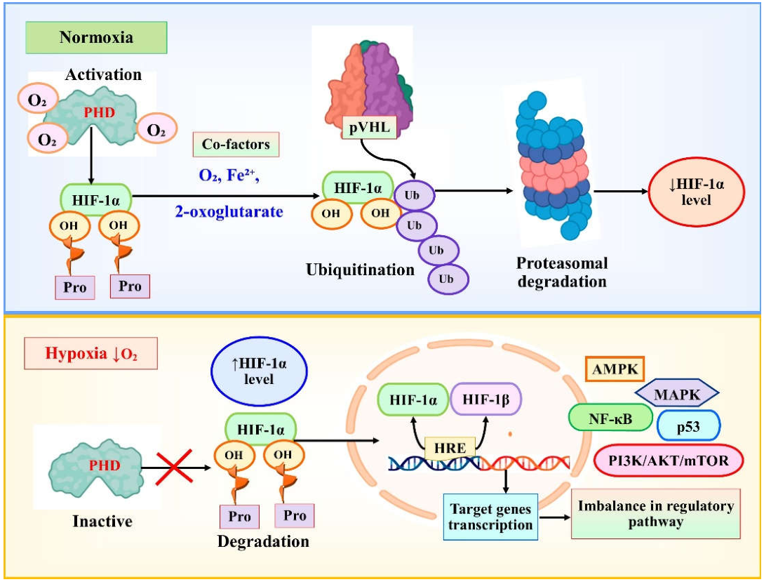
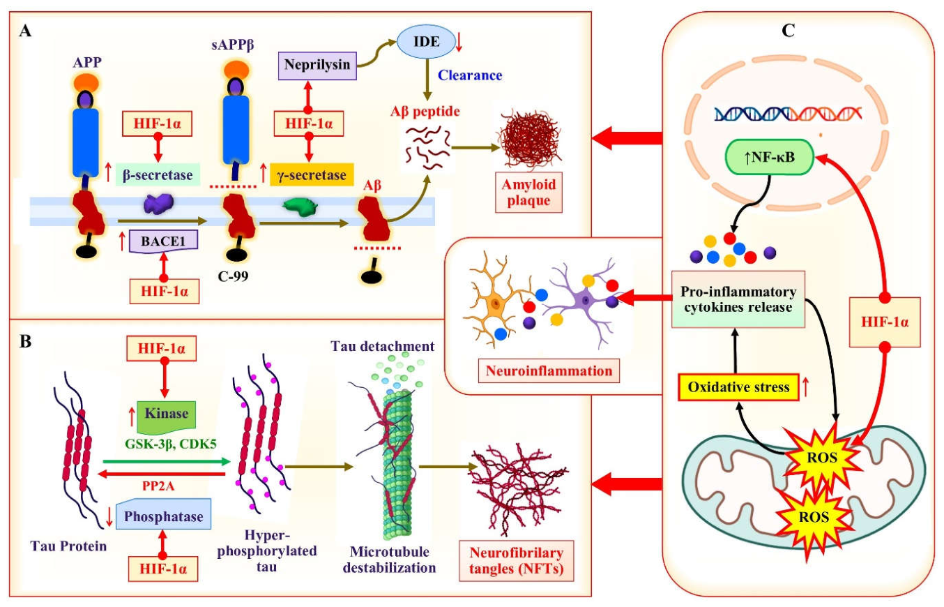
Figure 2.
HIF-1 mediated pathogenesis in AD; A: HIF-1 mediated modulation of Aβ pathogenesis; B: HIF-1 mediated modulation of tau pathology; and C: crosstalk among HIF-1, ROS, and AD pathophysiology.
Figure 2.
HIF-1 mediated pathogenesis in AD; A: HIF-1 mediated modulation of Aβ pathogenesis; B: HIF-1 mediated modulation of tau pathology; and C: crosstalk among HIF-1, ROS, and AD pathophysiology.
Under normoxic conditions, HIF-1α undergoes hydroxylation at specific proline residues by prolyl hydroxylase domain (PHD 1-3) enzymes, which require molecular oxygen (O₂), ferrous iron (Fe²⁺), and 2-oxoglutarate as cofactors. This hydroxylation tags HIF-1α for recognition by the von Hippel-Lindau tumor suppressor protein (pVHL), leading to ubiquitin-proteasomal degradation resulting in a half-life of approximately 5 minutes. Upon exposure to hypoxia, PHD activity gets inhibited leading to HIF-1α accumulation, nuclear translocation, dimerization with HIF-1β, and transcriptional activation of multiple target genes containing hypoxia-response elements in their promoters [1,36]. The HIF-1 pathway exhibits crosstalk with numerous signaling cascades, including PI3K/AKT/mTOR, NF-κB, and p53 pathways, and the magnitude, duration, and cellular context of HIF-1 activation determine whether the transcription promotes cell survival and adaptation or triggers cell death pathways, with mild and transient hypoxia eliciting protective responses whereas severe and prolonged hypoxia leading to HIF-1-mediated apoptosis [8,37,38].
The expression pattern of HIF-1α in the AD is complex and exhibits regional and cell-type specificity, with conflicting results indicating both upregulation and downregulation depending on the tissue analyzed, disease stage, and cellular context, stating the nuanced and context-dependent nature of HIF-1 involvement in AD progression. Immuno-histochemical studies have demonstrated significantly elevated HIF-1α immune-reactivity in brain micro-vessels isolated from AD patients, along with overexpression of pro-angiogenic proteins including angiopoietin-2 and matrix metalloproteinase-2 [39], suggesting that the cerebral vasculature undergoes profound hypoxic adaptation in AD that may represent either a compensatory response to impaired perfusion or a maladaptive process contributing to blood-brain barrier dysfunction. Similarly, triple transgenic AD mice (3 × Tg-AD) brains exhibit hypoxic vessels with upregulation of HIF-1α expression, which triggers the formation of NLRP1 inflammasome complexes that further stimulate HIF-1α expression, establishing an HIF-1α-NLRP1 vicious cycle exacerbating neuroinflammation and vascular pathology in a feed-forward manner [40].
In contrast, analyses of neuronal populations in the AD brain have revealed decreased HIF-1α levels, particularly in regions vulnerable to neurodegeneration, which correlate with reduced expression of glucose transporters GLUT1 and GLUT3, impaired glucose uptake, and abnormal tau hyperphosphorylation, indicating that neurons may experience a failure of adaptive hypoxic responses that predisposes them to energy failure and degeneration. The microglial compartment further illustrates this cell-type dichotomy, as the human AD hippocampus shows increased expression of HIF-1α and its target genes in microglia. This upregulation correlates with decreased microglial coverage of Aβ plaques and heightened plaque-associated neuropathology [41]. Conversely, complement C3a receptor-positive microglia in APP knock-in mice exhibit upregulated HIF-1 signaling with abnormal lipid droplet accumulation [42]. These reports demonstrate that HIF-1 activation in glial cells may contribute to dysfunctional immune responses and impaired Aβ clearance. Astrocytes derived from 5×FAD mice brains show compensatory mechanisms including increased HIF-1 expression, which is assumed to protect these cells against Aβ toxicity [43]. However, the chronic nature of this upregulation may ultimately impair astrocytic functions and contribute to the inflammatory milieu characteristic of the AD brain [2].
The differential expression of HIF-1α across cell types and brain regions seems to depend on the severity and duration of the underlying pathology. Patients with mild cognitive impairment show early adaptive HIF-1 responses, which may serve as a protective mechanism to maintain metabolic homeostasis. In contrast, patients with advanced AD exhibit dysregulated and potentially maladaptive HIF-1 signaling insufficient to support neuronal survival [3]. The temporal dynamics of HIF-1α expression in AD are complex. Early stages may involve hypoxic preconditioning and HIF-1 stabilization as protective responses. However, prolonged or severe hypoxia can overwhelm these mechanisms, triggering HIF-1-mediated p53 stabilization, BNIP3 activation, and apoptotic neuronal death, thus turning a potentially beneficial pathway into a driver of neurodegeneration [44]. Furthermore, emerging evidence suggests that HIF-1α expression patterns may be influenced by genetic factors, including APOE genotype, with APOE4 carriers demonstrating reduced HIF-1α and VEGF receptor-2 expression, explaining the enhanced vulnerability of these individuals to vascular pathology and neurodegeneration [45]. These complex and sometimes contradictory findings regarding HIF-1α expression in AD demand the necessity for region-specific and cell-type-specific analyses with careful consideration of disease stage, hypoxic severity, and genetic background when interpreting the role of HIF-1 in AD pathogenesis.
4.2. Mechanistic Insights into HIF-1-Mediated Pathogenesis in AD
4.2.1. HIF-1-Mediated Modulation of Aβ Pathology
The mechanistic insights of HIF-1 in AD pathogenesis involve multiple interconnected pathways directly influencing the generation and accumulation of Aβ peptides, the hyperphosphorylation and aggregation of tau protein, the activation of neuroinflammatory responses, and the disruption of cellular metabolism and mitochondrial function, collectively contributing to progressive neurodegeneration [2]. The most straightforward mechanistic link between HIF-1 and AD pathology is the transcriptional regulation of BACE1, the rate-limiting enzyme in amyloidogenic APP processing, wherein the BACE1 promoter contains functional hypoxia-response elements that bind HIF-1α under hypoxic conditions, leading to increased BACE1 expression, enhanced β-secretase activity, and resulting elevation of Aβ production (Figure 2 A) [46]. The HIF-1 mediated upregulation of BACE1 seems to be biphasic rather than transient, with an early phase of hypoxia characterized by mitochondrial electron transport chain-dependent ROS generation and a later phase characterized by HIF-1α stabilization and oxidative stress amplification, thereby offering multiple time points for therapeutic intervention.
In addition to β-secretase regulation, HIF-1 exerts profound effects on γ-secretase activity through both transcriptional and non-transcriptional mechanisms, including HIF-1α-mediated transactivation of the APH-1A gene, one of the critical components of the γ-secretase complex, and direct protein-protein interaction between HIF-1α and γ-secretase that enhances its catalytic activity without altering subunit expression levels, synergistically increasing the efficiency of the final cleavage that releases Aβ peptides from the C-99 fragment [47]. Moreover, hypoxia and HIF-1 signaling influence Aβ degradation pathways, as HIF-1 has been demonstrated to negatively regulate the expression of the Aβ-degrading enzyme neprilysin (NEP) through binding to hypoxia-response elements in the NEP promoter that exert repressive effects. Moreover, the expression of insulin-degrading enzyme is downregulated under hypoxic conditions, although direct involvement of HIF-1 remains unexplored [48]. The combined effect of increased production and decreased clearance of Aβ peptides under HIF-1 activation creates a feasible environment for Aβ accumulation, oligomerization, and plaque formation, supporting HIF-1 as a potent driver of amyloid pathology in the hypoxic condition.
4.2.2. HIF-1-Mediated Modulation of NITs Pathology
Regarding tau pathology, HIF-1 contributes to the hyperphosphorylation and aggregation of tau protein through multiple mechanisms that disrupt the kinase-phosphatase balance essential for maintaining tau in its properly phosphorylated state leading to boost the formation of NFTs and facilitating AD progression. Chronic hypoxia upregulates HIF-1α, which in turn decreases the activity of protein phosphatase 2A (PP2A), the principal phosphatase responsible for dephosphorylating tau, through LCMT1/PP2A deficiency, thereby tilting the equilibrium toward pathological hyperphosphorylation and subsequent aggregation of tau proteins (Figure 2 B) [49]. Simultaneously, hypoxia promotes the activation of protein kinases GSK-3β and CDK5 [50,51], which further promote tau phosphorylation at multiple sites, while HIF-1-mediated decreases in GLUT1 and GLUT3 expression reduce glucose uptake and O-GlcNAcylation, a post-translational modification that mediates inhibition of tau phosphorylation, thus further exacerbating the hyperphosphorylated tau [1,52]. The contribution of HIF-1 to tau pathology is particularly evident in the context of obstructive sleep apnea and chronic intermittent hypoxia, which induce alterations in tau phosphorylation suggestive of those observed in AD, and are associated with elevated serum levels of total and phosphorylated tau in patients with sleep-disordered breathing [53,54]. However, it must be noted that the effects of HIF-1 on tau phosphorylation appear to be context-dependent, as some studies have shown that HIF-1α may actually function as a "brake" to negatively regulate tau phosphorylation under certain conditions, such as in T-2 toxin-exposed microglia, where HIF-1α acts to suppress the T-2-induced hyperphosphorylation of tau [55], indicating a protective role in limiting tau pathology.
4.2.3. HIF-1-Mediated Modulation of Neuroinflammation
Another important mechanistic aspect is the effect of neuroinflammation by HIF-1 activation in AD, in which HIF-1 modulates the functions of microglia and astrocytes that may be either pro-inflammatory or anti-inflammatory depending on the duration and intensity of the stimulus. Chronic or prolonged HIF-1α activation in microglia induces cell cycle arrest, impairs proliferation and clustering around Aβ plaques, and compromises mitochondrial metabolism, leading to dysfunctional immune responses that are unable to effectively phagocytose Aβ deposits, hence, contributing to plaque accumulation and concurrent neurotoxicity [33,41]. However, acute HIF-1 activation in microglia can exacerbate phagocytosis of synaptic components and Aβ through feed-forward mechanisms [56], demonstrating a protective role in clearing pathological debris during early stages and in response to mild hypoxic stimuli in AD brain. The HIF-1 pathway also plays a pivotal role in the metabolic reprogramming of microglia, driving a shift from oxidative phosphorylation to glycolysis in response to Aβ exposure through mTOR-HIF-1α signaling, which may initially support the energy demands of inflammatory responses but ultimately leads to defective energy metabolism and diminished immune function with chronic Aβ exposure [57].
In astrocytes, HIF-1 activation are essential for maintaining neuronal metabolic support and neurotransmitter homeostasis, either promoting anti-inflammatory or pro-inflammatory responses depending on the context. Existing studies have demonstrated that HIF-1 stabilization can reverse Aβ-induced glial activation and glycolytic changes, suggesting a protective role, while chronic activation may contribute to astrocytic dysfunction and weakened support of neuronal health [58]. The cerebrovascular endothelial cell compartment is also significantly impacted by HIF-1 activation in AD. In chronic hypoxia, endothelial cells triggers HIF-1α-NLRP1 inflammasome formation, leading to overexpression of the adaptor molecule ASC (apoptosis-associated speck-like protein containing a CARD), caspase-1 activation, and IL-1β production, which further activates HIF-1α in a feed-forward loop that destroys the vascular system, impairs blood-brain barrier integrity, and compromises Aβ clearance [1,40,56], directly linking vascular dysfunction to the core pathological features of AD.
4.2.4. HIF-1-Mediated Redox Imbalance and Metabolic Reprogramming
The crosstalk among HIF-1, oxidative stress, and mitochondrial dysfunction constitutes another critical mechanistic aspect. HIF-1 not only responds to oxidative stress but also actively modulates the redox balance and metabolic reprogramming of cells in ways that can either protect against or exacerbate oxidative damage. Under hypoxic conditions, HIF-1 promotes a shift from mitochondrial oxidative phosphorylation to aerobic glycolysis through upregulation of pyruvate dehydrogenase kinase-1, which inhibits the TCA cycle, and lactate dehydrogenase A, which converts pyruvate to lactate, therefore reducing oxygen consumption and limiting mitochondrial ROS production, representing an adaptive response to protecting cells from oxidative damage (Figure 2 C) [59]. This metabolic reprogramming, however, is at the expense of reduced ATP generation and a dependence on glycolysis, which may be insufficient to satisfy the high energy requirements of neurons, particularly in the context of pre-existing age-related metabolic impairment, and may thus contribute to synaptic dysfunction and neuronal susceptibility in AD.
Furthermore, HIF-1 can upregulate antioxidant enzymes including heme oxygenase-1 (HO-1) and peroxiredoxins through hypoxia-response elements-mediated transcriptional induction, providing a direct mechanism for counteracting oxidative stress as well as reduction of ROS accumulation, which may be protective under mild hypoxic conditions [60]. Under severe or prolonged hypoxia, HIF-1-mediated stabilization of p53 through direct protein-protein interaction leads to p53-dependent apoptosis. Besides, HIF-1 upregulates the pro-apoptotic Bcl-2 family member BNIP3, which triggers mitochondrial permeability transition and cell death [61]. All these evidences indicate that the balance between antioxidant and pro-apoptotic activities of HIF-1 is exquisitely sensitive to the severity and duration of hypoxic stress.
The contribution of HIF-1 to iron imbalance is further associated with HIF-1 to oxidative stress mechanisms, as HIF-1 regulates transcription of transferrin receptor 1 (TfR1), the key mediator of iron distribution in the brain. Elevated HIF-1 signaling in AD is associated with increased TfR1 expression, iron accumulation, and consequent oxidative damage through Fenton chemistry, which drives lipid peroxidation, protein oxidation, and DNA damage [30,62,63]. The accumulation and dysregulation of iron under HIF-1 activation may promote ferroptosis, an iron-dependent form of programmed cell death linked to AD pathogenesis. Iron chelators that stabilize HIF-1 show varying effects on ferroptosis, depending on their specific mechanisms of action [64,65].
4.2.5. HIF-1: Friend or Foe? Temporal and Spatial Considerations in AD Pathogenesis
The temporal dynamics of HIF-1 activation in AD further complicate the interpretation of its mechanistic insight. Early hypoxic events can trigger protective HIF-1 responses that enhance glycolytic capacity, upregulate antioxidant defenses, and promote angiogenesis. However, prolonged hypoxia and chronic inflammation eventually overwhelm these adaptive mechanisms, leading to HIF-1-mediated apoptosis, exacerbated Aβ production, and amplified neuroinflammation. This duality is reported in the studies on ischaemic stroke, where the accumulation of HIF-1α during the acute (<24 hours) phase promotes cell death, whereas HIF-1α signaling during the later (>4 days) phase exerts pro-survival effects by limiting infarct size, suggesting that therapeutic strategies targeting HIF-1 must be carefully timed to optimize beneficial effects and avoid exacerbating damage [3,66,67]. Likewise, the neuroprotective and neurotoxic effects of HIF-1 in AD can be separated by temporal and spatial factors. HIF-1 activation in early or mild hypoxia may promote neuronal survival via enhanced glucose metabolism and antioxidant capacity, while in chronic or severe hypoxia, HIF-1 activation promotes Aβ deposition, tau hyperphosphorylation, neuroinflammation, and apoptosis, thus making HIF-1 a “double-edged sword” in AD pathophysiology (Figure 3) [2]. The molecular determinants that modulate these opposing outcomes include the magnitude of HIF-1α stabilization, the duration of hypoxic exposure, the cell types involved, the presence of co-existing stressors such as oxidative stress and inflammation, and also the genetic background of the individual, all of which must be considered in case of evaluating the mechanistic role of HIF-1 in AD and designing therapeutic interventions targeting this pathway.
5. Reported Interventions Targeting HIF-1 Modulation in AD Pathophysiology
The dichotomous nature of HIF-1 signaling in AD has been investigated by numerous researchers focusing on pharmacological, genetic, and physiological interventions that modulate this pathway, with the goal of harnessing its neuroprotective capacity while avoiding its pathogenic consequences. Table 1 represents a summary of diverse interventions categorized by their mechanisms of action, with neutoprotective and neurotoxic outcome to illustrate the context-dependent nature of HIF-1 modulation in AD.
5.1. Iron Chelators
Iron chelators are among the most thoroughly studied categories of HIF-1 modulators in AD, utilizing the iron-dependent characteristics of PHD enzymes to stabilize HIF-1α and activate downstream neuroprotective pathways. Deferoxamine (DFO), a clinically approved iron chelator, has demonstrated neuroprotective effects in APP/PS1 transgenic mice by upregulating HIF-1α through PHD inhibition and activating the P38/HIF-1α signaling cascade, resulting in reduced Aβ deposition, attenuated synaptic loss, and elevated expression of BDNF, transferrin receptor, and divalent metal transporter 1, as well as decreased iron levels in the hippocampus. The neuroprotective efficacy of DFO extends beyond simple iron chelation. It has been reported as promising candidate in AD therapy because of the ability to stabilize HIF-1α, promote metabolic reprogramming toward glycolysis, enhance antioxidant defenses, and support neuronal survival under conditions of amyloid toxicity [68,69,70].
M30, a multifunctional iron chelator with better BBB penetrating properties, has shown potential neuroprotective activity by stabilizing HIF-1α and upregulating its downstream targets, including aldolase A, enolase-1, and GLUT-1, along with reducing GSK-3β activity and tau phosphorylation, thereby protecting cortical neurons against Aβ25-35-induced toxicity in both APP/PS1 transgenic mice and primary neuronal cultures [71,72]. The dual mechanism of M30, combining iron chelation with HIF-1 stabilization and insulin signaling modulation, positions this compound as a potential candidate for targeting multiple pathological cascades simultaneously in AD therapeutics.
Transient iron chelation and HIF-1 stabilization offer neuroprotection, however, prolonged or excessive pharmacological modulation of the HIF pathway can produce neurotoxic outcomes. Chronic use of PHD inhibitors is advantageous, but short-term use leads to adverse effects when PHD inhibition is prolonged. Prolonged pharmacological stabilization of HIF-1α exacerbates neuroinflammation, promotes excessive angiogenesis with aberrant vascular remodeling, and alters neural progenitor cell status in preclinical AD models [73].
Besides, cobalt chloride (CoCl₂), a chemical hypoxia mimetic, stabilizes HIF-1α through PHD inhibition independent of oxygen status, leading to simulation of hypoxic conditions at the molecular level. Administration of CoCl₂ induces Aβ deposition, promotes tau hyperphosphorylation at Thr181, triggers dysregulated autophagy, and leads to hippocampal and cortical neurodegeneration in both in vivo mice models and neuronal cell lines [74].
5.2. Natural Phytochemicals and Bioactive Compounds
The plant kingdom has produced numerous bioactive compounds that modulate HIF-1 signaling and exhibit neuroprotective properties in AD models, offering the advantage of multi-target effects and favorable safety profiles. Icariin, a bioactive phytocompound isolated from Epimedii herba, has been identified through network pharmacology approaches as a direct modulator of the HIF-1 signaling pathway, demonstrating the ability to regulate Aβ1-42, Aβ1-40, BACE1 expression, tau phosphorylation, and inflammatory mediators, ultimately alleviating cognitive impairment in preclinical studies [75]. Ginsenoside Rg1, the active ingredient of Panax ginseng, exerts protective effects against Aβ-induced mitochondrial apoptosis in human endothelial cells by reducing HIF-1α expression, decreasing reactive nitrogen species (RNS), and attenuating protein nitrotyrosination, suggesting that suppression of excessive HIF-1 activation may be beneficial in certain contexts [76]. Salidroside, a glucoside of tyrosol derived from Rhodiola rosea, attenuates hypoxia-induced amyloidogenesis by reducing HIF-1α protein levels and decreasing BACE1 expression, thereby inhibiting β-secretase activity and enhancing sAPPα secretion while reducing Aβ generation in SH-SY5Y neuronal cells, highlighting the therapeutic potential of natural compounds that dampen HIF-1-mediated amyloidogenic processing [77]. α-Lipoic acid, a naturally occurring antioxidant compound, maintains brain glucose metabolism through the BDNF/TrkB/HIF-1α signaling pathway in P301S tauopathy mice, increasing the expression of GLUT3, GLUT4, VEGF, and HO-1, thereby enhancing glucose availability and improving cognitive function [78]. The NeuroProtect formula, a combination of geniposide and Panax notoginseng saponins, modulates HIF-1/PI3K-AKT signaling by targeting mTOR, HIF1A, TLR4, STAT3, and VEGFA, effectively reducing the Aβ42/Aβ40 ratio, increasing dendritic spine density, and reversing Aβ25-35-induced cell viability reduction in APP/PS1 transgenic mice and cell culture models [79].
Certain metabolic stressors derived from dietary sources can activate HIF-1-dependent amyloidogenic pathways and contribute to AD pathology. Palmitic acid-BSA (PA-BSA), a metabolic stressor representative of saturated fatty acid burden associated with high-fat diets, activates Akt/mTOR/HIF-1α and Akt/NF-κB signaling pathways, stimulating APP and BACE1 expression and accelerating Aβ production in SK-N-MC neuroblastoma cells [80]. This demonstrates how dietary factors can activate HIF-1-dependent amyloidogenic pathways, providing a mechanistic basis for the association between obesity, metabolic syndrome, and increased AD risk. High glucose conditions, mimicking metabolic stress observed in diabetes and metabolic syndrome, activate HIF-1α through ROS-dependent JNK pathway signaling, leading to upregulation of BACE1 expression, increased Aβ production, and decreased LXRα expression in SK-N-MC neuroblastoma cells and ZDF/ZLC rats [81]. This pathway provides a mechanistic link between diabetes, HIF-1 activation, and increased AD risk, underscoring the contribution of metabolic dysfunction to neurodegenerative pathology. Thiamine deficiency, a nutritional insufficiency, activates HIF-1α-mediated amyloidogenesis through transcriptional upregulation of BACE1 and increased β-secretase activity, while simultaneously inducing the pro-apoptotic protein BNIP3, triggering neurotoxicity and AD-like pathology in HT22 hippocampal neuronal cells [82]. This mechanism provides a molecular explanation for the epidemiological association between thiamine deficiency and cognitive impairment, establishing HIF-1 as a critical mediator linking nutritional status to AD pathogenesis.
5.3. Nutritional and Dietary Interventions
Numerous emerging studies support the neuroprotective potential of dietary supplements and nutritional interventions that modulate HIF-1 activity through multiple mechanisms. Lactoferrin, a milk-derived iron-binding glycoprotein, enhances α-secretase-dependent amyloid precursor protein processing through the ERK1/2-CREB and HIF-1α pathways in APP/PS1 double transgenic mice, effectively reducing Aβ aggregation and improving spatial learning while promoting non-amyloidogenic APP processing [83]. Coffee consumption, extensively documented for its neuroprotective properties, inhibits prolyl hydroxylation to activate HIF-1α and induces VEGF expression in SH-SY5Y human neuroblastoma cells, suggesting that coffee consumption reduces the risk of cognitive decline [84]. Probiotic interventions, the multi-strain formulation SLAB51, increase cerebral HIF-1α expression by decreasing PHD2 and iNOS levels in the brain and reducing nitric oxide concentrations in plasma, thereby attenuating oxidative stress and neuroinflammation in 3 × Tg-AD and wild-type mice [85]. The gut-brain axis, mediated through probiotic modulation of HIF-1 signaling, represents a novel therapeutic avenue for mitigating AD pathology through systemic and CNS effects.
Pathological conditions associated with oxygen deprivation represent the most significant damaging category of HIF-1 modulation. Chronic hypoxia, whether physiological or experimentally induced, promotes HIF-1α stabilization and consequent activation of amyloidogenic pathways, resulting in increased BACE1 expression and γ-secretase activity leading to elevated Aβ production along with tau hyperphosphorylation through downregulation of PP2A and activation of GSK-3β in rodent models and neuronal cell cultures [49,86]. Chronic intermittent hypoxia, which represents the recurrent oxygen desaturation episode characteristic of obstructive sleep apnea (OSA), activates chronic HIF-1 signaling and triggers a neuroinflammatory cascade involving microglial activation and astrogliosis. Studies in transgenic AD mice and OSA models have demonstrated that intermittent hypoxia elevates Aβ42 levels, exacerbates neuroinflammation, and contributes to cognitive decline through sustained HIF-1 activation [87,88]. The clinical relevance of these findings is supported by observations that patients with sleep-disordered breathing exhibit increased risk of cognitive decline and AD pathology.
5.4. Endogenous Proteins and Peptide Hormones
Several endogenous proteins and peptide hormones have been reported for the potential to modulate HIF-1 activity. Neuroglobin, a hypoxia-inducible cytoprotective protein, enhances neuroprotection and promotes neuronal survival under hypoxic conditions. A research demonstrated that shRNA-mediated knockdown and lentiviral vector-mediated overexpression of HIF-1α modulate neuroglobin expression in HN33 neural cells, suggesting a causal relationship between HIF-1 and neuroglobin induction [89]. Fibroblast growth factor 21 (FGF21), a liver-secreted peptide hormone, ameliorates neurodegeneration by inhibiting the PP2A/MAPKs/HIF-1α pathway triggered by Aβ25-35, effectively reducing tau hyperphosphorylation and attenuating oxidative stress in both SH-SY5Y cells and adult male Wistar rats [90]. Neuregulin 1, a neurotrophic factor, protects against hypoxia-induced neuronal damage by inhibiting CoCl₂-induced accumulation of HIF-1α and p53 leading to cell death and suppression of HIF-1α accumulation in SH-SY5Y human neuroblastoma cells, demonstrating that modulation of HIF-1 activity by endogenous growth factors may represent a physiological neuroprotective mechanism [91].
Heparin-binding EGF-like growth factor (HB-EGF), an endogenous growth factor induced by hypoxia and ischemia, promotes AD neuropathology through HIF-1-dependent mechanisms. HB-EGF increases HIF-1α expression leading to activation of matrix metalloproteinase-9 (MMP9), leading to blood-brain barrier disintegration and impaired Aβ clearance in bilateral common carotid artery occlusion (BCCAO) mice models [92,93]. This pathway establishes a mechanistic crosstalk amomg cerebral hypoperfusion, HIF-1 activation, vascular dysfunction, and impaired Aβ clearance, contributing to the progressive accumulation of Aβ pathology.
5.5. Gene Therapy and Genetic Interventions
Genetic approaches for modulating HIF-1 activity have emerged as promising next-generation strategies to attain sustained and cell-type-specific therapeutic effects in AD. Viral vector-mediated overexpression of HIF-1α, utilizing recombinant adeno-associated virus (rAAV) or lentiviral model, inhibits hippocampal neuronal apoptosis induced by Aβ plaque and promotes cell survival in primary hippocampal neurons and Sprague-Dawley rats, demonstrating the feasibility of gene therapy for enhancing HIF-1-mediated neuroprotection [94]. Conversely, HIF-1α siRNA-loaded astrocyte-derived extracellular vesicles have been employed to suppress HIF-1α expression through RNA interference, reducing APP, Aβ, Aβ fibrils, and phosphorylated tau, along with diminishing glial activation and partially restoring synaptic proteins in HIV-infected humanized mice models, suggesting that selective HIF-1α suppression may be beneficial in contexts where chronic HIF-1 activation drives neurodegeneration [95]. These complementary genetic approaches draw attention to the context-dependent nature of HIF-1 modulation in AD emphasizing the urge of precision therapy based on disease stage and pathological context.
HIF-1α overexpression through viral vector-mediated genetic constructs leading to constitutive HIF-1α stabilization and non-physiological pathway activation promotes BACE1 expression and γ-secretase activity while increasing Aβ production and activating pro-apoptotic pathways through p53 stabilization [96]. Studies in primary neurons and transgenic mice models have demonstrated that supra-physiological HIF-1α levels overwhelm adaptive protective mechanisms, converting HIF-1 from a survival factor into a promoter of neurodegeneration (Alexander et al., 2022). These findings support the critical importance of adopting appropriate HIF-1α expression levels in during developing gene therapy approaches for AD management.
5.6. Existing Drugs, Small Molecules, and Other Pharmacological Interventions
Several pharmacological agents, including clinically approved drugs, experimental small molecules, and physiotherapeutic interventions, have been demonstrated to modulate HIF-1 activity and exert differential impacts in AD models. Melatonin, an endogenous neurohormone, suppresses HIF-1α and BACE1 expression under oxygen-glucose deprivation and re-oxygenation (OGD/R) conditions, effectively inhibiting hypoxia-induced AD pathogenesis by downregulating the amyloidogenic pathway and reducing Aβ production in SH-SY5Y human neuroblastoma cells [97]. Low-dose simvastatin, a widely prescribed statin, reduces HIF-1α and BACE1 expression in sporadic AD and age-matched control neuronal mitochondrial cybrids, diminishing Aβ production and amyloidogenic processing [98]. Intermittent hypoxia-hyperoxia training (IHHT), a physiological intervention, modulates HIF-1α expression and reduces neuroinflammation, improving spatial learning and memory while decreasing Aβ, CYP2E1, HIF1α, and TNFα expression in the hippocampus of streptozotocin (STZ)-induced rat models of AD [99]. Neurotropin®, a non-protein extract of inflamed rabbit skin inoculated with vaccinia virus clinically used for neuropathic pain, regulates HIF-1α/MAPK signaling to lessen Aβ-induced oxidative damage, improve Aβ deposition in the hippocampus, and alleviate neuroinflammation in HT22 hippocampal cells and APP/PS1 transgenic mice [100].
All together, the diverse interventions modulating HIF-1 activity in AD models reveal a complex pharmacological landscape wherein transient or mild HIF-1 activation mediates neuroprotection through enhanced glucose metabolism, angiogenesis, antioxidant defense, and anti-inflammatory mechanisms. Conversely, the chronic, severe, or constitutively high HIF-1 activity exacerbates amyloid pathology, tau hyperphosphorylation, neuroinflammation, and neurodegeneration. The therapeutic potential of HIF-1 modulation is further complex by cell-type-specific responses, as neurons, microglia, astrocytes, and cerebrovascular endothelial cells each exhibit distinct transcriptions and metabolic reprogramming in response to HIF-1 activation. Pharmacological agents that stabilize HIF-1α through PHD inhibition, including iron chelators and naturally occurring compounds, have demonstrated the most consistent neuroprotective effects, while interventions that cause sustained HIF-1 activation or mimic severe hypoxia consistently produce detrimental outcomes.
6. Therapeutic Challenges and Future Perspectives
6.1. Challenges in HIF-1 Targeted Therapy for AD
Despite the promising preclinical evidence supporting HIF-1 modulation as a therapeutic strategy for AD, several significant challenges must be addressed before proceeding for clinical translation. The foremost challenge lies in the dichotomous nature of HIF-1 signaling itself, wherein the same pathway that confers neuroprotection under mild or transient hypoxic conditions becomes pathogenic when chronically or excessively activated. This requires precise control over the magnitude and duration of HIF-1 modulation to achieve therapeutic benefit without triggering detrimental consequences. The identification of an optimal therapeutic window for HIF-1 activation remains unclear and requires more research, as the threshold that separates neuroprotective effects from neurodegeneration appears to vary depending on the cell type, disease stage, and co-existing pathological conditions. All these factors are making it difficult to establish universal dosing guidelines for HIF-1-targeted therapies. Furthermore, the cell-type specific responses to HIF-1 activation pose a significant challenge, as HIF-1 stabilization in neurons may enhance survival by boosting glycolysis and antioxidant defense, whereas HIF-1 activation in microglia may worsen neuroinflammation and hinder amyloid clearance. HIF-1 in astrocytes can support or impair neuronal function depending on the context. The blood-brain barrier represents another significant obstacle, as many HIF-1 modulating agents, particularly iron chelators and small molecule inhibitors, exhibit limited brain penetration, necessitating the development of novel delivery strategies or the identification of peripherally acting agents that can indirectly modulate central HIF-1 activity [1,2,3]. The chronic nature of AD further complicates therapeutic development, as long-term HIF-1 modulation may lead to adaptive responses, tolerance, or off-target effects that diminish efficacy or produce adverse outcomes over time, requiring careful evaluation in extended preclinical studies before initiating clinical trials.
6.2. Advancing HIF-1 Therapeutics: Translational Opportunities
6.2.1. Isoform-Selective PHD Inhibitors
The development of isoform-selective PHD inhibitors that preferentially target specific PHD isoforms expressed in the brain represents a promising strategy for achieving more refined HIF-1 modulation. PHD2 is the predominant isoform in neurons, its selective inhibition may confer neuroprotection while avoiding the systemic effects associated with pan-PHD inhibition. This approach holds novelty because PHD2-selective inhibition would stabilize HIF-1α primarily in neurons, potentially harnessing the neuroprotective outcome of HIF-1 activation while reducing the neuroinflammatory consequences of HIF-1 stabilization in microglia and other cell types [101]. Furthermore, the development of PHD inhibitors with differential selectivity for the three PHD isoforms may enable fine-tuning of the HIF-1 response based on the specific pathological context and desired therapeutic outcomes, offering greater precision than currently available agents.
7.2.2. Nanoparticle-Based Targeted Delivery Systems
Nanoparticle-based delivery systems might be adopted to enhance brain bioavailability of HIF-1 modulators and achieve targeted delivery to specific cell types like astrocyte-targeted and neuron-targeted nanoparticles. This particular approach hold novelty for delivering HIF-1α stabilizers or inhibitors directly to the cells most relevant to AD pathology. The surface functionalization of nanoparticles with targeting ligands that recognize cell-specific receptors enables selective uptake by desired cell types, thereby distributing the therapeutic agent in the targeted cellular compartment where it is most needed rather than minimizing exposure to cells where HIF-1 modulation might produce adverse effects. Additionally, the development of stimuli-responsive nanoparticles that release the drug molecules in response to hypoxic conditions may enable more precise temporal control over HIF-1 modulation, ensuring that therapeutic effects are achieved only when and where required [102,103].
6.2.3. Gene Editing and Epigenetic Approaches
Gene therapy approaches, including CRISPR-based activation or repression of the HIF1A gene, offer the potential for sustained, cell-type-specific HIF-1 modulation, with the advantage of long-term therapeutic effects following a single administration. However, the concerns regarding off-target effects and immunogenicity remain to be addressed. The development of cell-type-specific promoters and viral vectors that selectively transduce neurons, astrocytes, or microglia would enable precise spatial control of HIF-1α expression, potentially achieving neuronal neuroprotection while avoiding HIF-1-mediated neuroinflammation in glial cells. Epigenetic modulation of HIF-1 target genes represents another emerging strategy, wherein drugs that modulate histone acetylation or DNA methylation at specific gene promoters may selectively enhance or suppress the expression of HIF-1 target genes that mediate beneficial effects without affecting pathogenic target genes [104].
6.2.4. Small Molecule Modulators of HIF-1 Target Genes
The identification of HIF-1 target genes that facilitate specific protective or detrimental effects has enabled the development of more selective therapeutic strategies capable of modulating individual downstream pathways rather than generalized HIF-1 activity, potentially achieving neuroprotection while circumventing the pathogenic consequences of non-selective HIF-1 activation. This approach involves screening for small molecules that selectively modulate the expression or function of specific HIF-1 target genes, such as those involved in glucose metabolism or antioxidant defense, without affecting HIF-1 target genes that promote amyloidogenesis or neuroinflammation. The development of such selective modulators would represent a significant advancement over current approaches, potentially overcoming the dichotomous nature of HIF-1 signaling and offering desired therapeutic benefit [105].
6.2.5. Combination Therapies
Combination therapies that pair HIF-1 modulation with other neuroprotective agents, such as anti-inflammatory drugs or antioxidants, may provide synergistic benefits by targeting multiple pathological pathways simultaneously with lower doses to minimize off-target effects. The concurrent administration of HIF-1 stabilizers with agents that reduce neuroinflammation or oxidative stress may enable neuroprotective HIF-1 activation while mitigating the inflammatory consequences arisen from chronic HIF-1 signaling. Similarly, the combination of HIF-1 inhibition with agents that promote amyloid clearance or reduce tau phosphorylation may address the pathological consequences of chronic HIF-1 activation with simultaneously targeting the core pathological features of AD, potentially achieving greater therapeutic efficacy than either approach alone [106].
6.3. Precision Medicine: Biomarkers-Specific HIF-1 Diagnostics and Therapeutics
The development of reliable biomarkers to monitor HIF-1 activity and predict treatment response is essential for successful clinical translation and personalized medicine approaches. Cerebrospinal fluid and plasma levels of HIF-1α and its downstream targets, including VEGF, EPO, and GLUT1, may serve as pharmacodynamic biomarkers to assess target specificity and guide dose selection in clinical trials. However, the relationship between peripheral and central HIF-1 activity must be carefully validated. Neuroimaging approaches, including positron emission tomography (PET) with hypoxia-specific tracers and functional magnetic resonance imaging (fMRI) to assess cerebral blood flow and oxygen metabolism, may provide non-invasive means to identify brain regions with hypoxic stress and monitor the effects of HIF-1-targeted interventions [107]. Genetic factors, including APOE genotype and polymorphisms in HIF-related genes, may influence individual responses to HIF-1 modulation and require patient stratification strategies, with APOE4 carriers potentially benefiting from different HIF-1 modulation approaches compared to non-carriers given the established association between APOE4 and impaired HIF-1 signaling [108]. The identification of patient subgroups based on disease stage, hypoxia, and inflammatory status may enable more precise targeting of HIF-1 modulation, with early-stage patients with mild hypoxic stress potentially benefiting from HIF-1 activation, while late-stage patients with chronic neuroinflammation may require HIF-1 inhibition. Longitudinal monitoring of biomarkers during treatment will be essential to assess whether HIF-1 modulation remains within the therapeutic window and to detect early signs of adverse effects, enabling timely adjustment of dosing or discontinuation of therapy if necessary [1,2,3,109].
6.4. Clinical Trial Considerations
The translation of HIF-1-targeted therapies from preclinical models to clinical trials requires careful consideration of trial design, patient selection, and outcome measures to maximize the likelihood of success. Dose optimization studies must be conducted to identify the optimal dose that achieves neuroprotective HIF-1 activation without triggering neurodegeneration, with dose escalation strategies with the help of pharmacodynamic biomarkers and monitoring for adverse effects. The selection of appropriate clinical endpoints is critical, as the neuroprotective effects of HIF-1 modulation may be best captured by measures of functional decline, cognitive performance, and biomarkers of neurodegeneration rather than by clinical endpoints that may be influenced by other factors. The duration of clinical trials must be adequate to monitor disease progression, necessitating longer follow-up periods than usual for acute therapies, due to the chronic nature of AD and the time needed for HIF-1 mediated neuroprotective mechanisms to translate into clinical benefits. The inclusion of biomarker endpoints, including measures of Aβ and tau pathology, neuroinflammation, and brain metabolism, will provide mechanistic insights and enable early assessment of target interaction, potentially facilitating go/no-go decisions in the development of AD therapeutics [1,2,3].
7. Concluding Remarks and Limitations
In conclusion, HIF-1 emerges as a critical molecular bridge connecting cerebral hypoperfusion, oxidative stress, and neuroinflammation to the Aβ plaques and neurofibrillary tangles, core pathological features of AD. The evidence demonstrates that HIF-1 exerts dual effects in the AD brain, offering neuroprotection through enhanced glucose metabolism, angiogenesis, and antioxidant defense under mild hypoxic conditions, while promoting neurodegeneration through amyloidogenesis, tau hyperphosphorylation, and apoptosis under severe or chronic oxygen deprivation. This dichotomy underscores the complexity of targeting this pathway therapeutically, as interventions must be carefully calibrated to maximize beneficial outcomes while avoiding detrimental consequences. Despite these mechanistic insights, several significant limitations must be acknowledged. The majority of mechanistic studies have been conducted in vitro using immortalized cell lines or primary neuronal cultures, which may not completely simulate the complex cell-cell interactions and chronic nature of AD pathology observed in the human brain. The temporal dynamics of HIF-1 activation remain poorly characterized, as most animal studies employ acute hypoxic exposures that do not adequately model the progressive cerebral hypoperfusion occurring over decades in the aging brain. Furthermore, the cell-type-specific contributions of HIF-1 signaling have not been systematically analyzed, and the relative importance of HIF-1 activation in neurons, microglia, astrocytes, and cerebrovascular endothelial cells for disease progression remains unclear. The clinical translation of HIF-1-targeted therapies faces substantial difficulties, including the development of highly selective modulators capable of penetrating the blood-brain barrier, the identification of appropriate therapeutic windows, and the establishment of reliable biomarkers to monitor treatment response. Additionally, the variability in HIF-1α expression patterns across AD patient populations suggests that HIF-1 targeted therapies may benefit from personalized medicine approach. Therefore, advanced future research on elucidating the precise molecular mechanisms underlying these interconnected pathways, identifying cell-type-specific HIF-1 target genes relevant to AD pathogenesis, and characterizing the temporal dynamics of HIF-1 responses in different stages of the disease will be essential for translating these mechanistic insights into effective therapeutic strategies for AD.
Author Contributions
Conceptualization, A.A. and K.M.; methodology, A.A., K.M., N.F.; software, A.A.; validation, K.M., N.F.; formal analysis, A.A.; investigation, A.A.; data curation, A.A., M.M.R.; writing—original draft preparation, A.A., N.F., M.M.R.; writing—review and editing, K.M., F.I., D.N.R.; visualization, A.A.; supervision, K.M. All authors have read and agreed to the published version of the manuscript.
Funding
This research received no external funding.
Institutional Review Board Statement
Not applicable.
Informed Consent Statement
Not applicable.
Acknowledgments
The authors have reviewed and edited the output and take full responsibility for the content of this publication.
Conflicts of Interest
The authors declare no conflicts of interest.
Abbreviations
The following abbreviations are used in this manuscript:
| 3 × Tg-AD | Triple Transgenic Alzheimer's Disease |
| 5×FAD | Five Familial Alzheimer's Disease Mutations |
| Aβ | Amyloid-Beta |
| AD | Alzheimer's Disease |
| AKT | Protein Kinase B |
| APOE | Apolipoprotein E |
| APP | Amyloid Precursor Protein |
| APP/PS1 | Amyloid Precursor Protein/Presenilin-1 |
| ARNT | Aryl Hydrocarbon Receptor Nuclear Translocator |
| ASC | Apoptosis-Associated Speck-Like Protein |
| BACE1 | β-Site APP-Cleaving Enzyme 1 |
| BBB | Blood-Brain Barrier |
| BCCAO | Bilateral Common Carotid Artery Occlusion |
| BDNF | Brain-Derived Neurotrophic Factor |
| BNIP3 | BCL2 Interacting Protein 3 |
| CDK5 | Cyclin-Dependent Kinase-5 |
| CNS | Central Nervous System |
| CoCl₂ | Cobalt Chloride |
| CSF | Cerebrospinal Fluid |
| DFO | Deferoxamine |
| DMT1 | Divalent Metal Transporter 1 |
| ECE-1 | Endothelin-Converting Enzyme-1 |
| EPO | Erythropoietin |
| ERK | Extracellular Signal-Regulated Kinase |
| EV | Extracellular Vesicle |
| FGF21 | Fibroblast Growth Factor 21 |
| FIH-1 | Factor Inhibiting HIF-1 |
| fMRI | Functional Magnetic Resonance Imaging |
| GLUT1 | Glucose Transporter 1 |
| GSK-3β | Glycogen Synthase Kinase-3 Beta |
| HB-EGF | Heparin-Binding Epidermal Growth Factor-Like Growth Factor |
| HIF-1 | Hypoxia-Inducible Factor-1 |
| HIV | Human Immunodeficiency Virus |
| HO-1 | Heme Oxygenase-1 |
| IHHT | Intermittent Hypoxia Hyperoxia Training |
| iNOS | Inducible Nitric Oxide Synthase |
| LDHA | Lactate Dehydrogenase A |
| MAPK | Mitogen-Activated Protein Kinase |
| MMP | Matrix Metalloproteinase |
| mTOR | Mammalian Target of Rapamycin |
| NEP | Neprilysin |
| NFTs | Neurofibrillary Tangles |
| NF-κB | Nuclear Factor Kappa B |
| NLRP1 | NLR Family Pyrin Domain-Containing 1 |
| NTP | Neurotropin® |
| OGD | Oxygen-Glucose Deprivation |
| OGD/R | Oxygen-Glucose Deprivation/Reoxygenation |
| OSA | Obstructive Sleep Apnea |
| PA-BSA | Palmitic Acid-Bovine Serum Albumin |
| PDK1 | Pyruvate Dehydrogenase Kinase 1 |
| PET | Positron Emission Tomography |
| PHD | Prolyl Hydroxylase Domain |
| PI3K | Phosphatidylinositol 3-Kinase |
| PP2A | Protein Phosphatase 2A |
| pVHL | von Hippel-Lindau Protein |
| rAAV | Recombinant Adeno-Associated Virus |
| RNS | Reactive Nitrogen Species |
| ROS | Reactive Oxygen Species |
| siRNA | Small Interfering RNA |
| SNP | Single Nucleotide Polymorphism |
| STAT3 | Signal Transducer and Activator of Transcription 3 |
| STZ | Streptozotocin |
| TBI | Traumatic Brain Injury |
| TfR1 | Transferrin Receptor 1 |
| Tg | Transgenic |
| TLR4 | Toll-Like Receptor 4 |
| TNF-α | Tumor Necrosis Factor Alpha |
| TrkB | Tropomyosin Receptor Kinase B |
| VEGF | Vascular Endothelial Growth Factor |
References
- Mitroshina, E.V.; Vedunova, M.V. The role of oxygen homeostasis and the HIF-1 factor in the development of neurodegeneration. Int. J. Mol. Sci. 2024, 25, 4581. [Google Scholar] [CrossRef] [PubMed]
- Lin, T.-K.; Huang, C.-R.; Lin, K.-J.; Hsieh, Y.-H.; Chen, S.-D.; Lin, Y.-C.; Chao, A.-C.; Yang, D.-I. Potential Roles of Hypoxia-Inducible Factor-1 in Alzheimer’s Disease: Beneficial or Detrimental? Antioxidants 2024, 13, 1378. [Google Scholar] [CrossRef] [PubMed]
- Mitroshina, E.V.; Savyuk, M.O.; Ponimaskin, E.; Vedunova, M.V. Hypoxia-inducible factor (HIF) in ischemic stroke and neurodegenerative disease. Front. Cell Dev. Biol. 2021, 9, 703084. [Google Scholar] [CrossRef] [PubMed]
- Bogetti, M.E.; Pozo Devoto, V.M.; Rapacioli, M.; Flores, V.; Fiszer de Plazas, S. NGF, TrkA-P and neuroprotection after a hypoxic event in the developing central nervous system. Int. J. Dev. Neurosci. 2018, 71, 111–121. [Google Scholar] [CrossRef] [PubMed]
- Aini, N.; Chu, H.; Banda, K.J.; Chen, R.; Lee, T.-Y.; Pien, L.-C.; Liu, D.; Lai, Y.-J.; Kang, X.L.; Chou, K.-R. Prevalence of sleep-related breathing disorders and associated risk factors among people with dementia: A meta-analysis. Sleep Med. 2023, 103, 51–61. [Google Scholar] [CrossRef] [PubMed]
- Akradi, M.; Farzane-Daghigh, T.; Ebneabbasi, A.; Bi, H.; Drzezga, A.; Mander, B.A.; Eickhoff, S.B.; Tahmasian, M.; Initiative, A.s.D.N. How is self-reported sleep-disordered breathing linked with biomarkers of Alzheimer’s disease? Neurobiol. Aging 2025, 154, 16–24. [Google Scholar] [CrossRef] [PubMed]
- Tao, B.; Gong, W.; Xu, C.; Ma, Z.; Mei, J.; Chen, M. The relationship between hypoxia and Alzheimer’s disease: an updated review. Front. Aging Neurosci. 2024, 16, 1402774. [Google Scholar] [CrossRef] [PubMed]
- Corrado, C.; Fontana, S. Hypoxia and HIF signaling: one axis with divergent effects. Int. J. Mol. Sci. 2020, 21, 5611. [Google Scholar] [CrossRef] [PubMed]
- Ahmed, A. Regulation of p53-dependent cell death responses in normoxia and hypoxia; UCL (University College London), 2011. [Google Scholar]
- Qian, L.; Tcw, J. Human iPSC-based modeling of central nerve system disorders for drug discovery. Int. J. Mol. Sci. 2021, 22, 1203. [Google Scholar] [CrossRef] [PubMed]
- Churilova, A.; Samoilov, M. The effect of different modes of hypobaric hypoxia on the expression of transcription factor pCREB and pro-survival proteins BDNF and BCL-2 in rat neocortex and hippocampus. SpringerPlus 2015, 4, L27. [Google Scholar] [CrossRef] [PubMed]
- Wodrich, A.P.; Scott, A.W.; Shukla, A.K.; Harris, B.T.; Giniger, E. The unfolded protein responses in health, aging, and neurodegeneration: recent advances and future considerations. Front. Mol. Neurosci. 2022, 15, 831116. [Google Scholar] [CrossRef] [PubMed]
- Yu, M.; Liu, J.; Shi, W.; Jiang, M.; Hou, H.; Xue, Y.; Xu, H.; Jiang, L.; Liu, H.; Cheng, D. Ubiquitination and Acetylation in Cellular Senescence: Mechanisms, Crosstalk, and Therapeutic Implications. FASEB J. 2025, 39, e71354. [Google Scholar] [CrossRef] [PubMed]
- Finder, V.H. Alzheimer's disease: a general introduction and pathomechanism. J. Alzheimer’s Dis. 2010, 22, S5–S19. [Google Scholar] [CrossRef]
- Kumar, A.; Singh, A. A review on Alzheimer's disease pathophysiology and its management: an update. Pharmacol. Rep. 2015, 67, 195–203. [Google Scholar] [CrossRef] [PubMed]
- Dos Santos Picanco, L.C.; Ozela, P.F.; de Fatima de Brito Brito, M.; Pinheiro, A.A.; Padilha, E.C.; Braga, F.S.; de Paula da Silva, C.H.; Dos Santos, C.B.R.; Rosa, J.M.; da Silva Hage-Melim, L.I. Alzheimer's disease: a review from the pathophysiology to diagnosis, new perspectives for pharmacological treatment. Curr. Med. Chem. 2018, 25, 3141–3159. [Google Scholar] [CrossRef] [PubMed]
- Azargoonjahromi, A. The duality of amyloid-β: its role in normal and Alzheimer’s disease states. Mol. Brain 2024, 17, 44. [Google Scholar] [CrossRef] [PubMed]
- Orobets, K.S.; Karamyshev, A.L. Amyloid precursor protein and Alzheimer’s disease. Int. J. Mol. Sci. 2023, 24, 14794. [Google Scholar] [CrossRef] [PubMed]
- Penke, B.; Bogár, F.; Fülöp, L. β-Amyloid and the pathomechanisms of Alzheimer’s disease: a comprehensive view. Molecules 2017, 22, 1692. [Google Scholar] [CrossRef] [PubMed]
- Mohamed, A.; Cortez, L.; Posse de Chaves, E. Aggregation state and neurotoxic properties of Alzheimer β-amyloid peptide. Curr. Protein Pept. Sci. 2011, 12, 235–257. [Google Scholar] [CrossRef] [PubMed]
- Naskar, S.; Gour, N. Realization of amyloid-like aggregation as a common cause for pathogenesis in diseases. Life 2023, 13, 1523. [Google Scholar] [CrossRef] [PubMed]
- Šimić, G.; Babić Leko, M.; Wray, S.; Harrington, C.; Delalle, I.; Jovanov-Milošević, N.; Bažadona, D.; Buée, L.; De Silva, R.; Di Giovanni, G. Tau protein hyperphosphorylation and aggregation in Alzheimer’s disease and other tauopathies, and possible neuroprotective strategies. Biomolecules 2016, 6, 6. [Google Scholar] [CrossRef] [PubMed]
- Mietelska-Porowska, A.; Wasik, U.; Goras, M.; Filipek, A.; Niewiadomska, G. Tau protein modifications and interactions: their role in function and dysfunction. Int. J. Mol. Sci. 2014, 15, 4671–4713. [Google Scholar] [CrossRef] [PubMed]
- Rawat, P.; Sehar, U.; Bisht, J.; Selman, A.; Culberson, J.; Reddy, P.H. Phosphorylated tau in Alzheimer’s disease and other tauopathies. Int. J. Mol. Sci. 2022, 23, 12841. [Google Scholar] [CrossRef] [PubMed]
- Braak, H.; Del Tredici, K. Neuroanatomy and pathology of sporadic Alzheimer's disease; 2014. [Google Scholar]
- Guha, S.; Johnson, G.V.; Nehrke, K. The crosstalk between pathological tau phosphorylation and mitochondrial dysfunction as a key to understanding and treating Alzheimer’s disease. Mol. Neurobiol. 2020, 57, 5103–5120. [Google Scholar] [CrossRef] [PubMed]
- Clavaguera, F.; Hench, J.; Goedert, M.; Tolnay, M. Invited review: prion-like transmission and spreading of tau pathology. Neuropathol. Appl. Neurobiol. 2015, 41, 47–58. [Google Scholar] [CrossRef] [PubMed]
- Mudher, A.; Colin, M.; Dujardin, S.; Medina, M.; Dewachter, I.; Alavi Naini, S.M.; Mandelkow, E.-M.; Mandelkow, E.; Buée, L.; Goedert, M. What is the evidence that tau pathology spreads through prion-like propagation? Acta Neuropathol. Commun. 2017, 5, 99. [Google Scholar] [CrossRef] [PubMed]
- Jeyabalan, J.B.; Pandi A, V.; Veintramuthu, S.; Sivasamy, R.; Dhanasekaran, M.; Justin, A. Converging pathologies in neurodegeneration: the mechanistic interplay between α-Synuclein and Tau in Alzheimer’s and Parkinson’s. Neurol. Sci. 2025, 46, 4779–4789. [Google Scholar] [CrossRef] [PubMed]
- Uddin, M.S.; Kabir, M.T.; Jalouli, M.; Rahman, M.A.; Jeandet, P.; Behl, T.; Alexiou, A.; Albadrani, G.M.; Abdel-Daim, M.M.; Perveen, A. Neuroinflammatory signaling in the pathogenesis of Alzheimer’s disease. Curr. Neuropharmacol. 2022, 20, 126–146. [Google Scholar] [CrossRef] [PubMed]
- Merelli, A.; Repetto, M.; Lazarowski, A.; Auzmendi, J. Hypoxia, oxidative stress, and inflammation: three faces of neurodegenerative diseases. J. Alzheimer’s Dis. 2021, 82, S109–S126. [Google Scholar] [CrossRef]
- Hambali, A.; Kumar, J.; Hashim, N.F.M.; Maniam, S.; Mehat, M.Z.; Cheema, M.S.; Mustapha, M.; Adenan, M.I.; Stanslas, J.; Hamid, H.A. Hypoxia-induced neuroinflammation in Alzheimer’s disease: potential neuroprotective effects of Centella asiatica. Front. Physiol. 2021, 12, 712317. [Google Scholar] [CrossRef] [PubMed]
- Wang, Y.-Y.; Huang, Z.-T.; Yuan, M.-H.; Jing, F.; Cai, R.-L.; Zou, Q.; Pu, Y.-S.; Wang, S.-Y.; Chen, F.; Yi, W.-M. Role of hypoxia inducible factor-1α in Alzheimer’s disease. J. Alzheimer’s Dis. 2021, 80, 949–961. [Google Scholar] [CrossRef]
- Palazon, A.; Goldrath, A.W.; Nizet, V.; Johnson, R.S. HIF transcription factors, inflammation, and immunity. Immunity 2014, 41, 518–528. [Google Scholar] [CrossRef] [PubMed]
- Chao, Y.; Zhong, Z.-F.; Sheng-Peng, W.; Chi-Teng, V.; Bin, Y.; Yi-Tao, W. HIF-1: structure, biology and natural modulators. Chin. J. Nat. Med. 2021, 19, 521–527. [Google Scholar] [CrossRef]
- Wollenick, K. HIF dependent and independent transcriptional regulation of the human PHD2 promoter; University of Zürich, 2011. [Google Scholar]
- Trejo-Solís, C.; Castillo-Rodríguez, R.A.; Serrano-García, N.; Silva-Adaya, D.; Vargas-Cruz, S.; Chávez-Cortéz, E.G.; Gallardo-Pérez, J.C.; Zavala-Vega, S.; Cruz-Salgado, A.; Magaña-Maldonado, R. Metabolic roles of HIF1, c-Myc, and p53 in glioma cells. Metabolites 2024, 14, 249. [Google Scholar] [CrossRef] [PubMed]
- Yeh, Y.-H.; Hsiao, H.-F.; Yeh, Y.-C.; Chen, T.-W.; Li, T.-K. Inflammatory interferon activates HIF-1α-mediated epithelial-to-mesenchymal transition via PI3K/AKT/mTOR pathway. J. Exp. Clin. Cancer Res. 2018, 37, 70. [Google Scholar] [CrossRef] [PubMed]
- Grammas, P.; Tripathy, D.; Sanchez, A.; Yin, X.; Luo, J. Brain microvasculature and hypoxia-related proteins in Alzheimer's disease. Int. J. Clin. Exp. Pathol. 2011, 4, 616. [Google Scholar] [PubMed]
- Jung, E.; Kim, Y.E.; Jeon, H.S.; Yoo, M.; Kim, M.; Kim, Y.-M.; Koh, S.-H.; Choi, Y.K. Chronic hypoxia of endothelial cells boosts HIF-1α-NLRP1 circuit in Alzheimer’s disease. Free Radic. Biol. Med. 2023, 204, 385–393. [Google Scholar] [CrossRef] [PubMed]
- March-Diaz, R.; Lara-Urena, N.; Romero-Molina, C.; Heras-Garvin, A.; Ortega-de San Luis, C.; Alvarez-Vergara, M.I.; Sanchez-Garcia, M.A.; Sanchez-Mejias, E.; Davila, J.C.; Rosales-Nieves, A.E. Hypoxia compromises the mitochondrial metabolism of Alzheimer’s disease microglia via HIF1. Nat. Aging 2021, 1, 385–399. [Google Scholar] [CrossRef] [PubMed]
- Gedam, M.; Comerota, M.M.; Propson, N.E.; Chen, T.; Jin, F.; Wang, M.C.; Zheng, H. Complement C3aR depletion reverses HIF-1α–induced metabolic impairment and enhances microglial response to Aβ pathology. J. Clin. Investig. 2023, 133. [Google Scholar] [CrossRef] [PubMed]
- van Gijsel-Bonnello, M.; Baranger, K.; Benech, P.; Rivera, S.; Khrestchatisky, M.; De Reggi, M.; Gharib, B. Metabolic changes and inflammation in cultured astrocytes from the 5xFAD mouse model of Alzheimer’s disease: Alleviation by pantethine. PLoS ONE 2017, 12, e0175369. [Google Scholar] [CrossRef] [PubMed]
- Valle Tenney, R.; Rebolledo, D.; Brandan, E. Hypoxia as an important feature of muscle fibrosis; cooperative effect of hypoxia and TGF-β1 is required to induce CTGF/CCN2 expression specifically in myotubes. In Proceedings of the KEYSTONE SYMPOSIA Therapeutic Targeting of Hypoxia-Sensitive Pathways, 2018; Oxford, United Kingdom: Location. [Google Scholar]
- Liu, A.; Wang, T.; Yang, L.; Zhou, Y. The APOE–Microglia Axis in Alzheimer’s Disease: Functional Divergence and Therapeutic Perspectives—A Narrative Review. Brain Sci. 2025, 15, 675. [Google Scholar] [CrossRef] [PubMed]
- Porel, P.; Bala, K.; Aran, K.R. Exploring the role of HIF-1α on pathogenesis in Alzheimer’s disease and potential therapeutic approaches. Inflammopharmacology 2025, 33, 669–678. [Google Scholar] [CrossRef] [PubMed]
- Li, T. The Regulation of Gamma Secretase by Hypoxia Inducible Factor-1α: Implications for Alzheimer's Disease and Cancer; Weill Medical College of Cornell University, 2021. [Google Scholar]
- Shahid, M.; Siddiqui, M.R.; Sajid, I. Ageing and oxidative stress: how they are related to developing neurodegenerative disorders. In Medicinal plants for the management of neurodegenerative diseases; CRC Press, 2024; pp. 20–48. [Google Scholar]
- Lei, L.; Feng, J.; Wu, G.; Wei, Z.; Wang, J.-Z.; Zhang, B.; Liu, R.; Liu, F.; Wang, X.; Li, H.-L. HIF-1α causes LCMT1/PP2A deficiency and mediates tau hyperphosphorylation and cognitive dysfunction during chronic hypoxia. Int. J. Mol. Sci. 2022, 23, 16140. [Google Scholar] [CrossRef] [PubMed]
- Beitner-Johnson, D.; Rust, R.T.; Hsieh, T.C.; Millhorn, D.E. Hypoxia activates Akt and induces phosphorylation of GSK-3 in PC12 cells. Cell. Signal. 2001, 13, 23–27. [Google Scholar] [CrossRef] [PubMed]
- Chen, G.-J.; Xu, J.; Lahousse, S.A.; Caggiano, N.L.; de la Monte, S.M. Transient hypoxia causes Alzheimer-type molecular and biochemical abnormalities in cortical neurons: potential strategies for neuroprotection. J. Alzheimer’s Dis. 2003, 5, 209–228. [Google Scholar] [CrossRef]
- Correia, S.C.; Moreira, P.I. Hypoxia-inducible factor 1: a new hope to counteract neurodegeneration? J. Neurochem. 2010, 112, 1–12. [Google Scholar] [PubMed]
- Yang, C.; Zhou, Y.; Liu, H.; Xu, P. The role of inflammation in cognitive impairment of obstructive sleep apnea syndrome. Brain Sci. 2022, 12, 1303. [Google Scholar] [CrossRef] [PubMed]
- Bhuniya, S.; Goyal, M.; Chowdhury, N.; Mishra, P. Intermittent hypoxia and sleep disruption in obstructive sleep apnea increase serum tau and amyloid-beta levels. J. Sleep Res. 2022, 31, e13566. [Google Scholar] [CrossRef] [PubMed]
- Li, X.; Long, J.; Yao, C.; Liu, X.; Li, N.; Zhou, Y.; Li, D.; Xiong, G.; Wang, K.; Hao, Y. The role of BTG2/PI3K/AKT pathway-mediated microglial activation in T-2 toxin-induced neurotoxicity. Toxicol. Lett. 2024, 400, 81–92. [Google Scholar] [CrossRef] [PubMed]
- Fischer, R.; Maier, O. Interrelation of oxidative stress and inflammation in neurodegenerative disease: role of TNF. Oxidative Med. Cell. Longev. 2015, 2015, 610813. [Google Scholar] [CrossRef]
- Huang, Q.; Wang, Y.; Chen, S.; Liang, F. Glycometabolic reprogramming of microglia in neurodegenerative diseases: insights from neuroinflammation. Aging Dis. 2024, 15, 1155. [Google Scholar] [PubMed]
- Chen, H.; Ma, D.; Yue, F.; Qi, Y.; Dou, M.; Cui, L.; Xing, Y. The potential role of hypoxia-inducible factor-1 in the progression and therapy of central nervous system diseases. Curr. Neuropharmacol. 2022, 20, 1651–1666. [Google Scholar] [CrossRef] [PubMed]
- Kierans, S.; Taylor, C. Regulation of glycolysis by the hypoxia-inducible factor (HIF): implications for cellular physiology. J. Physiol. 2021, 599, 23–37. [Google Scholar] [PubMed]
- Zhang, M.; Hou, M.; Ge, L.; Miao, C.; Zhang, J.; Jing, X.; Shi, N.; Chen, T.; Tang, X. Induction of peroxiredoxin 1 by hypoxia regulates heme oxygenase-1 via NF-κB in oral cancer. PLoS ONE 2014, 9, e105994. [Google Scholar] [CrossRef] [PubMed]
- Althaus, J.; Bernaudin, M.; Petit, E.; Toutain, J.; Touzani, O.; Rami, A. Expression of the gene encoding the pro-apoptotic BNIP3 protein and stimulation of hypoxia-inducible factor-1α (HIF-1α) protein following focal cerebral ischemia in rats. Neurochem. Int. 2006, 48, 687–695. [Google Scholar] [CrossRef] [PubMed]
- Petralla, S.; Saveleva, L.; Kanninen, K.M.; Oster, J.S.; Panayotova, M.; Fricker, G.; Puris, E. Increased expression of transferrin receptor 1 in the brain cortex of 5xFAD mouse model of Alzheimer’s disease is associated with activation of HIF-1 signaling pathway. Mol. Neurobiol. 2024, 61, 6383–6394. [Google Scholar] [CrossRef] [PubMed]
- Henning, Y.; Blind, U.S.; Larafa, S.; Matschke, J.; Fandrey, J. Hypoxia aggravates ferroptosis in RPE cells by promoting the Fenton reaction. Cell Death Dis. 2022, 13, 662. [Google Scholar] [CrossRef] [PubMed]
- Sanguigno, L.; Guida, N.; Anzilotti, S.; Cuomo, O.; Mascolo, L.; Serani, A.; Brancaccio, P.; Pennacchio, G.; Licastro, E.; Pignataro, G. Stroke by inducing HDAC9-dependent deacetylation of HIF-1 and Sp1, promotes TfR1 transcription and GPX4 reduction, thus determining ferroptotic neuronal death. Int. J. Biol. Sci. 2023, 19, 2695. [Google Scholar] [CrossRef] [PubMed]
- Liu, X.-Q.; Shi, M.-Z.; Bai, Y.-T.; Su, X.-L.; Liu, Y.-m.; Wu, J.-c.; Chen, L.-R. Hypoxia and ferroptosis. Cell. Signal. 2024, 122, 111328. [Google Scholar] [CrossRef] [PubMed]
- Yeh, S.H.; Ou, L.C.; Gean, P.W.; Hung, J.J.; Chang, W.C. Selective inhibition of early—but not late—expressed HIF-1α is neuroprotective in rats after focal ischemic brain damage. Brain Pathol. 2011, 21, 249–262. [Google Scholar] [PubMed]
- He, Q.; Ma, Y.; Liu, J.; Zhang, D.; Ren, J.; Zhao, R.; Chang, J.; Guo, Z.-N.; Yang, Y. Biological functions and regulatory mechanisms of hypoxia-inducible factor-1α in ischemic stroke. Front. Immunol. 2021, 12, 801985. [Google Scholar] [CrossRef] [PubMed]
- Schubert, D.; Soucek, T.; Blouw, B. The induction of HIF-1 reduces astrocyte activation by amyloid beta peptide. Eur. J. Neurosci. 2009, 29, 1323–1334. [Google Scholar] [CrossRef] [PubMed]
- Guo, C.; Zhang, Y.-X.; Wang, T.; Zhong, M.-L.; Yang, Z.-H.; Hao, L.-J.; Chai, R.; Zhang, S. Intranasal deferoxamine attenuates synapse loss via up-regulating the P38/HIF-1α pathway on the brain of APP/PS1 transgenic mice. Front. Aging Neurosci. 2015, 7, 104. [Google Scholar] [CrossRef] [PubMed]
- Fine, J.M.; Kosyakovsky, J.; Baillargeon, A.M.; Tokarev, J.V.; Cooner, J.M.; Svitak, A.L.; Faltesek, K.A.; Frey, W.H.; Hanson, L.R. Intranasal deferoxamine can improve memory in healthy C57 mice, suggesting a partially non-disease-specific pathway of functional neurologic improvement. Brain Behav. 2020, 10, e01536. [Google Scholar] [CrossRef] [PubMed]
- Avramovich-Tirosh, Y.; Bar-Am, O.; Amit, T.; Youdim, M.; Weinreb, O. Up-regulation of hypoxia-inducible factor (HIF)-1α and HIF-target genes in cortical neurons by the novel multifunctional iron chelator anti-Alzheimer drug, M30. Curr. Alzheimer Res. 2010, 7, 300–306. [Google Scholar] [CrossRef] [PubMed]
- Mechlovich, D.; Amit, T.; Bar-Am, O.; Mandel, S.; BH Youdim, M.; Weinreb, O. The novel multi-target iron chelator, M30 modulates HIF-1α-related glycolytic genes and insulin signaling pathway in the frontal cortex of APP/PS1 Alzheimer’s disease mice. Curr. Alzheimer Res. 2014, 11, 119–127. [Google Scholar] [CrossRef] [PubMed]
- Harten, S.K.; Ashcroft, M.; Maxwell, P.H. Prolyl hydroxylase domain inhibitors: a route to HIF activation and neuroprotection. Antioxid. Redox Signal. 2010, 12, 459–480. [Google Scholar] [CrossRef] [PubMed]
- Tang, J.; Li, Y.; Liu, X.; Yu, G.; Zheng, F.; Guo, Z.; Zhang, Y.; Shao, W.; Wu, S.; Li, H. Cobalt induces neurodegenerative damages through impairing autophagic flux by activating hypoxia-inducible factor-1α triggered ROS overproduction. Sci. Total Environ. 2023, 857, 159432. [Google Scholar] [CrossRef] [PubMed]
- You, M.; Yuan, P.; Li, L.; Xu, H. HIF-1 signalling pathway was identified as a potential new pathway for Icariin’s treatment against Alzheimer’s disease based on preclinical evidence and bioinformatics. Front. Pharmacol. 2022, 13, 1066819. [Google Scholar] [CrossRef] [PubMed]
- Yan, J.; Liu, Q.; Dou, Y.; Hsieh, Y.; Liu, Y.; Tao, R.; Zhu, D.; Lou, Y. Activating glucocorticoid receptor-ERK signaling pathway contributes to ginsenoside Rg1 protection against β-amyloid peptide-induced human endothelial cells apoptosis. J. Ethnopharmacol. 2013, 147, 456–466. [Google Scholar] [CrossRef] [PubMed]
- Li, Q.-Y.; Wang, H.-M.; Wang, Z.-Q.; Ma, J.-F.; Ding, J.-Q.; Chen, S.-D. Salidroside attenuates hypoxia-induced abnormal processing of amyloid precursor protein by decreasing BACE1 expression in SH-SY5Y cells. Neurosci. Lett. 2010, 481, 154–158. [Google Scholar] [CrossRef] [PubMed]
- Zhang, Y.-h.; Yan, X.-z.; Xu, S.-f.; Pang, Z.-q.; Li, L.-b.; Yang, Y.; Fan, Y.-g.; Wang, Z.; Yu, X.; Guo, C. α-Lipoic acid maintains brain glucose metabolism via BDNF/TrkB/HIF-1α signaling pathway in P301S mice. Front. Aging Neurosci. 2020, 12, 262. [Google Scholar] [CrossRef] [PubMed]
- Li, H.; Hua, Q.; Cheng, S.; Liu, X.; Cai, Q.; Zhang, J.; Peng, T.; Li, J.; Wang, C.; Liang, C. The neuroprotect formula: a preventive approach to AD targeting the HIF-1/PI3K-AKT signaling pathway evaluated through in vivo, in vitro, and network pharmacology approaches. Comb. Chem. High Throughput Screen. 2025, 28, 1039–1053. [Google Scholar] [CrossRef] [PubMed]
- Kim, J.Y.; Lee, H.J.; Lee, S.-J.; Jung, Y.H.; Yoo, D.Y.; Hwang, I.K.; Seong, J.K.; Ryu, J.M.; Han, H.J. Palmitic Acid-BSA enhances Amyloid-β production through GPR40-mediated dual pathways in neuronal cells: Involvement of the Akt/mTOR/HIF-1α and Akt/NF-κB pathways. Sci. Rep. 2017, 7, 4335. [Google Scholar] [CrossRef] [PubMed]
- Lee, H.J.; Ryu, J.M.; Jung, Y.H.; Lee, S.-J.; Kim, J.Y.; Lee, S.H.; Hwang, I.K.; Seong, J.K.; Han, H.J. High glucose upregulates BACE1-mediated Aβ production through ROS-dependent HIF-1α and LXRα/ABCA1-regulated lipid raft reorganization in SK-N-MC cells. Sci. Rep. 2016, 6, 36746. [Google Scholar] [CrossRef] [PubMed]
- Valle, M.L.; Anderson, Y.T.; Grimsey, N.; Zastre, J. Thiamine insufficiency induces Hypoxia Inducible Factor-1α as an upstream mediator for neurotoxicity and AD-like pathology. Mol. Cell. Neurosci. 2022, 123, 103785. [Google Scholar] [CrossRef] [PubMed]
- Guo, C.; Yang, Z.-H.; Zhang, S.; Chai, R.; Xue, H.; Zhang, Y.-H.; Li, J.-Y.; Wang, Z.-Y. Intranasal lactoferrin enhances α-secretase-dependent amyloid precursor protein processing via the ERK1/2-CREB and HIF-1α pathways in an Alzheimer’s disease mouse model. Neuropsychopharmacology 2017, 42, 2504–2515. [Google Scholar] [CrossRef] [PubMed]
- Kakio, S.; Funakoshi-Tago, M.; Kobata, K.; Tamura, H. Coffee induces vascular endothelial growth factor (VEGF) expression in human neuroblastama SH-SY5Y cells. Nutr. Neurosci. 2017, 20, 336–342. [Google Scholar] [PubMed]
- Bonfili, L.; Gong, C.; Lombardi, F.; Cifone, M.G.; Eleuteri, A.M. Strategic modification of gut microbiota through oral bacteriotherapy influences hypoxia inducible factor-1α: Therapeutic implication in Alzheimer’s disease. Int. J. Mol. Sci. 2021, 23, 357. [Google Scholar] [CrossRef] [PubMed]
- Zheng, L.; Mou, L.; Hao, L.; Chen, R.; Wang, Y.; Yu, M.; Zhang, X. Exploring the molecular characteristics of inflammatory bowel disease from the perspective of hypoxia-related genes. Front. Pharmacol. 2025, 16, 1612676. [Google Scholar] [CrossRef] [PubMed]
- Shiota, S.; Takekawa, H.; Matsumoto, S.-e.; Takeda, K.; Nurwidya, F.; Yoshioka, Y.; Takahashi, F.; Hattori, N.; Tabira, T.; Mochizuki, H. Chronic intermittent hypoxia/reoxygenation facilitate amyloid-β generation in mice. J. Alzheimer’s Dis. 2013, 37, 325–333. [Google Scholar] [CrossRef]
- Macheda, T.; Roberts, K.; Lyons, D.N.; Higgins, E.; Ritter, K.J.; Lin, A.-l.; Alilain, W.J.; Bachstetter, A.D. Chronic intermittent hypoxia induces robust astrogliosis in an Alzheimer’s disease-relevant mouse model. Neuroscience 2019, 398, 55–63. [Google Scholar] [CrossRef] [PubMed]
- Haines, B.; Demaria, M.; Mao, X.; Xie, L.; Campisi, J.; Jin, K.; Greenberg, D.A. Hypoxia-inducible factor-1 and neuroglobin expression. Neurosci. Lett. 2012, 514, 137–140. [Google Scholar] [CrossRef] [PubMed]
- Chen, S.; Chen, S.-T.; Sun, Y.; Xu, Z.; Wang, Y.; Yao, S.-Y.; Yao, W.-B.; Gao, X.-D. Fibroblast growth factor 21 ameliorates neurodegeneration in rat and cellular models of Alzheimer’s disease. Redox Biol. 2019, 22, 101133. [Google Scholar] [CrossRef] [PubMed]
- Yoo, S.-Y.; Yoo, J.-Y.; Kim, H.-B.; Baik, T.-K.; Lee, J.-H.; Woo, R.-S. Neuregulin-1 protects neuronal cells against damage due to CoCl2-induced hypoxia by suppressing hypoxia-inducible factor-1α and P53 in SH-SY5Y cells. Int. Neurourol. J. 2019, 23, S111. [Google Scholar] [CrossRef] [PubMed]
- Ashok, A.; Rai, N.K.; Raza, W.; Pandey, R.; Bandyopadhyay, S. Chronic cerebral hypoperfusion-induced impairment of Aβ clearance requires HB-EGF-dependent sequential activation of HIF1α and MMP9. Neurobiol. Dis. 2016, 95, 179–193. [Google Scholar] [CrossRef] [PubMed]
- Wu, M.-H.; Chao, A.-C.; Hsieh, Y.-H.; Lien, Y.; Lin, Y.-C.; Yang, D.-I. Protein kinase C-delta mediates cell cycle reentry and apoptosis induced by amyloid-beta peptide in post-mitotic cortical neurons. Int. J. Mol. Sci. 2024, 25, 9626. [Google Scholar] [CrossRef] [PubMed]
- Chai, X.; Kong, W.; Liu, L.; Yu, W.; Zhang, Z.; Sun, Y. A viral vector expressing hypoxia-inducible factor 1 alpha inhibits hippocampal neuronal apoptosis. Neural Regen. Res. 2014, 9, 1145–1153. [Google Scholar] [CrossRef] [PubMed]
- Ray, S.; Kumar, M.; Chemparathy, D.T.; Dash, P.K.; Sil, S. HIF-1 Targeting Intervention Renders Protection From Alzheimer's-Like Pathology in a Humanized Mice Model of HIV Infection. J. Extracell. Vesicles 2025, 14, e70191. [Google Scholar] [CrossRef] [PubMed]
- Alexander, C.; Li, T.; Hattori, Y.; Chiu, D.; Frost, G.R.; Jonas, L.; Liu, C.; Anderson, C.J.; Wong, E.; Park, L. Hypoxia Inducible Factor-1α binds and activates γ-secretase for Aβ production under hypoxia and cerebral hypoperfusion. Mol. Psychiatry 2022, 27, 4264–4273. [Google Scholar] [CrossRef] [PubMed]
- Singrang, N.; Nopparat, C.; Panmanee, J.; Govitrapong, P. Melatonin inhibits hypoxia-induced Alzheimer’s disease pathogenesis by regulating the amyloidogenic pathway in human neuroblastoma cells. Int. J. Mol. Sci. 2024, 25, 5225. [Google Scholar] [CrossRef] [PubMed]
- Jeong, J.-H.; Yum, K.S.; Chang, J.Y.; Kim, M.; Ahn, J.-y.; Kim, S.; Lapchak, P.A.; Han, M.-K. Dose-specific effect of simvastatin on hypoxia-induced HIF-1α and BACE expression in Alzheimer’s disease cybrid cells. BMC Neurol. 2015, 15, 127. [Google Scholar] [CrossRef] [PubMed]
- Tsagkaris, C.; Rayan, R.A.; Konstantara, E.; Matiashova, L.; Danilchenko, V. Hypoxic-Hyperoxic Training in Dementia. In Current Thoughts on Dementia: From Risk Factors to Therapeutic Interventions; Springer, 2022; pp. 465–485. [Google Scholar]
- Fang, W.L.; Zhao, D.Q.; Wang, F.; Li, M.; Fan, S.N.; Liao, W.; Zheng, Y.Q.; Liao, S.W.; Xiao, S.H.; Luan, P. Neurotropin® alleviates hippocampal neuron damage through a HIF-1α/MAPK pathway. CNS Neurosci. Ther. 2017, 23, 428–437. [Google Scholar] [CrossRef] [PubMed]
- Siddiq, A.; Aminova, L.R.; Troy, C.M.; Suh, K.; Messer, Z.; Semenza, G.L.; Ratan, R.R. Selective inhibition of hypoxia-inducible factor (HIF) prolyl-hydroxylase 1 mediates neuroprotection against normoxic oxidative death via HIF-and CREB-independent pathways. J. Neurosci. 2009, 29, 8828–8838. [Google Scholar] [CrossRef] [PubMed]
- Mazumder, K.; Hossain, M.E.; Aktar, A. Nanotherapeutics for the Treatment of Hepatocellular Carcinoma. In Nanotherapeutics for the Treatment of Hepatocellular Carcinoma; Bentham Science Publishers, 2022; pp. 219–247. [Google Scholar]
- Park, E.; Li, L.Y.; He, C.; Abbasi, A.Z.; Ahmed, T.; Foltz, W.D.; O'Flaherty, R.; Zain, M.; Bonin, R.P.; Rauth, A.M. Brain-Penetrating and disease site-targeting manganese dioxide-polymer-lipid hybrid nanoparticles remodel microenvironment of Alzheimer's disease by regulating multiple pathological pathways. Adv. Sci. 2023, 10, 2207238. [Google Scholar] [CrossRef]
- Wendeln, A.-C.; Degenhardt, K.; Kaurani, L.; Gertig, M.; Ulas, T.; Jain, G.; Wagner, J.; Häsler, L.M.; Wild, K.; Skodras, A. Innate immune memory in the brain shapes neurological disease hallmarks. Nature 2018, 556, 332–338. [Google Scholar] [CrossRef] [PubMed]
- Tsuru, M.; Ito, T.; Komai, K.; Kunitomo, F.; Nakayama, Y.; Murakami, T.; Ohuchi, K.; Shinkai, Y.; Kimura, T.; Miura, N. Hypoxia-Inducible Factor Prolyl Hydroxylase Inhibitor, FG4592, Induces Endogenous Metallothionein3 Expression in Human Neuronal Cell Line, ReNcell CX Cells. Biol. Pharm. Bull. 2025, 48, 137–143. [Google Scholar] [CrossRef] [PubMed]
- Patel, L.; Grossberg, G.T. Combination therapy for Alzheimer’s disease. Drugs Aging 2011, 28, 539–546. [Google Scholar] [CrossRef] [PubMed]
- Jamadar, S.D.; Ward, P.G.; Liang, E.X.; Orchard, E.R.; Chen, Z.; Egan, G.F. Metabolic and hemodynamic resting-state connectivity of the human brain: a high-temporal resolution simultaneous BOLD-fMRI and FDG-fPET multimodality study. Cereb. Cortex 2021, 31, 2855–2867. [Google Scholar] [CrossRef] [PubMed]
- Jiang, W.I.; Cao, Y.; Xue, Y.; Ji, Y.; Winer, B.Y.; Chandra, R.; Zhang, X.F.; Zhang, M.; Singhal, N.S.; Pierce, J.T. Suppressing APOE4-induced neural pathologies by targeting the VHL–HIF axis. Proc. Natl. Acad. Sci. 2025, 122, e2417515122. [Google Scholar] [CrossRef] [PubMed]
- Hunter, F.W.; Wouters, B.G.; Wilson, W.R. Hypoxia-activated prodrugs: paths forward in the era of personalised medicine. Br. J. Cancer 2016, 114, 1071–1077. [Google Scholar] [CrossRef] [PubMed]
Figure 3.
HIF-1 mediated differential responses in AD.
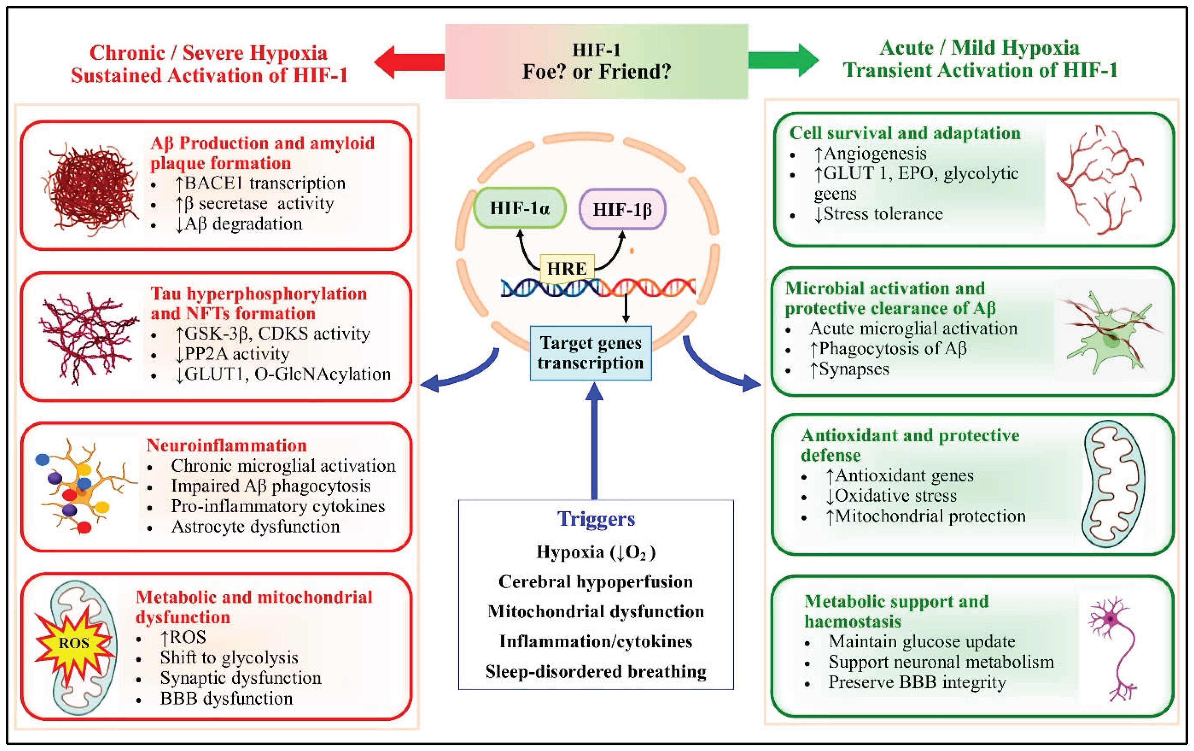

Table 1.
HIF-1 Modulating Interventions and Their Impacts on AD Pathology.
| Intervention | Nature/Source | Study Model | Mechanism of Action | Impacts on AD Pathology | Ref. |
| Deferoxamine (DFO) | Iron chelator | APP/PS1 transgenic mice (in vivo); APPsw cells (in vitro) | Upregulates HIF-1α via PHD inhibition; activates P38/HIF-1α pathway | ↓ Aβ deposition; ↓ synapse loss; ↑ BDNF, TFR, DMT1; ↓ Fe levels in hippocampus | [68,69,70] |
| M30 | Multifunctional iron chelator | APP/PS1 transgenic mice (in vivo); rat primary cortical neurons (in vitro) | Stabilizes HIF-1α; upregulates HIF-1 target genes; modulates insulin signaling | ↑ HIF-1α, aldolase A, enolase-1, GLUT-1; ↓ GSK-3β activity; ↓ Tau phosphorylation; protects against Aβ25-35 toxicity | [71,72] |
| Icariin | Phytocompound isolated from Epimedii herba | Preclinical studies; bioinformatics analysis | Targets HIF-1 signaling pathway (network pharmacology) | Regulates Aβ1-42, Aβ1-40, BACE1, tau, and p-tau; ↓ inflammatory mediators; alleviates cognitive impairment | [75] |
| NeuroProtect formula | Geniposide + Panax notoginseng saponins | APP/PS1 transgenic mice (in vivo); cell culture (in vitro) | Modulates HIF-1/PI3K-AKT signaling; targets mTOR, HIF1A, TLR4, STAT3, VEGFA | ↓ Aβ42/Aβ40 ratio; ↑ dendritic spine density; reverses Aβ25-35-induced cell viability reduction | [79] |
| α-Lipoic acid | Naturally occurring antioxidant | Tauopathy model P301S mice (in vivo) | Increases GLUT3, GLUT4, VEGF, and HO-1 expression through BDNF/TrkB/HIF-1α signaling | Maintains brain glucose metabolism; enhances glucose availability; improves cognitive function | [78] |
| Neuroglobin | Hypoxia-inducible cytoprotective protein | HN33 neural cell line (in vitro) | shRNA-mediated knockdown and lentiviral vector-mediated overexpression of HIF-1α | Enhances neuroprotection; promotes neuronal survival under hypoxic conditions | [89] |
| Lactoferrin | Mild derived iron-binding glycoprotein | APP/PS1 double Tg mice (in vivo) | Augments α-secretase-dependent APP processing through ERK1/2-CREB and HIF-1α pathways | Reduces Aβ aggregation; improves spatial learning; enhances non-amyloidogenic APP processing | [83] |
| Coffee | Natural beverage | SH-SY5Y human neuroblastoma cell line (in vitro) | Inhibits prolyl hydroxylation to activate HIF-1α; induces VEGF expression | Induces VEGF expression; potential neuroprotective effects; reduces risk of cognitive decline | [84] |
| SLAB51 | Multi-strain probiotic | 3 × Tg-AD and wild-type mice (in vivo) | Increases HIF-1α; decreases PHD2, iNOS in brain; reduces plasma NO | Reduces oxidative stress; attenuates neuroinflammation; mitigates AD pathology | [85] |
| Viral vector expressing HIF-1α | Genetic construct (rAAV, lentivirus) | Primary hippocampal neurons (in vitro); Sprague-Dawley rats (in vivo) | Overexpresses HIF-1α; activates HIF-1 target genes | Inhibits hippocampal neuronal apoptosis induced by Aβ protein; promotes cell survival | [85] |
| Fibroblast growth factor 21 (FGF21) | Endogenous protein hormone | SH-SY5Y cells (in vitro); adult male Wistar rats (in vivo) | Inhibition of PP2A/MAPKs/HIF-1α pathway triggered by Aβ25-35 | Ameliorates neurodegeneration; reduces tau hyperphosphorylation; attenuates oxidative stress | [90] |
| Neuregulin 1 | Endogenous neurotrophic factor | SH-SY5Y human neuroblastoma cell line (in vitro) | Inhibits CoCl₂-induced accumulation of HIF-1α and p53; attenuates cell death | Protects against hypoxia-induced neuronal damage; suppresses HIF-1α accumulation; reduces p53 stability | [91] |
| HIF-1α siRNA-loaded Astrocyte-derived EVs | Genetic construct (RNA interference) | HIV-infected humanized mice model (CD34+ NSG mice) | Suppresses HIF-1α expression via RNA interference | Reduces APP, Aβ, Aβ fibrils, and pTau; dampens glial activation (GFAP, IBA1); partially restores synaptic proteins; improves behavioral deficits | [95] |
| Ginsenoside Rg1 | Active ingredient of Panax ginseng | Human endothelial cells (in vitro) | Reduces Aβ-induced mitochondrial apoptosis; lessens HIF-1α expression; decreases RNS and protein nitrotyrosination | Protects against Aβ-induced endothelial apoptosis; reduces oxidative/nitrosative stress | [76] |
| Neurotropin® | Non-protein extract of inflamed rabbit skin inoculated with vaccinia virus | HT22 hippocampal cells (in vitro); APP/PS1 Tg mice (in vivo) | Regulates HIF-1α/MAPK signaling pathway | Lessens Aβ-induced oxidative damage; improves Aβ deposition in hippocampus; alleviates neuroinflammation | [100] |
| Salidroside | Glucoside of tyrosol from Rhodiola rosea | SH-SY5Y human neuroblastoma cell line (in vitro) | Reduces HIF-1α protein level under hypoxia; decreases BACE1 expression; inhibits β-secretase activity | Enhances sAPPα secretion; attenuates Aβ generation; reduces hypoxia-induced amyloidogenesis | [77] |
| Melatonin | Endogenous neurohormone | SH-SY5Y human neuroblastoma cell line (in vitro) | Suppresses HIF-1α and BACE1 expression under OGD and OGD/R conditions | Inhibits hypoxia-induced AD pathogenesis; downregulates amyloidogenic pathway; reduces Aβ production | [97] |
| Simvastatin (low dosage) | Statin (HMG-CoA reductase inhibitor) | Sporadic AD and age-matched control neuronal mitochondrial cybrids (in vitro) | Reduces HIF-1α and BACE1 expression | Attenuates Aβ production; reduces amyloidogenic processing; mitigates AD pathology | [98] |
| Intermittent Hypoxia-Hyperoxia Training (IHHT) | Physiological intervention | STZ-induced rat model of AD | Modulates HIF-1α expression; reduces neuroinflammation | Improves spatial learning and memory; ↓ Aβ, CYP2E1, HIF1α, and TNFα expression in hippocampus | [99] |
| Chronic hypoxia | Pathological condition | Rodent models; cell culture (SH-SY5Y, primary neurons) | HIF-1α stabilization; activation of amyloidogenic pathways | ↑ BACE1 expression; ↑ γ-secretase activity; ↑ Aβ production; ↑ tau hyperphosphorylation via ↓ PP2A and ↑ GSK-3β | [49,86] |
| Intermittent Hypoxia (Chronic) | OSA model | Transgenic AD mice; OSA models | Chronic HIF-1 activation; neuroinflammatory cascade | Induces microglial activation; promotes astrogliosis; ↑ Aβ42 levels; exacerbates neuroinflammation; contributes to cognitive decline | [87,88] |
| Heparin binding EGF-like growth factor (HB-EGF) | Endogenous growth factor | BCCAO mice model (in vivo) | Increases HIF-1α expression; activates MMP9 | Causes BBB disintegration; impairs Aβ clearance; promotes AD neuropathology | [92,93] |
| Cobalt chloride (CoCl₂) | Chemical hypoxia mimetic | Mice (in vivo); neuronal cell lines (in vitro) | Stabilizes HIF-1α via PHD inhibition; mimics hypoxic conditions | Induces Aβ deposition; promotes tau hyperphosphorylation at Thr181; triggers dysregulated autophagy; causes hippocampal and cortical neurodegeneration | [74] |
| HIF-1α overexpression (viral vectors) | Genetic construct | Primary neurons; transgenic mice models (in vivo) | Constitutive HIF-1α stabilization; non-physiological activation | Promotes BACE1 expression and γ-secretase activity; ↑ Aβ production; activates pro-apoptotic pathways through p53 stabilization | [96] |
| Prolyl hydroxylase domain enzymes inhibitors (Chronic) | Small molecule inhibitors | Preclinical AD models | Sustained HIF-1α stabilization; prolonged activation of HIF target genes | ↑ Neuroinflammation; ↑ angiogenesis; causes aberrant vascular remodeling; alters neural progenitor cell status | [73] |
| Thiamine Deficiency | Nutritional deficiency | HT22 hippocampal neuronal cell line | HIF-1α-mediated amyloidogenesis | Upregulates BACE1 transcription; ↑ β-secretase activity; induces BNIP3 expression; triggers neurotoxicity | [82] |
| High Glucose Condition | Metabolic stress condition | SK-N-MC neuroblastoma cells (in vitro); rats (in vivo) | ROS-dependent HIF-1α activation via JNK pathway | Upregulates BACE1 expression; ↑ Aβ production; ↓ LXRα expression | [81] |
| Palmitic Acid-BSA (PA-BSA) | Metabolic stressor | SK-N-MC neuroblastoma cells | Activation of Akt/mTOR/HIF-1α and Akt/NF-κB pathways | Stimulates APP and BACE1 expression; accelerates Aβ production and neurotoxicity | [80] |
Disclaimer/Publisher’s Note: The statements, opinions and data contained in all publications are solely those of the individual author(s) and contributor(s) and not of MDPI and/or the editor(s). MDPI and/or the editor(s) disclaim responsibility for any injury to people or property resulting from any ideas, methods, instructions or products referred to in the content. |
© 2026 by the authors. Licensee MDPI, Basel, Switzerland. This article is an open access article distributed under the terms and conditions of the Creative Commons Attribution (CC BY) license (http://creativecommons.org/licenses/by/4.0/).
Copyright: This open access article is published under a Creative Commons CC BY 4.0 license, which permit the free download, distribution, and reuse, provided that the author and preprint are cited in any reuse.


