

Submitted:
13 July 2026
Posted:
14 July 2026
You are already at the latest version
Abstract
Background/Objectives: Metabolic dysfunction-associated fatty liver disease (MASLD) is a metabolic disorder with an increasing incidence, for which pharmacotherapy is still very limited. One of the pharmacotherapeutic approaches for MASLD is to prevent oxidative stress processes, in which polyphenolic flavonoids, including quercetin, may play an extremely important role. Quercetin has been reported in several publications to have a potential hepatoprotective effect, but despite numerous research results, it has not lived up to the expectations placed on it so far. This study provides a comprehensive overview of the effects of quercetin on MASLD, including its role in autophagy during disease progression. Methods: This review describes the etiopathogenesis of MASLD and provides a comprehensive overview of the main physiological and pharmacological effects of quercetin, as well as its potential clinical applications. Conclusions: The effects of quercetin and commonly used biopharmaceuticals in folk medicine for the treatment of MASLD still do not show clear results. The extremely positive effects obtained in in vitro studies cannot be replicated or can only be replicated in a very limited form in pre- and clinical studies. Further research is needed to resolve this dilemma.
Keywords:
MASLD
; pharmacotherapy
; hepatoprotective
; autophagy
; and quercetin
1. Introduction
One of the greatest challenges of our time is the numerous so-called pathological lifestyle conditions, a wide spectrum of which affects an increasingly young, active, working age group. These disease groups manifest themselves as a result of modern social habits, lifestyle, reduced physical activity and environmental damage [1]. Several factors, including poor nutritional habits and poor food quality, combined with high calorie intake, high fat and carbohydrate intake, and low fiber and vitamin-deficient diets, contribute to the development of metabolic disorders [2]. In addition to dietary habits (diet) and physical activity (lifestyle changes), doctors and dietitians have increasingly studied polyphenol-based molecules found and extracted from plants. Polyphenols are not essential nutrients, but their antioxidant capacity prevents damage to biological systems from oxidative stress, which is an important health-preserving ability [3]. Polyphenols can be classified into two major groups: flavonoids and non-flavonoids [4].
Flavonoids are secondary metabolites that play a critical role in many plants in protecting them from environmental stressors – survival – and in performing essential physiological functions. Flavonoids are plant metabolites that are widely distributed in nature and perform protective functions for plants, such as protection against UV radiation or pathogens. In human nutrition, flavonoids are among the most important antioxidant compounds and play a significant role in maintaining health [5,6].
Quercetin is a natural flavonoid compound found in many plants - fruits (apples, plums, grapes, blueberries, blackberries, raspberries), vegetables (onions, red onions, garlic, kale, broccoli, cauliflower, cabbage, leafy vegetables - spinach, lettuce, arugula, spices - turmeric, ginger, cinnamon, capers (in very high amounts), and in smaller amounts in olive oil. It is found in moderate amounts in red wine and green tea. The name has been used since 1857 and comes from the Latin quercetum = oak forest (oak from Latin Quercus) [7,8]. It is also found in plants with proven medicinal properties, such as Ginkgo biloba, Hypericum perforatum, and elderberry [9,10,11].
Quercetin is of growing interest among researchers and health-conscious consumers alike, as its potential therapeutic applications range from the alleviation of allergic reactions to the prevention of cardiovascular diseases. In vitro and in vivo studies demonstrate quercetin’s antibacterial activity and its effective biofilm-inhibiting effect, which inhibits the expression of related genes. Furthermore, quercetin also has antitumor, antiangiogenic effects, and reduces mycotoxicity to cells.
In this article, the authors summarize the proven beneficial physiological effects of quercetin on the body, emphasizing its role in the increasingly widespread metabolic-associated fatty liver, diabetes, and cardio-metabolic pathologies that are proven to be responsible for the prevention of many chronic internal medicine diseases.
2. MASLD Etiopathogenesis–Molecular Cellular Mechanisms
The term metabolic dysfunction-associated fatty liver disease (MASLD) was given by the European Association for the Study of the Liver (EASL) and the American Association for the Study of Liver Disease (AASLD) to better fulfil the desire to describe the pathophysiologic background [12,13]. The development of this metabolic disease is determined by a spectrum of conditions, which are related to western-type lifestyle, including hypertension, hyperlipidemia, diabetes mellitus and overweight/obesity. These chain of pathologies can be influenced by unhealthy daily habits e.g., lack physical exertion, sedentary lifestyle, poor dietary choices and/or alcohol consumption [14]. The disease can progress from simple steatosis to steatohepatitis and potentially to cirrhosis and ultimately to hepatocellular carcinoma [15]. (Figure 1.)
MASLD can be often defined as a hepatic manifestation of a metabolic syndrome (MetS), also known as „X syndrome” [16] The traditional pathophysiologic development was defined by „two-hits” model. In this process the first „hit” represents the accumulation of triglycerides in hepatocytes, which leads to fatty liver disease (simple steatosis). This can be enforced by fatty acids imbalance between the input and output within hepatocytes, which often determines the appearance of several metabolic disorders such as central obesity and insulin resistance [17,18]. Overloading the hepatocyte with lipid will trigger additional pathophysiological processes, that will lead to chronic inflammation e.g., inflammatory cytokines, oxidative stress, bacterial endotoxins, and mitochondrial dysfunction. These number of inflammatory processes will worsen the liver damage – hepatocellular injury – that will eventually cause fibrosis. („second hit”) [19,20,21] Nowadays a novel, „multiple parallel hit” model was instated, representing the sequential process, where several factors act simultaneously driving the MASLD progression. These processes include genetic predisposition, gut-related bacterial dysbiosis, dyslipidemia, and adipocytokine imbalances, which will conclude in insulin resistance, oxidative stress and chronic inflammation development. Taking in consideration the multifaceted cause-and-effect relationship of MASLD development, the „multiple-hit” hypothesis defines the nature of this multifactorial disease, where metabolic, environmental, and genetic aspects collectively drive the development and progression of simple steatosis, inflammation, and fibrosis [19,20,22,23]. (Figure 1).
2.1. The Role of Lipid Metabolism Dysorder and Insulin Resistance Development
Excess caloric intake will lead to activation of several metabolic related signaling pathways, which will lead to lipid formation and deposition, generally in subcutaneous white adipose tissue. (WAT) In the course of disease progression from simple steatosis to MASLD and MASH (metabolic dysfunction-associated steatohepatitis) these forementioned mechanisms are impaired, ultimately causing ectopic fat accumulation, mainly in hepatocytes [19,24]. Free fatty acids are absorbed from the intestines reach the bloodstream, where they will be deposited mainly in the hepatocytes in form of triglycerides (TG). The main causes for liver steatosis development are the high-fat, high-sugar diet, de novo lipogenesis (DNL) in hepatocytes and the high lipid level stored in WAT. The main reasons for insulin resistance appearance and carbohydrate overloading are the high-fat dietary intake and the FFAs transport from WAT, and de novo lipogenesis. Insulin resistance (IR) has a central role in lipid metabolism disturbance, while insulin has an anti-lipolytic effect, and regulates the triglyceride accumulation in WAT and the storage of FFAs, leading to a decreased levels of TGs and FFAs in the liver. IR leads to downregulation of insulin receptor substrate 2 (IRS-2), which determines an overexpression of sterol regulatory element-binding protein 1c (SREBP-1c) and carbohydrate response element-binding protein (ChREBP). These transcriptional factors are related with genes encoding major enzymes mediating DNL – fatty acid synthase (FAS) and acetyl-CoA carboxylase (ACC) [19,25,26,27]
Peroxisome proliferator-activated receptor gamma (PPARγ) is a nuclear receptor transcription factor, having a central role in lipid metabolism, adipogenesis, and insulin sensitivity. In case of obesity, IR, and MetS, PPARγ is overexpressed in hepatocytes. The upregulation of PPARγ is enhanced by inflammatory cytokines, high-fat diet, and metabolic stress [28]. In case of PPARγ activation of several genes related in fatty acid transport – CD36 - and lipid storage – FABP4 (adipocyte fatty acid-binding protein) – are enhanced. PPARγ primes circulating FFAs uptake and their esterification into triglycerides. PPARγ regulates lipogenesis-related genes, including SREBP-1c, which drives de novo fatty acid synthesis. Generally, PPARγ improves insulin sensitivity (mainly in adipose tissue), while in the liver chronic activation causes IR. Besides lipid storage promotion, PPARγ has an anti-inflammatory role as well. PPARγ receptor activation reduces the pro-inflammatory cytokines expression – TNF-α (tumor necrosis factor-alpha), IL-6 (interleukin 6) – in both liver and adipose tissues [19,22].
2.2. Concomitent Roles of Oxidative Stress, Inflammation and Lipotoxicity
From both TGs and FFAs energy in form of ATP can be attained via β-oxidation process. In normal conditions, β-oxidation, happening inside the liver, is maintained by several regulatory factors, so the free electrons generated from lipid oxidation are neutralized, resulting in water. (H2O) In case of liver steatosis this electron transport chain process is disturbed, resulting in the production of reactive oxygen species (ROS) – in form of hydrogen peroxide (H2O2) or superoxide (O2.) [19,29,30] Normally, ROS are produced in macrophages, endothelial cells or pneumocytes. ROS mediates the immune response process and plays an important role in signal transmission. If ROS are produced in excess, they will trigger oxidative stress response, and will damage essential macromolecules like lipids, proteins and DNA. These will cause alteration in structure of several biomolecules, which will lead to loss or modification of their essential functions. Lipid peroxidation occurs in biological membranes due to the high content of polyunsaturated fatty acids (PUFAs). As consequence the end-products can determine DNA damage, denaturation of proteins, inhibition of enzyme activity and ultimately cell apoptosis [31].
The term, lipotoxicity’ mirrors the cause and effect between the high lipid levels inside the liver and their adverse consequences. Oxidative stress can trigger increased pro-inflammatory cytokines expression such as TNF-α, IL-6, IL-1β, and transforming growth factor beta (TGF-β). In oxidative stress lipooxygenase (LOX) and cyclooxygenase (COX) are upregulated. Chronic inflammation, caused by lipotoxicity determines the histopathological changes that are observed in MASH [32,33]. (Figure 1).
2.3. The Central Role of Autophagy
Autophagy represents a crucial cellular process, responsible for cellular homeostasis maintenance by transporting, degrading, and recycling damaged or dysfunctional small molecules, proteins or even entire organelles. Its central role is in hepatocellular lipid metabolism, which is crucial for liver health. In MASLD, there is a downregulated autophagy observed, adding to the increased lipid accumulation inside hepatocytes. The impaired autophagy occurs primarily due to the disturbance of autophagosome-lysosome fusion, resulting in autophagosome accumulation, and an inefficient intracellular lipid degradation [20,34]. Under normal conditions autophagy is responsible for degradation and formation of lipid droplets. These organelles in hepatocytes contain TGs and store them as a reservoir, for further use in several metabolic processes. Autophagy regulates the hydrolysis of intracellular TGs to FFAs and controls the efflux from lipid droplet and hepatocytes to peripheral tissues, mildening the liver steatosis condition. When autophagy is defective liver steatosis promotion and MASLD development occurs [35,36,37].
There are key autophagy proteins: Beclin 1 and Microtubule-associated proteins 1A/1B light chain 3 (LC3A). Beclin1 central role is in initiating autophagy, by forming complexes that promote autophagosome formation. LC3A protein role is in the elongation and closure of autophagosomes. Zhao et al. demonstrated that in case of hepatic steatosis both proteins are downregulated, which causes decreased autophagic activity and accumulation of lipid droplets inside the hepatocytes [38]. Another critical autophagy-related protein is Sequestosome-1 (SQSTM1/p62), which serves as a marker of impaired autophagy. SQSTM1 binds to ubiquitinated proteins, priming their degradation via autophagy. In case of impaired autophagy, SQSTM1 will accumulate, determining a defective autophagy, which will worsen MASLD, determining its progression [39]. Besides the forementioned autophagic proteins, there are other molecules, which also play critical roles in the MASLD development. CD36 is a fatty acid transporter that promotes lipid uptake into hepatocytes. In MASLD lipid accumulation is increased, which can be tied to the upregulation of CD36, that will cause liver damage. Perilipin 3 - regulates lipid droplet dynamics – is crucial for the mobilization of lipids inside hepatocytes. In case of Perilipin disturbance, lipid metabolism will become impaired, resulting in liver steatosis and steatohepatitis aggravation [40,41].
A subtype of autophagy, which targets mitochondria, is the so called, mitophagy’. Normally, mitophagy maintains mitochondria function, and removes damaged, injured and/or dysfunctional or excessive numbers of organelles. Main role of mitochondria is energy production, but they are exposed to ROS impact, representing a by-product of ATP synthesis [19,42,43]. In order to serve as an efficient identification and exclusion of damaged mitochondria, mitophagy requires certain signaling molecules to mark and dispose of them. PINK1 (PTEN-induced kinase 1)/PARKIN signaling pathway regulates mitophagy. PINK1 = Ser/Thr kinase, in injured mitochondria PINK1 is accumulated in the outer membrane, due to its depolarization, which makes it able for phosphorylation of ubiquitin and E ubiquitin ligase = PARKIN. PARKIN acts as a signaling molecule activating mitophagy process [19,44]. Both in vitro and in vivo experiments concluded that impaired mitophagy represents early sign of MASLD [45,46].
2.4. The Mediator Role of Gut Microbiota
Gut microbiota plays a multifactorial, complex role in mediating the pathogenesis of MASLD, impacting physiologic functions such as liver metabolism, inflammation and insulin sensitivity. Microbiota means a sum of bacteria, archaea and eukaryota collection found in the bowel system [47]. Besides influencing MASLD process, the gut microbiota plays a pivotal role in the function of immune system, determining the integrity of bowel mucosa and influences the metabolism of several nutrients and drugs. There is a bi-directional gut-liver axis signaling, via portal vein [48]. (Figure 1).
In case of poor alimentary habits (e.g., fructose-rich diet) an alteration in normal composition of the microbiota occurs, which is known as dysbiosis. In this condition a change in Firmicutes/Bacteroidetes ratio develops. In case of increased number of Firmicutes MASLD develops and can be associated with higher risk of type 2 diabetes and/or obesity [49].
Certain microbial-derived molecules can activate receptors and/or influence several metabolic processes. Such molecules are the short-chain fatty acids (SCFAs): propionate, butyrate and acetate (normally, they are present in 1:1:3 ratio) [50]. SCFAs have multiple roles, they represent energy sources for hepatocytes, they serve in maintaining the integrity of the mucosal lining and regulates the bile acid secretion [51,52,53]. They also act as immunomodulators and anti-inflammatory agents [54].
If alteration of SCFAs occurs the consequences trigger the development of chronic, low-grade inflammation. These entail a multi-step process, where the main pathophysiology phenomenon is the increased intestinal permeability (deterioration of intestinal barrier/, leaky gut’ phenomenon), and an enhanced bacterial endotoxin portal flux (LPS-mediated endotoxemia) that concludes in the overexpression of pro-inflammatory cytokines - TNF-α, IL-6, IL-1β - [55,56]. After absorption and translocation of LPS to the liver, hepatocytes recognize them via toll-like receptor 4 (TLR4) present in the cell membrane, and inside the hepatocytes upregulation of the nuclear factor kappa-light-chain enhancer of activated B cells (NF-κB) happens, resulting in overexpression of various pro-inflammatory cytokines [57]. (Figure 1).
Dysbiosis can trigger dysfunction of the bile acid metabolism by interacting with farnesoid X receptor (FXR) and Takeda G protein-coupled receptor 5 (TGR5) signaling pathways, causing disturbance in carbohydrate and lipid absorption, concluding in metabolic disorders [58].
The combination between dysbiosis and altered ratio of SCFAs can cause slowed bowel movements. In this process the leading cause is the increased secretion of peptide YY [59]. Another mechanism which causes slowed gut motility is the injured- or loss of enteric neurons, caused by LPS-induced neurotoxicity and lipotoxicity [60,61].
3. Main Physiological and Pharmacological Effects of Quercetin
Quercetin has the following physiological roles:
- antioxidant effects: it binds ROS and RNS products produced in the body, thereby protecting cells from the damaging effects of reactive oxidative products
- anti-inflammatory and immunomodulatory effects.
- antitumor effects.
- antibacterial, antiviral effects [62].
3.1. Physiological Effects of Free Radicals and Reactive Nitrogen Products
Quercetin has strong antioxidant and anti-inflammatory effects and plays a significant role in the regulation of inflammatory processes and in the protection of harmful processes against oxidative stress. These beneficial processes make it an excellent biopharmaceutical for the treatment of many chronic inflammatory processes, such as cardiovascular diseases and lipid metabolism disorders [20,63]. (Figure 2).
Free radicals are reactive molecules that have one or more unpaired electrons. They are formed during metabolic processes as essential mediators that play an important role in neurotransmitter or inflammatory processes, or as by-products that do not play an important role in the actual physiological processes. In aerobic life, oxygen reduction is crucial; in the basic state, oxygen combines with hydrogen to form water, a process that occurs in mitochondria during oxidative phosphorylation, but only 1–3% of the oxygen is partially reduced during the redox process. Consequently, free radicals or reactive nitrogen products are formed, which oxidize other cellular components or generate other free radicals [64].
Reactive oxygen species (ROS) and reactive nitrogen species (RNS) molecules can interact with all macromolecules (DNA, RNA, proteins, carbohydrates, lipids) and destroy their structure. In the first step, they abstract a hydrogen atom from the affected molecules, thereby converting the unpaired electron into a molecule with a more stable paired electron. In addition to abstracting the hydrogen molecule, an alternative solution is electron donation. From an electrochemical point of view, both the hydrogen and electron-donating molecules are oxidized. The free radicals or reactive molecules formed from this process are often called (pro)oxidants [65]. The consequences of oxidative stress caused by ROS and RNS molecules include DNA lesions, enzyme dysfunction, increased membrane permeability, impaired signal transduction, and ultimately necrosis or apoptosis [66,67,68,69].
In addition to the direct and indirect damage of reactive oxidative radicals to several biomolecules, there is also a connection with inflammatory processes. Inflammatory responses reflect the process of recognition, attack and elimination of exogenous and endogenous pathogens [70,71]. During evolution, in vertebrates, this immune response can be of two types, considering the speed and specificity of the response: innate and acquired (adaptive) immune response. The innate immune response directly protects the body from the spread of infections, preventing their occurrence (barrier formed by epithelial cells) or eliminating microbes (by phagocytosis, natural killer cells or the complement system) [71,72]. The adaptive immune response does not develop immediately, it is mediated by lymphocyte cells and the antibodies they produce. Antibodies produced by B lymphocytes can inhibit infection and eliminate microbes, while T lymphocytes directly destroy pathogens [70,71]. For the completeness of the defense, there is a cooperation between the innate and adaptive immune responses, for example, the maturation of dendritic cells is regulated by the innate immune response, which migrates to lymphoid organs, where they activate T lymphocytes, initiating the adaptive immune response [73]. A key factor in both immune responses is the production of cytokines (cytokines = glycoproteins), which are secreted by a variety of immune cells, including macrophages, neutrophil granulocytes, and T helper lymphocytes [70,74]. Cytokines can exert pro- and anti-inflammatory effects. TNF-α, one of the prominent pro-inflammatory cytokines, is produced by macrophages through the activation of nuclear transcription factor kappa-B (NF-κB) [75,76]. ROS can induce the production of several cytokines by activating the transcription factors NF-κB and activator protein-1 (AP-1) [77]. ROS-induced activation of the NF-κB transcription factor occurs by removal of its inhibitory moiety, IκBα. Activation occurs through phosphorylation, which results in degradation or translocation of the IκBα subunit, a process mediated by several IκBα kinase subtypes [78,79].
In vitro experiments on human leukocytes with H2O2 have shown that cytokine-induced NF-κB activation can be inhibited by reducing free radical production and by inactivating phosphorylation of the IκB subunit [80,81]. Alternatively, inhibition may be associated with an increased propensity for apoptosis, as cells are subjected to a dual traumatizing effect: on the one hand, an increasing amount of ROS, and on the other hand, an increase in pro-inflammatory cytokine levels. The conclusion of the experiment was a biphasic response of NF-κB to oxidative stress, where an effective, activated NF-κB was the response to intermediate levels of oxidative stress, while in the case of elevated levels of oxidative stress, cell death occurred [82,83]. TNF-α is an important cytokine that is also produced by the ROS-activated transcription factor NF-κB, but TNF-α itself also mediates NF-κB activation, increasing the activation of IκKs and the production of ROS radicals [79,84,85]. The increased ROS level occurs on the one hand, by stimulating oxidative phosphorylation in mitochondria, and on the other hand, by the respiratory burst [78,85,86,87,88]. TNF-α also stimulates gene transcription of other cytokines – IL-8. The amplification of TNF-α and NF-κB-mediated effects induced by increased ROS production can exert cytotoxic effects leading to apoptosis [79].
3.2. Physiological Effects of Quercetin in Preclinical Experimental Models
3.2.1. Antioxidant Effect
Reactive oxidative species (ROS) generated in the body during various metabolic processes play a pathological role in many pathological processes. Cell membrane damage and gene mutations occur, which greatly accelerate the aging process in the body, contributing to the development of many chronic diseases, such as heart disease, liver damage, and diabetes [96,97]. According to Hanasaki et al. quercetin is one of the most effective flavonoids in neutralizing free radicals. The four hydroxyl groups in the chemical structure of quercetin, which are found on the benzodihydropyran ring of the polyphenol, confer strong antioxidant capacity, which can directly react with ROS and RNS, such as superoxide, hydroxyl radical, and peroxynitrite. This prevents oxidative stress, which can damage DNA, proteins, and lipids, contributing to the prevention of many chronic diseases [98].
The antioxidant mechanisms of quercetin in vitro experimental models include:
- -
- indirectly neutralizing and scavenging free radicals: Manca et al. demonstrated that quercetin and glycerol nanoparticles incorporated into liposomes neutralize free radicals and protect human keratinocytes in vitro from hydrogen peroxide damage [99]. Experiments by Oh et al. demonstrated that quercetin has the highest antioxidant capacity known [100].
- -
- chelating metal ions: several studies have shown that quercetin, due to the catechol structure in its chemical structure, activates copper and iron ions, which exert antioxidant effects. Tang et al. demonstrated that quercetin could inhibit iron-induced lipid peroxidation in adult male C57BL/6J mice in a model of alcoholic liver disease. Quercetin binds iron, preventing iron accumulation and oxidative damage to cells [101]. Babenkova and colleagues demonstrated by chemiluminescence that iron incorporated into dihydroquercetin remained inactive, preventing the decomposition of hydrogen peroxide during the catalytic process, resulting in less hydroxyl free radical formation [102]
- -
- lipid peroxidation inhibitor: Lim et al. demonstrated that quercetin can inhibit the oxidative modification of low-density lipoprotein (LDL) as inferred from fluorescence intensity observations. They observed differences in intensity for thiobarbital, phosphatidylcholine hydroperoxides, and oxidized LDL [103]. Mbikay et al. verified the results of the previous group and concluded that at low concentrations, quercetin increases the expression of LDL receptors, reduces the secretion of the enzyme PCSK9 (proprotein convertase subtilisin/kexin type 9), thereby increasing LDL uptake and reducing oxidative damage to LDL. The enzyme PCSK9 is responsible for regulating circulating cholesterol concentration and the number of LDL receptors on the cell surface and has become a major molecular regulator of lipid metabolism, especially in the liver [104].
The antioxidant properties of quercetin in vivo experiments are gradient-dependent, with higher concentrations producing stronger antioxidant effects:
- -
- the antioxidant properties of quercetin are manifested in the regulation of glutathione (glutathione, a molecule with antioxidant capacity consisting of three amino acids: glutamic acid, cysteine, glycine) levels. In the case of reactive oxidative radicals (ROS) formed in the body, the enzyme sodium dismutase-2 (SOD-2) binds reactive oxygen (O2-) and converts it into hydrogen peroxide (H2O2). Glutathione peroxidase (GSH-Px) catalyzes the conversion of H2O2 into water molecules, which requires the presence of glutathione, which covers the hydrogen demand for the reaction [105].
- -
- effect on enzymatic activity: according to Odbayar et al. quercetin increases the expression of certain enzymes with antioxidant effects, such as glutathione transferase and aldo-keto-reductase. The level of expression is directly proportional to the amount of quercetin [106].
- -
- impact on signaling pathways: Wang et al. have shown in vitro and in vivo that quercetin exerts a protective effect on granulosa cells by increasing the expression of genes that protect against oxidative stress [107]. This observation is complemented by Granado-Serrano et al. and Kobori et al. have demonstrated that quercetin increased the expression of the transcription factor Nrf2 (nuclear factor erythroid related factor 2), activating the intracellular p38 MAPK (mitogen-activated protein kinase) signaling pathway, increasing intracellular glutathione levels, thereby increasing the antioxidant capacity of the cell [108,109].
3.2.2. Anti-Inflammatory and Immunomodulatory Effects
Anti-inflammatory effects: Quercetin has been shown to have significant anti-inflammatory effects in both animal and human experimental models [110,111]. Quercetin extract is a major component of several anti-allergic agents. Compared with Cromolyn (an anti-allergic drug, disodium cromoglycate representing the active ingredient), quercetin is a more potent inhibitor of IL-8 and can inhibit IL-6 and increase cytosolic calcium levels [112]. In addition to its broad-spectrum biochemical and pharmacological effects, quercetin exerts anti-inflammatory effects on the endothelium and the monocyte/macrophage system in vitro [113,114]. Li et al. [115] have shown that quercetin inhibits tumor necrosis factor alpha (TNF-α) production induced by lipopolysaccharide (LPS) released from macrophages in various animal models [110]. Furthermore, in vitro, quercetin inhibits LPS-induced IL-8 production in A549 lung cells [111] and in glial cells, quercetin inhibits TNF-α and IL-1α messenger RNA (mRNA) levels, thereby reducing neuronal death [116].
Quercetin exerts its anti-inflammatory effects in vitro and in vivo by the following molecular – cellular – mechanisms:
- -
- reduces the activity of pro-inflammatory cytokines – IL-6, IL-17, TNF-α, IL-1β – by inhibiting the NF-κB and AMPK signaling pathways. At the same time, it blocks the translocation of NF-κB into the cell nucleus (reduces the gene expression of pro-inflammatory mediators) [117,118,119,120,121,122,123,124,125,126] Regulation of the NF-κB, AP-1 and AMPK/SIRT1/NF-κB signaling pathways inhibits the gene expression of pro-inflammatory cytokines and the activity of oxidative enzymes. It actively increases the activity of the transcription factor Nrf2 (nuclear factor erythroid 2-related factor 2), supporting the body’s antioxidant defenses [127,128].
- -
- can inhibit the activity of inflammatory enzymes - cyclooxygenase (COX-2) and lipoxygenase (LOX) - thereby reducing the synthesis of pro-inflammatory mediators - prostaglandins, leukotrienes [127].
- -
- reduces the activity of inducible NO (iNOS) synthase, which results in reduced NO levels, and also reduces the level of RNA products, which has a significant anti-inflammatory effect. This is accompanied by supporting the activity of SOD, catalase and glutathione peroxidase enzymes, which play a role in the body’s antioxidant defense, neutralization of ROS radicals, and increases glutathione levels [127,129,130,131].
- -
- -
- -
- -
- reduces the levels of hydroxyl radicals, hydrogen peroxide, superoxide anion, NO, which are responsible for the appearance of oxidative stress. Effective neutralization of ROS molecules can be achieved by inhibiting the expression of NADP oxidase (NOX2), thereby reducing the degree of lipid peroxidation, DNA and protein damage [127,135].
- -
- regulates lipopolysaccharide metabolism, thereby reducing the activity of metabolites resulting from lipid peroxidation (MDA, 4-HNE). Quercetin inhibits structural modifications generated by lipopolysaccharides at the mitochondrial level, normalizing lipopolysaccharide homeostasis by activating vitamin D receptors (VDR) [127].
Immune system modulatory effect: several studies have shown that there is a relationship between quercetin and the immunomodulatory effect it generates. Nieman and his colleagues showed that cyclists in good physical condition, who received quercetin and epigallocatechin gallate (EGCG = catechin, with significant antioxidant properties, helping to protect against cell damage caused by free radicals) for 2 weeks, they became resistant to inflammation after three days of vigorous physical activity [136]. Clinical trials have examined quercetin, resveratrol, EGCG, and genistein (a natural compound found in soybeans and other plants, belonging to the flavonoid family, which, through their antioxidant properties, may help neutralize free radicals, thereby reducing the risk of chronic diseases). The research concluded that they had a positive effect on humoral and cellular immune responses [137].
3.2.3. Antitumor Activity
Several preclinical studies have demonstrated the influence of quercetin on cell cycle regulation, cell death promotion, and angiogenesis in vitro and in vivo experiments.
Cell cycle effects: Lee and his group demonstrated that quercetin inhibited the cell cycle in the G2 phase (late DNA synthesis phase) in human leukemia U937 cells [138]. Suh and colleagues observed the effect of quercetin on the G0/G1 (pre-DNA synthesis) phases of the cell cycle using 232B4 chronic lymphocytic leukemia cells and HOS osteosarcoma cells [139]. Chou and colleagues demonstrated the regulatory role of quercetin in p53 signaling pathways related to the tumor cell cycle. In their research, they discovered that quercetin alleviates endoplasmic reticulum (ER) stress and enhances p53 release, thereby inhibiting the activities of cyclin-dependent kinase 2 (cdk2), cyclin A, and cyclin B, causing MCF-7 breast cancer cells to become arrested in the S phase of the cell cycle [140]. Hamidullah and colleagues demonstrated that PC-3 and DU145 prostate cancer cells treated with certain doses of quercetin-6-C-β-D-glucopyranoside were arrested in the G0/G1 phase [141]. This phenomenon was associated with the downregulation of cyclin E and cyclin D, decreased expression of PCNA (proliferating cell nuclear antigen) and cdk-2 proteins, and increased expression of p21 and p27 proteins [142].
Effect on programmed cell death (apoptosis): Quercetin induces programmed cell death in tumor cells, thereby reducing tumor size and rendering the body tumor-free. Research has shown that at certain doses, quercetin can stimulate the expression of proapoptotic proteins and reduce the levels of antiapoptotic proteins [142]. Granato and colleagues observed that in pure erythroid leukemia (PEL), quercetin inhibited the phosphoinositol-3 kinase (PI3K/Akt/mTOR) and STAT 3 (signal transducer and activator of transcription protein 3) signaling pathways, which reduced the levels of c-FLIP (cellular FLICE (FADD-like IL-1β-converting enzyme) - inhibitory protein - an important antiapoptotic protein), cyclin D1, and c-Myc (transcription factor encoded by a proto-oncogene, plays an important role in cell growth) proteins involved in cell survival [143]. Deng and his research group demonstrated that quercetin inhibited the growth of MCF-7 breast cancer cells in a concentration-dependent manner, thereby exerting an inhibitory effect on breast cancer [144]. Teekaraman et al. investigated the effect of quercetin on the human metastatic ovarian cancer PA-1 cell line and found that quercetin induces the mitochondrial-mediated intrinsic apoptotic pathway, leading to tumor cell death [145]. Seo et al. investigated the effect of quercetin on apoptosis induction, and at a dose of 20 µM, inhibition of the STAT3 signaling pathway was achieved, suggesting that quercetin may be a potential effective treatment option for the prevention and treatment of HER2-expressing breast tumors [146].
3.2.4. Antimicrobial Effects
Antibacterial Activity
Several studies have reported the broad antibacterial spectrum of quercetin, which has inhibitory effects not only on bacteria but also on fungi. In several experiments, it has been shown to have effective inhibitory effects against bacteria such as Pseudomonas aeruginosa, Salmonella enteritidis, Staphylococcus aureus, Escherichia coli, Proteus, Aspergillus flavus [147,148]. Hossein and colleagues developed an artificially created synthetic compound (quercetin acyl glucoside), which effectively inhibited the growth of E. coli, S.aureus, P.aeruginosa [149]. An extract prepared from elderberry has shown significant antibacterial activity against Salmonella, Listeria, and Shigella [150].
Quercetin exerts its antibacterial activity by disrupting the cell membrane, modifying the permeability of the cell membrane, affecting protein synthesis and protein expression, reducing enzyme activity, and inhibiting nucleic acid synthesis. Wang and his research group used transmission electron microscopy to demonstrate how quercetin damages and destroys the cell membrane of S. aureus and demonstrated how quercetin kills E. coli [148]. Zhao and colleagues used pressed sugarcane extract (470 mg quercetin/g polyphenol) to investigate the bacteriostatic potential of quercetin against S.aureus, L.monocytogenes, E. coli, and S.typhimurium [151]. Plaper and colleagues found that quercetin negatively affects ATP activity, thereby reducing the growth of E. coli [152].
Quercetin can inhibit the adhesion of bacteria to surfaces, inhibit the function of transport channels on the cell membrane surface, and inhibit nucleic acid synthesis. Wang and colleagues showed that quercetin inhibits the formation of biofilms formed by Streptococcus pneumoniae [107]. Qayyum et al. similarly demonstrated the inhibitory effect of quercetin on biofilm formation, but in their case, they used Enterococcus faecalis strain MTCC 2729. The inhibition was detected by scanning electron microscopy (SEM) and confocal laser scanning microscopy [153]. Lee et al. demonstrated that quercetin exerts an inhibitory effect on genes that promote surface adhesion [154]. Cho and his research group demonstrated that quercetin significantly inhibits biofilm synthesis formed by the methicillin-susceptible S. aureus strain ATCC 6538 [155].
Streptococcus suis is a globally distributed Gram-positive bacterium [156,157]. S. suis-associated human infections, which are of porcine origin, can cause meningitis, septicemia, endocarditis, and deafness [158]. Suilysin cytotoxin produced by S. suis plays an important role in the pathogenesis of infections. In mouse experiments, quercetin has been shown to inhibit suilysin activity and reduce S. suis-induced cytotoxicity. Quercetin achieved its beneficial effects by reducing IL-1β, IL-6, and TNF-α levels [159]. In vitro experiments, performed on human leukocytes and in vivo - rat whole blood, quercetin was able to inhibit the production of ROS radicals and lipid peroxidation induced by gentamicin-induced oxidative stress. Quercetin was able to exert a more effective inhibitory effect than ascorbic acid, which was used as a reference [160].
The inhibitory effect of quercetin against fungi is not so clear, and several studies have established an additional (adjuvant) role [142]. Quercetin is ineffective against Clostridium neospora when used alone but potentiates the antifungal effect when used in combination with Amphotericin B [161]. Gao et al. demonstrated clinical efficacy against Candida albicans biofilm, suggesting potential for the treatment of Candida vaginitis. Quercetin has shown potent synergistic effects when used with fluconazole [162].
Mycotoxin Activity
Quercetin attenuates the effects of mycotoxins through the antioxidant and anti-inflammatory mechanisms. It protects cells from endoplasmic reticulum stress and mycotoxin-induced apoptosis by increasing glutathione peroxidase levels, enhancing superoxide dismutase (SOD) activity, and increasing catalase efficiency, thereby reducing lipid peroxidation, leading to reduced levels of reactive oxidative species (ROS) [142].
Aflatoxin B1, a common mycotoxin found in foods, has neurotoxic effects that lead to memory impairment. The protective effect of quercetin described above was demonstrated in mice exposed to aflatoxin B1, where quercetin was shown to reduce lipid peroxidation. This result correlates with the cognitive and behavioral impairments observed in experimental models of Parkinson’s disease [163] and chronic cerebral ischemia [164]. Quercetin significantly reduces the synthesis of aflatoxin B1 [142]. Quercetin inhibits the metabolism of tea polyphenols to the carcinogenic aflatoxin B1-8,9-epoxide [165]. A similar conclusion was reached in a study by Ghadiri et al. [166] Resveratrol and quercetin (both at 5 µM) inhibited the viability of cells treated with aflatoxin B1 (96–750 µM). Aflatoxin B1 consumption can also lead to toxicological consequences, such as hepatotoxicity and tumorigenicity. Quercetin inhibits these effects by regulating the activities of glutathione and superoxide dismutase enzymes, and by enhancing the function of mitochondria and lysosomes to protect against the cytotoxic effects generated by aflatoxin B1 [142].
Quercetin pretreatment can inhibit aflatoxin-induced cytotoxicity and oxidative stress, primarily by activating the Nrf2 signaling pathway, thereby regulating the changes induced by Aspergillus in the antioxidant defense system. Quercetin acts as an additional protective effect, reducing the genotoxic damage caused by Aspergillus toxin acting on DNA [142] Ramyaa et al. were the first to describe the protective effect of quercetin pretreatment, which inhibits ochratoxin-induced cytotoxicity and oxidative stress [167,168]. Schoneberg et al. showed that NO, TNF-α, IL-6, and IL-8 levels in ochratoxin-treated cell samples were significantly reduced by quercetin, thus demonstrating that quercetin has anti-inflammatory effects [169]. Studies have demonstrated the cytoprotective effects of quercetin against ochratoxin-induced oxidative stress, genotoxicity, and chronic lymphocytic infiltration [170].
Antiparasitic Activity
Quercetin has been shown to have antiparasitic activity in vitro against Leishmania, Trypanosoma, and Plasmodium, inhibiting parasite development/growth and thereby reducing disease progression [142,171,172,173,174,175,176,177]. Additional studies have investigated the efficacy of quercetin against Toxoplasma gondii, Giardia lamblia, and Entamoeba histolytica, which have been associated with mitochondrial dysfunction and dysfunction of several proteins and enzymes, such as heat shock proteins (HSPs), acetylcholinesterase, DNA topoisomerase, and kinases. The listed target proteins promote the death of parasites by apoptosis, which is evidenced by increased serum ROS levels and an increase in molecular markers of DNA degradation. Quercetin may also exert an indirect biological effect by generating microbicidal responses, which is evidenced by increased cytokine and NO levels [142,178,179,180,181,182,183,184].
4. Potential Clinical Applications of Quercetin
4.1. Effects on Metabolic Disorders
Quercetin is increasingly recommended as a dietary supplement for the treatment of several metabolic disorders, including diabetes, obesity, and circulatory disorders [185]. Quercetin has been shown to inhibit fat production in experimental conditions [186]. Studies have shown that quercetin has multiple, pleiotropic effects, including inhibition of glucose absorption in the small intestine, insulin secretion, and insulin sensitization mechanisms, as well as increased peripheral glucose utilization [187]. Quercetin helps reduce lipid peroxidation, platelet aggregation, and capillary permeability, making it an effective treatment for obesity and type 2 diabetes [188,189].
Quercetin reduces obesity-induced skeletal muscle atrophy by inhibiting pro-inflammatory receptors and their associated signaling pathways. Quercetin may help prevent obesity-induced skeletal muscle inflammation and sarcopenia [190]. Ying et al. reported that quercetin reduces malondialdehyde (MDA) and nitric oxide (NO) levels, alters the activity of antioxidant enzymes, stimulates the expression of genes involved in the PI3K/PKB (phosphoinositol-3-kinase/protein kinase B) signaling pathway, regulates glucose metabolism, reduces oxidative damage, and has a protective effect on ascorbic acid treatment [191].
Glucose and lipid metabolism are related to Akt – serine-threonine protein kinase – signaling [192]. Akt regulates the DNA-binding protein, SREBP (sterol regulatory element-binding protein), which regulates lipid metabolism [193], and modulates lipogenesis by regulating LDL receptors [194]. The silent information regulator sirtuin 1 (SIRT1) protein has a regulatory role in several metabolic processes: lipid accumulation and gluconeogenesis, aging, tumorigenesis, inflammation [195,196], and regulates Akt activity by inducing deacetylation [197,198]. SIRT1 protein exerts a protective effect on pancreatic β cells by neutralizing cytokine-induced toxicity and reducing the incidence of diabetic nephropathy [198,199,200]. Quercetin reduces blood glucose levels by increasing Akt phosphorylation and glycogen synthase kinase-3 (GSK-3) activity, while quercetin improves lipid metabolism by increasing the expression of SREBP and LDL receptors [198,201]. In type 2 diabetes, glucose uptake into skeletal muscle is reduced. Adenosine monophosphate-activated protein kinase (AMPK) is responsible for regulating glucose uptake into skeletal muscle, as AMPK induces the activity of the glucose transporter, GLUT4 [198,202]. The enzymes phosphoenolpyruvate carboxylase (PEPCK) and glucose-6-phosphatase (G6Pase) play a role in gluconeogenesis and glucose production. AMPK reduces the gene expression of these enzymes, reducing blood glucose levels [198,203]. Blood glucose levels are reduced by reduced blood glucose absorption at the level of the small intestine due to reduced activity of the maltase enzyme and the GLUT2 transporter, as well as increased insulin secretion from pancreatic β cells and increased glucokinase activity, which supports glucose storage in the liver. Quercetin inhibits α-glucosidase and increases adiponectin levels, which have been shown to improve insulin resistance [198,204,205].
One of the important health consequences of lipid metabolism disorders is the development of atherosclerosis, which is the underlying cause of most cardiovascular diseases, such as chronic ischemic heart disease and stroke [206,207]. Atherosclerosis is a complex, multifactorial disease with a chronic, progressive pathology, in which endothelial dysfunction, chronic low-grade inflammation in medium- and large-sized arteries, and lipid accumulation play important roles [208]. Clinically significant lipid transporters include classical LDL, HDL cholesterol, as well as oxidized LDL and small dense LDL cholesterol, lipoprotein(a), and apolipoprotein B (ApoB) [209]. The major LDL particles that cause cardiovascular events are elevated levels of LDL cholesterol and ApoB [210]. Oxidative modifications that occur in LDLs are responsible for the development of arterial stiffness. One of the most important pathological steps in arterial calcification is the structural modification of LDL to oxidized LDL. (Ox-LDL) Oxidized LDL becomes a chemoattractant, during which significant intracellular lipid deposits are formed. Macrophages derived from monocytes attach to Ox-LDL, which are responsible for the formation of foam cells, which constitute the center of atherosclerotic plaques [76,211].
In vitro and in vivo experiments with quercetin - in obese mice - have demonstrated the beneficial effects of quercetin on inflammation and glucose metabolism, which can be associated with its effect on lipid metabolism. In both studies, significant reductions in the levels of IL-1 and IL-6 cytokines were observed. These cytokines serve as inflammatory factors for adipocytes. In vivo studies also showed weight loss in mice. In addition, molecular findings revealed down-regulation of key adipose tissue factors, such as CCAT/enhancer binding protein (C/EBP), PPARγ, fatty acid-binding protein 4 (FABP4), and triglyceride synthase [76,212]. The primary signaling pathway involved in lipid metabolism is AMPK [76,213]. AMPK coordinates glucose metabolism by regulating glycolysis and gluconeogenesis. Lipid metabolism is regulated by modulating fatty acid synthesis and fatty acid oxidation [214]. Quercetin significantly increased AMPK protein expression, which was associated with PPARα and decreased PPARγ expression. PPARα, PPARβ, and PPARγ are found in higher abundance in tissues involved in lipid catabolism, such as liver, skeletal muscle, and brown adipose tissue. PPARs regulate fatty acid oxidation and degradation, lipid transport, and lipoprotein assembly [215]. Quercetin activates AMPK, which upregulates PPAR expression, enhances fat β-oxidation, and consequently reduces fat storage [216]. Quercetin activates the PPARγ signaling pathway and inhibits the activity of inflammatory factors generated by the MAPK signaling pathway, which activate leptin signaling in adipose tissue and accelerate fat oxidation [217].
Diabetes-related organ complications: quercetin may be a promising treatment tool for diabetes, as several mechanisms have been mapped to reduce blood sugar levels: it supports the action of insulin, promotes glycogen synthesis and improves insulin resistance.
- -
- insulin sensitizing effect: it stimulates the proliferation of pancreatic β cells, thereby improving glucose metabolism and insulin secretion [218]. Quercetin inhibits the enzymes α-glucosidase and α-amylase [219]. Quercetin has been shown to improve plasma insulin levels and reduce blood glucose in a streptozotocin (STZ)-induced diabetic animal model, maintaining β-cell function and number, thereby increasing circulating insulin activity in the serum. In an alloxan-treated diabetic animal model, quercetin reduced the dysfunction of the islets of Langerhans, supporting β-cell insulin secretion, preventing the development of diabetes, and reducing oxidative stress-induced damage [220]. Quercetin can restore the interference caused by hyperglycemia by modulating endothelial NOS (eNOS) and inducible NOS (iNOS) [221,222]. It is worth mentioning that quercetin can activate the SIRT1 transcription factor, increasing insulin sensitivity [223].
- -
- promotes glucose uptake: quercetin promotes glucose uptake by stimulating GLUT4 expression and endogenous GLUT4 translocation, which is achieved by upregulating estrogen receptor-α function, and simultaneously increases glucose uptake by skeletal muscle cells by phosphorylation of the phosphatidylinositol-3-kinase/Akt (PI3K/Akt) and AMP-activated protein kinase/Akt (AMPK/Akt) signaling pathways [224,225].
- -
- supports glucose utilization: quercetin enhances glucose utilization by acting on glucose transport and the insulin receptor signaling pathway, thereby acting as an agonist of glycogen phosphorylase (GP) and peroxisome proliferator-activated receptor γ (PPARγ) [226,227]. GP and PPARγ enzymes are target proteins associated with diabetes, and interactions between their ligands – quercetin, gallic acid, metformin – have shown equal affinity for binding to the target proteins (GP, PPARγ). Quercetin binds to both target proteins with equal efficacy [228].
Effect of quercetin on diabetic liver damage: CYP2E1 activity is increased in diabetes, and inhibition of this enzyme has a protective effect on the liver against oxidative damage. In a rat experiment with STZ-induced diabetes, it was found that in addition to hyperglycemia, weight loss, damaged liver cell ultrastructure, elevated protein levels and increased CYP2E1 activity were demonstrable. With the administration of 50 mg/kg body weight/day of Quercetin for 30 days, the prooxidant-antioxidant state became balanced with a marked decrease in CYP2E1 activity [229,230].
Effect of quercetin on diabetic nephropathy: quercetin has a beneficial effect on the structural damage of the kidney caused by hyperglycemia by inhibiting the activity of protein kinase C (PKC), downregulating the expression of transforming growth factor β1 (TGF-β1), reducing the formation of extracellular matrix and slowing down the process of renal hypertrophy [231]. Quercetin can prevent the manifestation of pathological changes at the renal level, slow down the progression of diabetic nephropathy and improve glycolipid metabolism disorders in rats with type 2 diabetes. Furthermore, quercetin prevents kidney tissue damage caused by diabetes-induced oxidative stress [232]. Quercetin can modify the tissue dyslipidemia status at the renal level by activating AMPK phosphorylation and inhibiting SREBP-1c transcription factor activity at the renal level, thereby protecting the progression of kidney damage [233].
Effect of quercetin on diabetic retinopathy: the pathological processes that cause diabetic retinopathy include oxidative stress, inflammation, neurodegeneration and damaged retinal vascular network [234]. Quercetin inhibited high glucose-induced cell proliferation by reducing vascular endothelial growth factor (VEGF) expression in human retinal endothelial cells [235]. Quercetin was protective against STZ-induced diabetes-induced retinal neurodegeneration by reducing oxidative damage and NF-κB-mediated inflammatory response, attenuating neurotrophic factor levels, and inhibiting neuronal cell death [236,237].
4.2. Beneficial Effects on Cardiovascular System
Classical cardiovascular disease risk factors include dyslipidemia, diabetes, hypertension, obesity, smoking, age, and genetic predisposition. Quercetin can be used as an effective and safe therapeutic option to reduce classical cardiovascular risk factors: it exerts lipid-lowering, blood pressure-lowering, blood sugar-lowering, and cardiotoxicity-protective effects, and its antioxidant, immunomodulatory, antibacterial, platelet aggregation inhibitory, and endothelial wall-protective effects make it a biopharmaceutical suitable for primary prevention and for the prevention of sudden cardiac death [76,187,238,239,240].
In addition to the proven anti-inflammatory and antioxidant effects, the protective mechanisms of quercetin include the following:
- -
- reduces systolic and diastolic blood pressure and thereby reduces mean arterial pressure
- -
- reduces lipid peroxidation in plasma and myocardial cells, thereby influencing the level of circulating free fatty acids, which is objectively manifested in decreasing total cholesterol and triglyceride levels.
- -
- has a positive effect on established metabolic disorders – regenerates blood vessels and reduces blood sugar levels.
- -
- reduces aortic wall thickness, protects against the development of atherosclerosis [142].
High blood pressure is one of the most important risk factors for cardiovascular diseases and is responsible for numerous organ complications – kidney, eye, vascular system. Quercetin has proven its beneficial effect on the vascular wall in numerous studies, which led to a decrease in blood pressure [142,241,242]. Increased ROS products contribute to the inhibition of the vasodilator effect of NO by inhibiting NOS activity [142,243]. Quercetin has a two-way effect on NOS: it reduces the level of ROS radicals and activates the endothelial NOS (eNOS) enzyme through AMPK signaling, which together causes vasodilation [142,244]. Additional vasodilator effects are caused by the activation of Calcium-activated potassium (KCa 1.1) channels, which cause vasorelaxation, which simultaneously causes inhibition of Ca channels [142,245].
Alugoju et al. have shown that in yeast cells, reducing caloric intake with quercetin significantly reduces hydrogen peroxide-mediated stress. Furthermore, it has been demonstrated that quercetin and calorie reduction increase the viability and chronological aging of yeast cells. The experiment demonstrated that quercetin modulates signaling pathways associated with carbohydrate metabolism, which positively influences cellular integrity and exerts a protective effect against the harmful effects of oxidative stress [142,246,247,248]. Edwards et al. reported a decrease in systolic, diastolic, and mean blood pressure in patients with stage 1 hypertension who consumed 730 mg of quercetin/day for 28 days [249]. Quercetin significantly reduces LDL oxidation and endothelium-dependent vasodilation and reduces the activity of adhesion molecules and other inflammatory markers [250]. In a clinical trial of 93 overweight or obese patients at high risk for metabolic syndrome, quercetin 150 mg daily for 6 weeks significantly reduced plasma oxidized LDL, thereby reducing systolic blood pressure and the development of atherosclerosis [251]. Wei and colleagues demonstrated in mice that quercetin can be successfully used to treat heart disease, as quercetin can treat LPS-induced cardiac abnormalities [252].
Quercetin has been shown to have a protective effect on lipid metabolism in rat hepatocytes in several studies [142]. Gnoni and colleagues studied the effects of quercetin on lipid metabolism in rat hepatocytes. 25 µM quercetin inhibited fatty acid synthesis within 30 minutes [253]. Tian and colleagues demonstrated that 50 µM 7-O-sialic acid was protective in human umbilical vein endothelial cells [254]. In a study in rats, quercetin was administered orally at a dose of 10 mg/kg for 7 days, successfully demonstrating that quercetin has a protective effect in preventing myocardial infarction induced under experimental conditions [255]. Kleemann et al. demonstrated in mice that quercetin reduces CRP expression and the levels of molecules involved in cardiovascular disease (serum amyloid protein, fibrinogen) [256].
It has also been shown to have a protective effect on the vascular system, and in mice fed a high-fat diet, quercetin was shown to protect against endothelial dysfunction, and in apolipoprotein E knock-out mice, quercetin was shown to protect against atherosclerosis [257].
The cellular and molecular mechanisms of quercetin in the pathomechanism of endothelial damage are as follows:
- -
- inflammation at the endothelial cell level occurs through the activation of the transcription factors NF-κB and AP-1, which quercetin effectively inhibits. It prevents potential endotheliitis by inhibiting the synthesis of pro-inflammatory cytokines responsible for the severity of inflammation – TNF-α, IL-1β and IL-6.
- -
- activates the Nrf2 gene, which is responsible for the production of antioxidant enzymes – SOD, catalase – and inhibits the activity of the NOX2 enzyme, reducing the formation of ROS radicals.
- -
- -
- -
- -
- -
- reduces the expression of adhesion molecules (ICAM-1, VCAM-1, E-selectin), which are responsible for the attachment of white blood cells to the endothelium and their migration into tissues. Quercetin thus plays an important role in the spread of inflammation and the prevention of atherosclerosis [76,270,271].
Quercetin has been shown to have a positive, protective effect on embryo, fetal, and placental development in several studies. Since there is no evidence of teratogenic or abortifacient effects, several studies have shown a potential protective effect in the prevention of gestational hypertension, and this direction is of great interest among researchers [272,273,274].
4.3. Neuroprotective Effects
Quercetin has protective effects against oxidative damage and neuroinflammation, is excellent for the treatment of neurological diseases and can be used as an adjunct treatment for cognitive impairment [275,276]. The neuroprotective role of quercetin has been demonstrated in several studies [277]. Ishisaka- [278] and Das et al. [279] found that oral doses of quercetin at 0.5-50 mg/kg exerted protective effects against several neurotoxicants in rodents.
Effect of quercetin on Alzheimer’s disease: quercetin has been shown to be neuroprotective against oxidative stress and to protect against excitotoxicity, thereby preventing neuronal death [229]. Neuroprotective effects are mediated by the following mechanisms:
- -
- inhibits amyloid-β aggregation
- -
- prevents intracellular neurofibril aggregation
- -
- inhibits amyloid precursor protein
- -
- inhibits the activity of the cleaving enzyme (β-site amyloid precursor protein cleaving enzyme 1 = BACE1)
- -
- reduces acetylcholinesterase activity [275]
Modulation of the PI3K, Akt/PKB tyrosine kinase and PKC signaling pathways plays a role in prolonging neuronal viability [276,281].
In vitro studies have demonstrated that the hydroxyl groups on the B ring in the chemical structure of quercetin play an important role in inhibiting amyloid-β aggregation by forming hydrogen bonds between the β-sheet structure, thereby breaking the cross-links formed between mature fibrils. 100 µM quercetin inhibited the activity of BACE1 in a cell-free environment [228,281,282,283].
Intracellular neurofibrils are formed by the τ (tau) protein, which appears after phosphorylation [276]. Quercetin effectively inhibits the pathological processes associated with tau protein:
- -
- reduces protein hyperphosphorylation
- -
- inhibits the activity of the GSK-3β (glycogen synthase kinase) enzyme, thereby inhibiting hyperphosphorylation
- -
An early symptom of Alzheimer’s disease is the decrease in cholinergic neurotransmitters; therefore, acetylcholinesterase can be used as an effective symptomatic treatment [284]. In vitro, quercetin competitively inhibited acetylcholinesterase and reduced enzyme activity in the hippocampal region [281]. In a diabetes-induced rat experiment, quercetin (50 mg/kg body weight) stopped the increased acetylcholinesterase activity in the cerebral cortex and hippocampus, thereby quercetin successfully restored enzyme function, thereby improving cholinergic signaling and memory impairment [285]. In another study, it was demonstrated that a single dose of quercetin successfully inhibited scopolamine-induced memory impairment [286].
4.4. Effects of Quercetin on Melanogenesis
Tyrosine kinase plays a key role in melanogenesis, which is associated with the function of three pigment-specific enzymes located in melanosomes, which are widely found in mammals, plants, bacteria and fungi [292,293,294]. Tyrosinase catalyzes the hydroxylation of monophenols to O-diphenols (monophenolase activity) and then converts O-quinone to melanin (diphenolase activity). Melanin accumulation leads to several pigmentary disorders, such as senile melasma, freckles and the development of melanoma [295]. Inhibition of tyrosinase overexpression can reduce melanin production. Quercetin can reversibly inhibit both monophenolase and diphenolase activity [296].
4.5. Effects on Inflammatory and Degenerative Joints-Related Diseases
The most common joint disease is arthritis, which causes limited function of the affected joint, local swelling, pain, stiffness, and local redness [297]. Treatment includes steroid-based medications, nonsteroidal anti-inflammatory drugs, and/or surgery [298,299]. Side effects of drug therapy include osteoporosis, bone fractures, stomach ulcers, and kidney damage. Chronic arthritis can lead to damage to the joint surface, requiring partial or total joint replacement (arthroplasty) [300].
Quercetin has been shown to reduce pain and inflammation associated with arthritis. In mice, quercetin dose-dependently reduced joint pain-related tenderness, edema, and white blood cell infiltration, and did not cause liver, kidney, or stomach damage. Quercetin has been shown to be beneficial in preventing oxidative stress-induced damage, inhibiting cytokine and COX-2 production, and preventing proteoglycan degradation, and inducing activation of the Nrf2/HO-1 (nuclear factor erythroid 2-related factor 2/heme oxygenase-1) signaling pathway [301].
Quercetin may be a potential adjuvant therapy in the treatment of rheumatoid arthritis. In mice, quercetin was as effective as monotherapy in combination with methotrexate. Its molecular basis was demonstrated in the reduction of pro-inflammatory cytokines (TNF-α, IL-1β, IL-17) and MCP-1 (monocyte chemoattractant protein-1) protein levels [302].
5. Discussion
Several biopharmacons have been recently in focus due to their various beneficial effects. The most impactful, common mechanism exhibits an antioxidant and/or anti-inflammatory effect. They can be useful in mitigating various chronic illnesses that a vast number of active, young adults struggles. This review targeted the role of quercetin in overcoming this forementioned goal. A huge number of chronic conditions starts with a dysbalanced metabolic process that occur on a cellular level. In lipid and/or carbohydrate metabolical dysfunctions liver serves as a common target. The most common non-alcoholic-related disease is MASLD. Generally, a multifactorial, paralel mechanisms primes the development of this condition. This review tried to chart out the major etiopathogenetic mechanism that triggers the MASLD development. The progression may even lead to primary hepatic carcinoma. In western societies this condition tends to become a ,,silent epidemic” issue, due to poor dietary choices (foods rich in fat and carbohydrate content) and an insufficient physical exertion. Taken all the etiological factors in cosideration overweight and/or obesity develops. The first step to treat this condition is lifestyle change. Besides the alimentary habit change and more follow-through exercise routine, a central focus is on the meditteranian diet. This diet has a high content in quercetin.
Given the pharmacotherapeutic options for the treatment of MASLD are still extremely limited, it is not possible to ignore the possibilities that are based on centuries of observations. However, individual differences in quercetin absorption and effective concentration in the body indicate that, despite its potential, it cannot yet provide a comprehensive solution for the treatment of the given disease. Further research is needed to summarize the beneficial effects on other organs that show comorbidity with fatty liver disease, and to consider their long-term effects.
Quercetin, as a polyphenol, represents the possible terapeutic option to treat several chronic metabolic-related conditions. This review mainly focuses on the metabolic dysfunctions, but the autors tried to extend the beneficial effects by providing an extended overview.
The autors focus on presenting up-to-date knowledge data regarding preclinical experiments and the encouraging results that can be translated in treatment of several human diseases. the vast information that we tried to provide in a narrative way highlights the multiple beneficial effects that can be implemented in everyday healthcare system.
6. Future Directions
There are several pro-inflammatory cytokines that trigger the appearance of chronic, low-grade inflammation. These molecules act as secondary messengers, which enhances several stress-related signaling pathways. To prevent the development of chronic inflammation several biomolecules such as quercetin should be implemented in various clinical trials.
Quercetin has been the subject of a very dynamic research period in the past decade. However, based on the results of the studies at that time, the potential possibilities of this drug have not been adequately translated into clinical practice. One of the goals of the authors of this review article is to successfully overcome the inhibiting factors that could allow this drug to be included in the pharmacological treatment options for MASLD, based on their planned preclinical results.
Quercetin intestinal absorption and the dosage should be carefully investigated. The first future goal is to resolve the absorbance rate of this ingested biomolecule. The second target is the proper dosage to have maximum efficiency. With these targets a wide group of patients can benefit from quercetin treatment.
Author Contributions
Conceptualization, A.T. and Z.K.; data curation, A.R.G. and N.G.; writing—original draft preparation, A.R.G.; writing—review and editing, A.R.G., A.V., G.P.., I.S., Z.K. and A.T.; visualization, Z.V.; supervision, A.T. and I.S. All authors have read and agreed to the published version of the manuscript.
Funding
This research received no external funding.
Institutional Review Board Statement
Not applicable.
Informed Consent Statement
Not applicable.
Data Availability Statement
No new data were created or analyzed in this study. Data sharing is not applicable to this article.
Acknowledgments
The authors have reviewed and edited the output and take full responsibility for the content of this publication.
Conflicts of Interest
The authors declare no conflicts of interest.
Abbreviations
The following abbreviations are used in this manuscript:
| MASLD | Metabolic dysfunction-associated steatotic liver disease |
| MetS | Metabolic syndrome |
| ROS | Reactive oxygen species |
| LDL | low-density lipoprotein |
| TNF-α | Tumor necrosis factor-α |
| IL-6 | Interleukin-6 |
| CRP | C-reactive protein |
| VLDL | Very low-density lipoprotein |
| HDL | High-density lipoprotein |
| TLRs | Toll-like receptors |
| SREBP-1C | Sterol regulatory element-binding protein 1c |
| ChREBP | Carbohydrate-responsive element-binding protein |
| NF-κB | Nuclear factor κB |
| PPAR-γ | Peroxisome proliferator-activated receptor gamma |
| PI3K | Phosphoinozitol 3 kinase |
| Akt | AKT serine/threonine kinase, also called protein kinase B (PKB) |
| LPS | Lipopolysaccharide |
| DNA | Deoxyribonucleic acid |
| IFN-γ | Interferon-γ |
| NK | Natural killer |
| FXR | Farnesoid X receptor |
| ER | endoplasmic reticulum |
| ATP | Adenosine triphosphate |
| SIRT1 | Sirtuin 1 |
| FFA | Free fatty acids |
| TG | Triglyceride |
| MASH | Metabolic dysfunction-associated steatohepatitis |
| IRS-1 | Insulin receptor substrate-1 |
| eNOS | Endogen nitric oxide synthase |
| HFpEF | Heart failure with preserved ejection fraction |
| TGF-β | Transforming growth factor-β |
| iNOS | Inducible nitric oxide synthase |
| SCFAs | Short-chain fatty acids |
| MAPK | Mitogen-activated protein kinase |
| PKC | Protein kinase C |
| AASLD | American Association for the Study of Liver Disease |
| BACE1 | β-site amyloid precursor protein cleaving enzyme 1 |
| Eph | Ephrin |
| Cav | caveolin |
| SOD | Sodium oxide dismutase |
| LOX | lipooxygenase |
| COX | ciclooxygenase |
| GSK-3β | glycogen synthase kinase |
| MCP-1 | monocyte chemoattractant protein-1 |
References
- Zhou, Y.; Qian, C.; Tang, Y.; Song, M.; Zhang, T.; Dong, G.; Lu, Y. Advance in the pharmacological effects of quercetin in modulating oxidative stress and inflammation related disorders. Phytother Res. 2023, 37(11), 4999–5016. [Google Scholar] [CrossRef] [PubMed]
- Manach, C.; Scalbert, A.; Morand, C.; Rémésy, C.; Jimenez, L. Polyphenols: food sources and bioavailability. Am. J. Clin. 2004, 79(5), 727–747. [Google Scholar] [CrossRef]
- Belscak-Cvitanovic, A.; Durgo, K.; Huđek, A.; Bacun-Druzina, V.; Komes, D. Overview of polyphenols and their properties. Polyphen. Prop. Recovery Appl. 2018, 3–44. [Google Scholar] [CrossRef]
- Ulusoy, H.G.; Sanlier, N. A minireview of quercetin: from its metabolism to possible mechanisms of its biological activities. Crit. Rev. Food Sci. Nutr. 2020, 60, 3290–3303. [Google Scholar] [CrossRef] [PubMed]
- Rajesh, R.U.; Dhanaraj, S. A critical review on quercetin bioflavonoid and its derivatives: Scope, synthesis, and biological applications with future prospects. Arab. J. Chem. 2023, 16(8), 104881. [Google Scholar] [CrossRef]
- Vollmannová, A.; Bojnanská, T.; Musilová, J.; Lidiková, J.; Cifrová, M. Quercetin as one of the most abundant represented biologically valuable plant components with remarkable chemoprotective effects - A review. Heliyon 2024, 10, e33342. [Google Scholar] [CrossRef] [PubMed]
- Anand David, V.; Arulmoli, R.; Parasuraman, S. Overviews of biological importance of quercetin: a bioactive flavonoid. Pharmacogn. Rev. 2016, vol. 10(no. 20), 84–89. [Google Scholar] [CrossRef]
- Manach; Williamson, G.; Morand, C.; Scalbert, A.; Remesy, C. Bioavailability and bioefficacy of polyphenols in humans. I. Review of 97 bioavailability studies. Am. J. Clin. Nutr. 2005, vol. 81(no. 1), 230S–242S. [Google Scholar] [CrossRef]
- Hakkinen, S. H.; Karenlampi, S. O.; Heinonen, I. M.; Mykkanen, H. M.; Torronen, A. R. Content of the flavonols quercetin, myricetin, and kaempferol in 25 edible berries. J. Agric. Food Chem. 1999, vol. 47(no. 6), 2274–2279. [Google Scholar] [CrossRef]
- Williamson, G.; Manach, C. Bioavailability and bioefficacy of polyphenols in humans. II. Review of 93 intervention studies. Am. J. Clin. Nutr. 2005, vol. 81(no. 1), 243S–255S. [Google Scholar] [CrossRef]
- Wiczkowski, 11 W.; Romaszko, J.; Bucinski, A.; et al. Quercetin from shallots (Allium cepa L. var. aggregatum) is more bioavailable than its glucosides. J. Nutr. 2008, vol. 138(no. 5), 885–888. [Google Scholar] [CrossRef]
- Rinella, M.E.; Lazarus, J.V.; Ratziu, V.; Francque, S.M.; Sanyal, A.J.; Kanwal, F.; Romero, D.; Abdelmalek, M.F.; Anstee, Q.M.; Arab, J.P.; et al. A multisociety Delphi consensus statement on new fatty liver disease nomenclature. Hepatology 2023, 78, 1966–1986. [Google Scholar] [CrossRef] [PubMed]
- Eslam, M.; Newsome, P.N.; Sarin, S.K.; Anstee, Q.M.; Targher, G.; Romero-Gomez, M.; Zelber-Sagi, S.; Wai-Sun Wong, V.; Dufour, J.F.; Schattenberg, J.M.; et al. A new definition for metabolic dysfunction-associated fatty liver disease: An international expert consensus statement. J. Hepatol. 2020, 73, 202–209. [Google Scholar] [CrossRef] [PubMed]
- Li, H.; Liang, J.; Han, M.; Gao, Z. Polyphenols Synergistic Drugs to Ameliorate Non-Alcoholic Fatty Liver Disease via Signal Pathway and Gut Microbiota: A Review. J. Adv. Res. 2025, 68, 43–62. [Google Scholar] [CrossRef] [PubMed]
- Benedict, M.; Zhang, X. Non-alcoholic fatty liver disease: An expanded review. World J. Hepatol. 2017, 9, 715–732. [Google Scholar] [CrossRef] [PubMed]
- Cataldo, I.; Sarcognato, S.; Sacchi, D.; Cacciatore, M.; Baciorri, F.; Mangia, A.; Cazzagon, N.; Guido, M. Pathology of Non-Alcoholic Fatty Liver Disease. Pathol.-J. Ital. Soc. Anat. Pathol. Diagn. Cytopathol. 2021, 113, 194–202. [Google Scholar] [CrossRef]
- Chen, L.; Liu, J.; Mei, G.; Chen, H.; Peng, S.; Zhao, Y.; Yao, P.; Tang, Y. Quercetin and Non-Alcoholic Fatty Liver Disease: A Review Based on Experimental Data and Bioinformatic Analysis. Food Chem. Toxicol. 2021, 154, 112314. [Google Scholar] [CrossRef] [PubMed]
- Gostyńska, A.; Buzun, K.; Żółnowska, I.; Krajka-Kuźniak, V.; Mańkowska-Wierzbicka, D.; Jelińska, A.; Stawny, M. Natural Bioactive Compounds–The Promising Candidates for the Treatment of Intestinal Failure-Associated Liver Disease. Clin. Nutr. 2024, 43, 1952–1971. [Google Scholar] [CrossRef] [PubMed]
- Markowska, J.; Kasprzak-Drozd, K.; Niziński, P.; Dragan, M.; Kondracka, A.; Gondek, E.; Oniszczuk, T.; Oniszczuk, A. Quercetin: A Promising Candidate for the Management of Metabolic Dysfunction-Associated Steatotic Liver Disease (MASLD). Molecules 2024, 19 29(22), 5245. [Google Scholar] [CrossRef]
- Katsaros, I.; Sotiropoulou, M.; Vailas, M.; Kapetanakis, E.I.; Valsami, G.; Tsaroucha, A.; Schizas, D. Quercetin’s Potential in MASLD: Investigating the Role of Autophagy and Key Molecular Pathways in Liver Steatosis and Inflammation. Nutrients 2024, 20 16(22), 3789. [Google Scholar] [CrossRef] [PubMed]
- Dowman, J.K.; Tomlinson, J.W.; Newsome, P.N. Pathogenesis of Non-Alcoholic Fatty Liver Disease. QJM An. Int. J. Med. 2010, 103, 71–83. [Google Scholar]
- Gál, A.R.; Szokodi, I.; Vizvári, Z.; Győrfi, N.; Vereczkei, A.; Petykó, Z.; Karádi, Z.; Tóth, A. The Pathophysiological Interrelationship Between Metabolic Dysfunction-Associated Steatotic Liver Disease and Cardiovascular Disease. Antioxidants 2026, 22 15(6), 710. [Google Scholar]
- Buzzetti, E.; Pinzani, M.; Tsochatzis, E.A. The multiple-hit pathogenesis of non-alcoholic fatty liver disease (NAFLD). Metab. Clin. Exp. 2016, 65, 1038–1048. [Google Scholar] [CrossRef] [PubMed]
- Guo, X.; Yin, X.; Liu, Z.; Wang, J. Non-Alcoholic Fatty Liver Disease (NAFLD) Pathogenesis and Natural Products for Prevention and Treatment. IJMS 2022, 23, 15489. [Google Scholar] [CrossRef] [PubMed]
- Zhao, M.; Chen, S.; Ji, X.; Shen, X.; You, J.; Liang, X.; Yin, H.; Zhao, L. Current Innovations in Nutraceuticals and Functional Foods for Intervention of Non-Alcoholic Fatty Liver Disease. Pharmacol. Res. 2021, 166, 105517. [Google Scholar] [CrossRef] [PubMed]
- Tanase, D.M.; Gosav, E.M.; Costea, C.F.; Ciocoiu, M.; Lacatusu, C.M.; Maranduca, M.A.; Ouatu, A.; Floria, M. The Intricate Relationship between Type 2 Diabetes Mellitus (T2DM), Insulin Resistance (IR), and Nonalcoholic Fatty Liver Disease (NAFLD). J. Diabetes Res. 2020, 26, 3920196. [Google Scholar]
- Cobbina, E.; Akhlaghi, F. Non-Alcoholic Fatty Liver Disease (NAFLD)- Pathogenesis, Classification, and Effect on Drug Metabolizing Enzymes and Transporters. Drug Metab. Rev. 2017, 49, 197–211. [Google Scholar] [CrossRef] [PubMed]
- Lange, N.F.; Graf, V.; Caussy, C.; Dufour, J.-F. PPAR-Targeted Therapies in the Treatment of Non-Alcoholic Fatty Liver Disease in Diabetic Patients. Int. J. Mol. Sci. 2022, 23, 4305. [Google Scholar] [CrossRef] [PubMed]
- Ameer, F.; Scandiuzzi, L.; Hasnain, S.; Kalbacher, H.; Zaidi, N. De Novo Lipogenesis in Health and Disease. Metabolism 2014, 63, 895–902. [Google Scholar] [CrossRef] [PubMed]
- Arroyave-Ospina, J.C.; Wu, Z.; Geng, Y.; Moshage, H. Role of Oxidative Stress in the Pathogenesis of Non-Alcoholic Fatty Liver Disease: Implications for Prevention and Therapy. Antioxidants 2021, 10, 174. [Google Scholar] [CrossRef] [PubMed]
- Su, L.-J.; Zhang, J.-H.; Gomez, H.; Murugan, R.; Hong, X.; Xu, D.; Jiang, F.; Peng, Z.-Y. Reactive Oxygen Species-Induced Lipid Peroxidation in Apoptosis, Autophagy, and Ferroptosis. Oxid. Med. Cell Longev. 2019, 5080843. [Google Scholar] [PubMed]
- Masarone, M.; Rosato, V.; Dallio, M.; Gravina, A.G.; Aglitti, A.; Loguercio, C.; Federico, A.; Persico, M. Role of Oxidative Stress in Pathophysiology of Nonalcoholic Fatty Liver Disease. Oxid. Med. Cell Longev. 2018, 9547613. [Google Scholar] [CrossRef] [PubMed]
- Delli Bovi, A.P.; Marciano, F.; Mandato, C.; Siano, M.A.; Savoia, M.; Vajro, P. Oxidative Stress in Non-Alcoholic Fatty Liver Disease. AnUpdated Mini Review. Front. Med. (Lausanne) 2021, 8, 595371. [Google Scholar] [CrossRef] [PubMed]
- Khambu, B.; Yan, S.; Huda, N.; Liu, G.; Yin, X.M. Autophagy in non-alcoholic fatty liver disease and alcoholic liver disease. Liver Res. 2018, 2, 112–119. [Google Scholar] [CrossRef] [PubMed]
- Scorletti, E.; Carr, R.M. A New Perspective on NAFLD: Focusing on Lipid Droplets. J. Hepatol. 2022, 76, 934–945. [Google Scholar] [CrossRef] [PubMed]
- He, Q.-J.; Li, Y.-F.; Zhao, L.-T.; Lin, C.-T.; Yu, C.-Y.; Wang, D. Recent Advances in Age-Related Metabolic Dysfunction-Associated Steatotic Liver Disease. World J. Gastroenterol. 2024, 36 30, 652–662. [Google Scholar] [CrossRef]
- Ramos, V. deM.; Kowaltowski, A.J.; Kakimoto, P.A. Autophagy in Hepatic Steatosis: A Structured Review. Front. Cell Dev. Biol. 2021, 9, 657389. [Google Scholar] [CrossRef] [PubMed]
- Zhao, Q.; Guo, Z.; Deng, W.; Fu, S.; Zhang, C.; Chen, M.; Ju, W.; Wang, D.; He, X. Calpain 2-mediated autophagy defect increases susceptibility of fatty livers to ischemia-reperfusion injury. Cell Death Dis. 2016, 7, e2186. [Google Scholar] [CrossRef] [PubMed]
- Cho, C.S.; Park, H.W.; Ho, A.; Semple, I.A.; Kim, B.; Jang, I.; Park, H.; Reilly, S.; Saltiel, A.R.; Lee, J.H. Lipotoxicity induces hepatic protein inclusions through TANK binding kinase 1-mediated p62/sequestosome 1 phosphorylation. Hepatology 2018, 68, 1331–1346. [Google Scholar] [CrossRef] [PubMed]
- Bai, Y.; Nan, Y.; Wu, T.; Zhu, A.; Xie, X.; Sun, Y.; Deng, Y.; Dou, Z.; Hu, X.; Zhou, R.; et al. Lipid Nanoparticle-Mediated Delivery of CRISPR-Cas9 Against Rubicon Ameliorates NAFLD by Modulating CD36 Along with Glycerophospholipid Metabolism. Adv. Sci. 2024, 11, e2400493. [Google Scholar] [CrossRef]
- Ren, Q.; Sun, Q.; Fu, J. Dysfunction of autophagy in high-fat diet-induced non-alcoholic fatty liver disease. Autophagy 2024, 20, 221–241. [Google Scholar] [PubMed]
- Ding, W.-X.; Yin, X.-M. Mitophagy: Mechanisms, Pathophysiological Roles, and Analysis. Biol. Chem. 2012, 393, 547. [Google Scholar] [CrossRef] [PubMed]
- Wang, S.; Long, H.; Hou, L.; Feng, B.; Ma, Z.; Wu, Y.; Zeng, Y.; Cai, J.; Zhang, D.-W.; Zhao, G. The Mitophagy Pathway and Its Implications in Human Diseases. Signal Transduct. Target. Ther. 2023, 8, 304. [Google Scholar] [CrossRef] [PubMed]
- Eiyama, A.; Okamoto, K. PINK1/Parkin-Mediated Mitophagy in Mammalian Cells. Curr. Opin. Cell Biol. 2015, 33, 95–101. [Google Scholar] [CrossRef] [PubMed]
- Dong, Y.; Hu, M.; Tan, K.; Dai, R. ZNF143 Inhibits Hepatocyte Mitophagy and Promotes Non-Alcoholic Fatty Liver Disease by Targeting Increased lncRNA NEAT1 Expression to Activate ROCK2 Pathway. Epigenetics 2023, 18, 2239592. [Google Scholar] [CrossRef] [PubMed]
- Undamatla, R.; Fagunloye, O.G.; Chen, J.; Edmunds, L.R.; Murali, A.; Mills, A.; Xie, B.; Pangburn, M.M.; Sipula, I.; Gibson, G.; et al. Reduced Mitophagy Is an Early Feature of NAFLD and Liver-Specific PARKIN Knockout Hastens the Onset of Steatosis, Inflammation and Fibrosis. Sci. Rep. 2023, 13, 7575. [Google Scholar] [CrossRef] [PubMed]
- Thursby, E.; Juge, N. Introduction to the Human Gut Microbiota. Biochem. J. 2017, 474, 1823–1836. [Google Scholar] [CrossRef] [PubMed]
- Hsu, C.L.; Schnabl, B. The Gut–Liver Axis and Gut Microbiota in Health and Liver Disease. Nat. Rev. Microbiol. 2023, 48 21, 719–733. [Google Scholar] [CrossRef]
- Zhou, X.; Zhang, X.; Niu, D.; Zhang, S.; Wang, H.; Zhang, X.; Nan, F.; Jiang, S.; Wang, B. Gut Microbiota Induces Hepatic Steatosis by Modulating the T Cells Balance in High Fructose Diet Mice. Sci. Rep. 2023, 13, 6701. [Google Scholar] [CrossRef] [PubMed]
- Louis, P.; Hold, G.L.; Flint, H.J. The Gut Microbiota, Bacterial Metabolites and Colorectal Cancer. Nat. Rev. Microbiol. 2014, 12, 661–672. [Google Scholar] [CrossRef] [PubMed]
- Sanna, S.; van Zuydam, N.R.; Mahajan, A.; Kurilshikov, A.; Vich Vila, A.; Võsa, U.; Mujagic, Z.; Masclee, A.A.M.; Jonkers, D.M.A.E.; Oosting, M.; et al. Causal Relationships among the Gut Microbiome, Short-Chain Fatty Acids and Metabolic Diseases. Nat. Genet. 2019, 51, 600–605. [Google Scholar] [CrossRef] [PubMed]
- Li, X.; He, M.; Yi, X.; Lu, X.; Zhu, M.; Xue, M.; Tang, Y.; Zhu, Y. Short-Chain Fatty Acids in Nonalcoholic Fatty Liver Disease: New Prospects for Short-Chain Fatty Acids as Therapeutic Targets. Heliyon 2024, 10, e26991. [Google Scholar] [CrossRef]
- Ji, Y.; Yin, Y.; Sun, L.; Zhang, W. The Molecular and Mechanistic Insights Based on Gut–Liver Axis: Nutritional Target for Non-Alcoholic Fatty Liver Disease (NAFLD) Improvement. Int. J. Mol. Sci. 2020, 21, 3066. [Google Scholar] [CrossRef] [PubMed]
- DiCiaula, A.; Baj, J.; Garruti, G.; Celano, G.; De Angelis, M.; Wang, H.H.; Di Palo, D.M.; Bonfrate, L.; Wang, D.Q.-H.; Portincasa, P. Liver Steatosis, Gut-Liver Axis, Microbiome and Environmental Factors. A Never-Ending Bidirectional Crosstalk. J. Clin. Med. 2020, 9, 2648. [Google Scholar]
- Chen, J.; Thomsen, M.; Vitetta, L. Interaction of Gut Microbiota with Dysregulation of Bile Acids in the Pathogenesis of Nonalcoholic Fatty Liver Disease and Potential Therapeutic Implications of Probiotics. J. Cell. Biochem. 2019, 120, 2713–2720. [Google Scholar] [PubMed]
- Kolodziejczyk, A.A.; Zheng, D.; Shibolet, O.; Elinav, E. The Role of the Microbiome in NAFLD and NASH. EMBO Mol. Med. 2019, 11, e9302. [Google Scholar] [PubMed]
- Ferro, D.; Baratta, F.; Pastori, D.; Cocomello, N.; Colantoni, A.; Angelico, F.; Del Ben, M. New Insights into the Pathogenesis of Non-Alcoholic Fatty Liver Disease: Gut-Derived Lipopolysaccharides and Oxidative Stress. Nutrients 2020, 12, 2762. [Google Scholar] [CrossRef] [PubMed]
- Ridlon, J.M.; Alves, J.M.; Hylemon, P.B.; Bajaj, J.S. Cirrhosis, Bile Acids and Gut Microbiota: Unraveling a Complex Relationship. Gut Microbes 2013, 4, 382–387. [Google Scholar] [CrossRef] [PubMed]
- Leung, C.; Rivera, L.; Furness, J.B.; Angus, P.W. The Role of the Gut Microbiota in NAFLD. Nat. Rev. Gastroenterol. Hepatol. 2016, 13, 412–425. [Google Scholar] [CrossRef] [PubMed]
- Fayfman, M.; Flint, K.; Srinivasan, S. Obesity, Motility, Diet, and Intestinal Microbiota-Connecting the Dots. Curr. Gastroenterol. Rep. 2019, 21, 15. [Google Scholar] [CrossRef] [PubMed]
- Rivera, L.R.; Leung, C.; Pustovit, R.V.; Hunne, B.L.; Andrikopoulos, S.; Herath, C.; Testro, A.; Angus, P.W.; Furness, J.B. Damage to Enteric Neurons Occurs in Mice That Develop Fatty Liver Disease but Not Diabetes in Response to a High-Fat Diet. Neurogastroenterol. Motil. 2014, 26, 1188–1199. [Google Scholar] [CrossRef] [PubMed]
- Fideles, S.O.M.; de Cássia Ortiz, A.; Buchaim, D.V.; de Souza Bastos Mazuqueli Pereira, E.; Parreira, M.J.B.M.; de Oliveira Rossi, J.; da Cunha, M.R.; de Souza, A.T.; Soares, W.C.; Buchaim, R.L. Influence of the Neuroprotective Properties of Quercetin on Regeneration and Functional Recovery of the Nervous System. Antioxidants 2023, 12, 149. [Google Scholar] [CrossRef] [PubMed]
- Chen, L.; Liu, J.; Mei, G.; Chen, H.; Peng, S.; Zhao, Y.; Yao, P.; Tang, Y. Quercetin and non-alcoholic fatty liver disease: A review based on experimental data and bioinformatic analysis. Food Chem. Toxicol. 2021, 154, 112314. [Google Scholar] [CrossRef] [PubMed]
- Kohen, R.; Nyska, A. Oxidation of biological systems: oxidative stress phenomena, antioxidants, redox reactions, and methods for their quantification. Toxicol. Pathol. 2002, 30, 620–650. [Google Scholar] [CrossRef] [PubMed]
- Diplock, A.T.; Charleux, J.L.; Crozier-Willi, G.; Kok, F.J.; Rice-Evans, C.; Roberfroid, M.; Stahl, W.; Vina-Ribes, J. Functional food science and defence against reactive oxidative species. Br. J. Nutr. 1988, 80 Suppl 1, 77–112. [Google Scholar]
- Spencer, J.P.; Whiteman, M.; Jenner, A.; Halliwell, B. Nitrite-induced deamination and hypochlorite-induced oxidation of DNA in intact human respiratory tract epithelial cells. Free Radic. Biol. Med. 2000, 28, 1039–1050. [Google Scholar] [CrossRef] [PubMed]
- Waris, G.; Ahsan, H. reactive Oxygen Species: Role in the development of cancer and various chronic conditions. J. Carcinog. 2006, 5, 14. [Google Scholar] [CrossRef] [PubMed]
- Sastre, J.; Pallardo, F.V.; Vina, J. Mitochondrial oxidative stress plays a role in aging and apoptosis. IUBMB Life 2000, 49, 427–435. [Google Scholar] [CrossRef] [PubMed]
- Kim, R.; Emi, M.; Tanabe, K.; Murakami, S.; Uchida, Y.; Arihiro, K. Regulation and interplay of apoptotic and non-apoptotic cell death. J. Pathol. 2006, 208, 319–326. [Google Scholar] [PubMed]
- Parkin, J.; Cohen, B. An overview of the immune system. Lancet 2001, 357, 1777–1789. [Google Scholar] [CrossRef] [PubMed]
- Abbas, A.K.; Lichtman, A.H. Basic Immunology: functions and disorders of the immune system; W.B. Saunders Company: Philadelphia, 2004. [Google Scholar]
- Liang, Y.; Zhou, Y.; Shen, P. NF-kappaB and its regulation on the immune system. Cell Mol. Immunol. 2004, 1, 343–350. [Google Scholar] [PubMed]
- Pasare, C.; Medzhitov, R. Toll-like receptors: linking innate and adaptative immunity. Microbes Infect. 2004, 6, 1382–1387. [Google Scholar] [CrossRef] [PubMed]
- Elenkov, I.J.; Iezoni, D.G.; Daly, A.; Harris, A.G.; Chrousos, G.P. Cytokine disregulation, inflammation and well-being. Neuroimmunomodulation 2005, 12, 255–269. [Google Scholar] [CrossRef] [PubMed]
- Tergaonkar, V. NFkappaB pathway: a good signaling paradigm and therapeutic target. Int. J. Biochem. Cell Biol. 2006, 38, 1647–1653. [Google Scholar] [CrossRef] [PubMed]
- Lucas, P.C.; McAllister-Lucas, L.M.; Nunez, G. NF-kappaB signaling in lymphocytes: a new cast of characters. J. Cell Sci. 2004, 117, 31–39. [Google Scholar] [CrossRef] [PubMed]
- Rahman, I.; Gilmour, P.S.; Jimenez, L.A.; MacNee, W. Oxidative stress and TNF-alpha induce histone acetylation and NF-kappaB/AP-1 activation in alveolar epithelial cells: potential mechanism in gene transcription in lung inflammation. Mol. Cell Biochem. 2002, 234-235, 239–248. [Google Scholar] [CrossRef] [PubMed]
- Chandel, N.S.; Trzyna, W.C.; McClintock, D.S.; Schumacker, P.T. Role of oxidants in NF-kappa B activation and TNF-alpha gene transcription induced by hypoxia and endotoxin. J. Immunol. 2000, 165, 1013–1021. [Google Scholar] [CrossRef] [PubMed]
- Schoonbroodt, S.; Piette, J. Oxidative stress interference with the nuclear factor-kappa B activation pathways. Biochem. Pharmacol. 2000, 60, 1075–1083. [Google Scholar] [CrossRef] [PubMed]
- Korn, S.H.; Wouters, E.F.; Vos, N.; Janssen-Heininger, Y.M. Cytokine-induced activation of nuclear factor-kappa b is inhibited by hydrogen peroxide through oxidative inactivation of I kappaB kinase. J. Biol. Chem. 2001, 276, 35693–35700. [Google Scholar] [CrossRef] [PubMed]
- Lahdenpohja, N.; Savinainen, K.; Hurme, M. Pre-exposure to oxidative stress decreases the nuclear factor-kappa B-dependent transcripition in T lymphocytes. J. Immunol. 1998, 160, 1354–1358. [Google Scholar] [CrossRef] [PubMed]
- Flescher, E.; Tripoli, H.; Salnikow, K.; Burns, F.J. Oxidative stress suppresses transcription factor activities in stimulated lymphocytes. Clin. Exp. Imunol. 1998, 112, 242–247. [Google Scholar] [CrossRef]
- Halliwell, B.; Gutteridge, J. Free radicals in biology and medicine; Oxford University Press: Oxford, 1999. [Google Scholar]
- Bowie, A.; O’Neil, L.A. Oxidative stress and nuclear factor-kappa B activation: a reassessment of the evidence in the light of recent discoveries. Biochem. Pharmacol. 2000, 59, 13–23. [Google Scholar] [CrossRef] [PubMed]
- Kaul, N.; Forman, H.J. Activation of NF kappa B by the respiratory burst of macrophages. Free Radic. Biol. Med. 1996, 21, 401–405. [Google Scholar] [CrossRef] [PubMed]
- Flohe, L.; Brigelius-flohe, R.; Saliou, C.; Traber, M.G.; Packer, L. Redox regulation of NF-kappa B activation. Free Radic. Biol. Med. 1997, 22, 1115–1126. [Google Scholar] [CrossRef] [PubMed]
- Me, Ginn-Pease; Whisler, R.L. Redox signals and NF-kappaB activation in T cells. Free Radic. Biol. Med. 1998, 25, 346–361. [Google Scholar]
- Ferrante, A. Activation of neutrophils by interleukins-1 and -2 and tumor necrosis factors. Immunol. Ser. 1992, 57, 417–436. [Google Scholar] [PubMed]
- MacNee, W. Oxidative stress and lung inflammation in airway diseases. Eur. J. Pharmacol. 2001, 429, 195–207. [Google Scholar] [CrossRef] [PubMed]
- Lee, K.W.; Lee, H.J. Biphasic effects of dietary antioxidants on oxidative stress-mediated carcinogenesis. Mech. Ageing Dev. 2006, 127, 424–431. [Google Scholar] [CrossRef] [PubMed]
- Soccio, M.; Toniato, E.; Evangelista, V.; Carluccio, M.; De Caterina, R. Oxidative stress and cardiovascular risk: the role of vascular NAD(P)H oxidase and its genetic variants. Eur. J. Clin. Invest. 2005, 35, 305–314. [Google Scholar] [CrossRef] [PubMed]
- Madamanchi, N.R.; Hakim, Z.S.; Runge, M.S. Oxidative stress in atherogenesis and arterial thrombosis: the disconnect between cellular studies and clinical outcomes. J. Thromb. Haemost. 2005, 3, 254–267. [Google Scholar] [CrossRef] [PubMed]
- Cave, A.C.; Brewer, A.C.; Narayanapanicker, A.; Ray, R.; Grieve, D.J.; Walker, S.; Shah, A.M. NADPH oxidases in cardiovascular health and disease. Antioxid. Redox Signal 2006, 8, 691–728. [Google Scholar] [CrossRef] [PubMed]
- Benzie, I.F. Evolution of antioxidant defence mechanism. Eur. J. Nutr. 2000, 39, 53–61. [Google Scholar] [CrossRef] [PubMed]
- Aslan, M.; Ozben, T. Oxidants in receptor tyrosine kinase signal transduction pathways. Antioxid. Redox Signal 2003, 5, 781–788. [Google Scholar] [CrossRef] [PubMed]
- Ullah, F.; Iqbal, N.; Ayaz, M.; et al. DPPH, ABTS free radical scavenging, antibacterial and phytochemical evaluation of crude methanolic extract and subsequent fractions of Chenopodium botrys aerial parts. Pak. J. Pharm. Sci. 2017, vol.30, no.3:761–766. [Google Scholar]
- Ghosh, N.; Chakraborty, T.; Mallick, S.; et al. Synthesis, characterization and study of antioxidant activity of quercetin magnesium complex. Spectrochim. Acta. Part A Mol. Biomol. Spectrosc. 2015, 151, 807–813. [Google Scholar] [CrossRef]
- Hanasaki, Y.; Ogawa, S.; Fukui, S. The correlation between active oxygens scavenging and antioxidative effects of flavonoids. Free Radic. Biol. Med. 1994, 16, no.6:845–850. [Google Scholar] [CrossRef]
- Manca, M. L.; Castangia, I.; Caddeo, C.; et al. Improvement of quercetin protective effect against oxidative stress skin damages by incorporation in nanovesicles. Colloids Surf. B Biointerfaces 2014, 123, 566–574. [Google Scholar] [CrossRef] [PubMed]
- Oh, W. Y.; Ambigaipalan, P.; Shahidi, F. Preparation of quercetin esters and their antioxidant activity. J. Agric. Food Chem. 2019, 67, no. 38:10653–10659. [Google Scholar] [CrossRef] [PubMed]
- Tang, Y.; Li, Y.; Yu, H.; et al. Quercetin attenuates chronic ethanol hepatotoxicity: implication of free iron uptake and release. Food Chem. Toxicol. 2014, 67, 131–138. [Google Scholar] [CrossRef] [PubMed]
- Babenkova, V.; Osipov, A. N.; Teselkin, Y. O. The effect of dihydroquercetin on catalytic activity of Iron (II) ions in the Fenton reaction. Bull. Exp. Biol. Med. 2018, 165, no.3:347–350. [Google Scholar] [CrossRef]
- Lim, B. Yu; Cho, S.; Her, E.; Park, D. The inhibition by quercetin and ganhuangenin on oxidatively modified low-density lipoprotein. Phyther. Res. 1998, 12, no.5:340–345. [Google Scholar] [CrossRef]
- Mbikay, M.; Sirois, F.; Simoes, S.; Mayne, J.; Chretien, M. Quercetin-3-glucoside increases low-density lipoprotein receptor (LDLR) expression, attenuates proprotein convertase subtilisin/kexin 9 (PCSK9) secretion, and stimulates LDL uptake by Huh7 human hepatocytes in culture. FEBS Open Bio 2014, 4, no.1:755–762. [Google Scholar] [CrossRef]
- Xu; Hu, M. J.; Wang, Y. Q.; Cui, Y. L. Antioxidant activities of quercetin and its complexes for medicinal application. Molecules 2019, 24, no.6:1123. [Google Scholar] [CrossRef] [PubMed]
- Odbayar, T. O.; Kimura, T.; Tsushida, T.; Ide, T. Isoenzyme-specific up-regulation of glutathione transferase and aldo-keto reductase mRNA expression by dietary quercetin in rat liver. Mol. Cell. Biochem. 2009, 325, no.1-2:121–130. [Google Scholar] [CrossRef]
- Wang, J.; Qian, X.; Gao, Q.; et al. Quercetin increases the antioxidant capacity of the ovary in menopausal rats and in ovarian granulosa cell culture in vitro. J. Ovarian Res. 2018, 11, no.1:51. [Google Scholar] [CrossRef]
- Granado-Serrano, A.B.; Martin, M. A.; Bravo, L.; Goya, L.; Ramos, S. Quercetin modulates Nrf2 and glutathionerelated defenses in HepG2 cells: involvement of p38. Chem.-Biol. Interact. 2012, 195, no.2:154–164. [Google Scholar] [CrossRef]
- Kobori, M.; Takahashi, Y.; Akimoto, Y.; et al. Chronic high intake of quercetin reduces oxidative stress and induces expression of the antioxidant enzymes in the liver and visceral adipose tissues in mice. J. Funct. Foods 2015, 15, 551–560. [Google Scholar] [CrossRef]
- K, R. M.; Ghosh, B. Quercetin inhibits LPS-induced nitric oxide and tumor necrosis factor-α production in murine macrophages. Int. J. Immunopharmacol. 1999, 21, no. 7:435–443. [Google Scholar]
- Geraets, L.; Moonen, H. J.; Brauers, K.; Wouters, E. F.; Bast, A.; Hageman, G. J. Dietary flavones and flavonoles are inhibitors of poly (ADP-ribose) polymerase-1 in pulmonary epithelial cells. J. Nutr. 2007, 137, no. 10:2190–2195. [Google Scholar] [CrossRef] [PubMed]
- Mlcek, J.; Jurikova, T.; Skrovankova, S.; Sochor, J. Quercetin and its anti-allergic immune response. Molecules 2016, 21, no. 5:623. [Google Scholar] [CrossRef] [PubMed]
- Boesch-Saadatmandi, C.; Loboda, A.; Wagner, A. E.; et al. Effect of quercetin and its metabolites isorhamnetin and quercetin-3-glucuronide on inflammatory gene expression: role of miR-155. J. Nutr. Biochem. 2011, 22, no. 3:293–299. [Google Scholar] [CrossRef]
- Lotito, S. B.; Frei, B. Dietary flavonoids attenuate tumor necrosis factor α-induced adhesion molecule expression in human aortic endothelial cells. J. Biol. Chem. 2006, 281, no. 48:37102–37110. [Google Scholar] [CrossRef] [PubMed]
- Li, Y.; Yao, J.; Han, C.; et al. Quercetin, inflammation and immunity. Nutrients 2016, 8, no.3:167. [Google Scholar] [CrossRef] [PubMed]
- Bureau, G.; Longpre, F.; Martinoli, M. G. Resveratrol and quercetin, two natural polyphenols, reduce apoptotic neuronal cell death induced by neuroinflammation. J. Neurosci. Res. 2008, 86, no. 2:403–410. [Google Scholar]
- Sul, O.J.; Ra, S.W. Quercetin Prevents LPS-Induced Oxidative Stress and Inflammation by Modulating NOX2/ROS/NF-kB in Lung Epithelial Cells. Molecules 2021, 26, 6949. [Google Scholar] [CrossRef] [PubMed]
- Koren Carmi, I.; Haj, R.; Yehuda, H.; Tamir, S.; Reznick, A.Z. The role of oxidation in FSL-1 induced signaling pathways of an atopic dermatitis model in HaCaT keratinocytes. Adv. Exp. Med. Biol. 2015, 849, 1–10. [Google Scholar] [PubMed]
- Chen, T.; Zhang, X.; Zhu, G.; Liu, H.; Chen, J.; Wang, Y.; He, X. Quercetin inhibits TNF-α induced HUVECs apoptosis and inflammation via downregulating NF-kB and AP-1 signaling pathway in vitro. Medicine 2020, 99, e22241. [Google Scholar] [CrossRef] [PubMed]
- Yang, H.; Yang, T.; Heng, C.; Zhou, Y.; Jiang, Z.; Qian, X.; Du, L.; Mao, S.; Yin, X.; Lu, Q. Quercetin improves nonalcoholic fatty liver by ameliorating inflammation, oxidative stress, and lipid metabolism in db/db mice. Phytother. Res. 2019, 33, 3140–3152. [Google Scholar] [CrossRef] [PubMed]
- Aghababaei, F.; Hadidi, M. Recent Advances in Potential Health Benefits of Quercetin. Pharmaceuticals 2023, 16, 1020. [Google Scholar] [CrossRef] [PubMed]
- Zhang, F.; Feng, J.; Zhang, J.; Kang, X.; Qian, D. Quercetin modulates AMPK/SIRT1/NF-κB signaling to inhibit inflammatory/oxidative stress responses in diabetic high fat diet-induced atherosclerosis in the carotid artery. Exp. Ther. Med. 2020, 20, 280. [Google Scholar] [CrossRef] [PubMed]
- Karuppagounder, V.; Arumugam, S.; Thandavarayan, R.A.; Sreedhar, R.; Giridharan, V.V.; Watanabe, K. Molecular targets of quercetin with anti-inflammatory properties in atopic dermatitis. Drug Discov. Today 2016, 21, 632–639. [Google Scholar] [CrossRef] [PubMed]
- Yu, J.; Fu, R.; Buhe, A.; Xu, B. Quercetin attenuates lipopolysaccharide-induced hepatic inflammation by modulating autophagy and necroptosis. Poult. Sci. 2024, 103, 103719. [Google Scholar] [CrossRef] [PubMed]
- Lv, P.; Han, P.; Cui, Y.; Chen, Q.; Cao, W. Quercetin attenuates inflammation in LPS-induced lung epithelial cells via the Nrf2 signaling pathway. Immun. Inflamm. Dis. 2024, 12, e1185. [Google Scholar] [CrossRef] [PubMed]
- Ke, X.; Chen, Z.; Wang, X.; Kang, H.; Hong, S. Quercetin improves the imbalance of Th1/Th2 cells and Treg/Th17 cells to attenuate allergic rhinitis. Autoimmunity 2023, 56, 2189133. [Google Scholar] [CrossRef] [PubMed]
- Ozorowski, M.; Wicinski, M.; Kuzminski, O.; Wojciechowski, P.; Siedlecki, Z.; Sniegocki, M.; Włodarczyk, E. The Effects of Quercetin on Vascular Endothelium, Inflammation, Cardiovascular Disease and Lipid Metabolism—A Review. Nutrients 2025, 17, 1579. [Google Scholar] [CrossRef] [PubMed]
- Deepika Maurya, P.K. Health Benefits of Quercetin in Age-Related Diseases. Molecules 2022, 27, 2498. [Google Scholar] [CrossRef]
- Wang, Y.; Wan, R.; Peng, W.; Zhao, X.; Bai, W.; Hu, C. Quercetin alleviates ferroptosis accompanied by reducing M1 macrophage polarization during neutrophilic airway inflammation. Eur. J. Pharmacol. 2023, 938, 175407. [Google Scholar] [CrossRef] [PubMed]
- Zhou, Y.; Qian, C.; Tang, Y.; Song, M.; Zhang, T.; Dong, G.; Zheng, W.; Yang, C.; Zhong, C.; Wang, A.; et al. Advance in the pharmacological effects of quercetin in modulating oxidative stress and inflammation related disorders. Phytother. Res. 2023, 37, 4999–5016. [Google Scholar] [CrossRef] [PubMed]
- Chen, J.; Zhang, H.; Yang, Y.; Chen, B. Quercetin regulates vascular endothelium function in chronic renal failure via modulation of Eph/Cav-1 signaling. Drug Dev. Res. 2022, 83, 1167–1175. [Google Scholar] [CrossRef] [PubMed]
- Yin, D.; Cao, J.Y.; Yang, Y.; Li, Z.T.; Liu, H.; Tang, T.T.; Ni, W.J.; Zhang, Y.L.; Jiang, W.; Wen, Y.; et al. Quercetin alleviates tubulointerstitial inflammation by inhibiting exosomes-mediated crosstalk between tubular epithelial cells and macrophages. Inflamm. Res. 2023, 72, 1051–1067. [Google Scholar] [CrossRef] [PubMed]
- Tribolo, S.; Lodi, F.; Connor, C.; Suri, S.; Wilson, V.G.; Taylor, M.A.; Needs, P.W.; Kroon, P.A.; Hughes, D.A. Comparative effects of quercetin and its predominant human metabolites on adhesion molecule expression in activated human vascular endothelial cells. Atherosclerosis 2008, 197, 50–56. [Google Scholar] [CrossRef] [PubMed]
- Vanani, A.R.; Mahdavinia, M.; Shirani, M.; Alizadeh, S.; Dehghani, M.A. Protective effects of quercetin against oxidative stress induced by bisphenol-A in rat cardiac mitochondria. Environ. Sci. Pollut. Res. Int. 2020, 27, 15093–15102. [Google Scholar] [CrossRef] [PubMed]
- Chen, X.; Li, H.; Wang, Z.; Zhou, Q.; Chen, S.; Yang, B.; Yin, D.; He, H.; He, M. Quercetin protects the vascular endothelium against iron overload damages via ROS/ADMA/DDAH/eNOS/NO pathway. Eur. J. Pharmacol. 2020, 868, 172885. [Google Scholar] [CrossRef] [PubMed]
- Nieman, C.; Henson, D. A.; Maxwell, K. R.; et al. Effects of quercetin and EGCG on mitochondrial biogenesis and immunity. Med. Sci. Sports Exerc. 2009, 41, no. 7:1467–1475. [Google Scholar] [CrossRef] [PubMed]
- Jantan, W. Ahmad; Bukhari, S. N. Plant-derived immunomodulators: an insight on their preclinical evaluation and clinical trials. Front. Plant Sci. 2015, 6, 655. [Google Scholar] [CrossRef] [PubMed]
- Lee, T. J.; Kim, O. H.; Kim, Y. H.; et al. Quercetin arrests G2/M phase and induces caspase-dependent cell death in U937 cells. Cancer Lett. 2006, 240, no.2:234–242. [Google Scholar] [CrossRef]
- Suh, D. K.; Lee, E. J.; Kim, H. C.; Kim, J. H. Induction of G1/S phase arrest and apoptosis by quercetin in human osteosarcoma cells. Arch. Pharmacal. Res. 2010, 33, no.5:781–785. [Google Scholar] [CrossRef]
- Chou, C. C.; Yang, J. S.; Lu, H. F.; et al. Quercetin-mediated cell cycle arrest and apoptosis involving activation of a caspase cascade through the mitochondrial pathway in human breast cancer MCF-7 cells. Arch. Pharmacal Res. 2010, 33, no.8:1181–1191. [Google Scholar] [CrossRef]
- Hamidullah; Kumar, R.; Saini, K. S.; et al. Quercetin-6- C - β-D-glucopyranoside, natural analog of quercetin exhibits anti-prostate cancer activity by inhibiting Akt-mTOR pathway via aryl hydrocarbon receptor. Biochimie 2015, 119, 68–79. [Google Scholar] [CrossRef] [PubMed]
- Yang, D.; Wang, T.; Long, M.; Li, P. Quercetin: Its Main Pharmacological Activity and Potential Application in Clinical Medicine”. Oxidative Med. Cell. Longev. 2020, 2–3. [Google Scholar]
- Granato, M.; Rizzello, C.; Gilardini Montani, M. S.; et al. Quercetin induces apoptosis and autophagy in primary effusion lymphoma cells by inhibiting PI3K/AKT/mTOR and STAT3 signaling pathways. J. Nutr. Biochem. 2017, 41, 124–136. [Google Scholar] [CrossRef] [PubMed]
- Deng, X. H.; Song, H. Y.; Zhou, Y. F.; Yuan, G. Y.; Zheng, F. J. Effects of quercetin on the proliferation of breast cancer cells and expression of survivin in vitro. Exp. Ther. Med. 2013, 6, no. 5:1155–1158. [Google Scholar] [CrossRef] [PubMed]
- Teekaraman, D.; Elayapillai, S. P.; Viswanathan, M. P.; Jagadeesan, A. Quercetin inhibits human metastatic ovarian cancer cell growth and modulates components of the intrinsic apoptotic pathway in PA-1 cell line. Chem.-Biol. Interact. 2019, 300, 91–100. [Google Scholar] [CrossRef] [PubMed]
- Seo, H. S.; Ku, J. M.; Choi, H. S.; et al. Quercetin induces caspase-dependent extrinsic apoptosis through inhibition of signal transducer and activator of transcription 3 signaling in HER2-overexpressing BT-474 breast cancer cells. Oncol. Rep. 2016, 36, no. 1:31–42. [Google Scholar] [CrossRef]
- Qin, X. R.; Zhang, M. J.; Gao, X. N.; Lin, Y.; Li, M. A.; Si-Yi, H. E. Study on the antibacterial activity of quercetin. Chem. Bioeng. 2009, 26, 55–57. [Google Scholar]
- Wang, S.; Yao, J.; Zhou, B.; et al. Bacteriostatic effect of quercetin as an antibiotic alternative in vivo and its antibacterial mechanism in vitro. J. Food Prot. 2018, 81, no. 1:68–78. [Google Scholar] [CrossRef]
- Hossion, A.M.; Zamami, Y.; Kandahary, R. K.; et al. Quercetin diacylglycoside analogues showing dual inhibition of DNA gyrase and topoisomerase IV as novel antibacterial agents. J. Med. Chem. 2011, 54, no. 11:3686–3703. [Google Scholar] [CrossRef] [PubMed]
- Yao, W. R.; Wang, H. Y.; Wang, S. T.; Sun, S. L.; Zhou, J.; Luan, Y. Y. Assessment of the antibacterial activity and the antidiarrheal function of flavonoids from bayberry fruit. J. Agric. Food Chem. 2011, 59, no.10:5312–5317. [Google Scholar] [CrossRef]
- Zhao, Y.; Chen, M.; Zhao, Z.; Yu, S. The antibiotic activity and mechanisms of sugarcane (Saccharum officinarum L.) bagasse extract against food-borne pathogens. Food Chem. 2015, 185, 112–118. [Google Scholar] [CrossRef] [PubMed]
- Plaper, A.; Golob, M.; Hafner, I.; Oblak, M.; Solmajer, T.; Jerala, R. Characterization of quercetin binding site on DNA gyrase. Biochem. Biophys. Res. Commun. 2003, 306, no.2:530–536. [Google Scholar] [CrossRef]
- Qayyum, S.; Sharma, D.; Bisht, D.; Khan, A. U. Identification of factors involved in Enterococcus faecalis biofilm under quercetin stress. Microb. Pathog. 2019, 126, 205–211. [Google Scholar] [CrossRef] [PubMed]
- Lee, J. H.; Park, J. H.; Cho, H. S.; Joo, S. W.; Cho, M. H.; Lee, J. Anti-biofilm activities of quercetin and tannic acid against Staphylococcus aureus. Biofouling 2013, 29, no. 5:491–499. [Google Scholar] [CrossRef] [PubMed]
- Cho, H. S.; Lee, J. H.; Cho, M. H.; Lee, J. Red wines and flavonoids diminish Staphylococcus aureus virulence with anti-biofilm and anti-hemolytic activities. Biofouling 2015, 31, no. 1:1–11. [Google Scholar]
- Xu, J. G.; Jing, H. Q.; Ye, C. Y. Highly virulent Streptococcus suis infection and problems involved in the disease prevention and control in China. Zhonghua Liu Xing Bing. Xue Za Zhi 2005, 26, 629−632. [Google Scholar]
- Gao, Z. Y.; Zhuang, H. Human infection due to Streptococcus suis. Zhonghua Liu Xing Bing. Xue Za Zhi 2005, 26, 645−648. [Google Scholar]
- Huang, Y. T.; Teng, L. J.; Ho, S. W.; Hsueh, P. R. Streptococcus suis infection. J. Microbiol. Immunol. Infect. 2005, 38, 306−313. [Google Scholar]
- Li, Y.; Chen, M.; Wang, J.; Guo, X.; Xiao, L.; Liu, P.; Liu, L.; Tang, Y.; Yao, P. Quercetin ameliorates autophagy in alcohol liver disease associated with lysosome through mTOR-TFEB pathway. J. Funct. Foods 2019, 52, 177−185. [Google Scholar] [CrossRef]
- Bustos, P. S.; Deza-Ponzio, R.; Paez, P. L.; Albesa, I.; Cabrera, J. L.; Virgolini, M. B.; Ortega, M. G. Protective effect of quercetin in gentamicin-induced oxidative stress in vitro and in vivo in blood cells. Effect on gentamicin antimicrobial activity. Environ. Toxicol. Pharmacol. 2016, 48, 253−264. [Google Scholar] [CrossRef]
- Oliveira, V. M.; Carraro, E.; Auler, M. E.; Khalil, N. M. Quercetin and rutin as potential agents’ antifungal against Cryptococcus spp. Braz. J. Biol. 2016, 76, no. 4:1029–1034. [Google Scholar] [CrossRef] [PubMed]
- Gao, M.; Wang, H.; Zhu, L. Quercetin assists fluconazole to inhibit biofilm formations of fluconazole-resistant Candida albicans in in vitro and in vivo antifungal managements of vulvovaginal candidiasis. Cell. Physiol. Biochem. 2016, 40, no.3-4:727–742. [Google Scholar] [CrossRef]
- Sriraksa, N.; Wattanathorn, J.; Muchimapura, S.; Tiamkao, S.; Brown, K.; Chaisiwamongkol, K. Cognitive-enhancing effect of quercetin in a rat model of Parkinson’s disease induced by 6-hydroxydopamine. Evid.-Based Complement. Altern. Med. 2012, vol.2012, 9 pages. [Google Scholar]
- Yao, Y.; Han, D. D.; Zhang, T.; Yang, Z. Quercetin improves cognitive deficits in rats with chronic cerebral ischemia and inhibits voltage-dependent sodium channels in hippocampal CA1 pyramidal neurons. Phyther. Res. 2010, 24, no. 1:136–140. [Google Scholar]
- Chirumbolo, S. Quercetin in cancer prevention and therapy. Integr. Cancer Ther. 2013, 12, no. 2:97–102. [Google Scholar]
- Ghadiri, S.; Spalenza, V.; Dellafiora, L.; et al. Modulation of aflatoxin B1 cytotoxicity and aflatoxin M1 synthesis by natural antioxidants in a bovine mammary epithelial cell line. Toxicology In Vitr. 2019, 57, 174–183. [Google Scholar] [CrossRef]
- Ramyaa, P.; Padma, V. V. Ochratoxin-induced toxicity, oxidative stress and apoptosis ameliorated by quercetin–modulation by Nrf2. Food Chem. Toxicol. 2013, 62, 205–216. [Google Scholar] [CrossRef] [PubMed]
- Ramyaa, P.; Krishnaswamy, R.; Padma, V. V. Quercetin modulates OTA-induced oxidative stress and redox signalling in HepG2 cells – up regulation of Nrf2 expression and down regulation of NF-κB and COX-2. Biochim. Et. Biophys. Acta 2014, 1840(no.1), 681–692. [Google Scholar] [CrossRef]
- Schoneberg, T.; Kibler, K.; Sulyok, M.; et al. Can plant phenolic compounds reduce Fusarium growth and mycotoxin production in cereals? Food Addit. Contam. Part A 2018, 35, no.12:2455–2470. [Google Scholar]
- Periasamy, R.; Kalal, I. G.; Krishnaswamy, R.; Viswanadha, V. Quercetin protects human peripheral blood mononuclear cells from OTA-induced oxidative stress, genotoxicity, and inflammation. Environ. Toxicol. 2016, 31, no.7:855–865. [Google Scholar] [CrossRef]
- Fonseca-Silva, F.; Inacio, J. D.; Canto-Cavalheiro, M. M.; Almeida-Amaral, E. E. Reactive oxygen species production and mitochondrial dysfunction contribute to quercetin induced death in Leishmania amazonensis. PLoS ONE 2011, 6, No. e14666. [Google Scholar] [CrossRef] [PubMed]
- Cataneo, A. H. D.; Tomiotto-Pellissier, F.; Miranda-Sapla, M. M.; Assolini, J. P.; Panis, C.; Kian, D.; Yamauchi, L. M.; Colado Simao, A. N.; Casagrande, R.; Pinge-Filho, P.; Costa, I. N.; Verri, W. A., Jr.; Conchon-Costa, I.; Pavanelli, W. R. Quercetin promotes antipromastigote effect by increasing the ROS production and antiamastigote by upregulating Nrf2/HO-1 expression, affecting iron availability. Biomed. Pharmacother. 2019, 113, No. 108745. [Google Scholar] [CrossRef] [PubMed]
- Ganesh, D.; Fuehrer, H. P.; Starzengruber, P.; Swoboda, P.; Khan, W. A.; Reismann, J. A.; Mueller, M. S.; Chiba, P.; Noedl, H. Antiplasmodial activity of flavonol quercetin and its analogues in Plasmodium falciparum: evidence from clinical isolates in Bangladesh and standardized parasite clones. Parasitol. Res. 2012, 110, 2289−2295. [Google Scholar] [CrossRef]
- Penna-Coutinho, J.; Aguiar, A. C.; Krettli, A. U. Commercial drugs containing flavonoids are active in mice with malaria and in vitro against chloroquine-resistant Plasmodium falciparum. Mem. Inst. Oswaldo Cruz 2018, 113, No. e180279. [Google Scholar] [CrossRef] [PubMed]
- Worthen, C.; Jensen, B. C.; Parsons, M. Diverse effects on mitochondrial and nuclear functions elicited by drugs and genetic knockdowns in bloodstream stage Trypanosoma brucei. PLoS Neglected Trop. Dis. 2010, 4, No. e678. [Google Scholar] [CrossRef]
- Mamani-Matsuda, M.; Rambert, J.; Malvy, D.; Lejoly-Boisseau, H.; Daulouede, S.; Thiolat, D.; Coves, S.; Courtois, P.; Vincendeau, P.; Mossalayi, M. D. Quercetin induces apoptosis of Trypanosoma brucei gambiense and decreases the proinflammatory response of human macrophages. Antimicrob. Agents Chemother. 2004, 48, 924−929. [Google Scholar] [CrossRef]
- Calzada, F.; Correa-Basurto, J.; Barbosa, E.; Mendez-Luna, D.; Yepez-Mulia, L. Antiprotozoal Constituents from Annona cherimola Miller, a Plant Used in Mexican Traditional Medicine for the Treatment of Diarrhea and Dysentery. Pharmacogn. Mag. 2017, 13, 148−152. [Google Scholar] [CrossRef]
- Jean-Moreno, V.; Rojas, R.; Goyeneche, D.; Coombs, G. H.; Walker, J. Leishmania donovani: differential activities of classical topoisomerase inhibitors and antileishmanials against parasite and host cells at the level of DNA topoisomerase I and in cytotoxicity assays. Exp. Parasitol. 2006, 112, 21−30. [Google Scholar] [CrossRef]
- Cortázar, T. M.; Coombs, G. H.; Walker, J. Leishmania panamensis: comparative inhibition of nuclear DNA topoisomerase II enzymes from promastigotes and human macrophages reveals antiparasite selectivity of fluoroquinolones, flavonoids and pentamidine. Exp. Parasitol. 2007, 116, 475−482. [Google Scholar] [CrossRef]
- Ganesh, D.; Fuehrer, H. P.; Jean-Moreno, V.; Rojas, R.; Goyeneche, D.; Coombs, G. H.; Walker, J. Leishmania donovani: differential activities of classical topoisomerase inhibitors and antileishmanials against parasite and host cells at the level of DNA topoisomerase I and in cytotoxicity assays. Exp. Parasitol. 2006, 112, no.1:21–30. [Google Scholar] [CrossRef]
- Worthen, C.; Jensen, B. C.; Parsons, M. Diverse effects on mitochondrial and nuclear functions elicited by drugs and genetic knockdowns in bloodstream stage Trypanosoma brucei. PLoS Neglected Trop. Dis. 2010, 4(no.5). [Google Scholar] [CrossRef]
- Ganesh, D.; Fuehrer, H. P.; Starzengrüber, P.; et al. Antiplasmodial activity of flavonol quercetin and its analogues in Plasmodium falciparum: evidence from clinical isolates in Bangladesh and standardized parasite clones. Parasitol. Res. 2012, 110(no. 6), 2289–2295. [Google Scholar] [CrossRef] [PubMed]
- Mead, J.; McNair, N. Antiparasitic activity of flavonoids and isoflavones against Cryptosporidium parvum and Encephalitozoon intestinalis. FEMS Microbiol. Lett. 2006, 259, no. 1:153–157. [Google Scholar] [CrossRef]
- de Sousa, L. R.; Wu, H.; Nebo, L.; et al. Natural products as inhibitors of recombinant cathepsin L of Leishmania mexicana. Exp. Parasitol. 2015, 156, 42–48. [Google Scholar] [CrossRef] [PubMed]
- D’Andrea. Quercetin: a flavonol with multifaceted therapeutic applications? Fitoterapia 2015, 106, 256–271. [Google Scholar] [CrossRef] [PubMed]
- Zhao, Y.; Chen, B.; Shen, J.; et al. The beneficial effects of quercetin, curcumin, and resveratrol in obesity. Oxidative Med. Cell. Longev. 2017, 2017, 8 pages. [Google Scholar] [CrossRef]
- Haddad, P.; Eid, H. The antidiabetic potential of quercetin: underlying mechanisms. Curr. Med. Chem. 2017, 24, no. 4:355–364. [Google Scholar] [CrossRef]
- Chen, S.; Jiang, H.; Wu, X.; Fang, J. Therapeutic effects of quercetin on inflammation, obesity, and type 2 diabetes. Mediat. Inflamm. 2016, 2016, 5 pages. [Google Scholar] [CrossRef]
- Portillo, M. Beneficial effects of quercetin on obesity and diabetes. Open Nutraceuticals J. 2011, 4, no. 1:189. [Google Scholar] [CrossRef]
- le, N. H.; Kim, C. S.; Park, T.; et al. Quercetin protects against obesity-induced skeletal muscle inflammation and atrophy. Mediat. Inflamm. 2014, 2014, 10 pages. [Google Scholar] [CrossRef]
- Ying, L.; Chaudhry, M. T.; Xiao, F.; et al. The effects and mechanism of quercetin dietary supplementation in streptozotocin-induced hyperglycemic arbor acre broilers. Oxidative Med. Cell. Longev. 2020, 11 pages. [Google Scholar]
- Wang, Y.; Dang, N.; Sun, P.; Xia, J.; Zhang, C.; Pang, S. The effects of metformin on fibroblast growth factor 19, 21 and fibroblast growth factor receptor 1 in high-fat diet and streptozotocin induced diabetic rats. Endocr. J. 2017, 64(5), 543–552. [Google Scholar] [CrossRef] [PubMed]
- Krycer, J. R.; Sharpe, L. J.; Luu, W.; Brown, A. J. The Akt–SREBPnexus: Cell signaling meets lipid metabolism. Trends Endocrinol. Metab. 2010, 21(5), 268–276. [Google Scholar] [CrossRef] [PubMed]
- Hao, J.; Chen, C.; Huang, K.; Huang, J.; Li, J.; Liu, P.; Huang, H. Polydatin improves glucose and lipid metabolism in experimental diabetes through activating the Akt signaling pathway. Eur. J. Pharmacol. 2014, 745, 152–165. [Google Scholar] [CrossRef] [PubMed]
- Bedalov, A.; Chowdhury, S.; Simon, J. Biology, chemistry, andpharmacology of sirtuins. Methods Enzymol. 2016, 574, 183–211. [Google Scholar] [CrossRef] [PubMed]
- Ye, X.; Li, M.; Hou, T.; Gao, T.; Zhu, W.; Yang, Y. Sirtuins in glucose and lipid metabolism. Oncotarget 2017, 8(1), 1845–1859. [Google Scholar] [PubMed]
- Pillai, V. B.; Sundaresan, N. R.; Gupta, M. P. Regulation of Akt signaling by sirtuins: Its implication in cardiac hypertrophy and aging. Circ. Res. 2014, 114(2), 368–378. [Google Scholar] [CrossRef] [PubMed]
- Hosseini, A.; Razavi, B. M.; Banach, M.; Hosseinzadeh, H. Quercetin and metabolic syndrome: A review. Phyther. Res. 2021, 35(10), 5352–5364. [Google Scholar] [CrossRef]
- Biason-Lauber, A.; Böni-Schnetzler, M.; Hubbard, B. P.; Bouzakri, K.; Brunner, A.; Cavelti-Weder, C.; Donath, M. Y. Identification of a SIRT1 mutation in a family with type 1 diabetes. Cell Metab. 2013, 17(3), 448–455. [Google Scholar] [CrossRef] [PubMed]
- Huang, K.; Huang, J.; Xie, X.; Wang, S.; Chen, C.; Shen, X.; Huang, H. Sirt1 resists advanced glycation end products-induced expres-sions of fibronectin and TGF-β1 by activating the Nrf2/ARE pathway in glomerular mesangial cells. Free Radic. Biol. Med. 2013, 5, 528–540. [Google Scholar] [CrossRef]
- Peng, J.; Li, Q.; Li, K.; Zhu, L.; Lin, X.; Lin, X.; Xie, X. Quercetin improves glucose and lipid metabolism of diabetic rats: Involvement of Akt signaling and SIRT1. J. Diabetes Res. 2017, 3417306. [Google Scholar] [PubMed]
- Dhanya, R.; Arya, A.; Nisha, P.; Jayamurthy, P. Quercetin, a leadcompound against type 2 diabetes ameliorates glucose uptake via AMPK pathway in skeletal muscle cell line. Front. Pharmacol. 2017, 8, 336. [Google Scholar] [CrossRef] [PubMed]
- Eid, H. M.; Nachar, A.; Thong, F.; Sweeney, G.; Haddad, P. S. Themolecular basis of the antidiabetic action of quercetin in cultured skeletal muscle cells and hepatocytes. Pharmacogn. Mag. 2015, 11(41), 74–81. [Google Scholar] [CrossRef] [PubMed]
- Jo, S.; Ka, E.; Lee, H.; Apostolidis, E.; Jang, H.; Kwon, Y. Comparison of antioxidant potential and rat intestinal a-glucosidases inhibitory activities of quercetin, rutin, and isoquercetin. Int. J. Appl. Res. Nat. Prod. 2009, 2(4), 52–60. [Google Scholar]
- Kobori, M.; Masumoto, S.; Akimoto, Y.; Oike, H. Chronic dietary intake of quercetin alleviates hepatic fat accumulation associated with consumption of a Western-style diet in C57/BL6J mice. Mol. Nutr. Food Res. 2011, 55(4), 530–540. [Google Scholar] [PubMed]
- Arabi, S.M.; Shahraki Jazinaki, M.; Chambari, M.; Bahrami, L.S.; Maleki, M.; Sukhorukov, V.N.; Sahebkar, A. The effects of Quercetin supplementation on cardiometabolic outcomes: An umbrella review of meta-analyses of randomized controlled trials. Phytother. Res. 2023, 37, 5080–5091. [Google Scholar] [CrossRef] [PubMed]
- Choy, P.C.; Siow, Y.L.; Mymin, D. OK Lipids and atherosclerosis. Biochem. Cell Biol. 2004, 82, 212–224. [Google Scholar] [CrossRef] [PubMed]
- Malekmohammad, K.; Bezsonov, E.E.; Rafieian-Kopaei, M. Role of Lipid Accumulation and Inflammation in Atherosclerosis: Focus on Molecular and Cellular Mechanisms. Front. Cardiovasc. Med. 2021, 8, 707529. [Google Scholar] [CrossRef] [PubMed]
- Poznyak, A.V.; Zhang, D.; Orekhova, V.; Grechko, A.V.; Wetzker, R.; Orekhov, A.N. A brief overview of currently used atherosclerosis treatment approaches targeting lipid metabolism alterations. Am. J. Cardiovasc. Dis. 2020, 10, 62–71. [Google Scholar] [PubMed]
- Spagnoli, L.G.; Bonanno, E.; Sangiorgi, G.; Mauriello, A. Role of inflammation in atherosclerosis. J. Nucl. Med. 2007, 48, 1800–1815. [Google Scholar] [CrossRef] [PubMed]
- Rafieian-Kopaei, M.; Setorki, M.; Doudi, M.; Baradaran, A.; Nasri, H. Atherosclerosis: Process, indicators, risk factors and new hopes. Int. J. Prev. Med. 2014, 5, 927–946. [Google Scholar] [PubMed]
- Nekohashi, M.; Ogawa, M.; Ogihara, T.; Nakazawa, K.; Kato, H.; Misaka, T.; Abe, K.; Kobayashi, S. Luteolin and quercetin affect the cholesterol absorption mediated by epithelial cholesterol transporter niemann-pick c1-like 1 in caco-2 cells and rats. PLoS ONE 2014, 9, e97901. [Google Scholar] [CrossRef] [PubMed]
- Pan, M.; Deng, Y.; Qiu, Y.; Pi, D.; Zheng, C.; Liang, Z.; Zhen, J.; Fan, W.; Song, Q.; Pan, J.; et al. Shenling Baizhu powder alleviates non-alcoholic fatty liver disease by modulating autophagy and energy metabolism in high-fat diet-induced rats. Phytomedicine 2024, 130, 155712. [Google Scholar] [CrossRef] [PubMed]
- Mao, Y.; Wang, B.; Wang, S.; Lu, H.; Ying, L.; Li, Y. Quercetin Improving Lipid Metabolism by Regulating Lipid Metabolism Pathway of Ileum Mucosa in Broilers. Oxidative Med. Cell. Longev. 2020, 2020, 8686248. [Google Scholar] [CrossRef]
- Hardie, D.G. Sensing of energy and nutrients by AMP-activated protein kinase. Am. J. Clin. Nutr. 2011, 93, 891S–896S. [Google Scholar] [CrossRef] [PubMed]
- Hardie, D.G.; Carling, D.; Gamblin, S.J. AMP-activated protein kinase: Also regulated by ADP? Trends Biochem. Sci. 2011, 36, 470–477. [Google Scholar] [CrossRef] [PubMed]
- Park, H.J.; Yun, J.; Jang, S.H.; Kang, S.N.; Jeon, B.S.; Ko, Y.G.; Kim, H.D.; Won, C.K.; Kim, G.S.; Cho, J.H. Coprinus comatus cap inhibits adipocyte differentiation via regulation of PPARγ and Akt signaling pathway. PLoS ONE 2014, 9, e105809. [Google Scholar] [CrossRef] [PubMed]
- Adewole, S. O.; Caxton-Martins, E. A.; Ojewole, J. A. Protective effect of quercetin on the morphology of pancreatic beta-cells of streptozotocin-treated diabetic rats. Afr. J. Tradit. Complement. Altern. Med. 2006, 4, 64−74. [Google Scholar]
- Kim, J. H.; Kang, M. J.; Choi, H. N.; Jeong, S. M.; Lee, Y. M.; Kim, J. I. Quercetin attenuates fasting and postprandial hyperglycemia in animal models of diabetes mellitus. Nutr. Res. Pract. 2011, 5, 107−111. [Google Scholar] [CrossRef]
- Shi, G. J.; Li, Y.; Cao, Q. H.; Wu, H. X.; Tang, X. Y.; Gao, X. H.; Yu, J. Q.; Chen, Z.; Yang, Y. In vitro and in vivo evidence that quercetin protects against diabetes and its complications: A systematic review of literature. Biomed. Pharmacother. 2019, 109, 1085− 1099. [Google Scholar] [CrossRef] [PubMed]
- Choi, H.-N.; Jeong, S.-M.; Huh, G. H.; Kim, J.-I. Quercetin ameliorates insulin sensitivity and liver steatosis partly by increasing adiponectin expression in ob/ob mice. Food Sci. Biotechnol. 2015, 24, 273−279. [Google Scholar] [CrossRef]
- Babacanoglu, C.; Yildirim, N.; Sadi, G.; Pektas, M. B.; Akar, F. Resveratrol prevents high-fructose corn syrup-induced vascular insulin resistance and dysfunction in rats. Food Chem. Toxicol. 2013, 60, 160−167. [Google Scholar] [CrossRef]
- Salehi, Bahare; Machin Galarza, Laura; Monzote, Lianet; Sharifi-Rad, Javad; Ezzat, Shahira; Salem, Mohamed; Merghany, Rana; Elsayyad, Nihal; Kılıç, Ceyda; Sytar, Oksana; Sharifi-Rad, Mehdi; Sharopov, Farukh; Cruz-Martins, Natália; Martorell, Miquel; Cho, Cs. Therapeutic Potential of Quercetin: New Insights and Perspectives for Human Health. ACS Omega 2020, 5, 11849−11872. [Google Scholar] [CrossRef]
- Hamilton, K. E.; Rekman, J. F.; Gunnink, L. K.; Busscher, B. M.; Scott, J. L.; Tidball, A. M.; Stehouwer, N. R.; Johnecheck, G. N.; Looyenga, B. D.; Louters, L. L. Quercetin inhibits glucose transport by binding to an exofacial site on GLUT1. Biochimie 2018, 151, 107−114. [Google Scholar] [CrossRef]
- Dai, X.; Ding, Y.; Zhang, Z.; Cai, X.; Bao, L.; Li, Y. Quercetin but not quercitrin ameliorates tumor necrosis factor-alpha-induced insulin resistance in C2C12 skeletal muscle cells. Biol. Pharm. Bull. 2013, 36, 788−795. [Google Scholar] [CrossRef]
- Moreira, L.; Araujo, I.; Costa, T.; Correia-Branco, A.; Faria, A.; Martel, F.; Keating, E. Quercetin and epigallocatechin gallate inhibit glucose uptake and metabolism by breast cancer cells by an estrogen receptor-independent mechanism. Exp. Cell Res. 2013, 319, 1784−1795. [Google Scholar] [CrossRef]
- Dhanya, R.; Arya, A. D.; Nisha, P.; Jayamurthy, P. Quercetin, a Lead Compound against Type 2 Diabetes Ameliorates Glucose Uptake via AMPK Pathway in Skeletal Muscle Cell Line. Front. Pharmacol. 2017, 8, 336. [Google Scholar] [CrossRef] [PubMed]
- Srinivasan, P.; Vijayakumar, S.; Kothandaraman, S.; Palani, M. Anti-diabetic activity of quercetin extracted from Phyllanthus emblica L. fruit: In silico and in vivo approaches. J. Pharm. Anal. 2018, 8, 109−118. [Google Scholar] [CrossRef]
- Dey, A.; Kumar, S. M. Cytochrome P450 2E1 and hyperglycemia-induced liver injury. Cell Biol. Toxicol. 2011, 27, 285−310. [Google Scholar] [CrossRef]
- Maksymchuk, O.; Shysh, A.; Rosohatska, I.; Chashchyn, M. Quercetin prevents type 1 diabetic liver damage through inhibition of CYP2E1. Pharmacol. Rep. 2017, 69, 1386−1392. [Google Scholar] [CrossRef]
- Xu, X.; Chen, P.; Zheng, Q.; Wang, Y.; Chen, W. Effect of pioglitazone on diabetic nephropathy and expression of HIF-1alpha and VEGF in the renal tissues of type 2 diabetic rats. Diabetes Res. Clin. Pract. 2011, 93, 63−69. [Google Scholar] [CrossRef]
- Chen, P.; Chen, J.; Zheng, Q.; Chen, W.; Wang, Y.; Xu, X. Pioglitazone, extract of compound Danshen dripping pill, and quercetin ameliorate diabetic nephropathy in diabetic rats. J. Endocrinol. Invest. 2013, 36, 422−427. [Google Scholar]
- Roth, G.; Kotzka, J.; Kremer, L.; Lehr, S.; Lohaus, C.; Meyer, H. E.; Krone, W.; Muller-Wieland, D. MAP kinases Erk1/2 phosphorylate sterol regulatory element-binding protein (SREBP)-1a at serine 117 in vitro. J. Biol. Chem. 2000, 275, 33302−33307. [Google Scholar] [CrossRef]
- Elbe, H.; Vardi, N.; Esrefoglu, M.; Ates, B.; Yologlu, S.; Taskapan, C. Amelioration of streptozotocin-induced diabetic nephropathy by melatonin, quercetin, and resveratrol in rats. Hum. Exp. Toxicol. 2015, 34, 100−113. [Google Scholar] [CrossRef]
- Thomas, A. A.; Feng, B.; Chakrabarti, S. ANRIL: A Regulator of VEGF in Diabetic Retinopathy. Invest Ophthalmol. Vis. Sci. 2017, 58, 470−480. [Google Scholar] [CrossRef]
- Kumar, B.; Gupta, S. K.; Srinivasan, B. P.; Nag, T. C.; Srivastava, S.; Saxena, R.; Jha, K. A. Hesperetin rescues retinal oxidative stress, neuroinflammation and apoptosis in diabetic rats. Microvasc. Res. 2013, 87, 65−74. [Google Scholar] [CrossRef]
- Ibarra, J.; Bland, M.; Gonzalez, M.; Garcia, C. Quercetin ameliorates hyperglycemia-induced inflammation and apoptosis in the retina and lateral geniculate nucleus in a rat model of type 2 diabetes mellitus (688.8). FASEB J. 2014, 28, 688–8. [Google Scholar] [CrossRef]
- Terao, J. Factors modulating bioavailability of quercetin-related flavonoids and the consequences of their vascular function. Biochem. Pharmacol. 2017, 139, 15–23. [Google Scholar] [CrossRef] [PubMed]
- Gormaz, J. G.; Quintremil, S.; Rodrigo, R. cardiovascular disease: a target for the pharmacological effects of quercetin. Curr. Top. Med. Chem. 2015, 15, no. 17:1735–1742. [Google Scholar] [CrossRef] [PubMed]
- Kravchenko, L.; Unhurian, L.; Tiuzhinska, S.K.; Ivanova, Y.; Obrazenko, M.; Zahorodnya, L.; Yamilova, T. Increasing the efficiency of hypolipidemic therapy with the combined use of quercetin in patients with non-alcoholic fatty liver disease on the background of the metabolic syndrome. Ceska Slov. Farm. 2024, 72, 297–303. [Google Scholar]
- Zhang, W.; Zheng, Y.; Yan, F.; Dong, M.; Ren, Y. Research progress of quercetin in cardiovascular disease. Front. Cardiovasc. Med. 2023, 10, 1203713. [Google Scholar] [CrossRef] [PubMed]
- WHO. Hypertension. Available online: https://www.who.int/news-room/fact-sheets/detail/hypertension.
- Wong, C.C.; Akiyama, Y.; Abe, T.; Lippiat, J.D.; Orfila, C.; Williamson, G. Carrier-mediated transport of quercetin conjugates: Involvement of organic anion transporters and organic anion transporting polypeptides. Biochem. Pharmacol. 2012, 84, 564–570. [Google Scholar] [CrossRef] [PubMed]
- Incalza, M.A.; D’Oria, R.; Natalicchio, A.; Perrini, S.; Laviola, L.; Giorgino, F. Oxidative stress and reactive oxygen species in endothelial dysfunction associated with cardiovascular and metabolic diseases. Vasc. Pharmacol. 2018, 100, 1–19. [Google Scholar] [CrossRef]
- Gormaz, J.G.; Quintremil, S.; Rodrigo, R. Cardiovascular Disease: A Target for the Pharmacological Effects of Quercetin. Curr. Top. Med. Chem. 2015, 15, 1735–1742. [Google Scholar] [CrossRef] [PubMed]
- Liu, C.; Huang, J.; Qiu, J.; Jiang, H.; Liang, S.; Su, Y.; Lin, J.; Zheng, J. Quercitrin improves cardiac remodeling following myocardial infarction by regulating macrophage polarization and metabolic reprogramming. Phytomedicine 2024, 127, 155467. [Google Scholar] [CrossRef] [PubMed]
- Vilaça, R.; Mendes, V.; Mendes, M.V.; Carreto, L.; Amorim, M.A.; de Freitas, V.; Moradas-Ferreira, P.; Mateus, N.; Costa, V. Quercetin protects Saccharomyces cerevisiae against oxidative stress by inducing trehalose biosynthesis and the cell wall integrity pathway. PLoS ONE 2012, 7, e45494. [Google Scholar] [CrossRef] [PubMed]
- Alugoju, P.; Periyasamy, L.; Dyavaiah, M. Protective effect of quercetin in combination with caloric restriction against oxidative stress-induced cell death of Saccharomyces cerevisiae cells. Lett. Appl. Microbiol. 2020, 71, 272–279. [Google Scholar] [CrossRef] [PubMed]
- Edwards, R. L.; Lyon, T.; Litwin, S. E.; Rabovsky, A.; Symons, J. D.; Jalili, T. Quercetin reduces blood pressure in hypertensive subjects. J. Nutr. 2007, 137, no. 11:2405–2411. [Google Scholar] [CrossRef] [PubMed]
- Brüll, V.; Burak, C.; Stoffel-Wagner, B.; et al. Acute intake of quercetin from onion skin extract does not influence postprandial blood pressure and endothelial function in overweight-to-obese adults with hypertension: a randomized, double-blind, placebo-controlled, crossover trial. European J. Nutr. 2017, 56, no. 3:1347–1357. [Google Scholar]
- Egert, S.; Bosy-Westphal, A.; Seiberl, J.; et al. Quercetin reduces systolic blood pressure and plasma oxidised lowdensity lipoprotein concentrations in overweight subjects with a high-cardiovascular disease risk phenotype: a doubleblinded, placebo-controlled cross-over study. Br. J. Nutr. 2009, 102, no.7:1065–1074. [Google Scholar] [CrossRef]
- Wei, X.; Meng, X.; Yuan, Y.; Shen, F.; Li, C.; Yang, J. Quercetin exerts cardiovascular protective effects in LPS-induced dysfunction in vivo by regulating inflammatory cytokine expression, NF-κB phosphorylation, and caspase activity. Mol. Cell. Biochem. 2017, 446, no. 1-2:43–52. [Google Scholar]
- Gnoni, V.; Paglialonga, G.; Siculella, L. Quercetin inhibits fatty acid and triacylglycerol synthesis in rat-liver cells. Eur. J. Clin. Investig. 2009, 39, no. 9:761–768. [Google Scholar] [CrossRef]
- Tian; Liu, Q.; Qin, S.; et al. Synthesis and cardiovascular protective effects of quercetin 7-O-sialic acid. J. Cell. Mol. Med. 2017, 21, no.1:107–120. [Google Scholar]
- Prince, P. S.; Sathya, B. Pretreatment with quercetin ameliorates lipids, lipoproteins and marker enzymes of lipid metabolism in isoproterenol treated cardiotoxic male Wistar rats. Eur. J. Pharmacol. 2010, 635, no. 1-3:142–148. [Google Scholar] [CrossRef] [PubMed]
- Kleemann, R.; Verschuren, L.; Morrison, M.; et al. Antiinflammatory, anti-proliferative and anti-atherosclerotic effects of quercetin in human in vitro and in vivo models. Atherosclerosis 2011, 218, no. 1:44–52. [Google Scholar] [CrossRef]
- Shen, Y.; Ward, N. C.; Hodgson, J. M.; et al. Dietary quercetin attenuates oxidant-induced endothelial dysfunction and atherosclerosis in apolipoprotein E knockout mice fed a high-fat diet: a critical role for heme oxygenase-1. Free Radic. Biol. Med. 2013, 65, 908–915. [Google Scholar] [CrossRef] [PubMed]
- Sahebkar, A. Effects of quercetin supplementation on lipid profile: A systematic review and meta-analysis of randomized controlled trials. Crit. Rev. Food Sci. Nutr. 2017, 57, 666–676. [Google Scholar] [PubMed]
- Vissenaekens, H.; Grootaert, C.; Raes, K.; De Munck, J.; Smagghe, G.; Boon, N.; Van Camp, J. Quercetin Mitigates Endothelial Activation in a Novel Intestinal-Endothelial-Monocyte/Macrophage Coculture Setup. Inflammation 2022, 41, 1600–1611. [Google Scholar] [CrossRef]
- Dagher, O.; Mury, P.; Thorin-Trescases, N.; Noly, P.E.; Thorin, E.; Carrier, M. Therapeutic Potential of Quercetin to Alleviate Endothelial Dysfunction in Age-Related senol cardiovascular diseases. Front. Cardiovasc. Med. 2021, 8, 658400. [Google Scholar] [CrossRef] [PubMed]
- Terao, J. Factors modulating bioavailability of quercetin-related flavonoids and the consequences of their vascular function. Biochem. Pharmacol. 2017, 139, 15–23. [Google Scholar] [CrossRef] [PubMed]
- Yamagata, K. Onion quercetin inhibits vascular endothelial cell dysfunction and prevents hypertension. Eur. Food Res. Technol. 2024, 250, 1–13. [Google Scholar]
- Peng, C.; Ai, Q.; Zhao, F.; Li, H.; Sun, Y.; Tang, K.; Yang, Y.; Chen, N.; Liu, F. Quercetin attenuates cerebral ischemic injury by inhibiting ferroptosis via Nrf2/HO-1 signaling pathway. Eur. J. Pharmacol. 2024, 963, 176264. [Google Scholar] [CrossRef] [PubMed]
- Li, J.X.; Tian, R.; Lu, N. Quercetin Attenuates Vascular Endothelial Dysfunction in Atherosclerotic Mice by Inhibiting Myeloperoxidase and NADPHOxidase Function. Chem. Res. Toxicol. 2023, 36, 260–269. [Google Scholar] [CrossRef] [PubMed]
- Suematsu, N.; Hosoda, M.; Fujimori, K. Protective effects of quercetin against hydrogen peroxide-induced apoptosis in human neuronal SH-SY5Y cells. Neurosci. Lett. 2011, 504, 223–227. [Google Scholar] [CrossRef] [PubMed]
- Zoico, E.; Nori, N.; Darra, E.; Tebon, M.; Rizzatti, V.; Policastro, G.; De Caro, A.; Rossi, A.P.; Fantin, F.; Zamboni, M. Senolytic effects of quercetin in an in vitro model of pre-adipocytes and adipocytes induced senescence. Sci. Rep. 2021, 11, 23237. [Google Scholar] [CrossRef] [PubMed]
- Shao, Z.; Wang, B.; Shi, Y.; Xie, C.; Huang, C.; Chen, B.; Zhang, H.; Zeng, G.; Liang, H.; Wu, Y.; et al. Senolytic agent Quercetin ameliorates intervertebral disc degeneration via the Nrf2/NF-κB axis. Osteoarthr. Cartil. 2021, 29, 413–422. [Google Scholar] [CrossRef]
- Ozyel, B.; Le Gall, G.; Needs, P.W.; Kroon, P.A. Anti-Inflammatory Effects of Quercetin on High-Glucose and Pro-Inflammatory Cytokine Challenged Vascular Endothelial Cell Metabolism. Mol. Nutr. Food Res. 2021, 65, e2000777. [Google Scholar] [CrossRef] [PubMed]
- Khoo, N.K.; White, C.R.; Pozzo-Miller, L.; Zhou, F.; Constance, C.; Inoue, T.; Patel, R.P.; Parks, D.A. Dietary flavonoid quercetin stimulates vasorelaxation in aortic vessels. Free Radic. Biol. Med. 2010, 49, 339–347. [Google Scholar] [CrossRef] [PubMed]
- Sharma, P.; Aggarwal, K.; Awasthi, R.; Kulkarni, G.T.; Sharma, B. Behavioral and biochemical investigations to explore the efficacy of quercetin and folacin in experimental diabetes induced vascular endothelium dysfunction and associated dementia in rats. J. Basic Clin. Physiol. Pharmacol. 2021, 34, 603–615. [Google Scholar] [CrossRef] [PubMed]
- Amadi, P.U.; Agomuo, E.N.; Adumekwe, C.W. Modulatory properties of cardiac and quercetin glycosides from Dacryodes edulis seeds during L-NAME-induced vascular perturbation. J. Basic Clin. Physiol. Pharmacol. 2020, 31, 20190116. [Google Scholar] [CrossRef]
- Vanhees, K.; Godschalk, R. W.; Sanders, A.; van Waalwijk van Doorn-Khosrovani, S. B.; van Schooten, F. J. Maternal quercetin intake during pregnancy results in an adapted iron homeostasis at adulthood. Toxicology 2011, 290, no. 2-3:350–358. [Google Scholar] [CrossRef] [PubMed]
- Maschio, B. H.; Gentil, B. C.; Caetano, E.; et al. Characterization of the effects of the shiitake culinary-medicinal mushroom, Lentinus edodes (Agaricomycetes), on severe gestational diabetes mellitus in rats. Int. J. Med. Mushrooms 2017, 19, no. 11:991–1000. [Google Scholar] [CrossRef] [PubMed]
- Liu, W.; Zhang, M.; Feng, J.; Fan, A.; Zhou, Y.; Xu, Y. The influence of quercetin on maternal immunity, oxidative stress, and inflammation in mice with exposure of fine particulate matter during gestation. Int. J. Environ. Res. Public Health 2017, 14, no.6:592. [Google Scholar] [CrossRef]
- Jakaria, M.; Azam, S.; Jo, S. H.; Kim, I. S.; Dash, R.; Choi, D. K. Potential therapeutic targets of quercetin and its derivatives: its role in the therapy of cognitive impairment. J. Clin. Med. 2019, 8, no. 11:1789. [Google Scholar] [CrossRef] [PubMed]
- Suganthy, N.; Devi, K. P.; Nabavi, S. F.; Braidy, N.; Nabavi, S. M. Bioactive effects of quercetin in the central nervous system: focusing on the mechanisms of actions. Biomed. Pharmacother. 2016, 84, 892–908. [Google Scholar] [CrossRef] [PubMed]
- Ossola, B.; Kaariainen, T. M.; Mannisto, P. T. The multiple faces of quercetin in neuroprotection. Expert Opin. Drug Saf. 2009, 8, no. 4:397–409. [Google Scholar] [CrossRef]
- Ishisaka, A.; Ichikawa, S.; Sakakibara, H.; et al. Accumulation of orally administered quercetin in brain tissue and its antioxidative effects in rats. Free Radic. Biol. Med. 2011, 51, no. 7:1329–1336. [Google Scholar] [CrossRef] [PubMed]
- Das, S.; Mandal, A. K.; Ghosh, A.; Panda, S.; Das, N.; Sarkar, S. Nanoparticulated quercetin in combating age related cerebral oxidative injury. Curr. Aging Sci. 2008, 1, no. 3:169–174. [Google Scholar] [CrossRef]
- Jung, S. H.; Murphy, E. A.; McClellan, J. L.; Carmichael, M. D.; Davis, J. M. The dietary flavonoid quercetin decreases neuroinflammation in a mouse model of Alzheimer’s disease. FASEB J. 2010, 24, 604−617. [Google Scholar] [CrossRef]
- Sabogal-Guáqueta, A. M.; Munoz-Manco, J. I.; Ramirez-Pineda, J. R.; Lamprea-Rodriguez, M.; Osorio, E.; Cardona-Gomez, G. P. The flavonoid quercetin ameliorates Alzheimer’s disease pathology and protects cognitive and emotional function in aged triple transgenic Alzheimer’s disease model mice. Neuropharmacology 2015, 93, 134−145. [Google Scholar] [CrossRef]
- Jiménez-Aliaga, K.; Bermejo-Bescos, P.; Benedi, J.; Martin-Aragon, S. Quercetin and rutin exhibit antiamyloidogenic and fibrildisaggregating effects in vitro and potent antioxidant activity in APPswe cells. Life Sci. 2011, 89, 939−945. [Google Scholar] [CrossRef]
- McGeer, P. L.; McGeer, E. G. The amyloid cascade inflammatory hypothesis of Alzheimer disease: implications for therapy. Acta Neuropathol. 2013, 126, 479−497. [Google Scholar] [CrossRef]
- Anand, R.; Gill, K. D.; Mahdi, A. A. Therapeutics of Alzheimer’s disease: Past, present and future. Neuropharmacology 2014, 76, 27−50. [Google Scholar] [CrossRef]
- Maciel, R. M.; Carvalho, F. B.; Olabiyi, A. A.; Schmatz, R.; Gutierres, J. M.; Stefanello, N.; Zanini, D.; Rosa, M. M.; Andrade, C. M.; Rubin, M. A.; Schetinger, M. R.; Morsch, V. M.; Danesi, C. C.; Lopes, S. T. A. Neuroprotective effects of quercetin on memory and anxiogenic-like behavior in diabetic rats: Role of ectonucleotidases and acetylcholinesterase activities. Biomed. Pharmacother. 2016, 84, 559−568. [Google Scholar] [CrossRef]
- Richetti, S. K.; Blank, M.; Capiotti, K. M.; Piato, A. L.; Bogo, M. R.; Vianna, M. R.; Bonan, C. D. Quercetin and rutin prevent scopolamine-induced memory impairment in zebrafish. Behav. Brain Res. 2011, 217, 10−15. [Google Scholar] [CrossRef]
- Hu, P.; Wang, M.; Chen, W. H.; et al. Quercetin relieves chronic lead exposure-induced impairment of synaptic plasticity in rat dentate gyrus in vivo. Naunyn-Schmiedeberg’s Arch. Pharmacol. 2008, 378, no. 1:43–51. [Google Scholar] [CrossRef]
- Barcelos, G. R.; Grotto, D.; Serpeloni, J. M.; et al. Protective properties of quercetin against DNA damage and oxidative stress induced by methylmercury in rats. Arch. Toxicol. 2011, 85, no. 9:1151–1157. [Google Scholar] [CrossRef] [PubMed]
- Sachdeva, S.; Pant, S. C.; Kushwaha, P.; Bhargava, R.; Flora, S. J. Sodium tungstate induced neurological alterations in rat brain regions and their response to antioxidants. Food Chem. Toxicol. 2015, 82, 64–71. [Google Scholar] [CrossRef] [PubMed]
- Lv, C.; Hong, T.; Yang, Z.; et al. Effect of Quercetin in the 1-Methyl-4-phenyl-1, 2, 3, 6-tetrahydropyridine- Induced Mouse Model of Parkinson’s Disease. Evid.-Based Complement. Altern. Med. 2012, 2012, 6 pages. [Google Scholar] [CrossRef]
- Lakroun, Z.; Kebieche, M.; Lahouel, A.; Zama, D.; Desor, F.; Soulimani, R. Oxidative stress and brain mitochondria swelling induced by endosulfan and protective role of quercetin in rat. Environ. Sci. Pollut. Res. Int. 2015, 22, no. 10:7776–7781. [Google Scholar] [CrossRef] [PubMed]
- Ochiai, A.; Tanaka, S.; Imai, Y.; Yoshida, H.; Kanaoka, T.; Tanaka, T.; Taniguchi, M. New tyrosinase inhibitory decapeptide: Molecular insights into the role of tyrosine residues. J. Biosci. Bioeng. 2016, 121, 607−613. [Google Scholar] [CrossRef]
- Hridya, H.; Amrita, A.; Mohan, S.; Gopalakrishnan, M.; Dakshinamurthy, T. K.; Doss, G. P.; Siva, R. Functionality study of santalin as tyrosinase inhibitor: A potential depigmentation agent. Int. J. Biol. Macromol. 2016, 86, 383−389. [Google Scholar] [CrossRef]
- Hu, Y. H.; Zhuang, J. X.; Yu, F.; Cui, Y.; Yu, W. W.; Yan, C. L.; Chen, Q. X. Inhibitory effects of cefotaxime on the activity of mushroom tyrosinase. J. Biosci. Bioeng. 2016, 121, 385−389. [Google Scholar] [CrossRef]
- Oyama, T.; Takahashi, S.; Yoshimori, A.; Yamamoto, T.; Sato, A.; Kamiya, T.; Abe, H.; Abe, T.; Tanuma, S. I. Discovery of a new type of scaffold for the creation of novel tyrosinase inhibitors. Bioorg. Med. Chem. 2016, 24, 4509−4515. [Google Scholar] [CrossRef]
- Fan, M.; Zhang, G.; Hu, X.; Xu, X.; Gong, D. Quercetin as a tyrosinase inhibitor: Inhibitory activity, conformational change and mechanism. Food Res. Int. 2017, 100, 226−233. [Google Scholar] [CrossRef]
- Athanasiou, K. A.; Darling, E. M.; Hu, J. C.; DuRaine, G. D.; Reddi, A. H. Articular Cartilage. In CRC Press; 2017. [Google Scholar]
- Laev, S. S.; Salakhutdinov, N. F. Anti-arthritic agents: Progress and potential. Bioorg. Med. Chem. 2015, 23, 3059−3080. [Google Scholar] [CrossRef]
- Cobelli, N.; Scharf, B.; Crisi, G. M.; Hardin, J.; Santambrogio, L. Mediators of the inflammatory response to joint replacement devices. Nat. Rev. Rheumatol. 2011, 7, 600−608. [Google Scholar] [CrossRef]
- Borghi, S. M.; Mizokami, S. S.; Pinho-Ribeiro, F. A.; Fattori, V.; Crespigio, J.; Clemente-Napimoga, J. T.; Napimoga, M. H.; Pitol, D. L.; Issa, J. P. M.; Fukada, S. Y.; Casagrande, R.; Verri, W. A., Jr. The flavonoid quercetin inhibits titanium dioxide (TiO2)-induced chronic arthritis in mice. J. Nutr. Biochem. 2018, 53, 81−95. [Google Scholar] [CrossRef]
- Haleagrahara, N.; Miranda-Hernandez, S.; Alim, M. A.; Hayes, L.; Bird, G.; Ketheesan, N. Therapeutic effect of quercetin in collagen induced arthritis. Biomed. Pharmacother. 2017, 90, 38−46. [Google Scholar] [CrossRef]
- Kravchenko, L.; Unhurian, L.; Tiuzhinska, S.K.; Ivanova, Y.; Obrazenko, M.; Zahorodnya, L.; Yamilova, T. Increasing the efficiency of hypolipidemic therapy with the combined use of quercetin in patients with non-alcoholic fatty liver disease on the background of the metabolic syndrome. Ceska Slov. Farm. 2024, 72, 297–303. [Google Scholar]
Figure 1.
MASLD etiopathogenesis, multiple parallel hit” model. Abbreviations: FFAs: free fatty acids, WAT: white adipose tissue, TGs: triglycerides, SCFAs: small chain fatty acids, ROS: reactive oxygen species, LOX: lipoxygenase, COX: cyclooxygenase, MASH: metabolic dysfunction-associated steatohepatitis, HCC: hepatocellular carcinoma.
Figure 1.
MASLD etiopathogenesis, multiple parallel hit” model. Abbreviations: FFAs: free fatty acids, WAT: white adipose tissue, TGs: triglycerides, SCFAs: small chain fatty acids, ROS: reactive oxygen species, LOX: lipoxygenase, COX: cyclooxygenase, MASH: metabolic dysfunction-associated steatohepatitis, HCC: hepatocellular carcinoma.
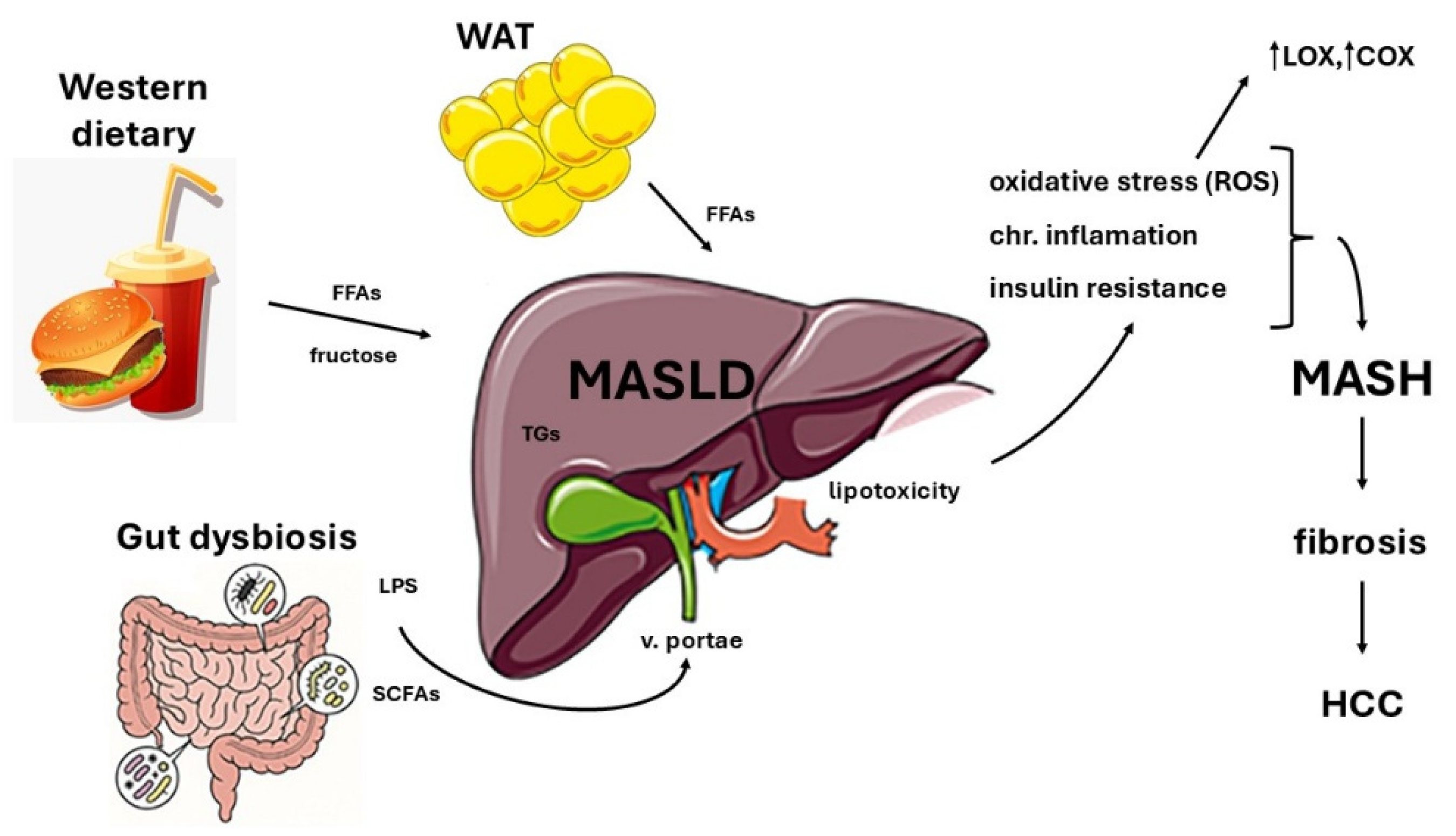
Figure 2.
The impact of Quercetin on MASLD.
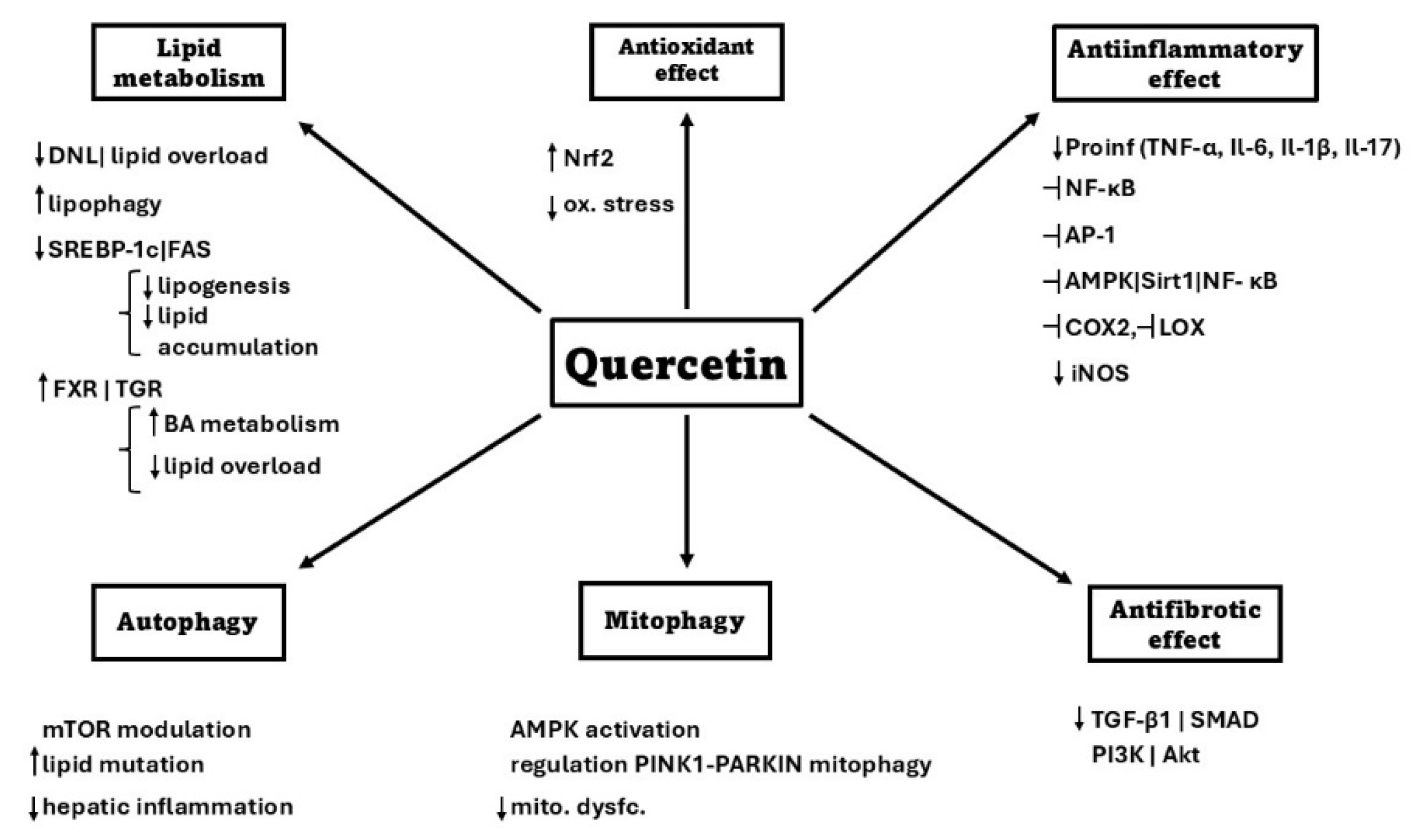
Disclaimer/Publisher’s Note: The statements, opinions and data contained in all publications are solely those of the individual author(s) and contributor(s) and not of MDPI and/or the editor(s). MDPI and/or the editor(s) disclaim responsibility for any injury to people or property resulting from any ideas, methods, instructions or products referred to in the content. |
© 2026 by the authors. Licensee MDPI, Basel, Switzerland. This article is an open access article distributed under the terms and conditions of the Creative Commons Attribution (CC BY) license.
Copyright: This open access article is published under a Creative Commons CC BY 4.0 license, which permit the free download, distribution, and reuse, provided that the author and preprint are cited in any reuse.


