Preprint
Article

This version is not peer-reviewed.

Colocasia esculenta corm Extract Attenuates Ethanol-induced Gastric Ulcer by Suppressing NF-κB Expression: An Integrated Network Pharmacology Approach and In Vivo Validation

Submitted:

13 July 2026

Posted:

14 July 2026

You are already at the latest version

Abstract

The corm of Colocasia esculenta (L.) Scott is widely used for food and treating gastric problems. However, its gastroprotective activity remains unexplored. This article aims to investigate the gastroprotective activity of the ethanol extract of C. esculenta corm (EECE) in ethanol-induced gastric ulcer in rats. EECE was analyzed for nutritional composition (proximate and vitamin C) and phytochemical composition (total phenol content, total flavonoid content, and quercetin levels), followed by metabolite profiling and an in vivo study. Male Wistar rats were randomly assigned to seven groups: (1) normal control, (2) negative control, (3) sucralfate group, (4) quercetin group, and three EECE groups at doses of (5) 200 mg/kg BW, (6) 400 mg/kg BW, and (7) 800 mg/kg BW. Following a two-week treatment period, all groups except the normal group were exposed to 70% ethanol. Post-mortem analysis included macroscopic and histopathological examination of gastric tissue, as well as Western blot analysis to investigate NF-κB p65 expression. EECE contains high moisture, ash, fat, and protein, and low carbohydrate levels, vitamin C at 417.73 mg/100 g, total phenol content of 833.43 mg GAE/100 g, total flavonoid content of 1,178.82 mg QE/100 g, and quercetin at 261.76 mg/100 g. UHPLC–HRMS/MS analysis revealed amino sugars, conjugated amino acids, lipids, phenolics, and minor cyanogenic glycosides. EECE significantly reduced ulcer area percentage in an ethanol-induced gastric ulcer model (p < 0.05). It increased mucosal thickness, reduced polymorphonuclear cell infiltration, and decreased NF-κB p65 expression. The gastroprotective effects of EECE are mediated by enhancing gastric mucosal defense and inhibiting NF-κB p65 expression, thus highlighting its potential as a promising gastroprotective adjunct therapeutic agent.
Keywords: 
;  ;  ;  ;  ;  

1. Introduction

Peptic ulcer disease (PUD) remains a major global health problem with a steadily increasing prevalence. In 2019, PUD affected approximately 8.09 million individuals worldwide, representing a 25.82% increase since 1990 [1]. In Indonesia, the prevalence of PUD is 274,396 cases (40.8% of the population), with PUD-related deaths reaching 0.13% of total deaths (https://www.worldlifeexpectancy.com/indonesia-peptic-ulcer-disease). PUD is characterized by gastric mucosal damage caused by excessive gastric acid and pepsin secretion, which may progress to submucosal ulcer, gastric bleeding, and perforation [2]. Major risk factors associated with PUD include Helicobacter pylori infection, psychological stress, long-term use of non-steroidal anti-inflammatory drugs (NSAIDs), alcohol consumption, and oxidative stress, all of which play critical roles in disease pathogenesis [3,4].
Gastric mucosal protection and healing occur through two principal mechanisms: physical protection via the formation of a mucosal barrier and molecular mechanisms that suppress inflammatory responses, particularly those mediated by the NF-κB signalling pathway [5,6]. The synergistic interaction between these mechanisms is essential for maintaining mucosal integrity and accelerating the repair of damaged gastric tissue [7]. Accordingly, enhancing gastroprotective mechanisms by modulating inflammation-related signalling pathways has become a key therapeutic target in the management of PUD.
Current pharmacological therapies for PUD include antacids, histamine H₂-receptor antagonists, mucosal protective agents, and proton pump inhibitors (PPIs) [8]. Although PPIs are generally effective, their long-term use has been associated with adverse effects such as renal dysfunction, cardiovascular disease, micronutrient deficiencies, and neurodegenerative disorders [9,10]. These concerns underscore the need for safer, more sustainable gastroprotective agents derived from natural sources [11].
Tuberous plants have been reported to exhibit gastroprotective properties due to their rich content of secondary metabolites, including flavonoids, tannins, alkaloids, and phenolic compounds, which possess antioxidant and anti-inflammatory activities [12,13,14]. Flavonoids contribute to gastric mucosal protection by stimulating mucus and bicarbonate secretion, exerting antioxidant effects, and inhibiting inflammatory responses and H. pylori infection [15,16]. In addition, tannins and alkaloids play important roles in mucosal defense by forming protective layers and reducing gastric acid secretion [17,18].
Taro (Colocasia esculenta L.) is a tuberous plant traditionally used for gastric disorders. It is known to contain a wide range of bioactive metabolites, including flavonoids, tannins, saponins, alkaloids, polysaccharides, and phenolic compounds [19,20,21,22]. Its corm has been reported to possess antioxidant and anti-inflammatory activities that protect the gastric mucosa against oxidative stress and inflammation, two key mechanisms underlying PUD pathogenesis [23,24,25]. Strong antioxidant capacity has also been demonstrated by DPPH and ABTS radical-scavenging assays, which are associated with its high phenolic and flavonoid content [26,27].
At the molecular level, flavonoids inhibit the PI3K/Akt/NF-κB signaling pathway, which plays a crucial role in the development of acid- or ethanol-induced gastric ulcers [6]. Quercetin, a major flavonoid, has been shown to reduce Akt phosphorylation and suppress NF-κB p65 expression, thereby exerting significant gastroprotective effects [28,29,30]. C. esculenta corm contains flavonoid compounds such as quercetin [31].
Despite these promising bioactivities, comprehensive studies evaluating the gastroprotective efficacy of C. esculenta corm extract in ethanol-induced gastric ulcer models—particularly those integrating metabolite profiling with NF-κB p65 pathway—remain limited. Therefore, this study aimed to investigate the gastroprotective effects of the ethanol extract of Colocasia esculenta corm (EECE) in an ethanol-induced gastric ulcer model, with a specific focus on the NF-κB signaling pathway. The novelty of this study lies in its integrative mechanistic approach combining UHPLC–HRMS/MS-based metabolite profiling with in vivo histopathological and molecular evaluation of NF-κB p65 suppression, providing new insights into EECE as a multifunctional natural anti-ulcer agent.

2. Results

2.1. Proximate Composition and Vitamin C Content of EECE

Proximate analysis revealed clear compositional differences between C. esculenta corm powder and the ethanol extract (EECE) (Table 1). Compared with the raw powder, EECE had higher moisture, ash, lipid, and protein contents, while the carbohydrate content was lower. In addition, EECE contained a high level of vitamin C, at 417.73 mg/100 g extract.

2.2. Total Phenolic Content (TPC) and Total Flavonoid Content (TFC)

The TPC of the EECE was 833.43 mg gallic acid equivalent (GAE)/100 g extract, and the TFC was 1,178.82 mg quercetin equivalent (QE)/100 g extract.

2.3. HPLC Analysis of Quercetin in EECE

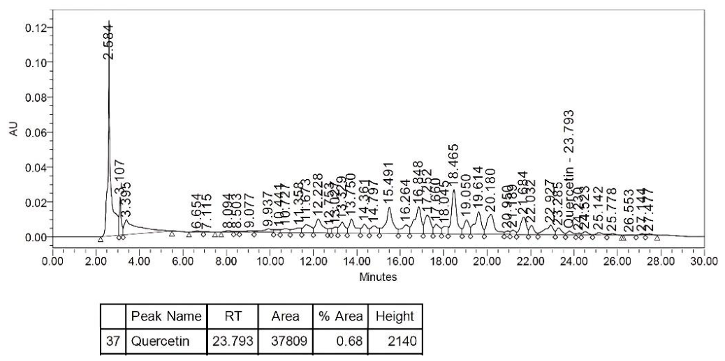
Quercetin in EECE was identified and quantified by HPLC compared with a quercetin standard (2 ppm), which eluted at a retention time (RT) of 24.515 min (Figure 1). As expected, the HPLC chromatogram of EECE showed constituent peaks of EECE, with quercetin observed at 23.973 min (Figure 2). Quantification was performed using a quercetin standard curve, yielding 261.76 mg per 100 g extract.

2.4. Metabolite Profiling and Putative Biological Implications of EECE

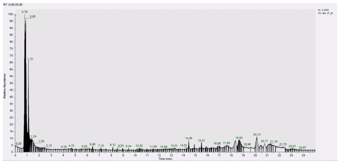
UHPLC-HRMS/MS of EECE (Figure 3) provided major metabolite groups (Table 2). The extract was dominated by amino sugars and conjugated amino acids, followed by lipids, nucleosides, and phenolic derivatives. The most abundant compounds belonged to conjugated amino acids and amino sugars, notably (2S)-3-phenyl-2-({[(3S,4S,5R)-2,3,4-trihydroxy-5-(hydroxymethyl) tetrahydro-2-furanyl] methyl} amino) propanoic acid (10.341%) and 1-[(3-carboxypropyl) amino]-1-deoxy-β-D-fructofuranose (9.695%).

2.5. Network Pharmacology-Based Mechanistic Insights

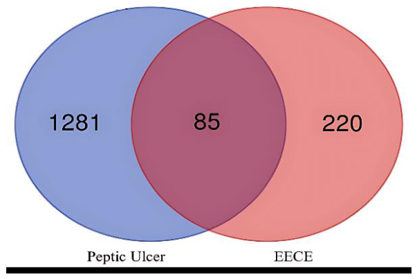
Based on metabolites identified by UHPLC-HRMS/MS analysis, a network pharmacology approach was applied to investigate the multi-compound, multi-target characteristics of EECE in the context of PUD. Compound annotation and target prediction identified 34 putatively bioactive compounds associated with 220 potential protein targets. A total of 1,281 PUD–related targets were retrieved from the GeneCards database. An intersection analysis between compound-related and disease-associated targets identified 85 common targets (Figure 4).
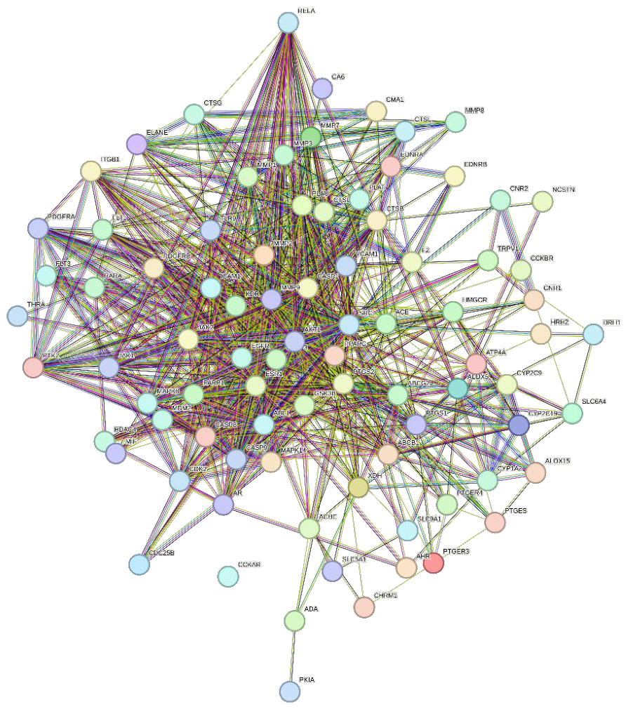
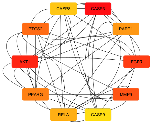
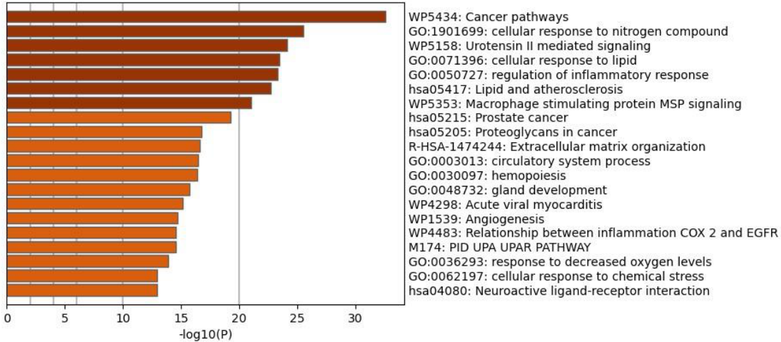
Protein–protein interaction (PPI) network analysis of the intersecting targets revealed a highly interconnected network (Figure 5). Using the Maximal Clique Centrality (MCC) algorithm implemented in CytoHubba, the top 10 hub genes were identified as CASP3, AKT1, EGFR, MMP9, PTGS2, PPARG, PARP1, RELA, CASP8, and CASP9 (Figure 6). GO enrichment and pathway analyses indicated that the intersecting targets were significantly enriched in multiple biological processes and signaling pathways related to peptic ulcer disease (Figure 7). The enrichment results demonstrated strong statistical significance, as indicated by −log10(p) values.

2.6. Macroscopic Evidence of Gastric Mucosal Protection by EECE

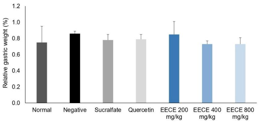
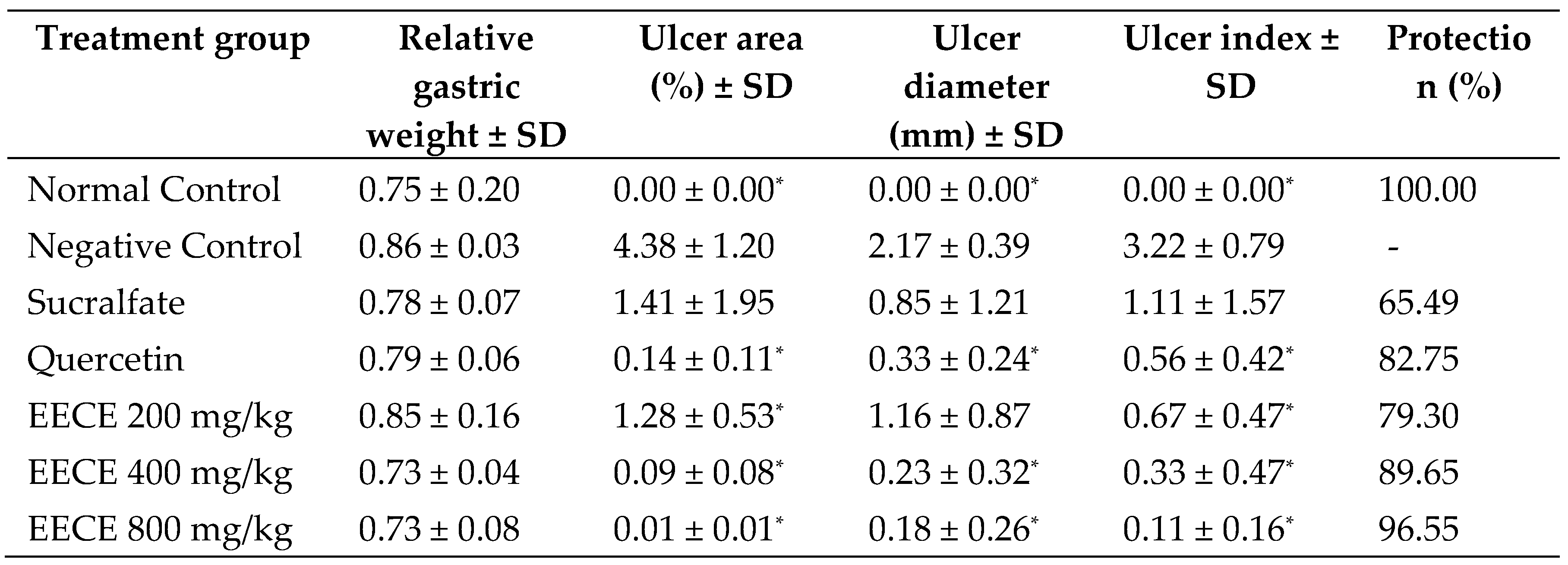
Relative gastric weight was evaluated as an initial macroscopic parameter to assess gross gastric responses following ethanol exposure and EECE treatment [32,33]. As shown in Figure 8 and Table 3, no significant differences in relative gastric weight were observed among the experimental groups, indicating that EECE administration at all tested doses did not induce gross gastric enlargement or abnormal changes in tissue mass.
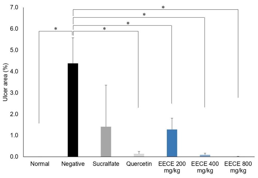
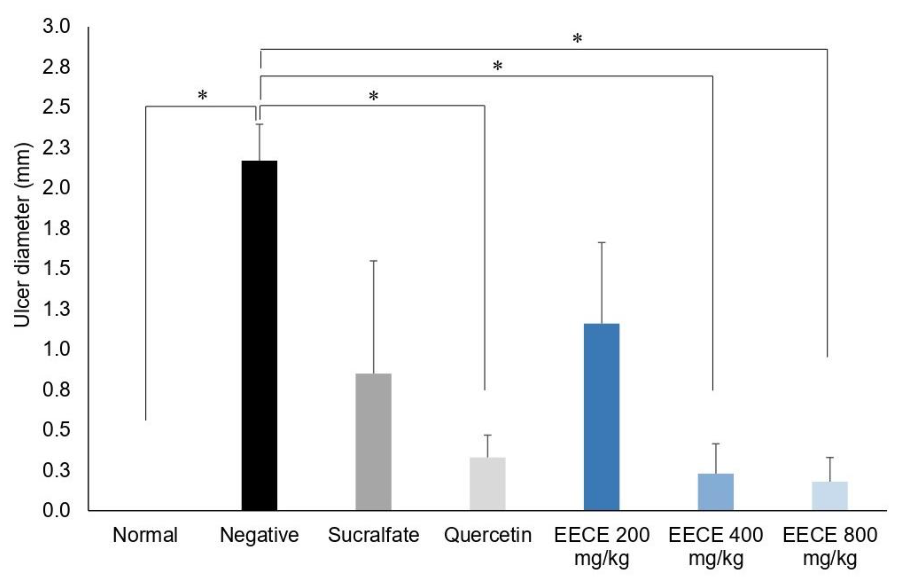
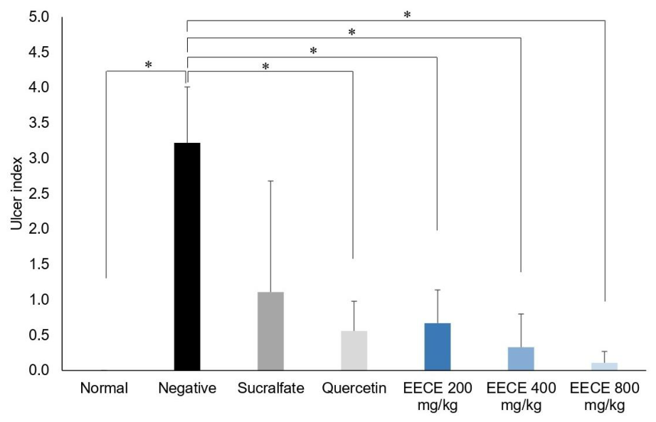
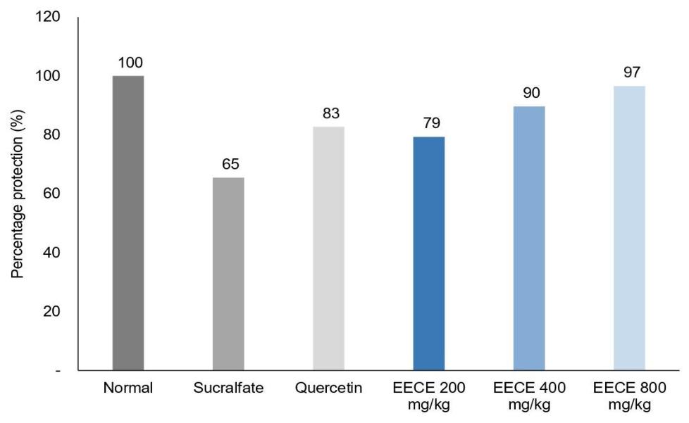
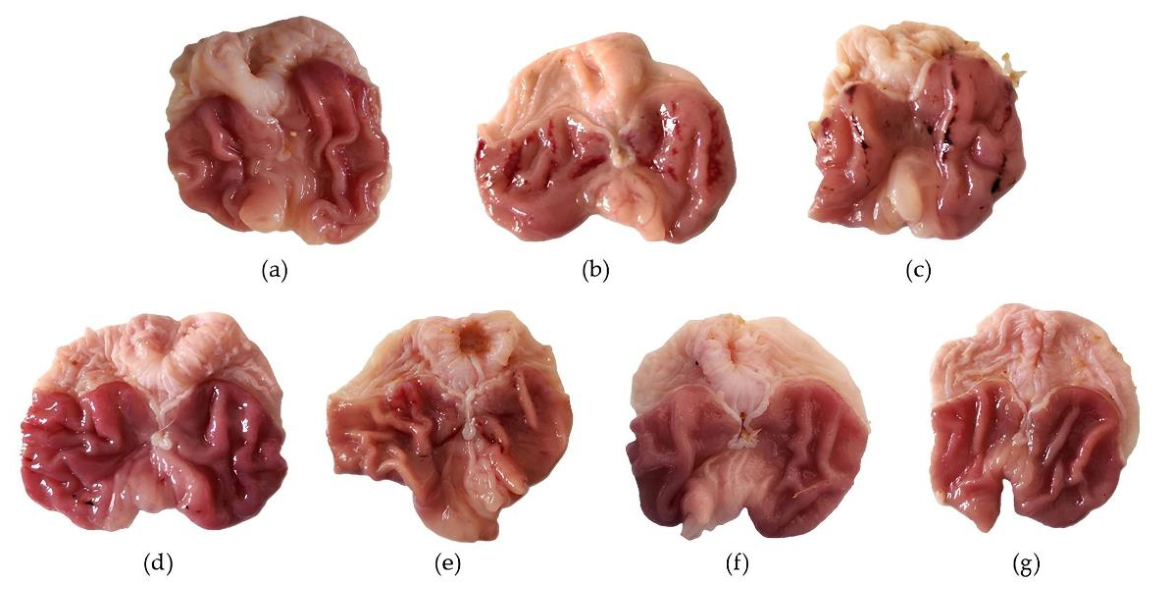
In contrast, macroscopic indicators of gastric mucosal injury were markedly altered following ethanol administration (Table 3). The negative control group exhibited substantial increases in ulcer area (Figure 9), ulcer diameter (Figure 10), and ulcer index (Figure 11), accompanied by a marked reduction in percentage protection (Figure 12). The macroscopic evaluation of gastric tissues revealed clear differences in ulcer severity among the experimental groups, as shown in Figure 13. Conversely, treatment with EECE resulted in pronounced attenuation of these macroscopic lesions, as evidenced by reduced ulcer area, ulcer diameter, and ulcer index, together with an increased percentage of gastric protection, demonstrating clear macroscopic evidence of gastric mucosal protection by EECE.

2.7. Histopathological Scoring, Mucosal Thickness, and PMN Cell Infiltration in Gastric Tissue

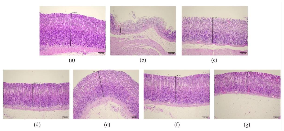
Histopathological evaluation (Table 4) showed that the negative control group exhibited the most severe gastric damage, as indicated by the highest histopathological score (6.0 ± 1.6), a marked reduction in mucosal thickness (697.23 ± 91.41 µm), and a significant increase in polymorphonuclear neutrophil (PMN) infiltration (24.1 ± 0.5 cells/HPF). Morphologically, gastric tissues from this group demonstrated epithelial erosion, submucosal edema, and dense infiltration of acute inflammatory cells, predominantly neutrophils (PMNs).
In contrast, the normal group displayed minimal histopathological alterations, with a low damage score (1.0 ± 0.8), preserved mucosal thickness (1111.51 ± 136.73 µm), and a low PMN count (2.4 ± 1.2 cells/HPF). The sucralfate-treated group showed marked improvement compared with the negative control, as evidenced by a reduced histopathological score (1.9 ± 0.7), a significant decrease in PMN count (0.7 ± 0.1 cells/HPF; p < 0.05), and partial restoration of mucosal thickness (1002.04 ± 116.44 µm). Quercetin treatment resulted in a low histopathological score (1.3 ± 0.7), a significant reduction in PMN infiltration (7.1 ± 0.3 cells/HPF; p < 0.05), and mucosal thickness comparable to that of the normal group (1105.05 ± 41.84 µm).
Administration of EECE at doses of 200, 400, and 800 mg/kg BW produced dose-dependent improvements in gastric histopathology. EECE at 200 mg/kg BW partially reduced the histopathological score (3.3 ± 1.3) and PMN count (10.6 ± 2.2 cells/HPF; p < 0.05), with mucosal thickness of 929.07 ± 38.32 µm. Increasing the dose to 400 mg/kg BW further improved histopathological parameters (score 2.5 ± 0.1; PMNs 10.2 ± 1.6 cells/HPF; p < 0.05), although mucosal thickness (1054.01 ± 35.69 µm) remained lower than that of the normal group. EECE at 800 mg/kg BW produced the most pronounced protective effect, as indicated by a low histopathological score (1.8 ± 1.0), a significant reduction in PMN infiltration (5.3 ± 0.9 cells/HPF; p < 0.05), and restoration of mucosal thickness (1061.71 ± 17.29 µm) approaching normal values. Representative histopathological images of gastric tissue are presented in Figure 14.

2.8. Western Blot Analysis of NF-κB p65 Expression

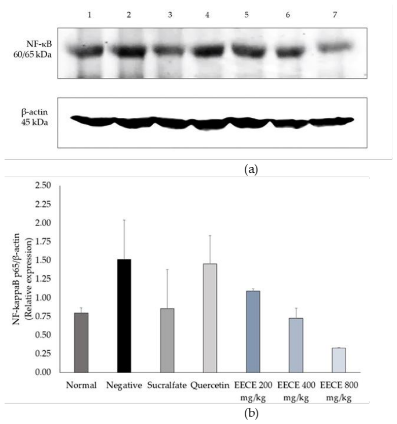
Western blot analysis (Figure 15) revealed differences in NF-κB p65 protein expression among experimental groups, which were further confirmed by quantitative analysis. The normal group exhibited low NF-κB p65 expression (0.79 ± 0.07). In contrast, ethanol induction in the negative control group resulted in increased NF-κB p65 expression (1.51 ± 0.53). In the treatment groups, NF-κB p65 expression was reduced relative to the negative control. The sucralfate and quercetin groups showed mean expression levels of 0.85 ± 0.52 and 1.46 ± 0.38, respectively.
Administration of EECE resulted in a dose-dependent reduction in NF-κB p65 expression, with mean values of 1.09 ± 0.03 at 200 mg/kg BW, 0.73 ± 0.13 at 400 mg/kg BW, and 0.33 ± 0.00 at 800 mg/kg BW. Although differences in NF-κB p65 expression among groups did not reach statistical significance (p > 0.05), a consistent downward trend was observed with increasing EECE dose.

3. Discussion

The present study demonstrates that the gastroprotective effects of EECE are closely associated with its chemical composition, which collectively modulates oxidative stress, inflammatory signaling, and mucosal integrity in the ethanol-induced gastric ulcer model. Ethanol-induced gastric injury is a multifactorial process involving direct epithelial disruption, excessive reactive oxygen species (ROS) generation, activation of inflammatory cascades, and impairment of mucosal defense mechanisms [34,35]. Therefore, effective gastroprotection requires simultaneous modulation of multiple biological targets rather than reliance on a single mechanism.
Proximate analysis revealed a substantial compositional shift between C. esculenta corm powder and EECE, indicating that 70% ethanol selectively enriches polar and semi-polar constituents while excluding insoluble macromolecules, particularly starch, which dominates the native corm matrix. The drastic decrease in carbohydrates confirms that ethanol-insoluble polysaccharides largely remained in the residual solid phase, whereas extractable organic compounds and mineral-associated fractions became proportionally concentrated in the dried extract [36]. This compositional shift is biologically relevant, as carbohydrate depletion does not compromise gastroprotection, while enrichment of antioxidant and anti-inflammatory constituents directly targets the key pathological drivers of ethanol-induced mucosal injury.
In parallel, EECE exhibited a high vitamin C content (417.73 mg/100 g extract), highlighting the effectiveness of hydroethanolic extraction in preserving hydrophilic antioxidants. Vitamin C is a potent ROS scavenger and plays a crucial role in limiting oxidative damage to gastric epithelial cells. Excessive ROS generation following ethanol exposure is known to trigger lipid peroxidation, mitochondrial dysfunction, and redox-sensitive activation of NF-κB signaling [37,38]. Thus, the high vitamin C content of EECE likely contributes to early suppression of oxidative stress, thereby attenuating upstream activation of inflammatory cascades.
The antioxidant capacity of EECE is further supported by its elevated TPC and TFC. Phenolic compounds and flavonoids are well documented to exert gastroprotective effects through ROS scavenging, inhibition of lipid peroxidation, and modulation of inflammatory signaling pathways, including NF-κB [39,40]. In comparison, Akyüz (2019) reported a lower TPC value of 240 mg GAE/100 g extract for an ethanolic extract of C. esculenta corms (Gölevez variety), highlighting the superior phenolic recovery achieved in the present study. Conversely, Nugroho et al. (2025) reported a markedly higher TPC value (3975 mg/100 g) for C. esculenta extracted using water [41], suggesting that extraction efficiency may be substantially enhanced through optimization of extraction parameters, such as solvent polarity, temperature, extraction duration, or the application of intensification techniques (e.g., sonication). Variations in TFC among studies are commonly attributed to differences in plant genotype, environmental growth conditions, post-harvest handling, and extraction and analytical methodologies [42,43,44,45].
Untargeted UHPLC–HRMS/MS profiling revealed a chemically diverse metabolite composition in EECE, including lipid-derived metabolites, aromatic compounds, phenolic derivatives, and indole-related molecules. Lipid-associated metabolites such as oleamide and related fatty acid amides have been reported to exert anti-inflammatory effects by suppressing iNOS and COX-2 expression and modulating NF-κB signaling [46,47]. In addition, lipid components contribute to maintaining gastric mucus hydrophobicity, which is essential for preventing hydrogen-ion back-diffusion and epithelial injury [48].
The presence of phenolic and indole derivatives, including trans-3-indoleacrylic acid, further supports the antioxidant potential of EECE. Even at relatively low concentrations, such compounds can synergistically attenuate oxidative stress and inflammatory amplification in ethanol-induced gastric injury [49]. Moreover, metabolomic analysis identified amino sugars and their derivatives, such as N-acetyl-D-quinovosamine and N-acetylglucosaminitol, which are structurally related to N-acetylglucosamine, a key precursor for mucin biosynthesis. Gastric mucus is a primary defensive barrier against ethanol-induced damage, and enhanced mucin production has been shown to improve mucosal resistance and ulcer healing [50,51].
The presence of aromatic metabolites such as trans-3-indoleacrylic acid, 5-amino-salicyluric acid, and other phenolic derivatives indicates active phenolic biosynthetic pathways that may contribute to the extract’s antioxidant potential [39]. The detection of compounds such as lotaustralin further suggests the presence of low-level cyanogenic glycosides, which are widely reported as characteristic defense-related metabolites in Araceae [52,53]. Overall, this complex metabolite profile supports the interpretation that EECE contains not only phenolic compounds quantified by total phenolic and flavonoid content analyses, but also other metabolite classes that may collectively contribute to its bioactive properties, including antioxidant, anti-inflammatory, and metabolic-modulatory activities [54,55].
Determining TFC and TPC in EECE provides initial quantitative insight into its chemical composition. EECE exhibited a TFC, indicating that flavonoids represent a major component of the extract. This finding is consistent with UHPLC–HRMS/MS results, which revealed multiple aromatic metabolites, phenolic derivatives, and indole compounds closely linked to flavonoid biosynthetic pathways in plants. Furthermore, the TPC value reflects the contribution of phenolic acids, aromatic indoles, and benzenoid derivatives detected through UHPLC–HRMS/MS. These results confirm that EECE contains not only flavonoids but also a broad spectrum of non-flavonoid phenolic compounds, in agreement with the characteristic chemical profile of tuber-derived plant materials.
In support of the role of phenolic constituents, the presence of quercetin, a major flavonol identified by HPLC, further substantiates the antioxidant potential of EECE. Quercetin is biochemically recognized for its strong capacity to scavenge reactive oxygen species and free radicals through both direct neutralization and modulation of endogenous antioxidant systems, thereby contributing to redox stabilization and attenuation of oxidative stress [56,57]. In addition, UHPLC–HRMS/MS identified several other phenolic metabolites that may synergize with quercetin to enhance the extract’s overall antioxidant activity.
Although HPLC analysis confirmed the presence of quercetin in EECE, this compound was not explicitly identified in the UHPLC–HRMS/MS dataset. This discrepancy can be attributed to fundamental differences between targeted and untargeted analytical approaches. HPLC is a targeted technique used to selectively detect and quantify predefined analytes via retention-time matching and authentic reference standards [2,58]. In contrast, UHPLC–HRMS/MS employs an untargeted strategy that broadly detects metabolites without prior target selection, followed by annotation based on accurate mass, ionization behavior, and spectral database matching [2,59].
In addition to methodological factors, the ionization behavior of phenolic compounds is critical to their detectability by LC–MS-based techniques. Flavonols, such as quercetin, ionize more efficiently in negative electrospray ionization (ESI–) mode, forming stable deprotonated [M–H]⁻ ions that enhance detection sensitivity in high-resolution mass spectrometry. Previous studies employing LC–MS analyses in ESI negative-ion mode have demonstrated improved detection of flavonoid aglycones and related phenolic compounds under such conditions, supporting the preferential detection of quercetin-type metabolites in ESI–mod [31].
Moreover, the chemical form of quercetin in plant tissues further influences its detectability. In plants, quercetin predominantly occurs as flavonoid glycosides (e.g., quercetin-3-O-glucoside, quercetin-3-O-rutinoside, and quercetin-3-O-rhamnoside) rather than as a free aglycone. These glycosylated forms represent the dominant natural state of quercetin and significantly affect its ionization efficiency and bioavailability [60]. Consequently, the aglycone form may exhibit low signal intensity or remain undetected in untargeted UHPLC–HRMS/MS profiling, which preferentially captures more stable and readily ionizable glycoside [61]. Therefore, the absence of quercetin in the UHPLC–HRMS/MS results may be attributed to its low concentration and/or suboptimal ionization of its aglycone or glycosidic forms under the applied analytical conditions. This interpretation is consistent with the HPLC quantification, which showed a relatively low quercetin content of 261.76 mg/100 g extract.
The metabolite profiling revealed multiple bioactive compounds, suggesting complex molecular interactions. Therefore, a network pharmacology analysis was employed to systematically explore the compound–target–pathway relationships underlying the observed biological effects. This analysis identified several hub genes, including RELA (NF-κB p65), AKT1, EGFR, PTGS2, MMP9, CASP3, CASP8, CASP9, and PARP1, as central regulators within the protein–protein interaction network. The prominence of these genes suggests that the therapeutic effects of EECE are mediated by coordinated regulation of inflammatory signaling, cell survival, apoptosis, and tissue remodeling, key processes involved in the pathogenesis and healing of peptic ulcer disease.
Among these hub genes, RELA (NF-κB p65) emerged as a critical regulatory node, underscoring the central role of NF-κB–mediated signaling in gastric mucosal injury and repair. NF-κB p65 functions as a master transcription factor that regulates the expression of pro-inflammatory cytokines, including TNF-α, IL-1β, and IL-6, which are known to exacerbate gastric inflammation and epithelial damage [62,63]. Dysregulation of NF-κB signaling has been widely implicated in gastric mucosal injury, in which exposure to proinflammatory agents, such as ethanol, induces NF-κB p65 activation and nuclear translocation, amplifying inflammatory cascades and promoting epithelial damage [64].
GO enrichment analysis further demonstrated significant enrichment of biological processes related to the regulation of inflammatory response, cellular response to chemical stress, and response to nitrogen-containing compounds. These processes are closely linked to NF-κB–dependent signaling pathways, highlighting the central role of NF-κB p65 as an upstream regulator linking chemical stress exposure to inflammatory damage in gastric tissues. Moreover, enrichment of biological processes associated with extracellular matrix organization, angiogenesis, and gland development indicates the involvement of tissue remodeling and mucosal regeneration during peptic ulcer healing. These regenerative events are functionally associated with NF-κB signaling through downstream effectors such as matrix metalloproteinase-9 (MMP-9), which plays a key role in extracellular matrix degradation and remodeling. Increasing evidence suggests that excessive activation of the NF-κB–MMP-9 axis contributes to impaired ulcer healing, sustained inflammation, and persistent mucosal injury, whereas controlled modulation of this pathway promotes angiogenesis, epithelial restitution, and restoration of gastric mucosal integrity [65,66,67,68].
Pathway enrichment analysis indicated significant involvement of COX-2–EGFR signaling, receptor-mediated pathways, and lipid mediator–related signaling cascades. RELA (NF-κB p65) functions as a key upstream transcriptional regulator of COX-2 expression, thereby enhancing prostaglandin-mediated lipid signaling and facilitating EGFR transactivation [62]. Prostaglandin E₂ signaling has been shown to play an essential role in epithelial cell survival, proliferation, and restitution processes [69]. Collectively, this coordinated NF-κB–COX-2/PGE₂–EGFR crosstalk has been increasingly recognized as a central regulatory mechanism governing the balance between gastric mucosal injury and repair [70]. Consistently, the identification of AKT1, EGFR, RELA, and MMP9 as hub genes in the cytoHubba analysis supports the presence of an NF-κB–AKT–EGFR signaling axis underlying ulcer pathogenesis.
Although several enriched pathways were annotated as cancer-related, these findings do not imply malignant transformation. Rather, they reflect the involvement of central hub genes such as AKT1, EGFR, RELA (NF-κB p65), and MMP9 that regulate inflammation, cell proliferation, survival, and tissue remodelling, core biological processes shared by chronic inflammatory disorders and cancer-associated signaling pathways. Consistent with this interpretation, GO and pathway enrichment analysis revealed that the predicted target genes are predominantly associated with inflammatory regulation, cellular stress responses, and tissue repair mechanisms relevant to the pathophysiology of peptic ulcer disease.
Collectively, these findings provide mechanistic support for the conclusion that the observed therapeutic effects are mediated by attenuation of excessive inflammation, protection of gastric epithelial cells from stress-induced injury and apoptosis, and promotion of mucosal healing. Notably, RELA (NF-κB p65) was identified as a central molecular hub integrating inflammatory signaling, stress responses, and tissue remodelling. Given the prominent involvement of NF-κB-centered pathways identified in silico, experimental validation using in vivo gastric ulcer models is warranted. Accordingly, subsequent in vivo studies were designed to evaluate gastric mucosal protection, inflammatory marker expression, and modulation of NF-κB p65 signaling.
These in silico predictions were strongly supported by in vivo findings, which provided functional validation of the involvement of inflammatory, oxidative stress–related, and tissue-remodeling pathways in gastric mucosal injury. In the negative control group, ethanol administration induced extensive hemorrhagic lesions, elevated ulcer indices, and severe mucosal erosion, confirming successful establishment of gastric injury and establishing a relevant biological context for assessing the gastroprotective effects of EECE. Consistent with the known pathophysiology of ethanol-induced gastric damage, characterized by epithelial disruption, excessive oxidative stress, and inflammatory cell infiltration leading to hemorrhagic lesions and mucosal erosion [35].
Ulcer diameter, which reflects the localized severity and depth of mucosal damage, decreased after EECE administration, with greater reductions at higher doses, suggesting a dose-related protective effect on gastric mucosal integrity. The absence of a statistically significant reduction at 200 mg/kg BW suggests that this dose was insufficient to effectively limit lesion expansion. In contrast, doses of 400 and 800 mg/kg BW markedly suppressed ulcer progression. This response pattern highlights the importance of sufficient exposure to bioactive compounds in counteracting ethanol-induced mucosal disruption. Comparable gastroprotective effects, with greater protection observed at higher doses or concentrations, have also been reported for plant-derived extracts rich in polyphenols and other bioactive metabolites [71].
Interestingly, although sucralfate provided partial protection, it did not significantly reduce ulcer area or index ulcer compared with the negative control. This observation may be explained by the primarily cytoprotective mechanism of sucralfate, which forms a physical barrier over the ulcer surface, thereby protecting the mucosa from acid, pepsin, and bile salts without substantially modulating oxidative stress or inflammatory signaling pathways [72]. In contrast, EECE and quercetin significantly reduced ulcer area and severity, suggesting broader protective mechanisms beyond surface shielding.
The negative control group exhibited pronounced gastric hemorrhage and extensive mucosal lesions, reflecting severe ethanol-induced gastric injury. Treatment with sucralfate provided partial protection, as gastric lesions and hemorrhagic areas remained evident, consistent with its primary cytoprotective mechanism. In contrast, quercetin administration effectively prevented the formation of visible gastric lesions, although mild hemorrhagic areas remained. Notably, EECE treatment provided the most pronounced gastroprotective effect, with a complete absence of gastric lesions across all treatment doses; at 800 mg/kg BW, the gastric mucosa appeared intact and free of hemorrhage. These macroscopic observations are consistent with reductions in ulcer area, ulcer diameter, and ulcer index, as well as increased gastric protection percentages observed in EECE-treated groups following ethanol-induced injury.
The superior gastroprotective effects observed in the quercetin and EECE-treated groups can be attributed to their antioxidant and anti-inflammatory properties. Quercetin, a major flavonol identified in EECE, effectively scavenges reactive oxygen species and enhances endogenous antioxidant defenses. Oxidative stress plays a pivotal role in ethanol-induced gastric injury by promoting lipid peroxidation, epithelial apoptosis, and microvascular dysfunction. Therefore, the high protective indices observed in EECE-treated animals are likely mediated, at least in part, by suppression of oxidative damage and preservation of mucosal redox balance [73,74].
Moreover, increasing evidence suggests that modulation of inflammatory signaling pathways is crucial for effective gastroprotection. Ethanol exposure activates the NF-κB pathway, leading to the transcription of pro-inflammatory mediators such as TNF-α, IL-1β, and COX-2, thereby exacerbating mucosal injury and delaying healing [34]. The significant reduction in ulcer severity and the high protection percentages observed with EECE administration are consistent with inhibition of NF-κB–mediated inflammatory responses, as further supported by the observed downregulation of NF-κB p65 expression. This mechanistic link strengthens the biological relevance of EECE as an anti-ulcer agent.
Beyond flavonoids, metabolomic profiling revealed that EECE contains substantial amounts of amino sugars and their derivatives, including N-acetyl-D-quinovosamine and N-acetylglucosaminitol. These compounds are structurally related to N-acetylglucosamine, a key building block in the synthesis of mucin glycoproteins. Gastric mucus constitutes the first line of defense against luminal aggressors, and enhanced mucin production has been shown to improve mucosal resistance to ethanol-induced injury [50,51]. The presence of acetylated amino sugars in EECE may therefore support mucin biosynthesis, thereby reinforcing the gastric mucus barrier and limiting penetration of acid and ethanol into the epithelium.
In addition, EECE contained amino acid conjugates and small peptides, such as prolyl leucine, which are implicated in epithelial regeneration and tissue repair. Amino acids play essential roles in regulating epithelial proliferation, differentiation, and barrier integrity in the gastrointestinal tract, thereby facilitating mucosal healing following injury [75,76]. The reduced lesion areas observed macroscopically in EECE-treated groups may thus reflect not only protective effects but also accelerated mucosal recovery.
Lipid-derived metabolites, including oleamide, were also detected in EECE and may contribute to its gastroprotective activity. Oleamide and related lipid amides have been reported to exert anti-inflammatory effects by suppressing iNOS and COX-2 expression and modulating NF-κB signaling [47,77]. Lipids further contribute to maintaining the hydrophobic properties of the gastric mucus layer, which are critical for preventing hydrogen-ion back-diffusion and preserving epithelial integrity [48]. These mechanisms collectively support the observed reduction in mucosal erosion and hemorrhage.
Although present at lower concentrations, phenolic and indole derivatives, such as trans-3-indoleacrylic acid, may further enhance EECE’s antioxidant capacity. Ethanol-induced gastric injury is strongly associated with excessive ROS generation, leading to lipid peroxidation and inflammatory amplification. Even minor amounts of antioxidant compounds can synergistically reduce oxidative burden and mitigate mucosal damage.61 Therefore, the combined action of multiple metabolite classes likely underlies the robust gastroprotective effect of EECE.
Taken together, the present findings demonstrate that the gastroprotective activity of EECE arises from a synergistic interplay among multiple bioactive metabolites rather than from a single compound. Amino sugars contribute to the reinforcement of the mucus barrier; lipids and amino acid derivatives support membrane stability and tissue repair; and flavonoids and phenolic compounds attenuate oxidative stress and inflammatory signaling. This multifaceted mechanism provides a strong scientific basis for the significant reduction in ulcer severity and high protection indices observed in the ethanol-induced gastric ulcer model, highlighting C. esculenta corm as a promising natural source of gastroprotective agents. To further substantiate this mechanistic interpretation at the tissue level, histopathological evaluation was performed to assess structural alterations and inflammatory cell infiltration in the gastric mucosa.
The severe histopathological alterations observed in the negative control group confirm successful induction of ethanol-induced gastric injury. Morphologically, this damage was characterized by epithelial erosion, submucosal edema, and infiltration by acute inflammatory cells, particularly neutrophils (PMNs), reflecting disruption of the mucosal barrier and activation of the acute inflammatory response [78,79]. High PMN infiltration indicates the active involvement of inflammatory cells in the release of pro-inflammatory mediators and reactive oxygen species (ROS), which play critical roles in exacerbating tissue damage, increasing oxidative stress, and accelerating the degradation of the gastric mucosal protective layer [80,81].
The minimal histological alterations observed in the normal group indicate intact mucosal architecture and absence of inflammatory activation. The improvement observed in the sucralfate-treated group is consistent with its established cytoprotective mechanism, which involves the formation of a viscous protective layer over the gastric mucosa via polymerization reactions under acidic conditions [72]. This layer functions as a barrier against aggressive factors such as gastric acid, pepsin, and bile salts, while simultaneously creating a microenvironment that supports mucosal healing and tissue regeneration [82]. With reduced mucosal exposure to irritants, activation of local inflammatory pathways can be suppressed, as indicated by decreased pro-inflammatory mediators and improved epithelial barrier integrity [51]. This condition reduces chemotactic signals that attract neutrophils to the injury site, resulting in fewer PMNs migrating to and accumulating in the damaged gastric tissue.
Quercetin-mediated gastroprotection is likely attributable to its combined antioxidant and anti-inflammatory properties, as reflected by the marked reduction in PMN infiltration and the preservation of gastric mucosal thickness. These findings indicate that quercetin effectively limits neutrophil recruitment and attenuates oxidative stress in gastric tissue, thereby maintaining mucosal structural integrity and facilitating accelerated healing [83,84]. This effect can also be explained by the antioxidant activity of quercetin, which acts as a ROS scavenger, neutralizing free radicals and reducing oxidative stress, both of which play a crucial role in gastric mucosal injury [56]. The reduction in oxidative stress contributes to the preservation of epithelial integrity, as oxidative stress and lipid peroxidation are known to increase mucosal permeability and exacerbate tissue damage under ulcerative conditions [83]. In addition, quercetin exhibits anti-inflammatory properties by inhibiting the activation of inflammatory pathways, including suppression of NF-κB p65 activation and reduced expression of pro-inflammatory mediators, thereby attenuating excessive inflammatory responses in gastric tissue [85]. These mechanisms ultimately contribute to reduced accumulation of inflammatory cells, as evidenced by reduced infiltration in ethanol-induced gastric ulcer models [83].
EECE treatment demonstrated dose-dependent gastroprotective effects, with higher doses providing more pronounced histological improvement. Partial protection at lower doses suggests early suppression of inflammatory infiltration, whereas optimal protection at 800 mg/kg BW was associated with both reduced PMN accumulation and restoration of mucosal thickness. This observation aligns with previous reports indicating that attenuation of inflammatory responses often precedes structural regeneration of gastric tissue [34]. The reduction in PMN infiltration, accompanied by increased mucosal thickness, underscores the close relationship between inflammatory control and tissue healing. Previous studies have shown that suppression of neutrophil recruitment is associated with inhibition of inflammatory pathways, including NF-κB p65, which plays a crucial role in the pathogenesis of gastritis and gastric ulcers [86,87].
Overall, integration of histopathological scoring, mucosal thickness, and PMN cell counts provides a comprehensive depiction of the extent of gastric mucosal injury and protection. The significant reduction in PMN numbers in the treated groups, particularly at higher EECE doses, confirms that modulation of inflammatory responses is a key mechanism for reducing tissue damage and supporting the recovery of gastric mucosal structure, likely mediated by regulation of inflammatory pathways, including NF-κB p65 [88,89]. To further elucidate the molecular basis underlying these histopathological improvements, NF-κB p65 expression was evaluated, given its central role in ethanol-induced gastric inflammation.
The increased expression of NF-κB p65 observed in the negative control group is consistent with the established mechanism of ethanol-induced gastric mucosal injury, which involves excessive ROS production and oxidative stress–mediated activation of NF-κB signaling. Activation of NF-κB p65 promotes the transcription of pro-inflammatory cytokines, including TNF-α, IL-1β, and IL-6, as well as inflammatory enzymes such as iNOS and COX-2, leading to inflammatory cell infiltration and exacerbation of gastric tissue damage [87,90]. Activation of NF-κB p65 has been widely recognized as a central event in the pathogenesis of ethanol-induced gastric mucosal injury, given its pivotal role in coordinating inflammatory responses and amplifying inflammatory signaling, culminating in progressive mucosal damage [38,88].
The reduced NF-κB p65 expression observed in the sucralfate-treated group suggests that, beyond its well-known cytoprotective action through formation of a physical barrier on the gastric mucosa, sucralfate may also exert anti-inflammatory effects by modulating NF-κB pathway activation. Inhibition of NF-κB signaling has been associated with decreased transcription of pro-inflammatory mediators that aggravate gastric mucosal injury [62,91,92]. Consistent with this, quercetin has been reported to attenuate gastric inflammatory responses by regulating the HMGB1/TLR4/NF-κB signaling pathway, thereby limiting amplification of inflammatory signals and the progression of mucosal injury in models of gastritis and gastric ulceration [93,94,95].
The dose-related reduction in NF-κB p65 expression following EECE administration suggests a modulatory effect on inflammatory signaling, particularly at higher doses. This observation aligns with previous studies reporting that natural compounds with anti-inflammatory properties can attenuate NF-κB p65 activation in experimental models of gastric ulceration [84,89]. Although changes in total NF-κB p65 protein expression did not reach statistical significance, the consistent downward trend is biologically relevant and may reflect increasing efficacy with higher exposure levels. In the pathophysiology of gastritis and gastric ulceration, modulation of NF-κB signaling is often more sensitively reflected in changes in pathway activation and downstream signaling events rather than in marked alterations in total protein expression, yet it still contributes to the attenuation of inflammatory responses [38,88].
Importantly, the observed trend of reduced NF-κB p65 expression is consistent with histopathological improvements, including decreased PMN infiltration, lower tissue injury scores, and restored mucosal thickness in the treatment groups. This concordance underscores the role of NF-κB p65 modulation in limiting inflammatory cell recruitment and restraining the progression of gastric mucosal injury. Collectively, these Western blot findings support the hypothesis that the gastroprotective effects of the treatments—particularly EECE—are mediated, at least in part, through suppression of inflammatory responses via regulation of the NF-κB p65 signaling pathway, even though these effects are not fully reflected as statistically significant changes in total protein expression [38,89].

4. Materials and Methods

4.1. Plant Material Collection and Identification

Corms were collected from a taro plantation in Cikeas Village, Sukaraja Subdistrict, Bogor Regency, West Java, Indonesia (Google Maps coordinates: 6°31’21” S, 106°51’01” E). The plant samples were identified by Arifin Surya Dwipa Irsyam (Scopus ID: 57211286941) at the Herbarium Bandungense, School of Life Sciences and Technology, Bandung Institute of Technology, Indonesia, and were confirmed as Colocasia esculenta (L.). Scott (family Araceae) [96,97], with document number 2455/IT1.C11.2/TA.00/2025, signed by Angga Dwiartama, Ph.D. The plant specimen was not deposited in the Herbarium Bandungense.

4.2. Preparation of Corm Powder

Fresh corms were peeled, washed thoroughly under running water, and sliced to facilitate drying. The slices were sun-dried for 2 days (8 hours per day), ground to a coarse powder, and sieved through a 60-mesh sieve (featuring 60 holes/linear inch). The resulting powder was stored in a tightly closed plastic container until extraction.

4.3. Preparation of Ethanol Extract (EECE)

Extraction was performed by briefly immersing the corm powder in 70% ethanol, prepared by diluting absolute ethanol (EMSURE®, Merck, Darmstadt, Germany; Cat. No. 1.00983), at a material-to-solvent ratio of 1:10 (w/v) for 3 × 24 h at room temperature, with occasional stirring during the first 6 h. The extracts were collected, filtered, and the solvent was removed by rotary evaporation at 40 °C, yielding a viscous extract (EECE).

4.4. Proximate Analysis

The proximate composition of EECE (moisture, lipid, ash, protein, carbohydrate) and the vitamin C content were determined. Moisture, lipid, ash, and protein were determined in accordance with SNI 01-2891:1992.

4.5. Total Phenol Content (TPC)

TPC was determined using the Folin–Ciocalteu reagent (Merck, Darmstadt, Germany; Cat. No. 1.09001), which quantifies phenolic compounds by reducing them to form a blue complex [98]. TPC was calculated using a gallic acid standard curve [99,100]. Briefly, EECE (0.2 g) was dissolved in methanol (EMSURE®, analytical grade, Merck, Darmstadt, Germany; Cat. No. 1.06007) and adjusted to a final volume of 25 mL. One mL of the sample or standard solution was mixed with 5.0 mL of diluted Folin–Ciocalteu reagent, then 4.0 mL of sodium carbonate solution (7.5%, w/v), prepared by dissolving sodium carbonate (Merck, Darmstadt, Germany) in distilled water, was added to the reaction mixture. After incubation, the absorbance was measured at 730 nm.

4.6. Total Flavonoid Content (TFC)

TFC was determined using the aluminum chloride (AlCl₃) colorimetric method, as previously described. EECE (0.2 g) was dissolved in ethanol (EMSURE®, Merck, Darmstadt, Germany; Cat. No. 1.00983) and adjusted to a final volume of 25 mL. An aliquot of 0.5 mL of the sample or standard solution was mixed with 1.5 mL of ethanol, 0.1 mL of AlCl₃ solution (10%, w/v) was prepared from aluminium chloride (AlCl₃, Sigma-Aldrich, St. Louis, MO, USA; Cat. No. 206911), 0.1 mL of sodium acetate solution (1 M), which was prepared from sodium acetate (EMSURE®, Merck, Darmstadt, Germany; Cat. No. 1.06268), and 2.8 mL of distilled water. The reaction mixture was incubated at room temperature for 30 min, and the absorbance was measured at 510 nm using an ultraviolet-visible spectrophotometer. A blank solution without AlCl₃ was prepared. TFC was calculated from the quercetin standard curve [101].

4.7. Determination of Quercetin in EECE Using RP-HPLC

Quercetin in EECE was identified and quantified by reverse-phase high-performance liquid chromatography (RP-HPLC) following recent validated methods [102,103]. Before injection, the sample and standard solutions were filtered through a membrane filter. Chromatographic separation was performed on an octadecylsilane C18 column (250 mm × 4.6 mm internal diameter) with a mobile phase of 0.1% trifluoroacetic acid (Sigma-Aldrich, St. Louis, MO, USA; Cat. No. T6508) in water [A], methanol (LiChrosolv®, gradient grade for HPLC, Merck, Darmstadt, Germany; Cat. No. 1.06035) [B], and acetonitrile (Fisher Scientific, Fair Lawn, NJ, USA; Cat. No. A996-4) [C], delivered at a flow rate of 1.0 mL/min. The injection volume was 10–20 μL, and detection was performed at the characteristic UV wavelength of quercetin (370 nm). Quantification was performed using a quercetin standard curve (Sigma-Aldrich, St. Louis, MO, USA; Cat. No. Q4951).

4.8. UHPLC-HRMS/MS-BASED METABOLITE PROFILING

The chemical profile of EECE was analyzed using an ultra-high-performance liquid chromatography-high-resolution mass spectrometry tandem mass spectrometry (UHPLC–HRMS/MS) system. Chromatographic separation was performed using a Thermo Scientific Vanquish™ UHPLC Binary Pump equipped with an Accucore™ Phenyl-Hexyl column (100 mm × 2.1 mm, 2.6 µm) maintained at 40 °C. The mobile phase consisted of MS-grade water containing 0.1% (v/v) formic acid (A) and MS-grade methanol containing 0.1% (v/v) formic acid (B), delivered at a flow rate of 0.3 mL/min using gradient elution. Methanol (Merck, Darmstadt, Germany; Cat. No. 632546) and formic acid (Merck, Darmstadt, Germany; Cat. No. 1.59013) were used. The gradient program started at 5% B, increased linearly to 90% B over 16 min, held at 90% B for 4 min, and then returned to the initial condition (5% B) for 25 min to re-equilibrate the column.
The sample was prepared by dissolving 1 mg of EECE in 1 mL of 100% methanol (Merck, Darmstadt, Germany; Cat. No. 632546), and 3 µL was injected into the system. Mass spectrometric detection was carried out using a Q Exactive™ Hybrid Quadrupole-Orbitrap™ high-resolution mass spectrometer operated in positive electrospray ionization (ESI+) mode. The capillary voltage was set to 3.30 kV, the capillary temperature to 320 °C, and mass spectra were acquired over an m/z range of 66.7–1000. Compound identification was performed by matching accurate mass and fragmentation patterns with reference databases including MzCloud, ChemSpider, and PubChem.

4.9. Network Pharmacology Analysis

Active compounds identified by UHPLC-HRMS/MS were subjected to target prediction using open-access databases. Disease-related targets for PUD were retrieved from the GeneCards database. Common targets shared by compounds and disease were identified using intersection analysis. Protein–protein interaction (PPI) networks were constructed using the STRING database (confidence score ≥ 0.4) and visualized in Cytoscape. Core targets were identified based on topological parameters, including degree and betweenness centrality. Gene Ontology (GO) and Kyoto Encyclopedia of Genes and Genomes (KEGG) pathway enrichment analyses were performed using Metascape, with significant terms defined by adjusted p-values < 0.05.

4.10. In Vivo Gastroprotective Study

4.10.1. Animal Ethics, Handling, and Maintenance

The study was conducted on male Wistar rats. The protocol for animal handling was approved on 8 August 2025 by the Research Ethics Committee of Universitas Padjadjaran, Indonesia (https://kep.unpad.ac.id/; approval document number 687/UN6.KEP/EC/2025, signed by Dr. Muhammad Hasan Bashari). The procedure was carried out by strictly adhering to The Guide for the Care and Use of Laboratory Animals (NRC 2011; eighth edition) (https://grants.nih.gov/grants/olaw/guide-for-the-care-and-use-of-laboratory-animals.pdf) (Guide for the Care and Use of Laboratory Animals 2011), and The ARRIVE guidelines 2.0 Animal Research: Reporting of In Vivo Experiments (https://arriveguidelines.org/arrive-guidelines). The procedures were carried out at the Pharmacology Laboratory, Faculty of Pharmacy, Universitas Padjadjaran, Indonesia.
Male Wistar rats with an approximate body weight of 180 g were purchased from the Animal Breeding Facility, Division of Animal Laboratories of PT. Biofarma, Jl. Kolonel Masturi Kav 10 Kertawangi, Cisarua, West Java, Indonesia, and housed under controlled environmental conditions (22–25 °C; 50–60% relative humidity), with a 12 h light/12 h dark cycle. Animals were kept at a density of 5 rats per cage, fed a standard rodent chow (containing approximately 18% crude protein and 5% fat), and given free access to drink water. All animals were acclimatized for 7 days before the experiment.

4.10.2. Experimental Grouping and Treatments

Rats were randomly allocated into six groups. Group 1: normal control-0.5% Na-CMC for 14 days; Group 2: negative control-ethanol induced PUD and 0.5% Na-CMC for 14 days; Group 3: ethanol-induced PUD and sucralfate 270 mg/kg in 0.5% Na-CMC for 14 days; Group 4: ethanol-induced PUD and quercetin 15 mg/kg in 0.5% Na-CMC for 14 days; Group 5: ethanol-induced PUD and EECE 200 mg/kg in 0.5% Na-CMC for 14 days; Group 6: ethanol-induced PUD and EECE 400 mg/kg in 0.5% Na-CMC for 14 days; Group 7: ethanol-induced PUD and EECE 800 mg/kg in 0.5% Na-CMC for 14 days.
Treatment with sucralfate (Meprofarm, Bandung, Indonesia), quercetin (Sigma-Aldrich, St. Louis, MO, USA; Cat. No. Q4951), or EECE was administered via oral gavage for 14 days. On day 14 (D14) after treatment, all the rats were fasted for 12 h. On D15, rats were PUD-induced using oral administration of ethanol 70% was prepared by diluting absolute ethanol (EMSURE®, Merck, Darmstadt, Germany; Cat. No. 1.00983) (5 mL/kg body weight) to all groups except the normal control. One hour after ethanol administration, rats were euthanized under anaesthesia according to institutional procedures, using a combination of ketamine (125 mg/kg body weight) and xylazine (10 mg/kg body weight) administered intraperitoneally. When rats were fully anesthetized, euthanasia was performed by trained personnel through cervical dislocation, and the stomachs were collected for evaluation. The remains were wrapped in medical waste plastics and buried in an animal waste burial.

4.10.3. Macroscopic Evaluation and Ulcer Index

The stomachs were excised, opened along the greater curvature, gently rinsed with 0.9% NaCl solution (Otsuka, Jakarta, Indonesia), spread flat, and the gastric mucosa was examined for hemorrhagic lesions [104]. Lesion severity was scored using a standardized scale [34]. The ulcer index (UI) was calculated as the total ulcer score divided by the number of animals presenting gastric lesions. The percentage of protection (protection ratio) was calculated using the ulcer index values of the control and treated groups, following a previous protocol by Oloyede et al. [105].

4.10.4. Histopathological Examination

Gastric tissues were fixed in 10% neutral buffered formalin (Indopath, Jakarta, Indonesia; Cat. No. IPPN05) for 24 h, dehydrated in graded ethanol (Merck, Darmstadt, Germany; Cat. No. 1.00983), cleared in xylene (Merck, Darmstadt, Germany; Cat. No. 1.08633), infiltrated with parrafin (Merck, Darmstadt, Germany; Cat. No. 1.07158), embedded, and sectioned (3–5 μm). Sections were stained with hematoxylin and eosin (H&E) (Merck, Darmstadt, Germany; Cat. Nos. 1.05174 and 1.15935) and examined under light microscopy (100× for general morphology; 400× for inflammatory cell assessment). Histological injury (epithelial loss, edema, hemorrhage, inflammatory infiltration, and lamina propria erosion) was scored according to established criteria [34,107]. The number of polymorphonuclear neutrophils (PMNs) was quantified by counting cells in five randomly selected high-power fields (HPFs) at 400× magnification and expressed as cells per HPF (cells/HPF).

4.10.5. Western Blot Analysis of NF-κB p65 Expression

Fresh gastric tissue (~25 mg) was homogenized in SDS lysis buffer containing Tris Base (Servicebio, Wuhan, China), Glycine (Servicebio, Wuhan, China), and 0.1% Tween-20 (Sigma-Aldrich, Darmstadt, Germany), and centrifuged at 15,000 rpm for 5 min. Protein samples were prepared with sample buffer (1:1), heated at 95 °C for 5 min, separated by SDS-PAGE using a 15% separating gel and stacking gel with TEMED (Sigma-Aldrich, Darmstadt, Germany), and transferred onto a nitrocellulose membrane at 200 mA for 30 min. Membranes were stained with Ponceau S solution (Sigma-Aldrich, Darmstadt, Germany), washed with PBST, and blocked with 5% BSA (Sigma-Aldrich, Darmstadt, Germany). Membranes were incubated overnight at 4 °C with primary antibodies against NF-κB p65 (ABclonal Technology, Wuhan, China; Cat. No. A19653, 1:1000) and β-actin (Minneapolis, MN, USA; Cat. No. MAB8529, 1:1000), the membranes were incubated with goat anti-mouse IRDye® 680RD and goat anti-rabbit IRDye® 800CW secondary antibodies (LI-COR Biosciences, Lincoln, NE, USA; Cat. Nos. 925-32210 and 926-32211) at a dilution of 1:15,000 for 2 h at room temperature. The molecular weight marker used was the BioHelix Prestained Protein Ladder PMB11-500 (BioHelix, Cat. No. PMB11-500).
The protein loading control was β-actin, with 20 µg of protein loaded per lane. Protein bands were visualized using the LI-COR Odyssey CLx Imaging System (LI-COR Biosciences) with a 6-minute exposure time on the 800 nm channel. Signals were quantified using ImageJ [107].

4.11. Statistical Analysis

Data were analyzed using SPSS v30. Normality was assessed before hypothesis testing. Normally distributed data were analyzed using one-way ANOVA followed by the LSD post hoc test. Non-normally distributed data were analyzed using Kruskal-Wallis followed by the Mann–Whitney test. Differences were considered statistically significant at p < 0.05.

5. Conclusions

EECE demonstrated marked gastroprotective activity against ethanol-induced gastric injury by effectively preserving gastric mucosal integrity and attenuating inflammatory damage. Macroscopic and histopathological evaluations confirmed significant reductions in ulcer severity and PMN cell infiltration following EECE treatment. Mechanistically, EECE suppressed NF-κB p65 activation, consistent with network pharmacology analysis, which identified RELA, the gene encoding NF-κB p65, as a key regulatory target associated with PUD. In addition, UHPLC-HRMS/MS profiling revealed bioactive metabolites with anti-inflammatory and antioxidant properties that may synergistically contribute to mucosal protection. Overall, these findings establish EECE as a promising natural gastroprotective agent against inflammation-mediated gastric mucosal injury.

Supplementary Materials

The following supporting information accompanies this manuscript: Figure S1. Representative full, uncropped Western blot images used for protein expression analysis.

Author Contributions

Conceptualization, S.A.S., J.L., and G.W.; methodology, S.A.S., J.L., and G.W.; formal analysis, R.P.; investigation, R.P.; data curation, R.P. and G.W.; resources, R.P., J.L., and G.W.; validation, S.A.S., J.L., and G.W.; funding acquisition, R.P.; project administration, S.A.S., G.W., and J.L.; writing—original draft preparation, R.P. and J.L.; writing—review and editing, R.P., J.L., S.A.S., and G.W. All authors have read and agreed to the published version of the manuscript.

Funding

The authors declare that financial support was received for this research and its publication. This research was funded by the Indonesian Education Scholarship (BPI), the Center for Higher Education Funding and Assessment, and the Indonesian Endowment Fund for Education (LPDP) under Grant No. 00480/BPPT/BPI.06/9/2024. The article processing charge (APC) was funded by Universitas Padjadjaran through the Indonesian Endowment Fund for Education (LPDP) on behalf of the Indonesian Ministry of Higher Education, Science and Technology and administered under the EQUITY Program (Contract Nos. 4303/B3/DT.03.08/2025 and 3927/UN6.RKT/HK.07.00/2025).

Institutional Review Board Statement

The animal study protocol was approved by the Research Ethics Committee of Universitas Padjadjaran, Indonesia (Approval No. 687/UN6.KEP/EC/2025; approval date: 8 August 2025). All animal procedures were conducted in accordance with the institutional guidelines for the care and use of laboratory animals.

Data Availability Statement

The datasets generated and analyzed during the current study are publicly available in the Science Data Bank. The raw polymorphonuclear (PMN) cell count data are available at https://doi.org/10.57760/sciencedb.27807, and the full, uncropped Western blot images are available at https://doi.org/10.57760/sciencedb.27808.

Acknowledgments

The authors gratefully acknowledge the Rector of Universitas Padjadjaran, West Java, Indonesia, for supporting the article processing charge (APC) of this publication. This study was conducted as part of the first author’s doctoral dissertation in the Doctoral Program in Pharmacy at the Faculty of Pharmacy, Universitas Padjadjaran, West Java, Indonesia. The first author also sincerely acknowledges financial support from the Indonesian Education Scholarship (Beasiswa Pendidikan Indonesia), the Center for Higher Education Funding and Assessment, the Ministry of Higher Education, Science, and Technology of the Republic of Indonesia, and the Indonesian Endowment Fund for Education (LPDP) for doctoral studies.

Conflicts of Interest

The authors declare no conflicts of interest.

Abbreviations

The following abbreviations are used in this manuscript:
EECE Ethanolic Extract of Colocasia esculenta
PUD Peptic Ulcer Disease
NF-κB Nuclear Factor Kappa B
TPC Total Phenolic Content
TFC Total Flavonoid Content
GAE Gallic Acid Equivalent
QE Quercetin Equivalent
UHPLC-HRMS Ultra-High-Performance Liquid Chromatography–High-Resolution Mass Spectrometry
RT Retention Time
LC-MS Liquid Chromatography–Mass Spectrometry
NSAIDs Nonsteroidal Anti-Inflammatory Drugs
Na-CMC Sodium Carboxymethyl Cellulose
GSH Reduced Glutathione
PMN Polymorphonuclear Neutrophils
ROS Reactive Oxygen Species
SOD Superoxide Dismutase
CAT Catalase
TLR4 Toll-Like Receptor 4
COX Cyclooxygenase
PGE2 Prostaglandin E₂
WB Western Blot
IHC Immunohistochemistry
H&E Hematoxylin and Eosin
HPF High-power fields
SDS-PAGE Sodium Dodecyl Sulfate–Polyacrylamide Gel Electrophoresis
PBST Phosphate-Buffered Saline with Tween 20
BSA Bovine Serum Albumin
CASP3 Caspase-3
AKT1 AKT Serine/Threonine Kinase 1
EGFR Epidermal Growth Factor Receptor
MMP9 Matrix Metallopeptidase 9
PTGS2 Prostaglandin-Endoperoxide Synthase 2
PPARG Peroxisome Proliferator-Activated Receptor Gamma
RELA RELA Proto-Oncogene, NF-κB Subunit (p65)
CASP8 Caspase-8
CASP9 Caspase-9

References

  1. Xie, X.; Ren, K.; Zhou, Z.; Dang, C.; Zhang, H. The global, regional and national burden of peptic ulcer disease from 1990 to 2019: A population-based study. BMC Gastroenterol. 2022, 22, 1–13. [CrossRef]
  2. Singh, S.; Singh, G.; Shafi, T.; Malik, S. Overview of peptic ulcer disease: Epidemiology, causes, pathophysiology, and clinical importance. Curr. Pharm. Res. 2025, 1, 68–79. [CrossRef]
  3. Levenstein, S.; Rosenstock, S.; Jacobsen, R.K.; Jørgensen, T. Psychological stress increases risk for peptic ulcer, regardless of Helicobacter pylori infection or use of nonsteroidal anti-inflammatory drugs. Clin. Gastroenterol. Hepatol. 2015, 13, 498–506.e1. [CrossRef]
  4. Popovic, D.; Stojanovic, M.; Milosavljevic, T.; Stojkovic-Lalosevic, M.; Glisic, T.; Savic, P.; et al. Oxidative stress in gastrointestinal ulcer disease: A gastroenterologist’s view. J. Gastrointestin. Liver Dis. 2023, 32, 277–282. [CrossRef]
  5. Boltin, D.; Niv, Y. Pharmacological and alimentary alteration of the gastric barrier. Best Pract. Res. Clin. Gastroenterol. 2014, 28, 981–994. [CrossRef]
  6. Gong, M.; Li, Q.; Guo, H.; Cui, B.; Liu, Y.; Wang, P.; et al. Protective effect of active components of Eucommia ulmoides leaves on gastric ulcers in rats: Involvement of the PI3K/Akt/NF-κB pathway. J. Food Sci. 2022, 87, 3207–3222. [CrossRef]
  7. Xie, L.; Luo, M.; Li, J.; Huang, W.; Tian, G.; Chen, X.; et al. Gastroprotective mechanism of modified Lvdou Gancao decoction on ethanol-induced gastric lesions in mice: Involvement of the Nrf2/HO-1/NF-κB signaling pathway. Front. Pharmacol. 2022, 13, 953885. [CrossRef]
  8. DiPiro, J.T. Pharmacotherapy: A Pathophysiologic Approach, 11th ed.; McGraw-Hill Medical: New York, NY, USA, 2020.
  9. Kinoshita, Y.; Ishimura, N.; Ishihara, S. Advantages and disadvantages of long-term proton pump inhibitor use. J. Neurogastroenterol. Motil. 2018, 24, 182–196. [CrossRef]
  10. Yibirin, M.; de Oliveira, D.; Valera, R.; Plitt, A.E.; Lutgen, S. Adverse effects associated with proton pump inhibitor use. Cureus 2021, 13, e12759. [CrossRef]
  11. van den Boom, R. Equine gastric ulcer syndrome in adult horses. Vet. J. 2022, 283–284, 105830. [CrossRef]
  12. Henneh, I.T.; Ahlidja, W.; Alake, J.; Mohammed, H.; Boapeah, S.O.; Kwabil, A.; et al. Acute toxicity profile and gastroprotective potential of ethanolic leaf extract of Manihot esculenta Crantz. Sci. Afr. 2022, 17, e01284. [CrossRef]
  13. Pasha, I.; Arshad, A.; Ahmad, F.; Raza, A. Antiulcerative potential of sweet potato (Ipomoea batatas) against aspirin-induced gastric ulcers in a rabbit model. Nutrition 2022, 103–104, 111799. [CrossRef]
  14. Castro Sánchez, C.D.; Ramírez Huangal, V.A. Efecto del Extracto Etanólico de Solanum tuberosum L. “Huayro” sobre Úlcera Gástrica Inducida en Mus musculus BALB/c. Bachelor’s Thesis, Universidad Nacional de Trujillo, Trujillo, Peru, 2024.
  15. Serafim, C.; Araruna, M.E.; Júnior, E.A.; Diniz, M.; Hiruma-Lima, C.; Batista, L. A review of the role of flavonoids in peptic ulcer (2010–2020). Molecules 2020, 25, 5431. [CrossRef]
  16. Cherrada, N.; Chemsa, A.; Gheraissa, N.; Laib, I.; Gueboudji, Z.; El-Shazly, M.; et al. Gastroprotective efficacy of North African medicinal plants: A review on their therapeutic potential for peptic ulcers. Food Sci. Nutr. 2024, 12, 8793–8824. [CrossRef]
  17. Demarque, D.P.; Callejon, D.R.; de Oliveira, G.G.; Silva, D.B.; Carollo, C.A.; Lopes, N.P. The role of tannins as antiulcer agents: A fluorescence-imaging-based study. Rev. Bras. Farmacogn. 2018, 28, 425–432. [CrossRef]
  18. Rizal, R.; Afriyeni, H.; Tari, M.N.Y. Effects of the ethanol extract of Momordica charantia L. leaves on gastroprotective activity in rats. JPPIE 2022, 1, 65–76. [CrossRef]
  19. Chakraborty, P.; Deb, P.; Chakraborty, S.; Chatterjee, B.; Abraham, J. Cytotoxicity and antimicrobial activity of Colocasia esculenta. J. Chem. Pharm. Res. 2015, 7, 627–635. Available online: https://www.jocpr.com/articles/cytotoxicity-and-antimicrobial-activity-of-colocasia-esculenta.pdf.
  20. Li, H.; Dong, Z.; Liu, X.; Chen, H.; Lai, F.; Zhang, M. Structure characterization of two novel polysaccharides from Colocasia esculenta (taro) and a comparative study of their immunomodulatory activities. J. Funct. Foods 2018, 42, 47–57.
  21. Ribeiro Pereira, P.; Bertozzi de Aquino Mattos, É.; Nitzsche Teixeira Fernandes Corrêa, A.C.; Afonso Vericimo, M.; Margaret Flosi Paschoalin, V. Anticancer and immunomodulatory benefits of taro (Colocasia esculenta) corms, an underexploited tuber crop. Int. J. Mol. Sci. 2021, 22, 265. [CrossRef]
  22. Esposito, T.; Pisanti, S.; Mauro, L.; Mencherini, T.; Martinelli, R.; Aquino, R.P. Activity of Colocasia esculenta (taro) corms against gastric adenocarcinoma cells: Chemical study and molecular characterization. Int. J. Mol. Sci. 2024, 25, 252. [CrossRef]
  23. Bhattacharyya, A.; Chattopadhyay, R.; Mitra, S.; Crowe, S.E. Oxidative stress: An essential factor in the pathogenesis of gastrointestinal mucosal diseases. Physiol. Rev. 2014, 94, 329–354. [CrossRef]
  24. Kang, D.W.; Choi, S.C.; Kang, J.E.; Park, J.S.; Lee, I.A. The anti-inflammatory effect of Colocasia esculenta water extract on mouse ear edema models induced by TPA. J. People Plants Environ. 2021, 24, 53–62. [CrossRef]
  25. Baro, M.R.; Das, M.; Kalita, A.; Das, B.; Sarma, K. Exploring the anti-inflammatory potential of Colocasia esculenta root extract in in vitro and in vivo models of inflammation. J. Ethnopharmacol. 2023, 303, 116021. [CrossRef]
  26. Akyüz, M. Determination of antioxidant activity of ethanol extract of Gölevez [Colocasia esculenta (L.)] tubers. Kahramanmaraş Sütçü İmam Univ. Tarım Doğa Derg. 2019, 22, 388–394. [CrossRef]
  27. Nur-Hadirah, K.; Arifullah, M.; Nazahatul, A.A.; Klaiklay, S.; Chumkaew, P.; Norhazlini, M.Z.; et al. Total phenolic content and antioxidant activity of an edible aroid, Colocasia esculenta (L.) Schott. IOP Conf. Ser. Earth Environ. Sci. 2021, 756, 012044. [CrossRef]
  28. Chiu, H.F.; Venkatakrishnan, K.; Golovinskaia, O.; Wang, C.K. Gastroprotective effects of polyphenols against various gastrointestinal disorders: A mini-review with special focus on clinical evidence. Molecules 2021, 26, 2090. [CrossRef]
  29. Sul, O.J.; Ra, S.W. Quercetin prevents LPS-induced oxidative stress and inflammation by modulating NOX2/ROS/NF-κB in lung epithelial cells. Molecules 2021, 26, 6949. [CrossRef]
  30. Jia, J.; Zhao, H.; Li, F.; Zheng, Q.; Wang, G.; Li, D.; et al. Research on drug treatment and the novel signaling pathway of chronic atrophic gastritis. Biomed. Pharmacother. 2024, 176, 116912. [CrossRef]
  31. Kumar, V.; Sharma, H.K. Process optimization for extraction of bioactive compounds from taro (Colocasia esculenta), using RSM and ANFIS modeling. J. Food Meas. Charact. 2017, 11, 704–718. [CrossRef]
  32. Caverzan, M.D.; Morales Vasconsuelo, A.B.; Cerchia, L.; Palacios, R.E.; Chesta, C.A.; Ibarra, L.E. Preclinical toxicological characterization of porphyrin-doped conjugated polymer nanoparticles for photodynamic therapy. Pharmaceutics 2025, 17, 593. [CrossRef]
  33. Elashmony, S.M.; Alhindi, Y.; Merzeban, D.H.; Mohammed, R.A.; Elsayed, A.M.; Sofi, M.A.; et al. Cranberry improves metabolic syndrome-related organ dysfunction in rats by modulating AMPK/SREBP1, ROCK1 and TGF-β1. Sci. Rep. 2025, 15, 32554. [CrossRef]
  34. Yuan, Y.; Wang, X.; Wang, Y.; Liu, Y.; Zhao, L.; Zhao, L.; et al. Mechanisms and impact on ethanol-induced acute gastric mucosal injury. Nutrients 2023, 15, 4866. [CrossRef]
  35. Boudebbaz, K.; Brouk, M.; Laalem, R.; Zabaiou, N. Gastroprotective properties of flavonoid-rich extract of Pulicaria odora against ethanol-induced gastric ulcer in mice. Heliyon 2025, 11, e41625. [CrossRef]
  36. Akwas, I.C.; Nwaka, A.C.; Chikwendu, C.J.; Ali, C.H.; Egbuna, C.; Ezekwueche, S.N. Proximate and phytochemical composition of ethanolic extracts from Chromolaena odorata and Zingiber officinale. Int. J. Innov. Biol. Sci. 2025, 4, 150–156. [CrossRef]
  37. Yin, X.; Chen, K.; Cheng, H.; Chen, X.; Feng, S.; Song, Y.; et al. Chemical stability of ascorbic acid integrated into commercial products: A review on bioactivity and delivery technology. Antioxidants 2022, 11, 153. [CrossRef]
  38. Wu, Y.; Duan, Z.; Qu, L.; Zhang, Y.; Zhu, C.; Fan, D. Gastroprotective effects of ginsenoside Rh4 against ethanol-induced gastric mucosal injury by inhibiting the MAPK/NF-κB signaling pathway. Food Funct. 2023, 14, 5167–5181. [CrossRef]
  39. Dai, J.; Mumper, R.J. Plant phenolics: Extraction, analysis and their antioxidant and anticancer properties. Molecules 2010, 15, 7313–7352. [CrossRef]
  40. Lin, K.; Deng, T.; Qu, H.; Ou, H.; Huang, Q.; Gao, B.; Wei, N. Gastric protective effect of Alpinia officinarum flavonoids: Mediating TLR4/NF-κB and TRPV1 signalling pathways and gastric mucosal healing. Pharm. Biol. 2023, 61, 50–60. [CrossRef]
  41. Nugroho, G.A.; Desmiaty, Y.; Sumiyati, Y.; Suherman, S.L.; Lim, H. Phenolic content and antioxidant activity of Colocasia esculenta in commercial herbal products. Sci. Phytochem. 2025, 4, 33–39. [CrossRef]
  42. Agati, G.; Azzarello, E.; Pollastri, S.; Tattini, M. Flavonoids as antioxidants in plants: Location and functional significance. Plant Sci. 2012, 196, 67–76. [CrossRef]
  43. Yang, L.; Wen, K.S.; Ruan, X.; Zhao, Y.X.; Wei, F.; Wang, Q. Response of plant secondary metabolites to environmental factors. Molecules 2018, 23, 762. [CrossRef]
  44. Rodríguez De Luna, S.L.; Ramírez-Garza, R.E.; Serna Saldívar, S.O. Environmentally friendly methods for flavonoid extraction from plant material: Impact of their operating conditions on yield and antioxidant properties. Sci. World J. 2020, 2020, 6792069. [CrossRef]
  45. Shraim, A.M.; Ahmed, T.A.; Rahman, M.M.; Hijji, Y.M. Determination of total flavonoid content by aluminum chloride assay: A critical evaluation. LWT 2021, 150, 111932. [CrossRef]
  46. Moon, S.M.; Lee, S.A.; Hong, J.H.; Kim, J.S.; Kim, D.K.; Kim, C.S. Oleamide suppresses inflammatory responses in LPS-induced RAW264.7 murine macrophages and alleviates paw edema in a carrageenan-induced inflammatory rat model. Int. Immunopharmacol. 2018, 56, 179–185. [CrossRef]
  47. Wisitpongpun, P.; Potup, P.; Usuwanthim, K. Oleamide-mediated polarization of macrophages and regulation of inflammasome activation in monocyte-derived macrophages. Front. Immunol. 2022, 13, 856296. [CrossRef]
  48. Paredes, S.D.; Hernández-Cortés, J.; Falahat, F.; Rancan, L.; Arias-Díaz, J.; Vara, E. Somatostatin mitigates gastric mucosal damage induced by LPS in a male Wistar rat model of sepsis. Biomolecules 2025, 15, 508. [CrossRef]
  49. Zhou, D.; Yang, Q.; Tian, T.; Chang, Y.; Li, Y.; Duan, L.R.; et al. Gastroprotective effect of gallic acid against ethanol-induced gastric ulcer in rats: Involvement of the Nrf2/HO-1 signaling and anti-apoptosis role. Biomed. Pharmacother. 2020, 126, 110075. [CrossRef]
  50. Kim, Y.S.; Lee, J.H.; Song, J.; Kim, H. Gastroprotective effects of Inulae Flos on HCl/ethanol-induced gastric ulcers in rats. Molecules 2020, 25, 5623. [CrossRef]
  51. Li, Y.; Pan, J.; Liu, H.; Liu, C. Intestinal mucin glycosylation: Structural regulation, homeostasis maintenance and disease association. Biomolecules 2025, 15, 1552. [CrossRef]
  52. Møller, B.L. Functional diversifications of cyanogenic glucosides. Curr. Opin. Plant Biol. 2010, 13, 338–347. [CrossRef]
  53. Gleadow, R.M.; Møller, B.L. Cyanogenic glycosides: Synthesis, physiology, and phenotypic plasticity. Annu. Rev. Plant Biol. 2014, 65, 155–185. [CrossRef]
  54. Dias, D.A.; Urban, S.; Roessner, U. A historical overview of natural products in drug discovery. Metabolites 2012, 2, 303–336. [CrossRef]
  55. Zhang, A.; Sun, H.; Wang, X. Mass spectrometry-driven drug discovery for development of herbal medicine. Mass Spectrom. Rev. 2018, 37, 307–320. [CrossRef]
  56. Aghababaei, F.; Hadidi, M. Recent advances in potential health benefits of quercetin. Pharmaceuticals 2023, 16, 1020. [CrossRef]
  57. Liang, Y.; He, Z.; Zhang, X.; Wang, F. Quercetin as a potent antioxidant: Mechanisms and therapeutic applications. Molecules 2022, 27, 6545. [CrossRef]
  58. Chen, L.; Zhong, F.; Zhu, J. Bridging targeted and untargeted mass spectrometry-based metabolomics via hybrid approaches. Metabolites 2020, 10, 348. [CrossRef]
  59. Beger, R.D.; Goodacre, R.; Jones, C.M.; Lippa, K.A.; Mayboroda, O.A.; O’Neill, D.; et al. Analysis types and quantification methods applied in UHPLC-MS metabolomics research: A tutorial. Metabolomics 2024, 20, 95. [CrossRef]
  60. Terao, J. Potential role of quercetin glycosides as anti-atherosclerotic food-derived factors for human health. Antioxidants 2023, 12, 258. [CrossRef]
  61. Rodríguez, E.P.; Li, Y.; Vaniya, A.; Shih, P.M.; Fiehn, O. Alternative identification of glycosides using MS/MS matching with an in silico modified aglycone mass spectra library. Anal. Chem. 2023, 95, 10618–10624. [CrossRef]
  62. Guo, Q.; Jin, Y.; Chen, X.; Ye, X.; Shen, X.; Lin, M.; et al. NF-κB in biology and targeted therapy: New insights and translational implications. Signal Transduct. Target. Ther. 2024, 9, 53. [CrossRef]
  63. Qiao, K.; Luo, J.; Liang, L.; Sun, B.G.; Sun, S.; Zhang, Y.; et al. Yeast protein hydrolysates alleviate ethanol-induced gastric mucosal injury via modulation of the NF-κB/AMPK signaling pathway. Food Biosci. 2025, 74, 107850. [CrossRef]
  64. Ren, S.; Chen, B.; Ma, Z.; Hu, H.; Xie, Y. Polygonum hydropiper extract attenuates ethanol-induced gastric damage through antioxidant and anti-inflammatory pathways. Braz. J. Med. Biol. Res. 2021, 54, e10841. [CrossRef]
  65. Ohara, R.; Dario, F.L.; Emílio-Silva, M.T.; Assunção, R.; Rodrigues, V.P.; Bueno, G.; et al. Citral modulates MMP-2 and MMP-9 activities on healing of gastric ulcers associated with high-fat diet-induced obesity. Int. J. Mol. Sci. 2023, 24, 4888. [CrossRef]
  66. Zeng, L.; Shen, L.; Fan, Y.; Luo, Q.; Hong, R.; Sun, X.; et al. Atractylodes processing products protect against gastric ulcers in rats by influencing the NF-κB–MMP-9/TIMP-1 regulatory mechanism and intestinal flora. Chem. Biodivers. 2023, 20, e202300068. [CrossRef]
  67. Shahzad, N.; Ibrahim, I.A.A.; Alzahrani, A.R.; Al-Ghamdi, S.S.; Alanazi, I.M.M.; Ahmad, M.P.; et al. A comprehensive review on phytochemicals as potential therapeutic agents for stress-induced gastric ulcer. J. Umm Al-Qura Univ. Appl. Sci. 2024, 10, 793–808. [CrossRef]
  68. El-Sayed, S.F.; Mahmoud, S.M.; Samy, W.; Wahid, R.M.; Talaat, A.; Seada, S.G. Vitamin D3 mitigates aspirin-induced gastric injury by modulating gastrokines, E-cadherin, and inhibiting NLRP3 and NF-κB/MMP-9 signaling pathway. Tissue Cell 2025, 93, 102724. [CrossRef]
  69. Gilman, K.E.; Limesand, K.H. The complex role of prostaglandin E2-EP receptor signaling in wound healing. Am. J. Physiol. Regul. Integr. Comp. Physiol. 2021, 320, R287–R296. [CrossRef]
  70. Tarnawski, A.S.; Ahluwalia, A. The critical role of growth factors in gastric ulcer healing: The cellular and molecular mechanisms and potential clinical implications. Cells 2021, 10, 1964. [CrossRef]
  71. Farzaei, M.H.; Abdollahi, M.; Rahimi, R. Role of dietary polyphenols in the management of peptic ulcer. World J. Gastroenterol. 2015, 21, 6499–6517. [CrossRef]
  72. Kudaravalli, P.; Patel, P.; John, S. Sucralfate. In StatPearls; StatPearls Publishing: Treasure Island, FL, USA, 2025.
  73. Gutiérrez Grijalva, E.P.; Moreno García, B.E.; Leyva López, N.; Soto Hernández, M.; Gutiérrez Dorado, R.; Santos-Buelga, C. A review of the role of flavonoids in peptic ulcer (2010–2020). Molecules 2020, 25, 5431. [CrossRef]
  74. Salehi, B.; Machin, L.; Monzote, L.; Sharifi-Rad, J.; Ezzat, S.M.; Salem, M.A.; et al. Therapeutic potential of quercetin: New insights and perspectives for human health. ACS Omega 2020, 5, 11849–11872. [CrossRef]
  75. Wu, G. Functional amino acids in nutrition and health. Amino Acids 2013, 45, 407–411. [CrossRef]
  76. Kong, S.; Zhang, Y.H.; Zhang, W. Regulation of intestinal epithelial cells properties and functions by amino acids. Biomed Res. Int. 2018, 2018, 2819154. [CrossRef]
  77. Ezzili, C.; Otrubova, K.; Boger, D.L. Fatty acid amide signaling molecules. Bioorg. Med. Chem. Lett. 2010, 20, 5959–5968. [CrossRef]
  78. im, H.R.; Kim, M.C.; Kang, E.J.; Choi, J.H.; Choi, Y.K.; Lee, I.B.; et al. The gastroprotective effect of Sicyos angulatus against hydrochloric acid/ethanol-induced acute gastritis and gastric ulcer in mice. J. Med. Food 2024, 27, 1219–1230. [CrossRef]
  79. Park, D.H.; Oh, Y.; Huh, W.S.; Kim, S.J.; Shin, M.S.; Seo, C.S.; et al. Protective effect of WON-21 on ethanol-induced gastritis in rats. J. Funct. Foods 2025, 132, 106977. [CrossRef]
  80. Raish, M.; Shahid, M.; Bin Jardan, Y.A.; Ansari, M.A.; Alkharfy, K.M.; Ahad, A.; et al. Gastroprotective effect of sinapic acid on ethanol-induced gastric ulcers in rats: Involvement of Nrf2/HO-1 and NF-κB signaling and antiapoptotic role. Front. Pharmacol. 2021, 12, 622815. [CrossRef]
  81. Ji, B.; Ma, Z.; Liu, S.; Yao, S.; Mizuno, K.; Terai, S.; et al. Pyroptosis in gastric mucosal injury-related diseases (Review). Int. J. Mol. Med. 2025, 56, 165. [CrossRef]
  82. Teng, G.; Liu, Y.; Wu, T.; Wang, W.; Wang, H.; Hu, F. Efficacy of sucralfate-combined quadruple therapy on gastric mucosal injury induced by Helicobacter pylori and its effect on gastrointestinal flora. Biomed Res. Int. 2020, 2020, 4936318. [CrossRef]
  83. Ahmed, Z.A. Gastroprotective effect of quercetin and misoprostol in ethanol-induced gastric ulcer in rats. Turk. J. Gastroenterol. 2024, 35, 822–830. [CrossRef]
  84. Prayoga, D.K.; Aulifa, D.L.; Budiman, A.; Levita, J.; Jiranusornkul, S. Etlingera elatior inflorescence extract mitigates acute gastric ulcers by suppressing the expression of inducible nitric oxide synthase in ethanol-induced Wistar rats. J. Exp. Pharmacol. 2025, 17, 343–357. [CrossRef]
  85. Aggarwal, D.; Chaudhary, M.; Mandotra, S.K.; Tuli, H.S.; Chauhan, R.; Joshi, N.C.; et al. Anti-inflammatory potential of quercetin: From chemistry and mechanistic insight to nanoformulations. Curr. Res. Pharmacol. Drug Discov. 2025, 8, 100217. [CrossRef]
  86. Kuang, W.; Xu, J.; Xu, F.; Huang, W.; Majid, M.; Shi, H.; et al. Current study of pathogenetic mechanisms and therapeutics of chronic atrophic gastritis: A comprehensive review. Front. Cell Dev. Biol. 2024, 12, 1513426. [CrossRef]
  87. Ali, D.E.; Al-Hawshabi, O.S.S.; Abd El-Aal, S.A.; Sheta, E.; Ibrahim, S.S.A.; El-Gazar, A.A.; et al. Gastroprotective effect of Arabincoside B isolated from Caralluma arabica against ethanol-induced gastric injury via modulating oxidative stress/SP/NK-1R/NF-κB loop. Inflammopharmacology 2025, 33, 4859–4869. [CrossRef]
  88. Abu-Baih, D.H.; Gomaa, A.A.R.; Abdel-Wahab, N.M.; et al. Apium extract alleviates indomethacin-induced gastric ulcers in rats via modulating the VEGF and IκB/NF-κB p65 signaling pathway: Insights from in silico and in vivo investigations. BMC Complement. Med. Ther. 2024, 24, 88. [CrossRef]
  89. Worapongpaiboon, R.; Kaikaew, K.; Werawatganone, P.; Somanawat, K.; Lerttanatum, N.; Klaikeaw, N.; Werawatganon, D. Gardenia jasminoides fruit extract alleviates non-steroidal anti-inflammatory drug-induced gastropathy in rats. BMC Complement. Med. Ther. 2024, 24, 401. [CrossRef]
  90. Kim, K.J.; Kim, E.; Kang, W.S.; Jeon, M.; Choi, H.; Lee, K.H.; et al. SR-5, the specific ratio of Korean multi-herbal formula: An evaluation of antiulcerogenic effects on experimentally induced gastric ulcers in mice. Dose Response 2021, 19, 15593258211044329. [CrossRef]
  91. Deng, P.; Li, Y.; Liang, X.; Zhao, L.; Zhou, X.; Liu, J.; et al. Pogostemon cablin acts as a key regulator of NF-κB signaling and has a potent therapeutic effect on intestinal mucosal inflammation. Mediators Inflamm. 2025, 2025, 9000672. [CrossRef]
  92. Manful, C.F.; Fordjour, E.; Ikumoinein, E.; Abbey, L.; Thomas, R. Therapeutic strategies targeting oxidative stress and inflammation: A narrative review. BioChem 2025, 5, 35. [CrossRef]
  93. Alzokaky, A.A.; Abdelkader, E.M.; El-Dessouki, A.M.; Khaleel, S.A.; Raslan, N.A. C-phycocyanin protects against ethanol-induced gastric ulcers in rats: Role of HMGB1/NLRP3/NF-κB pathway. Basic Clin. Pharmacol. Toxicol. 2020, 127, 265–277. [CrossRef]
  94. Frenț, O.D.; Stefan, L.; Morgovan, C.M.; Duteanu, N.; Dejeu, I.L.; Marian, E.; et al. A systematic review: Quercetin—secondary metabolite of the flavonol class, with multiple health benefits and low bioavailability. Int. J. Mol. Sci. 2024, 25, 12091. [CrossRef]
  95. Salem, M.B.; Saleh, A.M.; Seif El-Din, S.H.; Samir, S.; Hammam, O.A.; El-Lakkany, N.M. Molecular docking, characterization, ADME/toxicity prediction, and anti-ulcer activity of new quercetin derivatives on indomethacin-induced gastric ulcer in mice. Toxicol. Appl. Pharmacol. 2024, 484, 116880. [CrossRef]
  96. Backer, C.A.; Bakhuizen van den Brink, R.C. Flora of Java, Vol. 3; Noordhoff: Groningen, The Netherlands, 1968.
  97. Boyce, P.C.; Wong, S.Y. The Araceae of Malesia I: Introduction. Malayan Nat. J. 2012, 64, 33–67.
  98. Pérez, M.; Domínguez-López, I.; Lamuela-Raventós, R.M. The chemistry behind the Folin–Ciocalteu method for the estimation of (poly)phenol content in food: Total phenolic intake in a Mediterranean dietary pattern. J. Agric. Food Chem. 2023, 71, 17543–17553. [CrossRef]
  99. Singleton, V.L.; Rossi, J.A. Colorimetry of total phenolics with phosphomolybdic–phosphotungstic acid reagents. Am. J. Enol. Vitic. 1965, 16, 144–158.
  100. Domínguez-López, I.; Pérez, M.; Lamuela-Raventós, R.M. Total (poly)phenol analysis by the Folin–Ciocalteu assay as an anti-inflammatory biomarker in biological samples. Crit. Rev. Food Sci. Nutr. 2024, 64, 10048–10054. [CrossRef]
  101. Chang, C.C.; Yang, M.H.; Wen, H.M.; Chern, J.C. Estimation of total flavonoid content in propolis by two complementary colorimetric methods. J. Food Drug Anal. 2002, 10, 178–182. [CrossRef]
  102. Permana, A.D.; Maddeppungeng, N.M.; Asma, N.; Rahim, A.; Nainu, F.; Bahar, M.A.; et al. Validation of HPLC-UV method for simultaneous determination of quercetin and luteolin from Carthamus tinctorius L. in solid lipid nanoparticles incorporated in floating gel in situ formulation. Microchem. J. 2023, 194, 109373. [CrossRef]
  103. Mahanur, V.B.; Rajge, R.R.; Vishwas, S.; Chaitanya, M.V.L.; Salahuddin; Mishra, R.; et al. Development and validation of RP-HPLC method for estimation of quercetin present in hydroalcoholic extract of Erigeron bonariensis Linn. S. Afr. J. Bot. 2024, 167, 182–189. [CrossRef]
  104. Gusdinar, T.; Herowati, R.; Kartasasmita, R.E.; Adnyana, I.K. Sintesis kuersetin terklorinasi dan aktivitas perlindungan terhadap PUD. Maj. Farm. Indones. 2009, 20, 163–169.
  105. Oloyede, H.O.B.; Adaja, M.C.; Ajiboye, T.O.; Salawu, M.O. Anti-ulcerogenic activity of aqueous extract of Carica papaya seed on indomethacin-induced peptic ulcer in male albino rats. J. Integr. Med. 2015, 13, 105–114. [CrossRef]
  106. Yang, Y.; Yin, B.; Lv, L.; Wang, Z.; He, J.; Chen, Z.; et al. Gastroprotective effect of aucubin against ethanol-induced gastric mucosal injury in mice. Life Sci. 2017, 189, 44–51. [CrossRef]
  107. Pillai-Kastoori, L.; Schutz-Geschwender, A.R.; Harford, J. A systematic approach to quantitativ e Western blot analysis. Anal. Biochem. 2020, 593, 113608. [CrossRef]
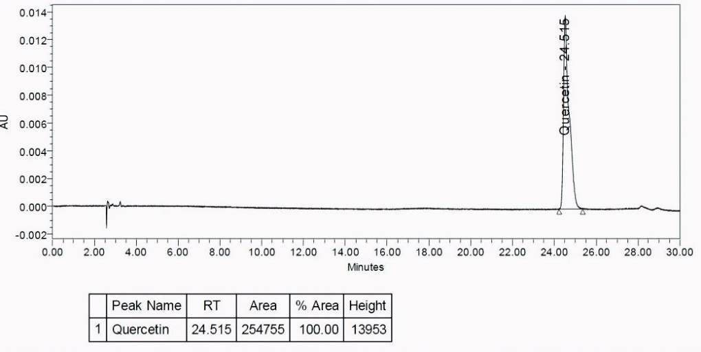
Figure 1. RP-HPLC chromatogram of quercetin standard, showing the quercetin peak at 24.515 min.
Preprints 223006 g001
Figure 2. RP-HPLC chromatogram of EECE, showing the quercetin peak at 23.793 min.
Preprints 223006 g002
Figure 3. UHPLC chromatogram of EECE (T = 40 °C; flow rate = 0.3 mL min⁻¹; sample: 1 mg mL⁻¹ in MeOH 100%; injection volume: 3 µL).
Preprints 223006 g003
Figure 4. Venn diagram showing the intersection between EECE-related targets and PUD–associated targets retrieved from the GeneCards database. A total of 85 common targets were identified and subjected to subsequent network pharmacology analysis.
Preprints 223006 g004
Figure 5. Protein–protein interaction (PPI) network of the intersecting targets constructed using the STRING database. Nodes represent proteins, and edges indicate protein–protein interactions.
Preprints 223006 g005
Figure 6. Protein–protein interaction (PPI) network of the intersecting targets constructed using the STRING database. Nodes represent proteins, and edges indicate protein–protein interactions.
Preprints 223006 g006
Figure 7. Bar chart of enriched Gene Ontology (GO) terms and biological pathways based on the intersecting targets. Bars are ranked by −log10(p), with higher values indicating greater statistical significance.
Preprints 223006 g007
Figure 8. Relative gastric weight of rats with ethanol-induced gastric ulcer (n=3). Values are expressed as mean ± SD (n = 3). Statistical analysis was performed using the Kruskal–Wallis test followed by the Mann–Whitney U test for post hoc pairwise comparisons.
Preprints 223006 g008
Figure 9. Ulcer area in rats with ethanol-induced gastric ulcer (n=3). Values are expressed as mean ± SD (n = 3). Statistical analysis was performed using the Kruskal–Wallis test followed by the Mann–Whitney U test for post hoc pairwise comparisons. * indicates a statistically significant difference compared with the negative control group (p < 0.05).
Preprints 223006 g009
Figure 10. Ulcer diameter of rats with ethanol-induced gastric ulcer (n=3). Values are expressed as mean ± SD (n = 3). Statistical analysis was performed using the Kruskal–Wallis test followed by the Mann–Whitney U test for post hoc pairwise comparisons. * indicates a statistically significant difference compared with the negative control group (p < 0.05).
Preprints 223006 g010
Figure 11. Ulcer index of rats with ethanol-induced gastric ulcer (n=3). Values are expressed as mean ± SD (n = 3). Statistical analysis was performed using the Kruskal–Wallis test followed by the Mann–Whitney U test for post hoc pairwise comparisons. * indicates a statistically significant difference compared with the negative control group (p < 0.05).
Preprints 223006 g011
Figure 12. Percentage protection of rats with ethanol-induced gastric ulcer (n=3).
Preprints 223006 g012
Figure 13. Macroscopic evaluation of gastric anti-ulcer effects. (a) Normal group; (b) negative control; (c) positive control treated with sucralfate; (d) positive control treated with quercetin; (e) EECE (200 mg/kg); (f) EECE (400 mg/kg); and (g) EECE (800 mg/kg).
Preprints 223006 g013
Figure 14. Histopathological examination of gastric tissue in experimental rats (100×). Representative hematoxylin and eosin (H&E)–stained sections of gastric mucosa showing (a) normal control with intact epithelial lining, (b) ethanol-induced group, (c) sucralfate-treated group, (d) quercetin-treated group, and (e–g) EECE-treated groups. Scale bar = 100 µm.
Preprints 223006 g014
Figure 15. (a) Representative Western blot of NF-κB p65 expression. Representative Western blot bands showing the expression of NF-κB p65 protein in gastric tissues from the different experimental groups following ethanol-induced gastric injury; (b) Quantification of NF-κB p65 protein expression. β-Actin was used as the internal loading control. Note: 1, Normal; 2, Negative; 3, Sucralfate; 4, Quercetin; 5, EECE 200 mg/kg; 6, EECE 400 mg/kg; 7, EECE 800 mg/kg.
Preprints 223006 g015
Table 1. Proximate composition of C. esculenta crude dried corm powder and EECE.
Proximate composition Content (%)
C. esculentacorm powder Ethanol extract of
C. esculentacorm
Moisture 9.61 28.93
Ash 2.94 44.22
Lipid 0.51 8.56
Protein 5.82 7.27
Carbohydrate 81.12 11.02
Table 2. Major metabolite classes identified in EECE by UHPLC–HRMS/MS analysis.
Metabolite class Metabolite name Relative abundance (%)
Amino sugars and derivatives 1-[(3-Carboxypropyl) amino]-1-deoxy-β-D-fructofuranose,
N-acetyl-D-quinovosamine,
N-acetylglucosaminitol
1.5–10
Conjugated amino acids Prolyl leucine, phenyl-propanoic acid derivatives 0.2–10
Lipids and fatty amides Oleamide, stearamide,
hexadecanamide, 1-stearoylglycerol
0.4–3.4
Nucleosides and purines Adenosine, adenine, ribofuranosyl thymine 0.9–2.2
Phenolic acid and indole derivatives Trans-3-indoleacrylic acid;
5-aminosalicyluric acid
1.0–1.4
Cyanogenic glycosides (minor) Lotaustralin ~0.8
Table 3. Quantitative assessment of gastric ulcer indices and protection percentage following EECE treatment (n = 3).
Preprints 223006 i001
Abbreviations: EECE, ethanolic extract of Colocasia esculenta; SD, standard deviation. Data are presented as median (interquartile range), n = 3. Statistical analysis was performed using the Kruskal–Wallis test followed by the Mann–Whitney U test. A p-value < 0.05 was considered statistically significant compared with the negative control group.
Table 4. Histopathological parameters of gastric tissue in ethanol-induced rats.
Treatment group Ulcer score ± SD Gastric mucosal thickness (µm) ± SD Number of PMN cells ± SD
Normal Control 1.0 ± 0.8 1111.51 ± 136.73 2.4 ± 1.2*
Negative Control 6.0 ± 1.6 697.23 ± 91.41 24.1 ± 0.5
Sucralfate 1.9 ± 0.7 1002.04 ± 116.44 0.7 ± 0.1*
Quercetin 1.3 ± 0.7 1105.05 ± 41.84 7.1 ± 0.3*
EECE 200 mg/kg 3.3 ± 1.3 929.07 ± 38.32 10.6 ± 2.2*
EECE 400 mg/kg 2.5 ± 0.1 1054.01 ± 35.69 10.2 ± 1.6*
EECE 800 mg/kg 1.8 ± 1.0 1061.71 ± 17.29 5.3 ± 0.9*
Abbreviations: EECE, ethanolic extract of Colocasia esculenta; PMN, Polymorphonuclear; SD, standard deviation. Data are presented as median (interquartile range). Statistical analysis was performed using the Kruskal–Wallis test followed by the Mann–Whitney U test. A p-value < 0.05 was considered statistically significant compared with the negative control group.
Disclaimer/Publisher’s Note: The statements, opinions and data contained in all publications are solely those of the individual author(s) and contributor(s) and not of MDPI and/or the editor(s). MDPI and/or the editor(s) disclaim responsibility for any injury to people or property resulting from any ideas, methods, instructions or products referred to in the content.
Copyright: This open access article is published under a Creative Commons CC BY 4.0 license, which permit the free download, distribution, and reuse, provided that the author and preprint are cited in any reuse.
Prerpints.org logo

Preprints.org is a free preprint server supported by MDPI in Basel, Switzerland.

Subscribe

© 2026 MDPI (Basel, Switzerland) unless otherwise stated

Accessibility

Disclaimer

Terms of Use

Privacy Policy

Privacy Settings