

Submitted:
11 July 2026
Posted:
14 July 2026
You are already at the latest version
Abstract
Background/Objectives: The Head Heave Test (HHT) was proposed approximately 25 years ago for the study of the linear translational vestibulo-ocular reflex (TVOR) of utricular origin. The exam consists of delivering a series of sudden, unpredictable movements along the interaural axis of the patient's head, with the patient asked to focus on the examiner's nose in front of them. It is considered abnormal if a clear corrective saccade is observed, visible only on one side, or significantly greater on one side compared to the other. The use of HHT has not become widespread due to the difficulties in assessing corrective saccades with the naked eye. The aim of this study is to propose a simple-to-use instrumental method that clearly documents corrective saccades following translational head impulses. To exclude the possibility that compensatory saccades could be due to simultaneous stimulation of the ipsilateral Lateral Semicircular Canal (LSC), we also recorded Video Head Impulse Test (VHIT). Methods: For this purpose, 110 consecutive outpatients evaluated for balance disorders underwent HHT, which was recorded using a VHIT device. The Video Head Heave Test (VHHT), performed in this manner, enabled the easy identification of compensatory saccades. Results: The patients were divided in three groups: A) without compensatory saccades (50,9%), B) with TVOR compensatory saccades on both sides (23,6%), C) with a prevalence of saccades on one side (25,5%). We found that VHHT compensatory saccades were present in patients without LSC deficit on VHIT, and conversely, we found patients with marked LSC impairment on VHIT without compensatory saccades at VHHT. Conclusions: The detected TVOR compensatory saccades are independent of any concomitant dysfunction of the ipsilateral LSC. We propose a simple method for evaluating utricular function that does not require dedicated equipment and instead uses the VHIT instruments commonly used in vestibular diagnostics.
Keywords:
VHIT
; video head impulse test
; head heave test
; TVOR
; translational vestibulo-ocular reflex
; compensatory saccades
; utricular function
1. Introduction
The utricle is responsible for numerous reflexes in response to static linear forces (e.g., gravity) and dynamic linear forces (e.g., translational accelerations).
In response to translational accelerations along the interaural axis, the utricle generates a compensatory ocular response that moves the eyes in the horizontal plane opposite to the head movement: the Translational Vestibulo-Ocular Reflex (TVOR).
The TVOR would originate from the lateral part of the utricle, which is stimulated by ipsilateral linear accelerations, and the response would be conveyed via a polysynaptic pathway to the contralateral abducens nucleus [1,2].
The TVOR has been studied in the laboratory using various methods (whole-body linear accelerations, centrifugal stimulation with the head in an eccentric position, and head translations); the easiest to reproduce in a clinical setting is lateral head translation.
Unlike angular movements, translational movements of the head do not cause retinal slip of a distant target (parallel lines meet at infinity). Linear compensatory eye movements are necessary only when the targets are close to the subject, and they are larger the closer the target is; therefore, tests for the TVOR are performed with the target approximately 15 cm from the patient [3,4].
Under these conditions, after a translational head impulse, a compensatory reflex eye movement appears (in the direction opposite to the head movement) with a latency of about 30ms, the magnitude of which is physiologically insufficient to keep the eyes on the target.
The average gain of the TVOR (relative to the ideal eye movement for compensation) in the healthy subject is insufficient, resulting in the need for a compensatory saccade to return to the target, which appears around 150ms after the onset of head movement, with any subsequent saccades occurring after the end of the head movement (about 300ms) [5].
After labyrinthectomy, an asymmetric response to impulsive translational stimuli is observed, with the decrease in reflexive eye velocity becoming more pronounced the closer the visual target is [2].
Within 3 months of labyrinthine damage, an asymmetry in response to impulses ipsilateral to the damaged labyrinth may be observed; this asymmetry disappears over time [1].
It is possible that the amplitude of the first corrective saccade could be a reliable index of utricular functional asymmetry [5].
Clinical Head Heave Test (HHT) is based on the visual detection of a clear compensatory saccade following a manually administered translational head impulse, with the operator standing in front of the patient, who is asked to fixate on the operator's nose. This test has been studied, together with the Head Impulse Test (HIT), in patients with Superior Vestibular Neuritis (SVN) in the acute phase. A significantly worse prognosis (assessed by recovery of labyrinthine function measured by the caloric test at 12 months) was observed in patients with both HHT and HIT positive; conversely, a non-pathological HHT was associated with a higher likelihood of functional recovery [6].
Bedside HHT can be difficult to interpret for several reasons: the TVOR is physiologically deficient, so most normal subjects show small corrective saccades in both directions of head movement.
Moreover, HHT depends on an individual's ability to focus attention and converge on a near target (the examiner's nose), and older individuals may have difficulty converging.
Ultimately, adaptation to the unilateral loss of utricular function occurs more rapidly than adaptation to the unilateral loss of lateral semicircular canal function [6].
From January 2024 to August 2025, 110 patients underwent HHT, with their head and eye movements recorded using a Video Head Impulse Test (VHIT) device; we called this the Video Head Heave Test (VHHT). The VHIT device we used employs a high-resolution, high-frame-rate camera positioned in front of the patient to measure head and eye velocities directly from video sequence analysis, without using accelerometers on the patient’s head.
The device measures head and eye movements, recording their respective velocities, whose curves are then superimposed. This clearly makes any refixation saccades visible as peaks in the eye velocity trace.
The calculation of the actual gain of the TVOR, understood as the ratio between the measured eye velocity and that of the ideal eye movement for compensation, is complex and cannot be automatically obtained with the VHIT device we used, which, instead, provides a gain measurement obtained simply by taking the ratio between the head and eye velocities.
This measurement, which we will refer to as Translational Gain (TG), is not comparable in absolute terms across subjects because it depends on the target's distance and eccentricity [1].
In clinical practice, the aspect of TVOR most readily studied is the compensatory saccade, whose presence and amplitude can be readily assessed using the VHHT.
For the evaluation of the results of this test, we therefore relied solely on the observation of compensatory saccades following the impulsive horizontal movement, as documented on the velocity trace.
In a normal subject, as reported in the literature, saccades may be absent or present bilaterally. Given this premise, the absence of a compensatory saccade indicates a normal reflex in both labyrinths, whereas its bilateral presence is not, in itself, indicative of a pathological condition.
Only the presence of compensatory saccades on one side alone or of clearly asymmetric amplitude indicates a unilateral TVOR deficiency.
This test cannot reveal symmetrical bilateral deficits of the TVOR.
A limitation of the HHT lies in the possibility of introducing head torsions during the translational impulse, which can stimulate the angular VOR and produce reflex eye movements of canal origin that may interfere with the TVOR. Although this interference can never be completely eliminated in the clinical test, the manual administration of the lateral translational impulse test remains valid as a bedside test because it has been found to be minimally contaminated by rotational components [5].
The aim of this study is to identify TVOR compensatory saccades with VHHT and to verify their possible relation with LSC deficit.
2. Materials and Methods
From January 2024 to August 2025, 110 patients with vestibular symptoms were initially evaluated with a thorough medical history and a careful bedside clinical vestibular examination. For each patient, a diagnostic hypothesis was proposed. In all patients, VHIT and VHHT were performed.
The VHHT was recorded using the VHIT device from Synapsys (Marseille, France), running the Video Head Impulse Test Ulmer II software, which is marketed by Inventis (Padova, Italy).
The patient is seated 90 cm from the camera, and a target is positioned 15 cm in front of the eyes (without glasses) at the level of the glabella, as shown in Photo 1.
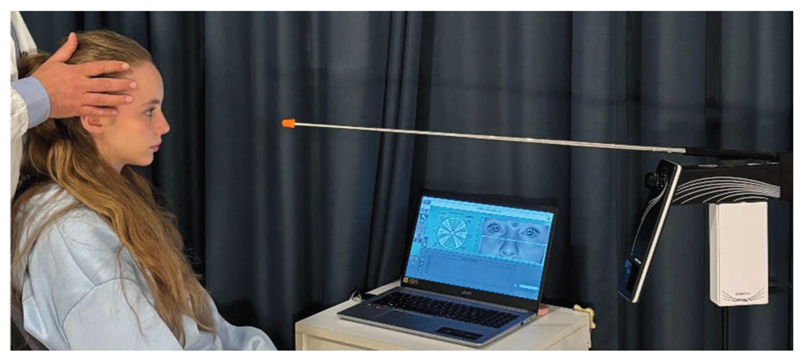
Photo 1. Patient position and proximal visual target.
The examiner, positioned behind the patient, places his hands on either side of the patient's head and imparts abrupt transverse movements (to the right and left) of approximately 5 cm to the patient's head in an unpredictable manner, acquiring at least 5 sequences for each side, taking particular care to avoid applying any torsion to the head on both the horizontal and frontal planes. (Photo 2)
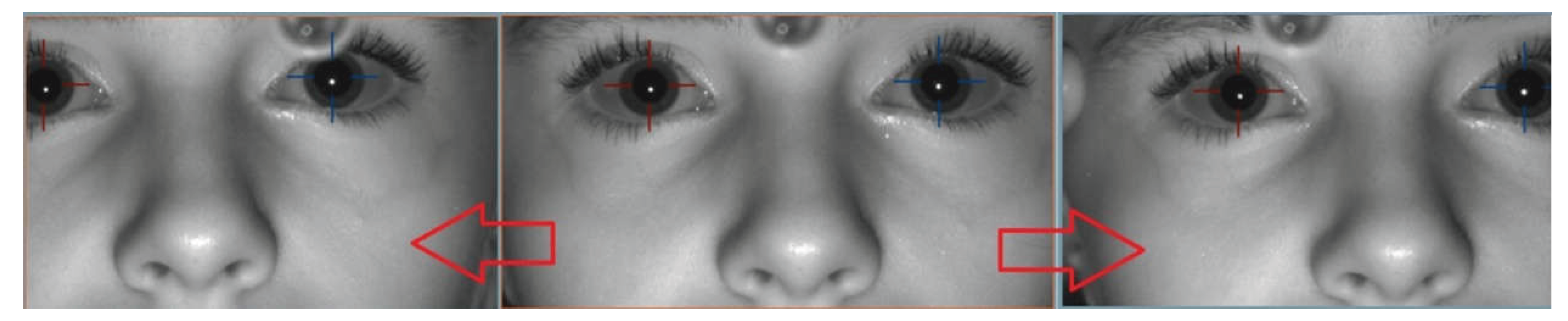
Photo 2. Patient's head transverse movements.
The VHIT device used is controlled by a PC, and the patient's head movement is continuously displayed on the screen. Therefore, the examiner can immediately assess the correctness of the movement by visually checking whether any rotations have been applied to the head.
The possibility of constant visual monitoring of the movements of the patient's head allows for a drastic reduction in torsional components, making any potential measurement errors in the TVOR due to the activation of the SCs negligible.
The acquisition window has been set to 600ms, and saccades have been identified on the velocity trace.
Lacking references, we considered arbitrarily the responses asymmetric when the number of refixation saccades observed was at least twice that on the other side, or when the saccade amplitude was double that on the other side.
Each patient, immediately before the VHHT, underwent a VHIT for all SCs using the same equipment mentioned earlier. For the VHIT, the distance from the camera is the same (90 cm) while the target is placed 2 meters away, at the patient's eye level; the examiner, always positioned behind the patient, imparts small, unpredictable angular impulsive movements to the patient's head on the horizontal plane, for the study of the LSCs, and on the oblique planes, (Left Anterior/Right Posterior and Right Anterior/Left Posterior) for the study of the vertical SCs.
According to the manufacturer's specifications, the angular VOR gain on the VHIT is considered pathological if it is below 0.85 for the LSC and below 0.75 for the Anterior Semicircular Canal (ASC) and Posterior Semicircular Canal (PSC).
The overall duration of the examination (VHIT + VHHT), performed by an experienced examiner, is only a few minutes (on average, 5 minutes).
Based on the results of the VHHT, the patients were divided into three groups: Group A, in which no compensatory saccades were observed in any direction of the stimulus; Group B, in which saccades were detected in both directions, without a significant difference between the two sides; Group C, with saccades present or predominant on only one side.
For each patient, we considered the following parameters, which are also reported in the results tables:
- 1)
- The absolute value of the Translational Gain (TG) for each side
- 2)
- The TG difference between the two sides is indicated as Absolute Translational Gain Difference (ATGD) (ATGD = prevailing side TG – minority side TG).
- 3)
- The TG difference between the two sides as a percentage of the overall response, indicated as Relative Translational Gain Difference (RTGD) (RTGD = 100 X ATGD/ (right TG + left TG)).
- 4)
- The VHIT gain of each Lateral Semicircular Canal (LCS)
- 5)
- The Relative Canal Gain Difference between the two LSCs (RCGD) evaluated at VHIT (RCGD = 100 X (prevailing LSC gain – minority LSC gain) / (LSC gain right+left)
3. Results
Group A (without TVOR compensatory saccades): 56 cases; 30 females and 26 males, aged between 18 and 85 years (mean 55) (Table A).
They exhibited a TG ranging from 0.92 to 1.29, with a mean of 1.07 and a standard deviation of 0.06. The ATGD between the two sides ranged from 0 to 0.15 (mean 0.04 ± 0.03), and the RTGD ranged from 0 to 7.4% (mean 1.7% ± 1.4).
The concomitant study of angular VOR with VHIT revealed unilateral deficits in one or both LSCs in 21 patients, with asymmetric impairment (RCGD > 10%) in 8.
Among these, 4 had RCGD > 30%: 1 Acute Unilateral Vestibular Deficit (AUVD) involving only the LSC; 1 had a three-canal deficit as an outcome of Temporal Bone Fracture (TBF), and 2 outcomes of SVN.
Another 4 patients had RCGD between 10-30%, all with isolated LSC deficit: 1 was in the acute phase (AUVD), 2 were outcomes of vestibulopathy of unknown origin (UNK), and 1 had Vestibular Migraine (VM) not in the ictal phase.
The resting 48 patients had RCGD below 10%: 15 UNK, 12 Benign Paroxysmal Positional Vertigo (BPPV), 7 VM, 6 Age-Related Vestibulopathy (ARV), 4 Meniére Disease (MD), 2 Central Nervous System Vestibulopathy (CNSV), 1 Anxious disturbance (ANX), 1 Otosclerosis (OTO).
Among these, 4 patients had unilateral isolated PSC impairment: 3 BPPV, 1VM.
Group B (with bilateral TVOR compensatory saccades): 26 cases; 15 females and 11 males, aged between 25 and 78 years (mean 58) (Table B).
The VHIT revealed a deficit of one or both LSC in 12 cases, with asymmetric impairment (RCGD > 10%) in only 3. Among these, 2 had RCGD > 30%; both were outcomes of AUVD (1 with a three-canals deficit, 1 with an isolated LSC deficit). 1 patient with RCGD of 10-30% had LSC together with PSC acute impairment (AUVD).
The remaining 23 patients had RCGD below 10%; among these, 7 had unilateral isolated PSC impairment (5 BPPV, 2 UNK), and 1 had isolated ASC impairment (BPPV).
15 patients had a normal VHIT (4 UNK, 2 MD, 2 VM, 4 BPPV, 1 Head Trauma (HT), 1 CNSV).
Subjects in group B showed a TG ranging from 0.9 to 1.29, with a mean of 1.08 (+/- 0.08). The ATGD ranged from 0 to 0.14 (mean 0.03 +/- 0.3), and the RTGD ranged from 0 to 5.74% (mean 0,68% +/- 1.58) while RCGD is highly variable, with a mean of 5.87% (+/- 9,3).
Group C (with unilateral TVOR compensatory saccades): 28 cases; 16 females and 12 males, aged between 18 and 80 years, average age 62 (Table C).
The VHIT revealed a concomitant deficit involving one or both LSC in 16 cases. In 10 of these, the LSC gain reduction exceeded 30%: 2 were outcomes of Intra-Tympanic Gentamicin treatment for Ménière's disease (ITG), 4 were acute SVN, and 4 were AUVD (1 involving the horizontal and posterior semicircular canals, 3 involving all three canals). One case of three-canal AUVD presented as a six-canal deficit with asynchronous onset, with a more recent (3-month) occurrence of labyrinthine damage on the side ipsilateral to the compensatory TVOR saccade. All these patients, had the compensatory TVOR saccade on the same side as the greater damage of the LSC.
Further 6 patients had RCGD below 10%: 4 BPPV, 1 VM, 1 ARV; TVOR compensatory saccade was found on both side (3 ipsilateral to the LSC deficit, 3 contralateral).
In 3 other cases, there was an isolated deficit of the PSC, on the same side of the TVOR saccades (1 BPPV, 1 VM, 1 UNK).
In 9 cases, the VHIT did not show canal deficits at VHIT (6 BPPV, 2 VM, 1 UNK).
Subjects in group C have a gain (TG) on the normal side in the VHHT ranging from 0.99 to 1.34 (mean 1.1 ± 0.1) and on the side of the TVOR saccade ranging from 0.72 to 1.22 (mean 1.02 ± 0.1). The absolute difference (ATGD) in TVOR gain between the two sides ranges from 0 to 0.36 (mean 0.08 ± 0.1), and the relative difference (RTGD) ranges from 0 to 19.1% (mean 4.03% ± 4.5).
In patients of group C, RCGD is highly variable, with a mean of 29.2% (+/- 41.8).
Table A
| A | age | sex | Diagnose | TG> | TG< | ATGD | RTGD | LSC> | LSC< | RCGD |
| 1 | 79 | M | UNK | 1,07 | 1,01 | 0,06 | 2,88 | 0,95 | 0,94 | 0,53 |
| 2 | 82 | M | UNK | 0,98 | 0,97 | 0,01 | 0,51 | 0,68 | 0,60 | 6,25 |
| 3 | 80 | M | ARV | 1,02 | 1,01 | 0,01 | 0,49 | 0,95 | 0,90 | 2,70 |
| 4 | 72 | F | UNK | 1,04 | 0,99 | 0,05 | 2,46 | 0,93 | 0,75 | 10,71 |
| 5 | 73 | F | UNK | 0,93 | 0,92 | 0,01 | 0,54 | 0,70 | 0,39 | 28,44 |
| 6 | 62 | F | VM | 1,12 | 1,07 | 0,05 | 2,28 | 0,88 | 0,72 | 10,00 |
| 7 | 65 | M | AUVD | 1,05 | 1,04 | 0,01 | 0,48 | 0,83 | 0,37 | 38,33 |
| 8 | 62 | F | ARV | 1,12 | 1,11 | 0,01 | 0,45 | 0,87 | 0,81 | 3,57 |
| 9 | 59 | M | BPPV | 1,04 | 1,03 | 0,01 | 0,48 | 0,93 | 0,88 | 2,76 |
| 10 | 70 | F | BPPV | 1,07 | 1,04 | 0,03 | 1,42 | 0,96 | 0,91 | 2,67 |
| 11 | 48 | M | BPPV | 1,11 | 1,10 | 0,01 | 0,45 | 0,95 | 0,94 | 0,53 |
| 12 | 77 | M | VM | 1,02 | 1,00 | 0,02 | 0,99 | 0,94 | 0,89 | 2,73 |
| 13 | 62 | F | BPPV | 1,00 | 1,00 | 0,00 | 0,00 | 0,91 | 0,82 | 5,20 |
| 14 | 65 | M | TBF | 1,08 | 1,07 | 0,01 | 0,47 | 0,85 | 0,05 | 88,89 |
| 15 | 77 | F | SVN | 1,01 | 0,94 | 0,07 | 3,59 | 0,66 | 0,21 | 51,72 |
| 16 | 75 | M | AUVD | 1,05 | 1,00 | 0,05 | 2,44 | 0,84 | 0,59 | 17,48 |
| 17 | 52 | F | UNK | 1,14 | 1,10 | 0,04 | 1,79 | 1,08 | 1,08 | 0,00 |
| 18 | 18 | M | UNK | 1,16 | 1,15 | 0,01 | 0,43 | 0,94 | 0,91 | 1,62 |
| 19 | 51 | M | UNK | 1,14 | 1,09 | 0,05 | 2,24 | 0,94 | 0,88 | 3,30 |
| 20 | 39 | M | MD | 1,07 | 1,05 | 0,02 | 0,94 | 1,04 | 0,91 | 6,67 |
| 21 | 44 | M | MD | 1,07 | 1,00 | 0,07 | 3,38 | 1,05 | 0,92 | 6,60 |
| 22 | 61 | F | UNK | 1,01 | 0,98 | 0,03 | 1,51 | 0,95 | 0,91 | 2,15 |
| 23 | 45 | F | VM | 1,03 | 0,96 | 0,07 | 3,52 | 0,83 | 0,81 | 1,22 |
| 24 | 21 | M | ANX | 1,13 | 1,10 | 0,03 | 1,35 | 0,91 | 0,90 | 0,55 |
| 25 | 59 | F | VM | 1,10 | 1,10 | 0,00 | 0,00 | 1,11 | 1,00 | 5,21 |
| 26 | 51 | M | UNK | 1,14 | 1,11 | 0,03 | 1,33 | 1,03 | 0,99 | 1,98 |
| 27 | 36 | F | UNK | 1,17 | 1,12 | 0,05 | 2,18 | 1,01 | 1,00 | 0,50 |
| 28 | 64 | F | UNK | 1,10 | 1,06 | 0,04 | 1,85 | 0,98 | 0,86 | 6,52 |
| 29 | 55 | M | BPPV | 1,29 | 1,19 | 0,10 | 4,03 | 0,86 | 0,81 | 2,99 |
| 30 | 32 | F | VM | 1,05 | 1,01 | 0,04 | 1,94 | 0,89 | 0,87 | 1,14 |
| 31 | 38 | F | UNK | 1,14 | 1,08 | 0,06 | 2,70 | 0,93 | 0,92 | 0,54 |
| 32 | 63 | F | UNK | 1,08 | 1,06 | 0,02 | 0,93 | 0,91 | 0,90 | 0,55 |
| 33 | 60 | M | UNK | 1,09 | 1,05 | 0,04 | 1,87 | 0,92 | 0,92 | 0,00 |
| 34 | 31 | M | UNK | 1,15 | 1,11 | 0,04 | 1,77 | 0,96 | 0,93 | 1,59 |
| 35 | 40 | F | OTO | 1,15 | 1,12 | 0,03 | 1,32 | 0,97 | 0,96 | 0,52 |
| 36 | 74 | F | UNK | 1,12 | 1,07 | 0,05 | 2,28 | 1,05 | 1,04 | 0,48 |
| 37 | 33 | M | UNK | 1,18 | 1,12 | 0,06 | 2,61 | 1,01 | 0,98 | 1,51 |
| 38 | 60 | M | UNK | 1,13 | 1,08 | 0,05 | 2,26 | 0,94 | 0,87 | 3,87 |
| 39 | 40 | M | MD | 1,14 | 1,09 | 0,05 | 2,24 | 0,95 | 0,84 | 6,15 |
| 40 | 42 | F | VM | 1,05 | 1,02 | 0,03 | 1,45 | 0,93 | 0,91 | 1,09 |
| 41 | 52 | F | VM | 1,13 | 1,13 | 0,00 | 0,00 | 0,94 | 0,89 | 2,73 |
| 42 | 32 | F | VM | 1,05 | 1,04 | 0,01 | 0,48 | 1,01 | 0,97 | 2,02 |
| 43 | 74 | M | UNK | 1,03 | 1,01 | 0,02 | 0,98 | 0,78 | 0,77 | 0,65 |
| 44 | 24 | F | MD | 1,15 | 1,13 | 0,02 | 0,88 | 0,99 | 0,94 | 2,59 |
| 45 | 48 | F | BPPV | 1,14 | 1,06 | 0,08 | 3,64 | 1,03 | 0,99 | 1,98 |
| 46 | 40 | F | BPPV | 1,12 | 1,11 | 0,01 | 0,45 | 0,94 | 0,84 | 5,62 |
| 47 | 56 | F | BPPV | 1,05 | 1,02 | 0,03 | 1,45 | 1,01 | 1,00 | 0,50 |
| 48 | 64 | F | BPPV | 1,01 | 0,93 | 0,08 | 4,12 | 0,82 | 0,82 | 0,00 |
| 49 | 54 | M | SVN | 1,14 | 1,12 | 0,02 | 0,88 | 1,04 | 0,20 | 67,74 |
| 50 | 32 | M | CNSV | 1,04 | 1,03 | 0,01 | 0,48 | 1,03 | 1,02 | 0,49 |
| 51 | 70 | F | ARV | 0,99 | 0,99 | 0,00 | 0,00 | 0,73 | 0,65 | 5,80 |
| 52 | 79 | F | BPPV | 1,03 | 1,00 | 0,03 | 1,48 | 1,00 | 0,93 | 3,63 |
| 53 | 73 | M | ARV | 1,06 | 1,02 | 0,04 | 1,92 | 0,79 | 0,77 | 1,28 |
| 54 | 85 | F | ARV | 1,06 | 0,98 | 0,08 | 3,92 | 0,92 | 0,82 | 5,75 |
| 55 | 75 | M | BPPV | 1,09 | 0,94 | 0,15 | 7,39 | 0,82 | 0,77 | 3,14 |
| 56 | 19 | F | BPPV | 1,15 | 1,08 | 0,07 | 3,14 | 0,94 | 0,91 | 1,62 |
Table B
| B | age | sex | Diagnose | TG> | TG< | ATGD | RTGD | LSC> | LSC< | RCGD |
| 1 | 58 | F | BPPV | 1,18 | 1,16 | 0,02 | 0,85 | 1,01 | 0,96 | 2,54 |
| 2 | 43 | F | AUVD | 1,13 | 1,08 | 0,05 | 2,26 | 1,01 | 0,79 | 12,22 |
| 3 | 66 | M | UNK | 1,10 | 1,01 | 0,09 | 4,27 | 0,70 | 0,67 | 2,19 |
| 4 | 52 | F | HT | 1,26 | 1,20 | 0,06 | 2,44 | 0,93 | 0,83 | 5,68 |
| 5 | 59 | M | UNK | 1,05 | 1,02 | 0,03 | 1,45 | 0,83 | 0,70 | 8,50 |
| 6 | 37 | M | MD | 1,26 | 1,24 | 0,02 | 0,80 | 0,98 | 0,97 | 0,51 |
| 7 | 65 | F | ARV | 1,01 | 1,00 | 0,01 | 0,50 | 0,93 | 0,87 | 3,33 |
| 8 | 78 | M | BPPV | 1,05 | 1,04 | 0,01 | 0,48 | 0,91 | 0,82 | 5,20 |
| 9 | 73 | M | AUVD | 1,06 | 1,03 | 0,03 | 1,44 | 1,61 | 0,78 | 34,73 |
| 10 | 64 | F | BPPV | 1,03 | 0,99 | 0,04 | 1,98 | 0,84 | 0,76 | 5,00 |
| 11 | 73 | M | BPPV | 1,04 | 1,00 | 0,04 | 1,96 | 0,81 | 0,76 | 3,18 |
| 12 | 53 | F | BPPV | 1,06 | 1,04 | 0,02 | 0,95 | 0,91 | 0,81 | 5,81 |
| 13 | 69 | F | BPPV | 1,05 | 1,02 | 0,03 | 1,45 | 1,06 | 0,99 | 3,41 |
| 14 | 67 | F | BPPV | 1,04 | 1,00 | 0,04 | 1,96 | 0,86 | 0,85 | 0,58 |
| 15 | 64 | F | UNK | 1,08 | 1,04 | 0,04 | 1,89 | 0,99 | 0,95 | 2,06 |
| 16 | 69 | F | BPPV | 0,90 | 0,96 | -0,06 | -3,23 | 0,98 | 0,94 | 2,08 |
| 17 | 57 | F | AUVD | 0,99 | 0,96 | 0,03 | 1,54 | 0,88 | 0,40 | 37,50 |
| 18 | 35 | F | VM | 1,16 | 1,16 | 0,00 | 0,00 | 0,99 | 0,95 | 2,06 |
| 19 | 49 | M | MD | 1,17 | 1,16 | 0,01 | 0,43 | 1,02 | 1,00 | 0,99 |
| 20 | 63 | M | UNK | 1,18 | 1,17 | 0,01 | 0,43 | 0,98 | 0,97 | 0,51 |
| 21 | 59 | F | VM | 1,15 | 1,10 | 0,05 | 2,22 | 0,93 | 0,91 | 1,09 |
| 22 | 61 | M | BPPV | 1,05 | 1,01 | 0,04 | 1,94 | 0,96 | 0,84 | 6,67 |
| 23 | 36 | F | BPPV | 1,11 | 1,11 | 0,00 | 0,00 | 1,02 | 1,01 | 0,49 |
| 24 | 25 | M | UNK | 1,29 | 1,15 | 0,14 | 5,74 | 0,88 | 0,82 | 3,53 |
| 25 | 61 | M | CNSV | 1,05 | 1,04 | 0,01 | 0,48 | 0,90 | 0,87 | 1,69 |
| 26 | 77 | F | UNK | 1,08 | 1,07 | 0,01 | 0,47 | 0,96 | 0,94 | 1,05 |
Table C
| C | age | sex | Diagnose | TG> | TG< | ATGD | RTGD | LSC< | LSC> | RCGD |
| 1 | 80 | F | ARV | 1,14 | 1,01 | 0,13 | 6,05 | 0,63 | 0,55 | 6,78 |
| 2 | 79 | F | BPPV | 1,09 | 1,02 | 0,07 | 3,32 | 0,76 | 0,79 | -1,94 |
| 3 | 59 | F | AUVD | 0,99 | 0,93 | 0,06 | 3,13 | 0,70 | 0,01 | 97,18 |
| 4 | 73 | M | BPPV | 1,02 | 1,00 | 0,02 | 0,99 | 0,89 | 0,92 | -1,66 |
| 5 | 68 | F | VM | 1,11 | 1,07 | 0,04 | 1,83 | 0,89 | 0,96 | -3,78 |
| 6 | 59 | F | AUVD | 1,10 | 1,00 | 0,10 | 4,76 | 0,03 | 0,01 | 50,00 |
| 7 | 65 | F | VM | 1,10 | 1,02 | 0,08 | 3,77 | 0,83 | 0,72 | 7,10 |
| 8 | 43 | M | ITG | 1,16 | 1,03 | 0,13 | 5,94 | 0,74 | 0,03 | 92,21 |
| 9 | 60 | M | BPPV | 1,13 | 1,12 | 0,01 | 0,44 | 0,98 | 0,97 | 0,51 |
| 10 | 56 | F | VM | 1,06 | 1,02 | 0,04 | 1,92 | 0,93 | 0,91 | 1,09 |
| 11 | 68 | M | BPPV | 1,03 | 1,03 | 0,00 | 0,00 | 0,83 | 0,95 | -6,74 |
| 12 | 59 | M | BPPV | 1,06 | 1,03 | 0,03 | 1,44 | 0,93 | 0,86 | 3,91 |
| 13 | 73 | F | BPPV | 1,11 | 1,03 | 0,08 | 3,74 | 0,93 | 1,03 | -5,10 |
| 14 | 59 | F | BPPV | 1,11 | 1,06 | 0,05 | 2,30 | 0,96 | 0,88 | 4,35 |
| 15 | 51 | F | UNK | 1,13 | 1,07 | 0,06 | 2,73 | 0,98 | 0,96 | 1,03 |
| 16 | 37 | F | BPPV | 1,06 | 1,04 | 0,02 | 0,95 | 0,97 | 1,01 | -2,02 |
| 17 | 72 | F | AUVD | 1,11 | 0,97 | 0,14 | 6,73 | 0,78 | 0,31 | 43,12 |
| 18 | 78 | M | SVN | 1,02 | 0,98 | 0,04 | 2,00 | 0,64 | 0,01 | 96,92 |
| 19 | 63 | M | SVN | 1,31 | 0,95 | 0,36 | 15,93 | 0,71 | 0,01 | 97,22 |
| 20 | 38 | M | SVN | 1,20 | 0,99 | 0,21 | 9,59 | 0,91 | 0,01 | 97,83 |
| 21 | 61 | F | BPPV | 1,14 | 1,07 | 0,07 | 3,17 | 0,95 | 0,91 | 2,15 |
| 22 | 48 | F | BPPV | 1,13 | 1,11 | 0,02 | 0,89 | 0,74 | 0,83 | -5,73 |
| 23 | 68 | M | BPPV | 1,05 | 1,07 | -0,02 | -0,94 | 0,81 | 0,78 | 1,89 |
| 24 | 74 | F | SVN | 1,00 | 1,00 | 0,00 | 0,00 | 0,82 | 0,04 | 90,70 |
| 25 | 18 | M | UNK | 1,34 | 1,22 | 0,12 | 4,69 | 1,04 | 1,01 | 1,46 |
| 26 | 71 | M | BPPV | 1,02 | 0,96 | 0,06 | 3,03 | 0,95 | 0,86 | 4,97 |
| 27 | 71 | M | ITG | 1,06 | 0,72 | 0,34 | 19,10 | 0,75 | 0,27 | 47,06 |
| 28 | 77 | F | AUVD | 1,10 | 0,99 | 0,11 | 5,26 | 0,98 | 0,01 | 97,98 |
Table legend: TG> (TVOR Gain of the prevailing side); TG< (TVOR Gain of the minority side); ATGD (Absolute TVOR Gain Difference); RTGD (Relative TVOR Gain Difference); LSC> (VOR gain of the prevailing LSC); LSC< (VOR gain of the minority LSC); RCGD (Relative Canalar Difference in VOR gain between the two LSCs). BPPV (Benign Paroxysmal Positional Vertigo), UNK (unknown); VM (Vestibular Migraine); ANX (anxiety disorder), ARV (Age Related Vestibulopathy); SVN (Superior Vestibular Neuritis); CNSV (Central Nervous System Vestibulopathies); MD (Meniére Disease); AUVD (Acute Unilateral Vestibular Deficit); OTO (Otosclerosis); TBF (Temporal Bone Fracture), HT (Head Trauma), ITG (Intra Tympanic Gentamicin).
4. Discussion
There are two types of VHIT systems: the first uses a camera mounted on a mask worn by the patient to evaluate eye movement and accelerometers placed on the head to read head movement; the second uses a camera placed in front of the patient to measure both eye and head movement directly from the video input.
To study TVOR, we used a VHIT system with a distal camera, considering that direct measurement of head and eye movements from the same source (video) can reduce the risk of measurement errors (e.g. accelerometers calibrated to measure angular movement).
The VHIT system software used is designed to calculate the gain for a visual target located 2 meters away, imparting angular movements to the head. The angular VOR gain thus calculated in a normal subject tends to have a value equal to 1.
In our case, given the proximity of the target and the difference in the cephalic movement (linear translation rather than angular), the gain value provided by the device cannot serve as an adequate indication of the TVOR function, as it depends on the target's distance and eccentricity.
For TVOR, the actual gain (with comparable absolute value across subjects) is expressed as the ratio of the recorded eye movement to the eye movement that would ensure correct aim maintenance for that target distance. This data cannot be obtained directly from VHIT systems.
VHIT systems calculate angular VOR gain as the ratio between ocular and cephalic velocity of a distant target (2 meters), and, with our device, it is considered normal if greater than 0.85 for impulses on the horizontal plane and greater than 0.75 for those on the vertical planes. This data, automatically calculated by the device, is not an adequate index for assessing TVOR functionality when the target is close, as it is not directly comparable across subjects.
In our sample, the gain calculated by the device (TG) proved to be an extremely variable index across subjects, while it was useful for comparing reflexes between the two sides of the same subject, although this data cannot always correlate with the presence of corrective saccades.
The average TG values observed in the three groups are similar (A: 1.07; B: 1.08; C: 1.1), and the average RTGD between the two sides is 1.73% (+/- 1.38) in group A, 0.68% (+/-1.58) in group B and only slightly higher in group C, albeit with wide variability (4.05% +/- 4.5).
Such a small numerical difference and wide variability among the three groups make the RG assessment insufficiently sensitive to correctly identify subjects with impulsive TVOR asymmetry, whereas direct identification of the refixation saccade on the velocity trace clearly highlights the three conditions.
Considering the above, we can still consider the TVOR gain difference values between the two sides (ATGD and RTGD) to be fairly indicative for the individual patient.
In group A, 8 out of 56 subjects had a unilateral LSC deficit with significant asymmetric LSC impairment (RCGD exceeding 30% in 4 patients and between 10-30% in 4 more patients).
In group B, only 3 cases had an asymmetric LSC deficit (RCGD greater than 30% in 2 and between 10 and 30% in 1).
In Group C, compensatory saccades at VHHT are associated with significant asymmetry between LSC in 10 cases (all with RCGD greater than 30%), while in another 11 cases the relative hypofunction of the LSC on the affected side is less than 10% (0.5-7.1%) and in another 7 cases the LSC on the side with TVOR compensatory saccades was prevalent (1.9%-6.7%).
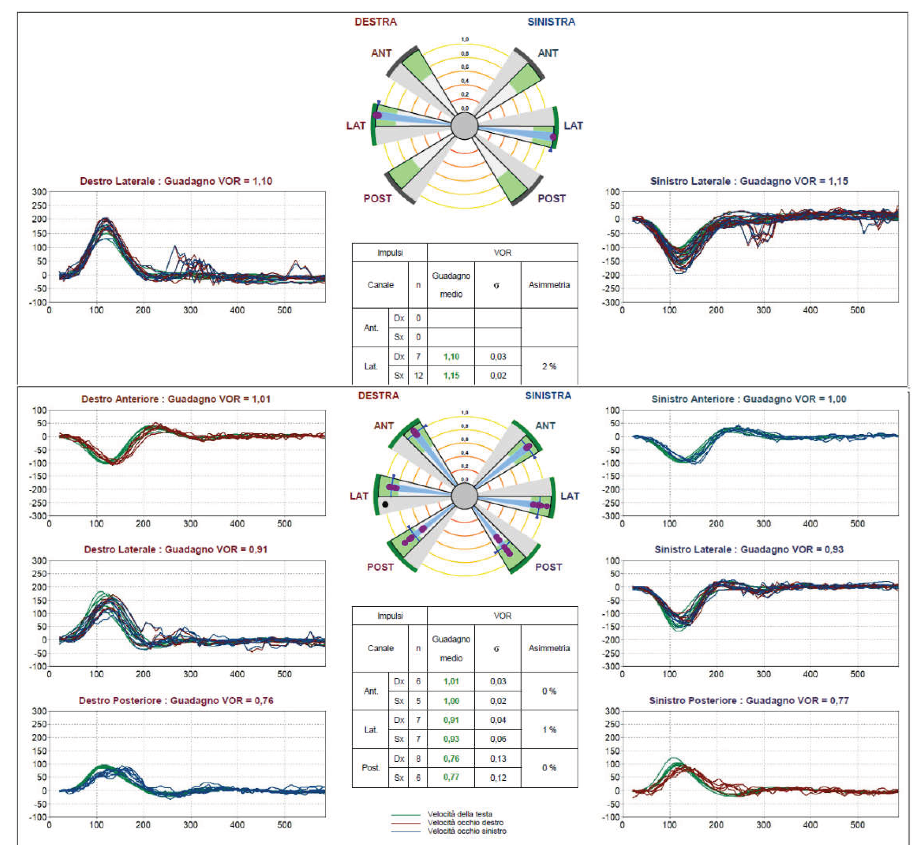
In conclusion, the compensatory saccade detected after translational impulse is not an expression of a deficit of the ipsilateral LSC (Figure 1).
In group C, out of a total of 26 cases, based on the diagnosis, and VHIT results (RCGD > 30%), indicating extensive labyrinth damage, an utricular deficit were expected in 10 cases (4 acute NVS, 2 acute complete VN, 1 acute asynchronous esacanalar deficit, 1 AUVD with impairment of LSC and PSC, 2 ITG) (Figure 2).
Other 3 patients have symmetrical LSC function and isolated PSC impairment (2 BPPV, 1 VM) (Figure 3).
In the remaining 15 cases, no asymmetric function of the semicircular canals was found on the VHIT, so a utricular deficit was unexpected.
In the remaining 15 cases, no VHIT asymmetries were found; among these 10 cases with BPPV, the TVOR saccade may reflect utricular damage responsible for otoconia detachment. In the remaining 5 cases (2 VM, 2 UNK, 1 ARV), positivity on VHHT reflects isolated utricular dysfunction without other clinical vestibular evidence (Figure 4).
In group A, a utricular deficit was expected in 4 patients, based on the diagnosis and VHIT results (1 AUVD with isolated LSC impairment, 1 TBF outcome, and 2 NVS outcomes). In these cases, the lack of saccades may reflect an actual dissociation between canalar and utricular damage, or it may be due to early recovery of macular function or reduced sensitivity of the method [1,6]. (Figure 5)
In group B, a possible utricular deficit was expected in 2 patients (both outcomes of prior AUVD: one with a three-canalar deficit, one with an isolated LSC deficit. (Figure 6)
According to the literature, TVOR is physiologically insufficient, so the finding of bilateral saccades on VHHT may reflect this normal condition, compensation for prior macular damage, or actual bilateral utricular failure (Figure 7)

VHHT does not allow for an assessment of the function of each single utricle, but only a comparison between the two sides. Therefore, it cannot highlight bilateral deficits, which cannot be ruled out in the case of bilateral saccades.
In group C, in 15 cases, VHHT positivity was not associated with significant unilateral damage at VHIT (RCGD <10% and absence of other SC-isolated deficits). Among these, BPPV (10) was the most common, VM (2) was less frequent, and 1 had ARV. In 2 cases, no diagnostic hypothesis was proposed (UNK); in these cases, VHHT positivity could reflect isolated idiopathic utricular damage unrelated to other vestibular pathologies.
The overall prevalence of VPPB across the three groups is as follows: group A, 12 cases (21%); group B, 10 (38%); and group C, 12 (43%). In our sample, BPPV is associated with concomitant utricular damage in approximately 35% of cases (12/34; groups A+B+C), consistent with the literature.
Indeed, in patients with BPPV studied with ocular Vestibular Evoked Myogenic Potentials (VEMPs), currently the gold standard for functional exploration of the utricle, an alteration in potential is found in highly variable percentages, ranging from 22% to 90%, depending on the criteria adopted for evaluating the results (absence of potential or asymmetry in amplitude) and the sample tested (first episode or recurrent), with a higher number of alterations found in patients with recurrent BPPV [8,9,10].
A total of 19 patients in all groups had extensive labyrinthine damage, in which utricular damage was be expected (10 AUVD, 6 NVS, 2 ITG, 1 TBF). Of these, 12 were evaluated within one month of the onset of labyrinthine damage (3 group A, 1 group B, 8 group C). Another 7 patients were seen at later stages (3 in group A, 1 in group B, 2 in group C).
Overall, 8/12 acute extensive labyrinthopathies were accompanied by unilateral TVOR saccades, while 5/7 chronic extensive labyrinthopathies did not present them.
These data are consistent with observations already reported in the literature, indicating that TVOR asymmetry is no longer measurable beyond the first 3 months after the onset of labyrinthine damage [1].
5. Conclusions
HHT is a clinical test that can detect functional asymmetries of the utricle. We suggest recording HHT using VHIT equipment (VHHT) to more easily highlight any compensatory saccades.
In our sample of 110 outpatients complaining of balance disorders, not selected for pathology, VHHT revealed asymmetrical TVOR compensatory saccades in 28 cases (25.4%), indicating a unilateral utricle deficit.
The detected compensatory saccades are independent of any concomitant dysfunction of the ipsilateral LSC.
The method we propose does not quantitatively measure TVOR gain but compares the two sides in the individual patient; therefore, it does not allow for the detection of bilateral utricular deficits.
VHHT is more sensitive in the acute phase of utricular damage.
The VHHT is a quick test of unilateral utricular dysfunction that does not require purchasing a dedicated device; instead, it uses a VHIT system, which is now widely used in outpatient clinical practice.
Author Contributions
Conceptualization and writing—original draft preparation, FdO, writing—review GN; editing, LN.; supervision, EA. All authors have read and agreed to the published version of the manuscript.
Funding
This research received no external funding.
Institutional Review Board Statement
: Ethical approval was not required for the studies involving humans because patients underwent routine vestibular testing in a private office setting. The studies were conducted in accordance with local legislation and institutional requirements. The research was conducted ethically, with all study procedures being performed in accordance with the requirements of the World Medical Association’s Declaration of Helsinki.
Informed Consent Statement
Written informed consent was obtained from each patient for study participation and data publication. All the patients authorised us to use the data and video recordings for scientific purposes.
Conflicts of Interest
The authors declare no conflicts of interest related to this study and no personal or financial relationships that could influence their work.
Abbreviations
The following abbreviations are used in this manuscript:
| HHT | Head Heave Test |
| VHHT | Video Head Heave Test |
| SC | Semicircular Canal |
| LSC | Lateral Semicircular Canal |
| ASC | Anterior Semicircular Canal |
| PSC | Posterior Semicircular Canal |
| VOR | Vestibulo-Ocular Reflex |
| TVOR | Translational Vestibulo-Ocular Reflex |
| HIT | Head Impulse Test |
| VHIT | Video Head Impulse Test |
| SVN | Superior Vestibular Neuritis |
| VN | Vestibular Neuritis |
| TG | Translational Gain |
| ATGD | Absolute Translational Gain Difference |
| RTGD | Relative Translational Gain Difference |
| RCGD | Relative Canal Gain Difference between the two LSCs |
| TBF | Temporal Bone Fracture |
| AUVD | Acute Unilateral Vestibular Deficit |
| UNK | vestibulopathy of unknown origin |
| VM | Vestibular Migraine |
| BPPV | Benign Paroxysmal Positional Vertigo |
| ARV | Age-Related Vestibulopathy |
| MD | Meniére Disease |
| CNSV | Central Nervous System Vestibulopathy |
| ANX | Anxious disturbance |
| OTO | Otosclerosis |
| ITG | Intra-Tympanic Gentamicin |
| AQEMs | Anti-Compensatory Quick Eye Movements |
| VEMPs | Vestibular Evoked Myogenic Potentials |
References
- Ramat, S.; Crane, B.T. Chapter 17 - Otolith testing: the translational vestibulo-ocular reflex (VOR). In Handbook of Clinical Neurophysiology; Elsevier, 2010; Volume 9, pp. 217–229. ISSN 1567-4231, ISBN 9780444529121. [Google Scholar] [CrossRef]
- Angelaki, D.E. Eyes on target: what neurons must do for the vestibuloocular reflex during linear motion. J. Neurophysiol. 2004, 92(1), 20–35. [Google Scholar] [CrossRef] [PubMed]
- Gresty, M.A.; Lempert, T. Pathophysiology and clinical testing of otolith dysfunction. Adv. Otorhinolaryngol. 2001, 58, 15–33. [Google Scholar] [CrossRef] [PubMed]
- Kessler, P.; Tomlinson, D.; Blakeman, A.; Rutka, J.; Ranalli, P.; Wong, A. The high-frequency /acceleration head heave test in detecting otolith diseases. Otol. Neurotol. 2007, 28(7), 896–904. [Google Scholar] [CrossRef] [PubMed]
- Ramat, S.; Zee, D.S.; Minor, L.B. Translational vestibulo-ocular reflex evoked by a "head heave" stimulus. Ann. N Y Acad. Sci. 2001, 942, 95–113. [Google Scholar] [CrossRef] [PubMed]
- Nuti, D.; Mandalà, M.; Broman, A.T.; Zee, D.S. Acute vestibular neuritis: prognosis based upon bedside clinical tests (thrusts and heaves). Ann. N Y Acad. Sci. 2005, 1039, 359–67. [Google Scholar] [CrossRef] [PubMed]
- Heuberger, M.; Sağlam, M.; Todd, N.S.; Jahn, K.; Schneider, E.; Lehnen, N. Covert Anti-Compensatory Quick Eye Movements during Head Impulses. PLoS ONE 2014, 9(4), e93086. [Google Scholar] [CrossRef] [PubMed]
- López-Viñas, L.; Rocío-Martín, E.; Santiago, E.R.; Pendolero, J.P.; Wix-Ramos, R. Diagnostic value of vestibular evoked myogenic potentials in benign paroxysmal positional vertigo. Acta Otorrinolaringol. Esp. (Engl Ed) 2024, 75(3), 192–196. [Google Scholar] [CrossRef] [PubMed]
- Martínez Pascual, P.; Amaro Merino, P. Otolithic damage study in patients with benign paroxysmal positional vertigo with vestibular evoked myogenic potentials. Acta Otorrinolaringol. Esp. (Engl Ed);Engl. Span. 2019, 70(3), 131–135. [Google Scholar] [CrossRef] [PubMed]
- Xu, H.; Liang, F.Y.; Chen, L.; Song, X.C.; Tong, M.C.; Thong, J.F.; Zhang, Q.Q.; Sun, Y. Evaluation of the utricular and saccular function using oVEMPs and cVEMPs in BPPV patients. J. Otolaryngol. Head. Neck Surg. 2016, 45, 12. [Google Scholar] [CrossRef] [PubMed] [PubMed Central]
Figure 1.
Absence of TVOR compensatory saccades in a pt with right LSC hypofunction at VHIT. Comparison between VHHT and VHIT velocity trace for patient no. 7 in Table A, suffering from AUVD with isolated LSC impairment. Green traces for head velocity, red traces for eye velocity; destro (Dx) = right; sinistro (Sx) = left. Guadagno = Gain. In the upper panel, the VHHT trace shows no saccades and has symmetrical gain values. In the lower panel, the VHIT traces for LSCs show compensatory saccades and a significantly reduced gain (0.37) on the right side.
Figure 1.
Absence of TVOR compensatory saccades in a pt with right LSC hypofunction at VHIT. Comparison between VHHT and VHIT velocity trace for patient no. 7 in Table A, suffering from AUVD with isolated LSC impairment. Green traces for head velocity, red traces for eye velocity; destro (Dx) = right; sinistro (Sx) = left. Guadagno = Gain. In the upper panel, the VHHT trace shows no saccades and has symmetrical gain values. In the lower panel, the VHIT traces for LSCs show compensatory saccades and a significantly reduced gain (0.37) on the right side.
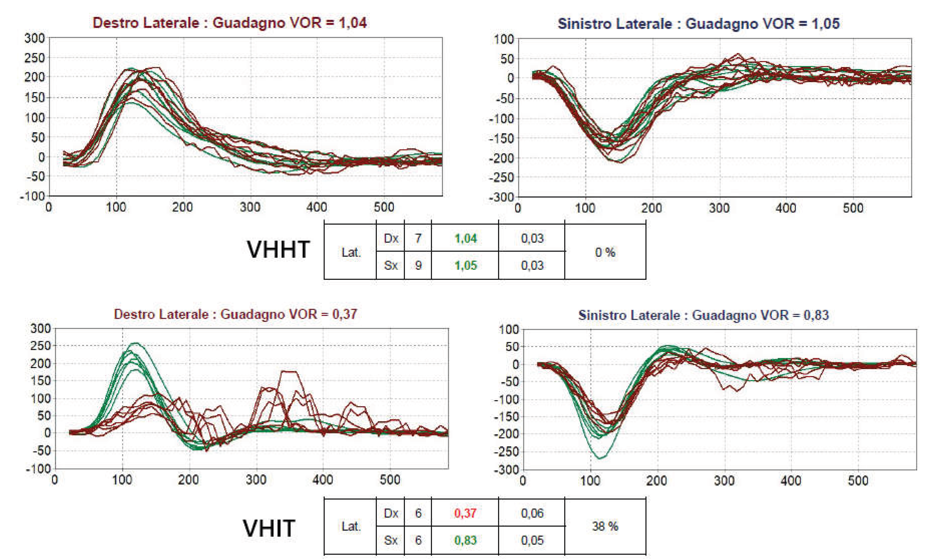
Figure 2.
Left TVOR compensatory saccades in a pt with acute left SVN. Comparison between VHHT and VHIT velocity traces (for all SCs) and relative gain values for patient no. 19 in Table C, suffering from acute SVN of the left side. In the upper panel, the VHHT traces show compensatory saccades on the left side and markedly asymmetric TG (left 0.95; right 1.31). In the lower panel, the VHIT traces show reduced saccades and gain values on the left-side ASC and LSC. The symmetrical reduction in gain and saccades on both PSCs is normal for age. Anti-Compensatory Quick Eye Movements (AQEM) on both the right-side PSC and LSC traces indicate an acute deficit in the left labyrinth [7].
Figure 2.
Left TVOR compensatory saccades in a pt with acute left SVN. Comparison between VHHT and VHIT velocity traces (for all SCs) and relative gain values for patient no. 19 in Table C, suffering from acute SVN of the left side. In the upper panel, the VHHT traces show compensatory saccades on the left side and markedly asymmetric TG (left 0.95; right 1.31). In the lower panel, the VHIT traces show reduced saccades and gain values on the left-side ASC and LSC. The symmetrical reduction in gain and saccades on both PSCs is normal for age. Anti-Compensatory Quick Eye Movements (AQEM) on both the right-side PSC and LSC traces indicate an acute deficit in the left labyrinth [7].
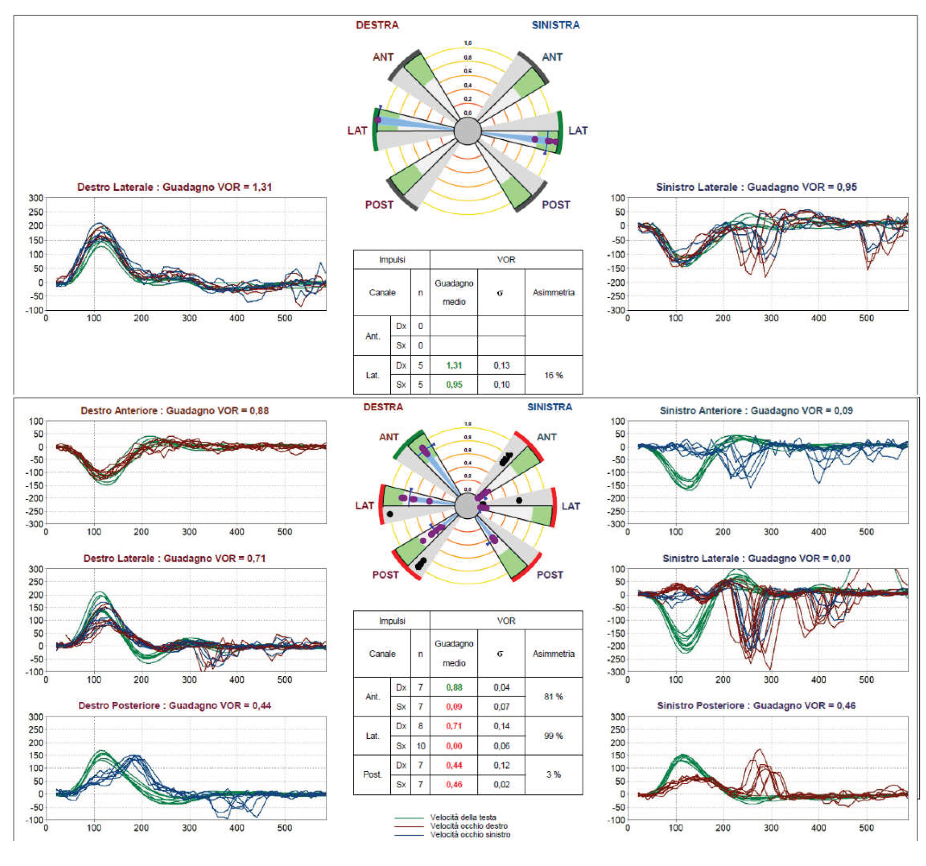
Figure 3.
Left TVOR compensatory saccades in a pt with left PSC hypofunction at VHIT. Comparison between VHHT and VHIT velocity traces (for all SCs) and relative gain values for patient no. 5 in Table C, diagnosed with VM. In the upper panel, the VHHT traces show compensatory saccades on the left side and slightly asymmetric TG (left 1.07; right 1.11). In the lower panel, the VHIT traces show saccades and reduced gain values exclusively on the left PSC.
Figure 3.
Left TVOR compensatory saccades in a pt with left PSC hypofunction at VHIT. Comparison between VHHT and VHIT velocity traces (for all SCs) and relative gain values for patient no. 5 in Table C, diagnosed with VM. In the upper panel, the VHHT traces show compensatory saccades on the left side and slightly asymmetric TG (left 1.07; right 1.11). In the lower panel, the VHIT traces show saccades and reduced gain values exclusively on the left PSC.
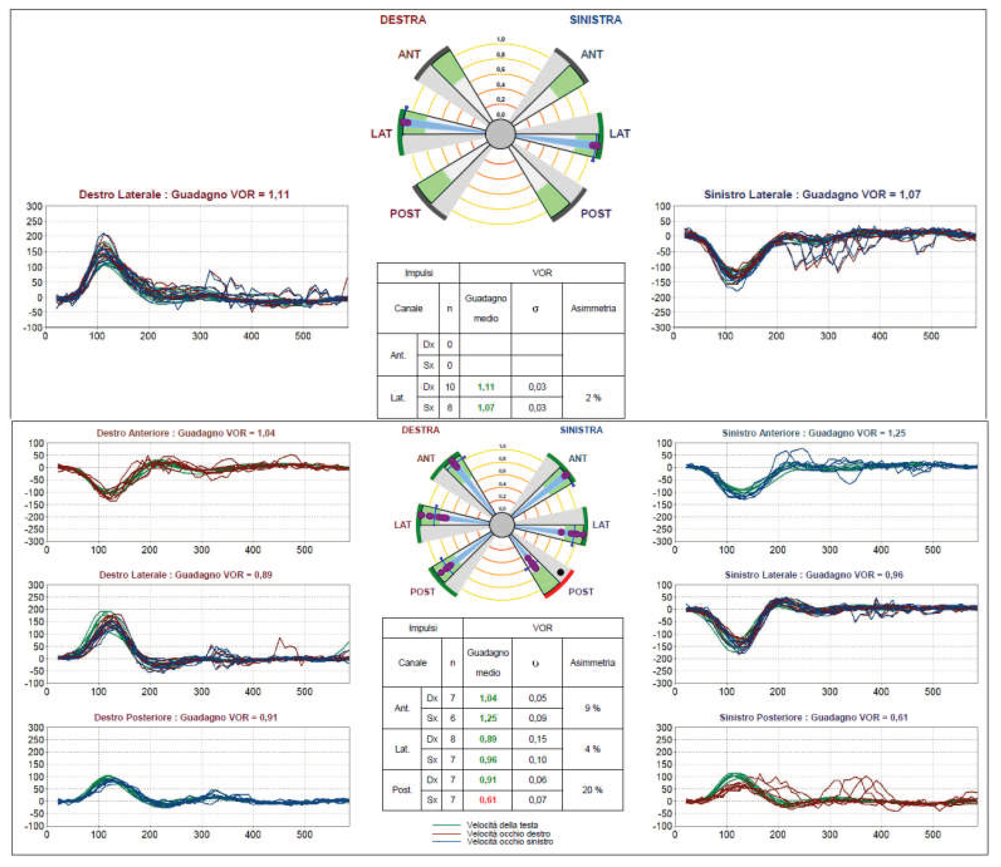
Figure 4.
Right TVOR compensatory saccades in a patient with normal bilateral semicircular canal function at VHIT. Comparison between VHHT and VHIT velocity traces (for all SCs) and relative gain values for patient no. 25 in Table C, with no clinical signs or history of specific vestibular disorders (UNK). In the upper panel, the VHHT traces show compensatory saccades on the right side and asymmetric TG (Left 1.34; Right 1.22). In the lower panel, the VHIT traces and gain values are normal for all six channels.
Figure 4.
Right TVOR compensatory saccades in a patient with normal bilateral semicircular canal function at VHIT. Comparison between VHHT and VHIT velocity traces (for all SCs) and relative gain values for patient no. 25 in Table C, with no clinical signs or history of specific vestibular disorders (UNK). In the upper panel, the VHHT traces show compensatory saccades on the right side and asymmetric TG (Left 1.34; Right 1.22). In the lower panel, the VHIT traces and gain values are normal for all six channels.
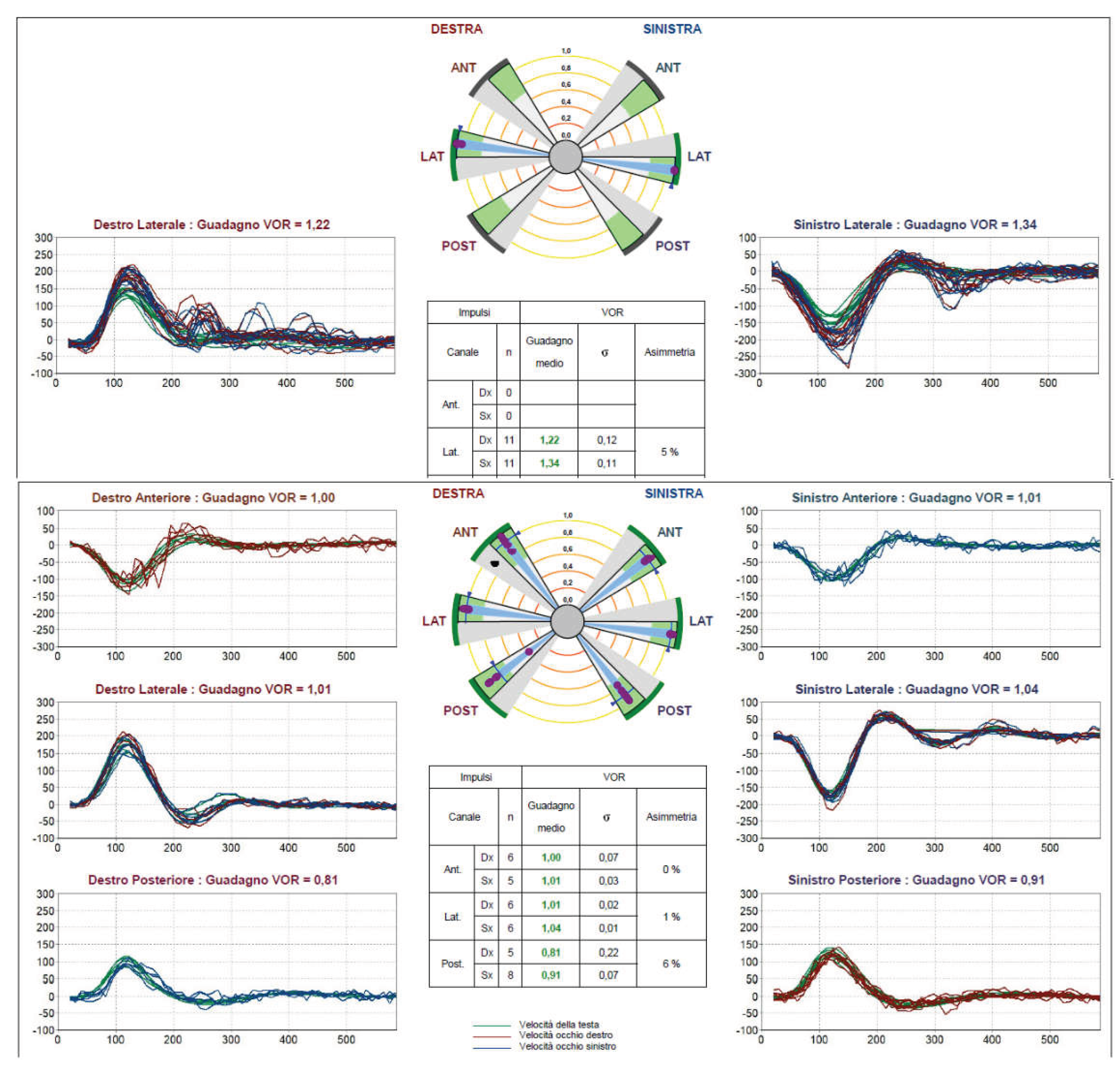
Figure 5.
Absence of TVOR compensatory saccades in a pt with right LSC and ASC hypofunction at VHIT. Comparison between VHHT and VHIT velocity traces (for all SCs) and relative gain values for patient no. 15 in Table A, a pt with late outcome of SVN on the right side. In the upper panel, the VHHT traces show no compensatory saccades, and the TG is symmetrical (left 0.84; right 0.82). In the lower panel, the VHIT traces show markedly reduced gain values and large covert saccades (indicating good compensation) on the right LSC. On the right ASC, despite the reduction in gain, there are no saccades. The symmetrical reduction in gain and saccades on both PSCs and the mild dysfunction of the left LSC were normal for his age. There are no AQEMs.
Figure 5.
Absence of TVOR compensatory saccades in a pt with right LSC and ASC hypofunction at VHIT. Comparison between VHHT and VHIT velocity traces (for all SCs) and relative gain values for patient no. 15 in Table A, a pt with late outcome of SVN on the right side. In the upper panel, the VHHT traces show no compensatory saccades, and the TG is symmetrical (left 0.84; right 0.82). In the lower panel, the VHIT traces show markedly reduced gain values and large covert saccades (indicating good compensation) on the right LSC. On the right ASC, despite the reduction in gain, there are no saccades. The symmetrical reduction in gain and saccades on both PSCs and the mild dysfunction of the left LSC were normal for his age. There are no AQEMs.
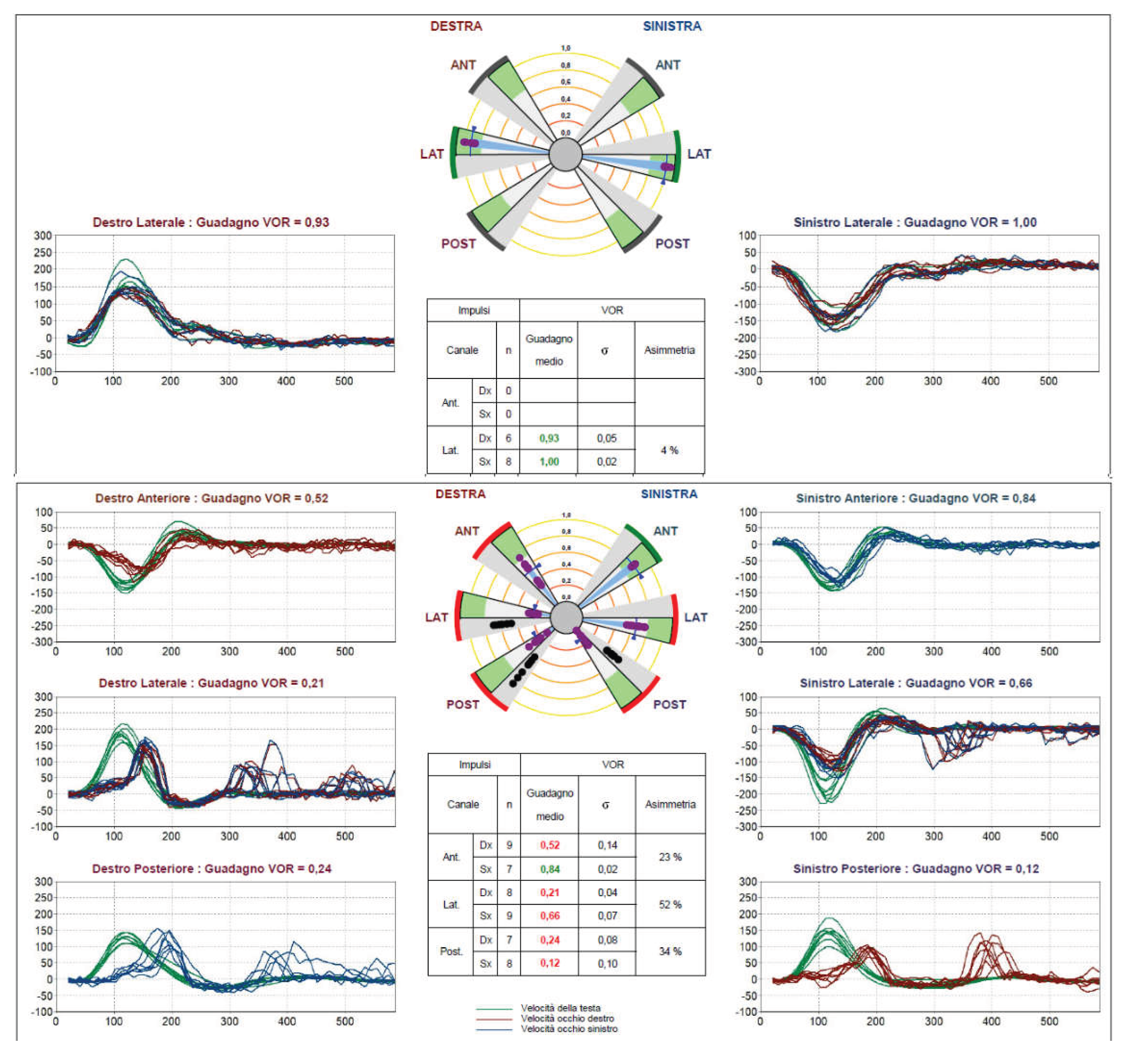
Figure 6.
Bilateral TVOR compensatory saccades in a patient with hypofunction of the three left semicircular canals at VHIT. Comparison between VHHT and VHIT velocity traces (for all SCs) and relative gain values for patient no. 17 in Table B, a left-sided AUVD late outcome involving all 3 SCs. In the upper panel, both VHHT traces show compensatory saccades and symmetrical TG (left 0.96; right 0.99). In the lower panel, the VHIT traces show saccades and a reduction in gain on all SCs on the left side. The presence of both covert and overt gathered saccades on the left LSC indicates good compensation. A slight dysfunction of the right PSC is normal for age. There are no AQEMs.
Figure 6.
Bilateral TVOR compensatory saccades in a patient with hypofunction of the three left semicircular canals at VHIT. Comparison between VHHT and VHIT velocity traces (for all SCs) and relative gain values for patient no. 17 in Table B, a left-sided AUVD late outcome involving all 3 SCs. In the upper panel, both VHHT traces show compensatory saccades and symmetrical TG (left 0.96; right 0.99). In the lower panel, the VHIT traces show saccades and a reduction in gain on all SCs on the left side. The presence of both covert and overt gathered saccades on the left LSC indicates good compensation. A slight dysfunction of the right PSC is normal for age. There are no AQEMs.
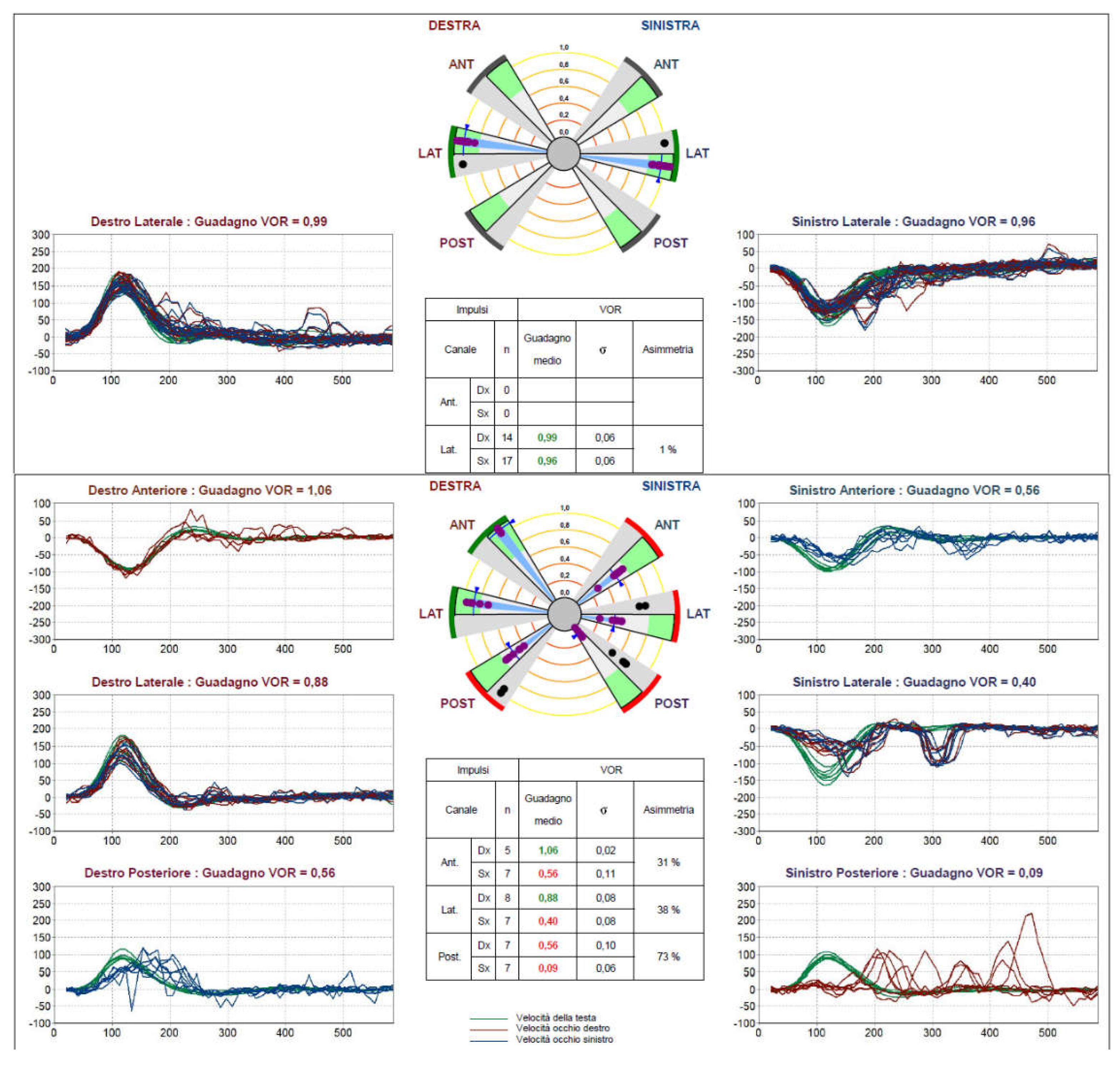
Figure 7.
Bilateral compensatory saccades of the TVOR in a patient with normal semicircular canal function at VHIT. Comparison between VHHT and VHIT velocity traces (for all SCs) and relative gain values for patient no. 21 in Table B, a case of VM. In the upper panel, both VHHT traces show compensatory saccades, and TG is slightly asymmetric (left 1.15; right 1.10). In the lower panel, the VHIT traces and gain values are normal for all six channels.
Figure 7.
Bilateral compensatory saccades of the TVOR in a patient with normal semicircular canal function at VHIT. Comparison between VHHT and VHIT velocity traces (for all SCs) and relative gain values for patient no. 21 in Table B, a case of VM. In the upper panel, both VHHT traces show compensatory saccades, and TG is slightly asymmetric (left 1.15; right 1.10). In the lower panel, the VHIT traces and gain values are normal for all six channels.
Disclaimer/Publisher’s Note: The statements, opinions and data contained in all publications are solely those of the individual author(s) and contributor(s) and not of MDPI and/or the editor(s). MDPI and/or the editor(s) disclaim responsibility for any injury to people or property resulting from any ideas, methods, instructions or products referred to in the content. |
© 2026 by the author. Licensee MDPI, Basel, Switzerland. This article is an open access article distributed under the terms and conditions of the Creative Commons Attribution (CC BY) license.
Copyright: This open access article is published under a Creative Commons CC BY 4.0 license, which permit the free download, distribution, and reuse, provided that the author and preprint are cited in any reuse.


