
Submitted:
10 July 2026
Posted:
14 July 2026
You are already at the latest version
Preprints on COVID-19 and SARS-CoV-2
Abstract
Post-exertional malaise (PEM) and cognitive dysfunction (“brain fog”) are among the most disabling features of Long COVID, yet approved disease-modifying treatments remain lacking. Emerging evidence implicates interacting disturbances in mitochondrial bioenergetics, redox regulation and neurovascular inflammation, although much of the supporting data derive from acute COVID-19, myalgic encephalomyelitis/chronic fatigue syndrome (ME/CFS), mitochondrial disease and mechanistic pharmacology rather than from direct Long COVID studies. This narrative review develops a mechanism-based translational hypothesis: that a pathway-targeted nutraceutical combination may modulate these three axes more effectively than single-agent antioxidant approaches. Candidate modules comprise coenzyme Q10 and alpha-lipoic acid for bioenergetic support; selenium, sulforaphane and resveratrol for Nrf2–thioredoxin redox regulation; and Boswellia serrata, luteolin and eicosapentaenoic acid for prostaglandin and resolution-pathway modulation. The mitochondrial redox modulator sonlicromanol provides pharmacological precedent for combined antioxidant and prostaglandin-directed mechanisms, although the proposed nutraceutical strategy remains clinically untested. We summarise the mechanistic rationale, critically appraise direct versus indirect clinical evidence with explicit certainty grading, outline safety and interaction considerations, and propose a stratified randomised trial framework. This model reframes nutraceuticals as pathway-specific candidate interventions and provides a falsifiable basis for future Long COVID trials.
Keywords:
long COVID
; post-acute sequelae of SARS-CoV-2
; post-exertional malaise
; brain fog
; mitochondrial dysfunction
; oxidative stress
; Nrf2
; mPGES-1
; nutraceuticals
; coenzyme Q10
1. Introduction
Long COVID—formally the post-acute sequelae of SARS-CoV-2 infection (PASC)—affects an estimated 6–10% of infected individuals and persists for months to years beyond the acute illness [1,2]. Among its heterogeneous manifestations, post-exertional malaise (PEM) and cognitive dysfunction (“brain fog”) are consistently ranked by patients as the most disabling and are strongly associated with failure to return to work and reduced quality of life [3]. PEM—a disproportionate and delayed worsening of symptoms after physical, cognitive or emotional exertion—is the cardinal feature shared with myalgic encephalomyelitis/chronic fatigue syndrome (ME/CFS), and its presence fundamentally constrains conventional graded-exercise rehabilitation [4].
Despite intense investigation, no licensed disease-modifying therapy exists for these symptoms. Symptomatic and pacing-based strategies remain the mainstay, yet they neither reverse the underlying pathophysiology nor restore exercise tolerance [5]. This therapeutic vacuum, combined with an increasingly coherent mechanistic picture, creates a rational opportunity for interventions that target the molecular drivers of PEM and brain fog directly.
Three pathological processes have emerged repeatedly and, importantly, appear mechanistically interconnected rather than independent: (i) mitochondrial dysfunction and bioenergetic insufficiency; (ii) a self-sustaining redox imbalance with impaired endogenous antioxidant defence; and (iii) persistent low-grade neurovascular inflammation [6,7]. Each is amenable, at least in principle, to modulation by specific nutraceutical agents whose molecular targets are now well characterised. The purpose of this review is to move beyond the generic framing of nutraceuticals as undifferentiated “antioxidants” and instead articulate a mechanism-based, pathway-specific hypothesis: that a rationally combined nutraceutical strategy may modulate all three axes and thereby attenuate PEM and brain fog. We develop the pathophysiological framework, map candidate agents to their molecular targets, appraise direct and indirect clinical evidence with explicit certainty grading, address safety, and propose a testable trial design.
2. Search Strategy
This narrative review was developed from targeted searches of PubMed, Scopus and Google Scholar (from database inception to July 2026) for literature on Long COVID, post-exertional malaise, mitochondrial dysfunction, oxidative stress, Nrf2, thioredoxin reductase, mPGES-1, prostaglandin E₂ and nutraceutical interventions, using combinations of these terms. Priority was given to human Long COVID studies, randomised controlled trials, systematic reviews and mechanistic studies directly relevant to the proposed pathways. Where direct Long COVID evidence was unavailable, indirect evidence from ME/CFS, primary mitochondrial disease, inflammatory disease and human nutraceutical studies was included and is explicitly identified as indirect throughout. Because this is a hypothesis-generating narrative synthesis rather than a systematic review, no formal risk-of-bias appraisal or quantitative pooling was undertaken; instead, the strength of evidence underpinning each principal claim is graded qualitatively in Section 6 (Table 2).
3. A Three-Axis Pathophysiological Framework
3.1. Mitochondrial Dysfunction and Bioenergetic Failure
SARS-CoV-2 interacts intimately with host mitochondria. Viral open reading frame and non-structural proteins localise to mitochondrial membranes, disrupting electron transport chain (ETC) assembly, promoting pathological fission, and provoking calcium dyshomeostasis with opening of the mitochondrial permeability transition pore [7,8]. A plausible downstream consequence, supported by related post-viral and ME/CFS literature, is functional restriction of pyruvate dehydrogenase (PDH) flux at the irreversible gateway converting pyruvate to acetyl-CoA. Oxidative modification of the PDH lipoyl cofactor, together with upregulation of PDH kinase, is proposed to force cells toward aerobic glycolysis—a Warburg-like shift that yields only two ATP per glucose molecule versus approximately 30–32 through oxidative phosphorylation, with attendant lactate accumulation [8,9].
Plasma metabolomic signatures in Long COVID support impaired fatty-acid oxidation, perturbed tricarboxylic-acid-cycle intermediates and reduced Complex I activity [8]. Additional PBMC and tissue/biomarker studies published in 2025 further support persistent mitochondrial abnormalities in Long COVID, including altered mitochondrial ATP-synthase behaviour, ultrastructural changes and disrupted mitophagy markers [10,11]. This bioenergetic insufficiency is present at rest but is unmasked catastrophically by exertion: invasive cardiopulmonary exercise testing demonstrates impaired systemic oxygen extraction rather than a primarily cardiopulmonary limitation [5]. Muscle biopsy studies further show that exercise precipitates mitochondrial and structural myofibre abnormalities specifically in the post-exertional window, providing a direct histological correlate of PEM [4]. The mechanistic congruence with ME/CFS—shared PDH blockade, glycolytic shift and exertional deterioration—strengthens the case for targeting mitochondrial bioenergetics [12].
3.2. Redox Imbalance and the Nrf2–Thioredoxin Axis
Reactive oxygen species (ROS) are generated in excess through ETC electron leak, NADPH-oxidase activation and uncoupled nitric oxide synthase, producing superoxide, hydrogen peroxide and peroxynitrite [13]. This oxidative burden appears to persist beyond viral clearance: Long COVID cohorts have shown increased oxidative-damage and redox-imbalance markers, including malondialdehyde/protein-carbonyl-related oxidative toxicity and elevated oxidative-stress index in patients with fatigue and brain fog [14,15]. SARS-CoV-2-mediated suppression of NRF2 signalling has been demonstrated in experimental systems [16]. Under physiological conditions the transcription factor Nrf2, released from its cytoplasmic repressor Keap1 upon oxidative cysteine modification, drives expression of more than 200 cytoprotective genes including thioredoxin reductase-1 (TrxR1), NAD(P)H quinone oxidoreductase 1 (NQO1) and the glutathione-synthetic enzyme glutamate-cysteine ligase (GCL) [17,18].
Persistence of SARS-CoV-2-mediated Nrf2 suppression in established Long COVID remains inferred rather than directly demonstrated. The thioredoxin reductase–thioredoxin–peroxiredoxin (TrxR–Trx–Prdx) cascade is the principal enzymatic system clearing hydrogen peroxide and lipid hydroperoxides [19,20]. Because TrxR is a selenoenzyme whose selenocysteine active site is itself vulnerable to irreversible oxidative inactivation, oxidative stress may disable the very machinery required to resolve it. Resulting peroxiredoxin inactivation would permit peroxide accumulation and mitochondrial DNA damage, further impairing Complex I—a self-amplifying redox–energetic loop that could plausibly underlie the chronicity of Long COVID [19,21].
3.3. Neurovascular Inflammation and a Putative mPGES-1/PGE₂ Contribution
Endothelial injury—via spike–ACE2 interactions, complement activation and neutrophil extracellular-trap formation—converges on nuclear factor kappa-B (NF-κB) activation in endothelium and macrophages [22,23]. Persistent endothelial dysfunction has been directly documented in Long COVID cohorts using non-invasive vascular assessment, confirming that vascular injury outlasts the acute infectious phase [24]. This drives transcription of pro-inflammatory cytokines and of the prostaglandin-biosynthetic enzymes cyclooxygenase-2 (COX-2) and microsomal prostaglandin E₂ synthase-1 (mPGES-1). Persistence of spike protein within CD16⁺ monocytes for up to fifteen months provides a plausible mechanism for sustained NF-κB signalling that outlasts viral clearance [25].
mPGES-1 is the terminal, inducible and rate-limiting enzyme converting the COX-2 product PGH₂ specifically to prostaglandin E₂ (PGE₂) [26]. Unlike non-selective COX inhibition—which also abolishes cardioprotective prostacyclin—selective mPGES-1 blockade targets PGE₂ excess while sparing other prostanoids. In experimental and inflammatory-disease settings, elevated PGE₂ acting through EP receptors causes pathological vasodilation, central sensitisation that amplifies fatigue and pain perception, and immunosuppression of cytotoxic lymphocytes [26,27]; the extrapolation of this cascade to Long COVID PEM and brain fog is at present inferential rather than directly established. The strongest Long COVID-specific support for the neurovascular arm comes from the direct demonstration of blood–brain-barrier disruption with sustained systemic inflammation in patients with cognitive impairment, which links peripheral inflammatory signalling to the substrate of brain fog but does not by itself confirm a prostaglandin-specific mechanism [28]. Amyloid-containing fibrin microclots that resist fibrinolysis may further entrap inflammatory mediators and propagate endothelial activation [29]. Because direct human evidence of an intracranial prostaglandin surge in Long COVID is currently scarce, verifying an elevated central prostanoid signature—for example, a raised cerebrospinal-fluid PGE₂ or a neuroimaging-based neuroinflammatory marker—should be regarded as a necessary early-phase step in testing, and a potential means of falsifying, this component of the hypothesis.
3.4. Convergence on Post-Exertional Malaise and Brain Fog
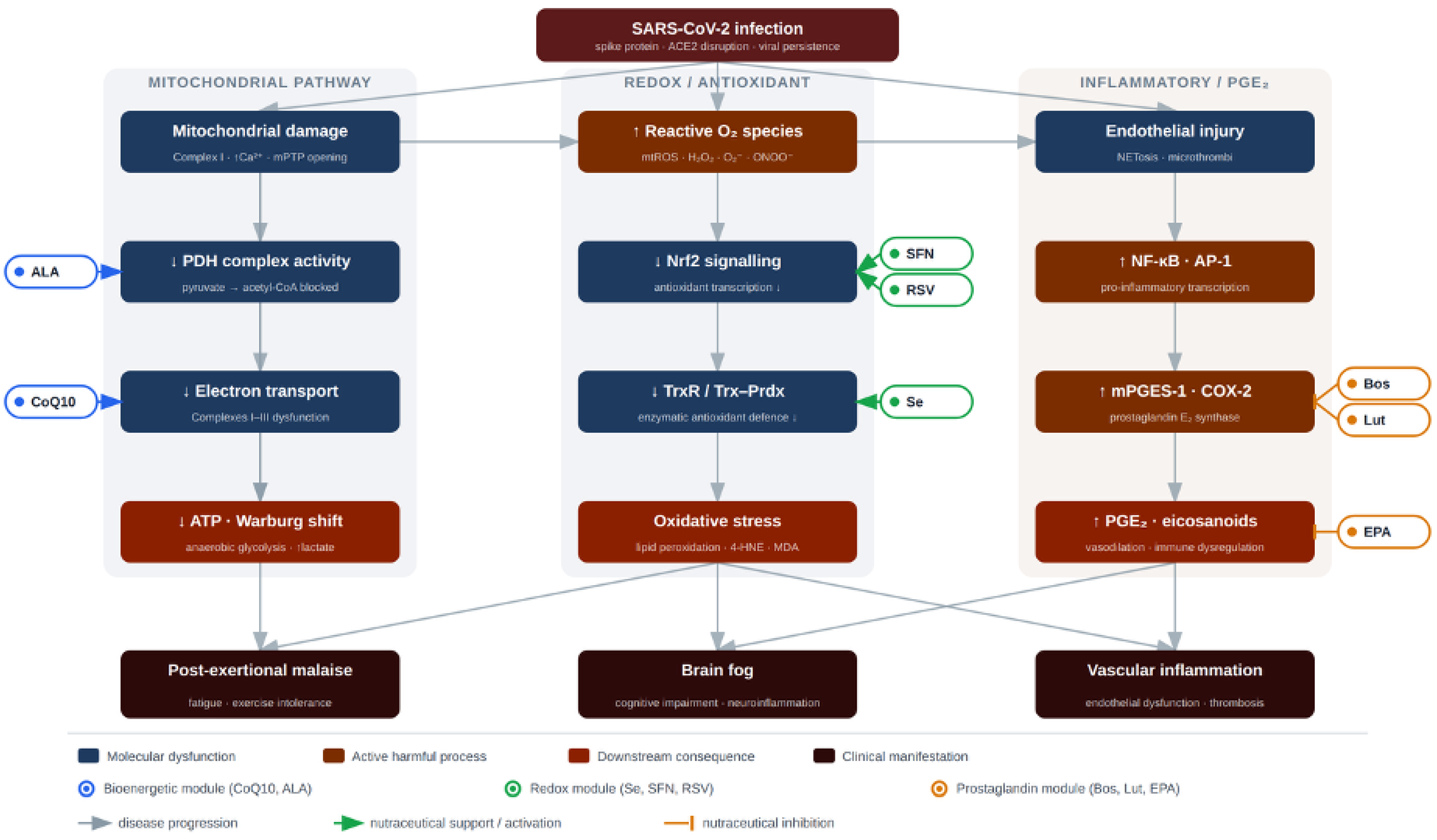
These axes are not parallel but interlocking (Figure 1). Mitochondrial ROS suppress Nrf2 and inactivate TrxR; the resulting oxidative stress activates NF-κB and amplifies prostaglandin synthesis; PGE₂ and cytokines in turn impair microvascular oxygen delivery and mitochondrial function. PEM emerges when exertion acutely increases metabolic demand upon a system already operating at the edge of bioenergetic reserve, precipitating an oxidative–inflammatory flare. Brain fog reflects the same triad expressed within a neuroinflamed, barrier-compromised central compartment. A therapeutic strategy addressing only one axis may therefore be undermined by the others—providing the central rationale for a combined, multi-target approach [6,7,30].

Table 1.
Candidate nutraceuticals, their principal molecular targets, proposed mechanisms in Long COVID, and indicative dose ranges for hypothesis-driven trial design.
Table 1.
Candidate nutraceuticals, their principal molecular targets, proposed mechanisms in Long COVID, and indicative dose ranges for hypothesis-driven trial design.
| Agent | Primary target | Proposed mechanism in Long COVID | Indicative dose range for trial design |
| Coenzyme Q10 | ETC Complexes I–III | Redox carrier restoring electron flow and ATP synthesis; reduces electron leak and secondary ROS generation | 100–200 mg twice daily (ubiquinol preferred) |
| Alpha-lipoic acid | PDH complex; ROS | Restores the PDH lipoyl cofactor, re-enabling pyruvate oxidation; amphiphilic antioxidant regenerating glutathione, vitamins C and E | 100–200 mg twice daily |
| Selenium | Thioredoxin reductase | Supplies the catalytic selenocysteine of TrxR, restoring Trx/Prdx peroxide clearance | 100–200 µg daily |
| Sulforaphane | Nrf2 (Keap1) | Electrophilic Nrf2 activation upregulating TrxR1, NQO1 and GCL; synergistic with selenium | 10–40 mg/day (stabilised sulforaphane, or glucoraphanin with active myrosinase) |
| Boswellia serrata (AKBA) | mPGES-1; NF-κB | Direct inhibition of microsomal PGE₂ synthase-1 and suppression of NF-κB/AP-1 transcription | 100–300 mg/day (≥30% AKBA; phytosome/lipid formulation preferred) |
| Luteolin | COX-2; mPGES-1 | Transcriptional suppression of COX-2 and mPGES-1; reduces PGE₂ and nitric oxide | 100–300 mg/day (bioavailability-enhanced) |
| EPA (omega-3) | COX-2 substrate; SPMs | Competitive displacement of arachidonic acid toward low-potency PGE₃; precursor to resolvins and protectins | 2–4 g EPA/day |
| Resveratrol | Nrf2 / NF-κB (SIRT1) | SIRT1-mediated Nrf2 activation and NF-κB inhibition; dual antioxidant and anti-inflammatory action | 150–500 mg/day (micronised or phytosome formulation) |
Doses are proposed for hypothesis-driven clinical trial design and should not be interpreted as individual medical advice. Final dosing requires protocol-specific safety review, medication-interaction screening and product-quality verification. AKBA, acetyl-11-keto-β-boswellic acid; ATP, adenosine triphosphate; COX-2, cyclooxygenase-2; EPA, eicosapentaenoic acid; ETC, electron transport chain; GCL, glutamate-cysteine ligase; mPGES-1, microsomal prostaglandin E₂ synthase-1; Nrf2, nuclear factor erythroid 2-related factor 2; NQO1, NAD(P)H quinone oxidoreductase 1; PDH, pyruvate dehydrogenase; ROS, reactive oxygen species; SPMs, specialised pro-resolving mediators; TrxR, thioredoxin reductase.
Table 2.
Qualitative evidence grading for the principal claims of the proposed framework, distinguishing direct human Long COVID evidence from indirect human and preclinical/mechanistic evidence.
Table 2.
Qualitative evidence grading for the principal claims of the proposed framework, distinguishing direct human Long COVID evidence from indirect human and preclinical/mechanistic evidence.
| Claim | Direct Long COVID human evidence | Indirect human evidence | Preclinical / mechanistic | Overall certainty |
| Mitochondrial dysfunction contributes to PEM | Moderate | Moderate (ME/CFS) | Strong | Moderate |
| PDH flux restriction is a central Long COVID lesion | Limited | Moderate (ME/CFS, metabolic) | Moderate | Low–moderate |
| Nrf2–thioredoxin suppression persists in Long COVID | Limited | Acute COVID / in vitro | Moderate | Low |
| mPGES-1/PGE₂ drives brain fog / PEM | Very limited | Inflammatory / neurovascular | Moderate | Low |
| CoQ10 + ALA improves Long COVID fatigue | Limited (non-randomised) | Metabolic support | Plausible | Low |
| High-dose EPA benefits Long COVID | Weak (feasibility pilot only) | Cardiovascular / inflammatory | Plausible | Low |
| Combined three-module therapy attenuates PEM/brain fog | None (untested) | Polytherapy rationale | Plausible | Very low |
Certainty reflects the author’s qualitative appraisal for this narrative review and is not derived from a formal GRADE assessment. ALA, alpha-lipoic acid; CoQ10, coenzyme Q10; EPA, eicosapentaenoic acid; ME/CFS, myalgic encephalomyelitis/chronic fatigue syndrome; mPGES-1, microsomal prostaglandin E₂ synthase-1; Nrf2, nuclear factor erythroid 2-related factor 2; PDH, pyruvate dehydrogenase; PEM, post-exertional malaise; PGE₂, prostaglandin E₂.
4. Nutraceutical Candidates and Their Molecular Targets
Table 1 summarises the eight candidate agents, their principal molecular targets, proposed mechanisms and indicative dose ranges for trial design. They are grouped below by the axis they predominantly address, although several act across more than one.
4.1. Mitochondrial Support: Coenzyme Q10 and Alpha-Lipoic Acid
Coenzyme Q10 (CoQ10) is an obligatory lipophilic electron carrier shuttling electrons between Complexes I/II and III; supplementation can restore ETC flux and ATP synthesis while reducing electron leak and secondary ROS generation [31]. Alpha-lipoic acid (ALA) is the physiological cofactor of the PDH complex and thus directly addresses the glycolytic-shift lesion; it is additionally an amphiphilic antioxidant that regenerates glutathione and vitamins C and E, bridging the mitochondrial and redox axes [32,33]. A non-randomised study of 174 patients reported resolution of severe fatigue in the majority receiving CoQ10 plus ALA, though a placebo-controlled trial of high-dose CoQ10 monotherapy over six weeks was negative—underscoring both the promise of the combination and the limitations of the current evidence [34,35].
4.2. Redox Restoration: Selenium, Sulforaphane and Resveratrol
Selenium is incorporated as selenocysteine into the catalytic site of TrxR and glutathione peroxidases; adequate selenium status is a stoichiometric prerequisite for a functional TrxR–Trx–Prdx system, and deficiency renders the enzyme inert regardless of upstream signalling [36]. Sulforaphane, the isothiocyanate generated by myrosinase-catalysed hydrolysis of the precursor glucoraphanin, is the most potent dietary Nrf2 activator: by alkylating Keap1 cysteines it triggers the Keap1–Nrf2 conformational switch that releases Nrf2 to upregulate TrxR1, NQO1 and glutathione synthesis, acting synergistically with selenium [37,38,39]. Because dietary myrosinase is heat-labile, any clinical protocol must specify either stabilised free sulforaphane or a standardised glucoraphanin–myrosinase co-delivery system to ensure reliable conversion. Resveratrol activates Nrf2 and inhibits NF-κB through SIRT1-mediated deacetylation, conferring dual antioxidant and anti-inflammatory activity that spans the redox and prostaglandin axes; being lipophilic and blood–brain-barrier-permeable, resveratrol and the flavonoid luteolin are particularly suited to the neuroinflammatory substrate of brain fog, although both undergo rapid phase-II metabolism and require bioavailability-enhanced (e.g., phytosome or micronised) formulations to reach meaningful plasma concentrations [40].
4.3. Prostaglandin Modulation: Boswellia, Luteolin and Eicosapentaenoic Acid
Boswellic acids—principally acetyl-11-keto-β-boswellic acid (AKBA) from Boswellia serrata—are among the few natural compounds shown to inhibit mPGES-1 directly, in addition to suppressing NF-κB/AP-1 transcription [41,42]. Luteolin, a dietary flavonoid, reduces COX-2 and mPGES-1 expression at the transcriptional level and lowers PGE₂ and nitric oxide output in activated macrophages [43,44]. Eicosapentaenoic acid (EPA) competitively displaces arachidonic acid from COX-2, shifting synthesis toward the low-potency PGE₃, and serves as the precursor to specialised pro-resolving mediators (resolvins, protectins) that actively terminate inflammation [45,46]. This last property is mechanistically important: emerging evidence reframes persistent PASC inflammation not merely as excessive pro-inflammatory signalling but as a failure of active resolution, making substrate provision for pro-resolving mediator synthesis a rational translational target rather than simple immunosuppression. Anti-inflammatory and pro-resolving effects require EPA doses (2–4 g/day) substantially above those in standard fish-oil preparations [47]. A cross-cutting translational caveat applies to three of the polyphenolic agents: resveratrol, luteolin and the boswellic acids all have low intrinsic oral bioavailability owing to rapid phase-II metabolism, so the model depends materially on advanced delivery technologies (phytosome complexes, micronisation or lipid-based nanocarriers). A trial employing standard raw powders would risk a false-negative result driven by sub-therapeutic plasma concentrations rather than by a genuine absence of effect.
5. Integrated Hypothesis and Combination Rationale
We propose that because the three axes are mutually reinforcing, an intervention must engage all three to interrupt the pathological loop durably. A single-axis approach—for example antioxidant monotherapy—may be neutralised by unaddressed inflammatory and bioenergetic drivers, a pattern consistent with the negative CoQ10 monotherapy trial [35]. The combination is therefore conceived as three coordinated modules: a bioenergetic module (CoQ10 + ALA), a redox module (selenium + sulforaphane, with resveratrol bridging), and a prostaglandin module (Boswellia + luteolin + EPA).
Pharmacological proof-of-concept for the dual antioxidant–anti-inflammatory mechanism is provided by sonlicromanol (KH176), a small-molecule mitochondrial-targeted redox modulator whose active metabolite KH176m both engages the thioredoxin/peroxiredoxin system to scavenge ROS and selectively inhibits mPGES-1-mediated PGE₂ biosynthesis [48,49]. In primary mitochondrial disease, sonlicromanol has progressed through phase 2 evaluation with signals of benefit on cognition and fatigue-related domains [50,51]. The nutraceutical strategy proposed here can be viewed as a food-derived, multi-agent approximation of this same dual mechanism: selenium and sulforaphane engage the thioredoxin arm that sonlicromanol augments, while Boswellia, luteolin and EPA engage the mPGES-1/PGE₂ arm it inhibits. Although no single nutraceutical replicates the engineered selectivity of KH176m, the combination reconstructs both mechanistic limbs from complementary agents.
6. Appraisal of Direct and Indirect Clinical Evidence
Direct clinical evidence in Long COVID remains sparse, and it is important to distinguish it clearly from the larger body of indirect and mechanistic support. The strongest direct signal is for the CoQ10–ALA combination: a non-randomised study of 174 patients reported resolution of severe fatigue in the majority of recipients [34]. This finding is susceptible to expectation and natural-recovery bias and cannot, alone, establish efficacy. Instructively, a rigorously conducted placebo-controlled trial of CoQ10 monotherapy was negative over a six-week exposure in a predominantly overweight cohort [35]. The contrast between a positive combination signal and a null monotherapy result is consistent with—though far from proof of—the central argument that single-axis intervention is insufficient; it must be interpreted cautiously given the differing designs, durations and populations.
The available Long COVID omega-3 study is best read as a feasibility trial: it primarily supports tolerability and recruitment feasibility while providing insufficient evidence for efficacy, particularly because it did not test an EPA-dominant anti-inflammatory dosing strategy [52]. It should therefore not be characterised simply as a “negative” efficacy trial. Indirect support for the wider framework is stronger: antioxidant and anti-inflammatory nutritional strategies have a coherent rationale in COVID-19 pathophysiology [30,53]; ALA improves metabolic and oxidative parameters across multiple conditions [54]; high-dose EPA has robust cardiovascular anti-inflammatory data [47]; and mitochondrial-disease literature provides a rationale for combined nutraceutical polytherapy rather than reliance on single agents [55]. The strength of evidence underpinning each principal claim is summarised in Table 2. Across the framework, direct human Long COVID evidence ranges from limited to very limited, mechanistic and preclinical support is moderate to strong, and overall certainty is correspondingly low to moderate. The central hypothesis—that a three-module combination attenuates PEM and brain fog—has not been tested and should be regarded as hypothesis-generating.
7. Safety, Interactions and Translational Considerations
A combination of eight agents raises real-world issues of adherence, tolerability, attribution of adverse effects, supplement contamination and cumulative interactions, and these must be addressed explicitly before any trial. Several specific considerations stand out. First, EPA at 2–4 g/day is pharmacologically distinct from low-dose “fish oil”: in the REDUCE-IT cardiovascular trial, high-dose eicosapentaenoic acid was associated with a higher incidence of atrial fibrillation/flutter requiring hospitalisation and a numerically higher rate of serious bleeding [47]. Second, EPA, resveratrol and Boswellia each carry antiplatelet potential; their combination, particularly alongside any NSAID use, warrants explicit bleeding-risk exclusion criteria. Third, selenium has a narrow, U-shaped therapeutic window, so baseline selenium status should be measured (or high background intake excluded) and total intake kept below the commonly cited adult upper limit of approximately 400 µg/day from all sources [36]. Fourth, ALA can lower blood glucose and requires glucose-risk screening in people with diabetes, on glucose-lowering therapy, or prone to hypoglycaemia [32,54]. Fifth, CoQ10 may attenuate warfarin anticoagulation [31], and sulforaphane induces cytochrome P450 1A2 via the aryl-hydrocarbon receptor, a material consideration for co-administered narrow-therapeutic-index CYP1A2 substrates [39]. Pregnancy, lactation and concurrent anticoagulant use should be explicit exclusions for a first trial, and product quality—given marked between-formulation variation in bioavailability—should be assured through independent certification.
8. Future Directions: A Proposed Trial Framework
Testing this hypothesis rigorously requires a stratified, biomarker-anchored randomised controlled trial. The population should comprise adults meeting the WHO post-COVID-condition definition—symptoms usually beginning within three months of infection, persisting at least two months and not explained by an alternative diagnosis—with PEM confirmed using a validated instrument (e.g., the DePaul Symptom Questionnaire–PEM) and fatigue or cognitive dysfunction above a pre-specified threshold [2,4]. Baseline phenotyping should capture PEM severity, a dysautonomia/POTS screen, inflammatory and oxidative-stress markers, omega-3 index and selenium status, with a documented medication and supplement washout.
A single primary endpoint—either a PEM/fatigue measure or a cognitive measure, but not both unless the study is powered accordingly—should be pre-specified, complemented by objective secondary measures such as actigraphy, wearable heart-rate/HRV data and, where safe and feasible, a submaximal exertional challenge; mechanistic biomarkers (post-exertional lactate kinetics or cardiopulmonary exercise-test oxygen extraction, plasma glutathione ratio, F₂-isoprostanes, high-sensitivity C-reactive protein and PGE₂ metabolites) should span all three axes [5,16,26]. To move beyond a simple active-versus-placebo comparison and begin isolating which modules matter, a stepwise four-arm design is preferable to an impractical full factorial: (i) placebo; (ii) bioenergetic module alone; (iii) bioenergetic plus redox modules; and (iv) the full three-module combination. A minimum twelve-week exposure is sensible, extended to 16–24 weeks where cognitive outcomes and omega-3 membrane incorporation are central, with EPA-dominant dosing to address the shortfalls of prior studies. Because EPA, Boswellia and sulforaphane can be difficult to blind owing to their distinct sensory signatures (fishy eructation, taste and sulfurous odour, respectively), the protocol should acknowledge and mitigate this with matched-flavour placebos and should formally measure blinding integrity—asking both participants and outcome assessors to guess their allocation at weeks 4 and 12 and reporting a blinding index—to safeguard internal validity. Stratification by baseline oxidative-stress and inflammatory burden may identify responsive endophenotypes.
A note on rehabilitation context is warranted. PEM constrains fixed-increment graded exercise approaches—NICE defines graded exercise therapy as establishing a baseline and then making fixed incremental increases premised on deconditioning, and recommends against it in ME/CFS [56]—and therefore necessitates pacing or symptom-titrated rehabilitation strategies; this does not imply that all carefully monitored rehabilitation is contraindicated in every Long COVID phenotype. Indeed, individually symptom-titrated exercise, delivered with cautious PEM monitoring, has improved fatigue and quality of life in selected post-COVID-condition patients [57]. A nutraceutical trial should be positioned as complementary to, not a replacement for, such individualised rehabilitation and standard care.
9. Conclusions
Post-exertional malaise and brain fog in Long COVID may arise from three interlocking pathological axes—mitochondrial bioenergetic failure, Nrf2–thioredoxin redox imbalance, and a putative mPGES-1/PGE₂-driven neurovascular inflammatory contribution. Reframing nutraceuticals as pathway-specific rather than generic antioxidants yields a coherent, mechanism-based hypothesis in which a three-module combination might engage all three axes together, partially recapitulating the dual mechanism of the mitochondrial redox modulator sonlicromanol. Current clinical evidence is limited and, for monotherapies, largely null, and the combination hypothesis remains untested; much of the supporting rationale is extrapolated from acute COVID-19, ME/CFS, mitochondrial disease and mechanistic pharmacology, as Table 2 makes explicit. The model is nonetheless biologically plausible, potentially suitable for clinical testing under defined safety, interaction and quality-control constraints, and readily falsifiable through the stratified trial framework proposed here. We advance it to catalyse the adequately powered, mechanism-anchored trials that patients with Long COVID urgently need.
Author Contributions
Conceptualisation, writing—original draft, review and editing, S.F.E.P. The author has read and agreed to the published version of the manuscript.
Funding
This research received no external funding.
Institutional Review Board Statement
Not applicable (narrative review; no human or animal data generated).
Informed Consent Statement
Not applicable.
Data Availability Statement
No new data were created or analysed in this study.
Conflicts of Interest
The author declares no financial conflict of interest relating to the agents discussed. No nutraceutical manufacturer had any role in the conception or writing of this manuscript.:
Abbreviations
AKBA, acetyl-11-keto-β-boswellic acid; ALA, alpha-lipoic acid; ATP, adenosine triphosphate; COX-2, cyclooxygenase-2; CoQ10, coenzyme Q10; EPA, eicosapentaenoic acid; ETC, electron transport chain; ME/CFS, myalgic encephalomyelitis/chronic fatigue syndrome; mPGES-1, microsomal prostaglandin E₂ synthase-1; NF-κB, nuclear factor kappa-B; Nrf2, nuclear factor erythroid 2-related factor 2; PASC, post-acute sequelae of SARS-CoV-2; PDH, pyruvate dehydrogenase; PEM, post-exertional malaise; PGE₂, prostaglandin E₂; Prdx, peroxiredoxin; ROS, reactive oxygen species; Trx, thioredoxin; TrxR, thioredoxin reductase.
References
- Davis, H.E.; McCorkell, L.; Vogel, J.M.; Topol, E.J. Long COVID: Major findings, mechanisms and recommendations. Nat. Rev. Microbiol. 2023, 21, 133–146. [Google Scholar] [CrossRef] [PubMed]
- Soriano, J.B.; Murthy, S.; Marshall, J.C.; Relan, P.; Diaz, J.V. WHO Clinical Case Definition Working Group on Post-COVID-19 Condition. A clinical case definition of post-COVID-19 condition by a Delphi consensus. Lancet Infect. Dis. 2022, 22, e102–e107. [Google Scholar] [CrossRef] [PubMed]
- Ceban, F.; Ling, S.; Lui, L.M.W.; et al. Fatigue and cognitive impairment in post-COVID-19 syndrome: A systematic review and meta-analysis. Brain Behav. Immun. 2022, 101, 93–135. [Google Scholar] [CrossRef] [PubMed]
- Appelman, B.; Charlton, B.T.; Goulding, R.P.; et al. Muscle abnormalities worsen after post-exertional malaise in long COVID. Nat. Commun. 2024, 15, 17. [Google Scholar] [CrossRef] [PubMed]
- Singh, I.; Joseph, P.; Heerdt, P.M.; et al. Persistent exertional intolerance after COVID-19: Insights from invasive cardiopulmonary exercise testing. Chest 2022, 161, 54–63. [Google Scholar] [CrossRef] [PubMed]
- Monje, M.; Iwasaki, A. The neurobiology of long COVID. Neuron 2022, 110, 3484–3496. [Google Scholar] [CrossRef] [PubMed]
- Molnar, T.; Lehoczki, A.; Fekete, M.; Varnai, R.; Zavori, L.; Erdo-Bonyar, S.; Simon, D.; Berki, T.; Csecsei, P.; Ezer, E. Mitochondrial dysfunction in long COVID: Mechanisms, consequences, and potential therapeutic approaches. GeroScience 2024, 46, 5267–5286. [Google Scholar] [CrossRef] [PubMed]
- Guntur, V.P.; Nemkov, T.; de Boer, E.; et al. Signatures of mitochondrial dysfunction and impaired fatty acid metabolism in plasma of patients with post-acute sequelae of COVID-19. Metabolites 2022, 12, 1026. [Google Scholar] [CrossRef] [PubMed]
- Nunn, A.V.W.; Guy, G.W.; Brysch, W.; Bell, J.D. Understanding Long COVID; Mitochondrial Health and Adaptation-Old Pathways, New Problems. Biomedicines 2022, 10, 3113. [Google Scholar] [CrossRef] [PubMed]
- Macnaughtan, J.; Chau, K.Y.; Brennan, E.; Toffoli, M.; Spinazzola, A.; Hillman, T.; Heightman, M.; Schapira, A.H.V. Mitochondrial function is impaired in long COVID patients. Ann. Med. 2025, 57, 2528167. [Google Scholar] [CrossRef] [PubMed]
- Szögi, T.; Borsos, B.N.; Masic, D.; et al. Novel biomarkers of mitochondrial dysfunction in Long COVID patients. GeroScience 2025, 47, 2245–2261. [Google Scholar] [CrossRef] [PubMed]
- Fluge, O.; Mella, O.; Bruland, O.; et al. Metabolic profiling indicates impaired pyruvate dehydrogenase function in myalgic encephalomyelitis/chronic fatigue syndrome. JCI Insight 2016, 1, e89376. [Google Scholar] [CrossRef] [PubMed]
- Paul, B.D.; Lemle, M.D.; Komaroff, A.L.; Snyder, S.H. Redox imbalance links COVID-19 and myalgic encephalomyelitis/chronic fatigue syndrome. Proc. Natl. Acad. Sci. USA 2021, 118, e2024358118. [Google Scholar] [CrossRef] [PubMed]
- Al-Hakeim, H.K.; Al-Rubaye, H.T.; Al-Hadrawi, D.S.; Almulla, A.F.; Maes, M. Long-COVID post-viral chronic fatigue and affective symptoms are associated with oxidative damage, lowered antioxidant defenses and inflammation: A proof of concept and mechanism study. Mol. Psychiatry 2023, 28, 564–578. [Google Scholar] [CrossRef] [PubMed]
- Mese, O.; Otsuka, Y.; Sakurada, Y.; Tokumasu, K.; Soejima, Y.; Morita, S.; Nakano, Y.; Honda, H.; Eguchi, A.; Fukuda, S.; Nojima, J.; Otsuka, F. Clinical evaluation of oxidative stress markers in patients with long COVID during the Omicron phase in Japan. Antioxidants 2025, 14, 1068. [Google Scholar] [CrossRef] [PubMed]
- Olagnier, D.; Farahani, E.; Thyrsted, J.; et al. SARS-CoV-2-mediated suppression of NRF2-signaling reveals potent antiviral and anti-inflammatory activity of 4-octyl-itaconate and dimethyl fumarate. Nat. Commun. 2020, 11, 4938. [Google Scholar] [CrossRef] [PubMed]
- Yamamoto, M.; Kensler, T.W.; Motohashi, H. The KEAP1-NRF2 system: A thiol-based sensor-effector apparatus for maintaining redox homeostasis. Physiol. Rev. 2018, 98, 1169–1203. [Google Scholar] [CrossRef] [PubMed]
- Hayes, J.D.; Dinkova-Kostova, A.T. The Nrf2 regulatory network provides an interface between redox and intermediary metabolism. Trends Biochem. Sci. 2014, 39, 199–218. [Google Scholar] [CrossRef] [PubMed]
- Beltrán-García, J.; Osca-Verdegal, R.; Pallardó, F.V.; et al. Oxidative stress and inflammation in COVID-19-associated sepsis: The potential role of anti-oxidant therapy. Antioxidants 2020, 9, 936. [Google Scholar] [CrossRef] [PubMed]
- Arnér, E.S.J.; Holmgren, A. Physiological functions of thioredoxin and thioredoxin reductase. Eur. J. Biochem. 2000, 267, 6102–6109. [Google Scholar] [CrossRef] [PubMed]
- Ntyonga-Pono, M.P. COVID-19 infection and oxidative stress: An under-explored approach for prevention and treatment? Pan Afr. Med. J. 2020, 35 (Suppl. 2), 12. [Google Scholar] [CrossRef]
- Nalbandian, A.; Sehgal, K.; Gupta, A.; et al. Post-acute COVID-19 syndrome. Nat. Med. 2021, 27, 601–615. [Google Scholar] [CrossRef] [PubMed]
- Merad, M.; Martin, J.C. Pathological inflammation in patients with COVID-19: A key role for monocytes and macrophages. Nat. Rev. Immunol. 2020, 20, 355–362. [Google Scholar] [CrossRef] [PubMed]
- Charfeddine, S.; Ibn Hadj Amor, H.; Jdidi, J.; et al. Long COVID-19 syndrome: Is it related to microcirculation and endothelial dysfunction? Insights from TUN-EndCOV study. Front. Cardiovasc. Med. 2021, 8, 745758. [Google Scholar] [CrossRef] [PubMed]
- Patterson, B.K.; Francisco, E.B.; Yogendra, R.; et al. Persistence of SARS-CoV-2 S1 protein in CD16+ monocytes in PASC up to 15 months post-infection. Front. Immunol. 2022, 12, 746021. [Google Scholar] [CrossRef] [PubMed]
- Koeberle, A.; Werz, O. Perspective of microsomal prostaglandin E2 synthase-1 as drug target in inflammation-related disorders. Biochem. Pharmacol. 2015, 98, 1–15. [Google Scholar] [CrossRef] [PubMed]
- Shimizu, T. Lipid Mediators in Health and Disease: Enzymes and Receptors as Therapeutic Targets for the Regulation of Immunity and Inflammation. Annu. Rev. Pharmacol. Toxicol. 2009, 49, 123–150. [Google Scholar] [CrossRef] [PubMed]
- Greene, C.; Connolly, R.; Brennan, D.; et al. Blood-brain barrier disruption and sustained systemic inflammation in individuals with long COVID-associated cognitive impairment. Nat. Neurosci. 2024, 27, 421–432. [Google Scholar] [CrossRef] [PubMed]
- Pretorius, E.; Vlok, M.; Venter, C.; et al. Persistent clotting protein pathology in Long COVID/PASC is accompanied by increased levels of antiplasmin. Cardiovasc. Diabetol. 2021, 20, 172. [Google Scholar] [CrossRef] [PubMed]
- Vollbracht, C.; Kraft, K. Oxidative stress and hyper-inflammation as major drivers of severe COVID-19 and Long COVID: Implications for the benefit of high-dose intravenous vitamin C. Front. Pharmacol. 2022, 13, 899198. [Google Scholar] [CrossRef] [PubMed]
- Hernández-Camacho, J.D.; Bernier, M.; López-Lluch, G.; Navas, P. Coenzyme Q10 supplementation in aging and disease. Front. Physiol. 2018, 9, 44. [Google Scholar] [CrossRef] [PubMed]
- Rochette, L.; Ghibu, S.; Muresan, A.; Vergely, C. Alpha-lipoic acid: Molecular mechanisms and therapeutic potential in diabetes. Can. J. Physiol. Pharmacol. 2015, 93, 1021–1027. [Google Scholar] [CrossRef] [PubMed]
- Packer, L.; Witt, E.H.; Tritschler, H.J. Alpha-lipoic acid as a biological antioxidant. Free Radic. Biol. Med. 1995, 19, 227–250. [Google Scholar] [CrossRef] [PubMed]
- Barletta, M.A.; Marino, G.; Spagnolo, B.; et al. Coenzyme Q10 + alpha lipoic acid for chronic COVID syndrome. Clin. Exp. Med. 2023, 23, 667–678. [Google Scholar] [CrossRef] [PubMed]
- Hansen, K.S.; Mogensen, T.H.; Agergaard, J.; et al. High-dose coenzyme Q10 therapy versus placebo in patients with post COVID-19 condition: A randomized, phase 2, crossover trial. Lancet Reg. Health Eur. 2023, 24, 100539. [Google Scholar] [CrossRef] [PubMed]
- Rayman, M.P. Selenium and human health. Lancet 2012, 379, 1256–1268. [Google Scholar] [CrossRef] [PubMed]
- Hu, C.; Eggler, A.L.; Mesecar, A.D.; van Breemen, R.B. Modification of Keap1 cysteine residues by sulforaphane. Chem. Res. Toxicol. 2011, 24, 515–521. [Google Scholar] [CrossRef] [PubMed]
- Fahey, J.W.; Zalcmann, A.T.; Talalay, P. The chemical diversity and distribution of glucosinolates and isothiocyanates among plants. Phytochemistry 2001, 56, 5–51. [Google Scholar] [CrossRef] [PubMed]
- Houghton, C.A. Sulforaphane: Its 'coming of age' as a clinically relevant nutraceutical in the prevention and treatment of chronic disease. Oxid. Med. Cell. Longev. 2019, 2019, 2716870. [Google Scholar] [CrossRef] [PubMed]
- Ramírez-Garza, S.L.; Laveriano-Santos, E.P.; Marhuenda-Muñoz, M.; et al. Health effects of resveratrol: Results from human intervention trials. Nutrients 2018, 10, 1892. [Google Scholar] [CrossRef] [PubMed]
- Verhoff, M.; Seitz, S.; Paul, M.; et al. Tetra- and pentacyclic triterpene acids from frankincense as inhibitors of microsomal prostaglandin E2 synthase-1. J. Nat. Prod. 2014, 77, 1445–1451. [Google Scholar] [CrossRef] [PubMed]
- Sengupta, K.; Alluri, K.V.; Satish, A.R.; et al. A double blind, randomized, placebo controlled study of the efficacy and safety of 5-Loxin for osteoarthritis of the knee. Arthritis Res. Ther. 2008, 10, R85. [Google Scholar] [CrossRef] [PubMed]
- Hämäläinen, M.; Nieminen, R.; Asmawi, M.Z.; et al. Effects of flavonoids on prostaglandin E2 production and on COX-2 and mPGES-1 expressions in activated macrophages. Planta Med. 2011, 77, 1504–1511. [Google Scholar] [CrossRef] [PubMed]
- Aziz, N.; Kim, M.Y.; Cho, J.Y. Anti-inflammatory effects of luteolin: A review of in vitro, in vivo, and in silico studies. J. Ethnopharmacol. 2018, 225, 342–358. [Google Scholar] [CrossRef] [PubMed]
- Calder, P.C. Omega-3 fatty acids and inflammatory processes: From molecules to man. Biochem. Soc. Trans. 2017, 45, 1105–1115. [Google Scholar] [CrossRef] [PubMed]
- Serhan, C.N.; Levy, B.D. Resolvins in inflammation: Emergence of the pro-resolving superfamily of mediators. J. Clin. Invest. 2018, 128, 2657–2669. [Google Scholar] [CrossRef] [PubMed]
- Bhatt, D.L.; Steg, P.G.; Miller, M.; et al. Cardiovascular risk reduction with icosapent ethyl for hypertriglyceridemia (REDUCE-IT). N. Engl. J. Med. 2019, 380, 11–22. [Google Scholar] [CrossRef] [PubMed]
- Beyrath, J.; Pellegrini, M.; Renkema, H.; et al. KH176 safeguards mitochondrial diseased cells from redox stress-induced cell death by interacting with the thioredoxin system. Sci. Rep. 2018, 8, 6577. [Google Scholar] [CrossRef] [PubMed]
- Jiang, X.; Renkema, H.; Pennings, B.; Pecheritsyna, S.; Schoeman, J.C.; Hankemeier, T.; Smeitink, J.; Beyrath, J. Mechanism of action and potential applications of selective inhibition of microsomal prostaglandin E synthase-1-mediated PGE2 biosynthesis by sonlicromanol's metabolite KH176m. Sci. Rep. 2021, 11, 880. [Google Scholar] [CrossRef] [PubMed]
- Janssen, M.C.H.; Koene, S.; de Laat, P.; et al. The KHENERGY study: Safety and efficacy of KH176 in mitochondrial. Clin. Pharmacol. Ther. 2019, 105, 101–111. [Google Scholar] [CrossRef] [PubMed]
- Smeitink, J.; van Es, J.; Bosman, B.; et al. Phase 2b program with sonlicromanol in patients with mitochondrial disease due to. Brain 2025, 148, 896–907. [Google Scholar] [CrossRef] [PubMed]
- Sarkar, A.; Speiser, E.; Dara, S.; Ogedegbe, C.; Chinnery, P.; Estanbouli, M.T.; et al. The feasibility of omega-3 supplementation compared to placebo in the management of long COVID symptoms among healthcare workers: A randomized controlled trial. Cureus 2024, 16, e76148. [Google Scholar] [CrossRef] [PubMed]
- Barrea, L.; Grant, W.B.; Frias-Toral, E.; et al. Dietary recommendations for post-COVID-19 syndrome. Nutrients 2022, 14, 1305. [Google Scholar] [CrossRef] [PubMed]
- Akbari, M.; Ostadmohammadi, V.; Lankarani, K.B.; et al. The effects of alpha-lipoic acid supplementation on glucose control and lipid profiles among patients with metabolic diseases: A systematic review and meta-analysis of randomized controlled trials. Metabolism 2018, 87, 56–69. [Google Scholar] [CrossRef] [PubMed]
- Tarnopolsky, M.A. The mitochondrial cocktail: Rationale for combined nutraceutical therapy in mitochondrial cytopathies. Adv. Drug Deliv. Rev. 2008, 60, 1561–1567. [Google Scholar] [CrossRef] [PubMed]
- National Institute for Health and Care Excellence (NICE). Myalgic Encephalomyelitis (or Encephalopathy)/Chronic Fatigue Syndrome: Diagnosis and Management; NICE Guideline NG206; NICE: London, UK, 2021; Available online: https://www.nice.org.uk/guidance/ng206 (accessed on 6 July 2026).
- Barz, A.; Berger, J.; Speicher, M.; Morsch, A.; Wanjek, M.; Rissland, J.; Jäger, J. Effects of a symptom-titrated exercise program on fatigue and quality of life in people with post-COVID condition-a randomized controlled trial. Sci. Rep. 2024, 14, 30511. [Google Scholar] [CrossRef] [PubMed]
Figure 1.
Three-axis pathophysiological model of post-exertional malaise and brain fog in Long COVID, showing the proposed sites of nutraceutical intervention. SARS-CoV-2 drives three interlocking cascades—mitochondrial (left), redox/antioxidant (centre) and inflammatory/prostaglandin (right)—that converge on the clinical manifestations (bottom row). Grey arrows denote disease progression. Coloured tags mark the proposed intervention site of each candidate agent, grouped as the bioenergetic module (blue: CoQ10, alpha-lipoic acid [ALA]), the redox module (green: selenium [Se], sulforaphane [SFN], resveratrol [RSV]) and the prostaglandin module (amber: Boswellia [Bos], luteolin [Lut], eicosapentaenoic acid [EPA]); arrowheads indicate metabolic support/activation and bars indicate enzymatic inhibition. The figure is conceptual and does not imply that each agent acts exclusively, or with proven clinical efficacy, at the indicated node in Long COVID; see Table 1 for the fuller target profile of each agent. COX-2, cyclooxygenase-2; mPGES-1, microsomal prostaglandin E₂ synthase-1; mPTP, mitochondrial permeability transition pore; Nrf2, nuclear factor erythroid 2-related factor 2; PDH, pyruvate dehydrogenase; PGE₂, prostaglandin E₂; ROS, reactive oxygen species; TrxR, thioredoxin reductase.
Figure 1.
Three-axis pathophysiological model of post-exertional malaise and brain fog in Long COVID, showing the proposed sites of nutraceutical intervention. SARS-CoV-2 drives three interlocking cascades—mitochondrial (left), redox/antioxidant (centre) and inflammatory/prostaglandin (right)—that converge on the clinical manifestations (bottom row). Grey arrows denote disease progression. Coloured tags mark the proposed intervention site of each candidate agent, grouped as the bioenergetic module (blue: CoQ10, alpha-lipoic acid [ALA]), the redox module (green: selenium [Se], sulforaphane [SFN], resveratrol [RSV]) and the prostaglandin module (amber: Boswellia [Bos], luteolin [Lut], eicosapentaenoic acid [EPA]); arrowheads indicate metabolic support/activation and bars indicate enzymatic inhibition. The figure is conceptual and does not imply that each agent acts exclusively, or with proven clinical efficacy, at the indicated node in Long COVID; see Table 1 for the fuller target profile of each agent. COX-2, cyclooxygenase-2; mPGES-1, microsomal prostaglandin E₂ synthase-1; mPTP, mitochondrial permeability transition pore; Nrf2, nuclear factor erythroid 2-related factor 2; PDH, pyruvate dehydrogenase; PGE₂, prostaglandin E₂; ROS, reactive oxygen species; TrxR, thioredoxin reductase.
Disclaimer/Publisher’s Note: The statements, opinions and data contained in all publications are solely those of the individual author(s) and contributor(s) and not of MDPI and/or the editor(s). MDPI and/or the editor(s) disclaim responsibility for any injury to people or property resulting from any ideas, methods, instructions or products referred to in the content. |
© 2026 by the author. Licensee MDPI, Basel, Switzerland. This article is an open access article distributed under the terms and conditions of the Creative Commons Attribution (CC BY) license.
Copyright: This open access article is published under a Creative Commons CC BY 4.0 license, which permit the free download, distribution, and reuse, provided that the author and preprint are cited in any reuse.


