

Submitted:
10 July 2026
Posted:
13 July 2026
You are already at the latest version
Abstract
Dysimmune inflammatory neuropathies (DINs), including Guillain-Barré syndrome (GBS), chronic inflammatory demyelinating polyradiculoneuropathy (CIDP), and multifocal motor neuropathy (MMN), are rare immune-mediated disorders of the peripheral nervous system associated with substantial diagnostic, therapeutic, functional, psychosocial, and policy-related unmet needs. This narrative review synthesises published evidence, clinical guidance, and patient-perspective inputs to characterise the European DIN patient pathway and identify priorities for improved care. The review highlights persistent challenges in early recognition, differential diagnosis, timely treatment initiation, long-term immunoglobulin use optimisation, rehabilitation, outcome measurement, and patient engagement. It also identifies important inequities in access to immunoglobulin therapy and emerging treatments, driven by plasma supply constraints, heterogeneous reimbursement criteria, affordability barriers, and inconsistent implementation of evidence-based care. Addressing these gaps will require harmonised DIN care pathways, systematic use of patient-reported outcomes, integrated rehabilitation, strengthened plasma supply resilience, real-world evidence generation, and structured patient involvement in clinical, regulatory, reimbursement, and policy processes. A coordinated European approach could improve outcomes for patients with DINs and provide a model for patient-centred policy reform in rare neurological diseases.
Keywords:
dysimmune inflammatory neuropathies
; Guillain-Barré syndrome
; chronic inflammatory demyelinating polyradiculoneuropathy
; multifocal motor neuropathy
; immunoglobulin therapy
; FcRn inhibitors
; patient-reported outcomes
; patient and carer disease burden
; rehabilitation
; access to care
; patient-centred care
Introduction
Dysimmune inflammatory neuropathies (DINs), also referred to as immune-mediated or autoimmune neuropathies, comprise a clinically heterogeneous group of peripheral nervous system disorders in which immune-mediated mechanisms contribute to nerve dysfunction, demyelination, axonal injury, or both [1]. These neuropathies can manifest with a range of clinical presentations, from acute, rapidly progressive paralysis as seen in Guillain-Barré syndrome (GBS), to a chronic, slowly progressive course as observed in chronic inflammatory demyelinating polyradiculoneuropathy (CIDP) and multifocal motor neuropathy (MMN) [2]. The interplay between the immune system and the peripheral nerves can lead to demyelination or axonal injury, resulting in impaired nerve conduction and subsequent neurological deficits [3,4]. The accurate diagnosis and management of DINs require a comprehensive understanding of their epidemiology, aetiology, pathogenesis, and available treatment modalities [5,6,7]. These disorders, while individually rare, collectively pose a significant burden on affected individuals, carers, and healthcare systems [8].
The purpose of this White Paper is twofold: to provide broad overview of the three main DINs (CIDP, GBS and MMN), through an extensive narrative literature review (epidemiology, disease burden, current and future therapeutic and rehabilitation strategies), as well as to contextualise them through real-world patient perspectives (patient experiences with the diseases, unmet patient needs, sources of inequity, and patient-centred recommendations). As such, this Paper is designed to provide comprehensive guidance for European patients, supporting their advocacy activities, offer a patient-centred view of the diseases for clinicians, support a more holistic and individualised treatment and rehabilitation strategies, and enable a reference for policy-makers at national and European levels, enhancing relevance and impact of policies affecting DIN patients.
Methodology
This paper was developed using a narrative literature review, complemented by patient-perspective inputs and structured multi-stakeholder consultation. A narrative review design was selected because the DIN field is characterised by heterogeneous evidence sources, including epidemiological studies, clinical trials, guidelines, rehabilitation literature, health services research, policy documents, and patient experience data, which are not readily amenable to a single systematic review methodology. The purpose of the review was therefore not limited to quantitative aggregation of findings, but rather to provide an interpretive and clinically meaningful synthesis across the full patient pathway.
The literature-based analysis was supplemented by real-world patient-reported experiences and expert insights from clinicians, patient organisation representatives, and policy experts. A triangulation approach was applied to compare and integrate findings across these complementary evidence streams, identify areas of convergence and divergence, and strengthen the validity, contextual relevance, and practical applicability of the conclusions. The manuscript was further refined through an iterative, multi-stage review process involving internal author review, external expert input, and extensive peer review by a multidisciplinary group of contributors, with additional comments from patient representatives and reviewers affiliated with the European Medicines Agency. This process served to challenge assumptions, improve balance, and validate the clinical, policy, and patient-centred relevance of the final text.
Epidemiology and Diagnostic Challenges
The epidemiology of DINs varies across different subtypes and geographical regions. The annual global incidence of GBS typically ranges from 0.81 to 1.91 cases per 100,000 person-years in adults, while the incidence in children is lower, at approximately 0.6 per 100,000 person-years [9]. While these figures represent the established baseline, data published in 2025 indicate that the global burden of GBS increased significantly during viral outbreaks including ZIKA and the COVID-19 pandemic [10,11]. Prospective surveillance has also described GBS following SARS-CoV-2 vaccination, although such findings should be interpreted cautiously and do not by themselves establish causality [14]. Age-standardised years-lived-with-disability (YLD) rates rose from 0.86 per 100,000 population in 2020 to 1.75 per 100,000 in 2021, a surge largely attributed to SARS-CoV-2 infections and observed most prominently in regions with lower socio-demographic index levels [10]. Many patients with GBS report antecedent immune triggers, most commonly infections, particularly with Campylobacter jejuni. Some patients have a recent vaccination history, however, this is mainly considered as a temporal association [12,13,14]. CIDP has a prevalence of approximately 2.8 to 7.7 per 100,000 individuals [15,16], with incidence rates ranging from 0.15 to 1.6 per 100,000 person-years [17]. The large variance in prevalence is believed to be driven by phenotypic overlap with other peripheral neuropathies, making precise epidemiological data for CIDP challenging to ascertain [18]. MMN has an estimated prevalence of 0.3 to 0.6 per 100,000 individuals [19]. Understanding the epidemiology of these conditions is crucial for healthcare resource allocation, early diagnosis, and targeted interventions [8]. Further epidemiological studies are warranted to refine incidence and prevalence estimates, particularly considering variations in diagnostic practice, case ascertainment, antecedent infectious exposures, and disease presentation.
In Europe, the estimated underdiagnosis of DINs ranges between 25% and 75% [20,21,22], underscoring the challenges in accurately capturing the true prevalence and incidence of these conditions, and highlighting the need for improved diagnostic strategies and awareness among clinicians [16]. Conversely, and in relation to atypical clinical presentations, concerns have been raised regarding the potential overdiagnosis of CIDP, particularly through the inappropriate reclassification of other neuropathies to justify initiation of therapies otherwise restricted to CIDP [23].
Diagnosis of the three main DINs is based on a combination of clinical presentation, electrophysiological studies, and cerebrospinal fluid analysis, with nerve biopsy occasionally providing supportive evidence in selected, atypical, or diagnostically challenging cases. While nerve conduction studies are non-invasive, the needle electromyography component is invasive and carries a small risk of discomfort, bleeding, infection, and, in very rare cases, nerve injury [24]. The 2021 European Academy of Neurology/Peripheral Nerve Society (EAN/PNS) guidelines provide revised diagnostic criteria to include imaging such as MRI and nerve ultrasound as supporting diagnostic tools. Furthermore, the detection of specific autoantibodies against nodal and paranodal proteins allows for differentiation from the autoimmune nodopathies, a separate dysimmune neuropathy group, no longer on the CIDP spectrum. These updated criteria aim to enhance diagnostic accuracy, particularly in atypical presentations. However, in some cases the use of gadolinium agents may carry an additional risk associated with gadolinium deposition in brain tissues as reported by EMA on 21 July 2017, following the 17–20 July 2017 CHMP meeting [25]. The evolving diagnostic landscape necessitates careful consideration of the benefit-risk profile of each modality, particularly when differentiating between variants or mimics [26].
Aetiology, Pathogenesis and Diagnostic Strategies
The DINs addressed in this paper (GBS, CIDP, and MMN) are heterogeneous immune-mediated disorders with complex and still incompletely understood pathogenic mechanisms. Despite major advances in molecular biology, pathology, immunology, and genetics, the immunopathogenesis of inflammatory neuropathies remains only partially elucidated, with substantial variability across disease subtypes and individual patients [1].
Current evidence suggests that the aetiology and pathogenesis of GBS, CIDP, and MMN arise from dynamic interactions between host susceptibility and immune triggers, particularly antecedent infections, and in selected cases possibly other environmental exposures, medications, or vaccines [27].
This biological complexity has important diagnostic consequences: no single test is sufficient, and diagnosis instead relies on the careful integration of clinical phenotype and disease course with electrodiagnostic studies, cerebrospinal fluid and serological findings, imaging, and the structured exclusion of important mimics. In practice, this makes the differential diagnosis particularly challenging, especially when distinguishing acute from chronic inflammatory neuropathies, identifying CIDP variants, recognising nodal/paranodal antibody-associated disorders, and separating MMN from motor neuron disease or other chronic immune-mediated neuropathies. Although electrodiagnostic testing remains a cornerstone of evaluation, its findings must always be interpreted in the broader clinical context to support accurate classification and timely treatment [1].
Guillain-Barré Syndrome
GBS is an autoimmune disease, meaning the body’s immune system mistakenly attacks its own tissues, specifically the peripheral nervous system [28]. GBS is frequently triggered by preceding infections, with Campylobacter jejuni being a prominent culprit [29]. A viral infection of cells may force them to release cell membrane particles containing gangliosides, which, in association with viral structures in an immunogenic context, could lead to a bystander-type autoimmune response [30]. Molecular mimicry, where antibodies directed against microbial antigens (such as lipo-oligosaccharides of C. jejuni) cross-react with nerve components, plays a pivotal role in GBS pathogenesis [31,32]. These autoantibodies can target gangliosides or other components on the nerve surface, leading to complement activation and nerve damage [7,33]. Both humoral (autoantibodies) and cellular (autoreactive T cells) immune mechanisms have been demonstrated to contribute to nerve injury in GBS. These immune responses lead to demyelination (damage to the myelin sheath), which slows or blocks nerve signals, causing rapid-onset progressive weakness and sensory disturbances that can result in complete paralysis requiring mechanical ventilation [34]. While less common in North America and Europe, some GBS cases involve axonal damage (the actual nerve fibre) instead of or in addition to demyelination [28]. There is also an increased prevalence and severity of GBS with increasing age, along with alterations in immune cell functioning that may play a role in differences in GBS with age, alongside general age-related declines in reparative processes [34]. The destruction of the neuromuscular junction may be the consequence of antiganglioside binding and complement activation and Fcγ receptor-mediated inflammation [35]. Because acute GBS is a neurological emergency, treatment should not be delayed while awaiting supportive laboratory or electrophysiological confirmation, provided the clinical presentation fulfils accepted diagnostic criteria [36]. Cerebrospinal fluid analysis, nerve conduction studies, and electromyography are useful to support the diagnosis, assess disease subtype and severity, and guide prognosis [37]. Measurement of anti-ganglioside antibodies, such as anti-GM1, anti-GD1a, or anti-GQ1b, may be helpful when acute motor axonal neuropathy (AMAN), Miller Fisher syndrome, or another GBS variant is suspected [38,39]. Miller Fisher syndrome is characterised by ophthalmoplegia, ataxia, and areflexia, and anti-GQ1b antibodies, are present in 95–98% of patients [40]. Given the urgency of treating suspected acute GBS, appropriate immunotherapy should not be delayed when the clinical presentation fulfils accepted diagnostic criteria; however, a broader differential diagnosis—including other acute neuropathies, infectious processes, and vascular events that may mimic GBS—should be actively pursued when clinical criteria are not fully met, atypical features are present, or the patient fails to respond as expected to appropriate therapy [41,42,43,44].
Chronic Inflammatory Demyelinating Polyradiculoneuropathy
CIDP, like GBS, is characterised by an immune-mediated attack on the myelin sheath of peripheral nerves. However, unlike GBS, which typically follows a single episode, CIDP is a chronic condition, meaning it usually but not always develops more slowly and lasts (usually developing over more than 8 weeks) [45,46]. The chronic nature of the immune attack in CIDP leads to progressive or relapsing weakness, sensory loss, impaired nerve conduction, and reduced or absent reflexes [47]. However, cranial nerves and respiratory involvement are rare. The precise aetiology of CIDP remains elusive, but it is thought to involve a combination of genetic predisposition and environmental triggers, such as preceding infections or other non-genetic exposures. Humoral immunity appears to be dominantly involved [48] but also cellular components of the immune system, including macrophage-induced demyelination and T cells, particularly CD4+ T helper cells, contribute to the inflammatory process by releasing cytokines that promote demyelination and axonal damage. CIDP may represent a spectrum of distinct but linked diseases, in which immunogenetic variations drive the manifestation of the disease in individual patients (phenotypic disparities) [15,49,50]. According to the 2021 EAN/PNS guideline, CIDP is classified as either typical CIDP or a CIDP variant, with the recognised variants including multifocal, focal, distal, motor, and sensory CIDP. Some CIDP variants may be driven by different immune pathways than typical CIDP [51]. Even within typical CIDP, different patients may experience varying timelines for symptom occurrence and different levels of disability [46]. The presence of specific autoantibodies to nodal and paranodal proteins (anti-neurofascin-155, anti-contactin-1, CASPR1) has led to the identification of the autoimmune nodopathies, which are no longer considered subtypes of CIDP [52]. The activation of the complement system has also been implicated in CIDP pathogenesis [53]. However, the precise mechanisms driving the breakdown of immune tolerance in CIDP are still being investigated.
Given that CIDP is often considered as a spectrum of related disorders, its diagnosis is based on a combination of clinical criteria, electrophysiological studies demonstrating demyelination, and responsiveness to immunomodulatory therapies [45]. This diagnostic complexity necessitates a careful differential diagnosis to distinguish CIDP from other neuropathies, including inherited demyelinating polyneuropathies, metabolic neuropathies, nodopathies and those associated with monoclonal gammopathies [54]. Although the European Academy of Neurology/Peripheral Nerve Society (EAN/PNS) criteria identify typical and CIDP and five variants by phenotype [45], the absence of specific biomarkers for each phenotype complicates diagnosis and classification [55]. Further complicating the diagnostic landscape, specific autoantibodies to nodal or paranodal proteins, while present in a minority of CIDP patients, do not currently serve as broadly applicable biomarkers for all forms of the disease [18]. The absence of universally accepted, specific biomarkers necessitates a reliance on comprehensive electrodiagnostic criteria, such as those established by the EAN/PNS, which classify CIDP into definite, probable, and possible categories based on a constellation of findings [56]. This diagnostic ambiguity might delay treatment initiation, exacerbate the disease burden and potentially contribute to irreversible neurological deficits [22]. This, in turn, highlights the need for improved diagnostic tools and a deeper understanding of the underlying immunological mechanisms that distinguish CIDP variants [18].
Multifocal Motor Neuropathy
Multifocal motor neuropathy is a distinct dysimmune neuropathy characterised by progressive, asymmetrical weakness in the limbs, without significant sensory involvement. Selective involvement of motor nerves (nerves responsible for movement) in MMN distinguishes it from other dysimmune neuropathies [57]. Another hallmark of the MMN is the presence of anti-GM1 IgM antibodies in blood/serum [58], found in 30%-50% of patients, which target motor neurons and activate complement at the nodes of Ranvier, leading to conduction block and axonal dysfunction. GM1 is a ganglioside, a type of molecule found in the outer layer of nerve cells. While its exact function is not fully understood, it appears to play a role in maintaining the structure and function of the nodes of Ranvier. These nodes are gaps in the myelin sheath and are critical for efficient nerve signal transmission. GM1 is thought to help cluster and stabilise ion channels at these nodes, which enables rapid “jumping” of nerve signals known as saltatory conduction [57]. Conversely, the anti-GM1 antibodies can disrupt the function of the nodes of Ranvier, potentially by interfering with the clustering and stabilisation of ion channels. This disruption, in turn, can lead to conduction block, a key electrophysiological feature of MMN, where nerve signals are slowed or blocked, causing muscle weakness. Furthermore, the paranodal region (area immediately adjacent to the nodes of Ranvier) is normally enriched in complement regulatory proteins that serve to prevent the activation of the complement system. The complement system is a part of the humoral, innate immune system and enhances the ability of antibodies and phagocytic cells to clear microbes and damaged cells from an organism, promote inflammation, and attack the pathogen’s cell membrane. It has been hypothesised that in MMN, the absence or dysfunction of regulatory proteins such as CD55 or CD59 may allow complement activation at the paranodes, contributing to nerve damage [57,59,60,61]. In other words, the disruption of GM1 function by antibodies, combined with potential complement activation due to dysfunctional regulatory proteins at the paranodes, may contribute to the conduction block and muscle weakness characteristic of MMN [57,61]. Activity of the classical pathway of complement and efficient complement-activating properties of anti-GM1 IgM antibodies have been demonstrated as important determinants of disease severity in patients with MMN [60]. It is crucial to distinguish MMN from motor neuron disease, particularly amyotrophic lateral sclerosis (ALS), because MMN is typically responsive to immunoglobulin-based immunotherapy, whereas ALS has no established comparable immunotherapy response [58]. The lack of relevant atrophy at the beginning of the disease, the absence of pyramidal and bulbar signs and the detection of conduction blocks heightens the suspicion of MMN [62,63]. Electrodiagnostic and serological testing are used to differentiate between these conditions.
Conduction block, defined as a significant reduction in compound muscle action potential amplitude or area between proximal and distal nerve stimulation sites, is a key diagnostic criterion, although specific thresholds remain debated [64]. This challenge is further exacerbated by the fact that conduction blocks can be activity-dependent or located in nerve segments not accessible by routine electrophysiological examinations [64]. The European Federation of Neurological Societies and Peripheral Nerve Society guidelines provide specific electrophysiological criteria for definite, probable, and possible MMN, emphasising the detection of definite or probable conduction blocks in at least two motor nerves outside of common entrapment sites [65]. Moreover, the diagnostic complexity of MMN is heightened by its potential to present with atypical distributions, including isolated upper limb involvement or focal weakness, which can further obscure its distinction from other peripheral neuropathies and even cervical radiculopathies [63]. In fact, the median delay to diagnosis for MMN can be substantial (over 3 years), underscoring the need for heightened clinical suspicion and meticulous electrophysiological assessment to avoid prolonged patient suffering and irreversible nerve damage [66].
Therapeutic Strategies for Dysimmune Inflammatory Neuropathies
Established Treatment Modalities and Current Guidelines
The cornerstone of treatment for GBS, CIDP, and MMN involves immunomodulatory therapies. IVIg, plasma exchange, and corticosteroids have been the mainstays of treatment for CIDP, IVIg and plasma exchange for GBS, and IVIg alone is an established first-line therapy for MMN [67].
IVIg and therapeutic plasma exchange (TPE) represent first-line therapies with strong recommendations for GBS [45]. TPE has been additionally endorsed by a consensus in severe GBS, and a systematic review suggests its benefits over IVIg [28]. Conversely, in CIDP, IVIg (rather than TPE) has been established for many years and after multiple studies as the preferred first-line treatment. Finally, for MMN IVIg has been demonstrated to be the only effective treatment [67]. The choice of treatment, therefore, depends on the specific diagnosis, disease severity, and individual patient factors.
IVIg is thought to work by modulating the immune system through a wide array of synergistic mechanisms, neutralising pathogenic antibodies via anti-idiotypic interactions, and inhibiting inflammatory pathways (blocking complement activation, modulating Fc receptor signalling, suppressing pro-inflammatory mediators and regulating T- and B-cell activity, dampening the overall inflammatory response; accelerating the breakdown of pathogenic autoantibodies by saturating the neonatal Fc receptor and promoting their clearance through immune complex formation) [68]. IVIg is administered intravenously over one or several days. However, several considerations complicate the use of IVIg in DINs; the considerable expense coupled with the inconvenience of prolonged intravenous infusions, can sometimes restrict patients’ quality of life. SCIg (subcutaneous formulation) is an alternative, with the practical advantage that it can be administered at home, thus avoiding hospital attendance and improving patient convenience. SCIg has a similar efficacy and safety profile to IVIg in CIDP, although in some cases the dose may need to be increased in selected patients [69,70].
TPE involves removing the patient’s plasma, which contains harmful antibodies and inflammatory mediators, and replacing it with donor plasma. The therapeutic effects of TPE could include the removal of pathological substances from the blood, such as monoclonal paraproteins and autoantibodies, as well as the replacement of deficient plasma components when plasma is used as a replacement fluid. Beyond these potential mechanisms, other possible mechanisms include possible alterations in lymphocyte proliferation and function that could sensitise these cells to immunosuppressant and chemotherapeutic agents and alterations in the immune system including changes in B and T cell numbers and activation, increased T suppressor function, and alteration in T-helper cell type 1/2 (Th1/Th2) ratio [71]. TPE is generally safe, but about 12%–38% of procedures have side effects, most commonly citrate toxicity (tingling, numbness) due to blood thinners, along with dizziness, fatigue, and hypotension. Severe, rare risks include allergic reactions, infections, bleeding, or blood clots. TPE is typically administered as a series of treatments over several days. In cases of GBS, plasma exchange has been shown to expedite recovery when compared to supportive care alone, underscoring its importance in managing the acute phase of the disease. In CIDP, the American Academy of Neurology (AAN) and the American Society for Apheresis (ASFA) conclude that plasma exchange is effective and can be offered as a first-line agent, where indicated, with a strong level of evidence [72]. However, IVIg is more widely used and is considered an effective treatment. Plasma exchange has not been proven effective in MMN and is therefore not recommended [57,73], making IVIg the only effective treatment to date.
Corticosteroids have also been widely used in CIDP due to their anti-inflammatory and immunosuppressive effects. In GBS and MMN, corticosteroids have been found to be ineffective [74] with some MMN cases where they may actually worsen the condition [75].
In some cases of CIDP, immunosuppressive agents, such as azathioprine, methotrexate, rituximab or cyclophosphamide, may be used [76]. These agents are mainly administered to patients who do not respond to first-line treatments or who experience significant side effects from IVIg or corticosteroids. For MMN, in certain healthcare settings or when IVIg is not readily accessible, immunosuppressants, such as azathioprine, methotrexate, or rituximab, are sometimes considered therapeutic options, although their efficacy remains controversial [77].
Acupuncture may also offer some benefits for neuropathy by addressing symptoms, and is considered a safe, non-pharmacological adjunctive treatment with minimal side effects. Other, alternative options could include acetylcarnitine [78], and vitamin B12 intake, specifically addressing neuropathic pain [79]., but their real-world effectiveness is subject to some controversy and is not evidenced through RCTs.
Current guidelines for the management of DINs vary across Europe but generally recommend a multidisciplinary approach involving physiotherapists, neurologists, immunologists, and rehabilitation specialists. For GBS, guidelines emphasise the importance of early diagnosis, supportive care, and prompt initiation of immunotherapy with IVIg or plasma exchange. The guidelines also advocate for vigilant monitoring of respiratory function and autonomic stability, with mechanical ventilation and cardiovascular support provided as needed [80]. For CIDP, guidelines recommend IVIg as a first-line treatment, with corticosteroids or immunosuppressants considered for patients who do not respond adequately to IVIg. The guidelines also recommend individualised treatment plans based on disease severity, response to treatment, and patient preferences [45]. For MMN, current guidance and recent reviews consistently identify IVIg as the first-line and principal disease-modifying treatment. Because treatment response and duration of benefit vary between patients, ongoing monitoring of disease activity is recommended, with IVIg dose and/or infusion interval adjusted according to clinical response, functional status, and recurrence of weakness before the next cycle [45,58] Unlike CIDP, MMN does not respond to plasma exchange or corticosteroid treatment [81].
International guidelines for CIDP, GBS, and MMN are available from the European Academy of Neurology and Peripheral Nerve Society (https://pnsociety.com/resources/guidelines/).
Evidence of Effectiveness and Safety of Immunoglobulin Treatments for DINs
Immunoglobulin therapies, both IVIg and SCIg, remain a cornerstone of treatment across dysimmune inflammatory neuropathies, with the strongest evidence in CIDP, more limited but still clinically meaningful evidence in MMN, and a comparatively more heterogeneous evidence base in GBS. In CIDP, multiple randomised controlled trials (9 trials and 372 participants), and recent systematic reviews support moderate-to-high confidence in benefit, showing that IVIg improves disability [46] and that SCIg can provide an effective maintenance alternative with advantages such as home administration and fewer systemic adverse events [82]. In MMN, although the evidence base is smaller (4 double-blind, placebo-controlled studies and 44 patients), the studies and longer-term follow-up data consistently show that IVIg improves muscle strength and reduces disability, making it the first-line and effectively the only established treatment, with many patients requiring ongoing therapy to prevent deterioration after withdrawal [64]. In GBS, where treatment is initiated early because of the acute and potentially severe course of disease, IVIg has been shown to hasten recovery and has broadly comparable efficacy to plasma exchange, although the overall quality of evidence remains lower (cross-sectional descriptive study) and treatment response may vary across patients and clinical settings [80,83]. Across these conditions, immunoglobulin therapy is generally well tolerated, but headache, thrombotic complications, and other systemic adverse events remain important considerations, particularly in older patients and those with vascular comorbidities, underscoring the need for careful patient selection and clinical monitoring.
The evidence base also highlights the importance of individualisation, maintenance planning, and appropriate discontinuation strategies. In MMN, many patients require continuous immunoglobulin therapy because muscle strength and functional status often deteriorate after withdrawal, making long-term maintenance a central component of care [64]. In CIDP, by contrast, although many patients benefit from sustained treatment, regular attempts to reduce or withdraw therapy in clinically stable patients are recommended in order to avoid overtreatment, with dose adjustments tailored to individual response [84]. In GBS, where the disease course is acute and monophasic, the emphasis is less on long-term maintenance and more on timely initiation of therapy during the early phase of disease [80]. Taken together, the evidence supports immunoglobulin therapy as an effective and generally safe intervention in DINs, while also highlighting important differences in route of administration, monitoring burden, durability of response, and the feasibility of tapering or discontinuation.
Comparative Analysis of Intravenous Immunoglobulin (IVIg) and Therapeutic Plasma Exchange (TPE) in DINs
The comparative effectiveness of IVIg and TPE, two immunomodulatory therapies, has been a subject of considerable interest in the management of neurological disorders; whilst both modalities have demonstrated efficacy in certain subpopulations and certain contexts, their relative advantages and disadvantages remain a topic of ongoing investigation. There is currently no data based on comparative RCTs which would enable a robust comparison of the two treatments; however there are numerous publications based on real-world cases, which provide information about the use of these treatments from health care providers.
In CIDP, TPE has demonstrated short-term efficacy in improving disability and nerve function, as reported in data from relatively small trials [72]. There is also a growing recognition of the potential synergies between IVIg and TPE, particularly in refractory cases, where sequential or combined therapy may offer enhanced clinical benefits [85]. Limited case-report evidence has suggested that sequential plasma exchange followed by IVIg may benefit some patients with severe or refractory CIDP, but this has not been established in controlled studies or endorsed as a standard superior strategy in current guidelines [86].
In GBS, TPE has also demonstrated short-term efficacy in improving disability and nerve function, comparable to the effect of IVIg. IVIg is generally preferred in patients with cardiovascular instability or limited venous access, whereas TPE may be favoured in cases where rapid removal of pathogenic antibodies is desired, such as some severe and acute presentations. Furthermore, factors such as cost, availability, and patient preference should influence the choice between IVIg and TPE in clinical practice [7].
In MMN, IVIg therapy is currently the only established treatment, whilst TPE shows no effectiveness [83,87,88,89]. However, many patients experience decreased responsiveness to IVIg over time, and require higher and more frequent dosing [90]. Hence, further research is needed to optimise treatment strategies and address the challenges of long-term management in MMN.
TPE, in specific cases, can be as effective as IVIg [91]; however, it may be less convenient to administer, as it is available only in specialised centres and has the added concerns of an invasive procedure that requires repeated venous access and indwelling catheters [7]. Moreover, complications can arise from TPE, including hypotension and in some cases even sepsis. In contrast, IVIg treatment is more readily available and has a more favourable safety profile relative to the severe side effects. It is worth noting that the mechanisms of action of TPE extend beyond simply removing circulating pathogenic antibodies, and similar to IgGs may have additional immunomodulatory effects, including the removal of immune complexes and cytokines, as well as altering the numbers of immune cells and the function of regulatory T cells (Tregs) and natural killer cells [72]. Literature points that the choice between IVIg and TPE should be patient-tailored, taking into account the underlying neurological disorder, disease severity, patient-specific factors, and available resources [36].
New and Emerging Therapies and Treatment Targets
Several new and emerging therapies are under investigation for the treatment of DINs, including complement inhibitors, B-cell depleting agents, and agents targeting neonatal Fc receptor (FcRn).
FcRn inhibitors represent an emerging class of immunomodulatory agents that act by altering the homeostatic regulation of immunoglobulin G (IgG). IgG antibodies are central components of adaptive humoral immunity, mediating pathogen neutralisation, opsonisation, complement activation, and immune effector functions. In several autoimmune disorders, however, a subset of IgG antibodies may acquire pathogenic properties through recognition of self-antigens, thereby contributing to tissue injury and disease perpetuation [92]. Typically, IgG antibodies have a relatively long lifespan in the circulation, weeks rather than days. This longevity is due to a specialised recycling mechanism mediated by the so-called neonatal Fc receptor. FcRn is expressed in various cell types, including endothelial cells lining blood vessels. It binds to IgG, protecting it from degradation and allowing it to return to circulation. FcRn inhibitors work by blocking the FcRn receptor [93], preventing IgG from binding to FcRn and being recycled, leading to increased lysosomal degradation of IgG, including pathogenic autoantibodies, and a reduction in overall and pathogenic IgG levels [94]. FcRn inhibitors can therefore alleviate autoimmune diseases. This mechanism is directly relevant to the treatment of IgG-mediated autoimmune diseases, including selected DINs, where pathogenic IgG autoantibodies are central to disease pathogenesis. Efgartigimod alfa is one example of an FcRn inhibitor. It is a modified fragment of a human IgG antibody that binds with high affinity to FcRn [95]. Efgartigimod alfa was approved by the FDA in June 2024 and by the EMA in June 2025 as monotherapy for adult patients with progressive or relapsing active chronic inflammatory demyelinating polyradiculoneuropathy (CIDP) after prior treatment with corticosteroids or immunoglobulins. The pivotal ADHERE randomised-withdrawal trial provides the key clinical evidence base for subcutaneous efgartigimod in CIDP [96].
Another target is the complement cascade, a highly conserved component of the innate immune system, which serves as the body’s primary line of defence against pathogens. However, in some autoimmune diseases, the complement system can become overactive, attacking the body’s own tissues and causing direct damage. C2 inhibitors trials (NCT05225675, NCT06742190, NCT06920004 for ARGX-117/empasiprubart) and also pilot studies with C1q (NCT04035135, NCT04701164 for ANX005/tanruprubart) and C5 inhibitors (NCT02493725, NCT04752566 for eculizumab) are investigated as a potential therapeutic option [97]. By inhibiting complement, these drugs aim to dampen down the entire complement pathway. In conditions such as GBS, CIDP and MMN there is literature that shows that complement activation contributes to the disease process [8] and studies, which conclude that targeting the complement pathway represents a rational therapeutic strategy [18].
In severe treatment-refractory CIDP, autologous haematopoietic stem cell transplantation (HSCT) has emerged as a potential rescue strategy and remains under clinical evaluation, with encouraging results reported in selected cohorts; however, its use is constrained by procedural toxicity and the need for careful patient selection. [98,99]. Essentially, HSCT aims to “reset” the immune system [100,101]. and involves collecting haematopoietic stem cells from the patient’s blood or bone marrow (autologous) or a donor (allogeneic), followed by high-dose immunosuppression to eliminate the existing immune cells. The reinfusion of collected stem cells aims to repopulate the immune system with restored self-tolerance, ideally eliminating the autoreactivity that targets the peripheral nerves [98,102]. It is important to note that HSCT for CIDP is generally reserved for severe, refractory cases [7], as it is associated with inherent risks of morbidity and mortality, including infection due to immune suppression [7]. Also, not all patients respond to HSCT, and further research is needed to identify those who benefit [102,103].
Finally, development of personalised treatment approaches based on individual patient characteristics and biomarkers is a major focus of current research efforts in DINs. The identification of biomarkers and improved diagnostics tools that can predict treatment response and disease progression would enable clinicians to tailor treatment regimens to maximise efficacy and minimise adverse effects. Pharmacogenomic studies are also undertaken to identify further genetic factors that influence drug metabolism and response, paving the way for novel dosing strategies for novel treatments and currently used regimens such as azathioprine, methotrexate, rituximab or cyclophosphamide [104]. The convergence of advances in diagnostics, therapeutics, precision and personalised medicine, and rehabilitation techniques holds promise for improving outcomes in patients with DINs, transforming their lives, and consequently diminishing the burden of the disease for the patient, the caregiver, and the healthcare system in general.
Disease Burden and Quality of Life Considerations
DINs pose a substantial individual burden for patients and an economic burden for caregivers, healthcare systems, and society, encompassing direct medical costs (expenses associated with diagnosis, treatment, hospitalisation, and rehabilitation) and indirect costs due to disability and lost productivity [105]. In DINs, direct healthcare costs are substantial and may reach tens of thousands of euros per patient annually, with costs rising markedly in patients requiring prolonged immunoglobulin-based maintenance therapy; in CIDP, recent estimates exceed €100,000 per patient per year [106]. GBS can lead to prolonged hospitalisation, mechanical ventilation, and long-term rehabilitation, often preventing return to work. CIDP is a chronic condition that requires ongoing treatment and monitoring, resulting in substantial healthcare expenditures, and a well-documented impact on carers’ quality of life [107]. MMN can cause progressive weakness and disability, affecting patients’ ability to work and perform daily activities, with over 50% of MMN patients reporting absenteeism [66,108]. Thus, the broader socio-economic impact of DINs extends far beyond healthcare expenditures, affecting employment rates, social support systems, and caregiver burden. Unfortunately, the impact of DINs on work productivity, social interactions, and family life is not well described [109]. As such, quantifying the full economic impact of DINs requires consideration of these multifaceted factors and their implications for individuals, families, and society.
Disease burden in dysimmune inflammatory neuropathies can be characterised using metrics such as disability-adjusted life years (DALYs), quality-adjusted life years (QALYs), and health-related quality of life (HRQoL), all of which capture the combined impact of reduced function, impaired well-being, and loss of independence. Patients with CIDP and MMN experience substantial quality-of-life impairment driven by weakness, fatigue, pain, reduced mobility, and limitations in social and occupational participation. In addition, the need for mobility aids, social exclusion, and caregiver support further underscores the impact of DINs on functional independence and, consequently, quality of life [22]. In CIDP, utility and HRQoL measures indicate a marked reduction in health status compared with healthy populations [110], while in MMN, utility values in the range of approximately 0.55–0.65 and the prominent impact of upper-limb dysfunction underscore a similarly important, though somewhat differently distributed, burden on daily functioning and independence. The SIGNS registry, in MMN, is reporting an even higher mean EQ-5D index of 0.8 ± 0.2 and confirms arm disability as the key driver of poorer QoL [111]. Taken together, these findings highlight a substantial QALY shortfall across both conditions and reinforce the broader societal and caregiving burden associated with long-term disability in DINs [22].
Beyond the quantifiable metrics, it is also crucial to acknowledge the profound emotional and psychological toll that DINs inflict on individuals and their families. The functional limitations can lead to feelings of isolation and depression, further diminishing patients’ overall quality of life, which can then be exacerbated by external triggers [112]. Additionally, the unpredictable nature of DINs, with periods of remission and relapse, can create uncertainty and anxiety for patients and their families. CIDP, in particular, has a notable influence on caregivers burden, encompassing emotional, physical, and financial challenges experienced by individuals responsible for caring for affected patients [22]. Any comprehensive management plan for DINs should address the psychological and social needs of patients and their caregivers, promoting coping strategies, support networks, and access to mental health services.
Thus, apart from developing more effective targeted therapies that address the specific pathogenic mechanisms underlying each DIN, future efforts should focus on implementing multidisciplinary approaches to optimise patient care and improve long-term functional outcomes. Standardised outcome measures and clinical practice guidelines should be designed to facilitate early diagnosis, monitor disease progression, and evaluate treatment effectiveness.
Living with CIDP in Europe: A Multi-Stakeholder View
An extensive literature review was conducted, examining existing studies on CIDP prevalence, diagnosis, treatment, socio-economic impact, and patient experiences. Of the three DIN conditions analysed in this paper, CIDP has been most thoroughly examined, including two recent studies; one with in-depth insights into Disease Burden provided directly by patients [22] and the other exploring all facets of “Living with CIDP in Europe” which encompassed data and insights from patient groups and clinicians [113]. The latter study is the most robust of its kind to date, using a mixed-methods approach that incorporates real-world evidence from 83 neurologists and 542 patients with CIDP across five European countries. The study captured the journey that patients have to go through, from diagnosis to treatment initiation and ongoing care. The quantitative data were complemented by qualitative insights from a Patient Advisory Board, convened in June 2024, comprising representatives from six patient organisations across six European countries.
CIDP presents diagnostic challenges due to overlap with other neuropathies like diabetic neuropathy, GBS [115] or its variants [114]. This can lead to misdiagnosis or delays, in some cases spanning years, before a correct diagnosis is reached [116]. Such delays in diagnosis and treatment initiation can result in severe, long-term disability and a significant impact on the patient’s and their family’s quality of life [46]. In a recent European study, 68% of patients were found to have received an incorrect initial diagnosis [21], with GBS being the most common misdiagnosis in 23% of patients. It should be noted, however, that GBS is sometimes initially almost indistinguishable from acute-onset CIDP. The Adelphi study [113] found that GBS misdiagnosis may be even more prevalent, in as many as 37% of cases. The same study indicated a mean time of 7.4 months between symptom onset and first consultation, and a mean of 6.5 months between first consultation and CIDP diagnosis. Patient representatives confirm that these delays align with their experiences. Timely diagnosis is crucial, as prompt intervention and treatment can halt or reverse demyelination, prevent axonal damage and permanent disability and maximise the potential for functional recovery, with consequences for the patients, their families and the society in general [116,117]. Conversely, treatment delays can result in persistent weakness, coordination problems, difficulty walking, nerve pain, chronic fatigue, and dependence on caregivers. Over- and underdiagnosis of CIDP is common. Diagnostic pitfalls include lack of attention to proximal muscle weakness as a diagnostic hallmark of CIDP, insufficient recognition of clinical atypical phenotypes, overreliance on CSF protein levels, misinterpretation of nerve conduction studies and poor adherence to electrodiagnostic criteria, and failure to exclude other causes of polyneuropathy [114]. The electrodiagnostic criteria are sometimes supplemented with additional diagnostic tests such as CSF examination, MRI, nerve biopsy, and somatosensory evoked potentials. However, the evidence for each of these additional diagnostic tests is limited, as supporting studies are often small and/or without the use of a clinically relevant control group.
Based on physician-reported data in the Adelphi study [113], the top 10 symptoms among those receiving treatment were: peripheral numbness (68%), distal muscle weakness (65%), peripheral tingling (64%), areflexia (52%), proximal muscle weakness (47%), neuropathic pain (37%), physical fatigue/low energy (36%), difficulty walking/maintaining gait (34%), loss of balance/falling (28%), and peripheral burning (23%). Considering neuropathic pain and peripheral burning as different types of pain, 60% of patients experience this symptom. Notably, this study also found that the average patient experiences a combination of six concurrent symptoms. These physician-reported symptoms contribute significantly to the patient-reported HRQoL, whereby as many as 38% of patients indicate moderate to extreme problems with usual daily activities, 34% indicate serious issues with mobility, and 32% report suffering from life-altering depression and anxiety. In addition to the symptoms captured in the study, patients raised the importance of several additional symptoms rarely reported in literature, in particular fatigability (to be distinguished from fatigue), difficulty with dexterity (functionality of hands), sensitive nerve impairment (raising the risk of burns when cooking, for example), recurrent nocturnal cramps and/or restless legs (having an impact on sleep), dysesthesia (making it impossible to wear certain garments or shoes). The patient advisory group confirmed that the symptoms with the most disabling impact are neuropathic pain, physical fatigue, muscle weakness, paraesthesia, and difficulty walking. These functional limitations impose substantial challenges on individuals, resulting in reduced participation in work, social activities, and daily life. Many CIDP patients rely on assistive devices such as wheelchairs, walkers, or braces to maintain mobility and independence, while occupational therapy and rehabilitation programmes play a crucial role in improving functional abilities and quality of life [46].
In terms of pharmacotherapy, 82% of the patients in the survey were on different maintenance treatments, including intravenous immunoglobulin (IVIg) (47%), corticosteroids (45%), rituximab (11%), subcutaneous immunoglobulin (SCIg) and plasmapheresis/ immunoadsorption (2%). Despite satisfactory physician-reported responses to IgG in over 70% of cases [23], certain patient subpopulations remain unresponsive.
All of the above treatment modalities carry a risk of adverse effects. With IVIg, the most common adverse effects are predominantly systemic and infusion-related. The reactions most often reported as occurring in more than 1 in 10 patients include headache, nausea, fever, fatigue/tiredness, rash, pain, and hypertension, while chills, dizziness, vomiting, arthralgia, hypotension, and flu-like symptoms are also well recognised. Severe but uncommon or rare IVIg-related complications include thromboembolic events, haemolysis, acute renal failure, aseptic meningitis, transfusion-related acute lung injury, and anaphylaxis, particularly in susceptible patients or at higher infusion rates [118].
With SCIg, adverse effects are more often local than systemic. Injection- or infusion-site reactions such as swelling, soreness, redness, induration, local heat, itching, bruising, and rash are the dominant adverse events and are classed as very common (≥10%). Similar to IVIg headache, nausea, vomiting, fatigue, pyrexia, pain, rash, pruritus, arthralgia, and musculoskeletal pain are generally common or very common depending on the product. In contrast, serious systemic reactions are less frequent with SCIg than with IVIg, although hypersensitivity, thromboembolic events, aseptic meningitis, and anaphylaxis remain possible. [119].
Corticosteroids are associated with a different adverse-event profile and, particularly with longer-term use, may cause weight gain, steroid myopathy, osteoporosis and fractures, increased susceptibility to infection, insomnia, anxiety, mood disturbance, hyperglycaemia, and other metabolic complications. For this reason, steroid treatment usually requires ongoing monitoring for bone health, infection risk, and neuropsychiatric or metabolic toxicity [18,120].
Rituximab, a monoclonal antibody targeting CD-20 positive B cells, is often a third-line treatment for refractory patients and is associated with common adverse events such as infusion reactions, cardiac disorders, cytokine-release syndrome, tumour-lysis syndrome and infections, with rare effects such as hypogammaglobulinaemia, or very rare effects such as progressive multifocal leukoencephalopathy [18].
Additional challenges include logistical issues related to hospital administration and corresponding travel time, as well as limited IVIg availability due to insufficient plasma collection and limitations within the healthcare system. Home administration of IVIg, where available, can reduce treatment burden and improve quality of life, but requires specific organisation, coordination between healthcare professionals, and patient monitoring. Subcutaneous immunoglobulin (SCIg) is increasingly used in stable CIDP patients, offering greater autonomy as it can be self-administered at home, but can result in an increase in dosage compared to the IVIg requirement. Corticosteroids, while low-priced and easily accessible, often cause side effects that limit or preclude their long-term use. Other therapies, such as rituximab, are mainly given in refractory cases [7]. Vyvgart (efgartigimod alfa) has recently been authorised in EU as monotherapy for the treatment of adult patients with progressive or relapsing active CIDP after prior treatment with corticosteroids or immunoglobulins. Given the challenges associated with approved CIDP therapies, novel treatments such as efgartigimod and other FcRn inhibitors may address unmet needs in certain subpopulations; however, the question of the additional benefit and associated risks is currently unclear and may require robust real-world evidence generation. Common adverse drug reactions include upper respiratory tract infections, urinary tract infections, bronchitis, nausea, myalgia, injection site reactions, and procedural headache [121].
The socio-economic and psycho-social impact of CIDP is profound, affecting work status, productivity, and extending to relationships, adversity, and mental health issues. Data from the Adelphi study, derived from a survey of 307 CIDP patients, shows that 24% were either unemployed, working part-time, or on long-term sick leave. In nearly half of these cases, patients indicated CIDP was the reason for their current employment status, and 48% indicated that even if employed, CIDP affected their ability to perform work satisfactorily, with many suffering from reduced income and/or responsibilities, and frequently needing to take time off work. Fatigue, pain, and mobility limitations contribute to reduced productivity, absenteeism, and presenteeism. Many individuals with CIDP experience financial strain due to medical expenses, loss of income, the need for assistive devices or home modifications, as well as high-intensity support from a caregiver. Among the caregivers, 41% made significant changes to their own working status due to caregiving responsibilities, with some reporting providing up to 60 hours of care per week, effectively preventing them from any full-time professional activity. All of the above factors also impact the patients’ psycho-social functioning and their mental health status. Depression and anxiety are highly prevalent among CIDP patients, linked to symptom severity, social isolation, financial uncertainty, and relationship breakdown. The strain exerted on the patients’ life partners, many of whom are also their primary caregivers, in as many as 50% of cases, leads to relationship difficulties or relationship breakdown, deepening the already severe set of psycho-social challenges.
Addressing the diverse needs of CIDP patients requires a multidisciplinary approach involving neurologists, nurses, physical therapists, occupational therapists, psychologists, and social workers, resulting in comprehensive management that includes pharmacological interventions, rehabilitation, psychological support, and social services [109]. Patient education and support groups can further empower individuals to manage their condition and improve their well-being.
Living with MMN: Global Patients’ Survey Results
MMN, like other immune-mediated neuropathies, presents substantial challenges for affected individuals and their families, extending beyond the physical symptoms to encompass psychological, social, and economic dimensions. Data from the first global Quality of Life (QoL) survey conducted by the Neuropathy Action Foundation in 2016 and completed by 211 individuals around the world highlight key aspects of the patient experience in MMN [66]. The survey underscores significant gaps in overall awareness of the condition, delays to diagnosis, and suboptimal management of MMN, which may lead to delayed or inappropriate interventions, functional decline, and consequently substantially reduced quality of life.
MMN typically presents with asymmetric motor weakness, often starting in the hands and forearms, and may initially affect a single limb or even a single nerve. The electrophysiological hallmark of MMN is conduction block; however, this is frequently overlooked or misinterpreted. The MMN QoL survey found that more than two-thirds of patients reported waiting over a year to receive a definitive diagnosis, with 44% indicating a delay of two to three years. A significant number of respondents experienced symptoms for over five years before receiving an accurate diagnosis. Misdiagnoses were common, including conditions such as amyotrophic lateral sclerosis, and nerve entrapment syndromes. Some patients recounted being dismissed entirely, with symptoms attributed to psychological distress, menopause, or aging.
Following diagnosis, treatment for MMN is commonly initiated with IVIg, which is currently the only modality with consistent physician-reported and patient-reported effectiveness in improving muscle strength, halting disease progression, and reducing symptoms such as weakness, cramping, and balance difficulties as early as one week of initiating the therapy [66]. Logistical and financial challenges are prevalent, mirroring the “delayed treatment initiation due to complex regulatory and reimbursement procedures” with nearly one-fifth of the surveyed patients reporting being unable to afford treatment at some point or being denied reimbursement by the payer/insurer. The location of treatment administration also significantly influences patient experience. While hospitals and infusion centres are commonly used, many patients express a preference for a home-based IVIg or SCIg, citing convenience and cost savings [66].
Beyond the clinical symptoms and treatment logistics, MMN profoundly affects patients’ functional and psycho-social abilities, translating into reduced quality of life. Over half of the respondents in the survey reported difficulty performing essential daily tasks, including walking up or down stairs (53%), walking for more than 15 minutes (51%), opening medicine bottles (58%), and standing for prolonged periods (36%). Simple tasks such as buttoning a shirt or turning a key presented challenges for nearly half of the sample. The physical limitations also extended to other domains, with high proportions reporting fatigue (75%), daytime sleepiness (78%), difficulty concentrating (46%), and reduced ability to maintain attention (38%). Consequently, over 50% stated that MMN regularly interfered with their ability to work or actively participate in social life. This highlights the significant societal costs associated with reduced productivity and disability, as already emphasised in the case of CIDP. The survey also highlights the profound impact of MMN on patients’ emotional well-being, with 46% reporting feeling depressed and 28% expressing feelings of hopelessness.
These patient survey insights highlight the importance of integrating Patient-Reported Outcome Measures (PROMs) into policy, clinical trials pre-specified protocols, and clinical practice, not only to improve health outcomes but also to enhance overall quality of life for those living with rare neuromuscular conditions. Similar to CIDP in the previous chapter, there is an urgent need for a comprehensive management strategies that address not only the physical aspects of MMN but also the psychological, social, and economic burdens.
Immunomodulatory, Regenerative and Supportive Effects of IgGs in DINs
The mechanisms underlying immunomodulation by IgGs are still largely unknown, although some emerging regularities and considerations have been identified [64,122]. Immunoglobulins have diverse mechanisms of action on innate and adaptive immune cells as well as on non-hematopoyetic cells processes [123]. A key feature of IgGs is its remarkable polyclonal antibody diversity, as each preparation is pooled from the plasma of thousands of healthy donors, resulting in a highly polyclonal mixture of IgGs with extensive variability in variable (V) regions, glycoforms, isoforms, and antibody specificities [124].
IgG increases IL-10 production, expands Tregs and promotes immune tolerance by activating tolerogenic innate cells. Tolerogenic innate cells interact with other cells, such as B-cells and NK-cells, modulating the production of pro-inflammatory cytokines, which may also play a role in maintaining T-cell tolerance. By activating these cells, IgGs can suppress excessive immune responses. These complex mechanisms are crucial for restoring immune homeostasis and mitigating the harmful inflammatory processes involved in dysimmune neuropathies [122,125].
There is also a growing body of evidence suggesting that IgGs play a crucial role in the stimulation of an immature myeloid population of dendritic cells (DCs), and in the elucidation of Fc-specific, HLA-restricted natural regulatory T cells (nTregs) [64]. Myeloid DCs are a type of immune cell that connect the innate and adaptive immune systems by capturing antigens and presenting them to T cells, thereby triggering an immune response. IgGs appear to stimulate a specific subset of immature myeloid DCs that produce IL-10, an anti-inflammatory cytokine that plays a crucial role in suppressing inflammation. By inducing DCs to produce IL-10, IgGs can dampen the inflammatory response. Fc-specific, HLA-restricted nTregs play a critical role in maintaining immune tolerance and preventing autoimmune responses [122,126].
Furthermore IVIg is thought to exert blockade of Fc receptors on macrophages, thereby preventing antibody-targeted attacks on Schwann cell membranes and/or axons [28]. IgGs may further suppress the production of pro-inflammatory cytokines, inhibiting the activation of autoreactive T cells, and promoting the expansion of Tregs [127]. IgG has also been shown to modulate the expression of FcRII receptors on B-cells, thereby preventing the differentiation of B-cells into IgG-producing plasma cells [64], and appears to exert a regulatory influence on the balance between Tregs and T helper 17 cells [122,125].
There is emerging evidence suggesting that IgG may exert direct regenerative and supportive effects within the nervous system. Specifically, IgGs have been shown in in vitro and animal studies to directly interact with neuronal receptors and ion channels to modulate synaptic transmission, and promote neurotrophic support [122,128,129]. It has also been suggested that by directly modulating neuronal excitability, IVIg may also alleviate neuropathic pain and improve nerve conduction velocity. Furthermore, IgGs have demonstrated the capacity to enhance the release of neurotrophic factors, such as nerve growth factor and brain-derived neurotrophic factor, which are essential for neuronal survival, growth, and regeneration [46].
The IgGs also have analgesic properties thought to stem from their ability to modulate pain signalling pathways, potentially through interactions with opioid receptors or the inhibition of pronociceptive mediators, augmenting IgGs’ position to include direct symptomatic relief. This comprehensive immunomodulatory capacity underscores the complex and multifaceted therapeutic utility of immunoglobulins in the treatment of dysimmune neuropathies [130].
The interplay between immunomodulation, regeneration and anti-inflammatory properties is increasingly recognised as a critical determinant of disease outcome in DINs, highlighting the need for therapeutic strategies that target both immune and neuronal pathways. However, there is a significant need to further explore the mechanisms of immunomodulation to reveal alternative new treatments and/or more targeted and personalised therapies.
Rehabilitation in Dysimmune Inflammatory Neuropathies
Rehabilitation, including different forms of physical therapy, occupational therapy, speech therapy (e.g., in bulbar GBS variants), psychological care and vocational rehabilitation, all play a crucial role in the management of DINs. While the initial treatment for these conditions focuses on immunomodulatory therapies, physical therapy is essential for regaining strength, mobility, and function, as well as improving overall quality of life. In some acute DINs, physical therapy can be considered as the first therapeutic strategy prior to initiating IgGs, and in rare cases where there are no other therapeutic options may be used as a stand-alone modality [131,132,133].
Physical therapy interventions must be tailored to the individual needs of each patient, taking into account the specific type of DIN, the severity of symptoms, and the presence of any comorbidities. Treatment strategies encompass a range of modalities, including therapeutic exercises, manual therapy, gait training, and assistive devices. These exercises may include progressive resistance training, stretching exercises, and aerobic conditioning activities. They are designed to strengthen specific muscles, and to exercise all muscles, thereby improving range of motion, and enhancing endurance. Specific manual therapy techniques, including soft tissue mobilisation, myofascial release, and joint articulation, can be strategically employed to alleviate pain, diminish fascial restrictions, and restore optimal biomechanical alignment, facilitating improved movement efficiency and reduced compensatory strain. Gait training helps patients improve their walking ability, balance, and coordination. Assistive devices, such as orthotics, braces, or walking aids, may be prescribed to provide support, stability, and protection, enabling patients to perform daily activities more safely and independently.
Rehabilitation programmes should start in most cases immediately after diagnosis, even during the acute phase of the illness (e.g., GBS), to prevent complications such as muscle atrophy, contractures, and joint stiffness. These programmes are tailored to the patient’s evolving needs and abilities as they progress through the various stages of recovery, while also accounting for the potential presence of comorbidities [134]. Moreover, patient education plays a pivotal role in empowering individuals to actively participate in their care, facilitating self-management strategies, and fostering long-term adherence to therapeutic regimens.
Given the intricate and multifaceted nature of DINs, the integration of personalised physical therapy interventions emerges as a cornerstone in the holistic management of these conditions, synergistically complementing medical treatments to optimise patient outcomes and enhance their overall well-being. Early rehabilitation efforts are crucial in preventing complications such as deep vein thrombosis, pressure sores, and contractures [135]. Ultimately, this approach not only addresses the immediate physical impairments but also equips individuals with the knowledge and skills necessary to navigate their condition effectively and maintain a fulfilling life.
Physical Therapy for GBS
In GBS, physical therapy interventions typically begin late in the plateau phase of the disease, once the progression of weakness has stabilised [132]. In the acute phase of GBS, where patients often experience profound weakness and paralysis, the implementation of gentle range-of-motion exercises and carefully graded, low-intensity strengthening exercises is paramount to mitigate the risks of muscle atrophy, prevent the development of contractures, and facilitate the gradual recovery of motor function. In the plateau phase, the focus is on:
- Range of motion exercises: To prevent joint contractures and maintain flexibility.
- Strengthening exercises: As muscle strength begins to return, exercises are progressed to improve muscle power and endurance.
- Aerobic exercise to improve aerobic capacity and reduce fatigue.
- Energy management to reduce fatigue, including pacing, energy conservation, graded exercise, and corresponding patient education.
- Functional mobility training: This includes activities like bed mobility, transfers, sitting balance, standing, and walking.
- Adaptive equipment: Assistive devices, such as walkers, canes, or wheelchairs, may be used to support mobility during recovery.
Physical Therapy for CIDP
Similar to GBS, physical therapy for CIDP aims to improve strength, mobility, and function. However, because CIDP is a chronic condition, physical therapy management needs to be ongoing to maintain function and prevent decline [136]. This could include exercises such as:
- Balance and coordination training: To reduce the risk of falls and improve stability.
- Endurance training: To improve stamina and reduce fatigue.
- Task-specific training: Practicing activities that are important for daily life, such as climbing stairs, dressing, or bathing.
- Adaptive equipment: Assistive devices, such as walkers, canes, or wheelchairs, hand orthoses.
Physical Therapy for MMN
Physical therapy for MMN also focuses on preserving strength, mobility, and function. Because MMN is a purely motor neuropathy, rehabilitation is typically directed at the specific muscle groups affected by weakness, most often in the distal upper limbs, while sensory impairment is usually not a major therapeutic target. Ongoing physiotherapy and occupational therapy are generally recommended to help maintain activities of daily living, support mobility, and reduce functional decline. However, exercise should be carefully individualised and monitored, as overexertion may lead to increased muscle pain, fatigue, or poorer muscle performance [62]. These could include:
- Targeted strengthening: Low-intensity, graded exercises focused on the specific muscle groups affected by weakness.
- Range-of-motion training: To maintain joint mobility, reduce stiffness, and prevent contractures.
- Functional and task-specific training: Practice of daily activities, particularly hand and fine motor tasks.
- Mobility and endurance support: Gait training, transfers, and gentle aerobic conditioning where appropriate.
- Fatigue management and adaptive support: Pacing, energy conservation, occupational therapy, and assistive devices to maintain independence.
General Considerations for Physical Therapy in DINs
- Individualised approach: Physical therapy programmes should be tailored to the individual’s specific needs and limitations.
- Gradual progression: Exercises should be progressed gradually as strength and function improve.
- Pain management: Pain can be a significant symptom in DINs, and physical therapists can use modalities like heat, ice, and manual therapy to manage pain.
- Fatigue management: Fatigue is also a common symptom, and physical therapists can help individuals develop strategies to manage their energy levels.
- Long-term management: For chronic conditions like MMN, long-term physical therapy may be necessary to maintain function and prevent decline [136].
Beyond Physical Therapy, Towards Holistic Rehabilitation
Beyond physical therapy, occupational, speech and psychological rehabilitation are all important components of comprehensive care in inflammatory neuropathies, particularly where weakness, fatigue, bulbar dysfunction, or loss of independence significantly affect daily functioning and quality of life.
Occupational therapy plays an important role in helping patients with inflammatory neuropathies maintain independence in activities of daily living and adapt to functional limitations caused by weakness, fatigue, impaired dexterity, or sensory-motor deficits. Interventions typically focus on upper-limb function, hand use, self-care activities, environmental adaptation, and energy conservation strategies. Occupational therapists may also recommend adaptive devices, splints, orthoses, or workplace adjustments to support safe and effective functioning at home, in the community, and at work [137].
Speech and language therapy is particularly relevant in patients with bulbar involvement, most notably in selected variants or severe presentations of Guillain-Barré syndrome, where dysarthria, dysphagia, and impaired communication may occur. Rehabilitation in this domain focuses on swallowing assessment and support, communication strategies, and exercises aimed at improving speech clarity and oropharyngeal function where appropriate. In addition to reducing the risk of aspiration and nutritional compromise, speech therapy can help preserve communication capacity and improve patient safety and quality of life during recovery [138].
Psychological rehabilitation is an essential but sometimes under-recognised component of care in disabling inflammatory neuropathies. Acute paralysis, chronic weakness, pain, fatigue, uncertainty of prognosis, and loss of independence may all contribute to anxiety, low mood, adjustment difficulties, or reduced motivation for rehabilitation. Psychological support may include counselling, psychoeducation, coping strategies, behavioural interventions, and where needed referral for formal mental health care. Integrated psychological care can improve emotional well-being, strengthen adherence to treatment and rehabilitation, and support broader recovery and reintegration [139].
Overall, rehabilitation in dysimmune inflammatory neuropathies is increasingly recognised as a core component of multidisciplinary care, delivered in coordination with neurologists, physiatrists, physiotherapists, occupational therapists, speech and language therapists, psychologists, and other relevant healthcare professionals. Its role extends beyond physical recovery alone, encompassing the restoration of functional ability, preservation of independence, reduction of long-term disability, and improvement of overall quality of life. At the same time, rehabilitation in DINs must be approached with particular clinical caution. Two important limiting factors should always be taken into account: first, the need for safety precautions, including close monitoring of vital signs, prevention of falls, and adaptation of therapy intensity to neurological status; and second, the presence of often profound, debilitating fatigue, especially in GBS and CIDP, which may substantially reduce a patient’s tolerance for exercise and active participation in rehabilitation programmes [140]. For this reason, rehabilitation should be individualised, closely supervised, and aligned with the phase and severity of disease. When delivered appropriately, it can make a substantial contribution not only to strength, mobility, and daily functioning, but also to confidence, autonomy, and successful reintegration into social and vocational life.
DIN Patient Perspective: Unmet Needs and Sources of Inequity
Unmet Needs Across the Patient Pathway in Dysimmune Inflammatory Neuropathies
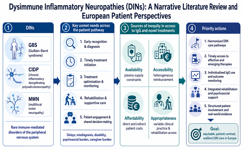
Given the clinical heterogeneity, diagnostic complexity, other associated risks, and fluctuating disease course characteristic of DINs such as CIDP, MMN, and GBS, it is unsurprising that unmet needs persist at nearly every juncture of the patient care continuum. These range from initial recognition and diagnostic delays to long-term treatment maintenance and system-level failures in patient engagement. The following sections outline key unmet needs across the DIN patient pathway, segmented into five critical stages. Each patient pathway stage has a unique set of unmet needs and corresponding solutions (see Table 1).
Screening and Diagnosis
DINs often present with nonspecific symptoms—weakness, fatigue, and sensory changes—that often overlap with a wide range of comorbidities such as neurological, infectious, metabolic, and psychological conditions. The absence of disease-specific biomarkers and the lack of a structured screening framework contribute significantly to the delayed and inaccurate diagnoses.
Patients with CIDP face a mean diagnostic delay of over 13 months: approximately 7.4 months between initial symptom onset and first medical consultation, and an additional 6.5 months from consultation to confirmed diagnosis [22,141]. This can lead not only to extended suffering (pain and fatigue are common symptoms), but also to irreversible nerve damage if appropriate therapy is not initiated in time. Misdiagnosis with conditions such as fibromyalgia, diabetic polyneuropathy, or GBS is common, and a considerable number of CIDP patients, especially if undiagnosed or misdiagnosed for extended periods of time, ultimately require assistive devices and intensive caregiver support—underscoring both the medical and socio-economic toll of diagnostic failure.
Similarly, patients with MMN often experience delays exceeding 15–24 months, frequently being misdiagnosed with more common neuromuscular disorders, such as amyotrophic lateral sclerosis (ALS) or cervical spondylotic myelopathy [81,142]. Non-neurological explanations - ranging from “nervous hysteria” to menopause or excessive caffeine intake - are sometimes proposed before appropriate neurological referral is made.
In the case of GBS, where early initiation of IgG therapy is directly correlated with better recovery, delays or errors in diagnosis may be particularly detrimental. Atypical presentations, including the pharyngeal-cervical-brachial variant, may be misattributed to bulbar strokes or cranial nerve pathologies. Other mimicking conditions, such as Lyme disease or West Nile virus, can further complicate clinical assessment, especially in non-specialist settings.
A major contributor to diagnostic inequity is the unequal distribution of specialised neurological services, particularly in rural or underserved regions, where generalists may lack familiarity with the nuanced presentations of DINs. This underscores the need for widespread professional education and greater diagnostic support—potentially including AI-assisted tools and clinical decision algorithms. European Reference Networks (ERNs), especially European Reference Network for Neuromuscular Diseases (ERN EURO-NMD), may also play a critical role by providing a unified platform for exchange of clinical and real-world data as well as an educational powerhouse accessible to all specialists across the EU.
Treatment Initiation
Even once a diagnosis is confirmed, initiating treatment can be hampered by systemic barriers, including complex regulatory frameworks, inconsistent reimbursement policies, and insufficient clinical guidance on treatment thresholds. Therefore, it should be emphasised that companies need to provide robust safety and efficacy data to allow for these treatments to be reimbursed and subsequently allow for patients’ access. These delays are particularly damaging in conditions such as GBS or rapidly progressive CIDP, where early intervention can dramatically alter prognosis.
Another common issue is sub-optimal dosing during and after treatment initiation. In CIDP, for example, insufficient IgG loading doses or inappropriate infusion intervals may lead to the misperception of treatment failure. This can prompt premature discontinuation or erroneous therapeutic switching to second-line agents—many of which carry greater risks and lower efficacy.
Clinical inertia and lack of familiarity with best-practice protocols often lead to corticosteroids being prescribed as the first-line therapy in some EU countries. This divergence from guidelines not only impairs patient outcomes but also contributes to unnecessary side effects and polypharmacy.
Treatment Maintenance
DINs are chronic conditions that require long-term disease control strategies. However, treatment maintenance is frequently undermined by rigid, non-personalised therapeutic regimens, especially in CIDP, where dosing and frequency must be continuously adjusted to accommodate disease variability, immunoglobulin pharmacokinetics, and patient-specific responses.
Patients often receive inflexible treatment plans, with fixed dosing intervals that fail to reflect real-world symptom fluctuation. For some, this leads to avoidable relapses and functional decline; for others, it results in overtreatment, with associated cost and adverse effects. Despite mounting evidence supporting the use of SCIg for stable maintenance therapy—especially in MMN and CIDP [82,143]—it remains underutilised in many European countries due to reimbursement restrictions, administrative complexity, or lack of provider’s familiarity. Similarly, too few countries allow IVIg care at home, which, when well organised, removes the burden of hospital visits from the patients.
Different sources estimate that 20–30% of CIDP patients show suboptimal or no response to IgG therapy [23], yet options for treatment-resistant disease remain limited. Immunosuppressants such as rituximab may be considered in refractory cases, but they carry a substantially higher risk profile, including infection and secondary malignancy. A review of existing therapies may be needed to assess their impact on DIN patients. A clear unmet need exists for prioritising treatment alternatives and other options as well as the development and approval of novel, targeted therapeutics that can be deployed when standard therapies fail.
Regular monitoring of long-term outcomes is also inconsistent. The use of validated clinical scores, patient-reported outcome measures, and biomarkers to titrate treatment remains patchy across different countries and different clinics within the same country. This contributes to the already-mentioned therapeutic inertia and inconsistency in long-term disease control.
Rehabilitation and Supportive Care
Despite the well-documented motor and sensory impairments associated with DINs, access to specialist rehabilitation services—including physiotherapy and occupational therapy—is sporadic and underfunded across Europe. This is particularly concerning for patients recovering from acute phases of GBS or those experiencing relapsing-remitting patterns in CIDP.
Inadequate rehabilitation not only impairs physical recovery but also increases dependency on caregivers, reduces employability, and contributes to social isolation. Yet, integration between neurology departments and rehabilitation services remains limited in many national health systems, with little to no dedicated care pathways for DIN patients.
The broader burden of associated complications—including fatigue, pain, depression, and anxiety—is also poorly addressed. These symptoms are often either medicalised inappropriately or dismissed as psychological sequelae, despite their direct link to disease pathophysiology and treatment side effects. Comprehensive, multidisciplinary care approaches that include mental health professionals, pain specialists, and social workers are rarely implemented in a structured manner.
Patient Engagement and Patient-Reported Evidence
The final, yet no less critical, area of unmet need concerns the systemic underrepresentation of patients in policy-making, guidelines development, and care pathways design. While the concept of patient-centred care is widely endorsed, meaningful patient involvement in the shaping of DIN care pathways remains limited.
Most notably, patient-reported outcomes, patient preferences, and patient experience, are seldom systematically collected or used to define treatment success (meaningful functional endpoints) or to inform evidence-based reimbursement decisions. Optimal rehabilitation must focus on individualised goal-setting and patient-reported functional goals (e.g., returning to work, being able to cook, resuming hobbies). Furthermore, there is minimal opportunity for DIN patient representatives to contribute to national HTA or European-level regulatory processes, as the main treatment with IgGs is considered “old” and thus very rarely subject to these proceedings. This absence limits the responsiveness of healthcare systems to real-world patient needs and distorts value assessments of therapies such as IgGs and the upcoming innovation that would benefit from accurate comparison with IgGs.
Patient-reported complications and comorbidities is a specific area, which lacks systematic appraisal and appropriate therapeutic strategies, and is frequently overlooked in care planning. Many DIN patients suffer from overlapping autoimmune disorders, cardiovascular risk factors, or metabolic conditions, yet integrated care models remain the exception rather than the norm. Holistically addressing comorbidities would not only improve quality of life but could also enhance therapeutic efficacy and ultimately lower the overall cost of illness.
Addressing Inequities in Access to Immunoglobulins and Novel Treatments for Patients with Dysimmune Inflammatory Neuropathies in Europe
Beyond the unmet needs across the patient pathway, DINs, as outlined throughout this paper, present a significant individual, clinical and socio-economic disease burden. IgG therapy, administered intravenously (IVIg) or subcutaneously (SCIg), is widely used as a treatment for many patients with DINs, having demonstrated superior efficacy and safety compared to alternatives such as corticosteroids in multiple studies detailed in the chapter on Evidence of Effectiveness and Safety of Immunoglobulins. Access to immunoglobulins across Europe present many challenges which can be categorised into four critical dimensions: availability, accessibility, affordability, and appropriateness (see Table 2).
Availability: Structural Constraints in Plasma Supply and Manufacturing
At the root of the availability crisis lies Europe’s systemic under-collection of human plasma, the essential raw material for immunoglobulin production. Current EU plasma collection volumes satisfy less than 60% of total IgG consumption. When assessed against actual medical need, the figure may fall as low as 30% [144], reflecting both underdiagnosis and underutilisation of IgGs in all appropriate indications, including DINs. Moreover, the plasma-derived medicinal products’ (PDMP) supply chain is inherently complex and fragile. The fractionation of plasma and the manufacture of IgG formulations require virus inactivation steps that rely on potent detergents, which have a notable environmental impact. As a result, the process is highly regulated, technologically complex, and time-consuming, typically requiring between 7 to 12 months from plasma collection to final product release. This makes the supply chain vulnerable to disruption, as seen during the COVID-19 pandemic. Compounding this vulnerability is Europe’s significant reliance on US-sourced plasma which relies on donors from across its border and accounts for approximately 40% of all plasma used in European PDMP manufacturing. Such ecosystem introduces geopolitical and logistical risks, undermining Europe’s pharmaceutical resilience.
Accessibility: Regulatory and Reimbursement Disparities Across Member States
Although the SmPCs of certain centrally authorised IVIg products approved through the European Medicines Agency (EMA) include indications such as GBS, CIDP and MMN, national payers still require robust comparative efficacy and safety data to support reimbursement decisions. In practice, the implementation of these decisions varies widely across member states. While some countries offer broad reimbursement for IgGs use in DIN indications, others apply highly restrictive eligibility criteria, limiting access to only a subset of patients. In several jurisdictions (e.g., Bulgaria, Croatia, Hungary, Italy, Lithuania, and Latvia), reimbursement may not extend to all DINs or is only partial, with patients required to pay out-of-pocket for a portion of their medicine. This creates an additional barrier for patients across all DINs and is particularly detrimental for those with MMN, for whom IgG is the only available medicine. Such variability in national policies undermines the principle of equity in access to these essential medicines.
Affordability: Financial Barriers and Risk of Catastrophic Expenditure
Even in countries where IgG medicine is formally reimbursed, patients often encounter indirect and direct costs that can be financially devastating. Out-of-pocket expenses may include treatment co-payments, costs for home carers, rehabilitation services not covered by insurance, equipment purchases and repeated travel to hospital-based IVIg infusion centres. For patients already suffering from diminished work capacity or unemployment due to physical disability, these costs pose a disproportionate burden. In this context, affordability is not merely about the drug price—it is about the total cost of living with a chronic, disabling illness. As such a better use of physiotherapy early in the patient pathway has the capacity to halt or off-set some of the costs that tend to multiply due to inappropriate or incomplete therapeutic options further down the disease progression. A narrow focus on pharmaceutical cost containment ignores the broader socio-economic burden faced by DIN patients and their families, as well as the value of appropriately treated DIN patients being able to return to productive employment and participate in a meaningful social life.
Appropriateness: Suboptimal Clinical Practices and Limited Rehabilitation
Clinical management of DINs varies significantly across Europe, not only in terms of access to IgGs, but also in terms of appropriateness of overall care provision. In certain countries, outdated treatment paradigms persist, with patients inappropriately receiving corticosteroids as the first-line therapy despite the established superiority of immunoglobulins. Where IgGs are used, the modalities of administration often fall short of optimal practice: low or inconsistent dosing, extended intervals between treatments, and underuse of SCIg despite its suitability for long-term home-based management. These inconsistencies reflect both clinical inertia and systemic gaps in guidance, negatively impact patients’ outcomes. While some countries benefit from national DINs treatment protocols, others lack clear, standardised recommendations. Access to rehabilitation, which provides crucial physical and psycho-social recovery strategies, is also uneven across Europe, often depending on local funding and infrastructure and in many cases requiring significant patient out-of-pocket expenses. As a result, treatment decisions may be shaped more by local habits, reimbursement policies, or economic constraints than by the adoption of individualised, evidence-based medicine.
Discussion, Conclusions and Recommendations
Discussion
Dysimmune inflammatory neuropathies (DINs), including Guillain–Barré syndrome (GBS), chronic inflammatory demyelinating polyradiculoneuropathy (CIDP), and multifocal motor neuropathy (MMN), represent a clinically heterogeneous group of rare but often disabling immune-mediated disorders of the peripheral nervous system. Although these conditions differ in their onset, natural history, pathophysiology, and treatment paradigms, they share a common pattern of substantial unmet needs across the patient pathways. These unmet needs extend from screening and diagnosis to treatment initiation, long-term disease control, rehabilitation, patient engagement, and system-level access to established and emerging therapies.
Each patient pathway stage is characterised by distinct, actionable gaps that require coordinated responses from clinicians, researchers, health authorities, and patient advocacy groups. Closing these gaps will necessitate investment in education and changes in clinical practice, enabling the most appropriate treatments in first- and subsequent-line settings, including greater use of physiotherapy and targeted use of novel therapies for specific patient subpopulations. Meaningful inclusion of the patient voice in system design with regards to diagnostic tools, alignment of treatment guidelines with clinical realities, expansion of access to comprehensive supportive care and rehabilitation should be considered. By addressing these unmet needs in an integrated and patient-centred fashion, European health systems have the opportunity not only to improve outcomes for individuals with DINs but also to serve as a model for equitable, evidence-based care in rare neurological diseases more broadly.
The inequities in access to immunoglobulin therapy for patients with DINs are equally multifactorial and persistent. They reflect not only reimbursement variation between countries, but also broader structural constraints in plasma supply, regulatory and payer heterogeneity, financial barriers for patients and families, and inconsistent implementation of appropriate clinical practice. Availability remains a foundational challenge because immunoglobulin therapy depends on a complex, highly regulated, and time-intensive plasma collection and manufacturing ecosystem. Europe’s under-collection of plasma and continuing reliance on imported plasma create a strategic vulnerability with direct implications for patients requiring IgG therapy. For rare, disabling conditions such as DINs, this vulnerability is not an abstract supply-chain issue; it directly affects treatment continuity, equity, and resilience of care.
Addressing sources of inequity will require a coordinated, multidimensional strategy at both EU and national levels. Strategic autonomy in plasma supply, regulatory harmonisation, equitable reimbursement, financial protection, and clinical standardisation all represent indispensable components of a future-proof policy response. At the policy level, these findings intersect with several ongoing European reform agendas, including the EU Regulation on Health Technology Assessment (Regulation (EU) 2021/2282), the Critical Medicines Act, the Substances of Human Origin framework, the General Pharmaceutical Legislation, and the proposed European Biotech Act. These initiatives create an opportunity to better align regulatory assessment, evidence generation, reimbursement decision-making, supply resilience, and patient-centred innovation. In particular, joint clinical assessment under the HTA Regulation could support earlier harmonisation of evidence expectations and reduce duplication across Member States, while still allowing national decision-makers to account for local health-system priorities. For DINs, such alignment could be especially valuable for emerging therapies, where robust comparative evidence, real-world data, and meaningful patient-reported outcomes will be essential to define appropriate use and to accelerate patient access, especially where high medical unmet needs persist.
Conclusions
DINs are rare, complex, and often disabling conditions that exert a disproportionate burden on patients, carers, healthcare systems, and society. Their impact extends beyond neurological impairment to include reduced independence, psychological distress, loss of productivity, caregiver burden, financial vulnerability, and diminished social participation. Although substantial progress has been made in understanding their diagnosis and treatment, important gaps remain across the entire care continuum.
The evidence reviewed in this paper points to a clear need for more coordinated, equitable, and patient-centred DIN care in Europe. Earlier recognition, timely referral, appropriate treatment initiation, individualised maintenance therapy, integrated rehabilitation, systematic outcome measurement, and meaningful patient involvement should form the foundation of future care pathways. At the same time, improving access to immunoglobulin therapy and emerging targeted treatments will require action beyond clinical practice alone, including stronger plasma supply resilience, more harmonised reimbursement frameworks, better financial protection, and greater consistency in the implementation of evidence-based patient-centred care.
Ensuring equitable access to existing therapeutic modalities and future innovations is not only a clinical necessity but a moral and a policy imperative. European health systems now have the opportunity to use DINs as a model for how rare neurological diseases can be addressed through integrated care pathways, patient-centred evidence generation, supply-chain resilience, and value-based access policies.
Placing patients with rare, disabling disorders at the centre of clinical, regulatory, reimbursement, and policy reform would not only improve outcomes for individuals with DINs, but also strengthen the broader European commitment to equity, resilience, and high-quality care.
Recommendations
- Patient Engagement: To embrace a patient-centred approach, national and supra-national institutions and bodies must ensure structured and permanent involvement of patient organisations in decision-making on regulatory approvals, reimbursement processes, guidelines creation, and medicines procurement.
- Unmet Needs across Patient Pathway: To close the numerous gaps and address the unmet needs in each of the DIN patient journey steps, European health systems must, as a pre-requisite, create and harmonise clear and detailed DIN Care Pathways. To do so, evidence-based protocols and educational materials should be created (or updated) to ensure all HCPs and stakeholders involved in DIN diagnosis, treatment, and rehabilitation have the necessary toolset, skillset, and mindset for the best-in-class and holistic disease management. The holistic disease management must include not only the golden standard treatment paradigms, but also disease-specific rehabilitation plans and psychosocial care for patients and carers alike. A pan-European tool (e.g. DIN barometer) could be introduced to assess and compare the patient journey in different Member States and identify outstanding gaps and unmet needs.
- Availability of Plasma-derived Immunoglobulins: To mitigate the systemic constraints to the availability of IgGs, the European Union must commit to achieving open strategic autonomy in plasma collection by gradually reducing and eventually eliminating the current high reliance on US plasma imports. This will require coordinated investment in national plasma collection infrastructure, harmonised donor recruitment frameworks, public awareness campaigns and greater coordination between the public and private sectors. More specifically, the EU Substances of Human Origin (SoHO) Coordination Board must effectively monitor and support increased national plasma collection efforts and include DIN patient experts in its working groups. Open strategic European autonomy must be underpinned by a national-level increase in plasma collection, and should be treated as a health security imperative, in line with the EU Critical Medicines Act (COM(2025) 102 final) proposed and EU Defence Autonomy.
- Accessibility of Effective and Safe Treatments: To start closing the access gap, EU member states should align the IgG regulatory and reimbursement status with the latest robust evidence from RCTs and real-world studies. European Reference Networks can facilitate and accelerate robust real-world evidence generation prospectively, in areas where clinical uncertainty persists. For the upcoming novel treatments, with strong evidence from ongoing randomised clinical trials, EU countries may consider early access schemes (similar to those systemically used in France and Germany) to minimise delays to provision of safe treatments for patients with currently no therapeutic options. FAST EU (Facilitating and Accelerating Strategic Clinical Trials in the EU/EEA) might also be used to ensure better evidence generation and greater early harmonisation between regulatory and HTA processes.
- Affordability of Care and Rehabilitation: To address issues with affordability of all facets of DIN care (treatment, supportive care, psychological care, physiotherapy), EU countries should adopt a more holistic approach to healthcare financing in high-burden diseases. This could include integrated “assisted living” packages that cover medication, holistic rehabilitation (including physiotherapy, psychotherapy, speech and occupational therapy), mobility aids. Physiotherapy or other adjunctive treatments, such as acupuncture, in specific cases, can be used as an early first-line treatment, which could improve patient outcomes by removing specific risks associated with invasive diagnostics and adverse effects of current treatment modalities. Funding models should recognise overall rehabilitation as an integral part of standard DIN care, not as optional or supplementary. Rehabilitation should not only target impairment reduction (strength, mobility) but also participation in daily life, work, and social roles, thus including vocational rehabilitation and ergonomic or environmental adaptations to support return to work and community participation. EU-level guidance on minimum financial protection standards for high-burden chronic diseases—including DINs—could also help mitigate the risk of catastrophic health expenditure.
- Appropriate use of Immunoglobulins and Novel Treatments: To ensure IgGs and other emerging therapeutic options are used safely and most effectively, clinicians, academic societies, policymakers and DIN patient advocates must work together to develop and implement comprehensive, pan-European treatment guidelines for treatment optimisation. These should encompass diagnostics, treatment initiation criteria, dosing regimens, monitoring protocols, and rehabilitation strategies. Importantly, these guidelines must be linked to implementation tools—such as education campaigns, prescribing support systems, and performance indicators—to ensure they translate into better patient outcomes. Additionally, real-world data should be better collected and analysed to support all of the above tools and systems, using existing platforms such as NEUROQUALI (https://neuroquali.sanoia.com/?lang=en) and ERN EURO-NMD (https://ern-euro-nmd.eu/) or creating new frameworks linking multiple ERNs focused on appropriate use of immunoglobulins across all relevant conditions.
- Future-proofing health systems and care pathways: EU should use the recent General Pharmaceutical Legislation (COM(2023) 192 final and COM(2023) 193 final) and the proposed European Biotech Act (COM(2025) 1022 final) as safeguards for the accelerated development of breakthrough innovation addressing unmet needs in DINs specifically and in rare diseases in general. The hallmarks of this approach should be to create a predictable innovative medicines’ evidence generation which leads to patients’ access. EU GPL could further strengthen incentives for the development of effective Orphan medicinal products, balancing patent and data exclusivity protection with a timely, equitable, and affordable patient access. The EU Biotech Act, in turn, should facilitate access to the innovative biotech therapies for DIN patients with well-assessed, fast-track regulatory pathways and clear investment signals to developers of innovative biotechnologies.
Author Contributions
Conceptualization, T.K. and J.-P.P.; methodology, T.K., A.K.-P. and J.-P.P.; formal analysis, T.K.; investigation, T.K.; resources, T.K. and J.-P.P.; data curation, T.K.; writing—original draft preparation, T.K.; writing—review and editing, T.K., A.K.-P., C.V.-G., C.S., P.P.W.L., M.S., N.V., S.S.-R., R.K. and J.-P.P.; visualization, T.K.; supervision, A.K.-P., C.S., M.S., S.S.-R. and J.-P.P.; project administration, T.K. and J.-P.P.; funding acquisition, J-P.P. All authors have read and agreed to the published version of the manuscript.
Funding
This research was funded by grants from argenx, CSL Behring, Grifols, Johnson & Johnson, LFB, Octapharma, Sanofi, Takeda, and Terumo. Grant numbers were not applicable or not provided. The APC was funded from the same grant support.
Institutional Review Board Statement
Ethical review and approval were waived for this study because the manuscript is a narrative literature review and patient-perspective synthesis, not an interventional or observational clinical study. The patient-perspective component was developed from secondary sources and through non-interventional consultation with EPODIN patient members and patient representatives, who provided lived-experience insights, contextual interpretation, and review of the manuscript from a patient-advocacy perspective. No patients were recruited as research subjects, no intervention was performed, no biological samples were collected, and no identifiable individual-level health data were analysed or reported. Patient input was used only in aggregated, non-identifiable form to contextualise the published evidence and strengthen the patient-centred interpretation of unmet needs.
Informed Consent Statement
Not applicable. This study did not involve the recruitment of human participants, the collection of new patient-level data, or the publication of identifiable individual patient information.
Data Availability Statement
No new clinical, experimental, or individual-level patient datasets were generated or analysed during the preparation of this manuscript. The scientific evidence discussed in this article is derived from publicly available literature cited in the reference list. Patient-perspective insights were obtained through non-interventional consultation and review by EPODIN patient members and representatives; these inputs were used in aggregated, non-identifiable form and are not publicly available in order to protect contributor privacy.
Acknowledgments
The authors acknowledge the European Patient Organisation for Dysimmune and Inflammatory Neuropathies (EPODIN) and its patient members and representatives for their lived-experience insights, patient-perspective review, and contribution to the interpretation of unmet needs across the DIN patient pathway. The authors also thank Steffen Thirstrup and Katerina Vucic of the European Medicines Agency for their scientific peer review and constructive comments on the manuscript. During the preparation of this manuscript, the authors used ChatGPT by OpenAI (GPT-5.5 Thinking) for language editing and consistency checks. The authors reviewed and edited the output and take full responsibility for the content of this publication.
Conflicts of Interest
This manuscript was funded by grants from argenx, CSL Behring, Grifols, Johnson & Johnson, LFB, Octapharma, Sanofi, Takeda, and Terumo. The funders had no role in the design of the review; in the collection, analysis, or interpretation of evidence; in the writing of the manuscript; or in the decision to submit or publish the work. The authors declare no other conflicts of interest.
References
- van Doorn, P.A.; Sommer, C.; Rajabally, Y.A. Autoimmune neuropathies: New guidelines and expert insights into clinical decision making. J. Neurol. Sci. 2025, 479, 125659. [Google Scholar] [CrossRef]
- Höftberger, R.; Lassmann, H. Inflammatory demyelinating diseases of the central nervous system. In Handbook of Clinical Neurology; Elsevier BV, 2017. [Google Scholar] [CrossRef]
- Ercolini, A.M.; Miller, S.D. Mechanisms of Immunopathology in Murine Models of Central Nervous System Demyelinating Disease. In The Journal of Immunology; American Association of Immunologists, 2006; 6, p. 176. [Google Scholar] [CrossRef] [PubMed]
- Mayo, L.; Quintana, F.J.; Weiner, H.L. The innate immune system in demyelinating disease. In Immunological Reviews; Wiley, 2012; 1, p. 248. [Google Scholar] [CrossRef] [PubMed]
- Cojocaru, I.M.; Cojocaru, M.; Siloşi, I.; Vrabie, C.D. Peripheral nervous system manifestations in systemic autoimmune diseases. PubMed 2014, 9(3), 289. Available online: https://pubmed.ncbi.nlm.nih.gov/25705295.
- Valentini, C.G.; Metafuni, E.; Gallo, L.; Giammarco, S.; Orlando, N.; Bianchi, M.; Sica, S.; Bacigalupo, A.; Chiusolo, P.; Teofili, L. ABO Mismatch in Allogeneic Hematopoietic Stem Cell Transplant: Effect on Short- and Long-term Outcomes. Transplant. Direct 2021, 7(8). [Google Scholar] [CrossRef] [PubMed]
- Lewis, R.A.; Muley, S.A. Chronic inflammatory demyelinating polyneuropathy: Treatment and prognosis. 2025. [Google Scholar] [CrossRef] [PubMed]
- Khadilkar, S.; Deshmukh, S.S.; Dhonde, P. Chronic dysimmune neuropathies: Beyond chronic demyelinating polyradiculoneuropathy. Ann. Indian Acad. Neurol. 2011, 14(2), 81. [Google Scholar] [CrossRef] [PubMed]
- Zheng, X.; Fang, Y.; Song, Y.; et al. Is there a causal nexus between COVID-19 infection, COVID-19 vaccination, and Guillain-Barré syndrome? Eur. J. Med. Res. 2023, 28, 98. [Google Scholar] [CrossRef] [PubMed]
- Gào, X.; Zhao, C.; Yang, J.; Yang, Z.; Feng, J.; Zhan, S.; Fan, D.; Liu, Z. Impact of COVID-19 vaccination coverage on global disability burden of Guillain-Barré syndrome. npj Vaccines 2025, 10(1), 182. [Google Scholar] [PubMed] [PubMed Central]
- Lee, H.J.; Abu Bakar, S.; Shin, O.S. An updated review of Zika virus vaccine development. Clin. Exp. Vaccine Res. 2025, 14(4), 325–334. [Google Scholar] [CrossRef] [PubMed] [PubMed Central]
- van Doorn, P.A.; Ruts, L.; Jacobs, B.C. Clinical features, pathogenesis, and treatment of Guillain-Barré syndrome. In The Lancet Neurology; Elsevier BV, 2008; 10, p. 7. [Google Scholar] [CrossRef] [PubMed]
- Tam, C.C.; O’Brien, S.J.; Rodrigues, L.C. Influenza, Campylobacter and Mycoplasma Infections, and Hospital Admissions for Guillain-Barré Syndrome, England. Emerg. Infect. Dis. 2006, 12(12), 1880–1887. [Google Scholar] [CrossRef] [PubMed]
- Tamborska, A.; Singh, B.; Leonhard, S.E.; Hodel, E.M.; Stowe, J.; Watson-Fargie, T.; Fernandes, P.M.; Themistocleous, A.C.; Roelofs, J.; Brennan, K.; et al. Guillain-Barré syndrome following SARS-CoV-2 vaccination in the UK: A prospective surveillance study. BMJ Neurol. Open 2022, 4, e000309. [Google Scholar] [CrossRef] [PubMed]
- Lehmann, H.C.; Burke, D.; Kuwabara, S. Chronic inflammatory demyelinating polyneuropathy: Update on diagnosis, immunopathogenesis and treatment. J. Neurol. Neurosurg. Psychiatry 2019, 90(9), BMJ. [Google Scholar] [CrossRef] [PubMed]
- Ieva, G.; Kalniņa, M.L.; Rots, D.; Roddate, M.; Gailīte, L.; Kurjāne, N.; Ķēniņa, V. Epidemiology and Clinical Course of Chronic Autoimmune Neuropathies During the SARS-CoV-2 Pandemic in Latvia. Open Neurol. J. 2023, 17. [Google Scholar] [CrossRef]
- Broers, M.C.; Bunschoten, C.; Nieboer, D.; Lingsma, H.F.; Jacobs, B.C. Incidence and prevalence of chronic inflammatory demyelinating polyradiculoneuropathy: A systematic review and meta-analysis. Neuroepidemiology 2019, 52, 161–172. [Google Scholar] [CrossRef] [PubMed]
- Brun, S.; de Sèze; Muller, S. CIDP: Current Treatments and Identification of Targets for Future Specific Therapeutic Intervention. Immuno 2022, 2(1), 118. [Google Scholar] [CrossRef]
- Löscher, W.N.; Oberreiter, E.M.; Erdler, M.; Quasthoff, S.; Culea, V.; Berek, K.; Embacher, N.; Grinzinger, S.; Hess, I.; Höger, F.S.; et al. Multifocal motor neuropathy in Austria: A nationwide survey of clinical features and response to treatment. J. Neurol. 2018, 265(12), 2834–2840. [Google Scholar] [PubMed] [PubMed Central]
- Broers, M.C.; de Wilde, M.; Lingsma, H.F.; van der Lei, J.; Verhamme, K.M.C.; Jacobs, B.C. Epidemiology of chronic inflammatory demyelinating polyradiculoneuropathy in The Netherlands. J. Peripher. Nerv. Syst. 2022, 27, 182–188. [Google Scholar] [CrossRef] [PubMed]
- Chaudhary, U.J.; Rajabally, Y.A. Underdiagnosis and diagnostic delay in chronic inflammatory demyelinating polyneuropathy. J. Neurol. 2020, 268(4), 1366. [Google Scholar] [CrossRef] [PubMed]
- Paci, S.; Arvin-Berod, C.; Wright, J.; Taylor, Y.; Ringland, C.; Sahar, R.; Plançon, J.-P. Burden of Illness and Unmet Need Among Patients with CIDP: Results from a Real-World Survey. 2024. [Google Scholar] [CrossRef] [PubMed]
- Querol, L.; Crabtree, M.; Herepath, M.; Priedane, E.; Viejo, I.V.; Agush, S.; Sommerer, P. Systematic literature review of burden of illness in chronic inflammatory demyelinating polyneuropathy (CIDP). In Journal of Neurology; 2020; 10, p. 268. [Google Scholar] [CrossRef] [PubMed]
- Gechev, A.; Kane, N.M.; Koltzenburg, M.; Rao, D.G.; van der Star, R. Potential risks of iatrogenic complications of nerve conduction studies (NCS) and electromyography (EMG). Clin. Neurophysiol. Pract. 2016, 1, 62–66. [Google Scholar] [CrossRef] [PubMed]
- European Medicines Agency (EMA). EMA’s final opinion confirms restrictions on use of linear gadolinium agents in body scans, 21 July 2017. Available online: https://www.ema.europa.eu/en/documents/press-release/emas-final-opinion-confirms-restrictions-use-linear-gadolinium-agents-body-scans_en.pdf EMA/457616/2017.
- Nascimento, O.J.M.; Marques, W.; Gonçalves, M.V.M.; Tomaselli, P.J.; Pupe, C.; França, M.C.; Gondim, F.d.A.A.; de Freitas, M.R.; Frezatti, R.S.S.; Oliveira, A.S.B.; et al. Brazilian Academy of Neurology recommendations for diagnosis, management, and treatment of chronic inflammatory demyelinating polyradiculoneuropathy (CIDP). Arq. De Neuro-Psiquiatr. 2025, 83(7), 1. [Google Scholar] [CrossRef] [PubMed]
- Caballero-Ávila, M.; Pascual-Goñi, E.; Lleixà, C.; Martín-Aguilar, L.; Collet-Vidiella, R.; Querol, L. The changing landscape of primary autoimmune neuropathies. Nat. Rev. Neurol. 2025, 21, 544–555. [Google Scholar] [CrossRef] [PubMed]
- Hughes, R.A.; Swan, A.V.; van Doorn, P.A. Intravenous immunoglobulin for Guillain-Barré syndrome [Review of Intravenous immunoglobulin for Guillain-Barré syndrome]. In Cochrane Library; Elsevier BV, 2014; 10, p. 2019. [Google Scholar] [CrossRef] [PubMed]
- Rahman, R.S.; Bauthman, M.S.; Alanazi, A.M.; Alsillah, N.N.; Alanazi, Z.M.; Almuhaysin, M.I.; Almutairi, R.K.; Binobaid, K.S.; Alharthi, Y.; Bawareth, R.M.; et al. Guillain–Barré syndrome: Pathophysiology, etiology, causes, and treatment. Int. J. Community Med. Public Health 2021, 8(7), 3624. [Google Scholar] [CrossRef]
- Cutillo, G.; Saariaho, A.-H.; Meri, S. Physiology of gangliosides and the role of antiganglioside antibodies in human diseases. In Cellular and Molecular Immunology; Springer Nature, 2020; 4, p. 17. [Google Scholar] [CrossRef] [PubMed]
- Waheed, S.; Bayas, A.; Hindi, F.; Rizvi, Z.; Espinosa, P.S. Neurological Complications of COVID-19: Guillain-Barre Syndrome Following Pfizer COVID-19 Vaccine. Cureus 2021. [Google Scholar] [CrossRef] [PubMed]
- Kaida, K.; Ariga, T.; Yu, R.K. Antiganglioside antibodies and their pathophysiological effects on Guillain-Barre syndrome and related disorders: A review. Glycobiology 2009, 19, 676–692. [Google Scholar] [CrossRef] [PubMed]
- Leonhard, S.E.; Papri, N.; Querol, L.; Rinaldi, S.; Shahrizaila, N.; Jacobs, B.C. Guillain-Barré syndrome. Nat. Rev. Dis. Prim. 2024, 10(1), 97. [Google Scholar] [PubMed]
- Hagen, K.M.; Ousman, S.S. The Neuroimmunology of Guillain-Barré Syndrome and the Potential Role of an Aging Immune System. In Frontiers in Aging Neuroscience; Frontiers Media, 2021. [Google Scholar] [CrossRef] [PubMed]
- He, L.; Zhang, G.; Liu, W.; Gao, T.; Sheikh, K.A. Anti-Ganglioside Antibodies Induce Nodal and Axonal Injury via Fcγ Receptor-Mediated Inflammation. J. Neurosci. 2015, 35(17), 6770–6785. [Google Scholar] [PubMed] [PubMed Central]
- Pinto, A.; Sèze, de; Jacob, A.; Reddel, S.W.; Yudina, A.; Tan, K. Comparison of IVIg and TPE efficacy in the treatment of neurological disorders: A systematic literature review. In Therapeutic Advances in Neurological Disorders; SAGE Publishing, 2023. [Google Scholar] [CrossRef] [PubMed]
- Rajabally, Y.A. Immunoglobulin and monoclonal antibody therapies in Guillain-Barré syndrome. Neurotherapeutics 2022, 19, 885–896. [Google Scholar] [CrossRef] [PubMed]
- Dash, S.; Pai, A.R.; Kamath, U.; Rao, P. Pathophysiology and diagnosis of Guillain-Barré syndrome—challenges and needs. Int. J. Neurosci. 2015, 125, 235–240. [Google Scholar] [CrossRef] [PubMed]
- Koike, H.; Chiba, A.; Katsuno, M. Emerging infection, vaccination, and Guillain-Barré syndrome: A review. Neurol. Ther. 2021, 10, 523–537. [Google Scholar] [CrossRef] [PubMed]
- Wakerley, B.R.; Uncini, A.; Yuki, N. Guillain–Barré and Miller Fisher syndromes—new diagnostic classification. In Nature Reviews Neurology; 2014; 9, p. 10. [Google Scholar] [CrossRef] [PubMed]
- Hughes, R.A.C.; Cornblath, D.R. Guillain-Barré syndrome. Lancet 2005, 366, 1653–1666. [Google Scholar] [CrossRef] [PubMed]
- Mosna, K.; Vadkerti, P.; Papp, L.; Palkovic, M.; Janega, P.; Babal, P. Guillain-Barré Syndrome with Lethal Outcome Following COVID-19 Vaccination - Case Report Supported by Autopsy Examination. Open Neurol. J. 2022, 16, e1874205X2207270. [Google Scholar] [CrossRef]
- Oshomoji, O.I.; Ajiroba, J.O.; Semudara, S.O.; Olayemi, M.A.; Adeoye, S.O. Autoimmune mechanisms in Guillain-Barré syndrome subtypes: A systematic review. Bull. Fac. Phys. Ther. 2024, 29, 86. [Google Scholar] [CrossRef]
- Stoian, A.; Bălașa, R.; Grigorescu, B.L.; Maier, S.; Andone, S.; Cocuz, I.G.; Bajko, Z.; Filep, C.R.; Stoian, M. Guillain-Barré syndrome associated with COVID-19: A close relationship or just a coincidence? Exp. Ther. Med. 2021, 22, 916. [Google Scholar] [CrossRef] [PubMed]
- den Bergh, P.V.; van Doorn, P.A.; Hadden, R.D.M.; Avau, B.; Vankrunkelsven, P.; Allen, J.A.; Attarian, S.; Blomkwist-Markens, P.H.; Cornblath, D.R.; Eftimov, F.; et al. European Academy of Neurology/Peripheral Nerve Society guideline on diagnosis and treatment of chronic inflammatory demyelinating polyradiculoneuropathy: Report of a joint Task Force—Second revision. Eur. J. Neurol. 2021, 28(11), 3556. [Google Scholar] [CrossRef] [PubMed]
- Bus, S.R.M.; de Haan, R.J.; Vermeulen, M.; van Schaik, I.N.; Eftimov, F. Intravenous immunoglobulin for chronic inflammatory demyelinating polyradiculoneuropathy. Cochrane Database Syst. Rev. 2024, 2(2), CD001797. [Google Scholar] [CrossRef] [PubMed]
- Rajabally, Y.A.; Attarian, S.; Delmont, E. Evolving immunologic perspectives in chronic inflammatory demyelinating polyneuropathy. J. Inflamm. Res. 2020, 13, 543–549. [Google Scholar] [CrossRef] [PubMed]
- Koike, H.; Katsuno, M. Pathophysiology of Chronic Inflammatory Demyelinating Polyneuropathy: Insights into Classification and Therapeutic Strategy Neurology and Therapy; Springer Healthcare: Adis, 2020; 2, p. 9. [Google Scholar] [CrossRef] [PubMed]
- Gable, K.L.; Li, Y. Chronic Inflammatory Demyelinating Polyneuropathy: How Pathophysiology Can Guide Treatment. Muscle Nerve 2025, 72, 201–211. [Google Scholar] [CrossRef] [PubMed]
- Mathey, E.K.; Park, S.B.; Hughes, R.A.; Pollard, J.D.; Armati, P.J.; Barnett, M.; Taylor, B.; Dyck, P.J.B.; Kiernan, M.C.; Lin, C.S.-Y. Chronic inflammatory demyelinating polyradiculoneuropathy: From pathology to phenotype. J. Neurol. Neurosurg. Psychiatry 2015, 86, 973–985. [Google Scholar] [CrossRef] [PubMed]
- Doneddu, P.E.; Dentoni, M.; Nobile-Orazio, E. Atypical chronic inflammatory demyelinating polyradiculoneuropathy: Recent advances on classification, diagnosis, and pathogenesis. In Current Opinion in Neurology; Lippincott Williams & Wilkins, 2021; 5, p. 34. [Google Scholar] [CrossRef] [PubMed]
- Collet-Vidiella, R.; De Lorenzo, A.; Querol, L. Autoimmune nodopathies: Emerging insights and clinical implications. Curr. Opin. Neurol. 2025, 38(5), 452–458. [Google Scholar] [PubMed]
- Querol, L.; Hartung, H.P.; Lewis, R.A.; van Doorn, P.A.; Hammond, T.R.; Atassi, N.; Alonso-Alonso, M.; Dalakas, M.C. The Role of the Complement System in Chronic Inflammatory Demyelinating Polyneuropathy: Implications for Complement-Targeted Therapies. Neurotherapeutics 2022, 19(3), 864–873. [Google Scholar] [CrossRef] [PubMed] [PubMed Central]
- Lewis, R.A. Tips in navigating the diagnostic complexities of chronic inflammatory demyelinating polyradiculoneuropathy. J. Neurol. Sci. 2022, 442, 120420. [Google Scholar] [CrossRef] [PubMed]
- Shimizu, S.; Iijima, M.; Fukami, Y.; Tamura, N.; Nakatochi, M.; Ando, M.; Nishi, R.; Koike, H.; Kaida, K.; Koga, M.; et al. Efficacy and Safety of Rituximab in Refractory CIDP With or Without IgG4 Autoantibodies (RECIPE): Protocol for a Double-Blind, Randomized, Placebo-Controlled Clinical Trial. JMIR Res. Protoc. 2020, 9(4). [Google Scholar] [CrossRef] [PubMed]
- Desai, U. How I Treat Chronic Inflammatory Demyelinating Polyneuropathy Podcast. Neurol. Ther. 2023, 12(5), 1409. [Google Scholar] [CrossRef] [PubMed]
- Multifocal motor neuropathy; Definitions, 2020. [CrossRef]
- Claytor, B.; Polston, D.; Li, Y. Multifocal Motor Neuropathy: A Narrative Review. Muscle Nerve 2025, 71, 512–534. [Google Scholar] [CrossRef] [PubMed]
- Piepers, S.; Jansen, M.; Cats, E.A.; van Sorge, N.M.; den Berg, L.H.va.; Pol, W.L.v.d. IVIg inhibits classical pathway activity and anti-GM1 IgM-mediated complement deposition in MMN. J. Neuroimmunol. 2010, 229, 256. [Google Scholar] [CrossRef] [PubMed]
- Vlam, L.; Cats, E.A.; Harschnitz, O.; Jansen, M.; Piepers, S.; Veldink, J.H.; Franssen, H.; Stork, A.C.J.; Heezius, E.; Rooijakkers, S.H.M.; et al. Complement activity is associated with disease severity in multifocal motor neuropathy. Neurol. Neuroimmunol. Neuroinflammation 2015, 2(4). [Google Scholar] [CrossRef] [PubMed]
- Yuki, N.; Watanabe, H.; Nakajima, T.; Späth, P. IVIG blocks complement deposition mediated by anti-GM1 antibodies in multifocal motor neuropathy. J. Neurol. Neurosurg. Psychiatry 2010, 82(1), 87. [Google Scholar] [CrossRef] [PubMed]
- Hameed, S.; Cascella, M. Multifocal Motor Neuropathy. In StatPearls [Internet]; Available from; StatPearls Publishing: Treasure Island (FL); NCBI Bookshelf, 2026. [Google Scholar]
- Wohnrade, C.; Seeliger, T.; Gingele, S.; Bjelica, B.; Skripuletz, T.; et al. Diagnostic value of neurofilaments in differentiating motor neuron disease from multifocal motor neuropathy. J. Neurol. 2024, 271, 4441–4452. [Google Scholar] [CrossRef] [PubMed]
- Keddie, S.; Efimov, F.; Berg, L.H.; van den; Brassington, R.; Haan, R.J.; de; Schaik, I.N.; van. Immunoglobulin for multifocal motor neuropathy (Review). 2022. [Google Scholar] [CrossRef] [PubMed]
- Joint Task Force of the EFNS and the PNS. European Federation of Neurological Societies/Peripheral Nerve Society guideline on management of multifocal motor neuropathy. Report of a joint task force of the European Federation of Neurological Societies and the Peripheral Nerve Society--first revision. J. Peripher Nerv. Syst. 2010, 15(4), 295–301. [Google Scholar] [CrossRef] [PubMed]
- Katz, J.; Lewis, R.A.; Spatafora, D. First Global Multifocal Motor Neuropathy (MMN) Quality of Life (QOL) Patient Survey Identifies Needs in Education and Treatment (P2.061). Neurology 2017, 88. [Google Scholar] [CrossRef]
- van Doorn, P.A.; Garssen, M.P.J. Treatment of immune neuropathies. In Current Opinion in Neurology; Lippincott Williams & Wilkins, 2002; 5, p. 15. [Google Scholar] [CrossRef] [PubMed]
- Fernández-Cruz, E.; Alecsandru, D.; Sánchez-Ramón, S. Mechanisms of action of immune globulin. Clin. Exp. Immunol. 2009, 157, 1–2. [Google Scholar] [CrossRef] [PubMed]
- Gardulf, A.; Nicolay, U. Replacement IgG therapy and self-therapy at home improve the health-related quality of life in patients with primary antibody deficiencies. Curr. Opin. Allergy Clin. Immunol. 2006, 6(6), 434–442. [Google Scholar] [CrossRef] [PubMed]
- Alsolaihim, A.; Baker, S.K. Dose Adjustment of Subcutaneous IgG in Chronic Inflammatory Demyelinating Polyneuropathy. Case Rep. Neurol. 2020, 12, 73–77. [Google Scholar] [CrossRef] [PubMed]
- Reeves, H.M.; Winters, J.L. The mechanisms of action of plasma exchange. Br. J. Haematol. 2014, 164(3), 342–351. [Google Scholar] [PubMed]
- Osman, C.; Jennings, R.; El-Ghariani, K.; Pinto, A. Plasma exchange in neurological disease. In Practical Neurology; BMJ, 2019; 2, p. 20. [Google Scholar] [CrossRef] [PubMed]
- Dalakas, M.C. Update on Intravenous Immunoglobulin in Neurology: Modulating Neuro-autoimmunity, Evolving Factors on Efficacy and Dosing and Challenges on Stopping Chronic IVIg Therapy. In Neurotherapeutics; Springer Science+Business Media, 2021; 4, p. 18. [Google Scholar] [CrossRef] [PubMed]
- Winer, J.B. Treatment of Guillain-Barré syndrome. 2023. Available online: https://academic.oup.com/qjmed/article/95/11/717/1543041.
- Abbas, A.M.; Rajabally, Y.A. Complications of Immunoglobulin Therapy and Implications for Treatment of Inflammatory Neuropathy: A Review. In Current Drug Safety; Bentham Science Publishers, 2018; 1, p. 14. [Google Scholar] [CrossRef] [PubMed]
- Rajabally, Y.A. Unconventional Treatments for Chronic Inflammatory Demyelinating Polyneuropathy. In Neurodegenerative Disease Management; Future Medicine, 2017; 5, p. 7. [Google Scholar] [CrossRef] [PubMed]
- Castle, D.; Robertson, N.P. Alternatives to intravenous immunoglobulin treatment in chronic inflammatory demyelinating polyradiculoneuropathy. In Journal of Neurology; Springer Science+Business Media, 2019; Vol. 266, Issue 9, p. 2338. [Google Scholar] [CrossRef] [PubMed]
- Li, S.; Li, Q.; Li, Y.; Li, L.; Tian, H.; Sun, X. Acetyl-L-Carnitine in the Treatment of Peripheral Neuropathic Pain: A Systematic Review and Meta-Analysis of Randomized Controlled Trials. PLoS ONE 2015, 10(3), e0119479. [Google Scholar] [PubMed] [PubMed Central]
- Julian, T.; Glascow, N.; Syeed, R.; Zis, P. B12 as a Treatment for Peripheral Neuropathic Pain: A Systematic Review. Nutrients 2020, 12(8), 2221. [Google Scholar] [CrossRef] [PubMed Central]
- van Doorn, P.A.; den Bergh, P.V.; Hadden, R.D.M.; Avau, B.; Vankrunkelsven, P.; Attarian, S.; Blomkwist-Markens, P.H.; Cornblath, D.R.; Goedee, H.S.; Harbo, T.; et al. European Academy of Neurology/Peripheral Nerve Society Guideline on diagnosis and treatment of Guillain–Barré syndrome. Eur. J. Neurol. 2023, 30(12), 3646. [Google Scholar] [CrossRef] [PubMed]
- Thyerlei, D.; Weiss, M. Pure Motor Chronic Inflammatory Demyelinating Polyneuropathy: Relationship to Multifocal Motor Neuropathy with Conduction Block. 2024. [Google Scholar]
- Goyal, N.; Karam, C.; Sheikh, K.A.; Dimachkie, M.M. Subcutaneous immunoglobulin treatment for chronic inflammatory demyelinating polyneuropathy [Review of Subcutaneous immunoglobulin treatment for chronic inflammatory demyelinating polyneuropathy]. In Muscle & Nerve; Wiley, 2021; 3, p. 64. [Google Scholar] [CrossRef] [PubMed]
- Cats, E.A.; der Pol, W.L.v.; Piepers, S.; Franssen, H.H.; Jacobs, B.C.; den Berg-Vos, R.M.v.; Kuks, J.B.M.; van Doorn, P.A.; van Engelen, B.G.M.; Verschuuren, J.J.G.M.; et al. Correlates of outcome and response to IVIg in 88 patients with multifocal motor neuropathy. Neurology 2010, 75(9), 818. [Google Scholar] [CrossRef] [PubMed]
- Adrichem, M.E.; Lucke, I.M.; Vrancken, A.F.J.E.; Goedee, H.S.; Wieske, L.; Dijkgraaf, M.G.W.; Voermans, N.C.; Notermans, N.C.; Faber, C.G.; Visser, L.H.; et al. Withdrawal of intravenous immunoglobulin in chronic inflammatory demyelinating polyradiculoneuropathy. Brain 2022, 145(5), 1641–1652. [Google Scholar] [CrossRef] [PubMed Central]
- Bajpayee, A.; Kaur, A.; Bajpai, N.; Kothari, N. Daily plasma exchange in severe Guillain–Barré syndrome helps in early weaning from the ventilator: A lesson from a case. Asian J. Transfus. Sci. 2017, 11(2), 206. [Google Scholar] [CrossRef] [PubMed]
- Walk, D.; Li, L.Y.; Parry, G.J.; Day, J.W. Rapid resolution of quadriplegic CIDP by combined plasmapheresis and IVIg. Neurology 2004, 62(1), 155–156. [Google Scholar] [CrossRef] [PubMed]
- Kuwabara, S.; Misawa, S.; Mori, M.; Iwai, Y.; Ochi, K.; Suzuki, H.; Nodera, H.; Tamaoka, A.; Iijima, M.; Toda, T.; et al. Intravenous immunoglobulin for maintenance treatment of multifocal motor neuropathy: A multi-center, open-label, 52-week phase 3 trial. J. Peripher. Nerv. Syst. 2018, 23(2), 115. [Google Scholar] [CrossRef] [PubMed]
- Kumar, A.; Patwa, H.; Nowak, R.J. Immunoglobulin therapy in the treatment of multifocal motor neuropathy. In Journal of the Neurological Sciences; Elsevier BV, 2017; p. 375. [Google Scholar] [CrossRef] [PubMed]
- Nobile-Orazio, E.; Meucci, N.; Barbieri, S.; Carpo, M.; Scarlato, G. High-dose intravenous immunoglobulin therapy in multifocal motor neuropathy. Neurology 1993, 43, 537. [Google Scholar] [CrossRef] [PubMed]
- Jovanovich, E.; Karam, C. Human immune globulin infusion in the management of multifocal motor neuropathy. In Degenerative Neurological and Neuromuscular Disease; Dove Medical Press, 2015. [Google Scholar] [CrossRef] [PubMed]
- Kozul, T.K.; Yudina, A.; Donovan, C.; Pinto, A.; Osman, C. Cost-minimisation analysis of plasma exchange versus IVIg in the treatment of autoimmune neurological conditions. BMC Health Serv. Res. 2022, 22(1). [Google Scholar] [CrossRef] [PubMed]
- Zhu, L.; Hou, H.; Wang, S.; Zhang, S.; Wang, G.; Guo, Z.; Wu, J. FcRn inhibitors: A novel option for the treatment of myasthenia gravis. In Neural Regeneration Research; Medknow, 2022. [Google Scholar] [CrossRef] [PubMed]
- Peter, H.; Ochs, H.D.; Cunningham-Rundles, C.; Vinh, D.C.; Kießling, P.; Greve, B.; Jolles, S. Targeting FcRn for immunomodulation: Benefits, risks, and practical considerations. In Journal of Allergy and Clinical Immunology; Elsevier BV, 2020; 3, p. 146. [Google Scholar] [CrossRef] [PubMed]
- Bussel, J.B.; Cines, D.B.; Blumberg, R.S. Neonatal Fc Receptor - Biology and Therapeutics. N Engl. J. Med. 2025, 392(16), 1621–1635. [Google Scholar] [CrossRef]
- Ulrichts, P.; Guglietta, A.; Dreier, T.; Bragt, T.V.; Hanssens, V.; Hofman, E.; Vankerckhoven, B.; Verheesen, P.; Ongenae, N.; Lykhopiy, V.; et al. Neonatal Fc receptor antagonist efgartigimod safely and sustainably reduces IgGs in humans. J. Clin. Investig. 2018, 128(10), 4372. [Google Scholar] [CrossRef] [PubMed]
- Allen, J.A.; Lin, J.; Basta, I.; Dysgaard, T.; Eggers, C.; Guptill, J.T.; Gwathmey, K.G.; Hewamadduma, C.; Hofman, E.; Hussain, Y.M.; et al. ADHERE Study Group. Safety, tolerability, and efficacy of subcutaneous efgartigimod in patients with chronic inflammatory demyelinating polyradiculoneuropathy (ADHERE): A multicentre, randomised-withdrawal, double-blind, placebo-controlled, phase 2 trial. Lancet Neurol. 2024, 23, 1013–1024. [Google Scholar] [CrossRef] [PubMed]
- Gorson, K.C. Evolving understanding of Guillain-Barré syndrome pathophysiology and the central role of the classical complement pathway in axonal injury. Front Neurol. 2025, 16, 1572949. [Google Scholar] [PubMed] [PubMed Central]
- Qin, Z.; Huang, Q.; Zou, J.; Tang, L.; Hu, Z.; Tang, X. Progress in Hematopoietic Stem Cell Transplantation for CIDP. In International Journal of Medical Sciences; Ivyspring International Publisher, 2020; 2, p. 17. [Google Scholar] [CrossRef] [PubMed]
- Greco, R.; Ruggeri, A.; McLornan, D.P.; et al. Indications for haematopoietic cell transplantation and CAR-T for haematological diseases, solid tumours and immune disorders: 2025 EBMT practice recommendations. Bone Marrow Transpl. 2025, 60, 1499–1525. [Google Scholar] [CrossRef] [PubMed]
- Burman, J.; Tolf, A.; Hägglund, H.; Askmark, H. Autologous haematopoietic stem cell transplantation for neurological diseases. J. Neurol. Neurosurg. Psychiatry 2017, 89(2), BMJ. [Google Scholar] [CrossRef] [PubMed]
- Lutter, L.; Spierings, J.; van Rhijn-Brouwer, F.C.C.-C.; van Laar, J.M.; van Wijk, F. Resetting the T Cell Compartment in Autoimmune Diseases With Autologous Hematopoietic Stem Cell Transplantation: An Update. In Frontiers in Immunology; Frontiers Media, 2018. [Google Scholar] [CrossRef] [PubMed]
- Burt, R.K.; Balabanov, R.; Tavee, J.; Han, X.; Sufit, R.; Ajroud-Driss, S.; Jovanovic, B.; Quigley, K.; Arnautovic, I.; Helenowski, I.; et al. Hematopoietic stem cell transplantation for chronic inflammatory demyelinating polyradiculoneuropathy. J. Neurol. 2020, 267(11), 3378. [Google Scholar] [CrossRef] [PubMed]
- Press, R.; Askmark, H.; Svenningsson, A.; Andersen, O.; Axelson, H.; Strömberg, U.; Wåhlin, A.; Isaksson, C.; Johansson, J.-E.; Hägglund, H. Autologous haematopoietic stem cell transplantation: A viable treatment option for CIDP. J. Neurol. Neurosurg. Psychiatry 2013, 85(6), 618. [Google Scholar] [CrossRef] [PubMed]
- Singh, D.B. The impact of pharmacogenomics in personalized medicine. Adv. Biochem Eng. Biotechnol. 2020, 171, 369–394. [Google Scholar] [CrossRef] [PubMed]
- Burt, R.K.; Tappenden, P.; Balabanov, R.; Han, X.; Quigley, K.; Snowden, J.A.; Sharrack, B. The Cost Effectiveness of Immunoglobulin vs. Hematopoietic Stem Cell Transplantation for CIDP. Front. Neurol. 2021, 12. [Google Scholar] [CrossRef] [PubMed]
- de Pedro, S.; Sánchez-Bayle, C.; Poveda, J.L.; Martínez-García, C.; Díaz-Manera, J. Cost analysis of immunoglobulin maintenance treatment of patients with chronic inflammatory demyelinating polyneuropathy in Spain. PharmacoEconomics Open. 2026, 10(2), 273–286. [Google Scholar] [CrossRef] [PubMed]
- Mengel, D.; Fraune, L.; Sommer, N.; Stettner, M.; Reese, J.P.; Dams, J.; Glynn, R.J.; Balzer-Geldsetzer, M.; Dodel, R.; Tackenberg, B. Costs of illness in chronic inflammatory demyelinating polyneuropathy in Germany. Muscle Nerve 2018, 58(5), 681. [Google Scholar] [CrossRef] [PubMed]
- Božović, I.; Perić, S.; Basta, I.; Kačar, A.; Nikolić, A.; Belanovic, B.; Lavrnić, D.; Rakočević-Stojanović, V.; Stević, Z. Quality of life in patients with multifocal motor neuropathy from Serbia. J. Neurol. Sci. 2019, 399, 151. [Google Scholar] [CrossRef] [PubMed]
- Allen, J.A.; Butler, L.M.; Levine, T.; Haudrich, A. A Global Survey of Disease Burden in Patients Who Carry a Diagnosis of Chronic Inflammatory Demyelinating Polyneuropathy. Adv. Ther. 2020, 38(1), 316. [Google Scholar] [CrossRef] [PubMed]
- Ryltoft, A.; Al-Zuhairy, A.; Sindrup, S.H.; Andersen, H.; Markvardsen, L.K. Quality of life in chronic inflammatory demyelinating polyneuropathy patients treated with subcutaneous immunoglobulin. Acta Neurol. Scand. 2020, 142(6), 637. [Google Scholar] [CrossRef] [PubMed]
- Stangel, M.; Gold, R.; Pittrow, D.; Baumann, U.; Borte, M.; Fasshauer, M.; Hensel, M.; Huscher, D.; Reiser, M.; Sommer, C. Treatment of patients with multifocal motor neuropathy with immunoglobulins in clinical practice: The SIGNS registry. Ther. Adv. Neurol. Disord. 2016, 9(3), 165–179. [Google Scholar] [CrossRef] [PubMed]
- Musche, V.; Bäuerle, A.; Jahre, L.M.; Schweda, A.; Dinse, H.; Moradian, S.; Weismüller, B.; Fink, M.; Wolters, A.; Fleischer, M.; et al. COVID-19-Related Burden and Risk Perception in Individuals with Chronic Inflammatory Demyelinating Polyneuropathy and Multifocal Motor Neuropathy: A Cross-Sectional Study. Neurol. Ther. 2022, 11(3), 1135. [Google Scholar] [CrossRef] [PubMed]
- Arvin-Berod, C.; Brackx, F.; Van de Veire, L.; Paci, S.; Taylor, Y.; Wright, J.; Del Real, A.P.; Plançon, J.P.; Sperry, R.; Fix, C.; et al. The journey to diagnosis for patients with CIDP: Results from a real-world international survey. Front Neurol. 2026, 16, 1748903. [Google Scholar] [PubMed] [PubMed Central]
- Broers, M.C.; Bunschoten, C.; Drenthen, J.; Beck, T.A.O.; Brusse, E.; Lingsma, H.F.; Allen, J.A.; Lewis, R.A.; van Doorn, P.A.; Jacobs, B.C. Misdiagnosis and diagnostic pitfalls of chronic inflammatory demyelinating polyradiculoneuropathy. Eur. J. Neurol. 2021, 28(6), 2065. [Google Scholar] [CrossRef] [PubMed]
- Pop-Busui, R.; Ang, L.; Boulton, A.J.M.; Feldman, E.L.; Marcus, R.L.; Mizokami-Stout, K.; Singleton, J.R.; Ziegler, D. Diagnosis and Treatment of Painful Diabetic Peripheral Neuropathy. ADA Clin. Compend. 2022, 2022(1), 1. [Google Scholar] [CrossRef] [PubMed]
- van Doorn, I.; Eftimov, F.; Wieske, L.; van Schaik, I.; Verhamme, C. Challenges in the Early Diagnosis and Treatment of Chronic Inflammatory Demyelinating Polyradiculoneuropathy in Adults: Current Perspectives. Ther. Clin. Risk Manag. 2024, 111. [Google Scholar] [CrossRef] [PubMed]
- Eftimov, F.; Lucke, I.M.; Querol, L.; Rajabally, Y.A.; Verhamme, C. Diagnostic challenges in chronic inflammatory demyelinating polyradiculoneuropathy [Review of Diagnostic challenges in chronic inflammatory demyelinating polyradiculoneuropathy]. In Brain; Oxford University Press, 2020; 11, p. 143. [Google Scholar] [CrossRef] [PubMed]
- European Medicines Agency. Kiovig, INN-human normal immunoglobulin (IVIg): EPAR product information [Internet]; Available from; EMA: Amsterdam; European Medicines Agency, 21 May 2026. [Google Scholar]
- European Medicines Agency. Hizentra, INN-human normal immunoglobulin: EPAR product information [Internet]; Available from; EMA: Amsterdam; European Medicines Agency, 2025. [Google Scholar]
- Yasir, M.; Goyal, A.; Sonthalia, S. Corticosteroid adverse effects. In StatPearls; Available from; StatPearls Publishing: Treasure Island (FL); NCBI Bookshelf.
- European Medicines Agency (EMA). Available online: www.ema.europa.eu/en/documents/product-information/vyvgart-epar-product-information_en.pdf.
- Hsieh, L.; Song, J.; Tremoulet, A.H.; Burns, J.C.; Franco, A. Intravenous immunoglobulin induces IgG internalization by tolerogenic myeloid dendritic cells that secrete IL-10 and expand Fc-specific regulatory T cells. Clin. Exp. Immunol. 2022, 208(3), 361. [Google Scholar] [CrossRef] [PubMed]
- Mausberg, A.K.; Heininger, M.K.; Meyer Zu Horste, G.; Cordes, S.; Fleischer, M.; Szepanowski, F.; Kleinschnitz, C.; Hartung, H.P.; Kieseier, B.C.; Stettner, M. NK cell markers predict the efficacy of IV immunoglobulins in CIDP. Neurol. Neuroimmunol. Neuroinflamm 2020, 7(6), e884. [Google Scholar] [PubMed] [PubMed Central]
- Galeotti, C.; Kaveri, S.V.; Bayry, J. IVIG-mediated effector functions in autoimmune and inflammatory diseases. Int. Immunol. 2017, 29(11), 491–498. [Google Scholar] [CrossRef]
- Manganotti, P.; Garascia, G.; Furlanis, G.; Stella, A.B. Efficacy of intravenous immunoglobulin (IVIg) on COVID-19-related neurological disorders over the last 2 years: An up-to-date narrative review. In Frontiers in Neuroscience; Frontiers Media, 2023. [Google Scholar] [CrossRef] [PubMed]
- Kaufman, G.; Massoud, A.H.; Dembele, M.; Yona, M.; Piccirillo, C.A.; Mazer, B. Induction of Regulatory T Cells by Intravenous Immunoglobulin: A Bridge between Adaptive and Innate Immunity Frontiers in Immunology; Frontiers Media, 2015. [Google Scholar] [CrossRef] [PubMed]
- Gong, Z.; Chen, Z.; Li, D.; Lu, X.; Wu, J.; Sun, H.; Wang, X.; Liu, S.; Xia, X.; Lu, F.; et al. Hydrogel loaded with cerium-manganese nanoparticles and nerve growth factor enhances spinal cord injury repair by modulating immune microenvironment and promoting neuronal regeneration. J. Nanobiotechnology 2025, 23(1). [Google Scholar] [CrossRef] [PubMed]
- Viengkhou, B.; Hofer, M.J. Breaking down the cellular responses to type I interferon neurotoxicity in the brain. In Frontiers in Immunology; Frontiers Media, 2023. [Google Scholar] [CrossRef] [PubMed]
- Chen, K.; Liu, J.; Cao, X. Regulation of type I interferon signaling in immunity and inflammation: A comprehensive review. In Journal of Autoimmunity; Elsevier BV, 2017; p. 83. [Google Scholar] [CrossRef] [PubMed]
- Brunetti, L.; Chapy, H.; Nahass, R.; Moore, R.; Wassef, A.; Adler, D.; Yurkow, E.J.; Kagan, L. Relationship between Body Composition and Serum Immunoglobulin Concentrations after Administration of Intravenous Immune Globulin–Preclinical and Clinical Evidence. Pharmaceutics 2023, 15(2), 510. [Google Scholar] [CrossRef] [PubMed]
- Kebede, M.A.; Tekle, A.B.; Eshetu, M.A.; Shash, E.P.; Berhanu, M.T.; Ahmed, E.; Negatie, H.M. Guillain-Barré syndrome following falciparum malaria infection: A case report. BMC Neurol. 2025, 25(1). [Google Scholar] [CrossRef] [PubMed]
- Physical Therapy Guide to Guillain-Barré Syndrome. 2023. Available online: https://www.choosept.com/guide/physical-therapy-guide-guillain-barre-syndrome.
- Soman, A.; Chikkanna, U.; Ramakrishna, K.K.; Bhargav, H.; Shivakumar, V.; Jasti, N.L.; Sharma, S.; Ganapathy, V.S.; Varambally, S. Integrative Medicine Enhances Motor and Sensory Recovery in Guillain–Barre Syndrome – A Case Study. Int. J. Yoga 2022, 15(1), 80. [Google Scholar] [CrossRef] [PubMed]
- Lange, U.; Rehart, S. Physikalische Therapieoptionen bei rheumatologischen Erkrankungen: Was gibt es an Evidenz? In Der Orthopäde; Springer Science+Business Media, 2018; 11, p. 47. [Google Scholar] [CrossRef] [PubMed]
- Li, X.; He, Y.; Da-wu, W.; Rezaei, M. Stroke rehabilitation: From diagnosis to therapy. In Frontiers in Neurology; Frontiers Media, 2024. [Google Scholar] [CrossRef] [PubMed]
- Hakim, R. CIDP and Physical Therapy: How Supervised Exercises Can Help Improve Quality of Life. 2024. Available online: https://ameripharmaspecialty.com/cidp/cidp-and-physical-therapy-how-supervised-exercises-can-help-improve-quality-of-life/.
- Houlahan, M.; Gintings, N.; Burdon, M.; Ashby, S. An exploratory international survey of the assessments and interventions used by occupational therapists and physiotherapists during the hospitalization of people with Guillain-Barré syndrome. Nurs. Health Sci. 2023, 25(3), 302–310. [Google Scholar] [PubMed]
- Dziewas, R.; Allescher, H.D.; Aroyo, I.; Bartolome, G.; Beilenhoff, U.; Bohlender, J.; Breitbach-Snowdon, H.; Fheodoroff, K.; Glahn, J.; Heppner, H.J.; et al. Diagnosis and treatment of neurogenic dysphagia - S1 guideline of the German Society of Neurology. Neurol. Res. Pract. 2021, 3(1), 23. [Google Scholar] [PubMed] [PubMed Central]
- Hillyar, C.; Nibber, A. Psychiatric Sequelae of Guillain-Barré Syndrome: Towards a Multidisciplinary Team Approach. Cureus 2020, 12(2), e7051. [Google Scholar] [PubMed] [PubMed Central]
- Garssen, M.P.J.; Bussmann, J.B.J.; Schmitz, P.I.M.; Zandbergen, A.; Welter, T.G.; Merkies, I.S.J.; Stam, H.J.; van Doorn, P.A. Physical training and fatigue, fitness, and quality of life in Guillain–Barré syndrome and CIDP. Cureus 2004. [Google Scholar] [CrossRef] [PubMed]
- Querol, L.; Rinaldi, S.; Borsi, A. Real-World Multinational Survey of Chronic Inflammatory Demyelinating Polyneuropathy: Disease Characteristics and Therapeutic Landscape. J. Peripher. Nerv. Syst. 2025, 30(no. 3), e70047. [Google Scholar] [CrossRef] [PubMed]
- Dimachkie, M.M.; Barohn, R.J.; Katz, J. Multifocal Motor Neuropathy, Multifocal Acquired Demyelinating Sensory and Motor Neuropathy, and Other Chronic Acquired Demyelinating Polyneuropathy Variants. In Neurologic Clinics; Elsevier BV, 2013; 2, p. 31. [Google Scholar] [CrossRef] [PubMed]
- Gentile, L.; Russo, M.; Rodolico, C.; Arimatea, I.; Vita, G.; Toscano, A.; Mazzeo, A. Long-term treatment with subcutaneous immunoglobulin in multifocal motor neuropathy. Sci. Rep. 2021, 11, 9219. [Google Scholar] [CrossRef] [PubMed]
- Kołtan, S.; Kostera-Pruszczyk, A.; Styczyński, J.; Hus, I.; Więsik-Szewczyk, E.; Heropolitańska-Pliszka, E.; Pac, M.; Lipowska, M.; Jahnz-Różyk, K.; Szepietowski, J.; et al. Appropriate Use Of Immunoglobulins In Poland - Key Considerations and Treatment Paradigms. J. Health Policy Outcomes Res. 2025. [Google Scholar] [CrossRef]

Table 1.
Unmet needs and potential solutions across the DIN patient pathway.
| Patient pathway stage | Key unmet needs | Consequences | Potential solutions |
| Early recognition and diagnosis | DINs often present with non-specific symptoms, including weakness, fatigue, sensory disturbance and pain. Limited disease-specific biomarkers, uneven access to neurophysiology and specialist expertise, and variable use of diagnostic criteria contribute to delayed or inaccurate diagnosis. | Diagnostic delay may lead to prolonged symptoms, irreversible axonal injury, avoidable disability, misdiagnosis, inappropriate treatment and increased caregiver dependence. In GBS and rapidly progressive CIDP, delayed recognition may affect prognosis. | Improve awareness among primary care, emergency and non-specialist physicians; promote use of EAN/PNS criteria; establish referral pathways; expand access to neuromuscular expertise, electrophysiology and CSF analysis; use ERN EURO-NMD to support education, and data exchange. |
| Treatment initiation | Treatment delayed by reimbursement restrictions, administrative complexity, inconsistent protocols and uncertainty in treatment thresholds. Suboptimal loading doses or infusion intervals may create false impression of treatment failure. | Delayed or inadequate treatment can reduce recovery potential, increase relapse risk, contribute to irreversible nerve damage and prompt premature switching to less effective or higher-risk therapies. | Align clinical access and reimbursement criteria with current evidence and guidelines; ensure timely access to IVIg, SCIg or TPE where indicated, and emerging therapies for eligible patients; standardise induction and loading-dose protocols. |
| Appropriate treatment use and optimisation | Clinical practice is heterogeneous, with variable use of IgG, corticosteroids, plasma exchange, SCIg and home-based care. Treatment is often insufficiently individualised to disease activity, pharmacokinetics, patient preference and long-term response. | Under-treatment may cause relapse and functional decline; overtreatment may increase adverse events, patient burden and inefficient use of scarce plasma-derived therapies. | Implement treat-to-target approaches using validated clinical scales, functional endpoints and PROMs; individualise IgG dose, route and interval; expand SCIg and home-based IVIg where clinically appropriate; periodically reassess stable patients to avoid unnecessary treatment. |
| Refractory or suboptimally responsive disease | A subset of patients, particularly with CIDP, show suboptimal or absent response to IgG therapy. Evidence-based sequencing after treatment failure remains limited, and access to novel targeted therapies may be delayed. | Persistent disease activity may lead to progressive disability, repeated treatment trials, exposure to higher-risk immunosuppressive therapies and sustained impairment in work, mobility and daily functioning. | Define inadequate response, partial response and treatment failure; reassess diagnosis in non-responders; support trials and real-world evidence for FcRn inhibitors, complement-directed and other targeted therapies; consider early access scheme for patients with high unmet need. |
| Long-term monitoring and evidence generation | Use of standardised clinical scores, biomarkers, PROMs and PREMs remains inconsistent across countries and centres. Long-term real-world data on outcomes, safety, treatment burden and caregiver impact are limited. | Inconsistent monitoring weakens treatment optimisation, limits comparability across health systems and may distort value assessment of both established and emerging therapies. | Define minimum European DIN outcomes dataset; routinely collect functional status, relapse frequency, treatment response, adverse events, fatigue, pain, HRQoL, work productivity and caregiver burden; integrate these into registries, HTA, and post-authorisation evidence generation. |
| Rehabilitation and supportive care | Access to physiotherapy, occupational therapy, speech and language therapy, psychological support, pain management and vocational rehabilitation is uneven and often poorly integrated with neurological care. | Insufficient rehabilitation may impair recovery, increase dependency, reduce employability, worsen fatigue and pain, and contribute to depression, anxiety and social isolation. | Embed rehabilitation into standard DIN care pathways; provide early, individualised and phase-appropriate rehabilitation; include fatigue and pain management, psychological support, assistive devices, workplace adaptation and vocational rehabilitation within multidisciplinary care models. |
| Patient engagement and shared decision-making | Patients remain insufficiently involved in guideline development, HTA, reimbursement decisions, care pathway design and policy discussions. Patient preferences and functional goals are not systematically incorporated into care. | Health systems may undervalue outcomes that matter most to patients, including independence, ability to work, hand function, walking, fatigue control, social participation and reduced caregiver reliance. | Ensure structured involvement of patient organisations in clinical, regulatory, HTA and policy processes; incorporate shared decision-making into treatment and rehabilitation planning; define treatment success using both clinical endpoints and patient-relevant functional outcomes. |
| Comorbidities and holistic care | Fatigue, pain, depression, anxiety, sleep disturbance, cardiovascular risk, metabolic disease and overlapping autoimmune conditions are often under-recognised or managed separately from DIN care. | Fragmented care may reduce quality of life, impair adherence, worsen functional outcomes and increase overall cost of illness. | Introduce structured comorbidity screening and periodic holistic review; integrate mental health care, pain medicine, sleep management, cardiovascular risk assessment and social support into DIN care pathways. |
Table 2.
Sources of inequity in access to IgGs and novel treatments for DIN patients.
| Equity dimension | Source of inequity | Mechanism/manifestation | Implications for DIN patients |
|
Availability |
Insufficient European plasma collection | EU plasma collection volumes remain below IgG consumption and may be substantially below estimated medical need, reflecting both underdiagnosis and underuse in appropriate indications. | Restricted IgG supply may increase vulnerability to shortages, constrain treatment initiation or maintenance, and disproportionately affect patients with DINs requiring long-term immunoglobulin therapy, particularly MMN. |
| Complex and fragile PDMP manufacturing | IgG production requires plasma fractionation, virus inactivation and extensive quality controls, with long lead times from plasma collection to product release. | Supply cannot be rapidly expanded in response to demand shocks, creating exposure to treatment interruption and limiting system resilience during crises. | |
| Dependence on imported plasma | European PDMP manufacturing remains significantly reliant on US-sourced plasma, introducing geopolitical, regulatory and logistical vulnerabilities. | Patients may face indirect consequences of global supply disruptions, trade restrictions, export constraints or changes in donor-policy environments outside Europe. | |
|
Accessibility |
Divergent reimbursement criteria across Member States | Although some centrally authorised IVIg products include DIN indications in their summaries of product characteristics, national reimbursement criteria and implementation practices vary substantially. | Patients with the same diagnosis may experience different levels of access depending on country, region, payer rules or local clinical interpretation. |
| Partial reimbursement and restrictive eligibility rules | Some jurisdictions reimburse IgG only for selected DIN indications, selected disease severities or selected patient subgroups; in others, patients may face co-payment or partial coverage. | Access barriers are especially consequential for MMN, where IgG is the only established disease-modifying treatment, and for rapidly progressive disease requiring timely intervention. | |
|
Affordability |
Direct and indirect patient costs | Even where IgG is reimbursed, patients may incur travel costs, co-payments, caregiver costs, rehabilitation expenses, assistive-device costs and income loss. | Financial burden may reduce adherence, delay care, worsen functional outcomes and increase the risk of catastrophic expenditure in patients already affected by disability or work impairment. |
| Hospital-centred administration burden | Limited availability of SCIg or organised home-based IVIg can oblige patients to attend hospital infusion centres repeatedly, often over long distances. | Treatment logistics may disrupt employment, family life and education, increase caregiver dependency, and create inequity for patients living far from specialist centres. | |
|
Appropriateness |
Heterogeneous clinical practice and outdated treatment paradigms | Treatment decisions may be driven by local habits, reimbursement constraints or limited familiarity with guidelines rather than individualised evidence-based care. | Patients may receive inappropriate first-line therapy, delayed escalation, avoidable adverse effects, or insufficient monitoring of treatment response and disease activity. |
| Suboptimal dosing, monitoring and route optimisation | Low or inconsistent IgG dosing, prolonged intervals, limited SCIg uptake and insufficient outcome measurement may lead to under-treatment or overtreatment. | Under-treatment may cause relapse and disability progression; overtreatment increases treatment burden, adverse events and inefficient use of scarce IgG products. | |
| Unequal access to rehabilitation and supportive care | Specialist physiotherapy, occupational therapy, psychological support, pain management, vocational rehabilitation and social care are unevenly funded and poorly integrated with neurological care. | Incomplete care may worsen disability, dependence, fatigue, pain, depression, anxiety and loss of work or social participation despite pharmacological disease control. | |
|
Cross-cutting |
Limited patient engagement in policy and evidence generation | Patient preferences, patient-reported outcomes and patient experience are not consistently captured in guidelines, HTA, reimbursement, procurement or care-pathway design. | Health systems may undervalue outcomes that matter most to patients, including independence, hand function, walking, fatigue control, ability to work and reduced caregiver reliance. |
Disclaimer/Publisher’s Note: The statements, opinions and data contained in all publications are solely those of the individual author(s) and contributor(s) and not of MDPI and/or the editor(s). MDPI and/or the editor(s) disclaim responsibility for any injury to people or property resulting from any ideas, methods, instructions or products referred to in the content. |
© 2026 by the authors. Licensee MDPI, Basel, Switzerland. This article is an open access article distributed under the terms and conditions of the Creative Commons Attribution (CC BY) license.
Copyright: This open access article is published under a Creative Commons CC BY 4.0 license, which permit the free download, distribution, and reuse, provided that the author and preprint are cited in any reuse.


