
Submitted:
10 July 2026
Posted:
13 July 2026
You are already at the latest version
Abstract
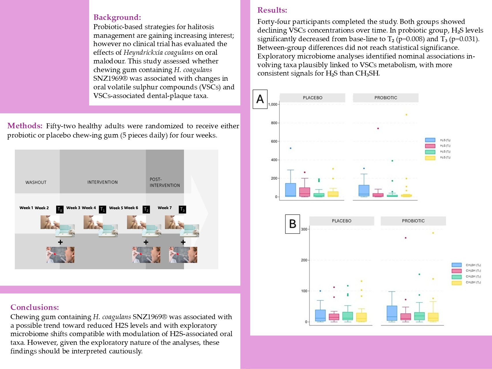
Background: Probiotic-based strategies for halitosis management are gaining increas-ing interest; however no clinical trial has evaluated the effects of Heyndrickxia coagulans on oral malodour. This study assessed whether chewing gum containing H. coagulans SNZ1969® was associated with changes in oral volatile sulphur compounds (VSCs) and VSCs-associated dental-plaque taxa. Methods: Fifty-two healthy adults were randomized to receive either probiotic or placebo chewing gum (5 pieces daily) for four weeks. VSCs were quantified at baseline and at three subsequent time points (T₁–T₃) using OralChroma™. Dental plaque samples were analyzed by 16S rRNA gene profiling to assess microbial diversity and taxonomic composition. Results: Forty-four participants completed the study. Both groups showed declining VSCs concentrations over time. In probiotic group, H₂S levels significantly decreased from baseline to T₂ (p=0.008) and T₃ (p=0.031). Between-group differences did not reach statistical significance. Exploratory microbiome analyses identified nominal associations involving taxa plausibly linked to VSCs metabolism, with more consistent signals for H₂S than CH₃SH. Conclusions: Chewing gum containing H. coagulans SNZ1969® was associated with a possible trend toward reduced H₂S levels and with exploratory microbiome shifts compatible with modulation of H₂S-associated oral taxa. However, given the exploratory nature of the analyses, these findings should be interpreted cautiously.
Keywords:
Heyndrickxia coagulans
; probiotic
; halitosis
; bad breath
; oral microbiome
; chewing gum
; metataxonomics
; volatile sulphur compounds
; hydrogen sulphide
1. Introduction
Halitosis, commonly referred to as bad breath, is a prevalent condition affecting approximately 30% of adolescents and adults worldwide. Its occurrence appears to be higher in low- and middle-income countries, and it may lead to considerable social discomfort and psychological distress [1,2,3,4]. It is defined as an unpleasant odor emanating from the oral cavity, with causes that may be intra-oral or extra-oral/systemic. In approximately 80–90% of cases, halitosis originates intra-orally and is primarily associated with microbial degradation of organic substrates such as desquamated epithelial cells, saliva proteins, and food debris [1,5]. This microbial metabolism can lead to the production of volatile sulphur compounds (VSCs), mainly hydrogen sulphide (H₂S) and methyl mercaptan (CH₃SH), which are considered major contributors to oral malodor [2]. Elevated levels of CH₃SH have been strongly associated with periodontal inflammation, whereas H₂S is more frequently linked to tongue coating and anaerobic bacterial activity on the dorsum of the tongue [3]. Since clinical studies have demonstrated that tongue coating, periodontal pockets and dental plaque are significantly correlated with VSCs levels and that mechanical tongue cleaning can reduce oral malodor [3,6], contemporary management strategies for halitosis focus on reducing the bacterial load, limiting substrate availability, neutralizing VSCs, and modulating the oral microbiota [1].
Chewing gum has been widely investigated as an adjunctive approach for controlling oral malodor. Clinical studies have shown that chewing gum can transiently reduce oral malodor and VSCs concentrations, largely due to increased salivary secretion and mechanical cleansing effects [7,8,9]. To enhance efficacy, various functional compounds have been incorporated into chewing gum formulations, such as plant extracts, e.g., green tea [10] or eucalyptus extract [11] or sodium bicarbonate [12,13]. In addition, probiotic-based strategies have emerged as a promising approach for the management of halitosis.
Probiotics are defined as live microorganisms that confer a health benefit on the host when administered in adequate amounts [14]. Clinical studies have reported that specific probiotic strains, such as Streptococcus salivarius K12, Weissella cibaria CMU, and Ligilactobacillus salivarius WB21, may reduce VSCs production and improve halitosis-related parameters [15,16,17,18,19,20,21,22,23].
Chewing gum may represent an interesting vehicle for probiotic delivery, as it can allow extended contact between the microorganism and oral tissues while also stimulating salivary flow [24]. Previous studies evaluating probiotic-containing chewing gums have reported reductions in salivary mutans streptococci and improvements in selected oral-health-related parameters [25,26]. Furthermore, clinical evidence suggests that probiotic chewing gum may contribute to reductions in oral malodor [7,27].
Heyndrickxia coagulans (formerly Bacillus coagulans) is an endospore-forming lactic-acid-producing bacterium that combines industrial robustness with a favourable safety profile, features that have supported its use in a range of probiotic applications [28]. Previous studies have explored the role of H. coagulans in supporting oral health in relation to several oral health conditions, including dental caries, gingivitis, and periodontitis [29]. To date no study has specifically investigated the potential impact of H. coagulans delivered through chewing gum on VSCs levels as markers of oral malodor. This gap in the literature is noteworthy, especially considering the increasing interest in probiotic-based products for managing oral malodor. Therefore, the aim of this study was to investigate whether a probiotic chewing gum containing H. coagulans could modulate oral VSCs concentrations, particularly H₂S and CH₃SH, in a randomized controlled trial. In addition, using dental-plaque 16S rRNA gene profiling data generated in the same trial and previously reported for biofilm ecology outcomes, we performed an exploratory secondary analysis focused on VSCs-associated oral microbial taxa.
2. Materials and Methods
2.1. Design of the Study
The present randomized controlled trial was originally designed and conducted at the Department of Biomedical, Surgical and Dental Sciences, University of Milan (Milan, Italy), in accordance with the principles of the Declaration of Helsinki. Ethical approval was obtained from the Ethics Committee of the University of Milan (February 13, 2024; protocol no. 24/24). This study was registered post-hoc at ISRCTN Registry (n. ISRCTN13055033; 04/12/2025). The present manuscript reports a secondary analysis of the same randomized, double-blind, placebo-controlled trial previously described by Cirio et al. [30], focusing specifically on oral VSCs and their exploratory associations with dental-plaque microbial taxa [29]. The CONSORT 2010 checklist for reporting randomized trials is provided in the supplementary material (Supplementary File S1).
2.2. Sample Selection
The study was conducted on healthy adult volunteers selected from the staff of Perfetti Van Melle SpA (PVM, Lainate, Italy) and from students attending the degree program in Dental Hygiene and in Dentistry and in the postgraduate program in Pediatric Dentistry of the University of Milan (Milan, Italy).
Because no prior data was available to estimate the expected effect of H. coagulans chewing gum on oral VSCs concentrations, no formal sample-size calculation was performed for the present secondary endpoint. The original trial enrolled 52 participants to ensure an adequate sample for exploratory clinical and microbiome analyses while accounting for potential dropouts. The inclusion criteria were: adult subjects aged 18 to 64 years with at least 24 natural teeth (excluding third molars), gingival index and plaque index scores ≤2, and a stimulated salivary flow rate between 1.5 and 2.0 mL/min. Exclusion criteria included: the presence of systemic diseases, pregnancy or lactation, history of drug abuse, smoking habits, use of fixed orthodontic appliances, and allergies to any ingredients contained in the chewing gums used. Brochures, providing a concise overview of the study objectives and participation procedures, were displayed near the lecture halls and in the break rooms to recruit potential participants. A total of 56 individuals responded to the recruitment call. Two participants refused enrolment owing to challenges in ensuring adherence to the study protocol. Subjects who gave their written consent to participate were interviewed to assess their eligibility based on the inclusion and exclusion criteria. They were then examined by a calibrated dentist (SC) to obtain their plaque index scores [31], gingival index score [32] and stimulated salivary flow rate. Two participants were excluded due to a potential allergy to the chewing gum components. Finally, fifty-two eligible subjects were identified and enrolled. Participants were randomized into two groups (26 in Probiotic group and 26 in Placebo group) using a computer-generated randomization system. Both participants and investigators were blinded to group allocation.
2.3. Chewing Gums Production
All chewing gums used in the study were produced and supplied by Perfetti Van Melle. The sugar-free chewing gums (weight 2.1 g) were formulated with gum base (Gum Base Co., Via Nerviano, 25, 20045 Lainate, Italy), food-grade polyols, excluding xylitol (proprietary blend; manufactured by Roquette Frères S.A. and Cargill Srl, 1 Rue de la Haute Loge, 62136 Lestrem, France), food-grade intensive sweeteners (Ajinomoto Co., Inc, 1-15-1 Kyobashi, Chuo-ku, Tokyo 104-8315, Japan), flavors (Mondarom Selegroven AG, Via Maito 8, 6804 Bironico, Svizzera) and incorporated Heyndrickxia coagulans SNZ1969® (provided by Sanzyme Biologics Ltd., Sattva Signature Tower, H.No. 8-2-472/1/A/B/SF-3, Road No. 1, Banjara Hills, Hyderabad, 500034 Telangana, India).
The chewing gum formulation and production process were based on the protocol previously described by Cirio et al. [30].
At the end of the production process, the mean count of H. coagulans in one pellet of chewing gum was 5 × 10⁸ CFU.
The placebo chewing gum was matched to the test gum in terms of shape, color, and composition, and was produced using the same process, but it did not contain any probiotics. Thus, the placebo controlled the mechanical and salivary-flow effects of chewing gum independently of probiotic administration.
2.4. Use of Chewing Gum
The study lasted a total of seven weeks. All enrolled subjects received instructions for at-home oral hygiene procedures, along with a manual toothbrush and a fluoride toothpaste (1450 ppm F) to be used throughout the study period. Participants were instructed not to use any mouthwash or other oral hygiene products aside from those provided by researchers. Additionally, participants were asked to avoid the use of antibacterial or antibiotic medications (topical or systemic), probiotics, chewing gum, and other xylitol-containing products during the study period. Participants requiring any of these products were instructed to notify the investigators and were subsequently withdrawn from the study.
The experimental period was structured as shown in Figure 1: an initial two-week washout phase, followed by a four-week intervention period, and concluding with a one-week post-intervention stage. During the four-week intervention period, participants were instructed to consume the assigned chewing gum five times per day (after breakfast, mid-morning, after lunch, mid-afternoon, and in the evening after dinner), at least 30 minutes after brushing their teeth.
To promote compliance and ensure correct product intake, participants were provided with blister packs containing the exact number of chewing gums required between two consecutive follow-up visits (n = 70). Participants were instructed to return the empty blister packs at the subsequent visit to verify adherence to the intervention. In addition, they received a paper diary in which they were asked to record each chewing gum intake. Adherence to the study protocol, at-home oral hygiene practices, and the occurrence of any adverse effects were monitored at each evaluation using a specifically designed questionnaire.
2.5. VSCs Detection from Oral Air Samples
VSCs in oral air samples were measured using a halitometer (OralChroma™, INSISTEC, Barcelona, Spain). Oral breath samples were collected using sterile single-use needleless syringes with a capacity of 1 mL. Participants were instructed to take a deep breath, place the tip of the sterile syringe inside the mouth, seal their lips around it, and keep it in position for 30 seconds. Care was taken to avoid contamination of the syringe with saliva. After the waiting period, the operator manipulated the syringe plunger to collect 1 mL of oral air, which was then immediately injected into the device for analysis. All assessments were performed between 8:00 and 10:00 a.m.
Before testing, participants were instructed to avoid garlic, onion, and other strong-flavored condiments, and to limit the consumption of margarine, milk, fried foods, sardines, salami, mortadella, sausages, red meat, cheese, sulphur-containing foods (e.g., cabbage, broccoli, cauliflower, and eggs), and alcohol. Participants were also asked not to brush their teeth during the 24 hours preceding the test and to refrain from consuming candies or chewing gum on the morning of the assessment. H₂S and CH₃SH concentrations were selected for analysis because of their stronger association with oral health, in accordance with previous studies [33,34]. Halitosis was recorded when levels H₂S was ≥112 ppb and CH₃SH was ≥26 ppb.
2.6. Metataxonomic Analysis via 16S rRNA Gene Profiling
Dental-plaque 16S rRNA gene profiling data used in the present manuscript were generated within the same randomized controlled trial previously reported by Cirio et al. [30] and were originally analyzed to assess the impact of H. coagulans SNZ1969® chewing gum on dental biofilm ecology. In the present study, these data were re-used for an exploratory secondary analysis focused on associations between dental-plaque microbial taxa and VSCs concentrations.
In brief, dental plaque was collected from buccal and lingual tooth surfaces using sterile FLOQSwabs® (Copan Italia S.p.A., Brescia, Italy) and immediately transferred into tubes containing 1 mL of nucleic acid preservation medium (eNAT®, Copan Italia S.p.A., Brescia, Italy). Samples were stored at 4 °C, transported within 2 h, and subsequently frozen at −80 °C until processing.
DNA extraction was performed using the QIAsymphony DSP Virus/Pathogen Midi Kit® (Qiagen, Milan, Italy) according to the manufacturer’s instructions. Microbial profiling was conducted by 16S rRNA gene sequencing targeting the V3–V4 region (primers 515F/806R) on an AVITI platform (Element Biosciences) using paired-end 2 × 300 bp chemistry at the Center for Omics Sciences (COSR, San Raffaele Hospital, Milan, Italy). Raw FASTQ files and run quality reports (FastQC) were provided by the sequencing facility. Sequence processing was performed in QIIME 2 (v2024.5). After demultiplexing, reads were quality filtered, denoised, merged, and chimera-checked using DADA2, yielding an ASV feature table. Quality filtering parameters were set based on per-base quality profiles. Alpha diversity was assessed using observed ASVs, Shannon index, Faith’s phylogenetic diversity, and Pielou’s evenness. Beta diversity was evaluated using Bray–Curtis, Jaccard, weighted UniFrac, and unweighted UniFrac metrics. Taxonomy was assigned using a Naive Bayes classifier trained on the SILVA 138/138.1 database (99% OTUs), trimmed to the V3–V4 region. Feature tables were aggregated to higher taxonomic levels (phylum to genus) for downstream analyses.
Since the generation and primary ecological analysis of these sequencing data have already been reported, for the present manuscript H₂S and CH₃SH concentrations were considered clinical exploratory outcomes. Microbiome-VSCs associations were considered secondary exploratory outcomes.
2.7. Statistical Analysis
Descriptive statistics were calculated for all variables and reported as mean ± standard deviation (SD) or median and interquartile range, as appropriate. The Shapiro–Wilk test was used to assess the normality of continuous data. Homoscedasticity was assessed when required for parametric comparisons. For between-group comparisons of continuous variables, the Mann–Whitney U test was applied due to non-normal data distributions. For categorical variables, Fisher’s exact test was used to evaluate differences between groups. Intra-group comparisons of repeated measures (T0, T1, T2, T3) were performed using the Wilcoxon signed-rank test for paired data. In specific subgroup analyses, Welch’s t-test was applied when assumptions for equal variances were violated. A p-value <0.05 was considered statistically significant. All statistical analyses were conducted using Stata/MP 19.0 for Windows.
Baseline associations between microbial taxa and VSCs (H₂S and CH₃SH) were evaluated using Spearman correlation on centered log-ratio (CLR)-transformed abundances across multiple taxonomic levels. To account for multiple testing, p-values were adjusted using the Benjamini–Hochberg false discovery rate (FDR) procedure within each endpoint and taxonomic level. Both nominal p-values and FDR-adjusted p-values were considered, with FDR-adjusted p-values < 0.05 interpreted as statistically significant. The effect of probiotic supplementation on VSCs-associated taxa over time was assessed using longitudinal regression models with subject-clustered robust standard errors, adjusting for visit, treatment group, age, and sex. Treatment-by-visit interactions were further examined within a difference-in-differences framework. In addition, between-group differences in changes in CLR-transformed abundances (ΔT2–T0 and ΔT3–T0) were analyzed using Mann–Whitney U tests.
Given the exploratory nature of the microbiome-VSCs analyses and the limited sample size, results from taxon-level analyses were interpreted as hypothesis-generating, particularly when statistical significance did not persist after FDR correction.
3. Results
3.1. Sample Characteristics
The study was conducted between September 2024 and April 2025. A total of 52 volunteers (26 per group) entered the 2-week washout period. Three participants in the probiotic group were excluded (one due to non-compliance and two due to intercurrent health conditions). Chewing gum administration was therefore initiated in 23 participants in the probiotic group and 26 in the placebo group.
During the intervention, one participant in placebo group discontinued before the first follow-up (T1) because of taste intolerance and two in the probiotic group because of gastrointestinal adverse effects. Prior to T2, two additional participants in the placebo group were excluded (gastrointestinal disorder and missed visit). Overall, 21 participants in the probiotic group and 23 in the placebo group completed the study (drop-out rate: 13.5%). A full flow diagram is reported in Figure 2.
Characteristics of participants, compliance and adverse effects reported in the two study groups during the experimental period have been reported in a previous study [30].
Final analyses included 44 participants, 21 in probiotic arm and 23 in placebo arm (mean age 28.4 years; 86.4% female), with no significant differences in age or sex between groups.
3.2. VSCs Concentrations
At baseline, no significant differences in H₂S or CH₃SH levels were observed between groups (Table 1).
Both H₂S and CH₃SH showed a general tendency to decrease over time, although the pattern was clearer for H₂S than for CH₃SH. The reduction in H₂S was more pronounced in the probiotic group, which showed significantly lower values at T₂ and T₃ compared with baseline (p = 0.008 and p = 0.031, respectively). Median values at these time points approached zero, with reduced dispersion compared with the placebo group (Table 1, Figure 3A). However, between-group comparisons did not show statistically significant differences at individual time points or in longitudinal changes.
CH₃SH showed a similar but less marked pattern, with no significant intra-group differences at any time point in either group (Table 1, Figure 3B). No statistically significant between-group differences were observed for changes in either H₂S or CH₃SH over time (Table 1), nor were any significant differences found in the reductions in values between follow-up intervals when comparing the probiotic and placebo groups (Table 2).
3.3. Microbial Associations with VSCs (Baseline)
No taxa remained significantly associated with baseline VSCs levels after correction for multiple testing using the Benjamini–Hochberg false discovery rate (FDR) procedure. All FDR-adjusted p-values were > 0.05. Therefore, the associations reported in Table 3 are nominal only and should be interpreted as exploratory.
For H₂S, nominal positive associations were observed with Kingella and an uncultured Prevotellaceae genus, while nominal negative associations were found for Atopobium spp., including A. rimae, and Veillonellaceae. For CH₃SH, Porphyromonas gulae was nominally positively associated, whereas Johnsonella ignava and Veillonellaceae showed nominal negative associations. None of these associations remained significant after FDR correction.These taxa were considered for exploratory interpretation based on biological plausibility in the oral ecosystem.
3.4. Longitudinal Microbial Modulation
Exploratory longitudinal models identified multiple taxa nominally associated with VSCs and potentially modulated by treatment. A higher number of signals was observed for H₂S-associated taxa compared with CH₃SH-associated taxa (Table 4).
Taxa showing both VSCs association and nominal treatment-related changes with coherent directionality were further examined. Overall, signals were more consistent for H₂S-related taxa.
Among the most biologically plausible taxa, members of Leptotrichiaceae (Leptotrichia spp. and L. hofstadii) and Kingella showed reductions in the probiotic group and increases in controls. Conversely, Actinomyces timonensis and Eubacterium sulci showed the opposite pattern (Table 5).
These directional changes were consistent with their baseline associations with H₂S, suggesting a possible probiotic-related modulation of taxa nominally associated with H₂S.
All longitudinal microbiome analyses were exploratory. The taxa reported in Table 5 were selected based on nominal significance, taxonomic interpretability, biological plausibility, and coherent directionality with baseline VSCs associations. These analyses were not considered confirmatory and were not interpreted as statistically significant after correction for multiple testing..
4. Discussion
This randomized, double-blind, placebo-controlled trial evaluated the effects of a chewing gum containing Heyndrickxia coagulans SNZ1969® on oral VSCs and oral microbiota composition. Overall, the results suggest a possible trend toward a greater reduction of H₂S in the probiotic group compared with placebo; however, between-group differences did not reach statistical significance, underscoring the exploratory nature of the study.
From a conceptual perspective, this comparison assesses whether the probiotic strain provides an additional effect beyond that of the chewing-gum vehicle alone, in which chewing gum acts as both delivery system and modulator of salivary flow and biofilm dynamics. Within this framework, the oral microbiome may be considered as a dynamic ecological network in which transient perturbations may alter functional outputs such as VSCs production rather than inducing stable compositional shifts.
Both study arms involved chewing gum use; therefore, the observed reductions in VSCs likely reflect, at least in part, the established effects of mastication on salivary stimulation and mechanical clearance [7,35]. However, the present design does not allow isolation of the specific contribution of chewing per se versus probiotic supplementation. Accordingly, all between-group differences should be interpreted as the additional effect of H. coagulans over a shared chewing gum vehicle, rather than as a direct comparison between masticatory and non-masticatory conditions.
Within this context, both groups exhibited reductions in VSCs over time, consistent with the short-term effects of chewing gum on oral malodor. The probiotic group showed a more pronounced intra-group decrease in H₂S, reaching statistical significance at later time points, with reduced inter-individual variability. Nevertheless, because the corresponding between-group comparisons were not statistically significant, this observation should not be interpreted as conclusive evidence of probiotic efficacy. In contrast, CH₃SH showed limited changes, consistent with its stronger association with subgingival, inflammation-related niches rather than supragingival biofilms and tongue coating. These compartment-specific differences could partly explain the higher responsiveness of H₂S to short-term ecological perturbations.
These findings are consistent with previous probiotic studies in halitosis, which more frequently report reductions in H₂S than CH₃SH, including interventions using Lactobacillus, Streptococcus, and Weissella species [36].
The present study adds preliminary evidence by evaluating H. coagulans delivered via chewing gum, a delivery system that ensures repeated oral exposure while simultaneously stimulating salivary flow. The delayed pattern of effect observed over time is compatible with the hypothesis that sustained ecological pressure may be required to modulate VSCs-associated microbial activity [37].
Microbiome analyses, although exploratory, provided additional ecological context. No taxa remained significantly associated with VSCs after FDR correction; however, coherent nominal associations were observed. Taxa positively associated with H₂S, including Leptotrichia, Leptotrichiaceae members, and Kingella, tended to decrease in the probiotic group, whereas taxa negatively associated with H₂S, such as Actinomyces timonensis and Eubacterium sulci, showed the opposite trend. Importantly, these changes are best interpreted as hypothesis-generating signals potentially reflecting shifts within a putative functional consortium associated with VSCs production, rather than as evidence for isolated taxonomic effects [38,39].
Mechanistically, H. coagulans may exert transient ecological pressure through competitive interactions and modulation of local environmental conditions, potentially influencing microbial metabolic activity. The chewing gum matrix likely enhances these effects by prolonging oral exposure and increasing salivary flow, thereby affecting substrate availability and biofilm turnover [35]. However, these mechanisms remain hypothetical and were not directly assessed in this study.
Strengths of the study include its randomized double-blind design, repeated longitudinal sampling, targeted quantification of VSCs, and integration of clinical and microbiome data. Baseline comparability and high completion rates further support internal validity. Importantly, the combined interpretation of VSCs dynamics and microbiome shifts provides a preliminary multi-level framework for generating hypotheses on the ecological modulation of oral malodor markers.
Several limitations should be acknowledged. The study was exploratory and not powered for between-group differences in either VSCs or microbiome outcomes. The relatively homogeneous population (young, predominantly female, orally healthy individuals) may have limited baseline variability and reduced detectable effect sizes, particularly for CH₃SH. Because participants were orally healthy adults and were not selected based on organoleptic halitosis diagnosis or elevated baseline VSCs levels, the findings cannot be directly generalized to patients with clinically manifest halitosis. Microbiome analyses involved multiple uncorrected comparisons and should therefore be interpreted cautiously. Moreover, the dental-plaque 16S rRNA gene profiling data were generated within the same trial and previously reported for biofilm ecology outcomes; therefore, their use here should be considered a secondary exploratory analysis focused on VSCs-associated taxa. In addition, the absence of organoleptic assessment limits translation of instrumental VSCs reductions into perceived halitosis. Finally, the study did not include a no-gum control arm, preventing separation of the effects of chewing itself from those of the placebo gum matrix.
5. Conclusions
Within the limitations of this exploratory secondary analysis of a randomized controlled trial, chewing gum containing Heyndrickxia coagulans SNZ1969® was associated with significant within-group reductions in H₂S levels and with nominal microbiome changes involving taxa plausibly linked to volatile sulfur compound (VSCs) metabolism. However, no statistically significant between-group differences were observed for either H₂S or CH₃SH, and none of the microbiome associations remained significant after adjustment for multiple testing. Accordingly, these findings should be considered hypothesis-generating rather than confirmatory. Future adequately powered randomized trials including participants with clinically confirmed halitosis, organoleptic assessment, prespecified VSCs endpoints, and longer follow-up periods are warranted to determine whether H. coagulans confers benefits beyond those attributable to the chewing-gum vehicle itself.
Supplementary Materials
The following supporting information can be downloaded at website of this paper posted on Preprints.org, S1. CONSORT 2010 checklist of information to include when reporting a randomized trial.
Author Contributions
Conceptualization, S.C., G.C. and M-G.C.; methodology, S.C., G.M and S.G.; software, C.S., G.M. and S.G.; validation, G.C., M.G.C. and S.C.; data curation, S.C., A.A., G.M. and S.G.; writing—original draft preparation, S.G., S.C. and M.G.C.; writing—review and editing, C.S., G.C. and A.A. All authors have read and agreed to the published version of the manuscript.
Funding
The present study was supported by Perfetti Van Melle S.p.A., which provided the test material (chewing gum), the laboratory analyses, and partially funded the research fellowship of the principal investigator.
Declaration of AI and AI-Assisted Technologies in the Writing Process
During the preparation of this manuscript, the authors utilized AI-based tools, including Grammarly (https://app.grammarly.com/) and ChatGPT-3.5 (https://chat.openai.com/), for grammar and style enhancement. Following the use of these tools, the authors thoroughly reviewed and edited all content, assuming full responsibility for the final publication’s accuracy and integrity.
Data Availability Statement
The data supporting the findings of this study are publicly available in the University of Milan Dataverse repository at the following DOI: https://doi.org/10.13130/RD_UNIMI/VATL7W. All datasets generated and analyzed during the current study have been deposited and can be accessed.
Acknowledgments
The authors would like to thank Andrea Sarrica, Natalja Kirika and Benedetta Massa for their valuable support in organizing the experimental activities and preparing the materials supplied by the company.
Conflicts of Interest
The authors declare no conflicts of interest.
Abbreviations
The following abbreviations are used in this manuscript:
| VSCs | Volatile sulphur compounds |
| H2S | Hydrogen sulphide |
| CH₃SH | Methyl mercaptan |
| ppb | Parts per billion |
| CFU | Colony-Forming Units |
| ppm | Parts Per Million |
| DNA | Deoxyribonucleic Acid |
| rRNA | Ribosomal Ribonucleic Acid |
| FDR | False Discovery Rate |
| CLR | Centered log-ratio |
| SD | Standard deviation |
References
- Porter, S.R.; Scully, C. Oral Malodour (Halitosis). BMJ 2006, 333, 632–635. [CrossRef]
- Tonzetich, J. Production and Origin of Oral Malodor: A Review of Mechanisms and Methods of Analysis. Journal of Periodontology 1977, 48, 13–20. [CrossRef]
- Yaegaki, K.; Sanada, K. Volatile Sulfur Compounds in Mouth Air from Clinically Healthy Subjects and Patients with Periodontal Disease. J of Periodontal Research 1992, 27, 233–238. [CrossRef]
- Quirynen, M.; Dadamio, J.; Van Den Velde, S.; De Smit, M.; Dekeyser, C.; Van Tornout, M.; Vandekerckhove, B. Characteristics of 2000 Patients Who Visited a Halitosis Clinic. J Clinic Periodontology 2009, 36, 970–975. [CrossRef]
- Villa, A.; Zollanvari, A.; Alterovitz, G.; Cagetti, M.; Strohmenger, L.; Abati, S. Prevalence of Halitosis in Children Considering Oral Hygiene, Gender and Age. Int J Dental Hygiene 2014, 12, 208–212. [CrossRef]
- Seemann, R.; Kison, A.; Bizhang, M.; Zimmer, S. Effectiveness of Mechanical Tongue Cleaning on Oral Levels of Volatile Sulfur Compounds. The Journal of the American Dental Association 2001, 132, 1263–1267. [CrossRef]
- Muniz, F.W.M.G.; Friedrich, S.A.; Silveira, C.F.; Rösing, C.K. The Impact of Chewing Gum on Halitosis Parameters: A Systematic Review. J. Breath Res. 2017, 11, 014001. [CrossRef]
- Rösing, C.K.; Gomes, S.C.; Bassani, D.G.; Oppermann, R.V. Effect of Chewing Gums on the Production of Volatile Sulfur Compounds (VSC) in Vivo. Acta Odontol Latinoam 2009, 22, 11–14.
- Hattab, F.N. Chewing Gum for Oral and Dental Health: A Review. Journal of Oral Health and Community Dentistry 2023, 17, 12–19. [CrossRef]
- Zeng, Q.C.; Wu, A.Z.; Pika, J. The Effect of Green Tea Extract on the Removal of Sulfur-Containing Oral Malodor Volatiles in Vitro and Its Potential Application in Chewing Gum. J. Breath Res. 2010, 4, 036005. [CrossRef]
- Tanaka, M.; Toe, M.; Nagata, H.; Ojima, M.; Kuboniwa, M.; Shimizu, K.; Osawa, K.; Shizukuishi, S. Effect of Eucalyptus-Extract Chewing Gum on Oral Malodor: A Double-Masked, Randomized Trial. Journal of Periodontology 2010, 81, 1564–1571. [CrossRef]
- Wild, J.E.; Nelson, B.J.; Hubbard, M.E.; Bowman, J.P. Oral Malodor Control Afforded through the Use of Sodium Bicarbonate-Containing Chewing Gum. Compend Contin Educ Dent 2001, 22, 43–46.
- Wåler, S.M. The Effect of Zinc-Containing Chewing Gum on Volatile Sulfur-Containing Compounds in the Oral Cavity. Acta Odontologica Scandinavica 1997, 55, 198–200. [CrossRef]
- Hill, C.; Guarner, F.; Reid, G.; Gibson, G.R.; Merenstein, D.J.; Pot, B.; Morelli, L.; Canani, R.B.; Flint, H.J.; Salminen, S.; et al. The International Scientific Association for Probiotics and Prebiotics Consensus Statement on the Scope and Appropriate Use of the Term Probiotic. Nat Rev Gastroenterol Hepatol 2014, 11, 506–514. [CrossRef]
- Bolos, A.; Bolos, O.C.; Maghet, E.; Danila, A.I.; Briceag, R.; Bumbu, B.A. Clinical and Microbiological Effects of Streptococcus Salivarius K12 Lozenges and Zinc Mouthrinse on Persistent Intra-Oral Halitosis. Microorganisms 2026, 14, 990. [CrossRef]
- He, L.; Yang, H.; Chen, Z.; Ouyang, X. The Effect of Streptococcus Salivarius K12 on Halitosis: A Double-Blind, Randomized, Placebo-Controlled Trial. Probiotics & Antimicro. Prot. 2020, 12, 1321–1329. [CrossRef]
- Burton, J.P.; Chilcott, C.N.; Moore, C.J.; Speiser, G.; Tagg, J.R. A Preliminary Study of the Effect of Probiotic Streptococcus Salivarius K12 on Oral Malodour Parameters. J Appl Microbiol 2006, 100, 754–764. [CrossRef]
- Lee, S.-K.; Lim, K.-O.; Yang, K.-I.; You, S.-J.; Kang, M.-S.; Lee, W.-P. Oral Probiotic Weissella Cibaria Improves Halitosis and Reduces Periodontal Pathogens: A Randomized, Double-Blind, Placebo-Controlled Trial. BMC Oral Health 2026, 26, 649. [CrossRef]
- Han, H.; Yum, H.; Cho, Y.-D.; Kim, S. Improvement of Halitosis by Probiotic Bacterium Weissella Cibaria CMU: A Randomized Controlled Trial. Front. Microbiol. 2023, 14, 1108762. [CrossRef]
- Passadakis, G.; Neophytou, C.; Davidopoulou, S.; Papadimitriou, K. Effectiveness of Probiotics in Managing Oral Halitosis: A Systematic Review of Randomized Controlled Trials. J Int Soc Prev Community Dent 2025, 15, 301–312. [CrossRef]
- Lee, D.-S.; Lee, S.-A.; Kim, M.; Nam, S.-H.; Kang, M.-S. Reduction of Halitosis by a Tablet Containing Weissella Cibaria CMU: A Randomized, Double-Blind, Placebo-Controlled Study. Journal of Medicinal Food 2020, 23, 649–657. [CrossRef]
- Iwamoto, T.; Suzuki, N.; Tanabe, K.; Takeshita, T.; Hirofuji, T. Effects of Probiotic Lactobacillus Salivarius WB21 on Halitosis and Oral Health: An Open-Label Pilot Trial. Oral Surgery, Oral Medicine, Oral Pathology, Oral Radiology, and Endodontology 2010, 110, 201–208. [CrossRef]
- Motta, P.D.B.; Gonçalves, M.L.L.; Gallo, J.M.A.S.; Sobral, A.P.T.; Motta, L.J.; Mayer, M.P.A.; Kawamoto, D.; De Andrade, D.C.; Santos, E.M.; Fernandes, K.P.S.; et al. Short Term Effect of Antimicrobial Photodynamic Therapy and Probiotic L. Salivarius WB21 on Halitosis: A Controlled and Randomized Clinical Trial. PLoS ONE 2024, 19, e0297351. [CrossRef]
- Cirio, S.; Salerno, C.; Guglielmetti, S.D.; Mezzasalma, V.; Sarrica, A.; Kirika, N.; Campus, G.; Cagetti, M.G. In Vivo Study on the Salivary Kinetics of Two Probiotic Strains Delivered via Chewing Gum. Microorganisms 2025, 13, 721. [CrossRef]
- Caglar, E.; Kavaloglu, S.C.; Kuscu, O.O.; Sandalli, N.; Holgerson, P.L.; Twetman, S. Effect of Chewing Gums Containing Xylitol or Probiotic Bacteria on Salivary Mutans Streptococci and Lactobacilli. Clin Oral Investig 2007, 11, 425–429. [CrossRef]
- Ericson, D.; Hamberg, K.; Bratthall, G.; Sinkiewicz-Enggren, G.; Ljunggren, L. Salivary IgA Response to Probiotic Bacteria and Mutans Streptococci after the Use of Chewing Gum Containing Lactobacillus Reuteri. Pathogens Disease 2013, 68, 82–87. [CrossRef]
- Keller, M.K.; Bardow, A.; Jensdottir, T.; Lykkeaa, J.; Twetman, S. Effect of Chewing Gums Containing the Probiotic Bacterium Lactobacillus Reuteri on Oral Malodour. Acta Odontologica Scandinavica 2012, 70, 246–250. [CrossRef]
- Jurenka, J. Bacillus Coagulans: Monograph. Alternative medicine review : a journal of clinical therapeutic 2012, 17, 76–81.
- Cirio, S.; Campus, G.; Salerno, C.; Allam, A.; Cagetti, M.G. Oral Health Benefits of Heyndrickxia Coagulans: A Systematic Review and Meta-Analysis of Current Evidence. Front. Oral Health 2026, 6, 1733955. [CrossRef]
- Cirio, S.; Mantegazza, G.; Salerno, C.; Guglielmetti, S.; Allam, A.; Campus, G.; Cagetti, M.G. Assessing the Impact of Heyndrickxia Coagulans Administered Through Sugar-Free Chewing Gum on Dental Biofilm: A Double-Blind Randomized Controlled Trial. Nutrients 2026, 18, 904. [CrossRef]
- Silness, J.; Loe, H. PERIODONTAL DISEASE IN PREGNANCY. II. CORRELATION BETWEEN ORAL HYGIENE AND PERIODONTAL CONDTION. Acta Odontol Scand 1964, 22, 121–135. [CrossRef]
- Löe, H.; Silness, J. Periodontal Disease in Pregnancy I. Prevalence and Severity. Acta Odontologica Scandinavica 1963, 21, 533–551. [CrossRef]
- Tangerman, A. Halitosis in Medicine: A Review. International Dental Journal 2002, 52, 201–206. [CrossRef]
- Tangerman, A.; Winkel, E.G. The Portable Gas Chromatograph OralChromaTM: A Method of Choice to Detect Oral and Extra-Oral Halitosis. J. Breath Res. 2008, 2, 017010. [CrossRef]
- Wessel, S.W.; Van Der Mei, H.C.; Maitra, A.; Dodds, M.W.J.; Busscher, H.J. Potential Benefits of Chewing Gum for the Delivery of Oral Therapeutics and Its Possible Role in Oral Healthcare. Expert Opinion on Drug Delivery 2016, 13, 1421–1431. [CrossRef]
- Tangerman, A.; Winkel, E.G. Intra- and Extra-oral Halitosis: Finding of a New Form of Extra-oral Blood-borne Halitosis Caused by Dimethyl Sulphide. J Clinic Periodontology 2007, 34, 748–755. [CrossRef]
- Perotti, S.; Mantegazza, G.; Pierallini, E.; Kirika, N.; Duncan, R.; Telesca, N.; Sarrica, A.; Guglielmetti, S. Human in Vivo Assessment of the Survival and Germination of Heyndrickxia Coagulans SNZ1969 Spores Delivered via Gummy Candies. Current Research in Food Science 2024, 9, 100793. [CrossRef]
- Takahashi, N. Oral Microbiome Metabolism: From “Who Are They?” To “What Are They Doing?” J Dent Res 2015, 94, 1628–1637. [CrossRef]
- Lamont, R.J.; Koo, H.; Hajishengallis, G. The Oral Microbiota: Dynamic Communities and Host Interactions. Nat Rev Microbiol 2018, 16, 745–759. [CrossRef]
Figure 1.
Chewing gum administration schedule and sample collection timeline.
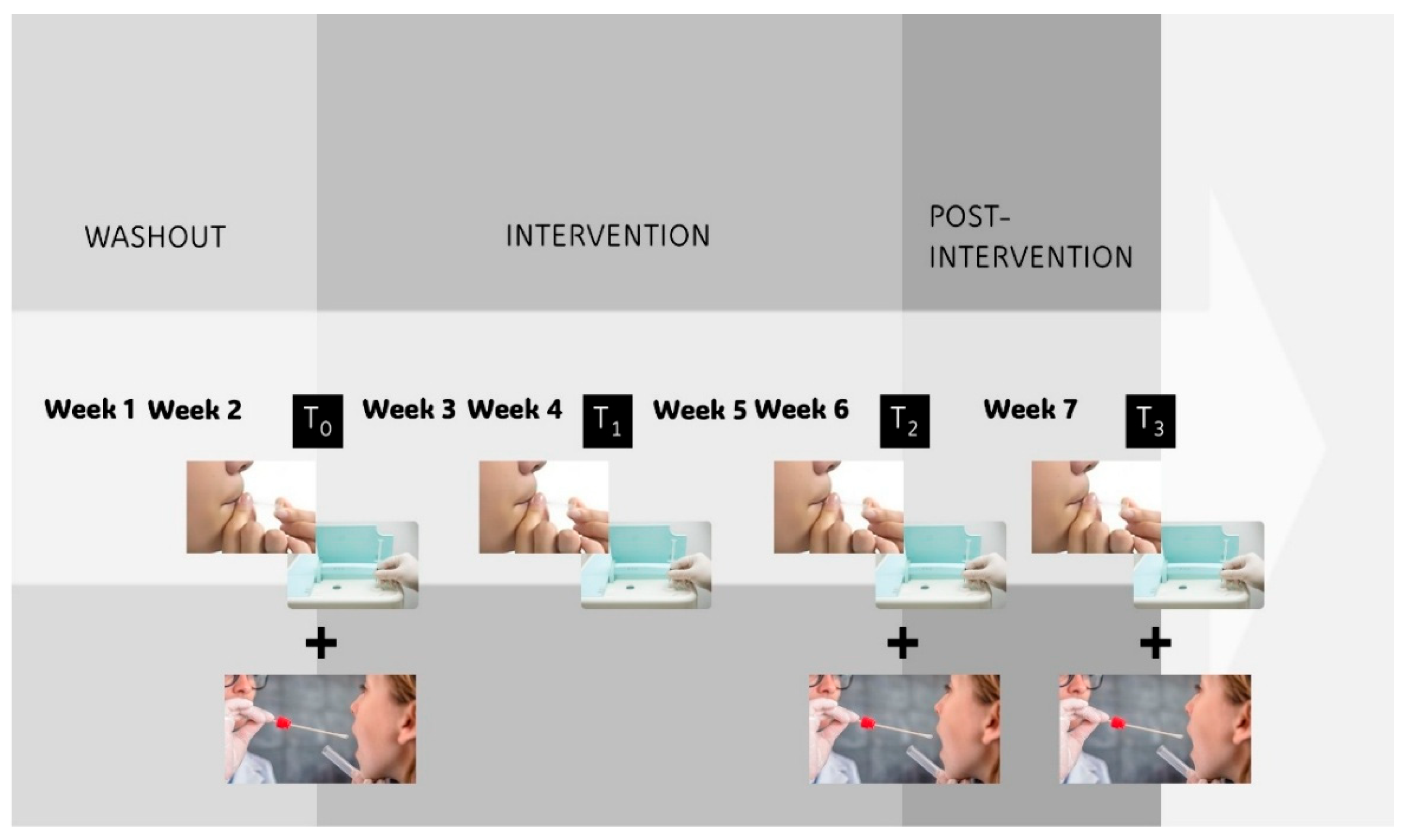
Figure 2.
Flow diagram of participant recruitment, randomization, and follow-up in the clinical trial.
Figure 2.
Flow diagram of participant recruitment, randomization, and follow-up in the clinical trial.
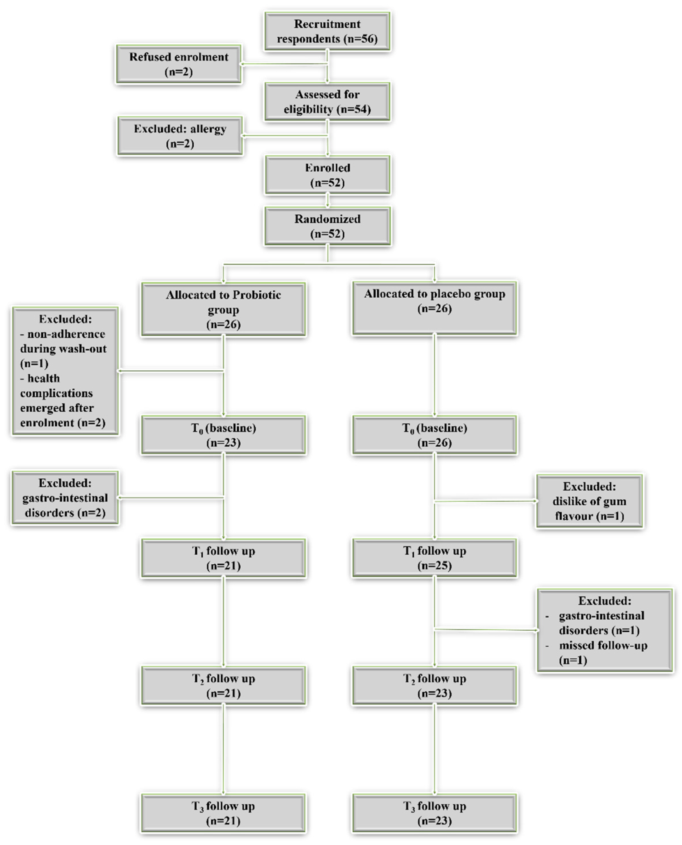
Figure 3.
Box plots of H₂S (A) and CH₃SH (B) concentrations (ppb) at baseline (T₀) and follow-up time points (T₁–T₃) in the placebo and probiotic groups.
Figure 3.
Box plots of H₂S (A) and CH₃SH (B) concentrations (ppb) at baseline (T₀) and follow-up time points (T₁–T₃) in the placebo and probiotic groups.
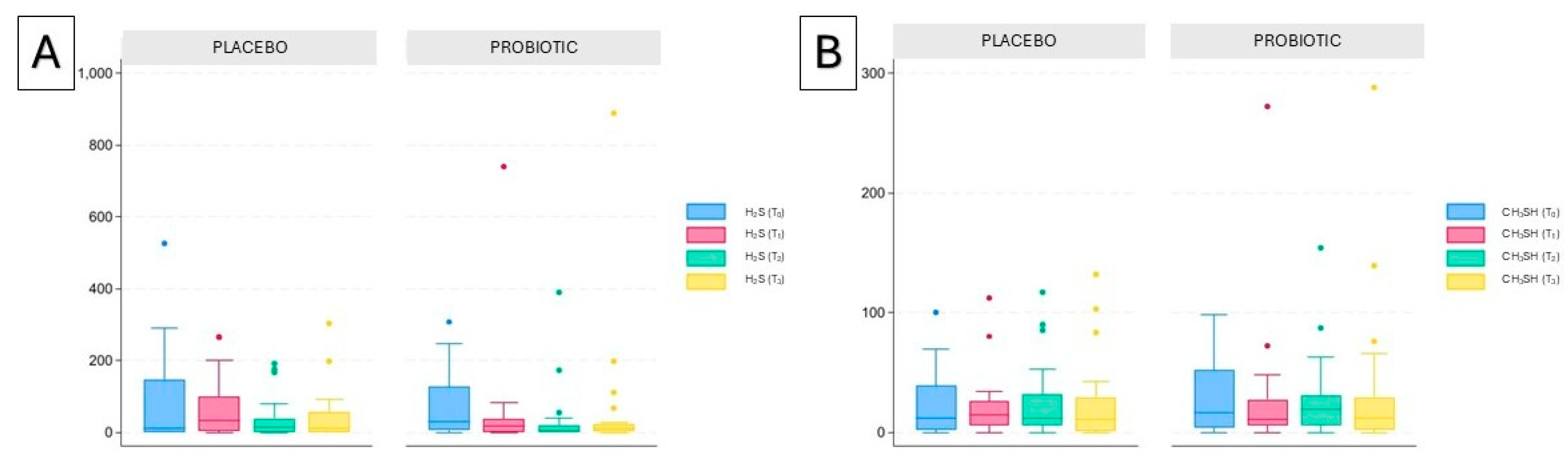

Table 1.
VSCs (H2S and CH3SH) mean values at baseline (T0), after 14 and 28 days from the beginning of administration (T1 and T2), and 7 days after the end of administration (T3) in the probiotic and placebo groups.
Table 1.
VSCs (H2S and CH3SH) mean values at baseline (T0), after 14 and 28 days from the beginning of administration (T1 and T2), and 7 days after the end of administration (T3) in the probiotic and placebo groups.
| Variable | Probiotic | Placebo | p-Value |
| N=21 | N=23 | ||
| H2S (ppb) mean (SD) | |||
| T0 | 76.81 (94.34) | 73.39 (124.54) | 0.394b |
| T1 | 53.67 (154.82) | 66.74 (79.45) | 0.070b |
| T2 | 36.90 (87.16) | 37.39 (57.51) | 0.327b |
| T3 | 67.62 (189.15) | 43.17 (71.12) | 0.868b |
| T0 vs T1 | 0.120c | 0.637c | |
| T0 vs T2 | 0.008c | 0.088c | |
| T0 vs T3 | 0.031c | 0.437c | |
| CH3SH (ppb) mean (SD) | |||
| T0 | 28.05 (30.62) | 22.65 (26.15) | 0.787b |
| T1 | 29.57 (56.98) | 20.52 (25.55) | 1.000b |
| T2 | 27.29 (35.07) | 25.26 (31.03) | 0.787b |
| T3 | 35.10 (65.40) | 23.78 (34.64) | 0.689b |
| T0 vs T1 | 0.728c | 0.749c | |
| T0 vs T2 | 0.835c | 0.704c | |
| T0 vs T3 | 0.917c | 1.000c |
N: number; SD: standard deviation. Normality and heteroskedasticity of continuous data were assessed with Shapiro-Wilk test. Comparisons between groups were performed using Fisher’s exact test (a); Mann-Whitney U test (b); Wilcoxon signed-rank test (c).
Table 2.
Changes (Δ) in VSCs levels from baseline to different time points across groups.
| Probiotic | Placebo | p Value | |
| N=21 | N=23 | ||
| H2S (ppb) mean (SD) | |||
| Δ T1-T0 | -23.14 (130.87) | -6.65 (99.06) | 0.165 |
| Δ T2-T0 | -39.90 (74.87) | -36.00 (114.10) | 0.541 |
| Δ T3-T0 | -9.19 (144.27) | -30.22 (142.18) | 0.398 |
| CH₃SH (ppb) mean (SD) | |||
| Δ T1-T0 | 1.52 (50.02) | -2.13 (32.57) | 0.912 |
| Δ T2-T0 | -0.76 (34.28) | 2.61 (40.80) | 0.604 |
| Δ T3-T0 | 7.05 (47.89) | 1.13 (32.78) | 0.968 |
Normality and heteroskedasticity of continuous data were assessed with Shapiro-Wilk test. Between-group differences were evaluated using the Mann–Whitney U test.
Table 3.
Association between taxa and VSCs levels assessed using Spearman correlation. FDR-adjusted p-values are reported. No association remained statistically significant after FDR correction; therefore, the taxa listed should be interpreted as nominal, exploratory findings.
Table 3.
Association between taxa and VSCs levels assessed using Spearman correlation. FDR-adjusted p-values are reported. No association remained statistically significant after FDR correction; therefore, the taxa listed should be interpreted as nominal, exploratory findings.
| Full_taxonomy | Rho | FDR-adjusted p-value |
|---|---|---|
| CH3SH | ||
| p_Actinobacteriota;c_Actinobacteria;o_Bifidobacteriales;f_Bifidobacteriaceae | -0.34 | 0.025 |
| p_Firmicutes;c_Negativicutes;o_Veillonellales-Selenomonadales;f_Veillonellaceae;g_Veillonellaceae | -0.33 | 0.028 |
| p_Firmicutes;c_Clostridia;o_Lachnospirales;f_Lachnospiraceae;g_Johnsonella;s_Johnsonella_ignava | -0.41 | 0.006 |
| p_Bacteroidota;c_Bacteroidia;o_Bacteroidales;f_Porphyromonadaceae;g_Porphyromonas;s_gulae | 0.37 | 0.013 |
| p_Patescibacteria;c_Gracilibacteria;o_JGI_0000069-P22;f_JGI_0000069-P22;g_JGI_0000069-P22;s_Candidatus_Gracilibacteria | 0.36 | 0.017 |
| p_Actinobacteriota;c_Actinobacteria;o_Propionibacteriales;f_Propionibacteriaceae;g_Pseudopropionibacterium;s_propionicum | 0.34 | 0.024 |
| p_Firmicutes;c_Negativicutes;o_Veillonellales-Selenomonadales;f_Veillonellaceae;g_Veillonellaceae;s_unidentified | -0.34 | 0.025 |
| H2S | ||
| p_Firmicutes;c_Negativicutes;o_Veillonellales-Selenomonadales;f_Veillonellaceae;g_Veillonellaceae | -0.37 | 0.014 |
| p_Firmicutes;c_Clostridia;o_Peptostreptococcales-Tissierellales;f_Anaerovoracaceae;g_Amnipila | -0.33 | 0.027 |
| p_Firmicutes;c_Clostridia;o_Lachnospirales;f_Lachnospiraceae;g_Butyrivibrio | -0.32 | 0.037 |
| p_Proteobacteria;c_Gammaproteobacteria;o_Burkholderiales;f_Neisseriaceae;g_Kingella | 0.31 | 0.041 |
| p_Actinobacteriota;c_Coriobacteriia;o_Coriobacteriales;f_Atopobiaceae;g_Atopobium | -0.31 | 0.041 |
| p_Bacteroidota;c_Bacteroidia;o_Bacteroidales;f_Prevotellaceae;g_uncultured | 0.30 | 0.046 |
| p_Firmicutes;c_Negativicutes;o_Veillonellales-Selenomonadales;f_Veillonellaceae;g_Veillonellaceae;s_unidentified | -0.38 | 0.012 |
| p_Actinobacteriota;c_Coriobacteriia;o_Coriobacteriales;f_Atopobiaceae;g_Atopobium;s_Atopobium_rimae | -0.33 | 0.027 |
| p_Firmicutes;c_Clostridia;o_Peptostreptococcales-Tissierellales;f_Anaerovoracaceae;g_Amnipila;s_uncultured_organism | -0.33 | 0.027 |
| p_Firmicutes;c_Clostridia;o_Peptostreptococcales-Tissierellales;f_Anaerovoracaceae;g_[Eubacterium]_nodatum_group;s_sulci | -0.30 | 0.048 |
Table 4.
Nominally VSCs-associated taxa.
| Endpoint | Level | Nominally VSCs-associated taxa | Taxa nominally modified by the probiotic | Taxa consistent in direction |
|---|---|---|---|---|
| CH3SH | Family | 36 | 11 | 10 |
| CH3SH | Genus | 64 | 9 | 7 |
| CH3SH | Species | 38 | 4 | 4 |
| H2S | Family | 74 | 27 | 25 |
| H2S | Genus | 116 | 25 | 24 |
| H2S | Species | 84 | 12 | 11 |
Table 5.
Exploratory H₂S-associated taxa showing nominal treatment-related changes with coherent directionality. Taxa were selected among nominally significant features that were taxonomically interpretable, compatible with the oral ecosystem, and directionally consistent with their baseline association with H2S levels.
Table 5.
Exploratory H₂S-associated taxa showing nominal treatment-related changes with coherent directionality. Taxa were selected among nominally significant features that were taxonomically interpretable, compatible with the oral ecosystem, and directionally consistent with their baseline association with H2S levels.
| Taxon | Δ time point | Δ median in PROBIOTIC group | Δ median in PLACEBO group | p-Value (Mann-Whitney U test ) | Interpretation |
|---|---|---|---|---|---|
| Leptotrichiaceae | T2-T0 | −0.32 | +0.49 | 0.015 | ↓ in probiotic group; ↑ in placebo group |
| Leptotrichiaceae | T3-T0 | −0.39 | +0.63 | 0.017 | Effect maintained even at T3 |
| Leptotrichia | T2-T0 | −0.29 | +0.40 | 0.010 | ↓ in probiotic group; ↑ in placebo group |
| Leptotrichia | T3-T0 | −0.35 | +0.65 | 0.011 | Effect maintained even at T3 |
| Kingella | T2-T0 | −0.51 | +0.93 | 0.021 | ↓ in probiotic group; ↑ in placebo group |
| Kingella | T3-T0 | −0.38 | +0.32 | 0.035 | ↓ in probiotic group; ↑ in placebo group |
| Actinomyces timonensis | T2-T0 | +0.12 | −0.08 | 0.021 | ↑ in probiotic group; ↓ in placebo group |
| Eubacterium sulci | T2-T0 | +0.18 | −0.09 | 0.010 | ↑ in probiotic group; ↓ in placebo group |
| Eubacterium sulci | T3-T0 | +0.08 | −0.16 | 0.010 | ↑ in probiotic group; ↓ in placebo group |
| Leptotrichia hofstadii | T2-T0 | −0.53 | +0.77 | 0.046 | ↓ in probiotic group; ↑ in placebo group |
↑: increasing; ↓: reduction.
Disclaimer/Publisher’s Note: The statements, opinions and data contained in all publications are solely those of the individual author(s) and contributor(s) and not of MDPI and/or the editor(s). MDPI and/or the editor(s) disclaim responsibility for any injury to people or property resulting from any ideas, methods, instructions or products referred to in the content. |
© 2026 by the authors. Licensee MDPI, Basel, Switzerland. This article is an open access article distributed under the terms and conditions of the Creative Commons Attribution (CC BY) license (http://creativecommons.org/licenses/by/4.0/).
Copyright: This open access article is published under a Creative Commons CC BY 4.0 license, which permit the free download, distribution, and reuse, provided that the author and preprint are cited in any reuse.


