
Submitted:
09 July 2026
Posted:
13 July 2026
You are already at the latest version
Abstract
Background/Objectives: Regenerative endodontic procedures have fundamentally changed the management of immature permanent teeth with pulpal necrosis by shifting the therapeutic focus from artificial apical barrier formation to biologically driven tissue regeneration. Unlike conventional apexification, revascularization aims to restore a functional intracanal environment that supports continued root maturation, dentinal wall thickening and apical closure. The present study describes the clinical application and long-term outcome of regenerative endodontic revascularization in a traumatically injured immature permanent maxillary central incisor. Methods: An 11-year-old patient presented with a traumatized immature permanent maxillary central incisor exhibiting pulpal necrosis and an open apex. Following clinical and radiographic assessment, a regenerative endodontic protocol was performed. Canal disinfection was achieved using 1.5% sodium hypochlorite irrigation and calcium hydroxide as the intracanal medicament. At the subsequent appointment, bleeding was intentionally induced beyond the apical foramen to establish an autologous blood clot scaffold. The canal was sealed coronally with a bioceramic material and restored with a definitive composite restoration. Clinical and radiographic follow-up, including cone-beam computed tomography, was performed over a three-year period. Results: Throughout the observation period, the treated tooth remained asymptomatic and functional, with no clinical or radiographic evidence of persistent apical pathology. Serial radiographs and CBCT examinations demonstrated progressive root maturation, increased dentinal wall thickness, continued root elongation and complete apical closure. Functional integrity and esthetic appearance were maintained during the entire follow-up period. Conclusions: This case demonstrates that regenerative endodontic revascularization is a predictable and biologically based treatment option for immature permanent teeth with pulpal necrosis following traumatic injury. When performed according to contemporary regenerative protocols, the procedure can facilitate continued root development and improve the long-term structural integrity of the tooth. Revascularization should be considered a valuable treatment option in appropriately selected immature permanent teeth, while long-term follow-up remains essential to evaluate healing and treatment stability.
Keywords:
regenerative endodontics
; pulp revascularization
; regenerative endodontic procedures (REPs)
; immature permanent tooth
; dental trauma
; pulp necrosis
; open apex
; root development
; stem cells from the apical papilla (SCAP)
; apexification
; case report
1. Introduction
Traumatic dental injuries (TDIs) are among the most common causes of pulpal necrosis in children and adolescents and frequently affect immature permanent anterior teeth. Owing to their incomplete root development, these teeth are characterized by thin dentinal walls, short roots and wide open apices, making conventional endodontic treatment technically demanding and compromising their long-term prognosis [1,2]. Preserving these teeth is of particular importance because premature tooth loss in young patients may impair esthetics, function, alveolar bone development and psychological well-being.
For several decades, apexification represented the standard treatment for immature permanent teeth with necrotic pulps. Traditionally performed with calcium hydroxide and more recently, with mineral trioxide aggregate (MTA) or other calcium silicate-based biomaterials, apexification successfully promotes the formation of an apical barrier that facilitates root canal obturation [3,4]. However, because root maturation ceases after pulpal necrosis, this treatment does not promote further root elongation or thickening of the dentinal walls. Consequently, the treated tooth often remains structurally weakened and susceptible to cervical or vertical root fracture during long-term function [5].
Regenerative endodontic procedures (REPs), commonly referred to as pulp revascularization or revitalization, have fundamentally changed the therapeutic concept for immature necrotic permanent teeth. Instead of replacing the lost pulp tissue with an inert filling material, regenerative therapy aims to establish a biologically favorable environment that enables the ingrowth of vascularized tissue into the disinfected root canal system. This process relies on effective canal disinfection while preserving stem cell viability, followed by induction of intracanal bleeding that provides a natural scaffold enriched with growth factors and mesenchymal stem cells originating primarily from the stem cells of the apical papilla (SCAP) [6,7,8,23]. Clinical studies have demonstrated that regenerative procedures may result in continued root development, increased dentinal wall thickness, apical closure and improved resistance to fracture compared with conventional apexification [2,6,9].
Despite these promising outcomes, regenerative endodontics remains an evolving field. Considerable debate still exists regarding the optimal irrigation protocols, intracanal medicaments, scaffold materials and coronal sealing techniques required to maximize biological regeneration. Furthermore, histological investigations have demonstrated that the newly formed intracanal tissue is often not identical to the original pulp–dentin complex but rather consists of varying proportions of fibrous connective tissue, cementum-like tissue and bone-like tissue [7,10]. Nevertheless, numerous clinical studies have consistently reported high survival rates and favorable radiographic healing, supporting regenerative endodontics as the preferred treatment strategy for immature permanent teeth with pulpal necrosis whenever case selection is appropriate [1,2,8].
The present case report describes the successful regenerative management of a traumatized immature permanent maxillary central incisor in an 11-year-old patient. The clinical protocol, biological rationale and radiographic findings obtained over a three-year follow-up period demonstrate continued root maturation, progressive thickening of the dentinal walls and complete apical closure. These findings further support regenerative endodontic therapy as a predictable and biologically based treatment option for preserving immature permanent teeth following traumatic pulp necrosis.
2. Materials and Methods
This manuscript presents a single-patient clinical case report describing the regenerative endodontic management of an immature permanent maxillary central incisor with pulp necrosis following traumatic dental injury. The treatment was performed according to contemporary regenerative endodontic principles and current recommendations of the American Association of Endodontists (AAE) and the European Society of Endodontology (ESE) [1,2,3].
An 11-year-old healthy patient was referred after sustaining dental trauma affecting the maxillary left central incisor tooth 21. The patient’s medical history was non-contributory and no systemic diseases or medications that could interfere with wound healing were reported.
Clinical examination included visual inspection, percussion, palpation, periodontal probing, mobility assessment and pulp sensibility testing. Radiographic assessment consisted of standardized periapical radiographs and cone-beam computed tomography (CBCT), which revealed an immature permanent tooth with incomplete root formation, a wide open apex, thin dentinal walls and a blunderbuss-shaped canal (Figure 2).
Initially, the tooth was diagnosed with pulpal shock secondary to trauma. Following a four-week observation period, repeated pulp sensibility testing using cold testing, laser Doppler flowmetry (LDF) and pulse oximetry demonstrated complete loss of pulpal vitality. Based on the clinical and radiographic findings, the diagnosis of pulp necrosis in an immature permanent tooth was established.
All clinical procedures were performed under rubber dam isolation using an aseptic technique.
After administration of local anesthesia containing 2% lidocaine with epinephrine (1:100,000), an endodontic access cavity was prepared using a sterile diamond bur. Necrotic pulp tissue was carefully removed (Figure 3) and the working length was established electronically and confirmed radiographically (Figure 4). Mechanical instrumentation of the root canal was intentionally minimized to preserve residual stem cells located within the apical tissues. Canal disinfection was achieved using 20 mL of 1.5% sodium hypochlorite (NaOCl), followed by sterile saline irrigation. Full-strength sodium hypochlorite (5.25%) was intentionally avoided because residual viable stem cells within the apical tissues are essential for regenerative healing. The use of a lower NaOCl concentration helps preserve stem cell viability while providing adequate canal disinfection [17]. Calcium hydroxide paste was subsequently placed as an intracanal medicament approximately 1–2 mm short of the working length before temporary restoration with glass ionomer cement.
After two weeks, the patient returned asymptomatic without clinical signs of inflammation. Local anesthesia without vasoconstrictor (3% mepivacaine) was administered to facilitate bleeding during the regenerative phase. Following removal of the calcium hydroxide dressing using ultrasonic activation, the canal was irrigated with 1.5% NaOCl followed by 17% aqueous EDTA to promote the release of dentin-derived growth factors [4].
Bleeding was intentionally induced by extending a sterile size #10 K-file approximately 1–3 mm beyond the apical foramen. The resulting blood clot was allowed to fill the canal up to the cemento-enamel junction (CEJ), serving as a natural scaffold containing mesenchymal stem cells and bioactive molecules. A resorbable collagen matrix (CollaPlug®, Zimmer Biomet, Warsaw, IN, USA) was carefully positioned over the blood clot to stabilize the scaffold.
The coronal barrier was created using EndoSequence® BC RRM Putty (Brasseler USA, Savannah, GA, USA) with an approximate thickness of 3 mm. Permanent coronal sealing was completed using a dual-cured resin composite restoration to ensure long-term prevention of bacterial leakage.
Clinical and radiographic examinations were performed postoperative (Figure 10), after 5 months, 8 months, 12 months, 24 months and 36 months (Figure 11).
Clinical evaluation included assessment of pain, tenderness to percussion, swelling, sinus tract formation, tooth mobility, periodontal status and functional integrity.
Radiographic assessment consisted of standardized periapical radiographs at every follow-up appointment. CBCT imaging was additionally performed to evaluate three-dimensional root development, dentinal wall thickening, apical closure and healing of the periapical tissues.
Treatment success was defined according to current AAE and ESE recommendations as the absence of clinical symptoms, resolution of periapical pathology, continued root maturation, increased dentinal wall thickness and progressive apical closure [2,3].
Written informed consent for treatment, clinical documentation, radiographic examinations and publication of anonymized clinical data and images was obtained from the patient’s legal guardians prior to treatment.
According to institutional and national regulations, ethical committee approval was not required for this single-patient case report. All procedures were conducted in accordance with the principles of the Declaration of Helsinki.
All clinical data, radiographs, CBCT images and photographic documentation supporting the findings of this case are presented within this article. Additional anonymized clinical information is available from the corresponding author upon reasonable request.
3. Results
The patient remained asymptomatic throughout the entire observation period. No spontaneous pain, swelling, sinus tract formation, tenderness to percussion or tenderness to palpation was observed at any follow-up examination. Physiological tooth mobility and healthy surrounding periodontal tissues were maintained during the three-year follow-up period.
The coronal restoration remained intact without evidence of marginal leakage, discoloration or secondary caries. Soft tissue healing was uneventful and the patient reported normal function without discomfort during mastication.
Standardized periapical radiographs obtained immediately after treatment confirmed adequate placement of the intracanal blood clot scaffold, collagen matrix, bioceramic coronal barrier and definitive composite restoration (Figure 6, Figure 7, Figure 8 and Figure 9).
Progressive radiographic changes were observed during follow-up examinations (Figure 11). At five months, initial signs of periapical healing and early hard tissue deposition were visible (Figure 11a). Eight months after treatment, continued root development with narrowing of the apical foramen became evident (Figure 11b). One year after treatment demonstrated further root elongation together with increased dentinal wall thickness (Figure 11c). At the two-year follow-up, complete apical closure and advanced maturation of the root were observed (Figure 11d).
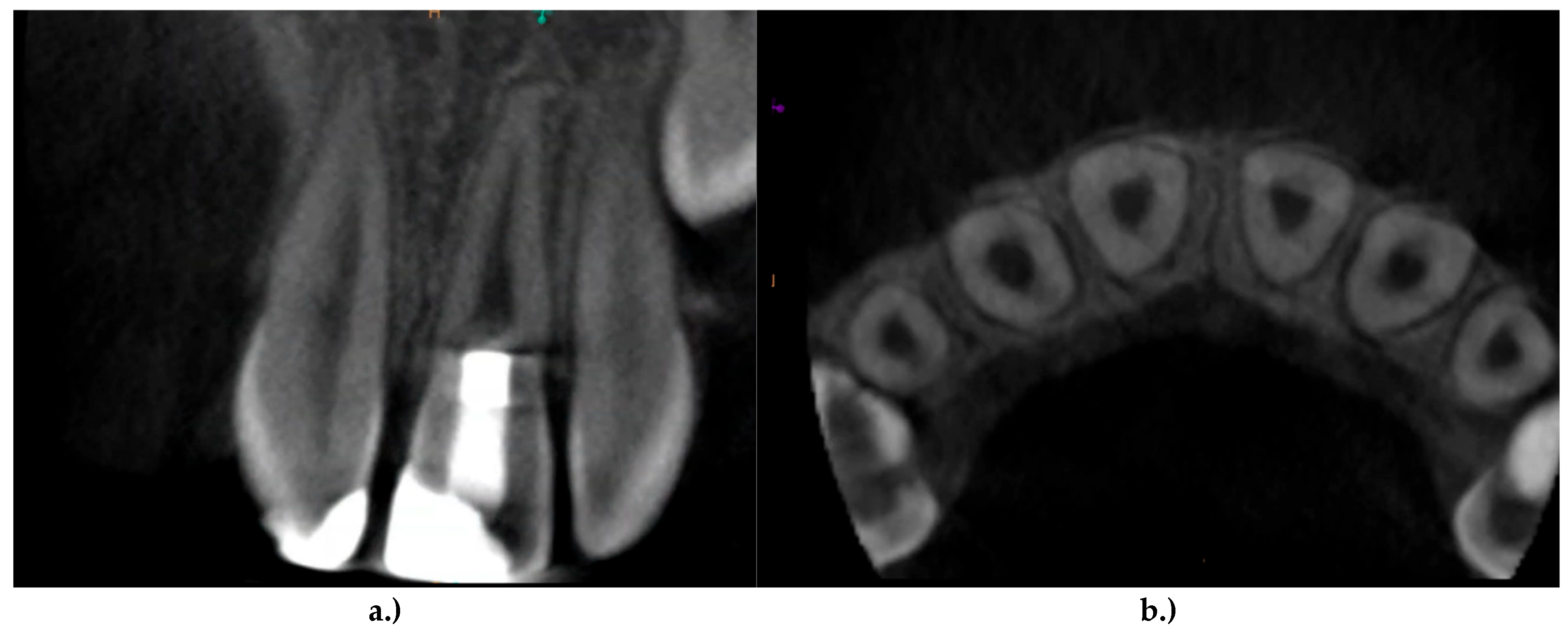
Three-year follow-up radiograph confirmed stable long-term outcomes without evidence of apical radiolucency, inflammatory root resorption, ankylosis or other pathological alterations (Figure 12).
CBCT examinations provided three-dimensional confirmation of the radiographic findings (Figure 13).
Compared with the baseline examination, CBCT demonstrated:
- Continued root elongation;
- Progressive thickening of the dentinal walls;
- Significant reduction of the root canal diameter;
- Complete apical closure;
- Resolution of the periapical lesion;
- Normal surrounding alveolar bone architecture.
No evidence of external inflammatory root resorption, replacement resorption, root fracture or newly developed periapical pathology was detected during the observation period.
According to current regenerative endodontic success criteria, the treatment fulfilled both primary and secondary outcome measures.
Primary outcomes included:
- Elimination of clinical signs and symptoms;
- Resolution of apical inflammation;
- Long-term tooth retention.
Secondary outcomes included:
- Continued physiological root maturation;
- Increased dentinal wall thickness;
- Progressive root lengthening;
- Complete apical closure;
- Stable functional and esthetic outcome.
The clinical and radiographic findings indicate successful regenerative healing and long-term preservation of the immature permanent tooth over a three-year follow-up period.
Figure 1.
Comparison of the post-treatment configuration and clinical outcome of regenerative endodontic treatment (revascularization) (left) and apexification (right).
Figure 1.
Comparison of the post-treatment configuration and clinical outcome of regenerative endodontic treatment (revascularization) (left) and apexification (right).
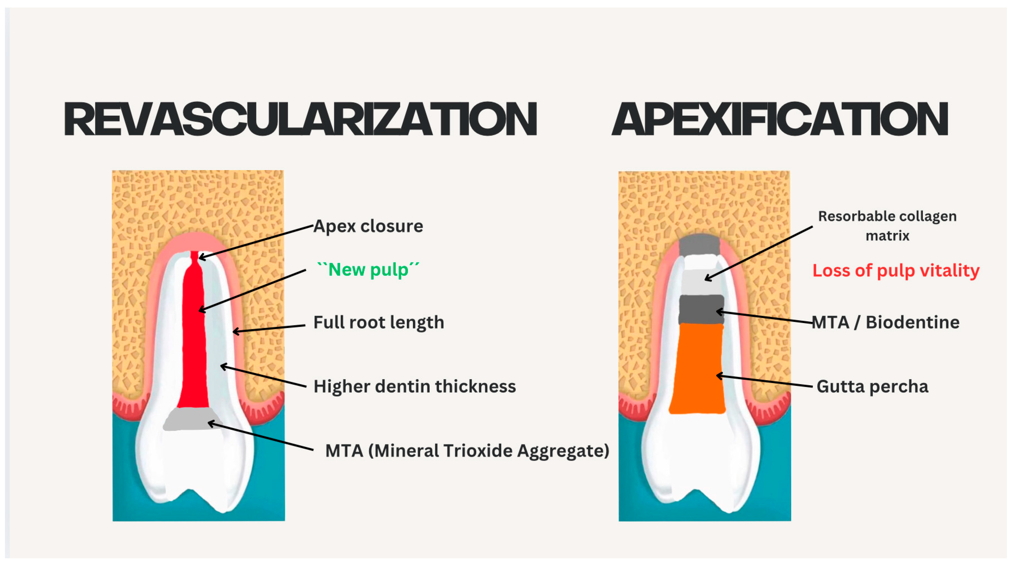
Figure 2.
Baseline radiographic examination of the maxillary left central incisor (tooth 21) at the initial visit before treatment a.) Periapical Radiograph b.) Sagittal CBCT view demonstrating an immature root with a wide open apex c.) Coronal CBCT view showing the characteristic blunderbuss-shaped root canal d.) Axial CBCT view illustrating the large pulp chamber and root canal space.
Figure 2.
Baseline radiographic examination of the maxillary left central incisor (tooth 21) at the initial visit before treatment a.) Periapical Radiograph b.) Sagittal CBCT view demonstrating an immature root with a wide open apex c.) Coronal CBCT view showing the characteristic blunderbuss-shaped root canal d.) Axial CBCT view illustrating the large pulp chamber and root canal space.
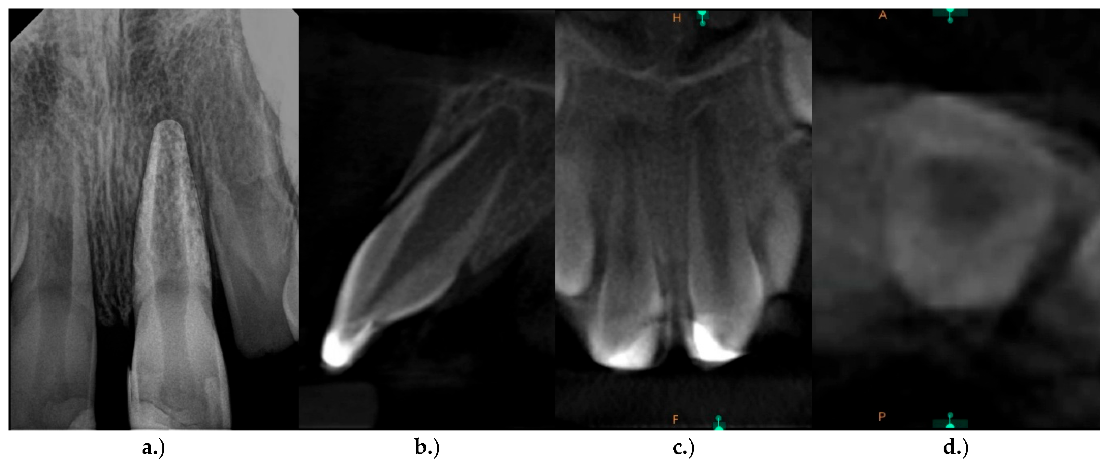
Figure 3.
a.) Preparation of the endodontic access cavity and b.) visualization of the necrotic pulp tissue.
Figure 3.
a.) Preparation of the endodontic access cavity and b.) visualization of the necrotic pulp tissue.
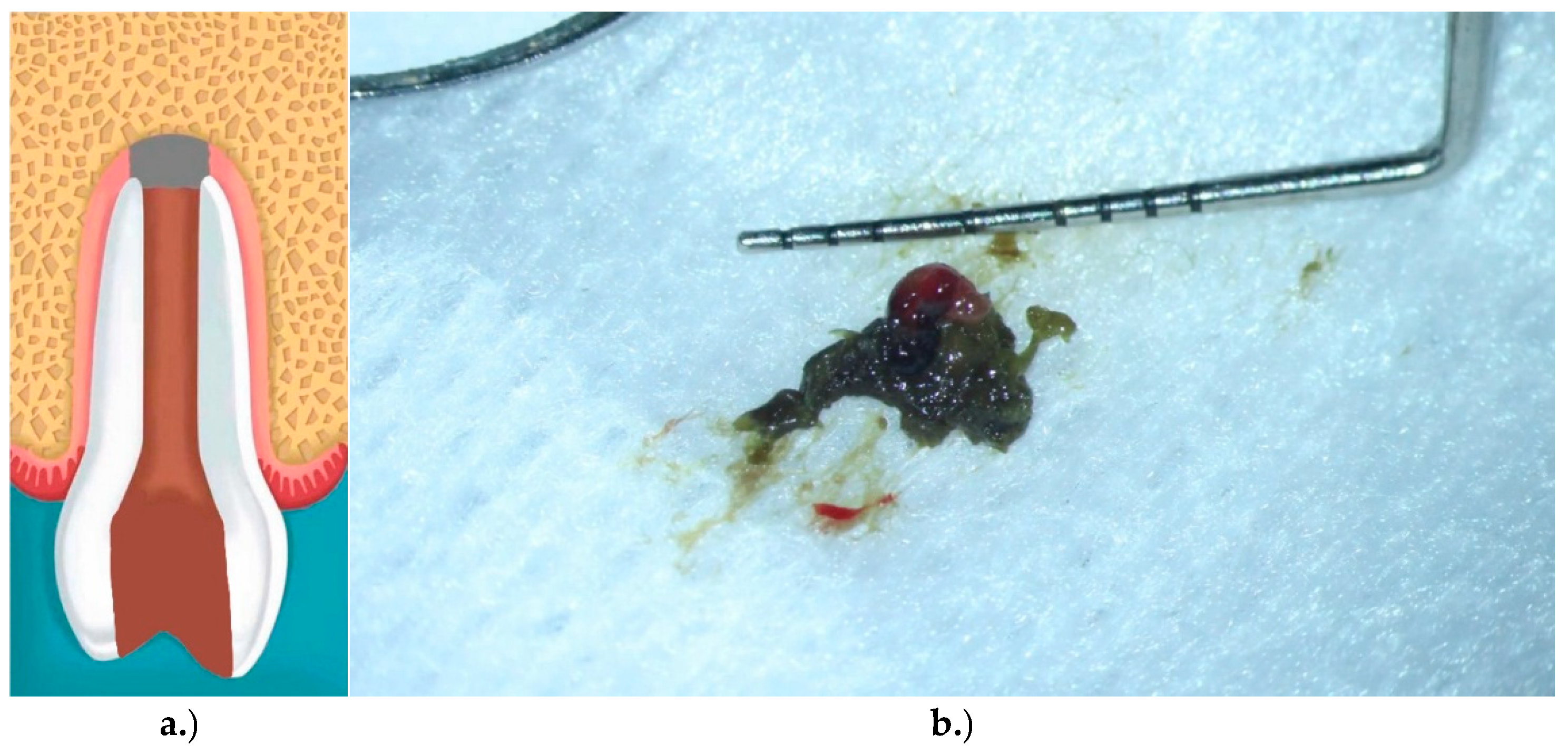
Figure 4.
Working length determination using an ISO size 100 K-File.
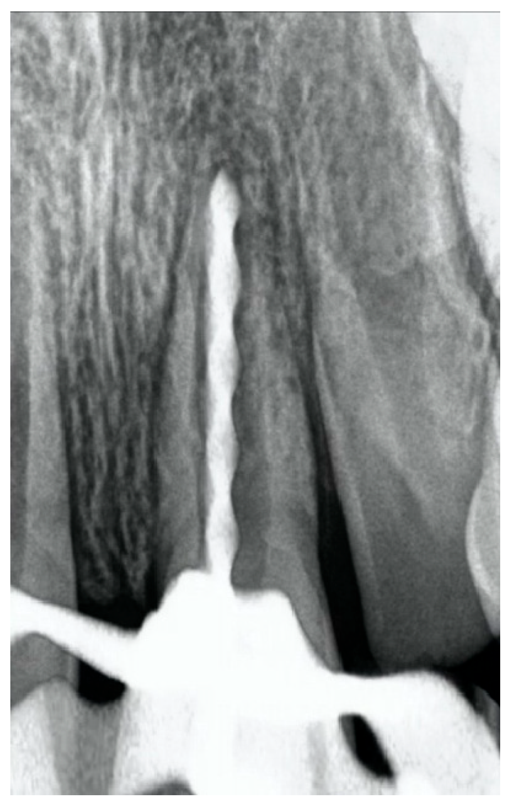
Figure 5.
Application of calcium hydroxide [Ca(OH)2] as an intracranial medicament, placed approximately 1-2mm short of the working length in tooth 21.
Figure 5.
Application of calcium hydroxide [Ca(OH)2] as an intracranial medicament, placed approximately 1-2mm short of the working length in tooth 21.
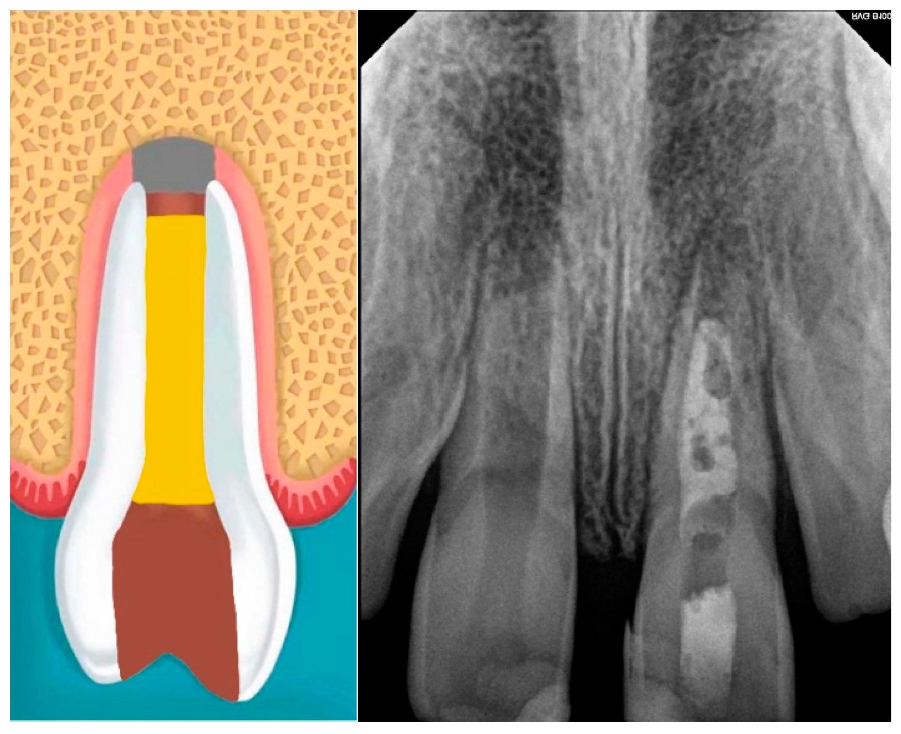
Figure 6.
(Left) Induction of apical bleeding by extending an ISO #10 K-file beyond the apical foramen. (Right) The resulting blood clot reached the level of the cementoenamel junction (CEJ).
Figure 6.
(Left) Induction of apical bleeding by extending an ISO #10 K-file beyond the apical foramen. (Right) The resulting blood clot reached the level of the cementoenamel junction (CEJ).
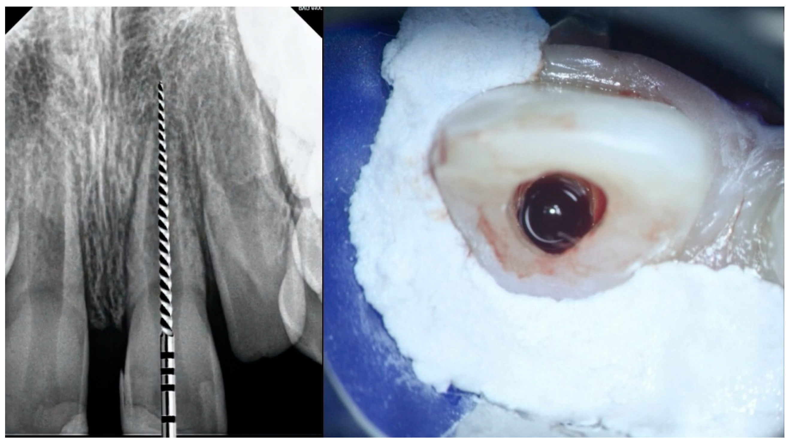
Figure 7.
Placement of a collagen sponge below the level of the cementoenamel junction.
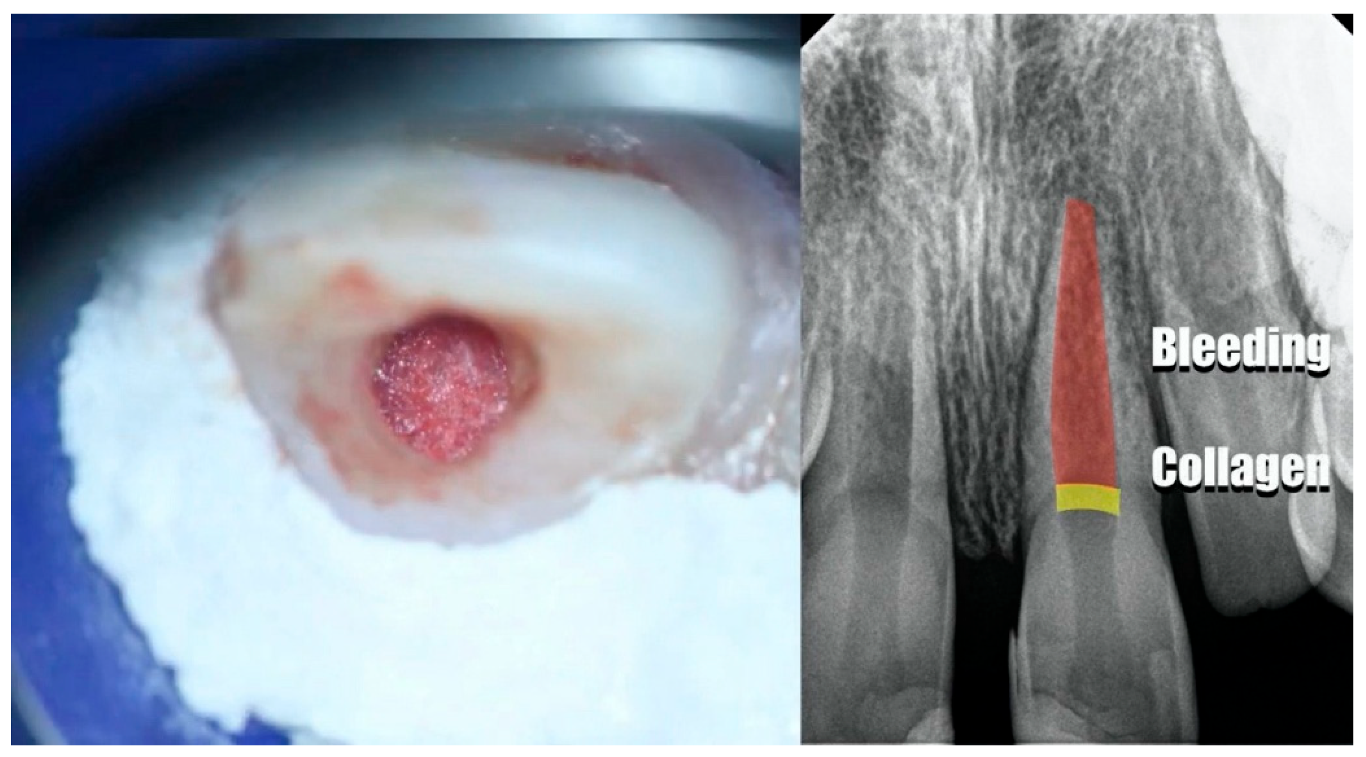
Figure 8.
Coronal sealing with a white bioceramic putty.
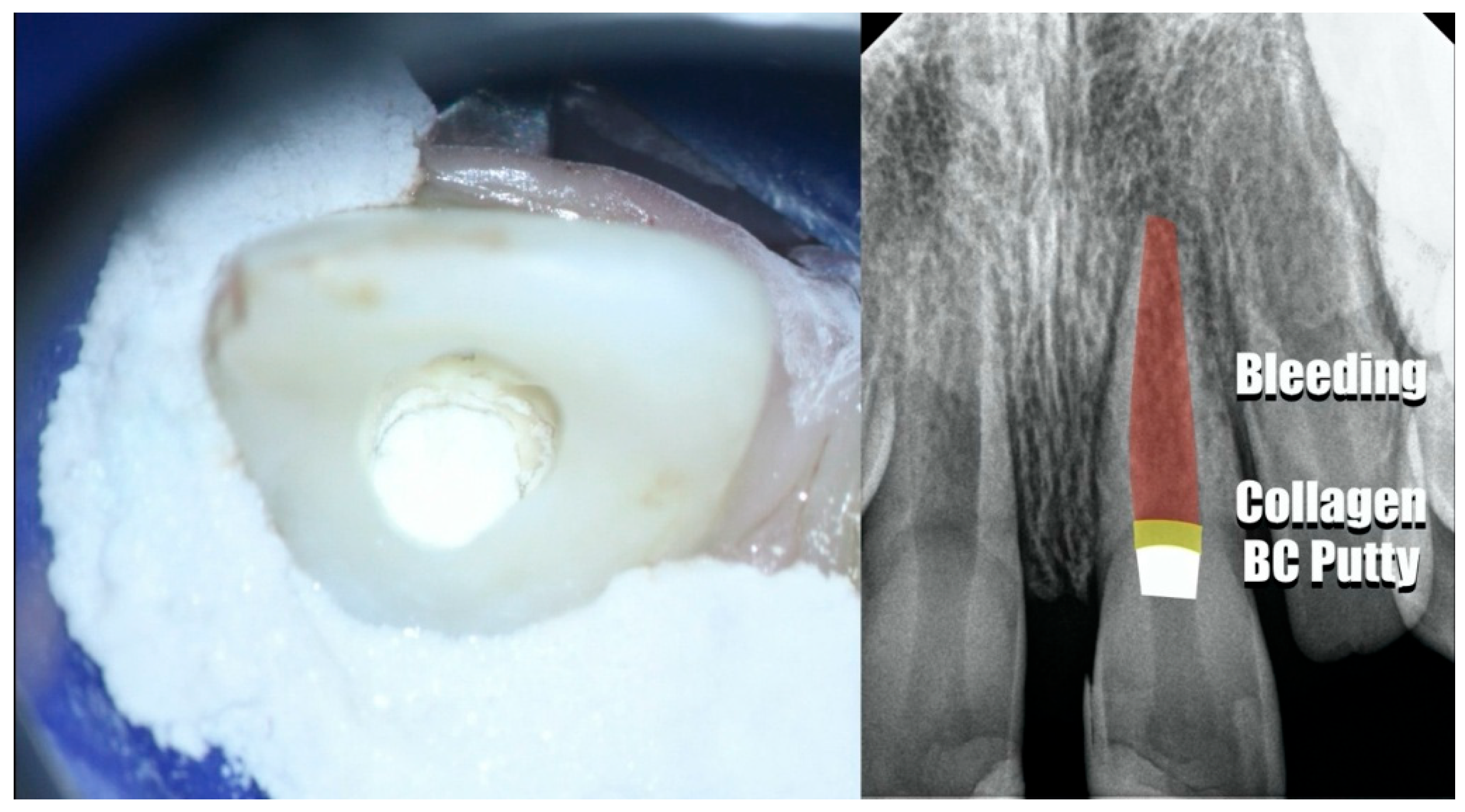
Figure 9.
Definitive restoration with a dual-cured composite resin.
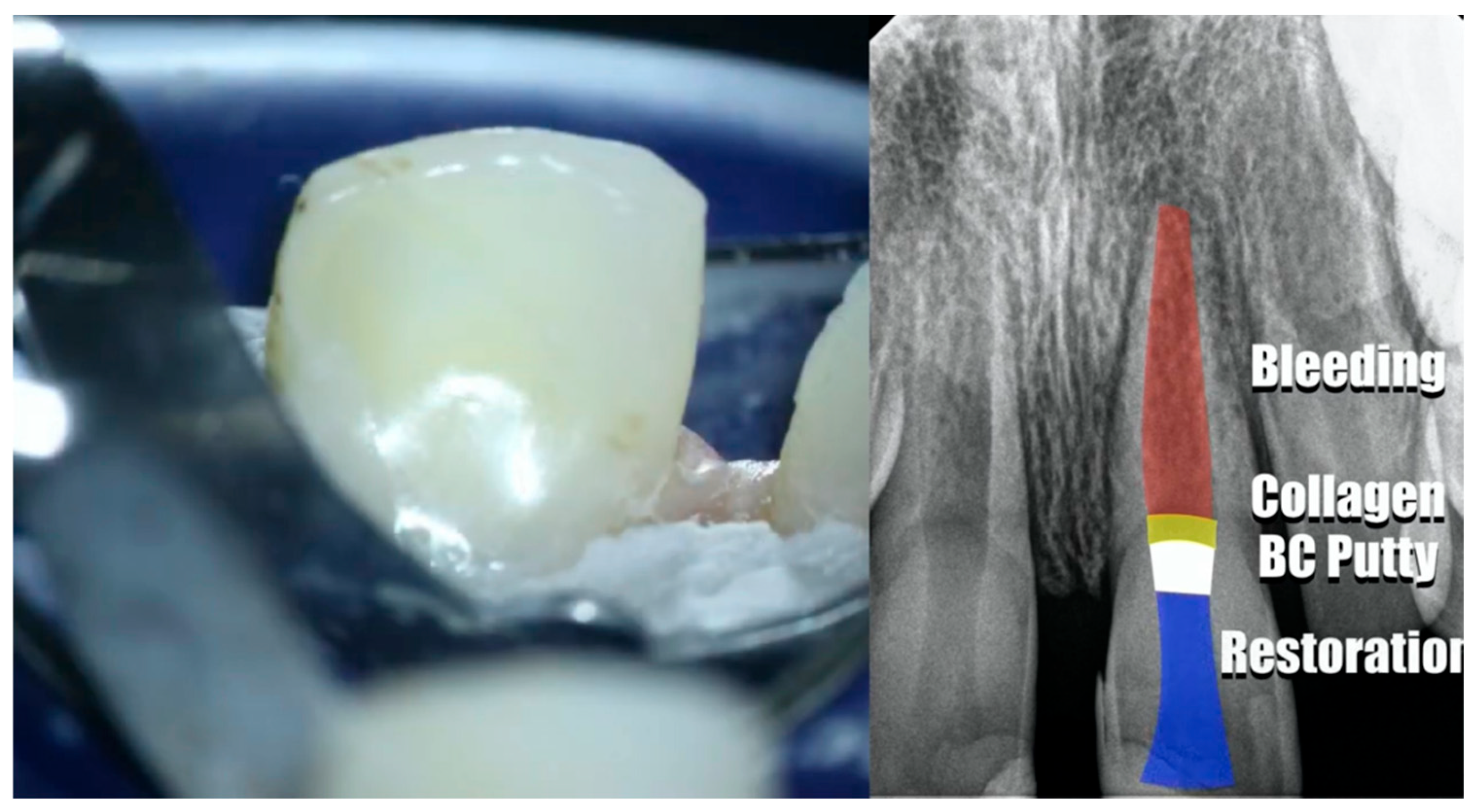
Figure 10.
Postoperative periapical radiograph of the maxillary central incisors (teeth #11 and #21).
Figure 10.
Postoperative periapical radiograph of the maxillary central incisors (teeth #11 and #21).
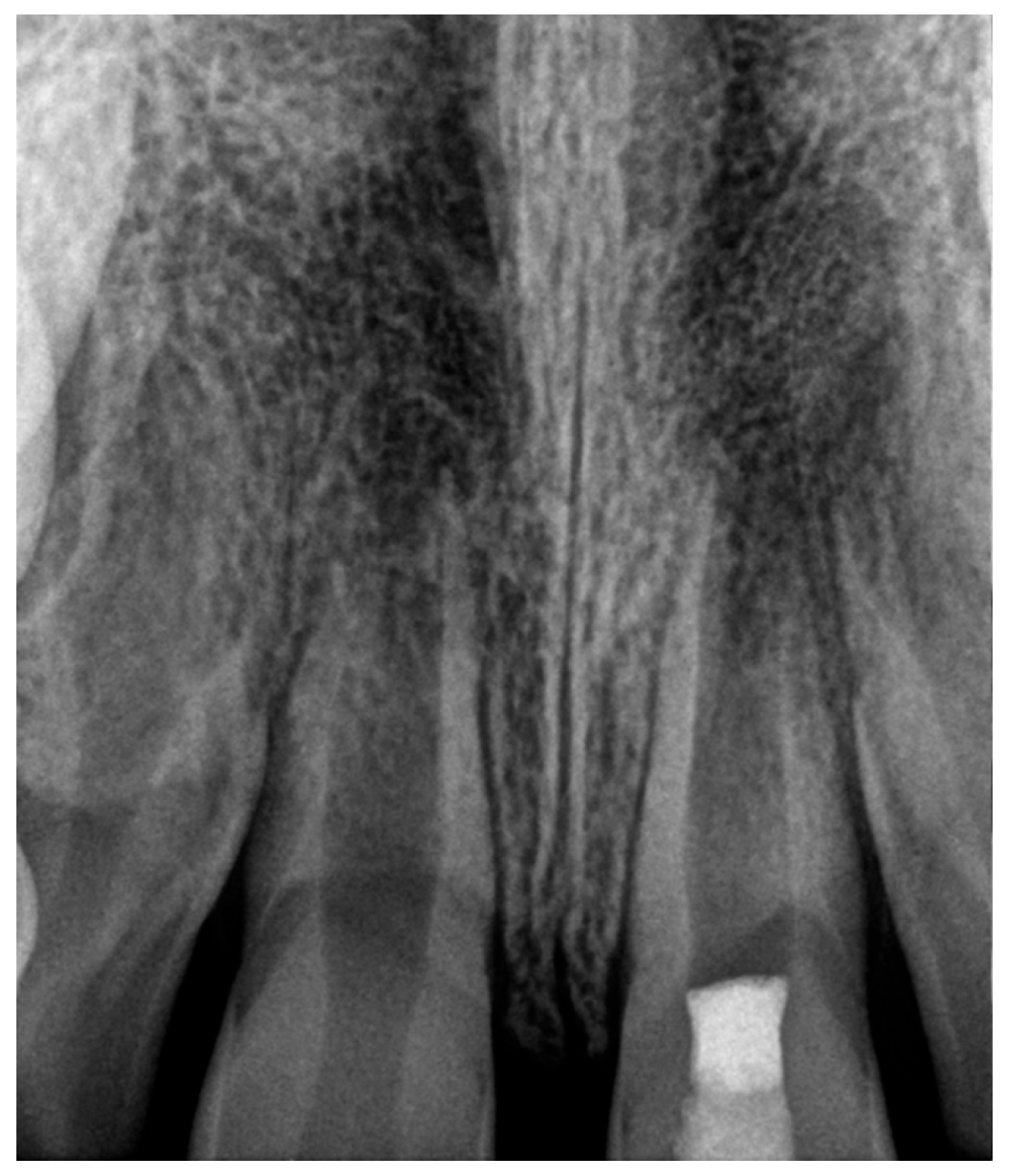
Figure 11.
Follow-up periapical radiographs obtained at a.) 5 months, b.) 8 months, c.) 1 year and d.) 2 years after regenerative endodontic treatment.
Figure 11.
Follow-up periapical radiographs obtained at a.) 5 months, b.) 8 months, c.) 1 year and d.) 2 years after regenerative endodontic treatment.
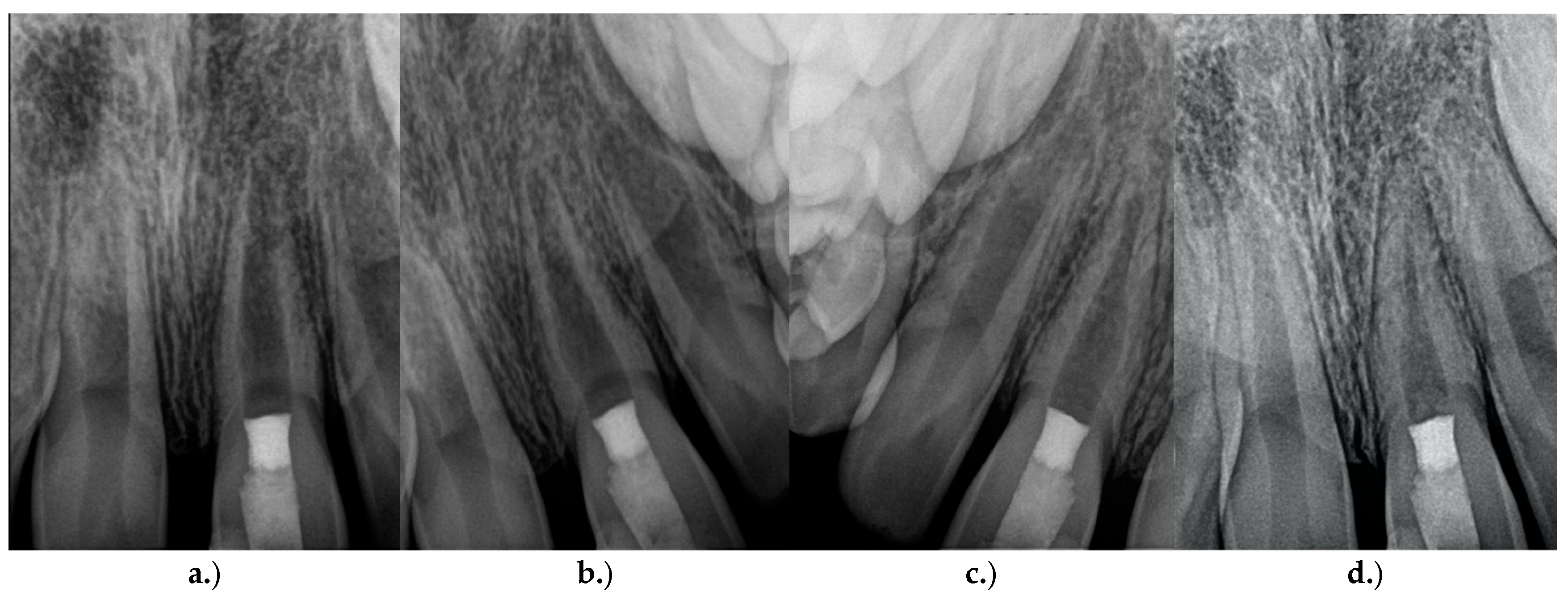
Figure 12.
a.) Extraoral photograph and b.) periapical radiograph obtained at the 3-year follow-up.
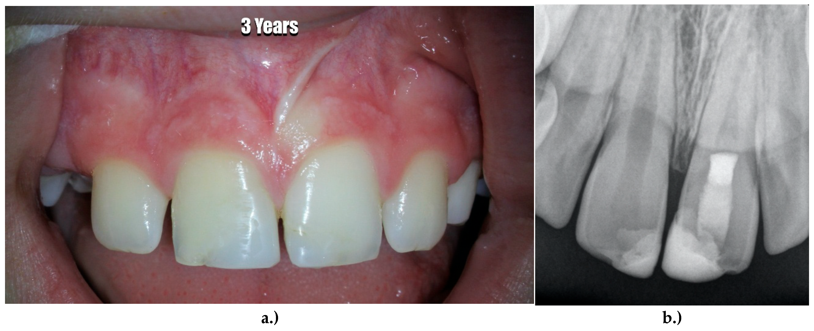
Figure 13.
CBCT images obtained at the 3-year follow-up: a.) coronal view and b.) axial view.
4. Discussion
Regenerative endodontic procedures (REPs) have fundamentally changed the management of immature permanent teeth with pulp necrosis by shifting the treatment concept from the creation of an artificial apical barrier toward biological regeneration of the root canal system. The present case demonstrated successful clinical and radiographic outcomes over a three-year follow-up period, including complete resolution of symptoms, continued root development, increased dentinal wall thickness and complete apical closure. These findings are consistent with the current evidence supporting regenerative endodontics as the preferred treatment modality for immature necrotic permanent teeth whenever appropriate biological conditions are present [1,2,3,4].
Unlike conventional apexification, regenerative endodontic treatment aims to preserve the long-term structural integrity of immature teeth by promoting continued root maturation (Figure 1). Although calcium hydroxide and mineral trioxide aggregate (MTA) apexification have demonstrated predictable apical barrier formation for many years, neither technique facilitates further root elongation nor reinforcement of the thin dentinal walls [3,5]. Consequently, teeth treated by apexification often remain susceptible to cervical and vertical root fractures throughout life. In contrast, regenerative procedures exploit the regenerative potential of the apical tissues, thereby improving both the biological and biomechanical prognosis of immature permanent teeth [6,7].
The favorable outcome observed in the present case agrees with the systematic review by Priya et al. [2], who reported high survival rates and favorable clinical outcomes following regenerative endodontic procedures in immature necrotic teeth. Similarly, Murray [1] concluded that regenerative endodontics should be considered the treatment of choice whenever continued root development can reasonably be expected. More recently, Verma et al. [16] confirmed through meta-analysis that regenerative procedures achieve significantly greater increases in root length and dentinal wall thickness compared with conventional apexification, while maintaining comparable tooth survival.
One of the most important biological factors contributing to treatment success is the preservation of stem cell viability during canal disinfection. Excessive mechanical instrumentation and high concentrations of sodium hypochlorite have been shown to reduce the survival of stem cells from the apical papilla (SCAP), thereby negatively affecting regenerative potential [6,11,17]. For this reason, the present treatment protocol deliberately employed minimal instrumentation together with irrigation using 1.5% sodium hypochlorite followed by 17% EDTA. This protocol has been recommended by both the American Association of Endodontists and the European Society of Endodontology because EDTA promotes the release of dentin-derived bioactive molecules, including transforming growth factor-β (TGF-β), which supports stem cell attachment, proliferation and odontoblastic differentiation [7,9,10].
The induction of intracanal bleeding remains a key step in regenerative endodontics (Figure 6). The resulting blood clot functions not only as a physical scaffold but also as a biologically active matrix containing platelets, cytokines, growth factors and mesenchymal stem cells that facilitate tissue regeneration [6,7]. SCAP are believed to represent the principal source of progenitor cells responsible for continued root maturation because of their high proliferative capacity and ability to differentiate into odontoblast-like cells capable of depositing mineralized tissue along the canal walls [11]. The progressive dentinal wall thickening and complete apical closure observed in the present case strongly support this biological concept.
The selection of calcium hydroxide as the intracanal medicament deserves particular attention (Figure 5). Historically, triple antibiotic paste was widely recommended for regenerative procedures; however, subsequent investigations demonstrated several disadvantages, including tooth discoloration, bacterial resistance and cytotoxic effects on stem cells [6,14]. Current AAE and ESE guidelines therefore recommend calcium hydroxide as a biologically more favorable intracanal medicament in many clinical situations [9,10]. The successful clinical outcome observed in the present case further supports these recommendations.
Despite increasing clinical success, regenerative endodontics continues to generate scientific discussion regarding the true nature of the regenerated tissue. Histological investigations have demonstrated that newly formed intracanal tissue rarely represents complete regeneration of the original pulp–dentin complex. Instead, varying proportions of fibrous connective tissue, cementum-like tissue, bone-like tissue and vascularized soft tissue have been reported [6,8]. Nevertheless, the primary objective of regenerative treatment is clinical success rather than histological perfection. In the present case, elimination of symptoms, continued root maturation and long-term preservation of the tooth were successfully achieved despite the unknown histological composition of the regenerated tissue.
Another frequently discussed issue concerns pulp canal obliteration following regenerative procedures. Progressive mineralized tissue deposition within the root canal has been interpreted by some clinicians as a potential complication requiring conventional root canal treatment [14]. However, current evidence suggests that canal obliteration alone should not be considered treatment failure. Unless associated with clinical symptoms or radiographic signs of apical disease, calcific tissue deposition most likely represents continued biological healing and should therefore be managed by regular clinical and radiographic observation rather than unnecessary intervention [1,15].
The present report has several limitations that should be acknowledged. As a single-case report, its findings cannot be generalized to all immature permanent teeth with pulp necrosis. Furthermore, histological confirmation of the regenerated tissue was not possible because extraction of a successfully functioning tooth would have been unethical. Nevertheless, comprehensive clinical examinations together with standardized periapical radiographs and three-dimensional CBCT imaging over a three-year observation period provide strong evidence supporting successful regenerative healing (Figure 13).
Future investigations should focus on improving the predictability of regenerative endodontic procedures through standardized treatment protocols, advanced scaffold materials, platelet concentrates, stem cell-based therapies, tissue engineering approaches and bioactive signaling molecules. Long-term multicenter prospective clinical studies are required to further clarify prognostic factors and optimize treatment outcomes in regenerative endodontics.
5. Conclusions
Regenerative endodontic procedures represent a biologically based treatment strategy for immature permanent teeth with pulp necrosis and open apices following traumatic injury. In the present case, revascularization resulted in complete resolution of clinical symptoms, continued root maturation, progressive thickening of the dentinal walls and complete apical closure during a three-year follow-up period. These findings demonstrate the capacity of regenerative endodontic therapy to promote functional healing while preserving the natural tooth.
Compared with conventional apexification, regenerative treatment offers the additional advantage of continued root development, thereby improving the long-term biomechanical stability of immature teeth. Careful case selection, effective disinfection, preservation of stem cell viability, induction of intracanal bleeding and an adequate coronal seal remain essential prerequisites for successful treatment.
Although histological confirmation of true pulp regeneration is not feasible in routine clinical practice, favorable clinical performance combined with progressive radiographic root maturation provides strong evidence supporting the effectiveness of regenerative endodontic procedures. This case further supports current recommendations of the American Association of Endodontists and the European Society of Endodontology, which advocate regenerative endodontics as the preferred treatment approach for immature permanent teeth with necrotic pulps whenever appropriate clinical conditions exist.
Long-term clinical and radiographic follow-up remains indispensable to monitor continued root development and to detect potential late complications. Future prospective multicenter studies with standardized treatment protocols and extended observation periods are required to further optimize regenerative procedures and strengthen the evidence base supporting their routine clinical application.
Overall, revascularization can be considered the preferred initial treatment option for immature permanent teeth with pulp necrosis, as it offers the potential for continued root development and biological healing. Importantly, if regenerative treatment does not achieve the desired clinical outcome, conventional apexification remains a predictable and viable secondary treatment option.
6. Patents
Supplementary Materials
Not applicable.
Author Contributions
“Conceptualization, Roland Frankenberger and Diyar Jolibagu; methodology, William Nudera; Roland Frankenberger and Diyar Jolibagu; formal analysis, Roland Frankenberger and Diyar Jolibagu; investigation, William Nudera and Diyar Jolibagu; resources, William Nudera; data curation, William Nudera and Diyar Jolibagu; writing—original draft preparation, Diyar Jolibagu; writing—review and editing, Roland Frankenberger; visualization, Roland Frankenberger and Diyar Jolibagu; supervision, Roland Frankenberger; project administration, Roland Frankenberger. All authors have read and agreed to the published version of the manuscript.”.
Funding
“This research received no external funding”.
Institutional Review Board Statement
Ethical review and approval were waived for this study because this manuscript reports a single clinical case treated according to established clinical standards without any experimental intervention. All procedures were performed in accordance with the Declaration of Helsinki.
Informed Consent Statement
Written informed consent was obtained from the patient’s legal guardians for treatment and for the publication of this case report and all accompanying clinical photographs and radiographic images.
Data Availability Statement
The data presented in this study are available within this article. Additional anonymized clinical data are available from the corresponding author upon reasonable request, subject to patient confidentiality and ethical restrictions.
Acknowledgments
The authors would like to express their sincere gratitude to Dr. William Nudera for performing the clinical treatment, providing the complete clinical documentation and long-term follow-up records and for his valuable support and willingness to share this exceptional clinical case for scientific publication. The authors also thank the patient and the patient’s legal guardians for their cooperation and for granting written informed consent for the publication of this case report and the accompanying clinical and radiographic documentation.
Conflicts of Interest
“The authors declare no conflicts of interest.”.
Abbreviations
The following abbreviations are used in this manuscript:
| AAE | American Association of Endodontists. |
| CBCT | Cone-Beam Computed Tomography |
| CEJ | Cementoenamel Junction |
| DAP | Double Antibiotic Paste |
| EDTA | Ethylenediaminetetraacetic Acid |
| ESE | European Society of Endodontology |
| GIC | Glass Ionomer Cement |
| LDF | Laser Doppler Flowmetry |
| MTA | Mineral Trioxide Aggregate |
| NaOCl | Sodium Hypochlorite |
| RCT | Root Canal Treatment |
| REP | Regenerative Endodontic Procedure |
| SCAP | Stem Cells from the Apical Papilla |
| TAP | Triple Antibiotic Paste |
| TDI | Traumatic dental injuries |
References
- Murray, P.E. Review of Guidance for the Selection of Regenerative Endodontics, Apexogenesis, Apexification, Pulpotomy, and Other Endodontic Treatments for Immature Permanent Teeth. Int. Endod. J. 2023, 56, 188–199. [Google Scholar] [PubMed]
- Priya, B.L.; Singh, N.; Mangalam, K.K.; et al. Success and Complication Rates of Revascularization Procedures for Immature Necrotic Teeth: A Systematic Review. Cureus 2023, 15, e51364. [Google Scholar] [CrossRef]
- Torabinejad, M.; Fouad, A.F.; Shabahang, S. Endodontics: Principles and Practice, 6th ed.; Elsevier: St. Louis, MO, USA, 2021. [Google Scholar]
- Kontakiotis, E.G.; Filippatos, C.G.; Tzanetakis, G.N.; Agrafioti, A. Regenerative Endodontic Therapy: A Data Analysis of Clinical Protocols. J. Endod. 2024, 50, 287–296. [Google Scholar]
- Torabinejad, M.; Walton, R.E.; Fouad, A.F. Endodontics: Principles and Practice, 5th ed.; Elsevier: St. Louis, MO, USA, 2015. [Google Scholar]
- Hargreaves, K.M.; Diogenes, A.; Teixeira, F.B. Treatment Options: Biological Basis of Regenerative Endodontic Procedures. J. Endod. 2021, 47, S19–S30. [Google Scholar]
- Galler, K.M. Clinical Procedures for Revitalization: Current Knowledge and Considerations. Int. Endod. J. 2022, 55, 934–956. [Google Scholar] [CrossRef]
- Chen, M.Y.H.; Chen, K.L.; Chen, C.A.; Tayebaty, F.; Rosenberg, P.A.; Lin, L.M. Responses of Immature Permanent Teeth with Infected Necrotic Pulps to Regenerative Endodontic Procedures. J. Endod. 2020, 46, 27–34. [Google Scholar] [CrossRef]
- European Society of Endodontology (ESE). Position Statement: Revitalization Procedures. Int. Endod. J. 2021, 54, 1975–1981. [Google Scholar]
- American Association of Endodontists (AAE). Clinical Considerations for a Regenerative Procedure. J. Endod. 2022, 48, 647–659. [Google Scholar]
- Diogenes, A.; Ruparel, N.B.; Shiloah, Y.; Hargreaves, K.M. Regenerative Endodontics: A Way Forward. J. Am. Dent. Assoc. 2016, 147, 372–380. [Google Scholar] [CrossRef] [PubMed]
- Galler, K.M.; Krastl, G.; Simon, S.; Van Gorp, G.; Meschi, N.; Vahedi, B.; Lambrechts, P. European Society of Endodontology Position Statement: Revitalization Procedures. Int. Endod. J. 2021, 54, 1975–1981. [Google Scholar] [CrossRef]
- Nagendrababu, V.; Duncan, H.F.; Bjørndal, L.; et al. Preferred Reporting Items for Case Reports in Endodontics (PRICE 2020). Int. Endod. J. 2020, 53, 1199–1205. [Google Scholar] [CrossRef] [PubMed]
- Kahler, B.; Rossi-Fedele, G.; Chugal, N.; Lin, L.M. An Evidence-Based Review of Regenerative Endodontic Therapy. J. Endod. 2017, 43, 668–681. [Google Scholar] [CrossRef]
- El Karim, I.; Duncan, H.F. Contemporary Concepts in Regenerative Endodontics. Dent. Clin. North Am. 2023, 67, 303–320. [Google Scholar]
- Verma, P.; Nosrat, A.; Kim, J.R.; et al. Outcome of Regenerative Endodontic Procedures: A Systematic Review and Meta-analysis. Int. Endod. J. 2024, 57, 421–438. [Google Scholar] [CrossRef]
- Ruparel, N.B.; Teixeira, F.B.; Ferraz, C.C.R.; Diogenes, A. Direct Effect of Intracanal Medicaments on Survival of Stem Cells of the Apical Papilla. J. Endod. 2012, 38, 1372–1375. [Google Scholar] [CrossRef] [PubMed]
- Saoud, T.M.A.; Sigurdsson, A.; Rosenberg, P.A.; Lin, L.M.; Ricucci, D. Treatment of Mature and Immature Teeth by Regenerative Endodontic Procedures: A Review. J. Endod. 2014, 40, S97–S101. [Google Scholar] [CrossRef]
- Lin, L.M.; Rosenberg, P.A. Repair and Regeneration in Endodontics. Int. Endod. J. 2011, 44, 889–906. [Google Scholar] [CrossRef] [PubMed]
- Lovelace, T.W.; Henry, M.A.; Hargreaves, K.M.; Diogenes, A. Evaluation of the Delivery of Mesenchymal Stem Cells into the Root Canal Space of Necrotic Immature Teeth after Clinical Regenerative Endodontic Procedures. J. Endod. 2011, 37, 133–138. [Google Scholar] [CrossRef] [PubMed]
- Bose, R.; Nummikoski, P.; Hargreaves, K. A Retrospective Evaluation of Radiographic Outcomes in Immature Teeth with Necrotic Root Canal Systems Treated with Regenerative Endodontic Procedures. J. Endod. 2009, 35, 1343–1349. [Google Scholar] [CrossRef] [PubMed]
- Wigler, R.; Kaufman, A.Y.; Lin, S.; Steinbock, N.; Hazan-Molina, H.; Torneck, C.D. Revascularization: A Treatment for Permanent Teeth with Necrotic Pulp and Incomplete Root Development. J. Endod. 2013, 39, 319–326. [Google Scholar] [CrossRef] [PubMed]
- Diogenes, A.; Ruparel, N.B.; Teixeira, F.B.; Hargreaves, K.M. Translational Science in Regenerative Endodontics. J. Endod. 2016, 42, S52–S57. [Google Scholar] [CrossRef]
Disclaimer/Publisher’s Note: The statements, opinions and data contained in all publications are solely those of the individual author(s) and contributor(s) and not of MDPI and/or the editor(s). MDPI and/or the editor(s) disclaim responsibility for any injury to people or property resulting from any ideas, methods, instructions or products referred to in the content. |
© 2026 by the authors. Licensee MDPI, Basel, Switzerland. This article is an open access article distributed under the terms and conditions of the Creative Commons Attribution (CC BY) license (http://creativecommons.org/licenses/by/4.0/).
Copyright: This open access article is published under a Creative Commons CC BY 4.0 license, which permit the free download, distribution, and reuse, provided that the author and preprint are cited in any reuse.


